Article

Site-Specific Addressing of Particles and Coatings via Enzyme-Mediated Destabilization

by
David Wedegärtner
and
Oliver I. Strube
*
Department of Chemistry–Biobased and Bioinspired Materials, Paderborn University, Warburger Street 100, 33098 Paderborn, Germany
*
Author to whom correspondence should be addressed.
Catalysts 2019, 9(4), 354; https://doi.org/10.3390/catal9040354
Submission received: 4 March 2019 / Revised: 1 April 2019 / Accepted: 10 April 2019 / Published: 12 April 2019

Abstract

:
Enzyme mediated addressing (EMA) is a highly specific and easy-to-apply technology for direction and deposition of particles and coatings on surfaces. Key feature of this process is an enzymatic reaction in direct proximity to the surface, which induces the deposition. The technique has previously shown great success in the handling of biological particles. In this study, addressing of non-biological nanoparticles, in particular plastics and metals, is presented. The respective particles are stabilized by an amphiphilic, enzyme-degradable block copolymer, consisting of poly(ethylene glycol) and poly(caprolactone). After contact with the enzyme pseudomonas lipase, the particles are destabilized, due to the loss of the hydrophilic part of the block copolymer. The lipase is therefore immobilized on glass supports. Immobilization is performed via adsorption or covalent bonding to epoxide groups. All deposition experiments show that addressing of individual particles occurs precisely within the predefined areas of enzyme activity. Depending on the material and reaction conditions, intact nanoparticles or coatings from such can be gained. The quintessence of the study is the indifference of the EMA regarding particle materials. From this rationale, the technique offers near unlimited materials compatibility within a precise, easy-to-apply, and upscalable process.

1. Introduction

1.1. Enzyme Mediated Addressing

Biomimetic abstraction of natural systems has become a guiding principle in many fields of materials science and technology in the desire for truly innovative products with supreme performance [1]. This approach is likewise valid in coatings technology although the ’’bio”-discussion centers here mainly around the utilization of renewable resources as substitutes for petrochemical raw materials. In this concept nature is seen as just another feedstock for chemicals to produce similar products which meet the established product quality and price point. This neglects the immense potential of nature which lies within the biological functionality of natural systems. Those are already adapted in other bioinspired materials that are often superior to conventional approaches. One of the main reasons behind many fascinating biological structures is the application of highly-specialized biocatalysts, the enzymes. This class of biological, polymeric catalysts is set apart from any other type of molecule class by its specificity, energy efficiency, and adaptability [2,3].
Not long ago, ex vivo enzymatic reactions were only applied by specialized scientists in the context of biochemistry. Since then though, the use of enzymes for special synthetic purposes has become ever more popular in numerous fields of applied sciences. Since the introduction of special biotechnological production techniques, such as genetic engineering, pure and highly reactive enzymes are more and more available at affordable prices. This has even led to several relevant industrial applications of enzymes in materials science, like the synthesis of polymers and the production of biopolymers from natural sources [4,5,6]. The ability of enzymes to build specific structures has however not yet been largely investigated although immense potential can be found here. This is especially true since processes for the formation of site-specific or patterned structures, like coatings and particle arrangements, are mostly energy-intensive, harsh, or slow. Moreover, they are often limited to small areas and specific materials. This renders them unsuitable for many industrial applications.
To overcome this, we previously introduced a novel technology which adapts the architectural skills of enzymes and enables defined deposition of colloidal particles in both a highly specific and easy-to-apply manner: the enzyme mediated addressing (EMA) [7]. Herein, enzymes control the destabilization or synthesis of the desired particles in close proximity to a support surface.
Deposition of those particles takes place only within the area of enzymatic activity; it is therefore site-specific [8]. The area of deposition is predefined by the selected method of enzyme immobilization. This branch of technology has seen huge success in the past, as it can enhance many properties of enzymes like stability, activity, selectivity, specificity, and resistance to inhibitors [9]. Which of those effects are gained depends not only on the specific enzyme but ofttimes also on sophisticated roadmaps for immobilization. The most relevant parameter is certainly orientation of the enzyme on the support, as was thoroughly investigated [10]. If the active site of an immobilized enzyme faces the support, its activity will at least be drastically reduced, if not halted completely. Suitable immobilization procedures give control over enzyme orientation if necessary [11].
Curiously, highly defined enzyme orientation is no critical factor for a successful application of the EMA process. Though only enzymes with proper orientation are assumedly active, specificity of deposition is fully confirmed in all previous experiments, despite utilization of immobilization techniques with only limited orientation control [12]. This is an important aspect as it results in an overall less demanding preparation procedure, which consequently increases feasibility in view of technical applications and upscalability.

1.2. Biomimetic Hybrid Particles

The initiation point of the EMA technology was the destabilization of casein micelles to generate specific coatings and nano-structures from this protein. The underlying principle is directly connected to the special structure of a casein micelle, which is comprised of a hydrophobic core, stabilized by hydrophilic parts of the amphiphilic κ-casein. Although the actual composition of a casein micelle is much more complex, this simplified model is sufficient to understand its colloidal properties and stabilization mechanisms [13,14].
Within the EMA process, the hydrophilic parts of the micelles are enzymatically cleaved by action of the peptidase chymosin, a highly specific reaction which is also the natural function of this enzyme. Performed in bulk, this reaction is long known for the sake of milk clotting. Performed within the specifics of the EMA, it is highly suitable for the production of defined protein structures on arbitrary surfaces, e.g., for applications in life sciences or adhesion [12,15].
The main limitation of this branch of the EMA technology has since then been its focus on this one specific protein as coating material. In this work, a biomimetic abstraction of the casein process is presented, to achieve the highly desired extension of EMA towards universal materials compatibility. To this end, stabilized, colloidal particles have to be produced, which mimic the structural features of a casein micelle. In the presented concept, the core is made from the desired coating material. The only condition here is the necessity for the core to be solvophobic (in this specific case hydrophobic), as this is the driving force for deposition. Within this range, all kinds of materials e.g., plastics (organic) or metals (inorganic) are suitable. The stabilizing layer of the micelle has two requirements. First, it has to be an amphiphilic polymer with the hydrophobic part binding to the core and the hydrophilic part stabilizing the micelle. Second, it has to be a substrate for an enzymatic catalysis, which must result in the loss of the hydrophilic part. Ideally, the necessary enzyme is readily available to increase economic attractivity for actual applications.
Figure 1 shows the schematic of such a biomimetic micelle and its behavior within the process of EMA, which is similar to the casein system: The micelles diffuse towards the support and enter the area of enzymatic activity (reaction zone). In consequence, the stabilizing block copolymer is degraded, leaving the hydrophobic core within an unfavorable energetic environment. Deposition on the support is thus preferred and takes place immediately. The process self-terminates, as soon as all enzymes are covered by coating particles. The specifics of this self-termination can be tuned to generate a wide variety of surface structures, from films with several microns film thickness to nano structures from single particles, as previously shown [16,17,18].

2. Results & Discussion

2.1. Examination of Stabilizing Block Copolymers

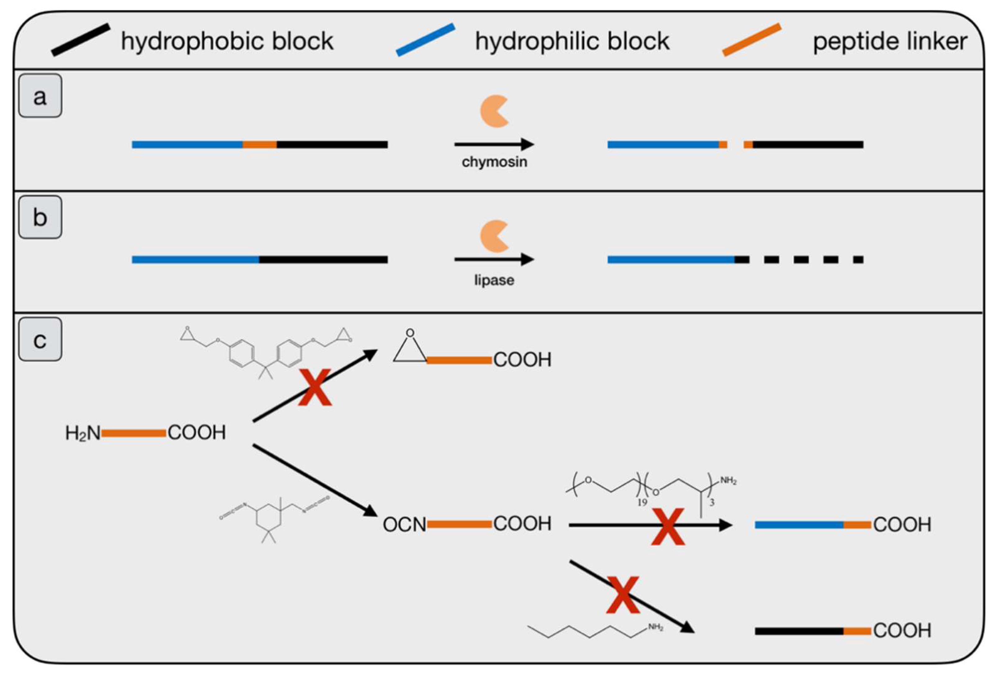
The first task towards biomimetic, casein-like micelles is the investigation of suitable polymer systems which are eligible as stabilizing layer. Two approaches were investigated to achieve this (Figure 2). The first approach closely mimics the natural casein micelle and consists of a hydrophilic and a hydrophobic polymer interconnected by a peptide linker. This linker resembles the amino acid sequence 103–110 in κ-casein. It is known from the literature, that this octapeptide is ideal for chymosin to selectively recognize it as substrate with high kinetic constants (kcat = 43.3 s−1 and km = 0.41 mM) [19]. Consequently, chymosin is once again the enzyme of choice for this variant.
The peptide linker obviously possesses an amine- and a carboxyl terminus. Those two distinct functionalities allow, in principle, the selective attachment of both polymer blocks [20]. The first concept was utilization of epoxy functionalized PEG to react with the amine terminus. However, already the test reaction with Bisphenol-A-Diglycidylether led to no substantial product. After that, a reaction of the amine terminus with a diisocyanate was considered, which gave the desired product. However, the amination of the now isocyanate-functionalized peptide did not yield relevant amounts of product, neither with a hydrophobic nor a hydrophilic block [21]. At this time, this approach was dismissed because the inherently complicated procedures and very low yields contradict the goal of an easy-to-apply, feasible process. All reaction steps are shown in Figure 2c.
The second approach dismisses the high similarity to the natural casein micelle, which is in the end not mandatory for a successful implementation. Instead, an enzyme degradable polymer as stabilizing layer was considered to be the most suitable variant. Again, the polymer has to be amphiphilic, so block copolymers are the most promising class of materials [22]. Additionally, it was decided that the enzymatic cleavage should occur within the hydrophobic part, as this immediately leads to the loss of the entire hydrophilic block. A partial cleavage of the hydrophilic block on the other side would not completely destabilize the micelle and lead to a retarded and less controllable process.
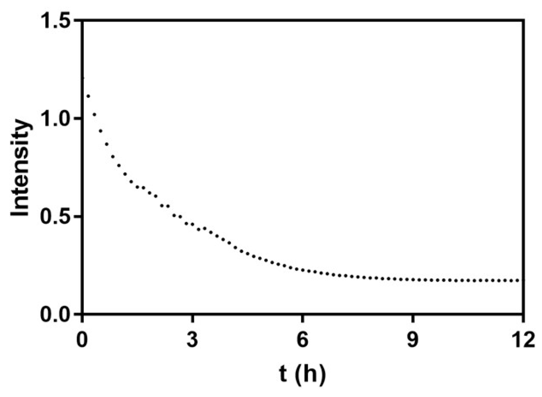
A block copolymer made of polycaprolactone (PCL) and polyethylene glycol (PEG) fulfills all necessary requirements and was thus chosen as model stabilizer. PCL shows high affinity to hydrophobic particles in aqueous solutions. The PEG block on the other hand is inert towards the enzyme and readily dissolves in the aqueous environment, once detached from the particle core. Moreover, the PCL block can easily be hydrolyzed by lipases, an affordable and readily available class of enzymes [23,24], which is consequently used as biocatalyst. Specifically, the lipase from pseudomonas bacteria was used [25], though most random lipases should work in a similar fashion. This can be utilized to perform the process under various reaction conditions, depending on the activity properties of the specific lipase. The degradation process of the micelles that are formed by the PEG-b-PCL block copolymers in aqueous solutions was monitored via UV-Vis spectroscopy [26]. Figure 3 shows the experiment for a dispersion of 1 mg/mL PEG(5000)-b-PCL(5000), with Mn of 10,000 g/mol in total and 5000 g/mol per block. 0.1 mg/mL lipase was added to the dispersion at t = 0. The spectrum confirms the intended lipase-induced degradation of the hydrophobic PCL blocks of the micelle. Degradation quickly starts and is completed after six hours, with the adsorption reaching a plateau caused by the remaining PEG polymers. As the application in the EMA process does not require a total degradation, this result suggests a very quick destabilization of the involved particles.

2.2. Synthesis of Nanoparticles Stabilized by Bomimetic Micelles

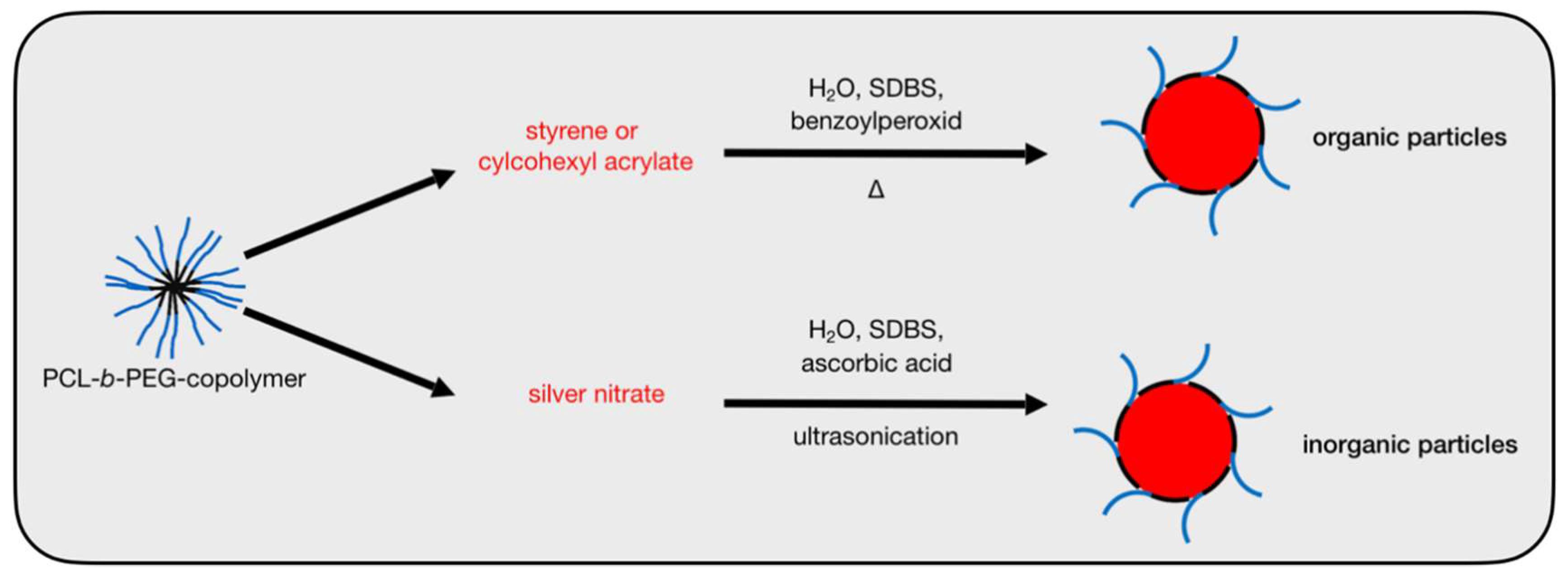
The PEG-b-PCL block copolymers were now used as stabilizer in the synthesis of three different types of nanoparticles, namely polystyrene (PS), poly(cyclohexyl acrylate) (PCHA), and silver (Figure 4). Polymeric particles were made via an emulsion polymerization pathway initiated by benzoylperoxide with styrene or cyclohexyl acrylate at pH 7.0 [27,28]. Stabilized silver particles were synthesized via reduction of silver nitrate by ascorbic acid in ultrapure water, aided by ultrasonication in the presence of the PEG-b-PCL block copolymer [29].
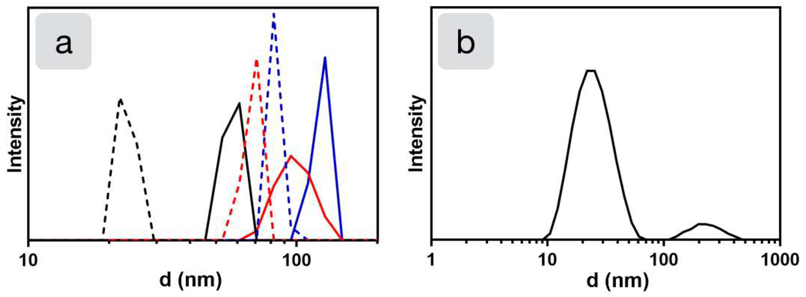
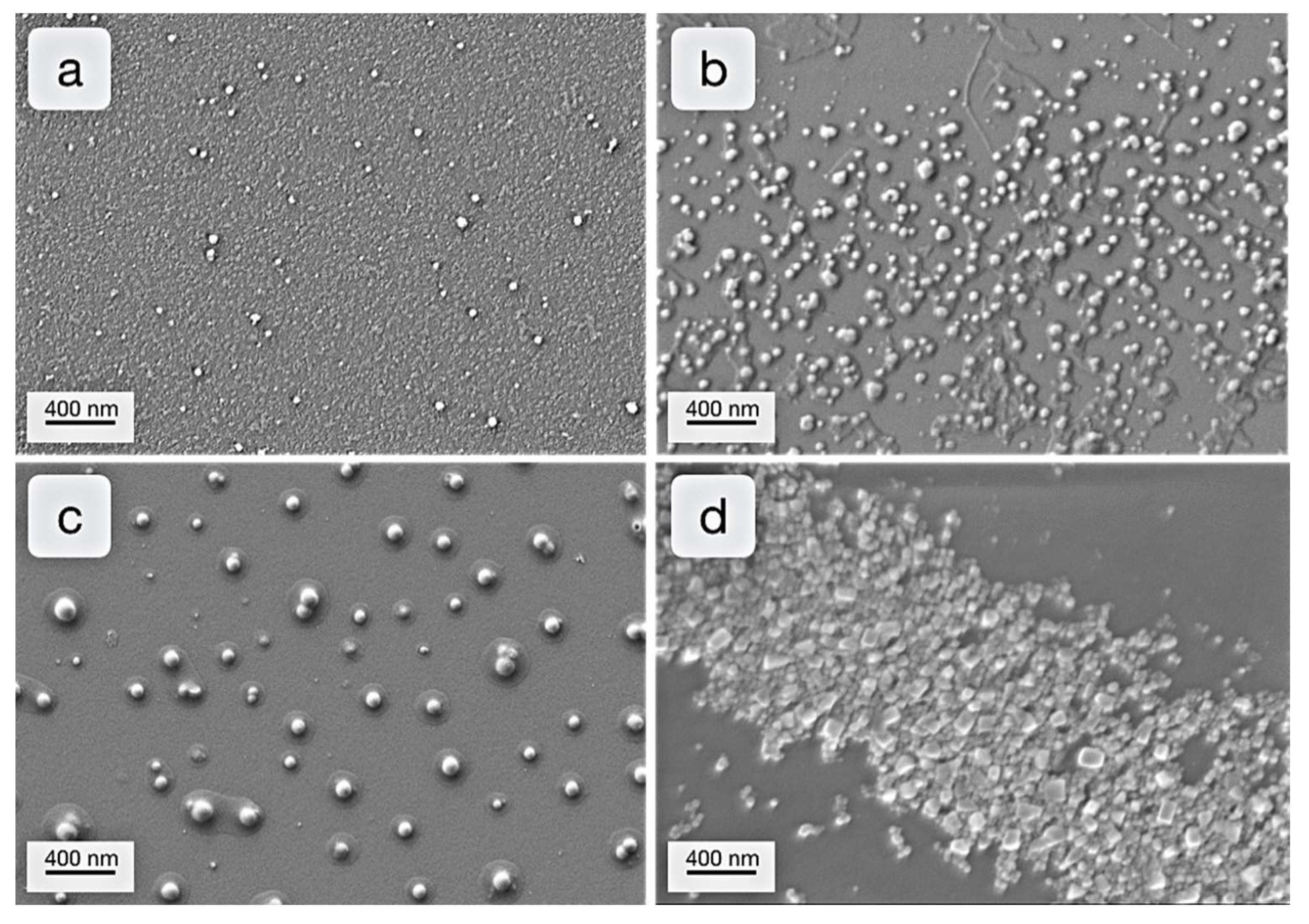
An important aspect of the process is the ability to tune the size of the desired nanoparticles. To test this, block copolymers with varying chain length were used in the synthesis of polystyrene particles. In total, three different block copolymers have been tested, namely PCL(2000)-b-PEG(2000), PCL(5000)-b-PEG(5000), and PCL(5000)-b-PCL(10000), with the numbers again showing Mn values of each block. First, the micellar sizes of pure block copolymers, dissolved in ultrapure water were determined via DLS-measurements. A clear tendency was observed. As expected, larger polymer blocks resulted in higher micellar diameters. If those values are compared to DLS measurements after particle synthesis, a direct correlation between micelle diameter and corresponding particle size is apparent (Figure 5a). The micelle size of the stabilized particles shifts from 22 nm to 53 nm PCL(2000)-b-PEG(2000), from 76 nm to 110 nm PCL(5000)-b-PEG(5000), and from 82 nm to 128 nm PCL(5000)-b-PEG(10000), respectively; all values are peak maxima. Additionally, the corresponding PS particles have been investigated with SEM, which confirms the results from DLS and thus the controllability of the particle size (Figure 6a−c).
Silver nanoparticles were made with PEG(2000)-b-PCL(2000) only, because the larger micelles yielded highly polydisperse particles or aggregated structures. Figure 5b shows that the majority of particles are of sizes around 30 nm. However, a small number of larger particles (around 250 nm) was also formed. The two specific particle sizes are confirmed by SEM, which shows largely the two mentioned sizes (Figure 6d). In summary it was possible to gain genuine nanoparticles from all desired materials, partly with controllable size.

2.3. Enzyme-Medatied Addressing

The stabilized particles from Section 2.2 were now used as precursor in the EMA process, with two variants of enzyme immobilization. The first one is an adsorption approach, where the enzyme solution is dropped on the blank glass surface. Adsorption is driven by a combination of van der Waals- and ionic interactions [30], the latter ones stemming from the pretreatment of the glass support [7]. After the enzyme solution is dried out non-adsorbed enzyme is washed off with ultrapure water. Nevertheless, the enzyme concentration varied and showed an accumulation at the drying edge of the former enzyme solution droplet. This effect could be disruptive for technical applications but is certainly reduceable by process optimizations. For the herein described scientific experiments though, it is in fact advantageous. It offers the possibility to demonstrate the influence of surface enzyme concentration on the resulting deposition.
On the other hand, to yield evenly distributed surface enzyme concentration, the enzyme was also covalently bound to the glass surface, via epoxy functionalized supports [13]. The molecular process of this coupling is well understood [31]. It is quite delicate and includes several steps. At the end though, exposed amino groups on the enzyme bind to the epoxy groups of the support. Application of high ionic strength in the immobilization buffer is known to be a feasible tool to drive favorable orientation to a certain degree [32]. In the context of EMA this level of control has proven to be sufficient, even for the production of genuine nano-structures over a macroscopic scale [15]. Therefore, this technique is also used in this work.
After enzyme immobilization, the functionalized glass supports were immersed vertically into the particle suspensions for 24 h. The temperature was set to 40 °C to achieve maximum biocatalytic activity [33]. Afterwards, the samples were washed with ultrapure water and dried.
Addressing of polystyrene particles represents the proof of concept for this novel branch of the EMA technology (Figure 7). The high concentration of polystyrene particles near the drying edge in the sample with adsorbed enzyme confirms the rationale behind EMA, that increased enzyme surface concentration leads to increased deposition of particles. The inner part of the reactive circle also shows deposition of polymer, in contrast to the outside area which is almost free of any particles. The important feature of site-specificity, observed in all previous varieties of EMA, can thus be confirmed for the herein presented approach as well [8,12,16,17].
However, amount of deposition is lower than observed in previous experiments (Figure 7a) [12]. This is explained by two factors. First, the accumulation of enzymatic activity on the edge proportionally reduces the amount on the inner area. Second, adhesion of polystyrene particles is less favorable on glass surfaces than the previously investigated materials. Optimizations of the procedure, e.g., variation of support materials and enzyme adsorption procedure, are thus necessary within future development. The samples with covalently bound enzyme result in a more homogeneous particle deposition (Figure 7b). This confirms the influence of evenly distributed enzyme activity. Again however, the monolayer shows several defects which can again be linked to the unfavorable compatibility between glass and PS. The general capability of the process, however, is additionally confirmed by corresponding blind samples without any enzyme and with deactivated enzyme, which both led to practically no particle deposition at all.
Another observation of the polystyrene samples is deposition in the form of intact particles without any kind of merging or film formation. This is contrasted by the apporach for EMA polymer films via in situ polymerization mechanism with the enzyme Horseraddish peroxidase [18]. The explanation for those differences are found within the distinct processes. In this apporach the polymeric particles are already formed and in a solid form when introduced to the deposition process. Thus film fomration cannot accur if the reation temperature is below the Minimum Film Forming Temperature (MFFT), which is realted to the glass transition temperature (Tg) of the polymer.
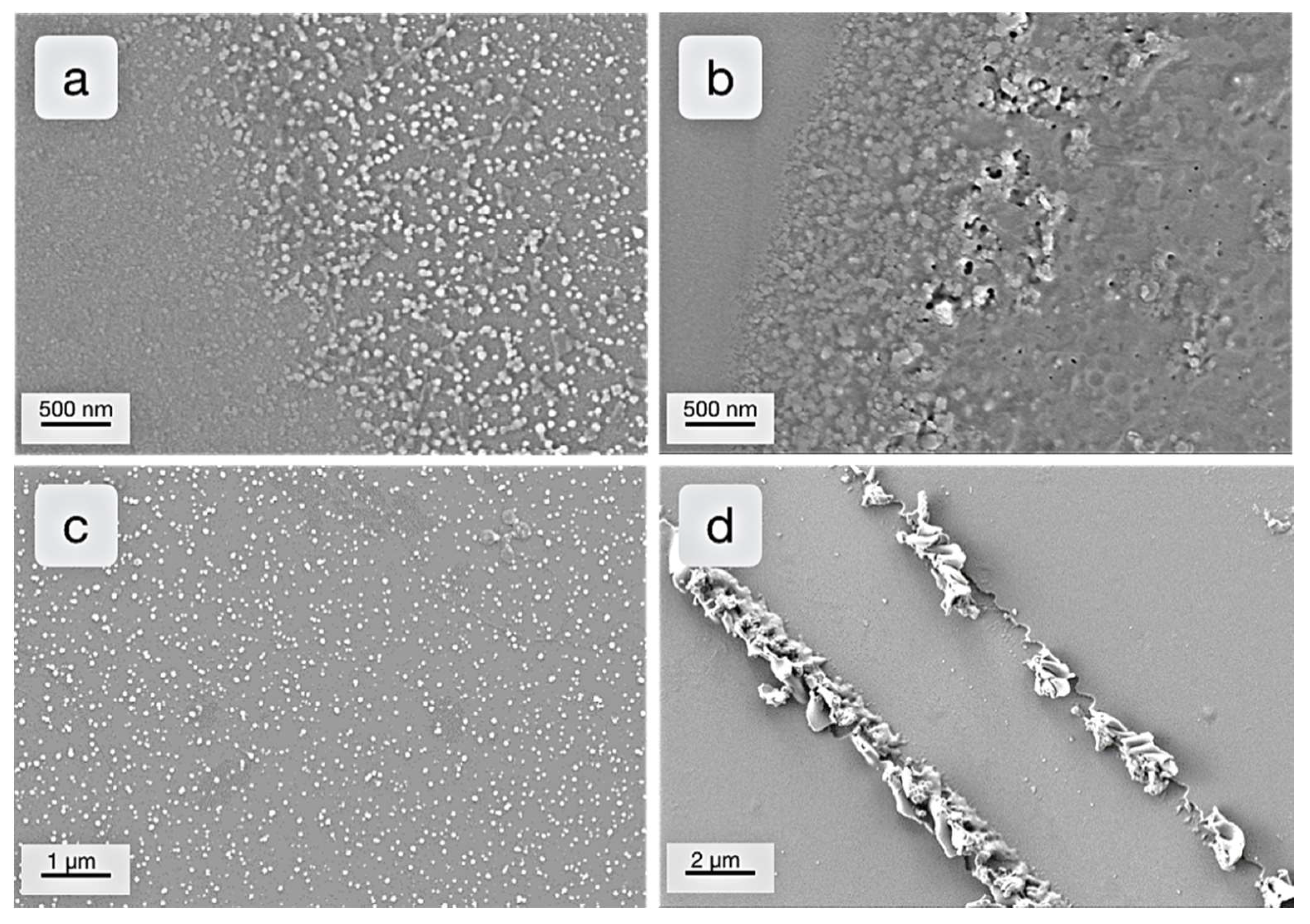
Although there might be a suitble appliaction for isolated polymer particles on surfaces, the main idea was to create coating structures, i.e., films. To achieve this initial goal, the influence of Tg was tested. To this end, particles from poly(cyclohexyl acrylate) (PCHA) were synthezised with the PEG-b-PCL stabilizer. In the literatue Tg of PCHA is given as 19 °C [34]. The particles should consequently merge into films at temperatures above this level. As the MFFT is usually located some degrees above Tg, the respective deposition experiments were performed at 15 °C and 40 °C with both, adsorbed and covalently bound enzyme.
The respective SEM pictures in Figure 8 reveal a significant influence of the temperature on the gained surface structures. At 15 °C, the same phenomenon as for the polystyrene counterpart is observed, with particles occuring as intact entities. At 40 °C, however, a different picture arises, as film formation clearly takes place, which again occurs in a site-specific manner. The amount of depostied matter is also increased, which indicates better adhesion of the polymer above Tg. Film formation at 40 °C with covalent enzyme immobilization shows higher homogeniety on the substrate, which was expected due to the more uniform enzyme functionalization. The thickness of all films was found to be in a range of a single or some few layers (up to 100 nm).
After successful implementation of polymer particles, deposition of inorganic matter, represented by silver nanoparticles, is considered next. The procedures are identical to the polymer samples. Reaction temperature was again set to 40 °C. Figure 9 shows the results for EMA with silver nanoparticles, which are highly similar to the ones with polystyrene. Again, use of adsorbed enzyme shows large amount of deposition in the area with increased enzyme activity, while covalent enzyme immobilization leads to a more homogeneous deposition of the silver nanoparticles. Additionally, all experiments showed high site-specificity and no deposition for respective blank tests. Site-specificity and influence of enzyme concentration can be observed in a very striking manner when comsidering a special artifact on the support (Figure 9c). In very minor amounts, immobilized enzyme can be found in highly aggregated forms, which often resemble microbial strucutres. Deposition patterns around such enzyme aggregates are highly specific and give a good impression on the influencing factors of the process. On top and around the high enzyme accumulation, lots of particles are found, whereas the density of deposition is reduced proportionally to the distance from the aggregate. This previously seen effect [13] is also valid for the EMA with silver particles, which again proves the indifference of the result from specific variations.
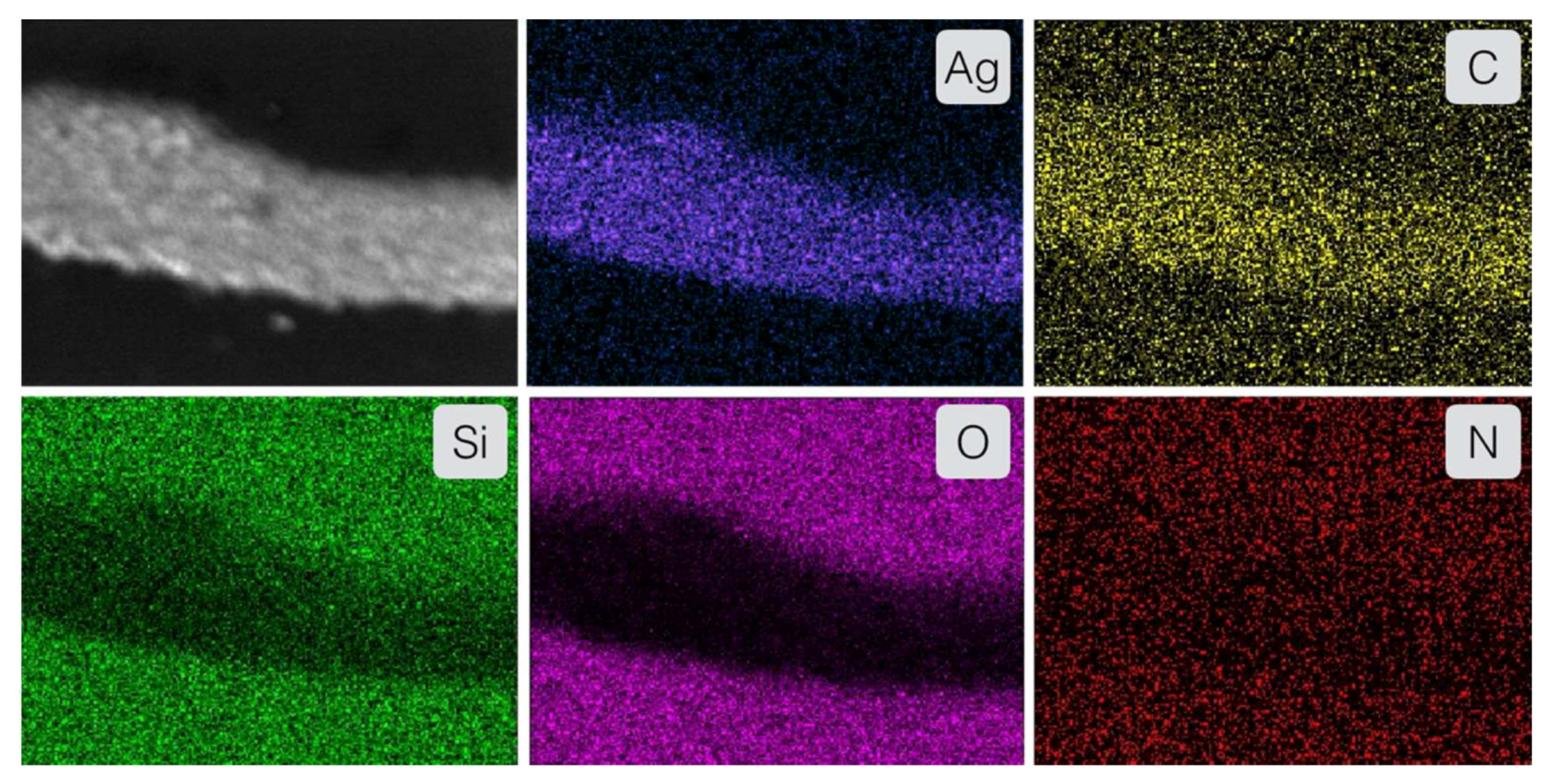
To prove the identity of deposited particles as metallic silver particles, EDX measurements were performed (Figure 10). The elemental maps show that the particles on the glass support consist in majority of silver, with minor traces of carbon, which derives from remaining stabilizer. The absence of nitrogen and oxygen is especially important, as it rules out the possible appearance of crystalline silver nitrate precursor. Silicon and oxygen are only found in the area around the particles and represent the SiO2 composition of the glass support.
A noteworthy aspect is the fate of the directing enzyme after completion of the process. As previously shown, the EMA process is self-limiting as the enzyme will finally be covered with deposited material [13]. Depending on the immobilization method, this effect is observed after deposition of a single or multiple layers. Consequently, the enzyme remains within or beneath the coating, but it is no longer active. This might be utilized to create a regenerative coating, if the enzyme can be reactivated by removal of the coating. If the enzyme is disruptive for the application however, methods for removal, e.g., thermal degradation, are applicable.

3. Materials and Methods

3.1. Materials

Freeze-dried pseudomonas lipase (41.3 U/mg, analytical grade) was obtained from VWR (Radnor, PA, USA) and used as provided. PEG-b-PCL block copolymers with various molecular weights were obtained from Creative PEGWorks (Durham, NC, USA). Sodium phosphate stock solution (500 mM, pH 7.0) was obtained by Cayman Chemical Company (Ann Arbor, MI, USA). The octapeptide LSFMAIPP was purchased from CASLO ApS (Lyngby, Denmark). All other chemicals were obtained from the VWR (Radnor, PA, USA) or Sigma Aldrich (St. Louis, MI, USA) and used without further purification.

3.2. Synthesis of Peptide Linked Stabilizers

For the buildup of a peptide linked stabilizer, three different approaches have been tested. (a) Equimolar amounts of the octapeptide LSFMAIPP and Bisphenol-A-diglycidylether are dissolved in DMF and the temperature is set to 40 °C while 1 wt.% dimethyl benzylamin is added as catalyst. The reaction mixture is stirred for 5 h. (b) Equimolar amounts of the octapeptide and isophorone diisocyanate are dissolved in DMF and the temperature is set to 60 °C. The reaction mixture is stirred for 3 h. (c) Afterwards Jeffamine M-1000 or hexamethylene amine are added to the solution.

3.3. PEG-b-PCL Stabilised Polymer Particle Syntheses

A typical synthesis involves the preparation of a solution consisting of 40 mg PEG-PCL (var. molecular weight) and 40 mg benzoyl peroxide dissolved in 50 mL DI water. Afterwards 4 mL sodium phosphate stock solution (2 mL) and 800 µL of the vinyl monomer are added. The solution is stirred for 30 min at room temperature. The temperature is set to 73 °C and the reaction mixture is stirred for additional 5 h.

3.4. PEG-b-PCL Stabilised Anorganic Particle Syntheses

For the silver particle synthesis, a modified reaction method of Moghimi-Rad et al. is used [29]. Therefore, a solution of 2 mg ascorbic acid, 20 mg PEG-PCL Copolymer and 1.25 mL sodium phosphate stock solution in 25 mL DI water was prepared. After ultrasonication, 2 mg AgNO3 in 12.5 mL DI water were added and again treated in an ultrasonic bath for 10 min.

3.5. Preparation of Supports

Glass supports were cleansed subsequently with EtOAc and DI water. To create a uniform and polar surface (and thus to facilitate adhesion of the enzyme), the glass supports were subsequently pretreated with an acidic and a basic cleaning solution, each for 10 min at about 70 °C. The order of the acidic and basic cleaning step varied depending on the particular experiment, but the supports were always thoroughly rinsed with DI water after each step. The acidic cleaning solution was a 1/1/5 (V/V/V) mixture of H2O2, conc. HCl and DI water, while the basic one was a 1/1/5 (V/V/V) mixture of H2O2, NH3 (25% W/V) and DI water. Polyethylene supports were cleansed subsequently with EtOAc and DI water only.

3.6. Adsorptive Enzyme Functionalization of Supports

Enzyme solutions with concentrations of 500 units/mL were prepared with ultrapure water. One drop (80 μL) of fresh enzyme solution was placed on each pretreated glass support using an Eppendorf pipette and left to dry. Supports with immobilized enzyme were stored at 5 °C. Non-adsorbed enzyme was rinsed off with ultrapure water prior to use.

3.7. Covalent Enzyme Functionalization of Supports

Immobilization of pseudomonas lipase was done by attachment to an epoxy-functionalized glass support, as described before [7].

3.8. Preparation of the Precursor Solution

The precursor solution contains 4 mL of the dispersion with the stabilised organic or inorganic particles and 11 mL ultrapure water with 0.2 mL SDBS solution is added to keep the pH at 7.

3.9. Coating of the Supports

The supports with immobilized enzyme were clamped by plastic tweezers, which were conjoined with the lit of the reaction vessel, so that the glass supports could be vertically held in place in the center of the beaker. The supports were put into the reaction solution for 24 h at varying temperatures.

3.10. Scanning Electron Microscopy (SEM) & Energy-Dispersive X-ray Spectroscopy (EDX)

SEM and EDX measurements were performed on a “Zeiss Neon 40 EsB CrossBeam” (Oberkochen, Germany). SEM images were recorded at an acceleration voltage of 2 kV using the InLens and the SE2 detector. EDX data were obtained at an acceleration voltage of 8 kV using mapping mode.

3.11. Dynamic Light Scattering (DLS) and Zeta-Potential

DLS and Zeta-Potential measurements were performed on a Zetasizer Nano-ZS by Malvern (Malvern Instruments Ltd., UK). The DLS measurements took place at a scattering angle of 90° and a temperature of 20 °C. The wavelength of the laser was 632.8 nm. The size distribution was obtained from the number plot autocorrelation function by regularization analysis, implemented in the Malvern Zetasizer software package.

3.12. UV/Vis Spectroscopy

UV/Vis-spectroscopy was performed with a Thermo Scientific Evolution 600 spectrometer (Waltham, MA, USA) using a 10 min cycle time. The bandwidth of the light source was set between 325 and 700 nm. The spectrum of DI water was used as a baseline.

4. Conclusions

Nanoparticles from polymers and metals, namely polystyrene, poly(cyclohexyl acrylate), and silver were successfully deposited on glass supports via the process of enzyme mediated addressing. As the necessary, enzyme-labile stabilizer, block copolymers with an enzyme-degradable hydrophobic block were added to the synthesis of nanoparticles. Immersion of enzyme-functionalized supports into a dispersion of any of those nanoparticles resulted in controlled, site-specific deposition of particles on enzyme-functionalized areas only.
The quantity of deposition was shown to be strongly dependent on the amount of enzyme on a specific area of the support. Areas without enzyme remained blank of any significant particle deposition. The process was generally independent of the specific type of particle, consequently the material plays only a passive role. However, adhesion between particle and support were found to be relevant. Moreover, setting the process temperature above Tg led to formation of films for the polymeric materials.
The presented experiments fill a significant gap in the concept of the EMA technology, as now its indifference with respect to particle materials is proven. The concept can thus be easily transferred to virtually any kind of particle-support system, if sufficient adhesion between the materials is ensured. Future optimizations can still occur in the more technical range, to enable a fully controlled and easy-to-apply technology platform for manifold applications.

Author Contributions

Conceptualization, O.I.S. and D.W.; methodology, D.W.; software, O.I.S. and D.W.; validation, D.W.; investigation, D.W.; writing—original draft preparation, O.I.S. and D.W.; supervision, O.I.S.; project administration, O.I.S.

Funding

This research received no external funding.

Conflicts of Interest

The authors declare no conflict of interest.

References

  1. Wanieck, K.; Fayemi, P.-E.; Maranzana, N.; Zollfrank, C.; Jacobs, S. Biomimetics and Its Tools. ICE publishing. 2017. Available online: https://sam.ensam.eu/bitstream/10985/12314/3/LCPI-BBN-WANIECK-2017.pdf (accessed on 4 March 2019).
  2. Vincent, J.F.V. Biomimetics—A review. Proc. Inst. Mech. Eng. Part H J. Eng. Med. 2009, 223, 919–939. [Google Scholar] [CrossRef] [PubMed]
  3. Marchetti, L.; Levine, M. Biomimetic Catalysis. ACS Catal. 2011, 1, 1090–1118. [Google Scholar] [CrossRef]
  4. Dicker, M.P.; Duckworth, P.F.; Baker, A.B.; Francois, G.; Hazzard, M.K.; Weaver, P.M. Green composites: A review of material attributes and complementary applications. Compos. Part A Appl. Sci. Manuf. 2014, 56, 280–289. [Google Scholar] [CrossRef]
  5. Banyard, D.A.; Bourgeois, J.M.; Widgerow, A.D.; Evans, G.R.D. Regenerative biomaterials: A review. Plast. Reconstr. Surg. 2015, 135, 1740–1748. [Google Scholar] [CrossRef]
  6. Dehsorkhi, A.; Hamley, I.W.; Seitsonen, J.; Ruokolainen, J. Tuning self-assembled nanostructures through enzymatic degradation of a peptide amphiphile. Langmuir 2013, 29, 6665–6672. [Google Scholar] [CrossRef] [PubMed]
  7. Strube, O.I.; Rüdiger, A.A.; Bremser, W. Enzymatically controlled material design with casein-from defined films to localized deposition of particles. J. Biotechnol. 2015, 201, 69–74. [Google Scholar] [CrossRef] [PubMed]
  8. Ruediger, A.A.; Bremser, W.; Strube, O.I. The enzyme-mediated autodeposition of casein: Effect of enzyme immobilization on deposition of protein structures. J. Coat. Technol. Res. 2016, 13, 597–611. [Google Scholar] [CrossRef]
  9. Sirisha, V.L.; Jain, A.; Jain, A. Enzyme Immobilization: An Overview on Methods, Support Material, and Applications of Immobilized Enzymes. Adv. Food Nutr. Res. 2016, 79, 179–211. [Google Scholar] [CrossRef] [PubMed]
  10. Li, Y.; Ogorzalek, T.L.; Wei, S.; Zhang, X.; Yang, P.; Jasensky, J.; Brooks, C.L.; Marsh, E.N.G.; Chen, Z. Effect of immobilization site on the orientation and activity of surface-tethered enzymes. Phys. Chem. Chem. Phys. PCCP 2018, 20, 1021–1029. [Google Scholar] [CrossRef]
  11. Manoel, E.A.; Dos Santos, J.C.S.; Freire, D.M.G.; Rueda, N.; Fernandez-Lafuente, r. Immobilization of lipases on hydrophobic supports involves the open form of the enzyme. Enzyme Microb. Technol. 2015, 71, 53–57. [Google Scholar] [CrossRef]
  12. Ruediger, A.A.; Terborg, E.; Bremser, W.; Strube, O.I. Influences on the film thickness in the enzymatic autodeposition process of casein. Prog. Org. Coat. 2016, 94, 56–61. [Google Scholar] [CrossRef]
  13. Ruediger, A.A.; Bremser, W.; Strube, O.I. Nanoscaled Biocoatings via Enzyme Mediated Autodeposition of Casein. Macromol. Mater. Eng. 2016, 301, 1181–1190. [Google Scholar] [CrossRef]
  14. Dalgleish, D.G. Proteolysis and aggregation of casein micelles treated with immobilized or soluble chymosin. J. Dairy Res. 1979, 46, 653. [Google Scholar] [CrossRef]
  15. Rüdiger, A.A.; Brassat, K.; Lindner, J.K.N.; Bremser, W.; Strube, O.I. Easily Accessible Protein Nanostructures via Enzyme Mediated Addressing. Langmuir 2018, 34, 4264–4270. [Google Scholar] [CrossRef]
  16. Strube, O.I.; Büngeler, A.; Bremser, W. Site-specific in situ synthesis of eumelanin nanoparticles by an enzymatic autodeposition-like process. Biomacromolecules 2015, 16, 1608–1613. [Google Scholar] [CrossRef] [PubMed]
  17. Strube, O.I.; Büngeler, A.; Bremser, W. Enzyme-Mediated In Situ Synthesis and Deposition of Nonaggregated Melanin Protoparticles. Macromol. Mater. Eng. 2016, 301, 801–804. [Google Scholar] [CrossRef]
  18. Appel, D.; Wedegärtner, D.; Lüther, M.; Strube, O.I. Enzyme-Mediated In Situ Buildup and Site-Specific Addressing of Polymeric Coatings. Macromol. Mater. Eng. 2019, 304, 1800536. [Google Scholar] [CrossRef]
  19. Visser, S.; van Rooijen, P.J.; Schattenkerk, C.; Kerling, K.E. Peptide substrates for chymosin (rennin). Kinetic studies with peptides of different chain length including parts of the sequence 101-112 of bovine k-casein. Biochim. Biophys. Acta 1976, 438, 265–272. [Google Scholar] [CrossRef]
  20. Reents, R.; Jeyaraj, D.A.; Waldmann, H. Enzymatically cleavable linker groups in polymer-supported synthesis. Drug. Discov. Today 2002, 7, 71–76. [Google Scholar] [CrossRef]
  21. Sabbioni, G.; Lamb, J.H.; Farmer, P.B.; Sepai, O. Reactions of 4 methylphenyl isocyanate with amino acids. Biomarkers 1997, 2, 223–232. [Google Scholar] [CrossRef]
  22. Glavas, L.; Olsén, P.; Odelius, K.; Albertsson, A.-C. Achieving micelle control through core crystallinity. Biomacromolecules 2013, 14, 4150–4156. [Google Scholar] [CrossRef]
  23. Gan, Z.; Yu, D.; Zhong, Z.; Liang, Q.; Jing, X. Enzymatic degradation of poly(ε-caprolactone)/poly(dl-lactide) blends in phosphate buffer solution. Polymer 1999, 40, 2859–2862. [Google Scholar] [CrossRef]
  24. Kulkarni, A.; Reiche, J.; Hartmann, J.; Kratz, K.; Lendlein, a. Selective enzymatic degradation of poly(ε-caprolactone) containing multiblock copolymers. Eur. J. Pharm. Biopharm. 2008, 68, 46–56. [Google Scholar] [CrossRef]
  25. Pencreac’h, G.; Leullier, M.; Baratti, J.C. Properties of free and immobilized lipase from Pseudomonas cepacia. Biotechnol. Bioeng. 1997, 56, 181–189. [Google Scholar] [CrossRef]
  26. Owen, S.C.; Chan, D.P.; Shoichet, M.S. Polymeric micelle stability. Nano Today 2012, 7, 53–65. [Google Scholar] [CrossRef]
  27. Bon, S.A.F.; Bosveld, M.; Klumperman, B.; German, A.L. Controlled Radical Polymerization in Emulsion. Macromolecules 1997, 30, 324–326. [Google Scholar] [CrossRef] [Green Version]
  28. Piirma, I. Nucleation in Monomer Droplets. In Emulsion Polymerization; Academic Press: New York, NY, USA, 1982; pp. 86–91. [Google Scholar]
  29. Moghimi-Rad, J.; Isfahani, T.D.; Hadi, I.; Ghalamdaran, S.; Sabbaghzadeh, J.; Sharif, M. Shape-controlled synthesis of silver particles by surfactant self-assembly under ultrasound radiation. Appl. Nanosci 2011, 1, 27–35. [Google Scholar] [CrossRef] [Green Version]
  30. Jesionowski, T.; Zdarta, J.; Krajewska, B. Enzyme immobilization by adsorption: A review. Adsorption 2014, 20, 801–821. [Google Scholar] [CrossRef]
  31. Mateo, C.; Fernández-Lorente, G.; Abian, O.; Fernández-Lafuente, R.; Guisán, J.M. Multifunctional Epoxy Supports: A New Tool To Improve the Covalent Immobilization of Proteins. The Promotion of Physical Adsorptions of Proteins on the Supports before Their Covalent Linkage. Biomacromolecules 2000, 1, 739–745. [Google Scholar] [CrossRef]
  32. Petrović, J.; Clark, R.A.; Yue, H.; Waldeck, D.H.; Bowden, E.F. Impact of surface immobilization and solution ionic strength on the formal potential of immobilized cytochrome C. Langmuir 2005, 21, 6308–6316. [Google Scholar] [CrossRef]
  33. Branco, R.V.; Gutarra, M.L.E.; Guisan, J.M.; Freire, D.M.G.; Almeida, R.V.; Palomo, J.M. Improving the thermostability and optimal temperature of a lipase from the hyperthermophilic archaeon Pyrococcus furiosus by covalent immobilization. BioMed. Res. Int. 2015, 2015, 250532. [Google Scholar] [CrossRef]
  34. Aldrich, S. Thermal Transitions of Homopolymers: Glass Transition & Melting Point. Available online: https://www.sigmaaldrich.com/technical-documents/articles/materials-science/polymer-science/thermal-transitions-of-homopolymers.html (accessed on 4 March 2019).
Figure 1. (a) Schematic composition of biomimetic micelles. (b) Schematic mechanism of enzyme mediated destabilization and deposition. Enzyme orientation is shown idealized.
Catalysts 09 00354 g001
Figure 2. Investigated approaches towards an enzyme addressable stabilizer. (a) Amphiphilic block copolymer with enzyme degradable peptide-linker; (b) amphiphilic block copolymer with enzyme degradable hydrophobic block; (c) examined reaction pathways towards the linker structure from (a).
Catalysts 09 00354 g002
Figure 3. UV/Vis-spectrum of a dispersion of PEG(5000)-b-PCL(5000) block copolymer micelles in aqueous solution at pH 7.0 and 40 °C after adding 0.1 mg/L pseudomonas lipase.
Catalysts 09 00354 g003
Figure 4. Reaction schemes for the syntheses of polymeric and silver nanoparticles, which result in respective, stabilized particles.
Catalysts 09 00354 g004
Figure 5. DLS measurements of pure block copolymer micelles and respective, stabilized nano particles. (a) Micelle size of the PEG-b-PCL block copolymers with various lengths (dashed lines) and stabilized polystyrene particles (solid lines); black: PEG(2000)-b-PCL(2000), red: PEG(5000)-b-PCL(5000), blue: PEG(5000)-b-PCL(10000), and (b) silver nanoparticles synthesized with PEG(2000)-b-PCL(2000).
Catalysts 09 00354 g005
Figure 6. SEM images with consistent magnification of synthesized nanoparticles. (a) Polystyrene @PEG(2000)-b-PCL(2000); (b) polystyrene @PEG(5000)-b-PCL(5000); (c) polystyrene @PEG(5000)-b-PCL(10000); (d) silver @PEG(2000)-b-PCL(2000).
Catalysts 09 00354 g006
Figure 7. SEM images of polystyrene particles deposited by the EMA process (a) in high amount near the drying edge of the adsorbed enzyme. (b) Homogeneously distributed with covalently immobilized enzyme.
Catalysts 09 00354 g007
Figure 8. SEM images of PCHA particles deposited by the EMA process (a) with adsorbed enzyme at 15 °C, (b) with adsorbed enzyme at 40 °C, (c) with covalently immobilized enzyme at 15 °C, and (d) with covalently immobilized enzyme at 40 °C.
Catalysts 09 00354 g008
Figure 9. SEM images of silver nanoparticles deposited by the EMA process (a) near the drying edge of the adsorbed enzyme, (b) with covalently bound enzyme, and (c) around an enzyme aggregate structure.
Catalysts 09 00354 g009
Figure 10. EDX mapping of addressed silver nanoparticles. The elements silver, carbon, silicon, oxygen, and nitrogen were analyzed.
Catalysts 09 00354 g010