Article

A Novel Method of Affinity Tag Cleavage in the Purification of a Recombinant Thermostable Lipase from Aneurinibacillus thermoaerophilus Strain HZ

by
Malihe Masomian
1,2,
Raja Noor Zaliha Raja Abd Rahman
1,2,* and
Abu Bakar Salleh
1,3
1
Enzyme and Microbial Technology Research Centre, Universiti Putra Malaysia, Serdang 43400, Selangor, Malaysia
2
Department of Microbiology, Faculty of Biotechnology and Biomolecular Science, Universiti Putra Malaysia, Serdang 43400, Selangor, Malaysia
3
Department of Biochemistry, Faculty of Biotechnology and Biomolecular Science, Universiti Putra Malaysia, Serdang 43400, Selangor, Malaysia
*
Author to whom correspondence should be addressed.
Catalysts 2018, 8(10), 479; https://doi.org/10.3390/catal8100479
Submission received: 20 August 2018 / Revised: 2 October 2018 / Accepted: 9 October 2018 / Published: 20 October 2018
(This article belongs to the Special Issue Biocatalysis for Industrial Applications)

Abstract

:
The development of an efficient and economical purification method is required to obtain a pure and mature recombinant protein in a simple process with high efficiency. Hence, a new technique was invented to cleave the tags from the N-terminal region of recombinant fusion HZ lipase in the absence of protease treatment. The recombinant pET32b/rHZ lipase was overexpressed into E. coli BL21 (DE3). Affinity chromatography was performed as the first step of purification. The stability of the protein was then tested in different temperatures. The fused Trx-His-S-tags to the rHZ lipase was cleaved by treatment of the fusion protein at 20 °C in 100 mM Tris-HCl buffer, pH 8.0. The precipitated tag was removed, and the mature recombinant enzyme was further characterized to specify its properties. A purification yield of 78.9% with 1.3-fold and 21.8 mg total purified mature protein was obtained from 50 mL starting a bacterial culture. N-terminal sequencing of purified recombinant HZ lipase confirmed the elimination of the 17.4 kDa tag from one amino acid before the native start codon (Methionine) of the protein. The mature rHZ lipase was highly active at 65 °C and a pH of 7.0, with a half-life of 2 h 15 min at 55 °C and 45 min at 60 °C. The rHZ lipase showed a preference for the hydrolysis of natural oil with a long carbon chain (C18) and medium-size acyl chain p-nitrophenyl esters (C10). The enzyme remained stable in the presence of nonpolar organic solvents, and its activity was increased by polar organic solvents. This study thus demonstrates a simple and convenient purification method which resulted in the high yield of mature enzyme along with unique and detailed biochemical characterization of rHZ lipase, making the enzyme favorable in various industrial applications.

1. Introduction

Lipases (E.C. 3.1.1.3) are widespread enzymes found mainly in the animal, plant and microbial kingdoms. They catalyze both the hydrolysis and synthesis of triglycerides containing long-chain fatty acids (C > 9) at the oil-water interface [1], which has markedly spurred their demand in the enzyme industry. Lipases are also used in the dairy and beverage industry, oleochemical industry, cosmetic industry, biosensors industry, detergents formulations and paper manufacturing, production of the biodiesel and organic synthesis [2]. Due to increasing demand for lipase across industries, the growth prospects of the global lipase market will remain positive. To date, several lipases from different genera of bacteria have been identified, purified and characterized [3,4].
To facilitate purification, remarkable research improvements have been reported in terms of sophistication and the variety of tags used for biological studies such as glutathione S-transferase, chitin-binding domain, Strep-tag, FLAG-tag, Thioredoxin (Trx) tag, self-cleaving intein, Elastin-like Polypeptide (ELP) or Polyhydroxybutyrate (PHB) [5,6,7]. Tags play an ever-widening role as a form of tremendous advantage [8]. However, for particular applications such as pharmaceutical industry or fundamental study, tags must be removed after the purification process. In some cases, the negative effects of additional tags to the target protein have been reported to result in alterations of the protein conformation, undesired flexibility in structural studies, changes in biological activity or activity inhibition, and toxicity [9]. Based on the system, tags may be removed by chemical cleavage, self-cleavable protein linker or enzymatic digestion [7].
Chemical cleavage is rather nonspecific when indicating the cutting side and may result in protein denaturation or cause a modification in the side chain of amino acids in the protein [7]. Enzymatic digestion requires protease treatment; then, the protease must be efficiently removed by the second step of purification to recover the pure mature target protein and also to avoid the contamination of the sample by proteases. In brief, all these methods are complicated, expensive and could lead to problematic effects on the protein properties.
A new technique has been introduced to overcome the above-mention limitation to cleave the tag of fusion recombinant HZ lipase (rHZ lipase). The rHZ lipase gene, isolated from Aneurinibacillus thermoaerophilus strain HZ, cloned into pET32b(+) plasmid and fused to the triple tags, Trx-His-S-tags, present in the plasmid [10,11]. The rHZ lipase and tags expressed as a single fusion protein. Fusion rHZ lipase has been purified using affinity chromatography. The tags can be removed using Enterokinase (EK) enzyme as plasmid contains a linker with a specific sequence, Asp-Asp-Asp-Asp-Lys-↓-X, Enterokinase site. Generally, one unit of Enterokinase is required to cleave 25 to 50 µg of the recombinant fusion proteins in a range of 16 to 20 h at 25 to 23 °C to 95% completion, for a total cost of 13,250 to 71,500 USD to cleave 1.0 g of the fusion protein. As the enzyme is expressed in a large amount, the huge cost of purification makes it unfavorable for industrial applications. Skala et al. [12] reported an alternative economical cleavage enzyme for commercial Enterokinase, do-it-yourself EK, which was produced by the expression of pentahistidine-tagged EK in E. coli and the refolding of the protein from bacterial inclusion bodies. However, using do-it-yourself EK requires another step of purification to remove the protease enzyme. To solve this problem, a simple and economical novel method was invented for tag removal from fusion rHZ lipase. The enzyme was further characterized to identify the properties of mature rHZ lipase.

2. Results

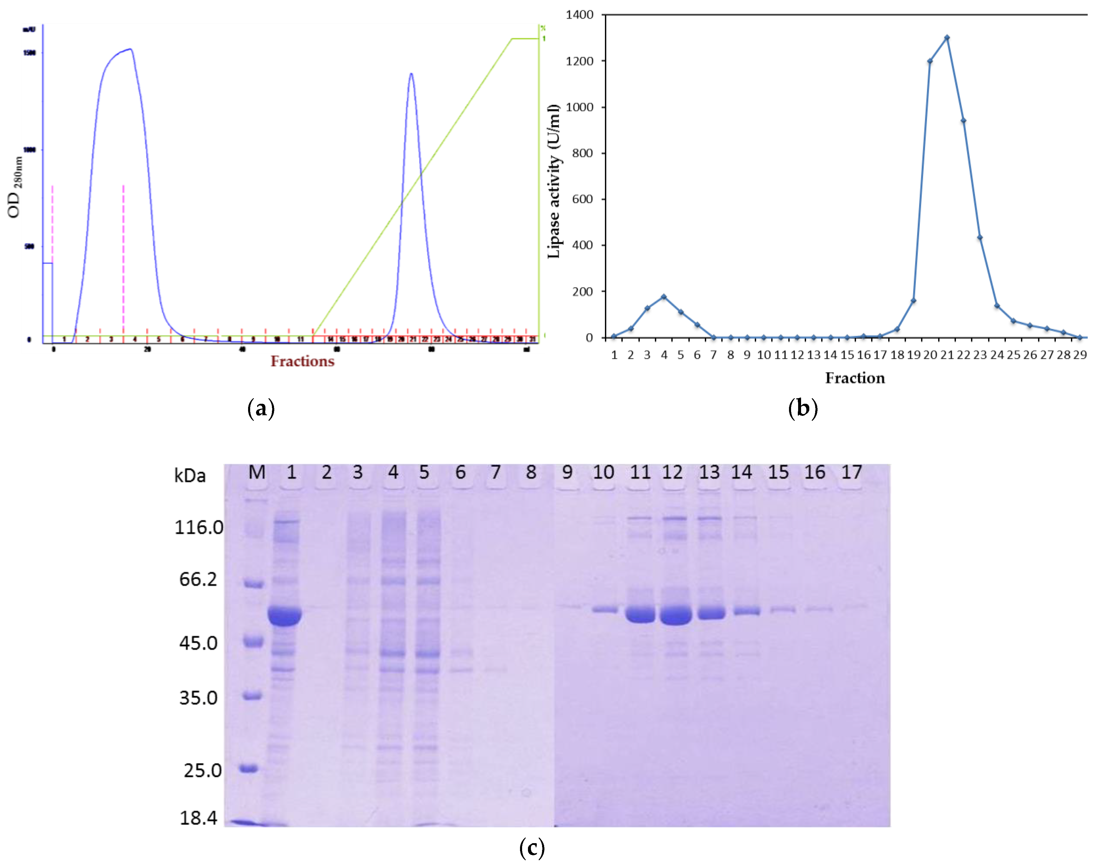
2.1. Purification of Recombinant Lipase

2.1.1. Affinity Chromatography
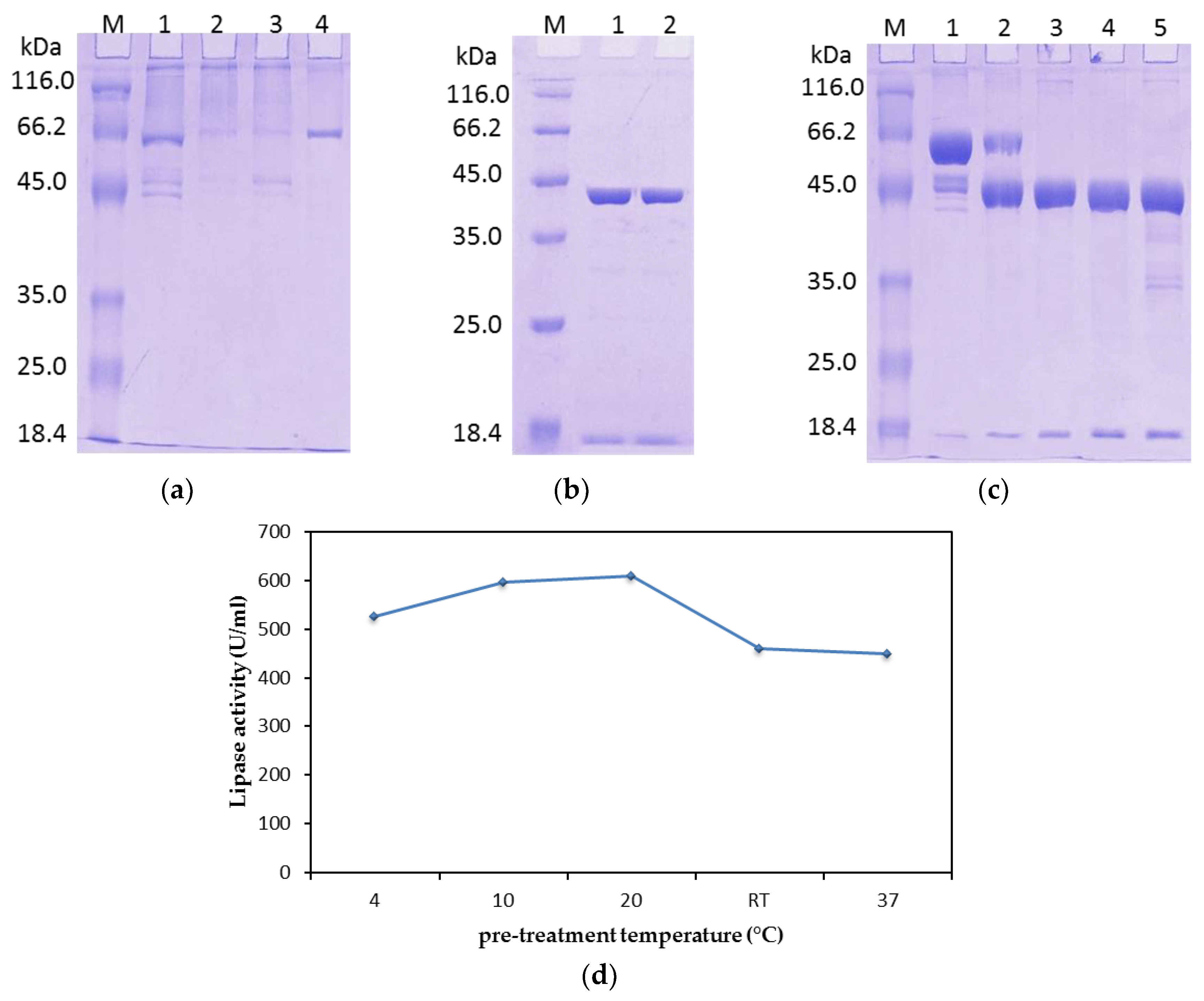
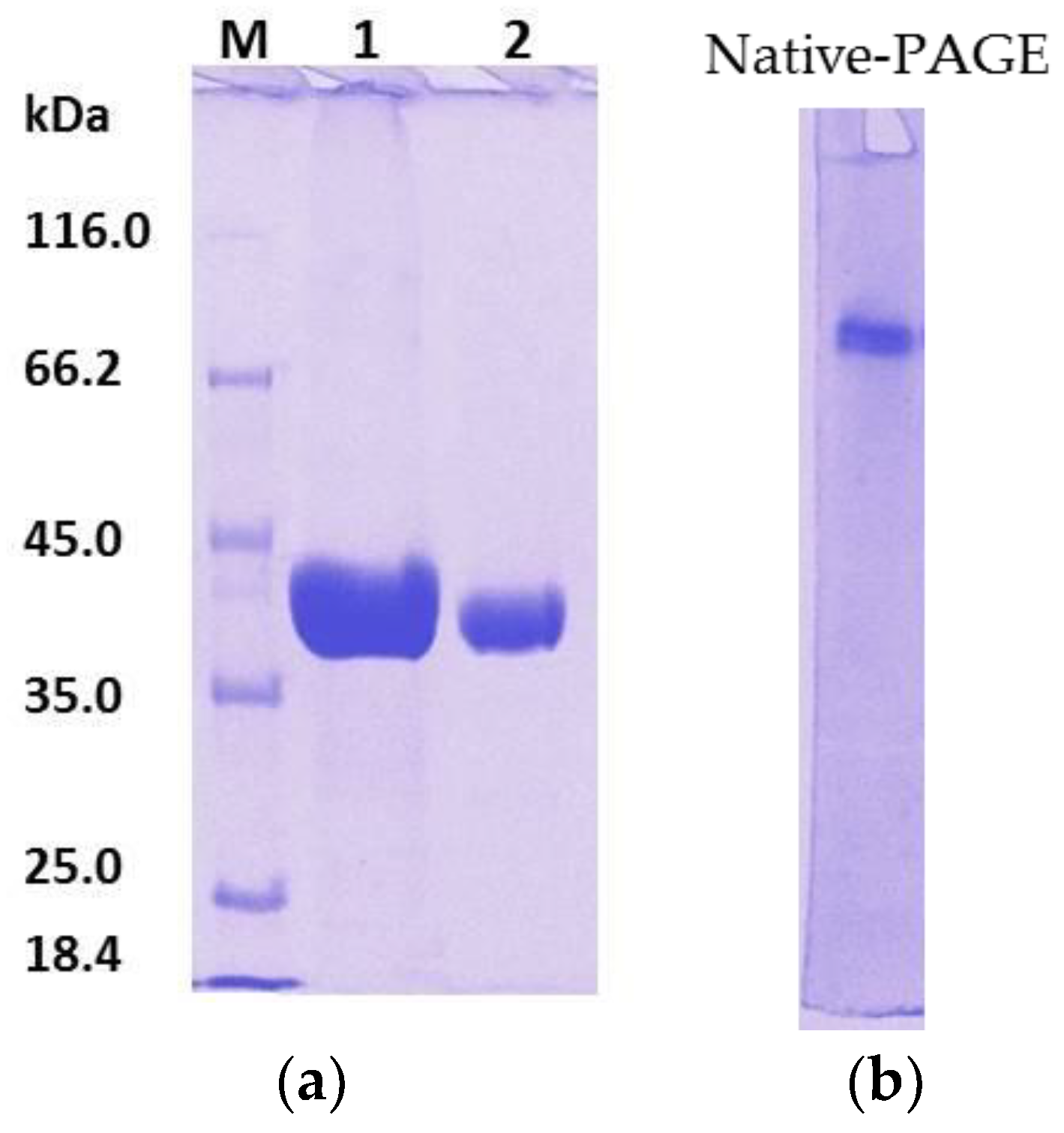
The fusion recombinant HZ lipase (rHZ lipase) was loaded onto a Ni-Sepharose chromatography column, and 82% of the loaded active enzyme adsorbed to the resin. To reduce nonspecific binding between the Ni+ ions (ligand attached to the matrix) and non-tagged proteins, imidazole was added to the washing buffer at a final concentration of 40 mM. In addition, NaCl at a final concentration of 500 mM was used in both washing and elution buffers to prevent unwanted ionic interaction. A linear gradient of imidazole up to 500 mM was used and the bound fusion rHZ lipase was eluted between 170 and 280 mM imidazole concentration, as shown in Figure 1a. All fractions were assayed for lipase activity (Figure 1b), and those fractions with enzyme activity run on sodium dodecyl sulfate–polyacrylamide gel electrophoresis (SDS-PAGE) (Figure 1c). Based on the results shown in Figure 1b,c, fractions 19 to 23 were pooled and used for the second step of the purification procedure. As shown in Table 1, the recovery obtained was 82% with a purification level of 1.1-fold. Additionally, the specific activity of purified fusion lipase increased from 230.60 U mg−1 (crude cell lysate) to 525.25 U mg−1 (purified fusion lipase).

2.1.2. Tag Removal

Prior to the tag cleavage, the purified fusion rHZ lipase was dialyzed against 100 mM Tris-HCl buffer, pH 8.0 for overnight. Then, 10 μg of the dialyzed fusion rHZ lipase was incubated with 3 different units of Recombinant Enterokinase (rEK), 01., 0.2 and 0.5 U, to determine the optimum and effective concentration of rEK to cleave the triple tags from fusion rHZ lipase. As shown in Figure 2a, 0.1 to 0.5 U of rEK was not enough to cleave the tag, so in the next step, the concentrations were increased to 1 and 1.5 U. The higher concentration of rEK was able to cleave the tag (Figure 2b). Hence, 50 U of rEK was required to cleave the tag of 2.5 mg of fusion rHZ lipase which was not economically even for the pilot experiment.
During long storage of the rHZ lipase in the chiller, the fusion protein showed degradation, while the activity of the protein was increased. So, the protein was incubated at different temperatures for 48 h, and then SDS-PAGE analysis (Figure 2c) was performed followed by enzyme activity assay (Figure 2d). The results showed that the tag could be auto-cleaved at 20 °C onward after 48 h incubation; however, the activity was highest (610.0 U mL−1) at 20 °C.
After treatment at 20 °C, the cleaved tag and protein aggregation were removed by centrifugation at 10,000× g for 2 min at 4 °C. The fusion rHZ lipase was purified by cycles of incubation at 20 °C and centrifugation (Figure 3a). From 50 mL of bacterial culture, 21.8 mg of the mature target protein was recovered after 48 h incubation. The electrophoresis of the purified protein on Native-PAGE confirmed the homogeneity of the mature protein (Figure 3b). At the final step of rHZ lipase purification, a 1.3-fold purification was achieved, with a final yield of 78.9% (Table 1). This method allowed easy and efficient separation of the pure, native goal protein without running extra chromatographic separations.

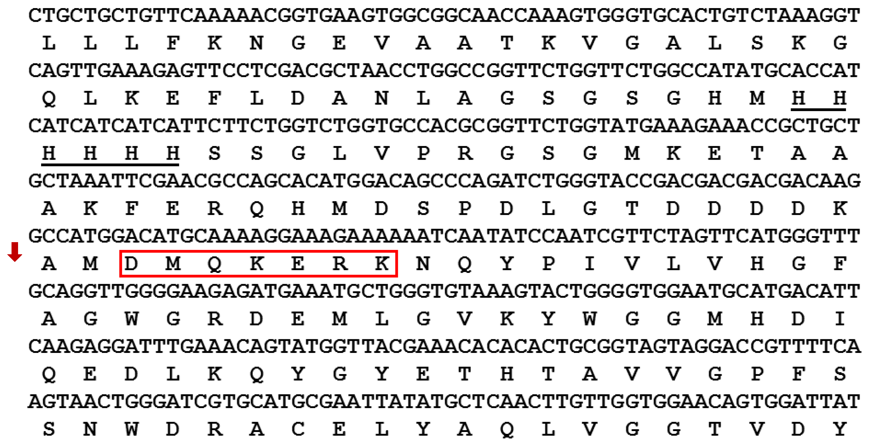
2.1.3. N-Terminal Sequencing

N-terminal sequencing was carried out using an Applied Biosystems 494 Procise Protein Sequencing System (Applied Biosystems, Foster City, CA, USA), to specify the amino acids at the N-terminus of the mature rHZ lipase after treatment at 20 °C to cleave the tags (Table 2). Sequential Edman degradation of the purified mature rHZ lipase showed that the tags were removed from one amino acid before the native start codon (Methionine) (Figure 4). Therefore, further Enterokinase cleavage was not required to remove the tags from the N-terminal part of the enzyme. The Edman degradation profile of mature rHZ lipase peak fraction is shown in Figure S1.

2.2. Characterization of Mature rHZ Lipase

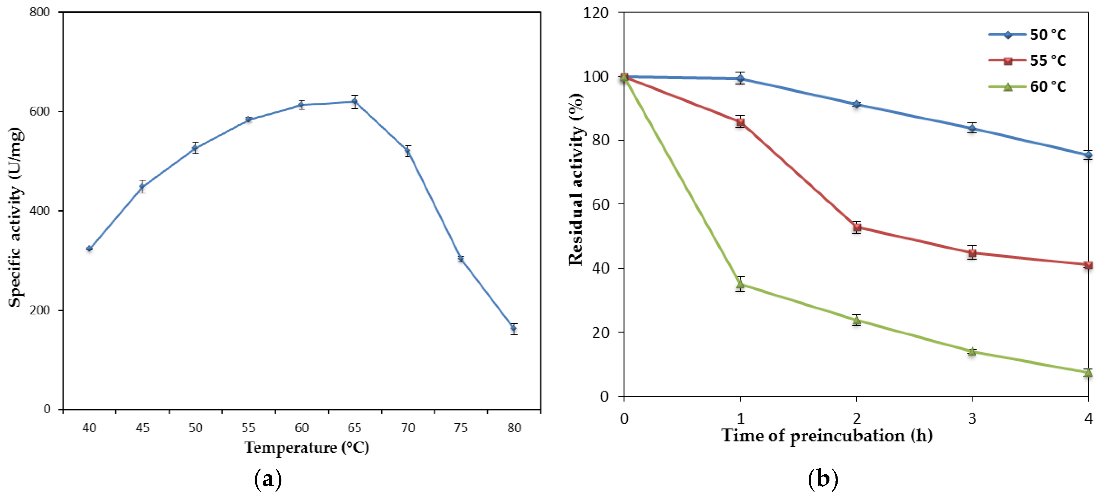
2.2.1. Effect of Temperature on the Lipase Activity and Stability

The influence of temperature on the mature rHZ lipase was studied in the range of 40 to 80 °C. The purified enzyme showed higher activity towards olive oil at 65 °C and had substantial activity over a wide range of temperatures (40–75 °C) (Figure 5a). A one-way analysis of variance (ANOVA) showed that the differences between the specific activity of rHZ lipase at different assay temperatures were significant (P < 0.05) except for the temperatures of 60 and 65 °C, which showed no statistical differences (P > 0.05) were observed. Therefore, the optimum temperature of rHZ lipase was between 60 and 65 °C. The specific activity of the purified enzyme increased gradually from 52.1% at 40 °C to the maximum of 100% at 65 °C, then steeply reduced to 26.3% at 80 °C. The result showed that lipase retained 84% of its original activity at 70 °C.
To analyze temperature stability, the mature rHZ lipase was pre-incubated at 50, 55 and 60 °C for 4 h before measuring the lipase activity. As shown in Figure 5b, the lipase was stable at 50 °C (> 70% residue activity) at up to 4 h of treatment. The half-lifes of mature rHZ lipase at 55 and 60 °C were 2 h 15 min and 45 min, respectively. The enzyme was stable up to 55 °C and lost only 14.2% of its native activity after treating at 55 °C for 1 h.

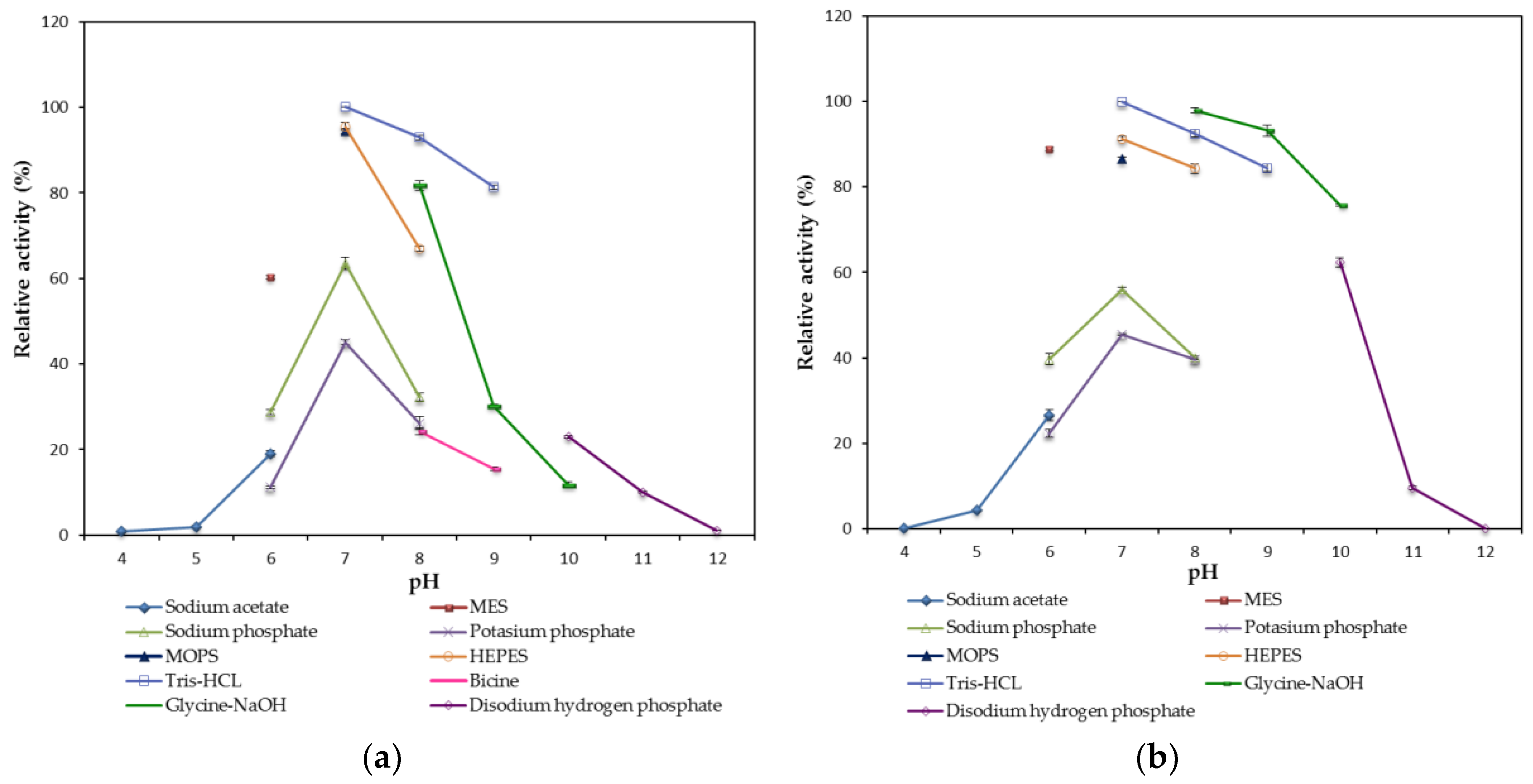
2.2.2. Effect of pH on the Lipase Activity and Stability

A study on the effects of pH on the activity of mature rHZ lipase revealed that the enzyme could operate in a wide range of pH (65 °C) (Figure 6a). The purified lipase showed more than 60% hydrolytic activity at pH 6.0 to 9.0 compared to the maximum at pH 7.0.
At pH 10.0, the activity of mature rHZ lipase was decreased to 23%, and there was no lipase activity at pH 12.0. At acidic pH (6.0) 60.3% of relative activity was observed compared to the optimum pH. However, the enzyme lost its activity at pH 4.0 and 5.0 using sodium acetate buffer. These results illustrated that higher rHZ lipase activity was at pH range of 6.0 to 9.0 and low activity was at extremely acidic or alkaline pH. The activity of rHZ lipase was very low in the presence of sodium phosphate, potassium phosphate and bicine compared to the other buffers such as MES, MOPS, HEPES, Tris-HCl and Glycine-NaOH.
The stability of purified mature rHZ lipase was studied at the pH (65 °C) range of 4.0–12.0 by pre-incubating the enzyme at 50 °C in buffers with different pH values for 30 min (Figure 6b). The enzyme was very stable at pH 6.0–10.0, and it was able to retain 89 and 75% of its activity after 30 min treatment at pH 6.0 and 10.0 compared to the control, respectively.

2.2.3. Effect of Metal Ions and Inhibitors on the Lipase Activity

The influence of different metal ions on the mature rHZ lipase activity at 1 mM and 5 mM final concentration was studied (Table 3). The metal ions from Group Ι of the periodic table (Li+, Na+, K+, Rb+, and Cs+) at 1 mM concentration did not strongly affect the rHZ lipase activity. In the presence of Li+, K+ and Cs+ lipase activity decreased 17.3, 14.8 and 4.7% compared to the control, respectively. But increasing the concentration of alkali metals up to 5 mM enhanced the lipase activity up to 18%. Metal ions from Group II of the periodic table (Mg2+, Ca2+, and Sr2+) at both concentrations of 1 and 5 mM significantly increased rHZ lipase activity. In the presence of 5 mM Ca2+ and Sr2+, rHZ lipase activity had 37.3 and 27.8% increments compared to the control, respectively.
Heavy metals (Fe2+, Ni2+, Cu2+, and Zn2+) had a reducing effect on rHZ lipase activity, except Mn2+ and Co2+. In the presence of 1 and 5 mM concentrations of Mn2+, lipase activity increased up to 5% and 12% compared to the control, respectively. At 1 mM concentration, Co2+ ion had no significant effect on rHZ lipase activity, while at 5 mM concentration, it had ~20% decreased on rHZ lipase activity. Fe2+, Ni2+, Cu2+, and Zn2+ inhibited the activity of rHZ lipase more than 42% at 1 and 5 mM concentrations.
One-way ANOVA analysis showed that the difference between lipase activity of control and other types of tested metal ions at 1 and 5 mM concentrations was significant (P < 0.05). However, there were no statistical differences between control and Na+, Rb+, Cs+, Mn2+ and Co2+ at 1 mM concentration. In addition, there were no statistical differences between Na+, K+, Rb+, Mg2+ and Mn2+ at 5 mM concentrations (P > 0.05).
Several metal-chelating agents, reducing agents, serine, and aspartic inhibitors were tested to investigate their inhibitory impact on rHZ lipase and verify the roles of certain amino acids in the structure and enzyme catalysis (Table 3). The metal chelating agent EDTA had a strong inhibitory effect on rHZ lipase activity, almost 100%. The reducing agents, DTT and 2-mercaptoethanol were the most effective agents in activation of the lipase around 21 and 12% at a concentration of 1 mM and around 18 and 6% at 5 mM concentration, respectively.
However, the rHZ lipase activity was inhibited by 19% and 39% in the presence of 1 and 5 mM pepstatin, compared to the control, respectively. The serine hydrolase inhibitor, PMSF showed 30% and 32% inhibitory effect on rHZ lipase activity at concentrations of 1 and 5 mM, respectively. A one-way ANOVA analysis showed that the difference between lipase activity of control and various inhibitors at 1 and 5 mM final concentrations was found to be significant (P < 0.05). However, there were no statistical differences between PMSF and pepstatin at a concentration of 5 mM (P > 0.05).

2.2.4. Substrates Specificity of the rHZ Lipase towards Triacylglycerols and Natural Oils

One of the important characteristics of lipases is their substrate which defines their ultimate usage and is a critical assessment. Substrate specificity of rHZ lipases was perused by comparing the hydrolytic rate of several synthetic triglycerides and natural fats to olive oil (Figure 7). In general, lipases prefer the natural oils compared to the synthetic triglycerides. The rHZ lipase showed broad substrate specificity towards triglycerides from C2 to C18 with maximum affinity to triolein (C18).
The rHZ lipase hydrolyzed all the tested natural oils with the highest activity toward olive oil except palm kernel oil, in which the major fatty acyl chain is lauric acid (C12:0). Other oils that caused relative activity of more than 40% as the substrate contained long, unsaturated fatty acyl chains, especially C18:1 and C18:2 (Table S1), such as olive oil, corn oil, rice bran oil, and soybean oil.
Moreover, a one-way ANOVA analysis showed that the differences between the lipase activity of olive oil and other tested substrates was statistically significant (P < 0.05). However, there were no significant differences between triolein, rice bran oil, soybean oil and corn oil (P > 0.05).

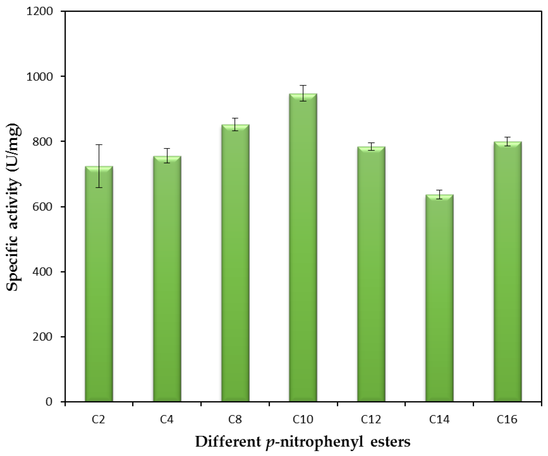
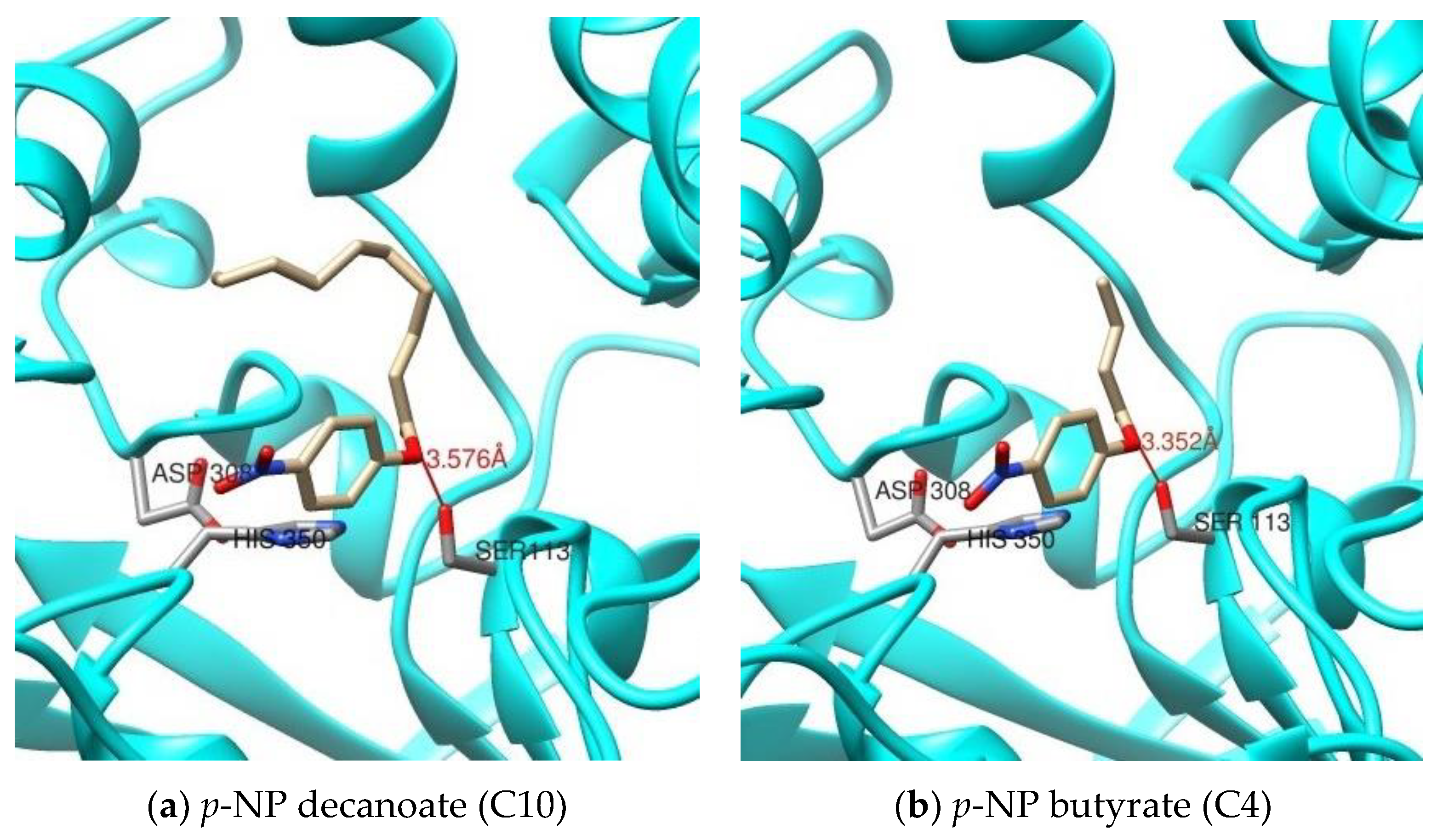
2.2.5. Substrate Specificity of the rHZ Lipase toward p-Nitrophenyl Ester Substrates and Molecular Docking

The rHZ lipase hydrolyzed all the p-nitrophenyl esters with different acyl chain lengths and the highest hydrolysis rate was observed for 4-nitrophenyl decanoate (C10) (Figure 8). The lowest hydrolysis occurred toward 4-nitrophenyl myristate (C14) which showed 67% specific activity compared to the maximum (4-nitrophenyl decanoate (C10)). A one-way ANOVA analysis showed that the differences between specific activity of 4-nitrophenyl decanoate (C10) and other p-nitrophenyl esters were found to be significant (P < 0.05). However, among natural substrates, the enzyme preferred those substrates with longer carbon chain such as olive oil.
Hence, to explain the substrate specificity of the rHZ lipase, protein-ligand docking was performed to determine the structure of the intra-molecular complex formed between the rHZ lipase and different p-nitrophenyl substrates. In YASARA, the output of the docking runs is organized based on the binding energy. YASARA docking gives positive binding energy. Therefore, a higher binding energy indicates a higher affinity between the receptor and ligand [13]. After clustering 25 runs for each substrate, the results were analyzed and the best complex conformation of the enzyme and substrate with higher binding energy and the lower distance between hydroxyl (Oγ-Ser113) and the carbonyl carbon of the substrate were chosen for further analysis (Table 4 and Figure S2).
The distances between hydroxyl (Oγ-Ser113) and the carbonyl carbon of the substrates ranged from 3.352 to 3.932 Å (Table 4). The protein-ligand interface for each substrate exhibited the highest binding energy of 4.444 kcal/mol for p-NP decanoate (C10) and the lowest binding energy of 3.201 kcal/mol for p-NP butyrate (C4) (Figure 9). In addition, the smaller dissociation constant (Kd) value indicated the greater the binding affinity of the ligand for its target which was calculated for p-NP decanoate (C10) compared to the other substrates. The molecular docking study results of rHZ lipase were in an agreement in terms of the hydrolysis rate of p-nitrophenyl esters in the wet experiment.

2.2.6. Determination of Km and Vmax of the rHZ Lipase

The effects of substrate concentration on rHZ lipase activity were determined using 4-nitrophenyl decanoate as a substrate. The rHZ lipase (0.5 mg mL−1) was assayed over a range of p-NP-decanoate concentrations from 0.1 to 0.22 mM. Figure S3 shows the progress curve of products formed at different substrate concentration as a function of time. The products formed increased as the substrate concentration increased. The curve reveals a linear gradient from 0 to 0.5 min. From this linear gradient, initial velocity for each substrate concentration was calculated to plot a Michaelis-Menten curve. GraphPad PRISM software (San Diego, CA, USA) was used to plot the Michaelis-Menten curve and least-squares nonlinear regression analysis was performed. The best-fit values of Km and Vmax from the analysis were 0.212 mM and 87.42 µmole min−1, respectively. The regression coefficient, R2 was at 0.9888. The Km value of the enzyme estimated from Lineweaver-Burk plot and Hanes-Woolf plot were 0.223 mM and 0.212 mM, respectively. The Vmax value obtained from the two plots were 89.29 µmole min−1 and 88.50 µmole min−1, respectively. Lineweaver-Burk plot provided the regression coefficient, R2 = 0.9378 while Hanes-Woolf was R2 = 0.8494. Table 5 provides the summary of Km and Vmax value from the three plotted graphs.

2.2.7. Effect of Organic Solvent on the Lipase Activity

In enzymatic reactions, using organic solvents as reaction media offers great advantages to industry compared to the usual aqueous reaction systems. The mature rHZ lipase was very stable in water-miscible solvents; glycerol (log P −0.3) and DMSO (log P −1.3) and it showed a 128% increase in activity in the presence of dimethyl sulfoxide (Table 6). The enzyme retained 67% of its activity in the presence of methanol (log P −0.76) compared to the control (without solvent), whereas almost all other alcohols with log P values of between −0.18 and 1.36 inactivated the enzyme.
In water-immiscible organic solvents such as benzene (log P 2.0), toluene (log P 2.5), isooctane (log P 4.7), n-tetradecane (log P 7.6) and n-hexadecane (log P 8.86), the relative activity of mature rHZ lipase increased around 2 to 8%. However, xylene (log P 3.15), octane (log P 5.2) and n-tridecane (log P 7.33) demonstrated minor (≤5%) inhibition effects on the rHZ lipase activity.
Subsequently, one-way ANOVA analysis showed that the differences between relative activity of control and organic solvents with log P -0.3 to 2.0 were found to be significant (P < 0.05). However, there were no statistical differences between toluene (log P 2.5), xylene (log P 3.15), hexane (log P 3.9), isooctane (log P 4.7), octane (log P 5.2), decane (log P 5.8), n-tridecane (log P 7.33), n-tetradecane (log P 7.6) and n-hexadecane (log P 8.86) (P > 0.05).

3. Discussion

Protein purification is a necessary step to characterize the function, structure and interactions analysis of protein for fundamental study or industrial purposes [14]. Such applications require a specific amount (from microgram to kilogram scales) of purified protein in different quality (e.g., partial or fully purified). The purification method could affect the quality and quantity of the protein, which must fit the intended use. For the majority of industrial purposes, the most important aspect is an economical, rapid, high yielding production process which could be shifted to large-scale operation. However, for fundamental studies, high purity of a protein is the first top priority, among others. To simplify the purification step(s) and detection, the expression of a recombinant protein attached to a tag of known size and biochemical properties, such as histidine tag, is one of the best options. The fusion His-tag proteins have the strong selective affinity toward metal ions such as Ni2+, Co2+, Cu2+, and Zn2+ which can be immobilized on chromatographic resin using chelating ligands [15]. Nevertheless, a fusion tag linked to the protein enzyme could alter the catalytic properties of the enzyme [16]; also, for crystallization purposes, only the mature protein is required so after affinity chromatography the tag needs to be cleaved.
There are a few reports that the fusion tag has been cleaved using protease treatment such as PF2001Δ60 lipase from Pyrococcus furiosus and T1 lipase from Geobacillus zalihae that cloned into pET-32a and pGEX plasmids, respectively [17,18]. In pET-32b vector using Enterokinase enzyme, tags can be cleaved from mature protein after lysine residue at its recognition site: DDDDK. However, the cost of the Enterokinase is prohibitively expensive for the production of one gram of mature protein.
To overcome this problem, after affinity the chromatography step, treatment of rHZ lipase at 20 °C was a straightforward and efficient technique that presented a non-enzymatic cleavage and non-chromatographic purification procedure. This method induced specific auto-cleavage of the target protein and precipitation of few undesirable E. coli proteins that eluted along with rHZ lipase. N-terminal sequencing confirmed that 17.4 kDa tag has been successfully removed from one amino acid before the methionine start amino acid, DDDDKAM↓DMQ, which is an advantage of this new technique. The obtained final yield was 79% which was higher than the purified T1 lipase expressed in E. coli systems with a final recovery of 51.5% [17], the purified BTL2 lipase expressed in a Pichia pastoris system with a final recovery of 18% [19], P1 lipase which purified through ultrafiltration and Q Hyper D column with a final recovery of 71% [20], and recombinant His-tagged L2 lipase that purified by single-step affinity chromatography with a final recovery of 76.1% [21].
A high kosmotropic salt (sodium, potassium, or ammonium sulfates) concentration enhances the hydrophobic interaction and protein precipitation due to higher “salting-out” or molar surface tension increment effects, while lowering the salt concentration weakens the hydrophobic interaction [22]. The fusion rHZ lipase was purified in 500 mM NaCl concentration; the salt was then removed by dialysis, which could be harsh to the Trx-His-S-tags protein. In addition, it is possible that the aggregation of the triple tag could not be reversed by removing the salt. Therefore, dialysis by incubation of the protein at 20 °C for a longer period (48 h) promoted the cleavage of the tag. However, the thermostable rHZ lipase structure was rigid enough not to undergo any conformational changes during dialysis and treatment at 20 °C.
The differences in the lipase activity of fusion and mature rHZ lipase (Figure 2d) showed that the presence of 17.4 kDa tag has a considerable effect on the active site of the enzyme. So, the biochemical characteristics of mature rHZ lipase which is the first member of subfamily I.9 true lipase from Aneurinibacillus thermoaerophilus strain HZ have been studied. Based on the gene sequence analysis, rHZ lipase is close to mesophilic Bacillus lipases. The predicted structure also revealed some differences between rHZ lipase and other thermostable lipases [10]. However, a study on the effect of temperature of rHZ lipase showed that the enzyme has high optimum activity and moderate stability at elevated temperatures which are comparable with thermostable lipases. The optimum reaction temperatures of 60–65 °C for rHZ lipase were higher than the majority of thermostable lipases, which had optimum temperatures of around 45–50 °C [23]. In addition, the rHZ lipase showed good stability at 50 °C which is lower than its optimum activity, but it maintained half of its activity for 45 min at 60 °C. The wild-type HZ lipase (wHZ lipase) has also an optimum temperature of 65 °C, but higher thermostability compared to the rHZ lipase, the half-life of 3 h 10 min at 65 °C [24]. Several lipases have been to be unstable at their optimum temperatures such as lipase from B. stearothermophilus MC7 [25] and BTL2 lipase from B. thermocatenulatus BTL2 [19]. To pursue any strategy to stabilize an enzyme such as preventing or decelerating of losing activity, it must be considered that enzyme inactivation may be related directly to shifting the native state (N) ⇄ unfolded enzyme (U) equilibrium or preventing the important irreversible process that happens after formation of unfolded protein [26]. To prevent these process and stabilize the enzyme, there are some soluble additives such as substrates, low molecular weight organic solutes, salts, polymeric solutes and synthetic polymers [27].
The maximum hydrolytic activity of mature rHZ lipase was noted at pH (65 °C) 7.0 using Tris-HCl buffer and the enzyme retained > 75% of its activity at the pH range of 6.0 to 10.0 after 30 min pre-incubation at 50 °C indicating that the purified enzyme is a neutral lipase. The wHZ lipase preferred potassium phosphide buffer pH (65 °C) 7.0 as an optimum buffer [24] while rHZ lipase lost more than 50% of its activity in the presence of potassium phosphide buffer pH (65 °C) 7.0 (Figure 6a). The optimal lipases activity of several thermostable lipases found in the moderate to highly alkaline pH range from 8.5–10.0, such as lipases from G. zalihae sp. T1 with the optimum pH at 9.0 and more than 60% stability at pH 9 to 11 [17], and B. stearothermophilus L1 [28] with optimum pH at 9–10 and 24 h stability at pH 5 to 11. On the other hand, two thermostable lipases from B. stearothermophilus SB-1 [29] and Bacillus megaterium CCOC-P2637 [30] showed optimum activity in a pH range of 3.0–6.0, lower than reports of optimum pH for Bacillus lipases. Meanwhile, using multiple buffer systems to cover a wide range of pH in this study enabled to detect the effect of buffer compound on the activity of the enzyme. Overlapping assay on a specific pH was done, when the buffer system was changed. The result indicated that the compound of buffers had also effect on the hydrolytic activity of rHZ lipase such as MES and phosphate buffers (Na+ and K+) at pH 6.0 and those other overlap buffers at pH 7.0 to 9.0 (Figure 6).
Changing the pH could alter the ionic bonds in the enzyme structure whereas these bonds are important to structural stability and help to determine the 3D feature and functionality of the protein enzyme. The solubility of the protein increases at pH values farther away from its isoelectric point due to greater electrostatic repulsions [31]. Therefore, rHZ lipase was less active at a pH near or far below/above its pI (5.74) as the enzyme might be inactivated or formed aggregates during treatment. These results help in choosing the appropriate buffer for enzyme crystallization.
Characteristic of mature rHZ lipase revealed that various metal ions had different impacts on the activity of the enzyme. Similar to mature rHZ lipase, most Bacillus lipases maintained their initial activities or were slightly activated (5–20% higher than the maximum activity) in the presence of up to 10 mM potassium ion. Calcium ions have been reported to increase the activity of most Bacillus lipases 10 to 50% compared to the maximum (Table S2), and the same results were also observed for rHZ and wHZ lipases [24]. Additionally, several reports have illustrated that calcium ion could maintain the activity of the lipases at elevated temperatures [23]. In addition, a Ca2+-binding motif, GXXGXD, reported in the protein sequence of Staphylococcus epidermidis lipase [32] and lipA from Acinetobacter sp. RAG-1 [33] was found in the gene sequence of rHZ lipase unlike the other mesophilic or thermostable Bacillus lipases. This motif shows the rHZ lipase can be a Ca2+-dependent lipase. The mature rHZ lipase lost its activity in the presence of most of the heavy metal ions tested in this study similar to the majority of thermostable Bacillus and Geobacillus lipases [23]. The inhibition of rHZ lipase activity in the presence of heavy metal ions might be due to the toxic effect of the excess amount of these ions in the reaction medium. The ionization of surface charged amino acid residues on the enzyme by the interaction of them with salt ions could significantly change the enzyme conformation followed by its activity [34].
Generally, some structural features of enzymes can be demonstrated by studying the impact of inhibitors. Unlike wHZ lipase, the complete activity inhibition of mature rHZ lipase by EDTA (metal-chelating agent) suggests that the enzyme is a metalloenzyme which supported that Ca2+-dependently of rHZ lipase. Moreover, the addition of DTT and 2-mercaptoethanol (reducing agents) to the rHZ lipase reaction promoted this activity which, suggests that there is no disulfide bond in rHZ lipase structure. Guncheva and Zhiryakova [23] reported that reducing agents decreased the activity of lipases from subfamily I.5, since disulfide bridges not only support the active site conformation, also play important roles on the thermostability of these enzymes. Hence, the activation of thermostable rHZ lipase by reducing agents can explain the structural differences of this enzyme and its lower thermostability compared to the other thermostable lipases belonging to subfamily I.5 of true lipases. The mature rHZ lipase activity inhibition by PMSF indicated the presence of a serine residue at the catalytic triad of the active site. The enzyme might belong to serine hydrolase. PMSF strongly inhibited the activity of the most of lipases from Bacillus genus by irreversible binding to the catalytic serine residue. However, it has also been reported that PMSF did not inhibit the activity of a thermostable lipase B from G. thermoleovorans ID-1 [35].
Most lipases contain a mobile lid domain placed over the active site which is also responsible for catalytic activity. The movements of amino acids in the lid are different in closed or open conformations; hence, the amino acid sequence of the lid defines the activity and specificity of lipases [36]. The mature rHZ lipase showed preference to hydrolyze the long chain triacylglycerides among all tested oils with the highest affinity to olive oil while wHZ lipase has 30% higher preference toward the sunflower oil compared to the olive oil [24]. Similarly, the lipases from G. zalihae sp. T1 and Bacillus sp. L2 exhibited a preference for natural oils such as olive, corn, palm, and soybean oils that mostly are contained unsaturated fatty acids [17,37] even though the sequence of amino acids in the lid domain of rHZ lipase and thermostable lipases from subfamily I.5 are different. The high activity of mature rHZ lipase to hydrolyze the natural oils can make it suitable for modification and improvement of physicochemical properties and nutritional value of the natural oils and formation of an extensive variety of saturated and polyunsaturated fatty acids [23]. Mesophilic and thermostable lipases from subfamily I.4 and I.5 have shown a different preference for p-nitrophenyl esters substrate, which could be due to their different protein amino acid sequences and biochemical characteristics. Lipase BTL2, Lip B and P1 lipase are thermostable enzymes which shown higher preference toward the p-nitrophenyl decanoate (C10) compared to the other p-nitrophenyl esters (Table S2). The highest activity of mature rHZ lipase towards p-nitrophenyl decanoate (C10) and olive oil was 947.7 and 620 U mg−1 at 65 °C, respectively, strongly indicating that rHZ lipase is a true lipase [38]. Besides, the enzyme showed more than 75% of hydrolysis activity toward both shorter (4-nitrophenyl acetate (C2)) and longer acyl chain (4-nitrophenyl palmitate (C16)) lengths p-nitrophenyl esters which was in line with the result of molecular docking study. Molecular docking helps to predict the possible position and orientation of ligand in protein binding site. The docking results of rHZ lipase showed that apart from the size of acyl chain length, the conformation of active site and the amino acid sequences were important parameters to determine the preference of rHZ lipase toward of p-nitrophenyl esters substrate. This makes the rHZ lipase an ideal catalyst for organic synthesis in aqueous and non-aqueous conditions.
Most enzymes applied in industrial activities have a range of Km values between 10−1 to 10−5 M when acting on biotechnological important substrates [39]. It has been reported that the Km and Vmax values of thermostable lipase BTID-B for the hydrolysis of tricaprylin was 6.24 mM and 63.3 μmol min−1 mg−1, respectively [35]. However, Massadeh et al., [40] reported that a lipase from Bacillus stearothermophilus HU1 had a Km and Vmax value of 0.235 mM and 161.2 nmol min−1, respectively when using p-NP-Palmitate as substrate. A similar study using p-NP-Palmitate was conducted on a purified thermostable lipase from Bacillus stearothermophilus MC7, which was found to have a Km and Vmax values of 0.33 mM and 188 μmol min−1 mg−1, respectively [25]. This suggests that the rHZ lipase had a high potential in industrial applications as it has a Km value between the required range.
In the presence of organic solvents, native enzymes generally show low activities and stabilities limiting enzymatic reactions [41]. Lipase performance in different solvents is associated with its efficiency in hydrolytic and synthetic reactions [23]. Exposure of the mature rHZ lipase to various organic solvents for 30 min ascertained that the enzyme was stable to both very polar and non-polar organic solvents. Polar water-miscible organic solvents destabilize the enzymes by eliminating the solvation water from the enzyme surface area [42] but increasing the activity of mature rHZ lipase in the presence of 25% DMSO could be due to the prevention of enzyme aggregating in the reaction mixture [30]. Lee et al. [35] reported that the activity of thermostable lipase BTID-B enhanced in the presence of 1% DMSO in the reaction medium. Nonpolar solvents are often less harmful to the enzymes because they contain two phases in water + water-immiscible organic solvent system; an immiscible organic solvent phase and an aqueous phase. The enzymatic reaction proceeds in the aqueous phase, which contains a dissolved enzyme. In the biphasic system, the enzyme is relatively stable due to the less direct contact of the enzyme with an organic solvent [43]. This phenomenon indicates the presence of more charged amino acid residues on the surface of rHZ lipase which is in an agreement with a report on the analysis of the predicted structure of the enzyme [10]. On the other hand, alcohol organic solvents decreased or inactivated the mature rHZ lipase. Guncheva and Zhiryakova [23] reported that most Bacillus lipases were inactivated by polar alcohol solvents. The majority of thermostable lipase from subfamily I.5 could not tolerate the presence of organic solvents or they tolerate a certain polarity of solvents. Interestingly, wHZ lipase has a closely similar organic solvent activity profile with rHZ lipase, except it stimulated wHZ lipase activity in the presence of methanol. In terms of the stability profile of the rHZ lipase for a wide polarity range of organic solvents, it showed a promising potential for industrial chemical processes.

4. Materials and Methods

4.1. Strains and Materials

The rHZ lipase gene was isolated by genomic DNA library construction and deposited in the GenBank database under accession number GU272057 [10]. Escherichia coli BL21 (De3), and pET32b (+) plasmids were purchased from Merck (Novagen, Germany). Ni-Sepharose 6 Fast Flow resin was obtained from GE Healthcare (Little Chalfont, Buckinghamshire, UK). All the chemicals used were of analytical grade.

4.2. Expression of rHZ Lipase

The expression of thermostable and organic solvent tolerant rHZ lipase was conducted in a transformed cell E. coli BL21 (De3) harboring pET-32b/rHZ. The recombinant clone was propagated in 200 mL of LB-broth supplemented with 50 µg mL−1 of ampicillin (Merck, Darmstadt, Germany) and incubated at 37 °C with 200 rpm shaking rate. When the OD600nm of the culture reached 0.5, isopropyl β-D-1-thiogalactopyranoside (IPTG) (Thermo Fisher Scientific, Waltham, MA, USA) was added to the final concentration of 0.025 mM to induce the expression of the cloned lipase gene. The cells were incubated further at 30 °C for 8 h.

4.3. Purification of Recombinant Lipase

4.3.1. Sample Preparation

Cells were harvested from 50 mL culture of recombinant E. coli BL21 (DE3) (pET32b/rHZ) culture by centrifugation at 10,000× g for 10 min at 4 °C. The supernatant was discarded, and the bacterial pellets were then resuspended in 15 mL binding buffer containing 100 mM Tris-HCl (pH 8.0, manufacturer, city and country) buffer, 500 mM NaCl and 40 mM imidazole. The cells were disrupted by sonication (Branson 250 sonifier: output 2, duty cycle 30, Branson Ultrasonics Corp., Danbury, CT, USA) on ice for approximately 4 min (in several short bursts). The supernatant was cleared by centrifugation (10,000× g for 10 min at 4 °C) and filtered through a 0.45 μm filter to remove cell debris or other particulate material and used as a crude enzyme.

4.3.2. Affinity Chromatography

The crude enzyme was applied onto 5.0 mL Ni-Sepharose 6 Fast Flow resin in an XK 16/20 column (GE Healthcare, Uppsala, Sweden), equilibrated with binding buffer (100 mM Tris-HCl (pH 8.0), 500 mM NaCl and 40 mM imidazole) at a flow rate of 0.5 mL/min. The column was washed with the same buffer until no protein was detected. The bound protein was eluted gradiently with 100 mM Tris-HCl (pH 8.0) buffer supplemented with 500 mM NaCl and 500 mM imidazole. Fractions (approximately 2.5 mL each) which exhibited lipase activities were collected and processed to the next step.

4.3.3. Treatment of the rHZ Lipase to Remove the Tags

The cleavage of the triple tags (Trx-His-S-tags) present at the N-terminal region of the rHZ lipase was optimized using Recombinant Enterokinase (Novagen, Madison, WI, USA) based on manufacturer’s instructions. The fusion protein was dialyzed against 100 mM Tris-HCl buffer, pH 8.0 for overnight. Then, the fusion rHZ lipase was treated with 4 different concentration of Recombinant Enterokinase. The three serial dilutions of rEK in rEK dilution/storage buffer was made to produce solutions having 0 (control), 0.1, 0.2, and 0.5 U enzymes per µL. Then, 4 reaction mixtures were prepared each containing 5 µL 10X rEK cleavage/capture buffer, 10 µg fusion rHZ lipase, 1 µL diluted rEK (each tube with 1 µL of a different enzyme dilution) and top up with deionized water to the final volume of 50 µL. The mixtures were incubated at 20 °C. After 16 h incubation, 10 µL of samples were taken and aliquots into 1 µL of 10X SDS sample buffer(10% w/v SDS, 10 mM β-mercaptoethanol, 30% v/v glycerol, 0.35 M Tris-HCl, pH 6.8, 0.1% w/v bromophenol blue (Merck, Darmstadt, Germany)) and the tag cleavage analyzed by SDS-PAGE. In the next experiment, 1 and 1.5 U of rEK was used to cleave the fusion rHZ lipase. The mixtures were incubated at 20 °C for 16 h followed up by SDS-PAGE analysis.
The cleavage rates and activity levels of the fusion rHZ lipase were analyzed by incubating the sample at 4, 10, 20, room temperature (~25) and 37 °C for 48 h. The fusion protein was dialyzed against 100 mM Tris-HCl buffer, pH 8.0 overnight. Then, the protein concentration was adjusted at 0.5 mg mL−1 using the same buffer and aliquoted into 5 different tubes and incubated for 48 h at above-mentioned temperatures. The control was the eluted fusion rHZ lipase from affinity chromatography without dialysis, kept at chiller (4 °C). The enzyme activities of the samples were determined using olive oil as a substrate at 65 °C. The SDS-PAGE analysis showed the cleavage rate of the samples after 48 h incubation.
The samples after treatment at 20 °C for 48 h were centrifuged at 10,000 × g for 2 min at 4 °C and analyzed by SDS-PAGE gel electrophoresis and Native-PAGE.

4.4. Lipase Assay

Determination of liberated free fatty acid was measured by the modified colorimetric method of Kwon and Rhee [24,44] with olive oil (Bertolli, Italy) as a substrate. The reaction mixtures, consisting of 5.0 µL of 0.5 mg mL−1 purified enzyme, 995.0 μL of 50 mM Tris-HCl buffer pH (65 °C) 7.0, 2.5 mL of olive oil emulsion (1:1 ratio of olive oil and 50 mM Tris-HCl buffer pH (65 °C) 7.0) and 20.0 µL of 20 mM CaCl2·2H2O, were incubated at 65 °C with shaking rate of 200 rpm for 30 min. Then, the reaction was stopped by adding 1.0 mL of 6 N HCl and 5.0 mL isooctane. The upper layer (4.0 mL) was pipetted into a test tube and 1.0 mL cupric acetate pyridine [copper (II) acetate monohydrate (5%; pH 6.1), pH adjusted by adding pyridine] was added. The free fatty acids dissolved in isooctane were determined by measuring the OD of the isooctane solution at 715 nm. One unit (U) of lipase activity is defined as the rate of fatty acid formation per min under standard assay condition. Each experiment was done in triplicate and value reported referred to means ± SE unless otherwise stated. The standard deviation was determined by the Microsoft Excel program.

4.5. Determination of Protein Content

The protein content was determined by the method of Bradford [45] using VWR Life Science AMRESCO kit (Radnor, PA USA) based on the manufacturer’s instruction. Bovine serum albumin (BSA, Sigma Chemical Co., St Louis, MO, USA) was used as a standard (Figure S4).

4.6. Gel Electrophoresis

After each step of purification, the purities of the protein fractions and their molecular weights were analyzed using 12% SDS-PAGE gel [46]. Besides, the purified protein was electrophoresed on Native-PAGE (12%), and the protein band was visualized using Coomassie brilliant blue R-250.

4.7. N-Terminal Sequencing

Purified mature rHZ lipase was transferred to 12% of SDS-PAGE gel. N-terminal sequencing was done by APAF Ltd.-Australian Proteome Analysis Facility (Macquarie University, Sydney, Australia). The protein in the gel band was passively eluted from the gel matrix using an SDS elution buffer overnight. The samples were then loaded onto a ProSorb filter cartridge (Applied Biosystems, Foster City, CA, USA) and washed with 0.1% TFA (2 × 100 μL) to remove the SDS and reduce the background contamination. The sample on the PVDF membrane was subjected to 7 cycles of Edman N-terminal sequencing. N-terminal sequencing utilized the well-established Edman degradation chemistry, sequentially removing amino acid residues from the N-terminus of the protein and identifying them by reversed phase HPLC [47]. Automated Edman degradation was carried out using an Applied Biosystems 494 Procise Protein Sequencing System. The performance of the sequencer was assessed routinely with 10 pmol β-Lactoglobulin standard.

4.8. Characterization of Purified rHZ Lipase

4.8.1. Effect of Temperature on the Lipase Activity and Stability

The apparent optimum temperature for mature rHZ lipase was measured by assaying its hydrolytic activities toward olive oil at various temperatures in the range of 40 to 80 °C at 5 °C intervals using 50 µL of the enzyme in assay reaction mixtures.
To study the thermostability of the mature rHZ lipase, the purified enzyme solutions were pre-incubated at 50 to 60 °C with the intervals of 5 °C for 4 h. Then, samples were removed and rapidly cooled in an ice bath before being assayed at 65 °C using olive oil as a substrate.

4.8.2. Effect of pH on the Lipase Activity and Stability

To determine the optimum pH for the mature rHZ lipase, lipase assays were carried out using olive oil emulsion as the substrate in different pH (65 °C) ranging from 4.0 to 12.0. Reaction mixtures were incubated at 65 °C for 30 min with 200 rpm shaking rate for lipase assay. The following buffers (Merck, Darmstadt, Germany) at a concentration of 50 mM were used: sodium acetate (pH 4.0 to 6.0), MES (pH 6.0), sodium phosphate (pH (65 °C) 6.0 to 8.0), potassium phosphate (pH (65 °C) 6.0 to 8.0), MOPS (pH (65 °C) 7.0), HEPES (pH (65 °C) 7.0 to 8.0), Tris-HCl (pH (65 °C) 7.0 to 9.0), bicine (pH (65 °C) 8.0 to 9.0), glycine-NaOH (pH (65 °C) 8.0 to 10.0), disodium hydrogen phosphate (pH (65 °C) 10.0 to 12.0).
The effect of pH on lipase stability was determined by pre-incubation aliquots of rHZ lipase in buffers with different pH values at 50 °C for 30 min. Residual activity was assayed using olive oil emulsion (olive oil in 50 mM Tris-HCl, pH (65 °C) 7.0) as substrate at 65 °C for 30 min.

4.8.3. Effect of Metal Ions and Inhibitors on the Lipase Activity

The effects of metal ions and inhibitors on mature rHZ lipase activity were determined by pre-incubating the 50 µL of enzyme at 50 °C for 30 min in 50 mM Tris-HCl buffer, pH (65 °C) 7.0, with 1 and 5 mM concentrations of various metal ions (Li+, Na+, K+, Rb+, Cs+, Mg2+, Ca2+, Sr2+, Mn2+, Fe2+, Co2+, Ni2+, Cu2+, and Zn2+) and inhibitors (dithiothreitol (DTT), 2-mercaptoethanol, phenylmethylsulfonyl fluoride (PMSF), ethylenediaminetetraacetic acid (EDTA) and pepstatin A). The residual activity was measured at 65 °C for 30 min and expressed as a percentage of the activity as compared to the control which contained no additional test compounds.

4.8.4. Substrates Specificity of the Mature rHZ Lipase towards Triacylglycerols and Natural Oils

To study the substrate specificity of the mature rHZ lipase enzyme, various synthetic triglycerides (triacetin, tributyrin, tricaprylin and triolein) and natural oils (coconut oil, palm kernel oil, palm oil, rice bran oil, olive oil, sesame oil, soybean oil, corn oil, and sunflower oil) were tested. The oils in the ratio of 1:1 were emulsified (v/v) in 50 mM Tris-HCl buffer pH (65 °C) 7.0 and used as substrates in the assay reaction. The lipase activity was assayed using 50 µL of the enzyme at 65 °C for 30 min colorimetrically as described in Section 4.4.

4.8.5. Substrate Specificity of the Mature rHZ Lipase Using p-NP Ester Substrates

In a wet experiment, the substrate specificity of the mature rHZ lipase was determined towards different length of acyl chain of p-nitrophenyl esters (p-NP); 4-nitrophenyl acetate (C2), 4-nitrophenyl butyrate (C4), 4-nitrophenyl octanoate (C8), 4-nitrophenyl decanoate (C10), 4-nitrophenyl laurate (C12), 4-nitrophenyl myristate (C14), 4-nitrophenyl palmitate (C16). The reaction mixture was contained 1 µL of the enzyme, 224 µL of Tris-HCl (pH (65 °C) 7.0), and 25 µL of substrate solution (2.5 mM p-nitrophenyl esters dissolved in dimethyl sulfoxide (DMSO) or isopropanol). The mixture was incubated at 65 °C for 10 min prior to measuring the absorbance at 410 nm. One unit of enzyme activity is defined as the rate of an enzyme that released 1 μmol of p-nitrophenyl per min under assay conditions.

4.8.6. Molecular Docking Study

An in silico study was used to predict the position and orientation of the different p-nitrophenyl esters binding to the rHZ lipase. Protein-ligand docking was performed using YASARA software, VINA approach (Vienna, Austria). The rHZ lipase structure was predicted via homology modeling through YASARA structure (17.4.17) and online SWISS-MODEL software (https://swissmodel.expasy.org/interactive) (Text S1). Template was selected using homology search from NCBI and PDB databases. The rHZ lipase is contains a lid domain with two helices, which protect the active site and is predominantly closed. For molecular docking, a structure with open lid conformation is necessary. Based on PSI-BLAST (Position-Specific Iterated BLAST) results, the closest lipase structure with open conformation to rHZ lipase was BTL2 lipase from Geobacillus thermocatenulatus with PDB ID: 2W22 that has 56% identity to rHZ lipase (Table S3). The structural information of chain ‘A’ of 3D structure of 2W22 (BTL2 lipase) was extrapolated to rHZ lipase by extracting the coordinate of the protein backbone of 2W22 crystal structure to the query sequence of rHZ lipase (Figure S5). Both structures of rHZ lipase predicted by YASARA sand SWISS-MODEL software (University of Basel, Basel, Switzerland) were validated using pdbsum (http://www.ebi.ac.uk/thornton-srv/databases/pdbsum/Generate.html) (Figure S6), Verify 3D (http://servicesn.mbi.ucla.edu/Verify3D/) (Figure S7) and ERRAT (http://servicesn.mbi.ucla.edu/ERRAT/) (Figure S8) programs (supplementary text). Based on the validation results, the predicated structure by YASARA was chosen for docking study (Table S4). Using the YASARA docking program, binding energy and dissociation constant (Kd) were extracted from atomic B-factor and atomic property. To fit the active site cleft and allow nonconstructive binding, the grid box size was set to 18 Å×15.5 Å×12.14 Å (x, y and z), with active site amino acids in the center. More positive energies indicate stronger binding, and negative energies mean no binding. For each substrate, 25 runs were performed, and the docking results cluster around certain hotspot conformations. The lowest energy complex in each cluster was saved.

4.8.7. Determination of Km and Vmax of the rHZ Lipase

The Km (Michaelis constant) and Vmax (maximum velocity) were determined by measuring the reaction velocities at different concentration of the substrate (0.1–0.22 mM) for 5 min at 65 °C. The enzyme assay was carried out using 4-nitrophenyl decanoate (C10) as substrate at 65 °C as described in Section 4.8.5. The values of Km and Vmax were calculated using GraphPad PRISM software (San Diego, CA, USA) and also Lineweaver-Burk and Hanes-Woolf plots.

4.8.8. Effect of Organic Solvents on the Lipase Activity

The stability of mature rHZ lipase in organic solvent was analyzed by measuring the residual activity after pre-incubating the 50 µL of purified enzyme in different organic solvents at 25% v/v concentration for 30 min at 50 °C with shaking at 150 rpm. The solvents were selected based on their log P values: glycerol (−3.0), DMSO (−1.3), methanol (−0.76), β-mercaptoethanol (−0.23), ethanol (−0.18), 1-propanol (0.28), pyridine (0.77),1-butanol (0.84), propyl acetate (1.2), isoamyl alcohol (1.36), benzene (2.0), toluene (2.5), xylene (3.15), hexane (3.9), 1-decanol (4.11), isooctane (4.7), octane (5.2), decane (5.8), n-tridecane (7.33), n-tetradecane (7.6), and n-hexadecane (8.86). The residual activity was measured at 65 °C for 30 min using olive oil as a substrate. The stability was expressed as the remaining lipolytic activity relative to control (without solvent).

5. Conclusions

This novel, simple and economical designed tag cleavage method was successfully able to remove the tag from the N-terminal region of fusion rHZ lipase in 48 h. This method is a protease-free treatment; hence, it does not require any further procedure to remove the protease or separate the tag form target protein which is the advantage of this method. Study on the activity and stability of the target protein in different temperatures showed that salt-free Tris-HCl buffer was suitable to remove the tag at 20 °C during 48 h incubation. An investigation toward a broad range of parameters, such as temperature, pH, metal ion, substrate, and inhibitors highlighted the differences in properties and some structural features of rHZ lipase compared to the other thermostable lipases in subfamily I.5 true lipases. The rHZ lipase showed sensitivity to the buffer compound, Ca2+ dependency, a lack of disulphide bond and a strong sensitivity to the chelating agent. Unlike the other thermostable lipases from subfamily I.5, greater than 50% enzyme activity at 40 °C and lower stability at high temperatures illustrated the less structural rigidity of the rHZ lipase. This high purification yield with an easy and economical approach and detailed characterization of mature rHZ lipase makes a significant contribution in the field of biotechnology, since the rHZ lipase is the first reported member of subfamily I.9 true lipases as yet characterized.

6. Patents

Rahman, R.N.Z.R.A., Masomian, M. (2014). Method for removing affinity tags. Malaysia. Pending Patent, Application No: PI 2014701848.

Supplementary Materials

The following are available online at https://www.mdpi.com/2073-4344/8/10/479/s1. Figure S1. N-terminal sequencing chromatogram. Figure S2. The best binding mode of the complexes between rHZ lipase and 4-nitrophenyl esters with higher binding energy and lower distance between hydroxyl (Oγ-Ser113) and the carbonyl carbon of the substrate. Figure S3. Progress curve of product formed against time. Figure S4. Calibration curve for determination of protein content by Bradford assay. Figure S5. Predicted 3D structure of rHZ lipase using the 3D structure of 2W22 (BTL2 lipase) by (a) YASARA and (b) SWISS-MODEL in the linear ribbon. Metal ions, Ca2+ and Zn2+ are shown as solid circles. α-helices (blue), β-sheets (red), turn (yellow) and 310-helix (green) are arranged in a single domain. Figure S6. Ramachandran plot of predicted rHZ lipase structures modeled by (a) SWISS-MODEL and (b) YASARA. The most favored region (red), additional allowed region (orange-brown), generously allowed region (dark yellow) and disallowed region (light yellow) were used to evaluate the quality of the structure. Note: Based on an analysis of 118 structures of resolution of at least 2.0 Angstroms and R-factor no greater than 20.0 a good quality model would be expected to have over 90% in the most favored regions [A, B, L]. Figure S7. Verify 3D structure evaluation of HZ lipase modeled by (a) YASARA and (b) SWISS-MODEL. Figure S8. Evaluation of predicted HZ lipase structure modeled by (a) YASARA and (b) SWISS-MODEL with ERRAT. Table S1. Fatty acid composition (%) of edible fats and oils. Table S2. Biochemical properties of Bacillus and Geobacillus lipases from subfamilies I.4 and I.5. Table S3. PSI blast of HZ lipase gene from A. thermoaerophilus strain HZ with lipases with open conformation. Table S4. Summary of evaluation of two predicted rHZ lipase structures.

Author Contributions

M.M. and R.N.Z.R.A.R. conceived and designed the experiments; M.M. performed the experiments; M.M, R.N.Z.R.A.R., and A.B.S. analyzed the data; R.N.Z.R.A.R. and A.B.S. contributed reagents and materials. M.M. wrote the paper. All authors have read and approved the final manuscript.

Funding

This work was supported by the Sciencefund Grant No. 10-01-04-SS007 from the Malaysian Ministry of Science/Technology and Innovation (MOSTI).

Acknowledgments

We would like to express our gratitude to late Prof. Mahiran Basri, one of the principal investigators in Enzyme and Microbial Technology Research Center, Faculty of Science, Universiti Putra Malaysia, for sharing her knowledge with us during the course of this research.

Conflicts of Interest

The authors declare no conflict of interest.

References

  1. Reis, P.; Holmberg, K.; Watzke, H.; Leser, M.E.; Miller, R. Lipases at interfaces: A review. Adv. Colloid Interface Sci. 2009, 147–148, 237–250. [Google Scholar] [CrossRef] [PubMed]
  2. Gause, G.; Dudnik, Y.; Laiko, A.; Netyksa, E. Induction of mutants with altered DNA composition: Effect of ultraviolet on Bacterium paracoli 5099. Science 1967, 157, 1196–1197. [Google Scholar] [CrossRef] [PubMed]
  3. Jonasson, P.; Liljeqvist, S.; Nygren, P.-A.K.; Ståhl, S. Genetic design for facilitated production and recovery of recombinant proteins in Escherichia coli. Biotechnol. Appl. Biochem. 2002, 35, 91–105. [Google Scholar] [CrossRef] [PubMed]
  4. Saxena, R.K.; Sheoran, A.; Giri, B.; Davidson, W.S. Purification strategies for microbial lipases. J. Microbiol. Methods 2003, 52, 1–18. [Google Scholar] [CrossRef]
  5. Zhao, X.; Li, G.; Liang, S. Several Affinity Tags Commonly Used in Chromatographic Purification. J. Anal. Methods Chem. 2013, 2013, 581093. [Google Scholar] [CrossRef] [PubMed]
  6. Garcia-Junceda, E.; Garcia-Garcia, J.F.; Bastida, A.; Fernandez-Mayoralas, A. Enzymes in the synthesis of bioactive compounds: The prodigious decades. Bioorg. Med. Chem. 2004, 12, 1817–1834. [Google Scholar] [CrossRef] [PubMed]
  7. García-Ruiz, J.M. Counterdiffusion methods for macromolecular crystallization. Meth. Enzymol. 2003, 368, 130–154. [Google Scholar] [PubMed]
  8. Makrides, S.C. Strategies for achieving high-level expression of genes in Escherichia coli. Microbiol. Rev. 1996, 60, 512–538. [Google Scholar] [PubMed]
  9. Diepen, M.T.-V. Crystallographic Studies of Modified Insulin; University of York: York, UK, 1996. [Google Scholar]
  10. Masomian, M.; Rahman, R.N.Z.R.A.; Salleh, A.B.; Basri, M. Analysis of comparative sequence and genomic data to verify phylogenetic relationship and explore a new subfamily of bacterial lipases. PLoS ONE 2016, 11, e0149851. [Google Scholar] [CrossRef] [PubMed]
  11. Galhardo, R.; Rocha, R.; Marques, M.; Menck, C. An SOS-regulated operon involved in damage-inducible mutagenesis in Caulobacter crescentus. Nucleic Acids Res. 2005, 33, 2603–2614. [Google Scholar] [CrossRef] [PubMed] [Green Version]
  12. Skala, W.; Goettig, P.; Brandstetter, H. Do-it-yourself histidine-tagged bovine enterokinase: A handy member of the protein engineer’s toolbox. J. Biotechnol. 2013, 168, 421–425. [Google Scholar] [CrossRef] [PubMed]
  13. Krieger, E.; Koraimann, G.; Vriend, G. Increasing the precision of comparative models with YASARA NOVA—A self-parameterizing force field. Proteins 2002, 47, 393–402. [Google Scholar] [CrossRef] [PubMed]
  14. Shanmugam, S.; Sathishkuma, T. Enzyme Technology; I.K. International Publishing House Pvt. Ltd.: New Delhi, India, 2009. [Google Scholar]
  15. Amersham Biosciences. Affinity Chromatography: Principles and Methods; RAK Design AB: Uppsala, Sweden, 2002. [Google Scholar]
  16. Mutsuda, M.; Michel, K.P.; Zhang, X.; Montgomery, B.L.; Golden, S.S. Biochemical properties of CikA, an unusual phytochrome-like histidine protein kinase that resets the circadian clock in Synechococcus elongates PCC 7942. J. Biol. Chem. 2003, 278, 19102–19110. [Google Scholar] [CrossRef] [PubMed]
  17. Leow, T.; Rahman, R.; Basri, M.; Salleh, A. A thermoalkaliphilic lipase of Geobacillus sp. T1. Extremophiles 2007, 11, 527–535. [Google Scholar] [CrossRef] [PubMed] [Green Version]
  18. Gavira, J.A.; Hernandez-Hernandez, M.; Gonzalez-Ramirez, L.A.; Briggs, R.A.; Kole, S.A.; Shaw Stewart, P.D. Combining counter-diffusion and microseeding to increase the success rate in protein crystallization. Cryst. Growth Des. 2011, 11, 2122–2126. [Google Scholar] [CrossRef]
  19. Quyen, D.T.; Schmidt-Dannert, C.; Schmid, R.D. High-level expression of a lipase from Bacillus thermocatenulatus BTL2 in Pichia pastoris and some properties of the recombinant lipase. Protein Expr. Purif. 2003, 28, 102–110. [Google Scholar] [CrossRef]
  20. Sinchaikul, S.; Sookkheo, B.; Phutrakul, S.; Pan, F.-M.; Chen, S.-T. Optimization of a thermostable lipase from Bacillus stearothermophilus P1: Overexpression, purification, and characterization. Protein Expr. Purif. 2001, 22, 388–398. [Google Scholar] [CrossRef] [PubMed]
  21. Shariff, F.M.; Rahman, R.N.Z.R.A.; Basri, M.; Salleh, A.B. A newly isolated thermostable lipase from Bacillus sp. Int. J. Mol. Sci. 2011, 12, 2917–2934. [Google Scholar] [CrossRef] [PubMed]
  22. Zhang, Y.; Cremer, P.S. Interactions between macromolecules and ions: The Hofmeister series. Curr. Opin. Chem. Biol. 2006, 10, 658–663. [Google Scholar] [CrossRef] [PubMed]
  23. Guncheva, M.; Zhiryakova, D. Catalytic properties and potential applications of Bacillus lipases. J. Mol. Catal. B Enzym. 2011, 68, 1–21. [Google Scholar] [CrossRef]
  24. Masomian, M.; Rahman, R.N.Z.R.A.; Salleh, A.B.; Basri, M. A new thermostable and organic solvent-tolerant lipase from Aneurinibacillus thermoaerophilus strain HZ. Process Biochem. 2013, 48, 169–175. [Google Scholar] [CrossRef]
  25. Kambourova, M.; Kirilova, N.; Mandeva, R.; Derekova, A. Purification and properties of thermostable lipase from a thermophilic Bacillus stearothermophilus MC 7. J. Mol. Catal. B Enzym. 2003, 22, 307–313. [Google Scholar] [CrossRef]
  26. Gray, C.J. Stabilization of enzymes with soluble additives. In Thermostability of Enzymes; Gupta, M.N., Ed.; Narosa: New Delhi, India, 1995; pp. 124–143. [Google Scholar]
  27. Klibanov, A.M. Why are enzymes less active in organic solvents than in water? Trends Biotechnol. 1997, 15, 97–101. [Google Scholar] [CrossRef]
  28. Kim, H.K.; Park, S.Y.; Lee, J.K.; Oh, T.K. Gene cloning and characterization of thermostable lipase from Bacillus stearothermophilus L1. Biosci. Biotechnol. Biochem. 1998, 62, 66–71. [Google Scholar] [CrossRef] [PubMed]
  29. Bradoo, S.; Saxena, R.K.; Gupta, R. Two acidothermotolerant lipases from new variants of Bacillus spp. World J. Microbiol. Biotechnol. 1999, 15, 87–91. [Google Scholar] [CrossRef]
  30. Lima, G.M.V.; Kreiger, N.; Mitchell, A.D.; Baratti, C.J.; Filippisde, I.; Fontana, D.J. Evaluation of the potential for the use in biocatalysis of a lipase from a wild strain of Bacillus megaterium. J. Mol. Catal. B Enzym. 2004, 31, 53–61. [Google Scholar] [CrossRef]
  31. Berg, J.M.; Tymoczko, J.L.; Strye, L. Biochemistry, 5th ed.; W H Freeman: New York, NY, USA, 2002. [Google Scholar]
  32. Simons, J.W.F.A.; Kampen, V.M.D.; Verheij, H.M. Cloning, purification and characterization of the lipase from Staphylococcus epidermidis comparison of the substrate specificity with those of other microbial lipases. Eur. J. Biochem. 1998, 253, 675–683. [Google Scholar] [CrossRef] [PubMed]
  33. Snellman, E.A.; Sullivan, E.R.; Colwell, R.R. Purification and properties of the extracellular lipase, LipA, of Acinetobacter sp. RAG-1. Eur. J. Biochem. 2002, 269, 5771–5779. [Google Scholar] [CrossRef] [PubMed]
  34. Dong, H.; Gao, S.; Han, S.; Cao, S. Purification and characterization of a Pseudomonas sp. lipase and its properties in non-aqueous media. Appl. Microbiol. Biotechnol. 1999, 30, 251–256. [Google Scholar]
  35. Lee, D.W.; Kim, H.W.; Lee, K.W.; Kim, B.C.; Choe, E.A.; Lee, H.S.; Kim, D.S.; Pyun, Y.R. Purification and characterization of two distinct thermostable lipases from the gram positive thermophilic bacterium Bacillus thermoleovorans ID-1. Enzyme Microb. Technol. 2001, 25, 363–372. [Google Scholar] [CrossRef]
  36. Khan, F.I.; Lan, D.; Durrani, R.; Huan, W.; Zhao, Z.; Wang, Y. The lid domain in lipases: Structural and functional determinant of enzymatic properties. Front. Bioeng. Biotechnol. 2017, 5, 16. [Google Scholar] [CrossRef] [PubMed]
  37. Sabri, S.; Rahman, R.N.Z.R.A.; Leow, T.C.; Basri, M.; Salleh, A.B. Secretory expression and characterization of a highly Ca2+-activated thermostable L2 lipase. Protein Expr. Purif. 2009, 68, 161–166. [Google Scholar] [CrossRef] [PubMed]
  38. Yang, W.; He, Y.; Xu, L.; Zhang, H.; Yan, Y. A new extracellular thermo-solvent-stable lipase from Burkholderia ubonensis SL-4: Identification, characterization and application for biodiesel production. J. Mol. Catal. B Enzym. 2016, 126, 76–89. [Google Scholar] [CrossRef]
  39. Sharma, R.; Soni, S.K.; Vohra, R.M.; Gupta, L.K.; Gupta, J.K. Purification and characterization of a thermostable alkaline lipase from a new thermophilic Bacillus sp. RSJ-1. Process Biochem. 2002, 37, 1075–1084. [Google Scholar] [CrossRef]
  40. Massadeh, M.; Sabra, F.; Dajani, R.; Arafat, A. Purification of lipase enzyme produced by Bacillus Stearothermophilus HU1. In Proceedings of the International Conference on Eco-Systems and Biological Sciences, Penang, Malaysia, 19–20 May 2012; pp. 34–37. [Google Scholar]
  41. Doukyu, N.; Ogino, H. Organic solvent-tolerant enzymes. Biochem. Eng. J. 2010, 48, 270–282. [Google Scholar] [CrossRef]
  42. Castro-Ochoa, L.D.; Rodríguez-Gómez, C.; Valerio-Alfaro, G.; Oliart Ros, R. Screening, purification and characterization of the thermoalkalophilic lipase produced by Bacillus thermoleovorans CCR11. Enzyme Microb. Technol. 2005, 37, 648–654. [Google Scholar] [CrossRef]
  43. Ghatorae, A.S.; Bell, G.; Halling, P.J. Inactivation of enzymes by organic solvents: New technique with well-defined interfacial area. Biotechnol. Bioeng. 1994, 43, 331–336. [Google Scholar] [CrossRef] [PubMed]
  44. Kwon, D.Y.; Rhee, J.S. A simple and rapid colorimetric method for determination of free fatty acid for lipase assay. J. Am. Oil Chem. Soc. 1986, 63, 69–92. [Google Scholar] [CrossRef]
  45. Bradford, M.M. A rapid and sensitive method for the quantitation of microorganism quantities of protein utilizing the principles of protein-dye binding. Anal. Biochem. 1976, 72, 248–254. [Google Scholar] [CrossRef]
  46. Laemmli, U.K. Cleavge of structure protein during assembly of the head of bacteriophage T4. Nature 1970, 277, 680–685. [Google Scholar] [CrossRef]
  47. Edman, P.; Begg, G. A protein sequenator. Eur. J. Biochem. 1967, 1, 80–91. [Google Scholar] [CrossRef] [PubMed]
Figure 1. Affinity chromatography of fusion rHZ lipase using Ni Sepharose 6 Fast Flow resin. (a) Elution profile of the fusion rHZ lipase. A linear gradient up to 500 mM Imidazole was used. The optical density (OD) at 280 nm was recorded by the AKTA Explorer every 2.56 s. (b) Lipase activity profile of the fusion rHZ lipase. The lipase activity was measured at 65 °C for 30 min using olive oil as a substrate. (c) SDS-PAGE analysis of the fusion rHZ lipase after affinity chromatography. Lane M: Protein marker, lane 1: crude cell lysate, 2.5 µL sample loaded, lanes 2–4: fractions 1–3 (sample injection), 5 µL sample loaded, lanes 5–8: fractions 4–7 (wash unbound protein) 5 µL sample loaded, lanes 9–17: fractions 17–25 (Elution of bind protein), 1 µL sample loaded.
Catalysts 08 00479 g001
Figure 2. Effect of Recombinant Enterokinase and temperature on the fusion rHZ lipase tag-cleavage. (a) Optimization of minimum rEK concentrations to cleave the tags of the target protein. The mixtures were incubated at 20 °C for 16 h. Lane M: Protein marker, lane 1: 0.1 U rEK, lane 2: 0.2 U rEK, lane 3: 0.5 U rEk, lane 4: 0 U rEK (control). Note: After 16 h of incubation with different rEK concentrations, the lipase activity of the samples was tested to confirm the presence of rHZ lipase and all showed similar results (data not shown). (b) Optimization of maximum rEK concentrations to cleave the tags of the target protein. The mixtures were incubated at 20 °C for 16 h. Lane M: Protein marker, lane 1: 1 U rEK, lane 2: 1.5 U rEK. (c) Optimization on the effect of different temperatures on tag-cleavage in fusion rHZ lipase. The dialyzed protein against 100 mM Tris-HCl buffer, pH 8.0 were incubated at 4, 10, 20, RT (room temperature) and 37 °C for 48 h. The samples were loaded on an SDS-PAGE gel before centrifugation. Lane M: Protein marker, lane 1: 4 °C, lane 2: 10 °C, lane 3: 20 °C, lane 4: RT, lane 5: 37 °C. (d) Lipase activity of treated rHZ lipase after 48 h incubation at different temperatures.
Catalysts 08 00479 g002
Figure 3. Gel electrophoresis analysis of mature rHZ lipase. (a) SDS-PAGE analysis of rHZ lipase after treatment at 20 °C and centrifugation. Lane M: Protein marker, lane 1: fraction no. 12 of affinity chromatography, lane 2: fraction no. 13 of affinity chromatography. (b) Native-PAGE analysis of purified rHZ lipase.
Catalysts 08 00479 g003
Figure 4. N-terminal sequencing situation of purified mature rHZ lipase. The seven amino acids detected in N-terminal sequencing are bracketed and the His6-tag is underlined. The red arrow indicates the cleavage point.
Catalysts 08 00479 g004
Figure 5. Effect of temperature on the mature rHZ lipase. (a) The optimum temperature of mature rHZ lipase. The enzyme was incubated with substrate (olive oil) at various temperatures for 30 min. (b) Effect of temperature on mature rHZ lipase stability. The rHZ lipase was pre-incubated at various temperatures ranging from 50 to 60 °C at 5 °C intervals for 4 h. The residual activity was measured at 65 °C (optimum temperature) every 1 h. Data are means ± standard deviations of three determinations (n = 3) and the standard deviations are indicated as error bars.
Catalysts 08 00479 g005
Figure 6. Effects of pH on the mature rHZ lipase. (a) The optimum pH of mature rHZ lipase activity. The enzyme was incubated with substrate (olive oil) emulsion prepared in various buffers with pHs from 4 to 12 at 65 °C for 30 min. The activities of the rHZ lipase against different buffer are shown as values relative to the optimum pH (pH 7.0). (b) Effect of pH on the mature rHZ lipase stability. The enzyme was pre-incubated in various buffers with pHs ranging from 4 to 12 at 50 °C for 30 min. Lipase assay was done at 65 °C for 30 min using the substrate emulsion of olive oil and Tris-HCl, pH (65 °C) 7.0. The relative activities of the rHZ lipase against different buffer are shown as values relative to the optimum pH (pH 7.0). The buffers used were at 50 mM concentration. Data are means ± standard deviations of three determinations (n = 3) and the standard deviations are indicated as error bars. When the error bar cannot be seen, the deviation is less than the size of the symbol.
Catalysts 08 00479 g006
Figure 7. Effect of substrate on the mature rHZ lipase activity. The mature rHZ lipase was assayed with different oil emulsion (v/v, 1:1) as substrate at 65 °C for 30 min. Data are means ± standard deviations of three determinations (n = 3) and the standard deviations are indicated as error bars. When the error bar cannot be seen, the deviation is less than the size of the symbol.
Catalysts 08 00479 g007
Figure 8. Effect of substrate specificities toward different p-nitrophenyl esters on mature rHZ lipase activity. Data are means ± standard deviations of three determinations (n = 3) and the standard deviations are indicated as error bars. When the error bar cannot be seen, the deviation is less than the size of the symbol.
Catalysts 08 00479 g008
Figure 9. The binding mode of the complexes between rHZ lipase and p-NP decanoate (C10) (a)and p-NP butyrate (C4) (b). The catalytic residues contained Ser113, Asp308, and His350.
Catalysts 08 00479 g009
Table 1. Summary of purification of rHZ lipase.
StepTotal Activity (U)Total Protein (mg)Specific Activity (U mg−1)Recovery (%)Purification Fold
Crude16,776.0036.38461.20100.01.0
Affinity13,787.8126.25525.2582.21.1
Dialysis13,338.6523.70562.8079.51.2
Treatment at 20 °C13,235.0221.78607.5878.91.3
Table 2. N-terminal sequencing result of mature rHZ lipase after auto-removal of the tags.
CycleMajor SequenceMinor Sequence
1DM
2MQ, G
3QK, N
4KE
5ER, G
6R-
7KN, A, T
Notes: A main sequence of approximately 9 pmol was found. There was also the same sequence as a minor sequence but starting at M; that is, without the N-terminal D.
Table 3. Effect of various metal ions and inhibitors on the activity of mature rHZ lipase.
Metal Ions/InhibitorsConcentration (mM)Relative Activity (%) ± SE
Control-100
Li+182.7 ± 0.8
5118.4 ± 1.1
Na+1100.1 ± 1.9
5108.3 ± 1.2
K+185.2 ± 3.0
5112.3 ± 1.2
Rb+1100.3 ± 1.1
5112.6 ± 2.0
Cs+195.3 ± 1.1
5115.7 ± 3.8
Mg2+1115.7 ± 1.4
5113.0 ± 2.4
Ca2+1127.9 ± 0.9
5137.3 ± 1.3
Sr2+1111.6 ± 1.2
5127.8 ± 2.6
Mn2+1105.2 ± 4.3
5112.2 ± 1.2
Fe2+147.2 ± 3.5
51.5 ± 0.3
Co2+1100.4 ± 1.2
579.3 ± 4.3
Ni2+151.4 ± 1.8
557.9 ± 2.9
Cu2+110.5 ± 1.0
59.8 ± 0.4
Zn2+18.3 ± 1.0
56.9 ± 1.5
PMSF170.7 ± 2.9
567.8 ± 2.1
DTT1121.4 ± 1.1
5118.2 ± 1.3
β-mercaptoethanol1113.0 ± 1.7
5106.6 ± 3.5
EDTA1 0.5 ± 0
50.3 ± 0
pepstatin1 81.3 ± 1.0
560.8 ± 1.8
Notes: The mature rHZ lipase was pre-incubated with various metal ions and inhibitors at 50 °C for 30 min prior to lipase assay. The lipase activity was measured at 65 °C for 30 min using olive oil as a substrate. The activity is expressed as a percentage of the activity relative to the appropriate control value (with no addition of the test compounds). Values are means of three replicates ± SE.
Table 4. Molecular docking of rHZ Lipase with the different p-nitrophenyl substrate using YASARA software.
p-NP SubstratesDistance between Oγ-Ser113 and Carbonyl Carbon of the Substrate [Å]Binding Energy [kcal/mol]Dissociation Constant [pM]
4-nitrophenyl acetate (C2)3.4853.2324274768384.000
4-nitrophenyl butyrate (C4)3.3523.2014504389120.000
4-nitrophenyl hexanoate (C6)3.5653.9251327201408.000
4-nitrophenyl octanoate (C8)3.6444.257757840192.000
4-nitrophenyl decanoate (C10)3.5764.444552719552.000
4-nitrophenyl laurate (C12)3.9323.8671463697792.000
4-nitrophenyl myristate (C14)3.9653.6971950127744.000
4-nitrophenyl palmitate (C16)3.7064.0681042596992.000
Table 5. Summary of Km and Vmax determined from three.
PlotVmax (μmole min−1)Km (mM)
Michaelis-Menten87.420.212
Lineweaver-Burk89.290.223
Hanes-Woolf88.500.212
Table 6. Effects of organic solvent on the stability of the mature rHZ lipase.
SolventsLog PRelative Activity (%) ±SE
Control-100
Glycerol−3.0109.35 ± 2.61
DMSO−1.3128.25 ± 3.36
Methanol−0.7667.05 ± 1.37
Ethanol−0.1815.49 ± 1.23
1-Propanol0.281.44 ± 0.00
Pyridine0.772.74 ± 0.10
1-Butanol0.841.00 ± 0.01
Propyl acetate1.22.45 ± 0.53
Isoamyl alcohol1.360.62 ± 0.02
Benzene2.0107.85 ± 2.67
Toluene2.5105.82 ± 4.79
Xylene3.1595.73 ± 2.18
Hexane3.9100.31 ± 3.71
Isooctane4.7104.85 ± 2.97
Octane5.295.66 ± 2.55
Decane5.8100.19 ± 2.86
n-Tridecane7.3395.70 ± 2.65
n-Tetradecane7.6102.11 ± 2.24
n-Hexadecane8.86102.04 ± 0.34
Note: The remaining lipase activity was expressed relative to the control (no organic solvent in the assay reaction mixture).

