Article

Immobilization of Chitosanases onto Magnetic Nanoparticles to Enhance Enzyme Performance

1
College of Food Science and Engineering, Ocean University of China, Qingdao 266003, China
2
Laboratory for Marine Drugs and Bioproducts of Qingdao National Laboratory for Marine Science and Technology, Qingdao 266237, China
*
Authors to whom correspondence should be addressed.
Catalysts 2018, 8(9), 401; https://doi.org/10.3390/catal8090401
Submission received: 7 September 2018 / Revised: 16 September 2018 / Accepted: 17 September 2018 / Published: 18 September 2018
(This article belongs to the Special Issue Biocatalysis for Industrial Applications)

Abstract

:
In this study, chitosanase cloning from Streptomyces albolongus was fermented and purified by a Ni-NTA column. Fe3O4-SiO2 magnetite nanoparticles (MNPs) were synthesized by the co-precipitation method coating with silica via a sol-gel reaction and were then amino functioned by treating with 3-aminopropyltriethoxysilane. Chitosanases were immobilized onto the surface of MNPs by covalent bonding (MNPs@chitosanase). Transmission electron microscopy (TEM), Fourier transform infrared spectrometer (FT–IR), and magnetic measurements were used to illustrate the MNPs and immobilized chitosanase. The optimal conditions of immobilization were studied. The thermal, pH, and stabilities of immobilized chitosanase were tested and the results showed that the stabilities were significantly enhanced compared with free chitosanase. After being recycled 10 times, the residual activity of the immobilized chitosanase was 43.7% of the initial activity.

1. Introduction

Chitosan oligosaccharides (COS) are depolymerized derivatives of chitosan with degrees of polymerization less than 20, which consist of D-glucosamine via β-1,4-glycoside linkages [1]. Chitosan-oligosaccharides have some physiological activities, including antimicrobial activities [2,3,4], immuno-enhancing effects [5], and anti-tumour activities [6]. COS can be produced by the hydrolysis of chitosan using chemical methods or enzymatic catalysis [7,8]. In comparison with chemical methods, enzyme catalysis is more environmentally friendly and efficient [9]. Chitosanase is a kind of hydrolase enzyme that hydrolyzes the β-1, 4 glycosidic bond of chitosan [10]. There is a large demand for COS as a functional food and pharmaceutical raw material, so studies on chitosanases have received much attention [11].
However, free enzymes have some limits in industrial production, including the loss of catalytic activity after one cycle, low stability, and low activity in the organic phase. They also cannot be separated from the substrate or product [12,13]. The immobilization of enzymes on the carrier can overcome these drawbacks efficiently. Basically, there are three immobilization categories: binding with a support (e.g., adsorption, ionic binding, and covalent binding), cross-linking, and entrapment [12,14]. The covalent binding is a practical method for enzyme immobilization, while the protection effect of the carrier can enhance the stability of the enzyme. In addition, the leakage of the enzyme can be reduced by increasing the strength of the bonds [15].
In spite of advantages of enzyme immobilization, previous research shows that only a few immobilized enzymes can be used in industrial biocatalytic processes [16]. Simplicity and cost-effectiveness are the key properties of immobilization techniques for industrial application [17]. Magnetic nanoparticles (MNPs) have been proved to be efficient materials for enzyme immobilization due to their nanoscale size and nonchemical separation from the reaction mixture by applying a magnetic field [18]. Various modifications of MNPs have been used, such as silanization, carbodiimide activation, and PEG or PVA spacing, with the aim to make the surface of the nanoparticles function [19]. Enzymes were covalently immobilized onto the MNPs by functional groups or cross-linkers, such as glutaraldehyde, carbodiimide activity, and active epoxy groups [20,21,22]. Among carriers for enzyme immobilization, amino-functionalized magnetic nanoparticles were proved to be simple, cost-effective and with high performance [23]. In previous studies, MNPs were developed as a tool to immobilize lipase [15], and co-immobilize β-agarase, and α-neoagarobiose hydrolase [24]. The carrier showed excellent performance in both research, which proved its potential for immobilizing other industrial important enzymes.
In this research, MNPs were synthesized and coated by silica. They were then amino-functioned with 3-aminopropyltriethoxysilane followed by activation. The characteristics of MNPs were measured by transmission electron microscopy (TEM), Fourier transform infrared spectrometry (FT–IR), and a vibrating sample magnetometer (VSM). The optimum conditions for immobilization were determined and the stability, recycling, and catalytic efficient of immobilized enzymes were investigated.

2. Results and Discussion

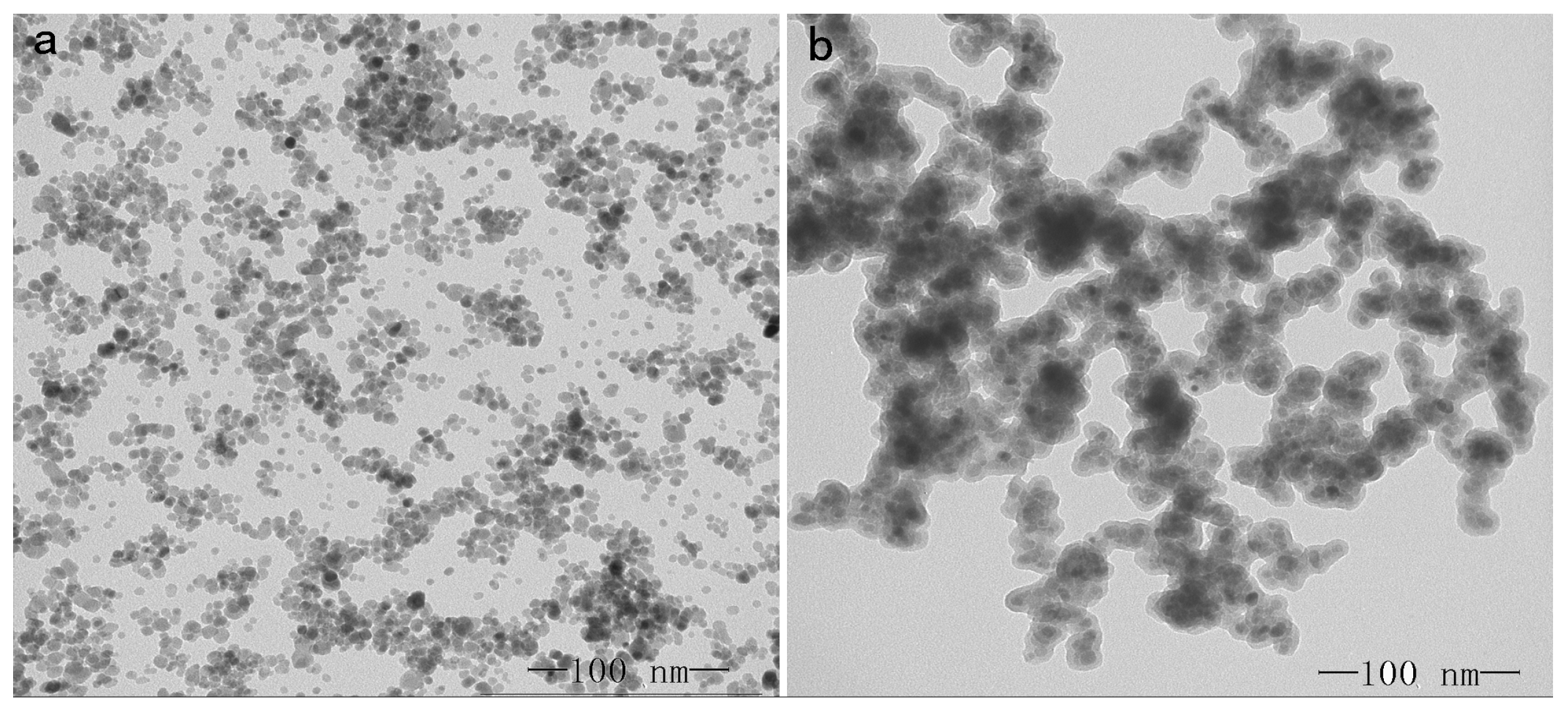
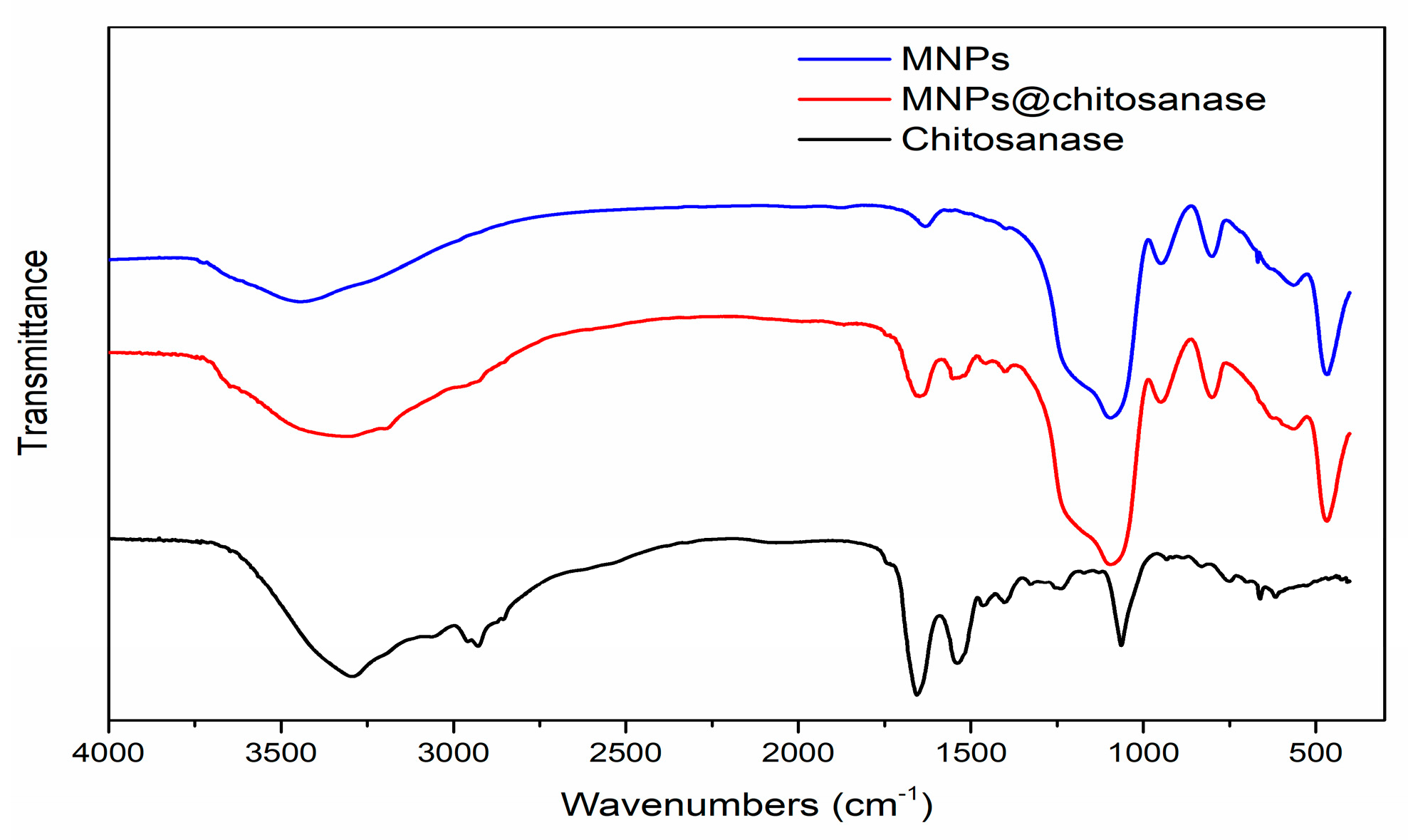
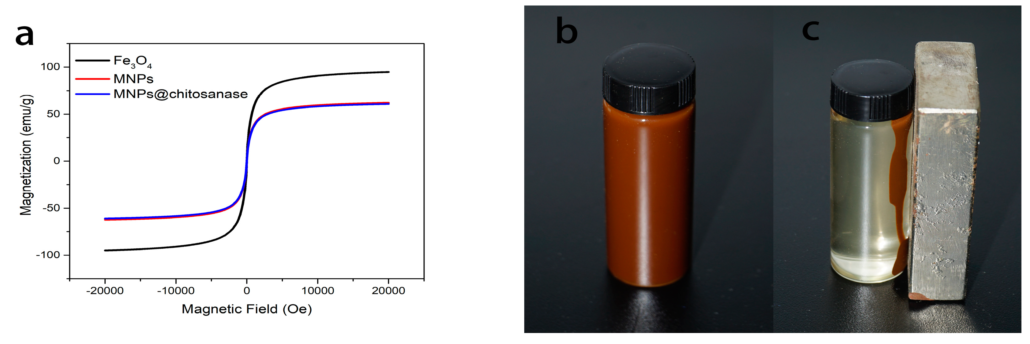
2.1. Characterization of Magnetic Nanoparticle

Transmission electron microscopy (TEM) was used to observe the size and morphology of Fe3O4 and Fe3O4-SiO2 nanoparticles. Figure 1a showed the picture of Fe3O4, where the size of nanoparticles ranged from 9 to 11 nm. As shown in Figure 1b, Fe3O4-SiO2 nanoparticles have a core-shell structure, and the size is from 30 to 50 nm.
Figure 2 showed the FT-IR spectra of MNPs, MNPs@chitosanase and chitosanase. The spectrum of immobilized chitosanase appeared strong band at 1646 cm−1, which should be attributed to the covalent binding between MNPs and chitosanases. In addition, the absorption at 1539 cm−1 showed the chitosanases were immobilized on the MNPs successfully.
The magnetic behaviours were confirmed by the vibrating sample magnetometer, which were typical plot of magnetization against applied magnetic field at room temperature, and crossed the zero point, inductive of the superparamagnetic properties. Figure 3a showed the hysteresis loops of the Fe3O4, MNPs and MNPs@chitosanase, the saturation magnetization values were 94.7, 62.1, and 60.8 emu/g, respectively. The decreased superparamagnetic behaviours of the MNPs compared with Fe3O4 could be explained due to SiO2 coated on the surface of the Fe3O4. There was only 2.1% loss of magnetization after immobilization, indicating that the immobilized chitosanases could be separated from the reaction medium rapidly and easily in an external magnetic field. MNPs were well dispersed in aqueous solution (Figure 3b) and the superparamagnetic properties were high enough to make the carrier separated from the solution in the presence of an external magnetic field (Figure 3c) in 15 s.

2.2. Characterization of Immobilized Chitosanases

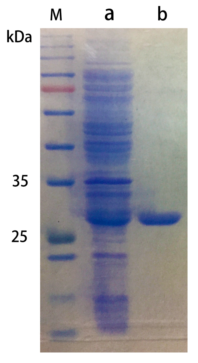
2.2.1. Purification of Chitosanase

The crude chitosanase extract was purified using a Ni-NTA column (10 cm × 2 cm) and analyzed by SDS-PAGE, which showed only one pure band in purified chitosanase (Figure 4, Lane b). And it was determined that the concentration of purified chitosanase was 0.2 mg/mL, which was appropriate for immobilization.

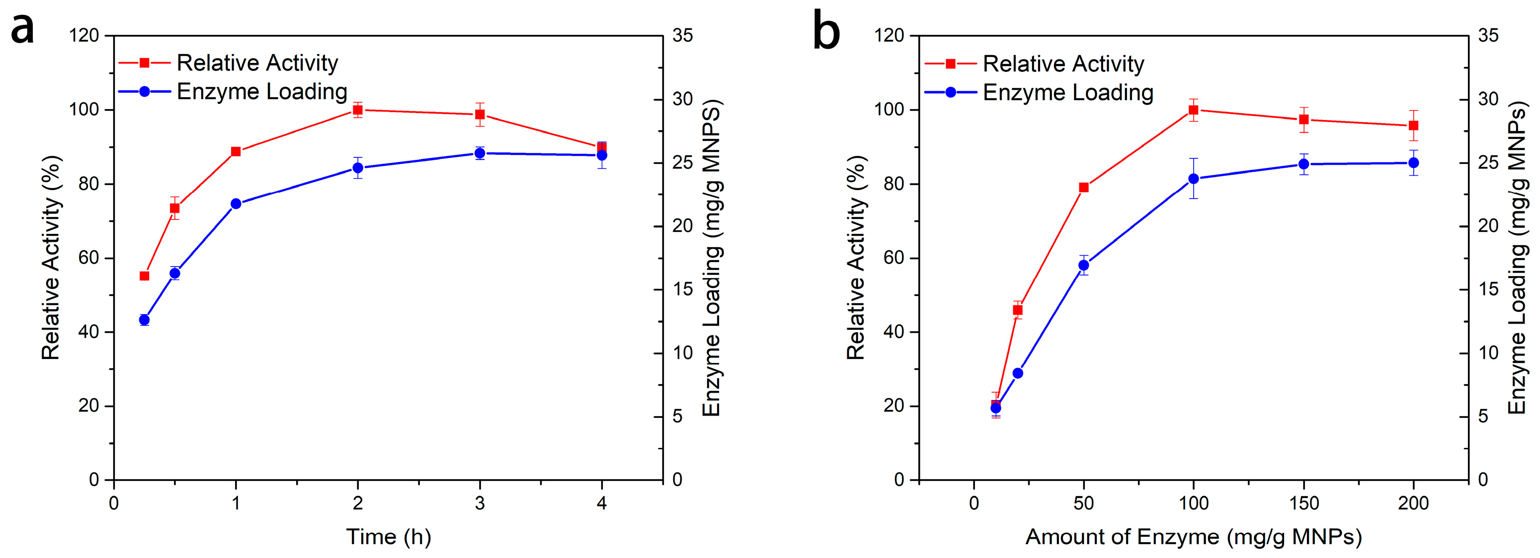
2.2.2. Optimum Conditions for Immobilization

A schematic diagram of the immobilization was illustrated in Figure 5. The effect of reaction time on the relative activity and enzyme loading during the immobilizing process was shown in Figure 6a. The results showed that by increasing the reaction time from 1 to 4 h, the enzyme loading rapidly increased at first. Then the speed of growth became slow, and the activity reached its highest at 2 h and then slightly decreased. This is because most functional groups on the surface of MNPs were covalently bonded with chitosanases, so there was not enough space for the additional chitosanases to be attached [25]. As time went on, the enzymes became denatured and the activity of the enzymes was gradually reduced. The effect of enzyme amount on immobilization was shown in Figure 6b, where enzyme loading and relative activity were illustrated. Various amounts of chitosanases (10–200 mg chitosanase/g MNPs) were added to the carriers. The amount of chitosanase immobilized increased along with the chitosanase added into the reaction and the highest value was 25.1 mg chitosanase/g MNPs. The relative activity reached its highest at 100 mg/g MNPs when the amount of chitosanase immobilized was 23.8 mg chitosanase/g MNPs and then it slightly declined. This is probably due to the interaction of the chitosanases on the surface of the MNPs. 2 h of reaction time and 100 mg/g MNPs of enzyme were selected as the optimized conditions for enzyme immobilization.

2.2.3. Stability and of Immobilized and Free Chitosanases

Figure 7a–c illustrated the pH stability of free and immobilized chitosanases. The immobilized chitosanases exhibited better stability compared to free chitosanases. The results suggested that MNPs protected the enzyme from alkaline and acid to enhance the pH stability [26], which could widen its application in the industry.
Sometimes in industrial applications, a higher reaction temperature can enhance the productivity [27], so the thermal stability of enzymes is important for its industrial application. Figure 7d showed the thermal stability of free chitosanase and MNPs@chitosanase. The immobilized chitosanases displayed a better stability than the free chitosanases with a residual activity of 19.4% while the free chitosanases is 8.4% after being incubated for 50 h at 50 °C.
The long-term storage ability of free and MNPs@chitosanase was tested, as shown in Figure 7e,f. After being stored for 28 d, the immobilized chitosanases showed better stability than that of free chitosanases. The residual activity of immobilized chitosanases was 77.9%, while the residual activity of free chitosanases was 70.2% at 4 °C; the residual activity of immobilized chitosanases was 44.7%, while the residual activity of free chitosanases was 15.8% at room temperature. This is because the covalent bonding that connects enzymes onto the carriers was able to protect the enzymes from denaturalization [28].

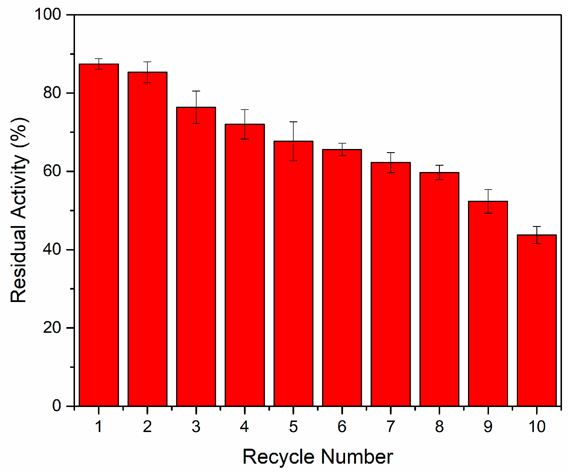
2.2.4. Reusability of Immobilized Chitosanases

The enzymes immobilized on the MNPs were able to be separated from the substrate by magnetic force, so it was easy to recycle and reuse the immobilized enzymes after reaction. The reusability is an advantage compared with free enzymes in industrial production for building a cost-effective enzymatic process. Figure 8 showed the reusability of the MNPs@chitosanase. After 10 cycles of reaction, the residual activity was 43.7%. The decrease in the activity might be the immobilized chitosanases dropped from the carrier [29], and as the reaction proceeded, part of the enzyme was deactivated.

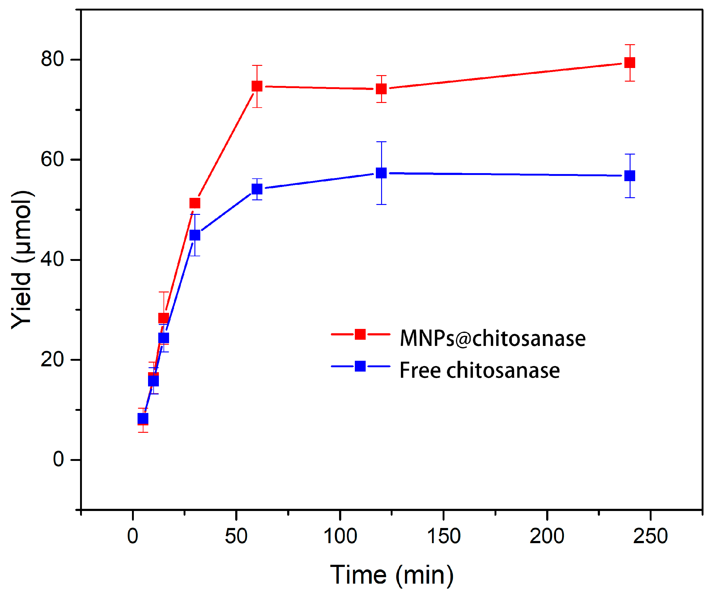
2.2.5. Catalytic Efficiency of Free and Immobilized Chitosanases

The amount of COS was investigated to characterize the catalytic performance of free and immobilized chitosanases. In order to compare the catalytic efficiency, the initial activities of the two kinds of enzymes were the same. Figure 9 shows the time courses of the hydrolysis of chitosanase using the free and immobilized chitosanases. Chitosanases immobilized onto MNPs showed much better catalytic ability, which was attributed to the better stability during the hydrolysis process. The amount of COS hydrolyzed by free chitosanases were 56.8 μmol while that of immobilized chitosanases were 79.4 μmol after 240 min reaction, which was 1.4 times higher than that of free chitosanases.

3. Materials and Methods

3.1. Materials

Bovine Serum Albumin (BSA) as well as Coomassie Brilliant Blue G-250, Tris, and imidazole were purchased from Solarbio (Beijing, China); Chitosan, sodium chloride (NaCl), hydrochloric acid (HCl), ammonium hydroxide (NH3·H2O), ferric chloride hexahydrate (FeCl3·6H2O), ferrous chloride tetrahydrate (FeCl2·4H2O), ethanol, tetraethyl orthosilicate (TEOS), and tetrahydrofuran (THF) were obtained from Sinopharm Group Co. Ltd. (Shanghai, China); 3-aminopropyltriethoxysilane (APTES) and 2,4,6-trichloro-1,3,5-triazine were purchased from Sigma Co. (Houston, TX, USA); Yeast Extract and Tryptone were bought from Oxoid Ltd. (Basingstoke, UK).

3.2. Synthesis of Fe3O4 Nanoparticles

The synthesis of Fe3O4 nanoparticles has been widely researched [30,31]. In this study, the co-precipitation method was used as a simple and convenient method. To synthesize Fe3O4 nanoparticles, 0.8 g of FeCl2·4H2O and 2.7 g of FeCl3·6H2O were dissolved in 50 mL of deionized water under nitrogen gas at room temperature. Then 4 mL of 25% NH3·H2O was added into the solution followed by violent stirring for 30 min. The Fe3O4 nanoparticles were separated by an external magnetic force and washed 3–4 times with deionized water and ethanol followed by vacuum drying under room temperature.

3.3. Preparation of Silica Coated Magnetite Nanoparticles

Fe3O4-SiO2 nanoparticles were prepared by the hydrolysis of tetraethyl orthosilicate (TEOS). 0.5 g of Fe3O4 was mixed with 150 mL of ethanol and then ultrasonification occurred for 10 min under nitrogen gas at room temperature. Then, 20 mL of deionized water and 10 mL of 25% NH3·H2O were added to the suspension followed by stirring and the addition of 6 mL of 28% tetraethyl orthosilicate (TEOS) at room temperature with additional stirring for 5 h. Fe3O4-SiO2 nanoparticles were separated by an external magnetic force and washed 3–4 times with deionized water and ethanol. They were then vacuum dried under room temperature.

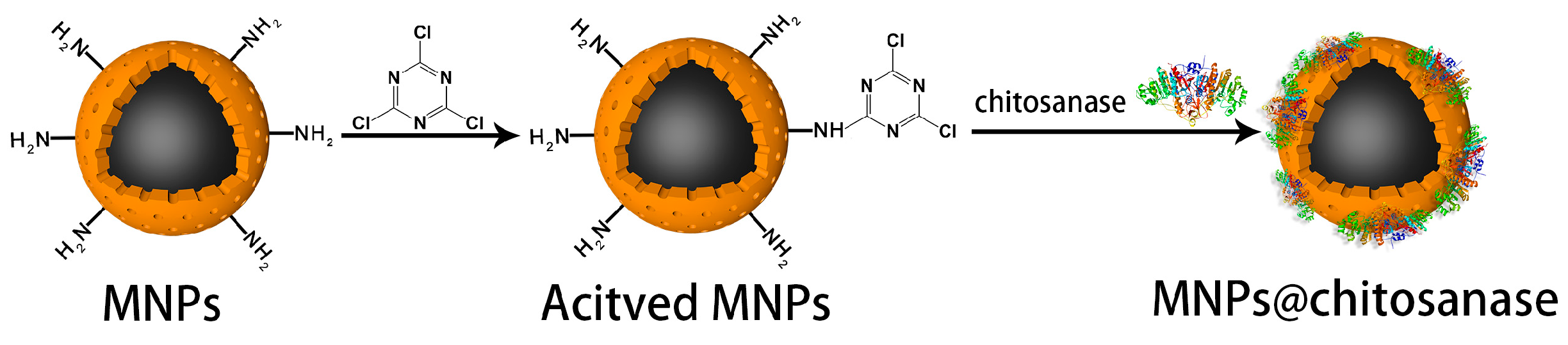
3.4. Nanoparticles Surface Modification

In this research, the method developed by Wang et al. was used to modify the Fe3O4-SiO2 nanoparticles [15,32]. Fe3O4-SiO2 nanoparticles (100 mg) and 30 mL 3-aminopropyltriethoxysilane (APTES) were added into 50 mL of ethanol. Then the suspension was stirred for 2 h at room temperature. Next, the temperature was raised to 50 °C for 1.5 h for amino functionalization. The amine-functionalized Fe3O4-SiO2 nanoparticles were separated by an external magnetic force and washed 3–4 times with ethanol and tetrahydrofuran (THF). Then the functioned nanoparticles were treated with 4 g of 2,4,6-trichloro-1,3,5-triazine in 100 mL of tetrahydrofuran (THF) at room temperature for 3 h to activate the amine-functionalized Fe3O4-SiO2 nanoparticles. The obtained MNPs were washed 3–4 times with THF, ethanol, and deionized water, and then vacuum dried under room temperature.

3.5. Purification of Chitosanase

Chitosanase was obtained by recombinant expression from Streptomyces alblongus in Escherichia coli, which was cloned and constructed using pET-21a vector in our laboratory. To prepare chitosanase, the recombinant E. coli was cultivated into ZYP5052 medium composed of 1% tryptone, 0.5% yeast extract, 0.2% MgSO4, 1.2% glycerin, 0.1% glucose, and 10% α-galactose with Ampicillin (100 μg/mL), and by shaking at 180 rpm for 48 h at 20 °C. A culture broth was centrifuged at 8000 rpm for 5 min. The precipitate was collected, re-suspended in a Tris-HCl buffer (50 mM, pH 8.0), and ultrasonicated for 30 min. The debris was removed by centrifuging at 9000 rpm for 20 min, and the supernatant was used as the crude chitosanase. The crude extract was loaded on a Ni-NTA column (10 cm × 2 cm), which was pre-equilibrated with buffer A (50 mM Tris-HCl buffer pH 8.0 containing 10 mM imidazole and 500 mM NaCl). After binding, the weakly bound impurities were washed with buffer B (50 mM Tris-HCl buffer pH 8.0 containing 50 mM imidazole and 500 mM NaCl), followed by buffer C (50 mM Tris-HCl buffer pH 8.0 containing 100 mM imidazole and 500 mM NaCl). The eluted fractions that showed high chitosanase activity were collected. SDS-PAGE was used to determine the purification of chitosanase, and the concentration of chitosanase was measured by Bradford method [33].

3.6. Characterization

TEM images were recorded by transmission electron microscopy (MIC-JEM 1200EX); the FT–IR spectra were recorded by a Fourier transform infrared spectroscopy (Thermo Scientific Nicolet iS10, Waltham, MA USA); the magnetization curves of the Fe3O4, MNPs, and MNPs@chitosanase were measured with a vibrating sample magnetometer (PPMS-9, Quantum Design, San Diego, CA, USA) at room temperature.

3.7. Chitosanase Immobilization

MNPs (5.0 mg) were dispersed in 400 μL of a Tris-HCl buffer (50 mM, pH 8.0). Then, various volumes of the chitosanase solution were added into the carrier and the mixture was shaken at 20 °C. The immobilized chitosanases were removed by magnetic decantation and washed three times with a Tris-HCl buffer (50 mM, pH 8). The amount of chitosanase immobilized on MNPs was determined by measuring the initial and final concentration of chitosanase in the immobilization medium using the Bradford method.

3.8. Assay of Chitosanase Activity

Immobilized and free chitosanase activities were determined by measuring the reducing sugars liberated during the hydrolysis of the chitosan with the 3, 5-dinitrosalicylic acid (DNS) method [34]. The substrate solution was prepared by mixing 100 μL chitosan solution (2 g chitosan predissolved in 100 mL of 1% acetic acid) with a 100 μL Tris-HCl buffer (50 mM, pH 8.0). Then, free and immobilized chitosanase were added to the substrate solution at the condition of 50 °C. After reaction, the absorbance at 540 nm was measured against a blank. The residual activity (%) was determined by calculating the ratio of the activity of sample to initial activity; sample with the highest activity was defined as 100% relative activity (%) [35].

3.9. Stability and Reusability of Immobilized Chitosanases

The thermal stability of the free and MNPs@chitosanase was evaluated by measuring the residual activities of the free and immobilized chitosanases incubating in Tris-HCl buffer (50 mM, pH 8.0) with various temperatures and times. The pH stability was evaluated by measuring the residual activity of free and immobilized chitosanases incubated in various pH and times. The storage stabilities of the immobilized and free chitosanases were determined by measuring their residual activity after incubation at 4 °C and room temperature in the Tris-HCl buffer (50 mM, pH 8.0).
The reusability of the immobilized chitosanases was evaluated by measuring the residual activity after recycling 10 times. After each hydrolysis cycle, the immobilized chitosanases were separated and washed with Tris-HCl buffer (50 mM, pH 8.0) by adding a magnetic force.

3.10. Catalytic Efficiency of Free and Immobilized Chitosanases

The catalytic performance of free and MNPs@chitosanase were evaluated by hydrolysis of chitosan. Free and immobilized chitosanases (the initial activity of chitosanases immobilized was the same as that of free chitosanases) were incubated in chitosan in a Tris-HCl buffer (50 mM, pH 8.0). The catalytic performance of the free and immobilized chitosanases were investigated by measuring the hydrolysed chitosan.

4. Conclusions

This research developed a method for immobilizing chitosanases on the surface of Fe3O4-SiO2 magnetic nanoparticles with covalent bonding. The TEM picture showed the silica was successfully coated on the surface and the size of the Fe3O4-SiO2 was about 30 nm to 50 nm. The hysteresis loops showed the MNPs@chitosanase have high superparamagnetic propertiy. The results of the FT–IR spectra indicated the enzyme was immobilized on the carrier. The thermal, pH, and storage abilities were improved after immobilization. The immobilized chitosanases were also used for recycling. After it was recycled 10 times, the residual activity was 43.74%. The enhancement of the chitosanases proved the immobilization can expand the use of enzymes in industrial applications.

Author Contributions

W.W. and N.G. performed the experiment; X.M. and Z.Z. analysed the data; and W.W. and W.H. wrote the paper.

Funding

This work was supported by the China Agriculture Research System (CARS-48), the Major Special Science and Technology Projects in Shandong Province (2016YYSP016), and the Shandong Natural Science Foundation (no. ZR2015CQ021).

Conflicts of Interest

The authors declare no conflict of interest.

References

  1. Kim, S.K.; Rajapakse, N. Enzymatic production and biological activities of chitosan oligosaccharides (COS): A review. Carbohydr. Polym. 2005, 62, 357–368. [Google Scholar] [CrossRef]
  2. Choi, Y.J.; Kim, E.J.; Piao, Z.; Yun, Y.C.; Shin, Y.C. Purification and characterization of chitosanase from Bacillus sp. strain KCTC 0377BP and its application for the production of chitosan oligosaccharides. Appl. Environ. Microbiol. 2004, 70, 4522–4531. [Google Scholar] [CrossRef] [PubMed]
  3. Lin, S.; Mao, S.; Guan, Y.; Luo, L.; Luo, L.; Pan, Y. Effects of dietary chitosan oligosaccharides and Bacillus coagulans on the growth, innate immunity and resistance of koi (Cyprinus carpio koi). Aquaculture 2012, 342–343, 36–41. [Google Scholar] [CrossRef]
  4. Lu, Y.; Slomberg, D.L.; Schoenfisch, M.H. Nitric oxide-releasing chitosan oligosaccharides as antibacterial agents. Biomaterials 2014, 35, 1716–1724. [Google Scholar] [CrossRef] [PubMed] [Green Version]
  5. Zou, P.; Yang, X.; Wang, J.; Li, Y.; Yu, H.; Zhang, Y.; Liu, G. Advances in characterisation and biological activities of chitosan and chitosan oligosaccharides. Food Chem. 2016, 190, 1174–1181. [Google Scholar] [CrossRef] [PubMed]
  6. Suzuki, K.; Mikami, T.; Okawa, Y.; Tokoro, A.; Suzuki, S.; Suzuki, M. Antitumor effect of hexa-N-acetylchitohexaose and chitohexaose. Carbohydr. Res. 1986, 151, 403–408. [Google Scholar] [CrossRef]
  7. Pires, C.T.; Vilela, J.A.; Airoldi, C. The effect of chitin alkaline deacetylation at different condition on particle properties. Procedia Chem. 2014, 9, 220–225. [Google Scholar] [CrossRef]
  8. Pareek, N.; Vivekanand, V.; Saroj, S.; Sharma, A.K.; Singh, R.P. Purification and characterization of chitin deacetylase from Penicillium oxalicum, SAEM-51. Carbohydr. Polym. 2012, 87, 1091–1097. [Google Scholar] [CrossRef]
  9. Naqvi, S.; Cord-Landwehr, S.; Singh, R.; Bernard, F.; Kolkenbrock, S.; EI Gueddari, N.E.; Moerschbacher, B.M. A recombinant fungal chitin deacetylase produces fully defined chitosan oligomers with novel patterns of acetylation. Appl. Environ. Microbiol. 2016, 82, 6645–6655. [Google Scholar] [CrossRef] [PubMed]
  10. Dahiya, N.; Tewari, R.; Hoondal, G.S. Biotechnological aspects of chitinolytic enzymes: A review. Appl. Microbiol. Biotechnol. 2006, 71, 773–782. [Google Scholar] [CrossRef] [PubMed]
  11. Thadathil, N.; Velappan, S.P. Recent developments in chitosanase research and its biotechnological applications: A review. Food Chem. 2014, 150, 392–399. [Google Scholar] [CrossRef] [PubMed]
  12. Sheldon, R.A. Enzyme Immobilization: The quest for optimum performance. Adv. Synth. Catal. 2007, 349, 1289–1307. [Google Scholar] [CrossRef]
  13. Trevan, M.D. Enzyme immobilization by adsorption. In New Protein Techniques; Walker, J.M., Ed.; Humana Press: New York, NY, USA, 1988; Volume 3, pp. 481–489. ISBN 978-1-59259-490-0. [Google Scholar]
  14. Zhou, Z.; Hartmann, M. Recent progress in biocatalysis with enzymes immobilized on mesoporous hosts. Top. Catal. 2012, 55, 1081–1100. [Google Scholar] [CrossRef]
  15. Ranjbakhsh, E.; Bordbar, A.K.; Abbasi, M.; Khosropour, A.R.; Shams, E. Enhancement of stability and catalytic activity of immobilized lipase on silica-coated modified magnetite nanoparticles. Chem. Eng. J. 2012, 179, 272–276. [Google Scholar] [CrossRef]
  16. Straathof, A.J.; Panke, S.; Schmid, A. The production of fine chemicals by biotransformations. Curr. Opin. Biotechnol. 2012, 13, 548–556. [Google Scholar] [CrossRef]
  17. Vaghari, H.; Jafarizadeh-Malmiri, H.; Mohammadlou, M.; Berenjian, A.; Anarjan, N.; Jafari, N.; Nasiri, S. Application of magnetic nanoparticles in smart enzyme immobilization. Biotechnol. Lett. 2016, 38, 223–233. [Google Scholar] [CrossRef] [PubMed]
  18. Johnson, P.A.; Park, H.J.; Driscoll, A.J. Enzyme nanoparticle fabrication: Magnetic nanoparticle synthesis and enzyme immobilization. In Enzyme Stabilization and Immobilization; Humana Press: New York, NY, USA, 2011; pp. 183–191. ISBN 978-1-60761-895-9. [Google Scholar]
  19. Liao, M.H.; Chen, D.H. Immobilization of yeast alcohol dehydrogenase on magnetic nanoparticles for improving its stability. Biotechnol. Lett. 2001, 23, 1723–1727. [Google Scholar] [CrossRef]
  20. Hu, B.; Pan, J.; Yu, H.L.; Liu, J.W.; Xu, J.H. Immobilization of Serratia marcescens lipase onto amino-functionalized magnetic nanoparticles for repeated use in enzymatic synthesis of Diltiazem intermediate. Process Biochem. 2009, 44, 1019–1024. [Google Scholar] [CrossRef]
  21. Xie, W.; Ma, N. Enzymatic transesterification of soybean oil by using immobilized lipase on magnetic nano-particles. Biomass Bioenergy 2010, 34, 890–896. [Google Scholar] [CrossRef]
  22. Lei, L.; Liu, X.; Li, Y.; Cui, Y.; Yang, Y.; Qin, G. Study on synthesis of poly(GMA)-grafted Fe3O4/SiOX magnetic nanoparticles using atom transfer radical polymerization and their application for lipase immobilization. Mater. Chem. Phys. 2011, 125, 866–871. [Google Scholar] [CrossRef]
  23. Mrówczyński, R.; Nan, A.; Liebscher, J. Magnetic nanoparticle-supported organocatalysts—An efficient way of recycling and reuse. RSC Adv. 2014, 4, 5927–5952. [Google Scholar] [CrossRef]
  24. Wang, Q.; Sun, J.; Liu, Z.; Huang, W.C.; Xue, C.; Mao, X. Co-immobilization of β-agarase and α-neoagarobiose hydrolase for enhancing the production of 3,6-anhydro-l-galactose. J. Agric. Food Chem. 2018, 66, 7087–7095. [Google Scholar] [CrossRef] [PubMed]
  25. Gao, J.; Kong, W.; Zhou, L.; He, Y.; Ma, L.; Wang, Y.; Yin, L.; Jiang, Y. Monodisperse core-shell magnetic organosilica nanoflowers with radial wrinkle for lipase immobilization. Chem. Eng. J. 2017, 309, 70–79. [Google Scholar] [CrossRef]
  26. Shareghi, B.; Farhadian, S.; Zamani, N.; Salavati-Niasari, M.; Gholamrezaei, S. Stability and enzyme activity of lysozyme in the presence of Fe3O4 nanoparticles. Monatshefte Chem. 2016, 147, 465–471. [Google Scholar] [CrossRef]
  27. Arroyo, M.; Sánchez-Montero, J.M.; Sinisterra, J.V. Thermal stabilization of immobilized lipase B from Candida antarctica, on different supports: Effect of water activity on enzymatic activity in organic media. Enzyme Microb. Technol. 1999, 24, 3–12. [Google Scholar] [CrossRef]
  28. Mateo, C.; Palomo, J.M.; Fernandez-Lorente, G.; Guisan, J.M.; Fernandez-Lafuente, R. Improvement of enzyme activity, stability and selectivity via immobilization techniques. Enzyme Microb. Technol. 2007, 40, 1451–1463. [Google Scholar] [CrossRef]
  29. Barbosa, O.; Torres, R.; Ortiz, C.; Berenguer-Murcia, Á.; Rodrigues, R.C.; Fernandez-Lafuente, R. Heterofunctional supports in enzyme immobilization: From traditional immobilization protocols to opportunities in tuning enzyme properties. Biomacromolecules 2013, 14, 2433–2462. [Google Scholar] [CrossRef] [PubMed] [Green Version]
  30. Khosroshahi, M.E.; Ghazanfari, L. Synthesis and functionalization of SiO2, coated Fe3O4, nanoparticles with amine groups based on self-assembly. Mater. Sci. Eng. C 2012, 32, 1043–1049. [Google Scholar] [CrossRef]
  31. Jang, J.H.; Lim, H.B. Characterization and analytical application of surface modified magnetic nanoparticles. Microchem. J. 2010, 94, 148–158. [Google Scholar] [CrossRef]
  32. Wang, Y.; Liu, B. Conjugated polymer as a signal amplifier for novel silica nanoparticle-based fluoroimmunoassay. Biosens. Bioelectron. 2009, 24, 3293–3298. [Google Scholar] [CrossRef] [PubMed]
  33. Bradford, M.M. A rapid and sensitive method for the quantitation of microgram quantities of protein utilizing the principle of protein-dye binding. Anal. Biochem. 1976, 72, 248–254. [Google Scholar] [CrossRef]
  34. Miller, G.L. Use of dinitrosalicylic acid reagent for determination of reducing sugar. Anal. Chem. 1959, 31, 426–428. [Google Scholar] [CrossRef]
  35. Batule, B.S.; Park, K.S.; Kim, M.I.; Park, H.G. Ultrafast sonochemical synthesis of protein-inorganic nanoflowers. Int. J. Nanomed. 2015, 10, 137. [Google Scholar] [CrossRef]
Figure 1. Transmission electron microscopy (TEM) images of Fe3O4 (a) and Fe3O4-SiO2 (b) nanoparticles.
Catalysts 08 00401 g001
Figure 2. FT–IR spectra of MNPs, MNPs@chitosanase and chitosanase.
Catalysts 08 00401 g002
Figure 3. (a) Hysteresis loops of Fe3O4, MNPs and chitosanases immobilized on MNPs. Photographs of (b) the MNPs dispersed in aqueous solution and (c) the MNPs attracted by an external magnetic field for 15 s.
Catalysts 08 00401 g003
Figure 4. SDS-PAGE analysis of purified chitosanase. Lane M, protein markers; Lane a, crude extract before purification; Lane b, purified chitosanase.
Catalysts 08 00401 g004
Figure 5. Schematic diagram of the immobilization of chitosanases onto the MNPs.
Catalysts 08 00401 g005
Figure 6. Effect of reaction times (a) and amount of enzyme (b) on enzyme loading and relative activity. The reaction was from the condition of 20 °C and Tris-HCl buffer (50 mM, pH 8.0); the reaction time was 2 h.
Catalysts 08 00401 g006
Figure 7. Stability of MNPs@chitosanase and free chitosanase. (a) The stability of the free and MNPs@chitosanase at pH 3, (b) the stability of free and MNPs@chitosanase at pH 7, (c) the stability of free and MNPs@chitosanase at pH 9, (d) the thermal stability of free and MNPs@chitosanase at 50 °C, the storage stability of free and MNPs@chitosanase at (e) 4 °C and (f) room temperature.
Catalysts 08 00401 g007
Figure 8. Reusability of MNPs@chitosanase.
Catalysts 08 00401 g008
Figure 9. Time courses of the hydrolysis of chitosan using the free and MNPs@chitosanase.
Catalysts 08 00401 g009

