Article

Significantly Enhanced Aqueous Cr(VI) Removal Performance of Bi/ZnO Nanocomposites via Synergistic Effect of Adsorption and SPR-Promoted Visible Light Photoreduction

College of Materials Science and Engineering, Chongqing Jiaotong University, Chongqing 400074, China
*
Author to whom correspondence should be addressed.
Catalysts 2018, 8(10), 426; https://doi.org/10.3390/catal8100426
Submission received: 25 August 2018 / Revised: 12 September 2018 / Accepted: 18 September 2018 / Published: 29 September 2018
(This article belongs to the Special Issue Environmental Applications of Photocatalytic Processes)

Abstract

:
Bismuth nanoparticles (BiNPs) and Zinc Oxide photocatalysts (BiNPs/ZnO) with different Bi loadings were successfully prepared via a facile chemical method. Their morphology and structure were thoroughly characterized by powder X-ray diffraction (XRD), scanning electron microscopy (SEM), high-resolution transmission electron microscopy (HRTEM), X-ray photoelectron spectroscopy (XPS), UV-Vis (Ultraviolet-Visible) diffuse reflectance spectroscopy (DRS), photoluminescence spectra (PL), and electrochemical impedance spectroscopy (EIS). The results showed that a modification of hexagonal wurtzite-phase ZnO nanoparticles with Bi is achievable with an intimate interfacial interaction within its composites. The performance of the photocatalytic Cr(VI) removal under visible light irradiation indicated that BiNPs/ZnO exhibited a superior removal performance to bare ZnO, Bi, and the counterpart sample prepared using a physical mixing method. The excellent performance of the BiNPs/ZnO photocatalysts could be ascribed to the synergistic effect between the considerable physical Cr (VI) adsorption and enhanced absorption intensity in the visible light region, due to the surface plasmon resonance (SPR) as well as the effective transfer and separation of the photogenerated charge carriers at the interface.

1. Introduction

Hexavalent chromium (Cr(VI)) has been proven to be one of the most toxic pollutants by the US Environmental Protection Agency (EPA), because of its flagrant toxic, mutagenic, as well as teratogenic effects to human being [1,2]. In order to remove Cr(VI) from the wastewater, a good deal of methods have been applied, including bioremediation, chemical precipitation, and photocatalytic reduction [3,4]. As for high efficiency, safety, and energy saving, photocatalytic reduction is the most attractive method for Cr(VI) removal [5,6,7].
Among many oxide-based photocatalysts, ZnO has received extensive attention in the field of environmental restoration, and because it has the similar band gap and photocatalytic mechanism as TiO2, ZnO is considered to be an ideal photocatalyst to replace TiO2 [8,9]. Though a few studies have been published on the photoreduction activity of Cr(VI) using ZnO-based photocatalysts [10,11,12,13], owing to the 3.2 eV band gap of ZnO, it is difficult to be excited under visible light. Additionally, a high recombination of photo-excited carriers also makes its real applications blocked [14,15].
Very recently, as a semimetal, bismuth (Bi) has been excavated to have direct plasma photocatalytic ability mediated by SPR (surface plasmon resonance) [16]. This property would cause the absorbed electrons to resonate inside the metal, thereby increasing the absorption of photons, and this resonance mostly occurs in the visible light region [17,18,19,20]. Similar to noble metal elements, for instance Au [21,22], Ag [23,24], and Pt [25,26], Bi is also found to be a potential candidate to activate the wide band gap photocatalysts, and is thus widely applied as a cocatalyst due to its SPR effect; many groups have done research in this area [27,28], exploring cocatalysts Bi/CdS [29], Bi/g-C3N4 [18], Bi/Bi2O3 [30], and Bi/(BiO)2CO3 [28]. Wang et al., described that Bi/CdS microspheres could efficiently elevate the visible light photocatalytic activity for methyl orange (MO) degradation, because of its enhanced visible light absorption and quickly charged separation caused by the introduction of Bi [29]. Dong et al., proclaimed that incorporating Bi into the semiconductor g-C3N4 nanosheets showed an improved photodegradation capability of NO under visible light illumination, which could be assigned to the suppression of photodriven electrons–holes pairs, because Bi served as the recipient and communicator of the electron [18].
Although photocatalytic degradation of organic pollutants via photo-oxidation has been widely disclosed in most of cases where Bi-based catalysts were employed [31,32], work about the wastewater remediation via photoreduction using Bi cocatalysts was rarely found. In this work, the BiNPs/ZnO composites were prepared herein by an easy eco-friendly synthesis method compared to that reported with a solid-state method [33]. The resulting BiNPs/ZnO composites showed significantly enhanced aqueous Cr(VI) removal performance under visible light conditions (up to 99.3% removal ratio). The detailed mechanism demonstrated that the improved Cr(VI) removal activity was owed to the synergistic effect between the excellent physical Cr(VI) adsorption of BiNPs/ZnO composites and the SPR effect of Bi phase of the composites under visible light.

2. Results and Discussion

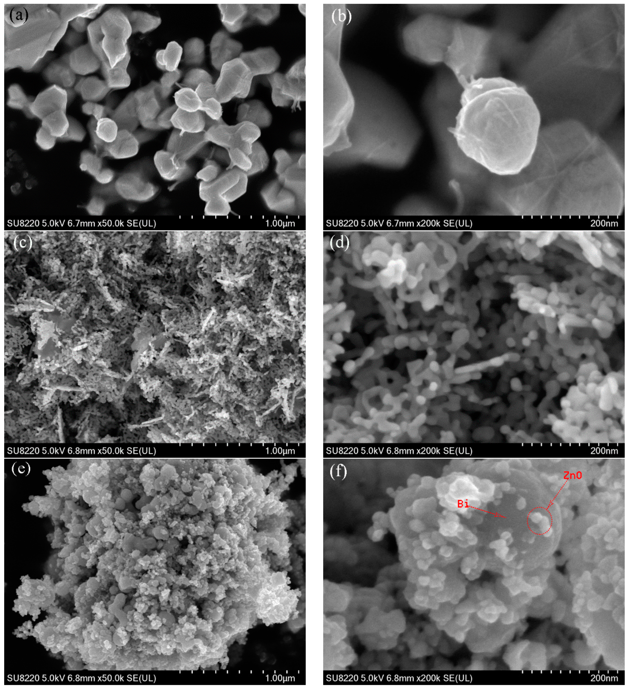
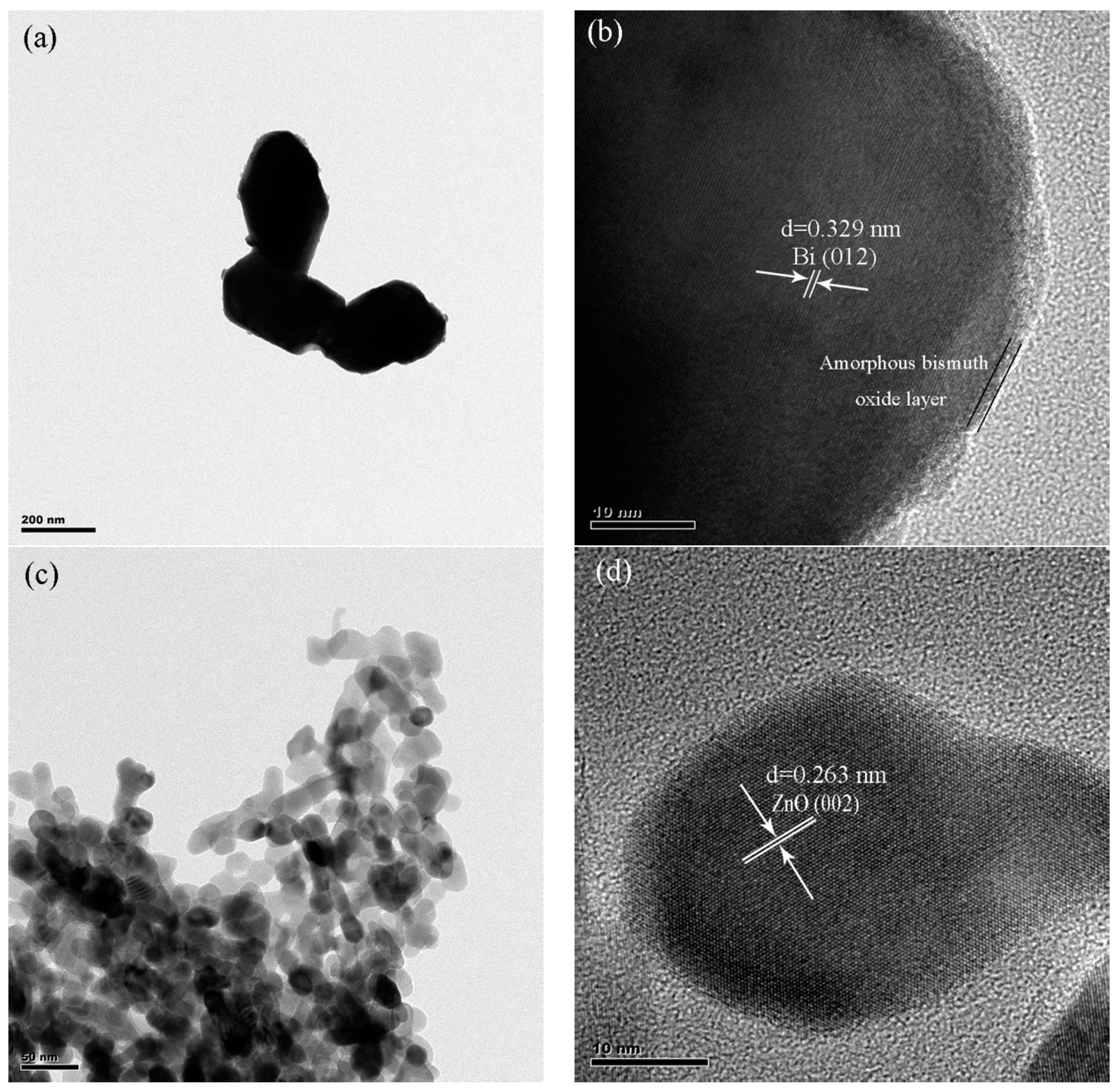
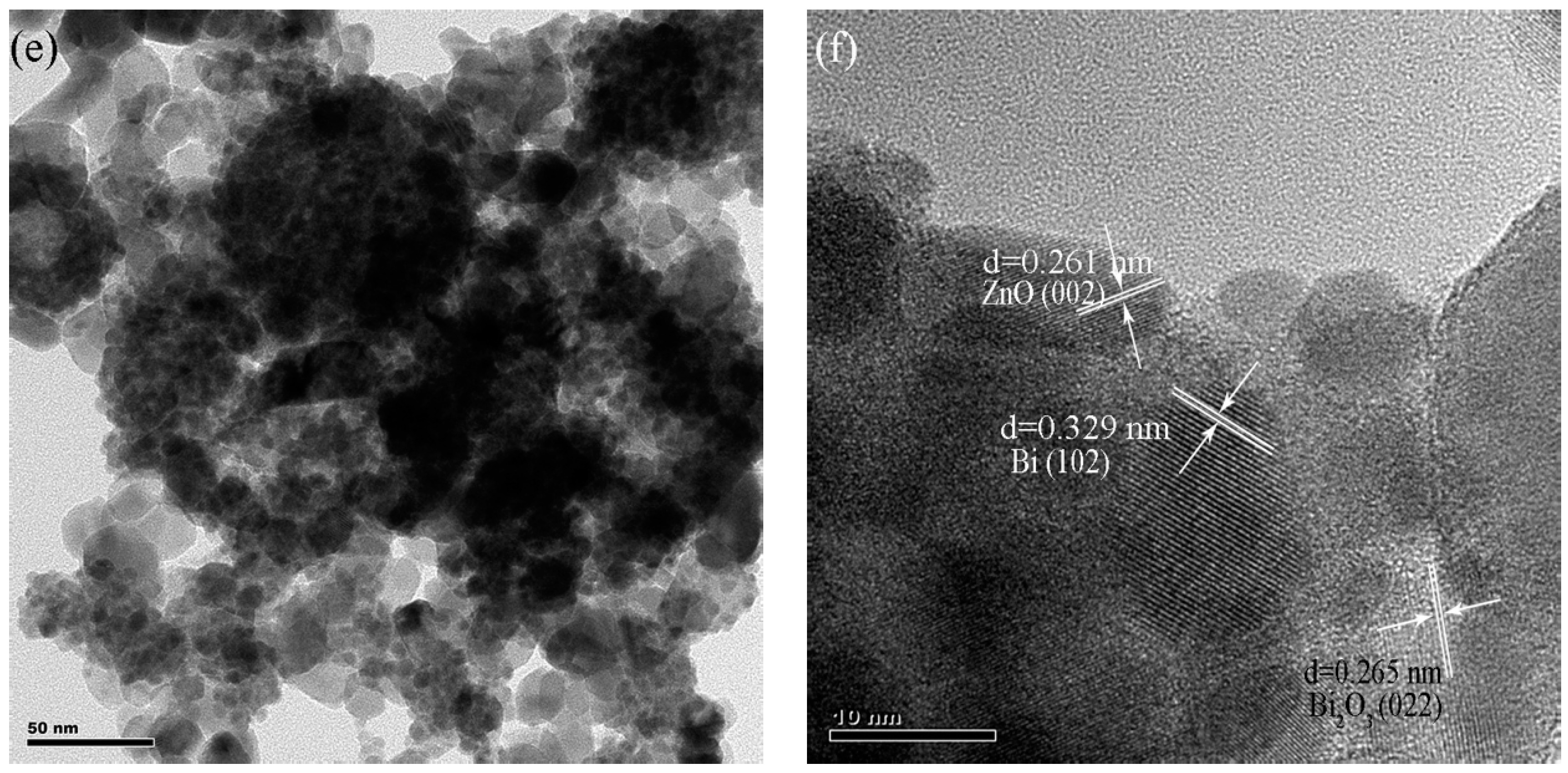
2.1. Characterization of BiNPs/ZnO Nanocomposites

Figure 1 exhibits the XRD spectrums of ZnO, Bi, and Bi-ZnO with different Bi loadings. For pristine metal Bi, the characteristic peaks at 23.4° (003), 27.1° (012), 37.9° (104), 39.8° (110), 44.8° (015), 45.2° (006), 57.3° (024), 63.4° (112), and 73.1° (009) were exactly in agreement with the typical rhombohedral phase of Bi (JCPDS-No. 44-1246). For bare ZnO, the peaks at 31.8° (100), 34.5° (002), 36.42° (010), 47.6° (102), 56.58° (110), 63.05° (103), and 67.9° (112) were highly identical with those of the ZnO hexagonal wurtzite phase (JCPDS-NO. 36-1451) [34]. For the BiNPs/ZnO with different Bi contents, the characteristic diffraction peaks of ZnO were weakened with an increased content of Bi metal. Compared to that at ca. 27.1° assigned to (012) diffraction reflection characteristic of Bi semimetal crystal, the peak in the XRD patterns for all Bi/ZnO composites exhibited a red shift of about 1.1°, due to the growth of the Bi particle on ZnO as a nuclei substrate, suggesting an interfacial interaction between the two phases of the composites [35,36], which was favorable to the transfer of photogenerated e/h+ pairs across the interface within the composites.
Figure 2 exhibits the typical SEM images of Bi, ZnO, as well as Bi-ZnO-80. From the holistic morphology of the Bi nanoparticle (Figure 2a,b), a good deal of Bi irregular particles with an average size of ca. 230 nm were detected. In Figure 2c, pure ZnO exhibited flocculent-like structure due to the nanosize effect-induced aggregation and Figure 2d showed that interconnected ZnO nanoparticles of around 10 nm were also clearly observed. Figure 2e,f of Bi-ZnO-80 demonstrated that some nanoparticles were randomly attached on the surface of large particles. It could be reasonable to deduce that ZnO nanoparticles were deposited onto the Bi particles from their dimensions of each phases of the Bi-ZnO-80 composite.
Figure 3 exhibits the representative HRTEM images of Bi, ZnO and Bi-ZnO-80. The bare Bi particles appeared on atactic spherical morphology with a size diameter in the range of 200–250 nm (Figure 3a) and the lattice fringes of 0.328 nm were also in accordance with (012) the facet of the bismuth (Figure 3b). Nevertheless, a 2–5 nm thick amorphous surface layer appeared at the edge of the composite (Figure 3b), which might have arisen from contact with air due to the easy oxidation of Bi [37]. As shown in Figure 3c, the ZnO nanoparticles exhibited a flocculent-like structure consisting of aggregated spherical ZnO nanoparticles with an average particle size of approximately 10 nm, which was in line with the SEM observations. The lattice fringes were measured to be 0.263 nm (Figure 3d), which was in agreement with the (002) facet of the hexagonal wurtzite ZnO phase [34]. From Figure 3e, it was observed that the Bi-ZnO-80 composite was composed of two kinds of constituent particles, one with a large diameter Bi and the other with a small ZnO. Two different types of lattice fringes were distinctly detected as well (Figure 3f), one of d = 0.322 nm consistent with the (012) plane of Bi, the other of d = 0.261 nm corresponding to bare wurtzite ZnO nanoparticles as presented in Figure 3f, further demonstrating that the resulting composite was composed of Bi and ZnO phases. The clear lattice fringe of 0.265 nm in HRTEM (Figure 3f) could be indexed to the (002) plane of Bi2O3. In addition, the junction interface formed by the combination Bi and ZnO phase in the composite was easily observed in Figure 3f. ZnO nanoparticles played the part of bridges for the connection between the Bi particles. The tight junction between the two components facilitates the separation of photogenerated carriers and it can increase the photocatalytic activity of the catalyst to some extent.
Figure 4 shows the XPS survey spectra of the Bi4f of the Bi-ZnO-80 composite as well as the Zn2p of ZnO and its composite. The survey spectrum in Figure 4a expressly demonstrated that the composition for the Bi-ZnO-80 included the element with Zn, O, and Bi. The Bi4f XPS spectra of Bi-ZnO-80 are shown in Figure 4b. The characteristic peaks of the Bi-Bi bonds of the element Bi normally appeared at 156.8 and 162.1 eV, respectively [30,38], and the respective peaks at 158.9 and 164.2 eV were in accordance with the characteristic peaks of Bi-O bonds [39,40]. There was a notably significant shift of approximately 0.4 eV to the higher binding energies which could be detected in the Bi4f peaks of the Bi-ZnO-80 compared to the original Bi, and this shift indicated a strong interfacial interaction existing between Bi and ZnO phase [41], which was in line with those observed in the XRD pattern. The Zn2p spectrum of the Bi-ZnO-80 (Figure 4c) exhibited two peaks at 1021.2 and 1044.3 eV, respectively, which was in agreement with ZnO2p3/2 and ZnO2p1/2 as well as with the Zn2p of the pure ZnO [34].
The UV-Vis DRS (diffuse reflectance spectroscopy) spectra of ZnO and BiNPs/ZnO nanocomposites of different Bi loadings are shown in Figure 5a. In accordance with our expectation, a sharp fundamental absorption edge for ZnO rose at ca. 400 nm, putting down the bandgap to 3.22 eV. After depositing the Bi nanoparticles onto ZnO, the absorption threshold of these as-prepared composites was hardly affected by the Bi loadings. However, compared with bare ZnO, all BiNPs/ZnO composites exhibited stronger adsorption intensity in the visible light region. At the same time, the band gap energy estimated from the intercepts of the tangents to the plots of (Ahν)2 vs. photon energy was shown in Figure 5b [42,43], which also suggested that the band gap energy of the Bi-ZnO-80 composite was not significantly changed, as compared with that of pure ZnO. Therefore, the enhancement of absorption in the visible light region of BiNPs/ZnO was not due to the change of the band gap energy. Notably, a strong absorption centered at ca. 500 nm (inset of Figure 5a) was seen, which could be put down to the SPR effect of the Bi metal [27]. In other words, the SPR effect allowed the absorbed photons to resonate inside the Bi metal and enhanced the absorption of light, and this normally occurred in the visible region. Such phenomena concerning the SPR effect have been widely reported by others groups [17,19]. However, since the resonance wavelength was related to the shape and size of the nanoparticles, the interparticle distance, and the dielectric constant of the environment, the strong absorption caused by the resonance did not appear at the same wavelength for different Bi contents. To date, Bi as a base metal has been demonstrated to display the SPR effect in the near ultraviolet and visible light range [16,44].
The PL (photoluminescence spectra) spectrum is an effective method to monitor the separation efficiency of photogenerated electron–hole pairs for a semiconductor under light exposure [45]. The PL spectrum of pure ZnO exhibited an emission peak centered at ca. 443 and 451 nm (~2.8 eV) under excitation at 280 nm (Figure 6a), which were largely ascribed to the quenched of band gap transition. It was noteworthy that the peak intensity of the Bi-ZnO-80 hybrid was weaker than bare ZnO, which might be attributed to the difference of work functions in ZnO and Bi, leading to the most of the electrons from the conduction band of ZnO being rapidly transferred to the surface of the metal Bi particles under UV light, resembling to the Mott-Schottky effect [20,46,47,48]. At the same time, this was done to further detect the separation efficiency of photoinduced charges of photocatalysts under visible light [49]. We recorded the EIS (electrochemical impedance spectroscopy) plots of pure ZnO and Bi-ZnO-80 composites, as depicted in in Figure 6b. ZnO exhibited a much larger arc radius than Bi-ZnO-80 in EIS plots, which indicated a more efficient separation of photo-induced electron–hole pairs and faster charge transfer across the junction interface within Bi-ZnO-80 [23,49]. These results indicated that the separation of photogenerated carriers at their interfaces is more efficient with ZnO after modifying the Bi particles.

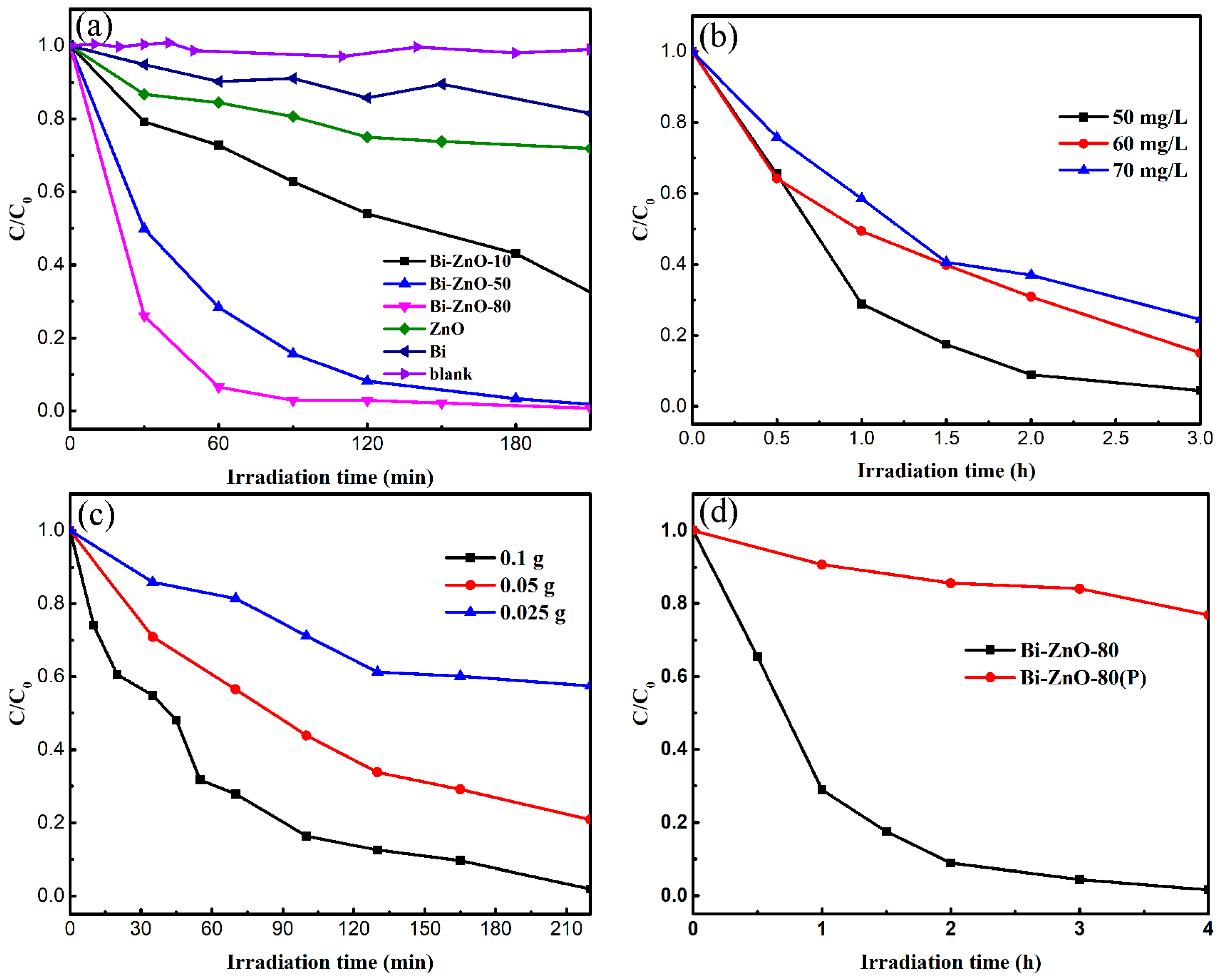
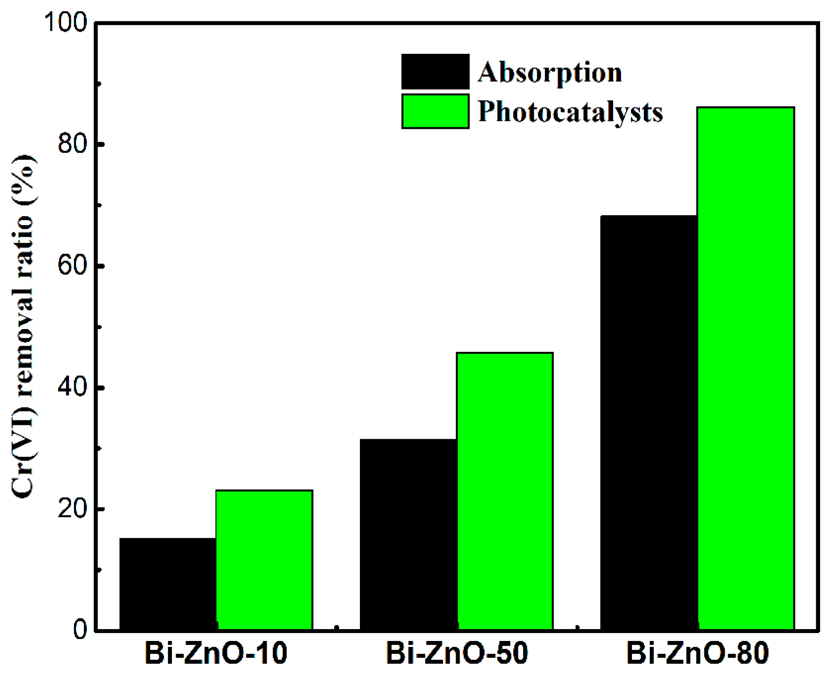
2.2. Photocatalytic Activity of Bi/ZnO Composites

Figure 7a shows the photocatalytic activity of the BiNPs/ZnO toward reduction of Cr(VI) under visible light irradiation. A blank experiment proved that the aqueous Cr(VI) was extremely steady under visible light irradiation. The reduction ratio of Cr(VI) by pure ZnO and Bi after 210 min visiblelight irradiation reached 28% and 18% respectively. Notably, after the ZnO nanoparticles were modified by the Bi particles, the reduction ratio of Cr(VI) was attained at 67%, 98%, and 99.3% for Bi-ZnO-10, Bi-ZnO-50, and Bi-ZnO-80, respectively, within the same condition. It has been strongly demonstrated that the modification with Bi nanoparticles could greatly enhance the photocatalytic reduction activity of BiNPs/ZnO composites, which could be ascribed to the enhanced visible light absorption, due to the SPR effects of the metal Bi and the accelerated photoinduced electron–holes separation driven across the interfaces of the BiNPs/ZnO composites. Moreover, we also observed that the mass ratio of Bi exerted a notable effect on the photocatalytic activity. The photoreduction efficiency of Cr(VI) on BiNPS/ZnO composites increased as the content of Bi increased. This was understandable because as the amount of Bi deposition increased, the visible light absorption active site increased due to its SPR effect, thus resulting in a considerable photocatalytic performance.
Correlations between initial solution concentration and photocatalytic performance were obtained by changing the initial concentration of Cr(VI) from 50 to 70 mg/L on the Bi-ZnO-80 composite. As illustrated in Figure 7b, the reduction ratio of Cr(VI) was elevated from 75.5% to 99.5% with the decrease of initial concentration of Cr(VI). As expected, as the initial concentration of Cr(VI) increased, more Cr(VI) ions were adsorbed onto the surface of the BiNPs/ZnO composite, and eventually the photocatalytic efficiently would be inhibited due to the decrease on the light absorption site [50].
The effect of photocatalyst dosage on the photocatalytic performance was investigated by changing the dosage of the Bi-ZnO-80 from 0.025 to 0.1 g (Figure 7c). The photocatalytic Cr(VI) reduction ratio increased from 42.6% and 80% to 98.1% as the photocatalyst dosage increased. This phenomenon was caused by the increased absorption sites on the photocatalyst, and thus more free electrons would be conducted during photocatalysis [50].
Figure 7d shows the photocatalytic activities of Bi-ZnO-80(P) and Bi-ZnO-80 under visible light irradiation. Compared to 30.8% Cr(VI) reduction ratio using Bi-ZnO-80(P) under 4 h irradiation, the photocatalytic activity of Bi-ZnO-80 was greatly enhanced (94% reduction ratio), strongly suggesting that a stronger interfacial interaction existed in the chemically synthesized sample (Bi-ZnO-80) than the simple physical mixing (Bi-ZnO-80(P)), because the ZnO nanoparticles could act as nuclei substrates for the growth of Bi particles in the chemical process. It is expected that this intimate interface within the as-prepared composite was very beneficial to the separation of photo-inducted e/h+ pairs as well as prolonged the lifetime of photogenerated carriers, ultimately enhancing the photocatalytic activity of the resulting BiNPs/ZnO composites. This phenomenon of the photocatalytic performance of simple physical mixing of samples was much poorer than chemical methods, due to the relatively weak interface force that has been reported in many studies [35,51,52,53,54].
The physical adsorption process acts a vital part of the whole photocatalysis, as the photocatalytic reaction rate is determined by the concentration of molecules adsorbed onto the surface of the solid catalyst, which is dependent on its diffusion rate from bulk solution to the catalyst surface. The physical adsorption of high rate and high capacity facilitates the Cr(VI) accumulation around the photocatalyst and thus favors the subsequent photocatalytic Cr(VI) reduction. To explore the role of the physical adsorption of Cr(VI) onto BiNPs/ZnO composites in the entire photoreduction process, the dark adsorption experiments using 0.05 g BiNPs/ZnO composites was conducted alongside the photocatalytic tests, and the outcomes were presented in Figure 8. For photocatalytic tests using Bi-ZnO-10, Bi-ZnO-50, and Bi-ZnO-80 catalysts, removal ratios of 23.01%, 45.7%, and 86.2% (respectively) were calculated by the difference of Cr(VI) concentration before and after irradiation. For the physical adsorption of Cr(VI) without light irradiation, the adsorption equilibrium was established with 60 min and the physical adsorption accounted for nearly 65.6%, 68.5%, and 79.0% of total removal, respectively (Figure 8). One can easily see that the physical Cr(VI) adsorption onto the surface of BiNPs/ZnO composites was not ignored at all during the photoreduction using the BiNPs/ZnO composites. It is suggested that the high-performance physical adsorption process made highly efficient photocatalytic Cr(VI) reduction eventually possible. In other words, in comparison with those of bare Bi and ZnO, the significantly enhanced photocatalytic removal activity of BiNPs/ZnO nanocomposites toward aqueous Cr(VI) under visible light irradiation was achieved via the excellent physical Cr(VI) adsorption of BiNPs/ZnO composites and the SPR effect of Bi phase of the composites under visible light.

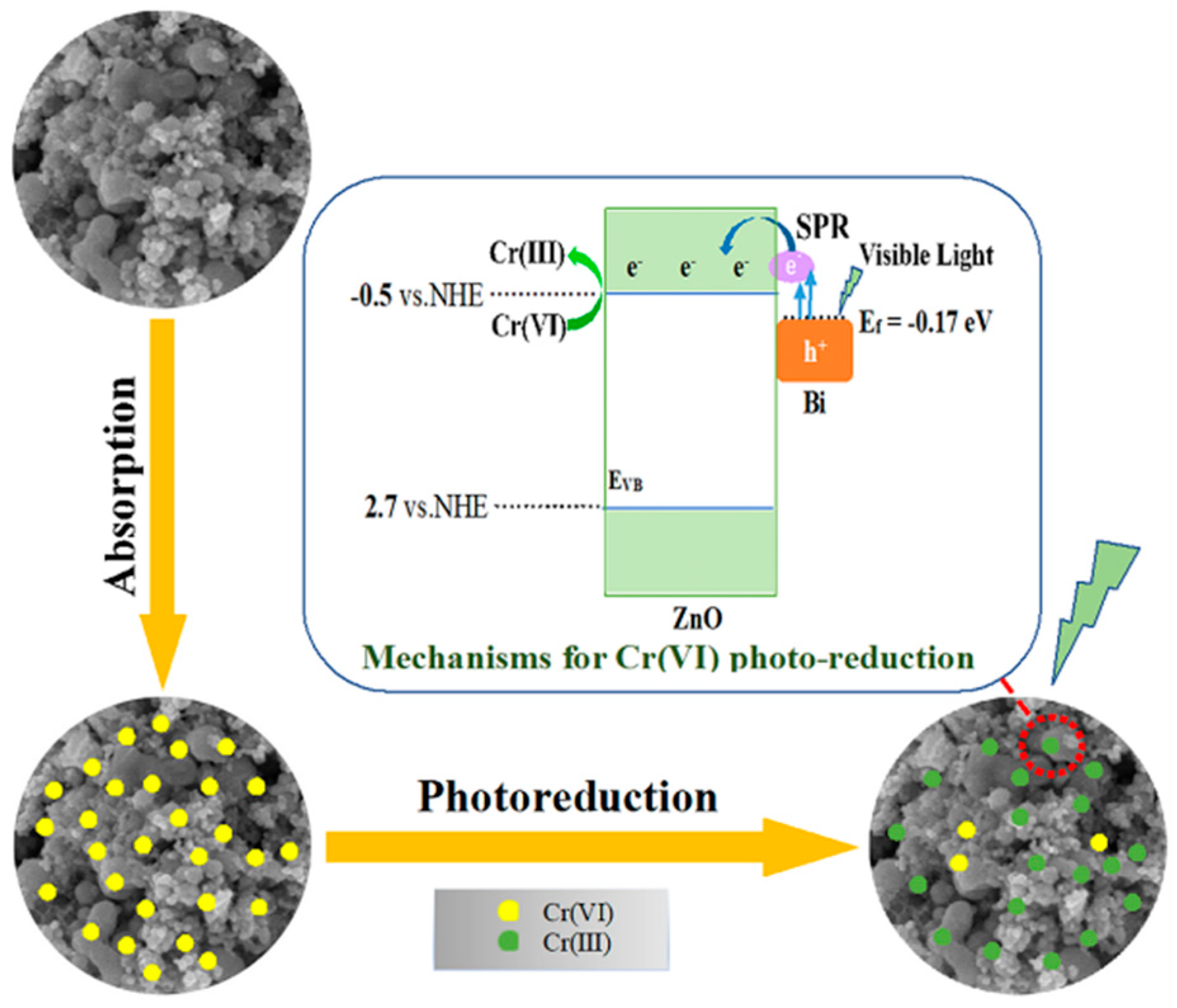
2.3. Proposed Mechanism

Based on the systematic investigation, the removal mechanism for Cr(VI) over BiNPs/ZnO under visible light illumination was proposed as shown in Figure 9. The high-efficient removal of Cr(VI) could be assigned to the synergistic effect of physical adsorption and SPR-promoted visible light photoreduction. Firstly, Cr(VI) was rapidly adsorbed and enriched onto the surface of BiNPs/ZnO composites. As the adsorption enrichment and photocatalytic reduction of Cr(VI) occurred at the adjacent sites (inset of Figure 9), the rapid enrichment of Cr(VI) on the surface of the photocatalyst reduced its diffusion distance in solution, which greatly increased the rate of photocatalysis. The BiNPs exhibited unusual noble metal-like behavior with increased visible light absorption ability due to its the SPR effect. The e/h+ pairs could be generated via the SPR of Bi [16]. The SPR exciation of Bi metal took place along with the light harvesting, after which the plasmon decay could ensue in one of three ways [19,55,56]: (1) the photon’s elastic radiation is reemission, in which one of the channels for the molecules that adsorbs onto the plasma to extract energy absorbs reemission photons with higher energy levels from the plasma emission, (2) non-radiative Landau damping, which converts photon energy into single as well as excited state e/h+ pairs; the excited electrons generate more electrons by Coulomb inelastic scattering and other electrons interacting with each other, thereby promoting the photoreduction reaction, or (3) the interaction of the excited surface plasma with the adsorbate induces direct electron injection into the adsorbate. Therefore, it can be inferred that plasma decay of the Bi/ZnO occurred at about 500 nm by (2) and (3). As for the BiNPs/ZnO samples, the excited electrons generated from the BiNPs through mechanism (2) was quickly injected into the tightly bound ZnO through the mechanism (3). Afterwards, the photo-induced electron would be reduced from Cr2O72− to Cr3+ on the surface of the ZnO particle. As shown in Figure 9, this transfer facilitated the e/h+ separation and caused lots of electrons on the surface of ZnO and holes on Bi, respectively, thereby greatly promoting the photocatalytic reduction of the Cr(VI) solution.

3. Experimental

3.1. Synthesis of BiNPs/ZnO Nanocomposite

ZnO nanoparticles were prepared using our previous work [34]. Virgin Bi particles were prepared using Dong’s method [16]. The BiNPs/ZnO nanocomposites with various loadings of Bi were synthesized by the following typical procedure: sodium hypophosphite (10.60 g) was first dissolved in distilled water (20 mL) in a beaker and then stirred for 15 min to make the solids dissolve completely. Afterwards, tartaric acid (0.05 g), sodium hydroxide (0.25 g), and distilled water (37.5 mL) were added to the above solution by stirring. After all reactants were dissolved, 1.0 g of ZnO was added to the solution. Then, the mixture was ultrasonically treated for 30 min to obtain a well-dispersed solution (solution A). Meanwhile, distilled water (17.6 mL), concentrated nitric acid (2.4 mL), and bismuth nitrate pentahydrate (9.7 g) were successively added to in a 50 mL glass beaker while continuously stirring. The resulting solution was marked as solution B. A 2.5 mL dose of solution B was dropped into solution A while stirring. Ten minutes later, the resulting precursor suspension was treated at 60 °C for 6 h. The resulting white solid was filtered, washed with water and ethanol four times, and dried at 40 °C for 12 h to obtain the final BiNPs/ZnO, labeled as Bi-ZnO-50 in which the mass ratio of Bi to ZnO was 50%. Just by changing the amount of ZnO in the procedure mentioned above, the BiNPs/ZnO composites with 10%, 50%, 80% mass ratio of Bi-to-ZnO were also prepared, respectively. For comparison, the as-prepared Bi and ZnO with a mass ratio of 80% were added to the ethanol solution. After sonicating for 30 min, the resulting sample was filtered, washed with ethanol, and dried at 40 °C for 12 h to give the physically mixed sample BiNPs/ZnO marked as Bi-ZnO-80(P).

3.2. Characterization

The XRD was used to phase analysis of the sample, which was conducted on PANalytical X’pert Pro powder (Almelo, The Netherlands) diffractometer through Cu-Ka radiation (λ = 1.5418 Å) with a scan step of 0.013°. In order to analyze the morphology of the sample, SEM was conducted on a FEI F250 scanning electron microscope with acceleration voltage of 30 kV. To further understand the phase and morphology, HRTEM was carried out on a FEI Tecnai G2 F20 (Hillsboro, OR, USA) field-emission transmission electron microscope at an accelerating voltage of 200 kV. XPS was used to analyze the composition of the surface of the sample, and it was performed on an ESCALAB 250 Xi photoelectron (Waltham, MA, USA) spectrometer using a monochromatic Al Kα X-ray source (hν = 1486.6 eV). The UV-vis DRS was carried out on a Hitachi U-4100 (Hitachi Ltd., Tokyo, Japan) UV-vis spectrophotometer using BaSO4 as the reference specimen, which was normally conducted to detect the band gap of the photocatalyst. In order to further explore the separation of electron–hole pairs after excited by visible light, we conducted PL and EIS tests. PL were recorded on a HITACHI F-7000 (Tokyo, Japan) to explore the optical properties of the obtained specimens. Photoelectrochemical measurements were conducted using in a three electrodes quartz cell with 0.1 M Na2SO4 electrolyte solution on the electrochemical system (CHI-760E, Shanghai, China). Platinum wire was used as counter, saturated calomel electrode (SCE) used as reference electrodes and ZnO, Bi-ZnO-80 electrodes on ITO served as the working electrode.

3.3. Photocatalytic Tests

The photocatalytic performances of BiNPs/ZnO composites were assessed by photoreduction of Cr(VI) solution under different conditions. All the experiments were carried out using a BILON-CHX-V photoreactor (Shanghai Bilon Instruments Manufacture Co., Ltd., Shanghai, China) and a 500 W Xenon lamp with working current 5 A, with the maximum wavelength emission at 470 nm as the visible light source (Shanghai Jiguang Special Lighting Factory, Shanghai, China). The mixture of Cr(VI) (50 mg/L) was made by dissolving K2Cr2O7 in distilled water. In each experiment, a 100-mg photocatalyst was mixed with 50 mL of aqueous Cr(VI). Before irradiation, the mixture solution was vigorously stirred in the dark for 1 h to reach the adsorption–desorption equilibrium. At the time of irradiation, 3 mL of the clear reaction aqueous was obtained by separating the as-prepared samples using a centrifugation a the solution at a certain time interval. The concentration of Cr(VI) was at the discretion of the outcome, which resulted in a 1,5-diphenylcarbazide method at a wavelength of 540 nm using UV-1000 spectrophotometer [57]. As a point of comparison, blank experiments (without the addition of as-prepared samples) wer conducted. In order to detect the physical adsorption capacity of composites, dark experiments were performed under the identical conditions. C was defined as the concentration of Cr(VI) after the irradiation t, and C0 was the concentration of Cr(VI) after adsorption equilibrium, and thus the removal ratio of Cr(VI)(η (%)) was determined by (1 − C/C0) × 100%.

4. Conclusions

In general, BiNPs/ZnO photocatalysts were successfully synthesized in an ordinary eco-friendly synthesis method. An intimate interfacial interaction was constructed between ZnO and Bi phases in the prepared photocatalysts. Bi/ZnO nanocomposites showed outstanding Cr(VI) removal activity, excelling in virgin ZnO, Bi, and the sample prepared employing the physical mixing method. The improvement of photocatalytic performance was derived from the synergy effect of the excellent Cr(VI) physical adsorption from an aqueous solution and enhanced light absorption in visible light regions due to the SPR effect of Bi, leading to the effective separation of photogenerated carriers across the junction interface within the composites. The Bi/ZnO photocatalyst prepared in this paper is being further assessed by degrading other contaminants and exhibit potential applications for water treatment and remediation.

Author Contributions

X.Y. and Z.F. conceived and designed the experiments; Z.F.; J.Z.; J.N.; J.L.; D.P., and X.C. performed the experiments and analyzed the data; X.Y. and Z.F. wrote the paper.

Funding

This research was funded by National Natural Science Foundation of China, 51402030 and Natural Science Foundation Project of CQ CSTC: cstc2017jcyjB0028.

Acknowledgments

The authors would like to gratefully acknowledge the financial supports from National Natural Science Foundation of China (No. 51402030) and Natural Science Foundation Project of CQ CSTC (cstc2017jcyjB0028).

Conflicts of Interest

The authors declare no conflict of interest.

References

  1. Langård, S. Chromium carcinogenicity; A review of experimental animal data. Sci. Total Environ. 1988, 71, 341–350. [Google Scholar] [CrossRef]
  2. Katz, S.; Salem, H. The toxicology of chromium with respect to its chemical speciation: A review. J. Appl. Toxicol. 1993, 13, 217–224. [Google Scholar] [CrossRef] [PubMed]
  3. Kabra, K.; Chaudhary, R.; Sawhney, R.L. Treatment of hazardous organic and inorganic compounds through aqueous-phase photocatalysis: A review. Ind. Eng. Chem. Res. 2004, 43, 7683–7696. [Google Scholar] [CrossRef]
  4. Barrera-Diaz, C.E.; Lugo-Lugo, V.; Bilyeu, B. A review of chemical, electrochemical and biological methods for aqueous Cr(VI) reduction. J. Hazard. Mater. 2012, 223, 1–12. [Google Scholar] [CrossRef] [PubMed]
  5. Wang, Y.J.; Jiang, L.J.; Feng, C.G. Photocatalytic reduction of Cr(VI). Prog. Chem. 2013, 25, 1999–2010. [Google Scholar]
  6. Jing, L.Q.; Zhou, W.; Tian, G.H.; Fu, H.G. Surface tuning for oxide-based nanomaterials as efficient photocatalysts. Chem. Soc. Rev. 2013, 42, 9509–9549. [Google Scholar] [CrossRef] [PubMed]
  7. Daghrir, R.; Drogui, P.; Robert, D. Modified TiO2 for environmental photocatalytic applications: A review. Ind. Eng. Chem. Res. 2013, 52, 3581–3599. [Google Scholar] [CrossRef]
  8. Zhang, H.J.; Chen, G.H.; Bahnemann, D.W. Photoelectrocatalytic materials for environmental applications. J. Mater. Chem. 2009, 19, 5089–5121. [Google Scholar] [CrossRef]
  9. Hernandez-Alonso, M.D.; Fresno, F.; Suarez, S.; Coronado, J.M. Development of alternative photocatalysts to TiO2: Challenges and opportunities. Energy Environ. Sci. 2009, 2, 1231–1257. [Google Scholar] [CrossRef]
  10. Peng, H.; Liu, X.; Tang, W.; Ma, R. Facile synthesis and characterization of ZnO nanoparticles grown on halloysite nanotubes for enhanced photocatalytic properties. Sci. Rep. 2017, 7, 2250. [Google Scholar] [CrossRef] [PubMed]
  11. Liu, L.; Sun, T.; Zhang, H.; Chen, H. Scalable and facile preparation of optical-magnetic dual function 3d Ni@graphene-ZnO for high efficiency removal of hexavalent chromium. Ceram. Int. 2017, 43, 3792–3796. [Google Scholar] [CrossRef]
  12. Yang, Y.; Li, H.; Hou, F.; Hu, J.; Zhang, X.; Wang, Y. Facile synthesis of ZnO/Ag nanocomposites with enhanced photocatalytic properties under visible light. Mater. Lett. 2016, 180, 97–100. [Google Scholar] [CrossRef]
  13. Li, S.; Cai, J.; Wu, X.; Zheng, F. Sandwich-like TiO2@ZnO-based noble metal (Ag, Au, Pt, or Pd) for better photo-oxidation performance: Synergistic effect between noble metal and metal oxide phases. Appl. Surf. Sci. 2018, 443, 603–612. [Google Scholar] [CrossRef]
  14. Pirhashemi, M.; Habibi-Yangjeh, A.; Pouran, S.R. Review on the criteria anticipated for the fabrication of highly efficient ZnO-based visible-light-driven photocatalysts. J. Ind. Eng. Chem. 2018, 62, 1–25. [Google Scholar] [CrossRef]
  15. Gu, X.; Li, C.; Yuan, S.; Ma, M.; Qiang, Y.; Zhu, J. ZnO based heterojunctions and their application in environmental photocatalysis. Nanotechnology 2016, 27, 402001. [Google Scholar] [CrossRef] [PubMed]
  16. Dong, F.; Xiong, T.; Sun, Y.J.; Zhao, Z.W.; Zhou, Y.; Feng, X.; Wu, Z.B. A semimetal bismuth element as a direct plasmonic photocatalyst. Chem. Commun. 2014, 50, 10386–10389. [Google Scholar] [CrossRef] [PubMed]
  17. Li, J.; Dong, X.A.; Sun, Y.; Cen, W.; Dong, F. Facet-dependent interfacial charge separation and transfer in plasmonic photocatalysts. Appl. Catal. B 2018, 226, 269–277. [Google Scholar] [CrossRef]
  18. Dong, F.; Zhao, Z.W.; Sun, Y.J.; Zhang, Y.X.; Yan, S.; Wu, Z.B. An advanced semimetal-organic Bi spheres-g-C3N4 nanohybrid with SPR-enhanced visible-light photocatalytic performance for no purification. Environ. Sci. Technol. 2015, 49, 12432–12440. [Google Scholar] [CrossRef] [PubMed]
  19. Kale, M.J.; Avanesian, T.; Christopher, P. Direct photocatalysis by plasmonic nanostructures. ACS Catal. 2014, 4, 116–128. [Google Scholar] [CrossRef]
  20. Li, J.T.; Cushing, S.K.; Bright, J.; Meng, F.K.; Senty, T.R.; Zheng, P.; Bristow, A.D.; Wu, N.Q. Ag@Cu2O core-shell nanoparticles as visible-light plasmonic photocatalysts. ACS Catal. 2013, 3, 47–51. [Google Scholar] [CrossRef]
  21. Yu, J.; Yue, L.; Liu, S.; Huang, B.; Zhang, X. Hydrothermal preparation and photocatalytic activity of mesoporous Au-TiO2 nanocomposite microspheres. J. Colloid Interface Sci. 2009, 334, 58–64. [Google Scholar] [CrossRef] [PubMed]
  22. Wu, L.; Li, F.; Xu, Y.; Zhang, J.W.; Zhang, D.; Li, G.; Li, H. Plasmon-induced photoelectrocatalytic activity of au nanoparticles enhanced TiO2 nanotube arrays electrodes for environmental remediation. Appl. Catal. B 2015, 164, 217–224. [Google Scholar] [CrossRef]
  23. Yu, J.; Sun, D.; Wang, T.; Li, F. Fabrication of Ag@AgCl/ZnO submicron wire film catalyst on glass substrate with excellent visible light photocatalytic activity and reusability. Chem. Eng. J. 2018, 334, 225–236. [Google Scholar] [CrossRef]
  24. Yu, J.; Dai, G.; Huang, B. Fabrication and characterization of visible-light-driven plasmonic photocatalyst Ag/AgCl/TiO2 TiO2 nanotube arrays. J. Phys. Chem. C 2009, 113, 16394–16401. [Google Scholar] [CrossRef]
  25. Cai, J.; Huang, Y.; Huang, B.; Zheng, S.; Guo, Y. Enhanced activity of pt nanoparticle catalysts supported on manganese oxide-carbon nanotubes for ethanol oxidation. Int. J. Hydrogen Energy 2014, 39, 798–807. [Google Scholar] [CrossRef]
  26. An, Y.; Xu, B.; Liu, Y.; Wang, Z.; Wang, P.; Dai, Y.; Qin, X.; Zhang, X.; Huang, B. Photocatalytic overall water splitting over MIL-125(Ti) upon CoPi and Pt Co-catalyst deposition. Chemistryopen 2017, 6, 701–705. [Google Scholar] [CrossRef] [PubMed]
  27. Weng, S.; Chen, B.; Xie, L.; Zheng, Z.; Liu, P. Facile in situ synthesis of a Bi/BiOCl nanocomposite with high photocatalytic activity. J. Mater. Chem. A 2013, 1, 3068–3075. [Google Scholar] [CrossRef]
  28. Sun, Y.; Zhao, Z.; Dong, F.; Zhang, W. Mechanism of visible light photocatalytic NOx oxidation with plasmonic Bi cocatalyst-enhanced (BiO)2CO3 hierarchical microspheres. Phys. Chem. Chem. Phys. 2015, 17, 10383–10390. [Google Scholar] [CrossRef] [PubMed]
  29. Wang, B.; Feng, W.H.; Zhang, L.L.; Zhang, Y.; Huang, X.Y.; Fang, Z.B.; Liu, P. In situ construction of a novel Bi/CdS nanocomposite with enhanced visible light photocatalytic performance. Appl. Catal. B 2017, 206, 510–519. [Google Scholar] [CrossRef]
  30. Liu, X.; Cao, H.; Yin, J. Generation and photocatalytic activities of Bi@Bi2O3 microspheres. Nano Res. 2011, 4, 470–482. [Google Scholar] [CrossRef]
  31. Zou, J.-P.; Ma, J.; Luo, J.-M.; Yu, J.; He, J.; Meng, Y.; Luo, Z.; Bao, S.-K.; Liu, H.-L.; Luo, S.-L.; et al. Fabrication of novel heterostructured few layered WS2-Bi2WO6/Bi3.84W0.16O6.24 composites with enhanced photocatalytic performance. Appl. Catal. B 2015, 179, 220–228. [Google Scholar] [CrossRef]
  32. Zou, J.-P.; Ma, J.; Huang, Q.; Luo, S.-L.; Yu, J.; Luo, X.-B.; Dai, W.-L.; Sun, J.; Guo, G.-C.; Au, C.-T.; et al. Graphene oxide as structure-directing and morphology-controlling agent for the syntheses of heterostructured graphene-Bi2MoO6/Bi3.64Mo0.36O6.55 composites with high photocatalytic activity. Appl. Catal. B 2014, 156–157, 447–455. [Google Scholar] [CrossRef]
  33. Labib, S. Study of photocatalytic properties of pure and doped ZnO powders. Mater. Werkst. 2016, 47, 19–28. [Google Scholar] [CrossRef]
  34. Yuan, X.; Zhou, C.; Jing, Q.; Tang, Q.; Mu, Y.; Du, A.-K. Facile synthesis of g-C3N4 nanosheets/ZnO nanocomposites with enhanced photocatalytic activity in reduction of aqueous chromium(VI) under visible light. Nanomaterials 2016, 6, 173. [Google Scholar] [CrossRef] [PubMed]
  35. Li, X.B.; Zhang, H.S.; Luo, J.M.; Feng, Z.J.; Huang, J.T. Hydrothermal synthesized novel nanoporous g-C3N4/MnTiO3 heterojunction with direct z-scheme mechanism. Electrochim. Acta 2017, 258, 998–1007. [Google Scholar] [CrossRef]
  36. Jiang, D.; Yu, H.; Yu, H. Modified g-C3N4/TiO2 nanosheets/ZnO ternary facet coupled heterojunction for photocatalytic degradation of p-toluenesulfonic acid (p-tsa) under visible light. Phys. E Low-Dimens. Syst. Nanostruct. 2017, 85, 1–6. [Google Scholar] [CrossRef]
  37. Warren, S.C.; Jackson, A.C.; Cater-Cyker, Z.D.; DiSalvo, F.J.; Wiesner, U. Nanoparticle synthesis via the photochemical polythiol process. J. Am. Chem. Soc. 2007, 129, 10072–10073. [Google Scholar] [CrossRef] [PubMed]
  38. Morgan, W.E.; Stec, W.J.; Van Wazer, J.R. Inner-orbital binding-energy shifts of antimony and bismuth compounds. Inorg. Chem. 1973, 12, 953–955. [Google Scholar] [CrossRef]
  39. Dong, F.; Lee, S.C.; Wu, Z.; Huang, Y.; Fu, M.; Ho, W.-K.; Zou, S.; Wang, B. Rose-like monodisperse bismuth subcarbonate hierarchical hollow microspheres: One-pot template-free fabrication and excellent visible light photocatalytic activity and photochemical stability for no removal in indoor air. J. Hazard. Mater. 2011, 195, 346–354. [Google Scholar] [CrossRef] [PubMed]
  40. Li, R.; Chen, W.; Kobayashi, H.; Ma, C. Platinum-nanoparticle-loaded bismuth oxide: An efficient plasmonic photocatalyst active under visible light. Green Chem. 2010, 12, 212–215. [Google Scholar] [CrossRef]
  41. Cheng, K.; Sun, W.; Jiang, H.-Y.; Liu, J.; Lin, J. Sonochemical deposition of au nanoparticles on different facets-dominated anatase TiO2 single crystals and resulting photocatalytic performance. J. Phys. Chem. C 2013, 117, 14600–14607. [Google Scholar] [CrossRef]
  42. Venkataswamy, P.; Jampaiah, D.; Kandjani, A.E.; Sabri, Y.M.; Reddy, B.M.; Vithal, M. Transition (Mn, Fe) and rare earth (La, Pr) metal doped ceria solid solutions for high performance photocatalysis: Effect of metal doping on catalytic activity. Res. Chem. Intermed. 2018, 44, 2523–2543. [Google Scholar] [CrossRef]
  43. Singh, M.; Jampaiah, D.; Kandjani, A.E.; Sabri, Y.M.; Della Gaspera, E.; Reineck, P.; Judd, M.; Langley, J.; Cox, N.; van Embden, J.; et al. Oxygen-deficient photostable Cu2O for enhanced visible light photocatalytic activity. Nanoscale 2018, 10, 6039–6050. [Google Scholar] [CrossRef] [PubMed]
  44. Toudert, J.; Serna, R.; Jimenez de Castro, M. Exploring the optical potential of nano-bismuth: Tunable surface plasmon resonances in the near ultraviolet-to-near infrared range. J. Phys. Chem. C 2012, 116, 20530–20539. [Google Scholar] [CrossRef]
  45. Dong, F.; Li, Q.Y.; Sun, Y.J.; Ho, W.K. Noble metal-like behavior of plasmonic bi particles as a cocatalyst deposited on (BiO)2CO3 microspheres for efficient visible light photocatalysis. ACS Catal. 2014, 4, 4341–4350. [Google Scholar] [CrossRef]
  46. Guo, L.T.; Cai, Y.Y.; Ge, J.M.; Zhang, Y.N.; Gong, L.H.; Li, X.H.; Wang, K.X.; Ren, Q.Z.; Su, J.; Chen, J.S. Multifunctional Au-Co@CN nanocatalyst for highly efficient hydrolysis of ammonia borane. ACS Catal. 2015, 5, 388–392. [Google Scholar] [CrossRef]
  47. Yu, Y.; Cao, C.; Liu, H.; Li, P.; Wei, F.; Jiang, Y.; Song, W. A Bi/BiOCl heterojunction photocatalyst with enhanced electron–hole separation and excellent visible light photodegrading activity. J. Mater. Chem. A 2014, 2, 1677–1681. [Google Scholar] [CrossRef]
  48. Li, Y.D.; Wang, J.W.; Deng, Z.X.; Wu, Y.Y.; Sun, X.M.; Yu, D.P.; Yang, P.D. Bismuth nanotubes: A rational low-temperature synthetic route. J. Am. Chem. Soc. 2001, 123, 9904–9905. [Google Scholar] [CrossRef] [PubMed]
  49. Zhang, H.; Zong, R.; Zhu, Y. Photocorrosion inhibition and photoactivity enhancement for zinc oxide via hybridization with monolayer polyaniline. J. Phys. Chem. C 2009, 113, 4605–4611. [Google Scholar] [CrossRef]
  50. Shirzad-Siboni, M.; Farrokhi, M.; Darvishi Cheshmeh Soltani, R.; Khataee, A.; Tajassosi, S. Photocatalytic reduction of hexavalent chromium over ZnO nanorods immobilized on kaolin. Ind. Eng. Chem. Res. 2014, 53, 1079–1087. [Google Scholar] [CrossRef]
  51. Xu, H.; Zhao, H.; Song, Y.; Yan, W.; Xu, Y.; Li, H.; Huang, L.; Yin, S.; Li, Y.; Zhang, Q.; et al. g-C3N4/Ag3PO4 composites with synergistic effect for increased photocatalytic activity under the visible light irradiation. Mater. Sci. Semicond. Process. 2015, 39, 726–734. [Google Scholar] [CrossRef]
  52. Zhang, F.-J.; Xie, F.-Z.; Zhu, S.-F.; Liu, J.; Zhang, J.; Mei, S.-F.; Zhao, W. A novel photofunctional g-C3N4/Ag3PO4 bulk heterojunction for decolorization of Rh.B. Chem. Eng. J. 2013, 228, 435–441. [Google Scholar] [CrossRef]
  53. Zhang, Y.C.; Yao, L.; Zhang, G.S.; Dionysiou, D.D.; Li, J.; Du, X.H. One-step hydrothermal synthesis of high-performance visible-light-driven SnS2/SnO2 nanoheterojunction photocatalyst for the reduction of aqueous Cr(VI). Appl. Catal. B 2014, 144, 730–738. [Google Scholar] [CrossRef]
  54. Wang, X.J.; Wang, Q.; Li, F.T.; Yang, W.Y.; Zhao, Y.; Hao, Y.J.; Liu, S.J. Novel BiOCl-C3N4 heterojunction photocatalysts: In situ preparation via an ionic-liquid-assisted solvent-thermal route and their visible-light photocatalytic activities. Chem. Eng. J. 2013, 234, 361–371. [Google Scholar] [CrossRef]
  55. Warren, S.C.; Thimsen, E. Plasmonic solar water splitting. Energy Environ. Sci. 2012, 5, 5133–5146. [Google Scholar] [CrossRef]
  56. Christopher, P.; Xin, H.; Marimuthu, A.; Linic, S. Singular characteristics and unique chemical bond activation mechanisms of photocatalytic reactions on plasmonic nanostructures. Nat. Mater. 2012, 11, 1044–1050. [Google Scholar] [CrossRef] [PubMed]
  57. Yuan, X.; Jing, Q.; Chen, J.; Li, L. Photocatalytic Cr(VI) reduction by mixed metal oxide derived from ZnAl layered double hydroxide. Appl. Clay Sci. 2017, 143, 168–174. [Google Scholar] [CrossRef]
Figure 1. XRD (X-ray diffraction pattern) spectrums of ZnO, Bi and BiNPs/ZnO with different Bi contents.
Catalysts 08 00426 g001
Figure 2. SEM (scanning electron microscopy) images of (a,b) Bi, (c,d) ZnO, and (e,f) Bi-ZnO-80.
Catalysts 08 00426 g002
Figure 3. HRTEM (high-resolution transmission electron microscope) images of (a,b) Bi, (c,d) ZnO, and (e,f) Bi-ZnO-80.
Catalysts 08 00426 g003aCatalysts 08 00426 g003b
Figure 4. (a) XPS survey spectra, (b) Bi4f, and (c) Zn2p peaks of the Bi-ZnO-80 composite.
Catalysts 08 00426 g004aCatalysts 08 00426 g004b
Figure 5. (a) DRS spectra and (b) plots of (Ahν)2 versus hv for ZnO and BiNPs/ZnO anocomposites.
Catalysts 08 00426 g005
Figure 6. (a) PL spectra of ZnO and Bi-ZnO-80 photocatalysts and (b) EIS plots of the ZnO and Bi-ZnO-80 photocatalysts with light on/off cycles under the irradiation of visible light (λ > 420 nm) [Na2SO4 = 0.1 M].
Catalysts 08 00426 g006
Figure 7. (a) Photoreduction of aqueous Cr(VI) using BiNPs/ZnO, Bi and ZnO photocatalysts under visiblelight irradiation, (b) Photocatalytic reduction of Cr(VI) of different initial concentration by Bi-ZnO-80 composite, (c) Photocatalytic reduction of Cr(VI) by different dosage of Bi-ZnO-80 composite, (d) Photoreduction of aqueous Cr(VI) using Bi-ZnO-80 composite synthesized by Bi-ZnO-80 and Bi-ZnO-80(P) under visiblelight irradiation.
Catalysts 08 00426 g007
Figure 8. Removal ratio by physical adsorption and photocatalysis of Cr(VI) over Bi-ZnO-10, Bi-ZnO-50, and Bi-ZnO-80.
Catalysts 08 00426 g008
Figure 9. Mechanism for Cr(VI) removal catalyzed by BiNPs/ZnO nanocomposites under visible light irradiation.
Catalysts 08 00426 g009

