Article

Synergistic Enhancement of Photoelectrochemical Hydrogen Evolution, Antimicrobial, and Cytotoxic Activities in a ZIF-8/Aspergillus nidulans Extract Nanocomposite


by
Amira Ben Gouider Trabelsi
1,
Fatemah H. Alkallas
1,
Abdelaziz M. Aboraia
2,3,*,
Mohamed E. Abouelela
4,
Mohammad H. A. Hassan
5 and
Abdallah M. A. Hassane
6,*
1
Department of Physics, College of Science, Princess Nourah bint Abdulrahman University, P.O. Box 84428, Riyadh 11671, Saudi Arabia
2
Physics Department, Faculty of Science, Al-Azhar University, Assiut Branch, Assiut 71524, Egypt
3
College of Health and Medical Technology, Al-Ayen Iraqi University, An Nasiriyah 64001, Thi-Qar, Iraq
4
Department of Pharmacognosy, Faculty of Pharmacy (Boys), Al-Azhar University, Cairo 11884, Egypt
5
Department of Medical Laboratory Technology, Higher Technological Institute for Applied Health Sciences, Minya 71511, Egypt
6
Department of Botany and Microbiology, Faculty of Science, Al-Azhar University, Assiut Branch, Assiut 71524, Egypt
*
Authors to whom correspondence should be addressed.
Catalysts 2026, 16(8), 712; https://doi.org/10.3390/catal16080712
Submission received: 13 June 2026 / Revised: 1 August 2026 / Accepted: 3 August 2026 / Published: 6 August 2026

Abstract

Pushing ahead in materials chemistry means building tiny substances that work hard and take into account clean power, planet care, life science all at once. From this effort comes a new direction: ZIF-8, a metal-linked cage structure, now fused with active components extracted from the common fungus Aspergillus nidulans. Not just mixed, but grown together with fungal extracts tucked neatly into the skeleton of the material while keeping its orderly shape intact. Three versions appeared—loaded at 2%, 4%, and 6 weight percent—and each one was mapped out using X-ray signals, sharp images from electron scans, and element tracing. A light flickered on and off during tests in which electricity flowed through these new composites set between three points, designed to split water and release hydrogen gas. Surprisingly, the ZIF-8@2% nidulans blend showed strong teamwork between electricity- and light-driven biology, creating a sharp spike in temporary current while cutting down reaction delay to just 310 mV/dec—pushing hydrogen release through a faster molecular handshake. In this mix, natural compounds from fungi act like tiny solar collectors, helping electrons move more freely, which is reflected in impedance scans as lower resistance. On top of that, higher doses of the material effectively blocked harmful microbes, thanks to ZIF-8 breaking cell walls and active fungal ingredients punching holes as well. Instead of relying on harsh chemicals, it uses a nature-inspired design in which molds meet synthetic frameworks, creating a single system with potential for sustainable energy generation and antimicrobial applications.

1. Introduction

In the face of a finite supply of fossil fuels and growing environmental problems, the world’s scientific community has turned to alternatives that are both sustainable and environmentally friendly, with zero-emission options taking center stage [1]. The most promising clean energy carrier is hydrogen (H2), with the highest gravimetric energy density of 142 kJ/g and an entirely harmless combustion product (water) [2,3]. Solar energy-based photoelectrochemical (PEC) water splitting and the hydrogen evolution reaction (HER) are elegant and green approaches for directly converting solar radiation into chemical fuel among various hydrogen production technologies [4,5,6]. The commercialization of PEC-HER is, however, greatly hindered by the scarcity of cost-effective, stable and highly active electrocatalysts that can induce charge separation and promote electron transfer at the interfaces [7,8]. Metal–organic frameworks (MOFs) are interesting porous crystalline materials composed of metal coordination centers connected by organic molecules, and have attracted considerable interest in the past decade for their use in energy conversion and storage [9,10]. In particular, the Zinc ion framework ZIF-8 (Zinc ions (Zn2+) coordinated to 2-methylimidazole linkers) is widely recognized for its outstanding thermal stability, permanent porosity, large surface area, and easily tunable structural topology [11,12]. While there are many advantages, the pristine is severely limited when used in the direct application of photoelectrochemical systems. ZIF-8 has a very large electron band gap (3.3–3.4 eV), which renders it sensitive to a small portion of the solar spectrum (ultraviolet light) and has poor electrical conductivity, which produces high recombination rates of photo-excited electron–hole (e/h+) pairs and low current densities [13,14].
To circumvent these drawbacks, researchers have focused on modifying the electronic structure of ZIF-8. Traditional strategies include doping the framework with foreign transition-metal ions (such as Cobalt, Iron, or Copper) or fabricating semiconductor heterojunctions with carbon-based structures such as graphitic carbon nitride (g-C3N4) or graphene derivatives [15,16]. Concurrently, another critical global challenge is the rising threat of antimicrobial resistance (AMR) among pathogenic microbes, which requires the urgent engineering of advanced biocidal agents [17,18]. Interestingly, nanoscale zinc-based frameworks naturally exhibit intrinsic antimicrobial activity through the slow, controlled release of localized Zn2+ ions, which induce oxidative stress and rupture of microbial cell walls. Consequently, designing a single bifunctional catalyst that simultaneously drives energy generation (PEC-HER) and exhibits strong antimicrobial efficacy represents a highly desirable, atom-economic objective.
Recently, the integration of biological entities with synthetic nanomaterials—often termed “bio-nanocomposites”—has emerged as a highly sustainable methodology [19,20]. Fungal species, particularly Aspergillus nidulans, are rich factories of diverse secondary metabolites, proteins, amino acids, and extracellular enzymes [21]. These bio-organic molecules possess complex functional groups (such as carboxyl, hydroxyl, and amine bands) that can act as natural reducing, stabilizing, and capping agents. More importantly, certain bioactive fungal extracts possess unique conjugated molecular chains that absorb visible light and act as organic charge-shuttling antennas when coupled with wide-bandgap inorganic materials [22]. Despite the extensive exploration of ZIF-8 modifications, several critical gaps remain in the current literature: Reliance on Toxic Synthetic Chemicals: Most classical methods used to sensitize or alter the band structure of ZIF-8 rely on harsh, toxic chemical reducing agents, surfactants, or expensive noble metals, which raise environmental concerns and limit scalability.
Underutilization of Biomass and Fungal Extract Co-catalysts: While green synthesis of simple metal oxide nanoparticles using plant extracts is common, the deliberate incorporation of crude or refined fungal extracts from Aspergillus nidulans directly into a crystalline MOF matrix as a functional photo-biochemical co-catalyst remains virtually unexplored. Lack of Dual-Functional Platforms: Most research decouples energy-conversion materials from biomedical agents. There is a profound scarcity of single-phase hybrid materials engineered to simultaneously maximize PEC solar-hydrogen kinetics and deliver potent antimicrobial protection through a unified, synergistic mechanism. To address these challenges, this study explores the “closed-loop” valorization of biological extracts by introducing a novel, eco-friendly bio-hybrid nanocomposite framework. The primary objective is to evaluate the synergistic enhancement of both photoelectrochemical hydrogen evolution kinetics and antimicrobial performance achieved by anchoring Aspergillus nidulans extract within a ZIF-8 matrix. We systematically investigate the effects of extract loading (2%, 4%, and 6 wt%) on phase integrity, morphological transformation, interfacial charge storage (CV), charge-transfer resistance (Rct via EIS), and transient photocurrent dynamics. Through this systematic approach, we establish the fundamental mechanism underlying this bio-inorganic coupling, optimizing the configuration to achieve high-efficiency solar-to-chemical conversion while maintaining reliable biocidal activity.

2. Results & Discussion

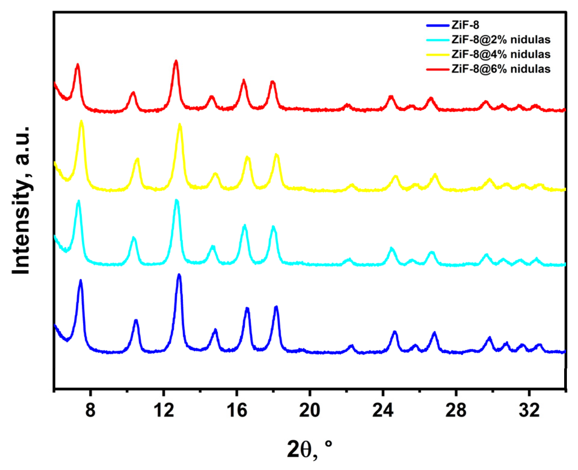
The crystalline structure and phase purity of pristine ZIF-8 and the fabricated ZIF-8@nidulans nanocomposites with varying extract fractions (2%, 4%, and 6%) were evaluated via X-ray diffraction (XRD), as illustrated in Figure 1. Pristine ZiF-8 exhibits sharp, well-defined diffraction peaks at 2 theta values of approximately 7.30°, 10.35°, 12.70°, 14.70°, 16.40°, and 18.00°, which correspond to the (011), (002), (112), (022), (013), and (222) crystal planes of a sodalite-type cubic framework, respectively [23]. This profile is in perfect agreement with the simulated and reported literature for crystalline ZIF-8. Upon the integration of Aspergillus nidulans extract (2%, 4%, and 6%), the positions of all major characteristic peaks of the ZIF-8 host matrix remain virtually unaltered. This preservation indicates that incorporating the biological extract molecules does not collapse or severely distort the long-range crystalline framework of the metal–organic framework (MOF). A slight decrease in peak intensities and a subtle broadening are observed with increasing extract content, which can be attributed to the amorphous or highly disordered nature of the encapsulated organic biomolecules/peptides from the A. nidulans extract, as well as slight changes in domain size due to host–guest interactions.
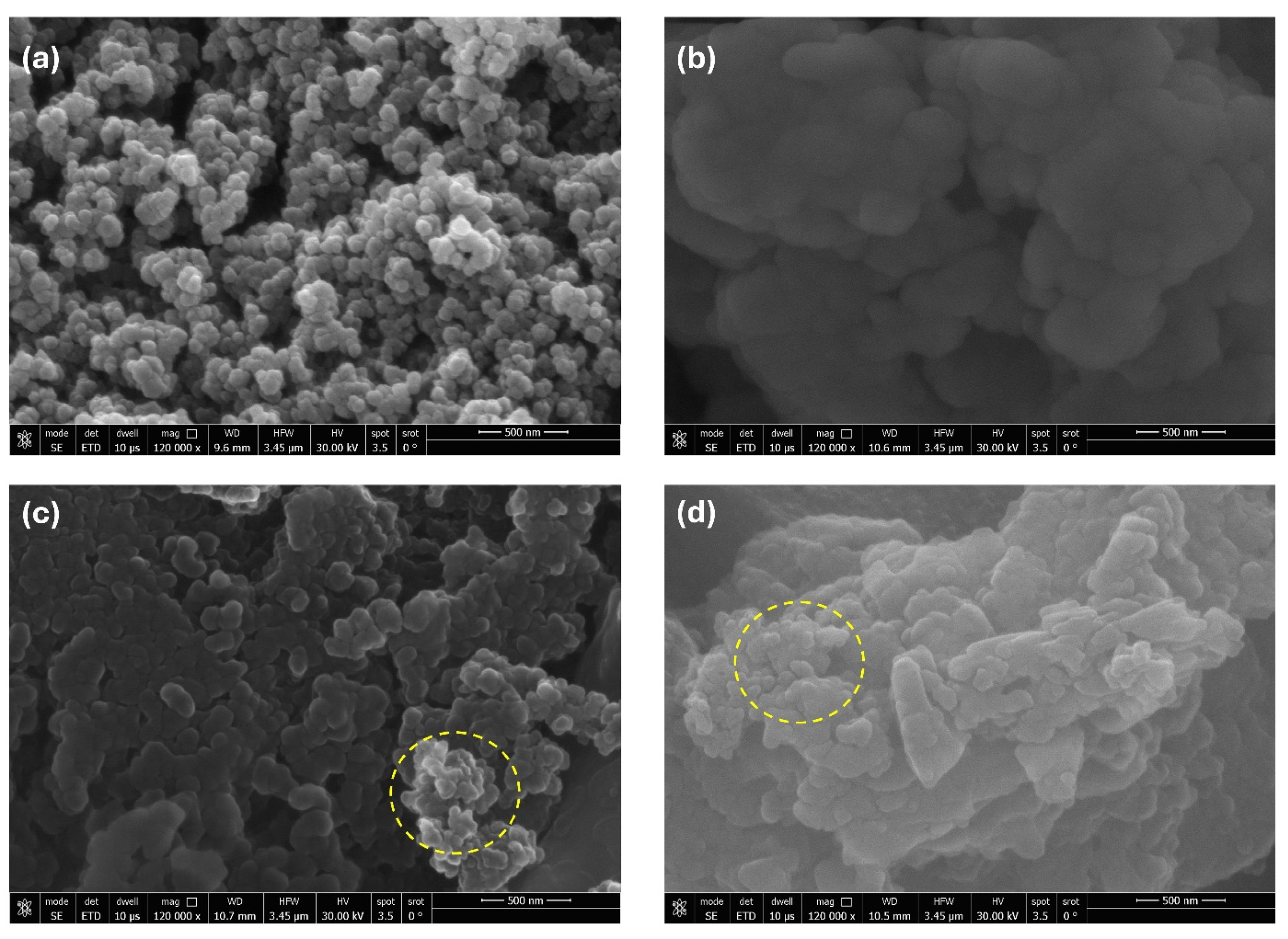
Field-emission scanning electron microscopy (FESEM) was employed to monitor morphological changes across the sample set, as shown in Figure 2. Figure 2a confirms that pristine ZIF-8 consists of uniform, nanoscale pseudo-spherical/polyhedral particles densely packed with well-defined grain boundaries, with an average size well below 100 nm. Figure 2b shows the amorphous, highly aggregated, and bulk-like matrix morphology typical of crude dried biological extracts (A. nidulans). For the ZIF-8@nidulans nanocomposites (Figure 2c,d), a progressive morphological transition is apparent. At optimal loading (Figure 2c, highlighted in yellow circles), the primary ultra-fine ZIF-8 nanoparticles remain distinct but are anchored onto or encapsulated by the thin organic sheets of the extract. At higher loading (Figure 2d), significant aggregation occurs; the excess biomaterial forms an overlying surface coating, transforming the particulate framework into larger, fused agglomerates. This dense organic capping layer directly correlates with the decreased surface accessibility and mass-transport bottlenecks identified in subsequent performance tests.
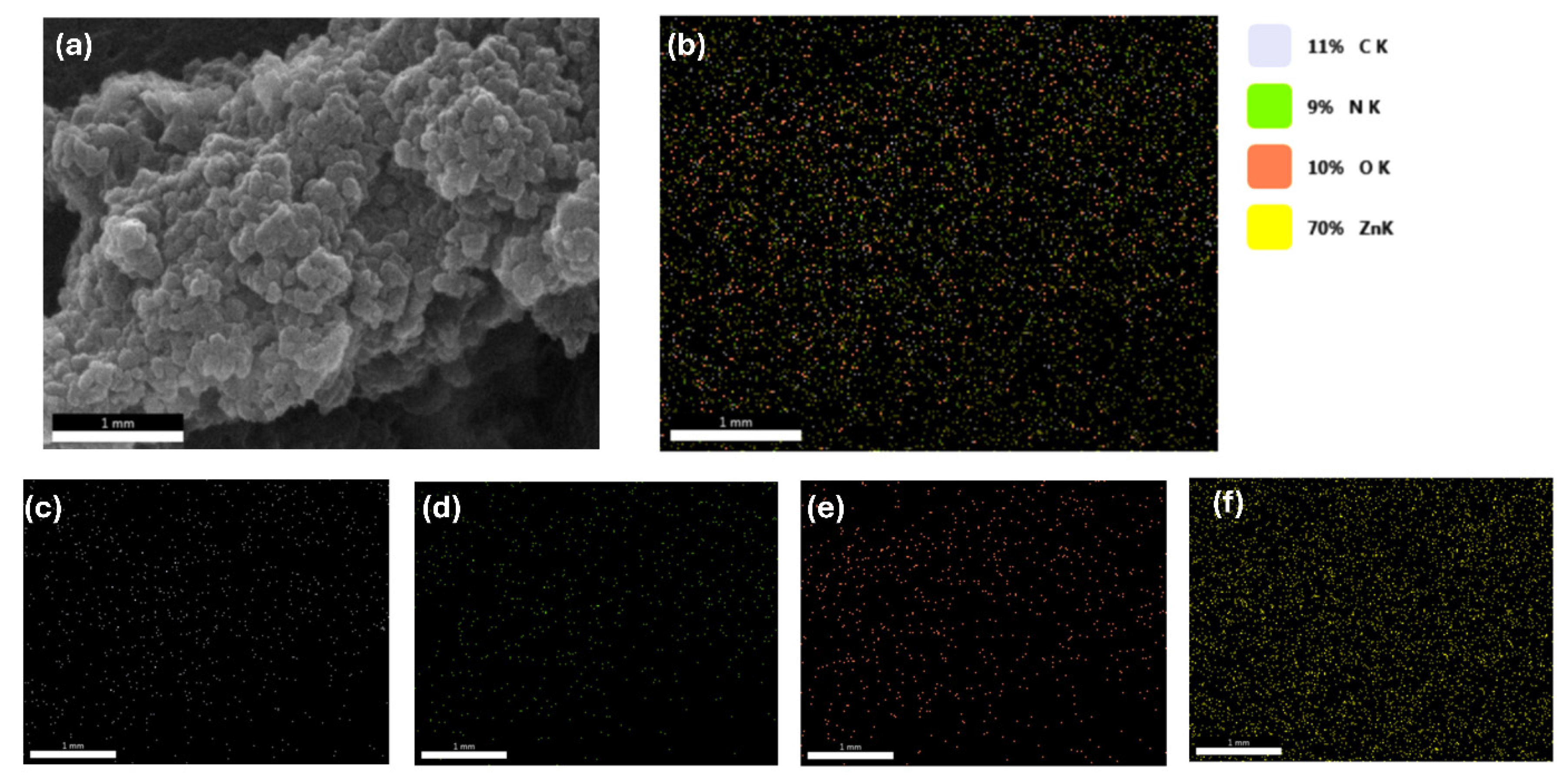
To verify the homogeneous distribution of the biomolecules within the MOF, energy-dispersive X-ray spectroscopy (EDS) elemental mapping was performed on a selected composite zone (Figure 3). The elemental breakdown reveals a predominant Zinc signal (70 wt.% Zn K-shell emission) originating from the coordination centers of the MOF network. Concurrently, carbon (11%), nitrogen (9%), and oxygen (10%) are distributed with high uniformity throughout the mapped region. The prominent presence of oxygen (O K channel), which typically originates from amino acid residues, proteins, and secondary metabolites in the fungal extract, confirms the successful chemical integration of A. nidulans into the porous matrix of the ZIF-8 support.
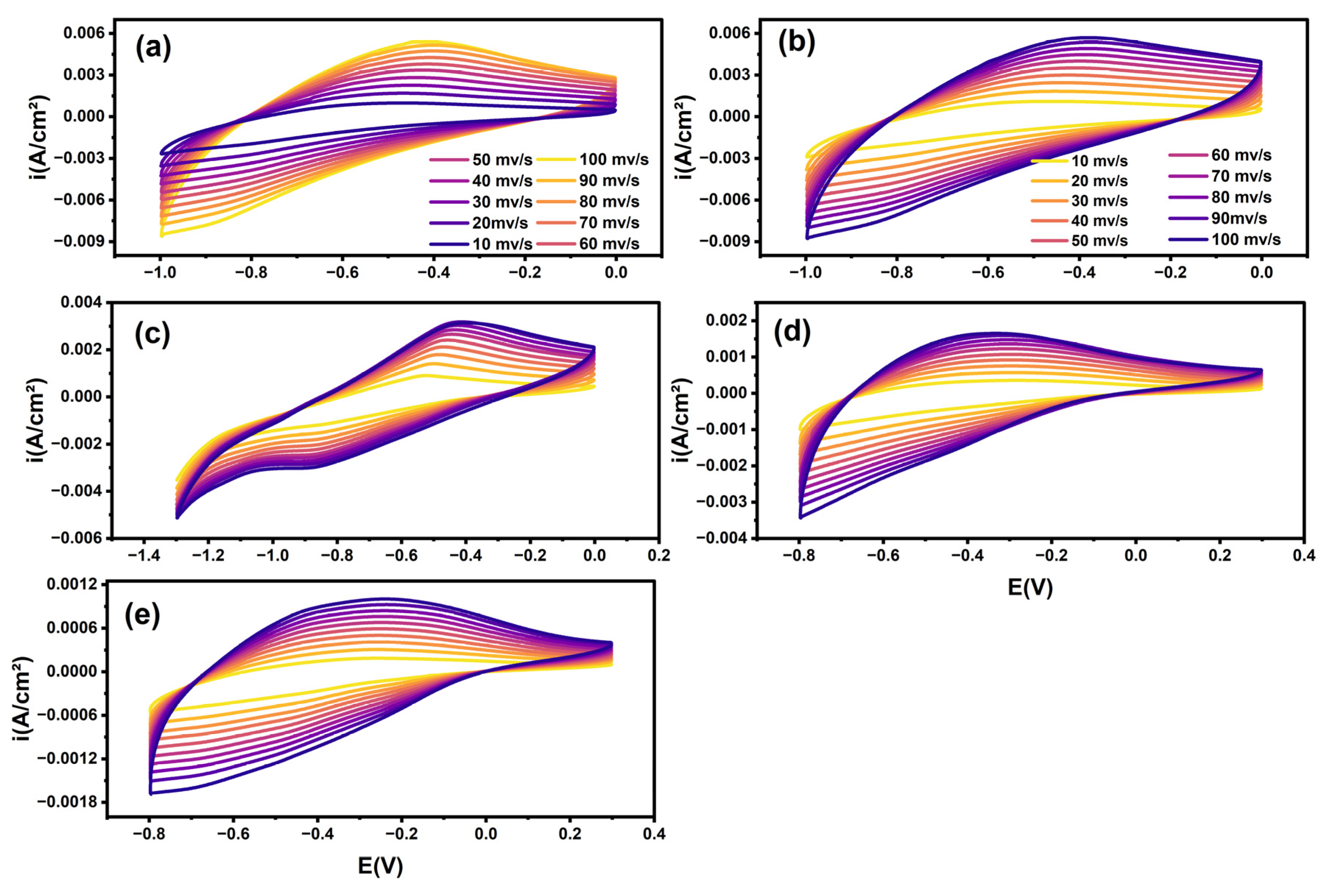
To explore the electrochemical profiles and charge-transfer behavior before evaluating gas evolution, Cyclic Voltammetry (CV) was recorded at scan rates of 10–100 mV/s (Figure 4). The CV loops across the samples deviate from the ideal rectangular shape of pure electric double-layer capacitors (EDLCs), demonstrating pseudo-capacitive/faradaic redox features. Both pristine ZIF-8 (Figure 4a) and pure A. nidulans (Figure 4b) show stable, widening current envelopes as scan rates ramp up, though their integrated current responses differ significantly. The ZIF-8@2% nidulans variant (Figure 4c) maintains broad redox-active current profiles, demonstrating optimized exposure of active surface sites. However, as the extract concentration increases to 4% and 6% (Figure 4d,e), the overall current density decreases significantly by nearly an order of magnitude (dropping down to peak currents near 0.0006−0.0012 A/cm2). This dramatic decline stems from the excessive insulating biopolymer layer that wraps the active sites, thereby restricting the transport of free ions at the electrode–electrolyte interface.
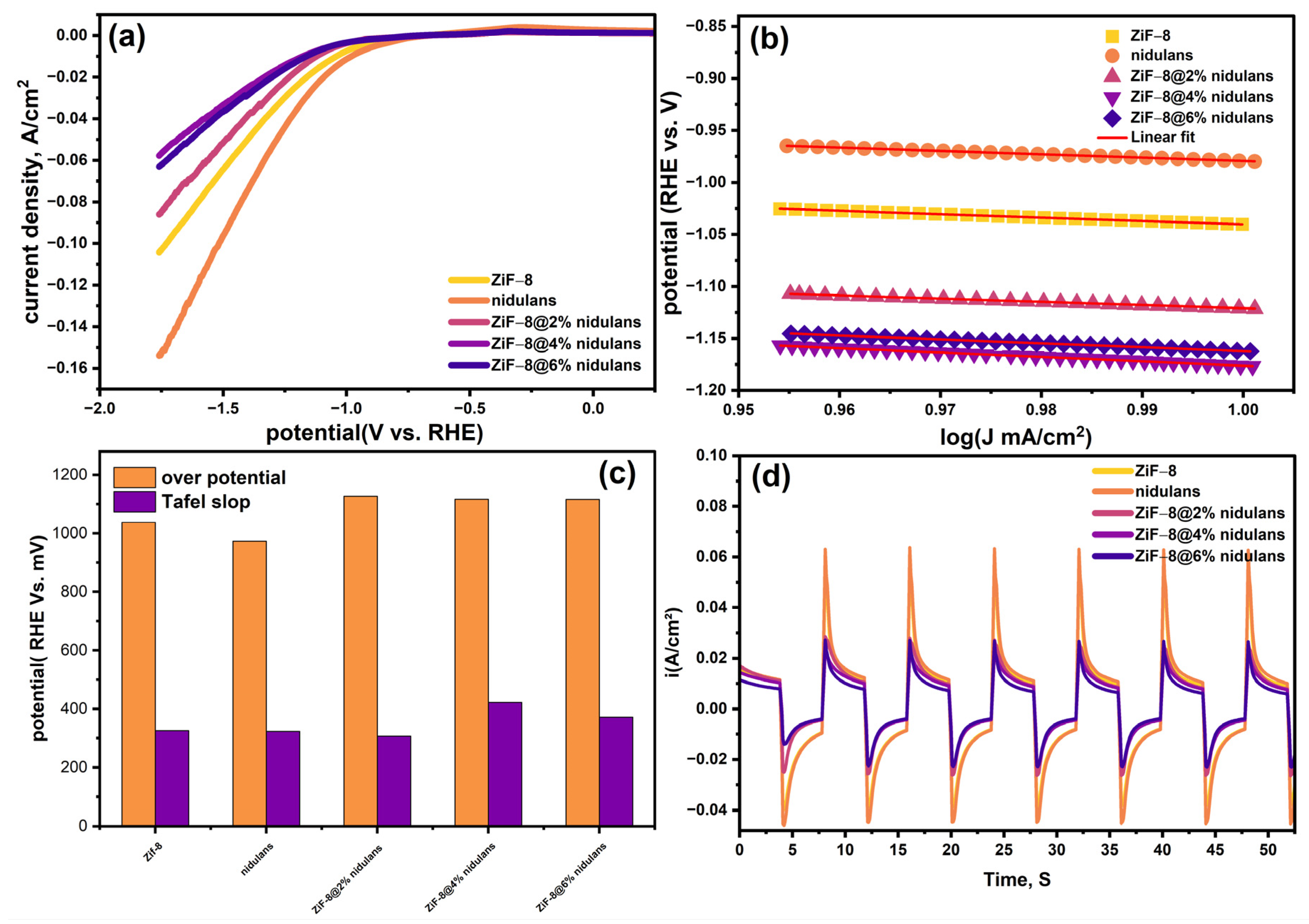
The performance of the fabricated electrodes for the photoelectrochemical hydrogen evolution reaction (PEC-HER) was systematically studied using linear sweep voltammetry (LSV), Tafel kinetic analysis, and transient photocurrent measurements under chopped-light conditions (Figure 5). Polarization Characteristics: As shown in Figure 5a, pure A. nidulans extract exhibits the highest cathodic current density, reaching approximately −0.15 A/cm2 at −1.75 V vs. RHE, along with a favorable positive onset potential. Pristine ZiF-8 exhibits a lower current-density response. When introducing the extract to the MOF support, the ZiF-8@2% nidulans composite demonstrates a clear synergetic improvement over pristine ZiF-8. However, exceeding these thresholds (4% and 6%) leads to a progressive loss of current density and a negative shift in the onset potential, confirming that excessive organic bio-loading impedes the catalytically active centers. (Figure 5b,c) Tafel Kinetics and Overpotential Trends. To extract the reaction kinetics, Tafel slopes (vs. log J) were evaluated (Figure 5b,c). The estimated Tafel values for ZIF-8, pure nidulans, ZIF-8@2%, ZIF-8@4%, and ZIF-8@6% are 328, 328.3, 310, 422, and 371 mV/dec, respectively. The minimized Tafel slope for ZiF-8@2% nidulans (310 mV/dec) implies that the hydrogen evolution mechanism at this optimum loading operates under the Tafel recombination pathway (where two adsorbed H intermediates recombine to liberate H2).
The low slope indicates an accelerated interfacial electron injection rate, triggered by the matching electronic bands between the bio-organic component and the MOF skeleton. Transient Photocurrent Inferences: The light-harvesting synergy was evaluated using transient photocurrent i-t traces over multiple on/off illumination steps (Figure 5d). Pristine ZIF-8 shows a negligible photo-response due to its wide bandgap. In contrast, the pure A. nidulans extract shows powerful photocurrent spikes up to 0.06 A/cm2, highlighting its ability to capture light energy. Among the composite formulations, ZIF-8@2% nidulans yields the highest photocurrent. This confirms that the fungal extract functions as a built-in proposed mechanistic hypothesis system, effectively transferring photogenerated charge carriers into the ZIF-8 framework to suppress electron–hole recombination.
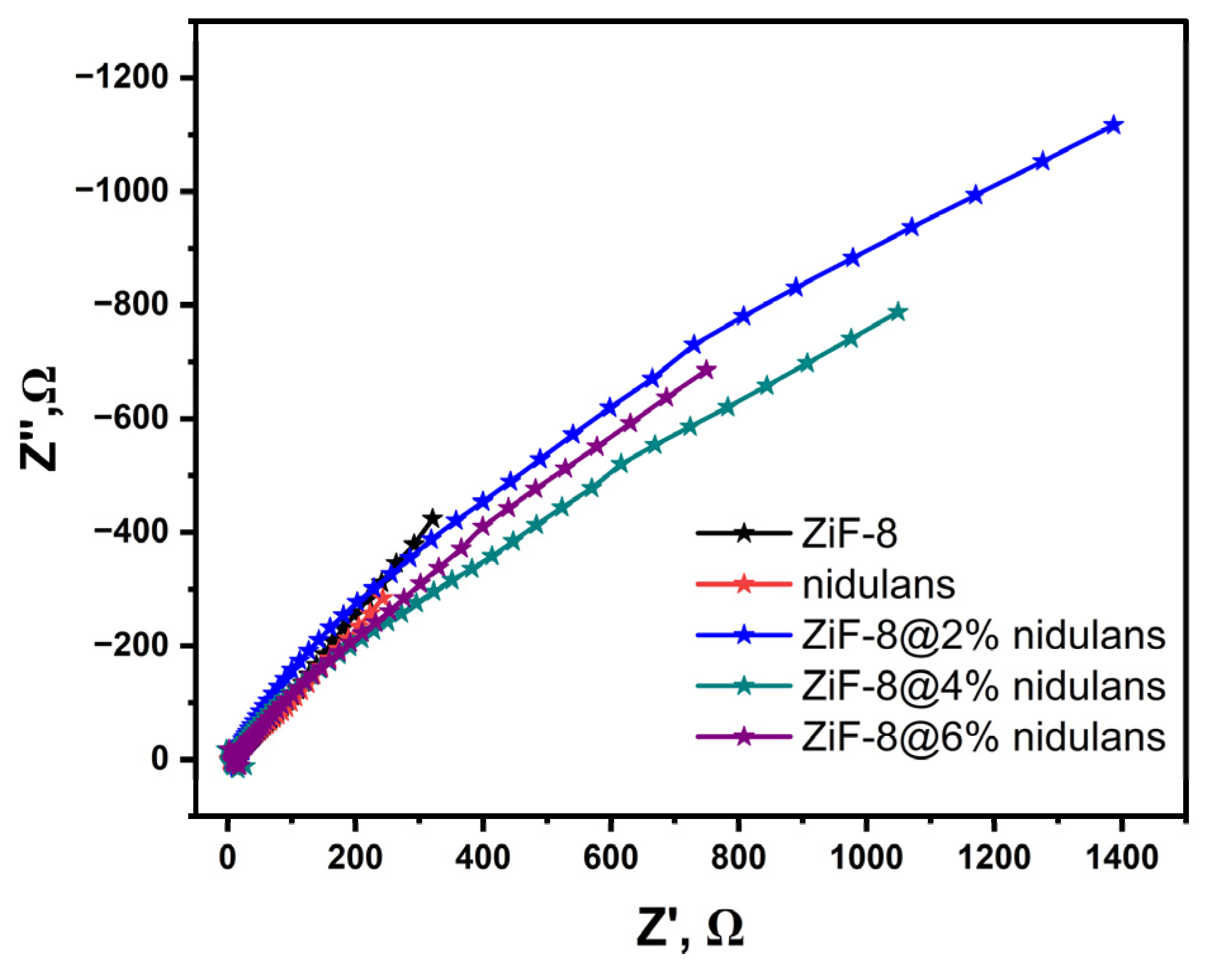
To decouple the internal resistance parameters and confirm the charge-transfer mechanism, Electrochemical Impedance Spectroscopy (EIS) Nyquist plots were obtained (Figure 6). The high-frequency intercept along the real axis (Z′) represents the solution/equivalent series resistance (Rs), while the diameter of the arc reflects the charge transfer resistance (Rct) across the electrode/electrolyte boundary. All samples show an incomplete, large capacitive arc extending into the low-frequency range, revealing combined charge transfer and diffusion control. Pristine ZiF-8 (black stars) possesses a highly restricted arc structure due to its poorly conducting framework. Strikingly, the ZIF-8@2% nidulans composite (blue stars) presents a tailored, linear-shifting arc slope that transitions efficiently across a wide impedance range. This demonstrates a reduced Rct compared with the higher-loading counterparts (4% and 6%). At excessive loadings (4% and 6%), the arcs contract or present irregular impedance shifts, matching the CV drop-offs. This confirms that the 2% configuration yields the most conductive pathways, facilitating rapid electron transport to feed the cathodic hydrogen evolution reaction.
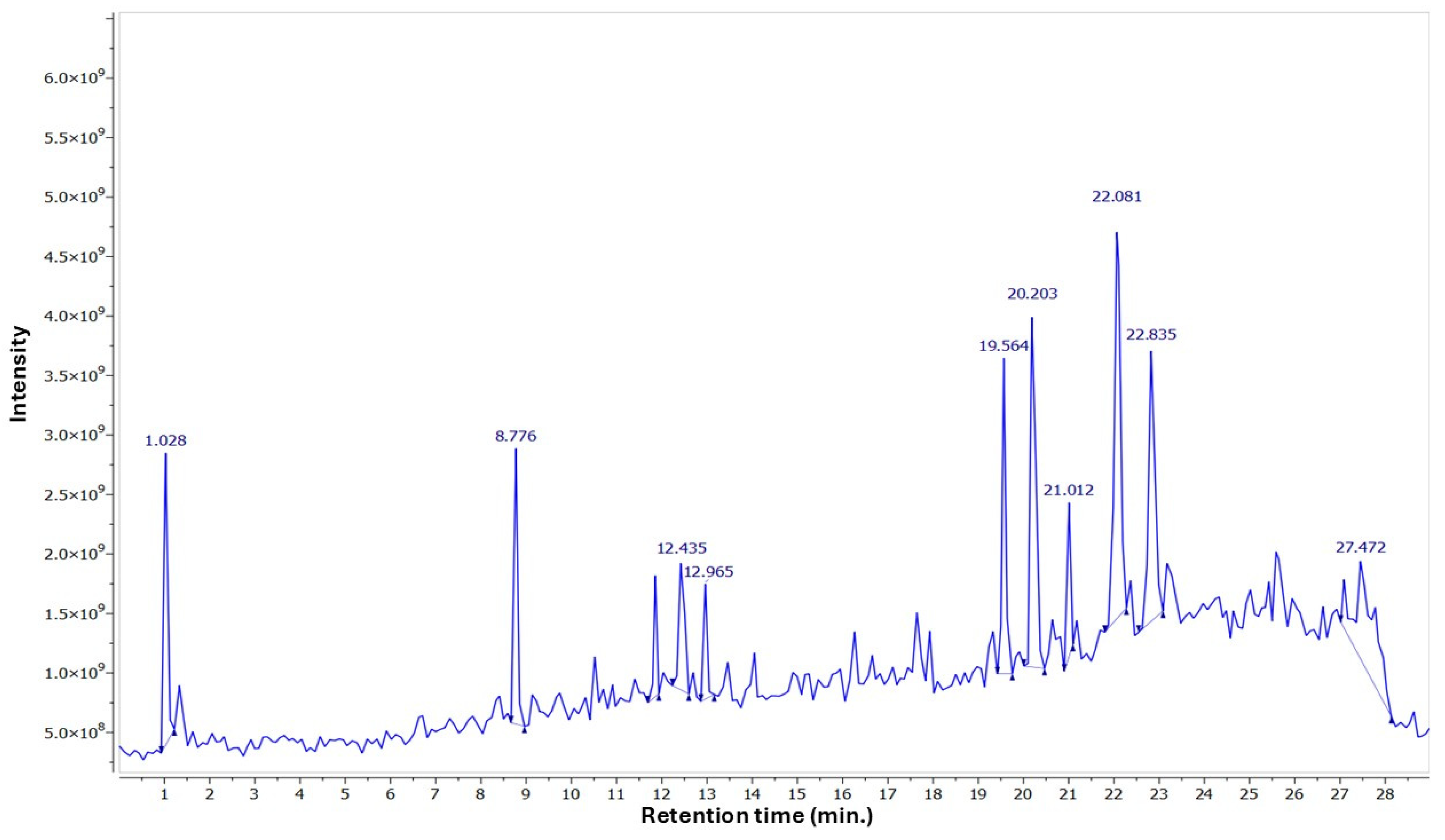
Aspergillus nidulans ethyl acetate extract was analyzed for the chemical components using two-stage mass spectrometry to derive the molecular mass, molecular formula, and characteristic fragment ions of the detected metabolites. Previously separated Aspergillus compounds were used to compare the obtained molecular formula with published data. Twenty chemical secondary metabolites were detected in negative ion mode and identified based on their precursor ions and MS2 fragmentation patterns, compared with the Competitive Fragmentation Modeling for Metabolite Identification online database. The mass spectrometric analysis of compounds extracted from various Aspergillus species reveals a striking diversity in molecular structures and fragmentation patterns, highlighting the complexity inherent in fungal metabolites. The compounds are arranged by retention time, accompanied by their previously reported occurrence in fungal species (Table 1, Figure 7).
Many of these previously reported compounds have shown promising biological activities. Acetophthalidin, derived from A. fumigatus BM923, was reported to exhibit inhibition of the mammalian cell cycle by [24]. Manniche et al. (2004) [25] demonstrated moderate antimalarial activity against Plasmodium falciparum by the Karanakafuran A and B isolated from A. karnatakaensis IBT 22154. Auroglaucine from Eurotium repens UM-031509 showed good binding affinity for the human cannabinoid receptor [26]. Aspergillus wentii-derived Aspewentin B displayed cytotoxicity against marine zoo- and phytoplanktons, as recorded by [27], and demonstrated cytotoxicity against HL-60 and K562 cell lines by 3-hydroxydiorcinol from A. sydowii FNA026. Aspergoterpenin B retrieved from A. versicolor produced antimicrobial activity on Erwinia carotovora. Aspericacid A, isolated from Aspergillus sp. LS78, presented significant antifungal activity as reported by [28].
Table 1. List of tentatively identified compounds from A. nidulans EtOAc extract analyzed by LC-MS-MS.
No.tRM–HMFMS2Compound NameReported
Source
Reference
17.41207.03C10H8O5189, 165, 149, 137, 123AcetophthalidinAspergillus fumigatus BM923[24]
27.69245.12C15H18O3227, 201, 199, 183, 169, 155, 139Aspergillusene BAspergillus sydowii PSU-F154[29]
37.79243.09C11H16O6197, 169, 153Tensyuic acid FAspergillus niger FKI-2342[30]
410.75313.14C19H22O4295, 277, 257, 251, 223, 213, 171Karnatakafuran AAspergillus karnatakaensis IBT 22154[25]
511.64229.11C11H18O5211, 183, 167, 141(−)-9-hydroxyhexylitaconic acidAspergillus aculeatus CRI322-03[31]
613.04243.12C12H20O5225, 207, 199, 195, 181, 167, 1539-hydroxyhexylitaconic acid-4-methyl esterAspergillus aculeatus CRI322-03[31]
714.08265.07C13H14O6219, 193, 179, 177, 165, 159Ruakuric acidAspergillus fumigatus[32]
814.85295.13C19H20O3277, 251, 171, 155Karnatakafuran BAspergillus karnatakaensis IBT 22154[25]
916.44297.15C19H22O3279, 265, 251,243, 197AuroglaucineEurotium repens UM-031509[26]
1019.60309.06C14H14O8291, 277, 253, 237, 209Dikojiacid BAspergillus flavus GZWMJZ-288[33]
1121.04404.16C23H23N3O4402, 357, 320Asperdiazapinone CAspergillus sp. PSU-RSPG185[34]
1222.30339.13C20H20O5293, 255, 163Aurgosin IPenicillium sp. JP-1[35]
1323.15279.16C16H24O4249, 233, 207, 189, 163, 133Decumbenone AAspergillus sulphureus KMM 4640[36]
1425.61283.17C19H24O2241, 199Aspewentin BAspergillus wentii na-3[27]
152.28195.02C10H12O4177, 151, 149, 133, 1213,5-Dimethylorsellinic acidAspergillus nidulans[37]
166.66193.00C11H14O3165, 149, 147, 137, 121, 1052-ethyl-4,6-dihydroxy-3,5-dimethylbenzaldehydeAspergillus nidulans[37]
1710.94245.01C14H14O4138, 123, 1093-hydroxydiorcinolAspergillus sydowii FNA026[38]
1814.08265.07C13H14O6219, 193, 179, 177, 163, 153, 137Antafumicin A/BAspergillus niger NH-401[39]
1914.85295.13C15H20O6277, 251, 233, 155, 137Aspergoterpenin BAspergillus versicolor[33]
2024.47281.16C16H26O4263, 237, 209, 187Aspericacid AAspergillus sp. LS78[28]
The antibacterial results of the study demonstrated the efficacy of A. nidulans extract, MOF, and MOF + extract against B. cepacian, E. coli, and S. typhimurium. Notable variations among the tested agents were observed where MOF + extract at different concentrations exhibited the most potent antibacterial activity with inhibition zones of 19.33, 13.67, and 10.67 mm at concentration 10%, 5%, and 2.5%, respectively, compared with A. nidulans extract and MOF individually (Table 2). Meanwhile, no antibacterial activities were demonstrated against E. coli and S. typhimurium at all concentrations of the tested agents.
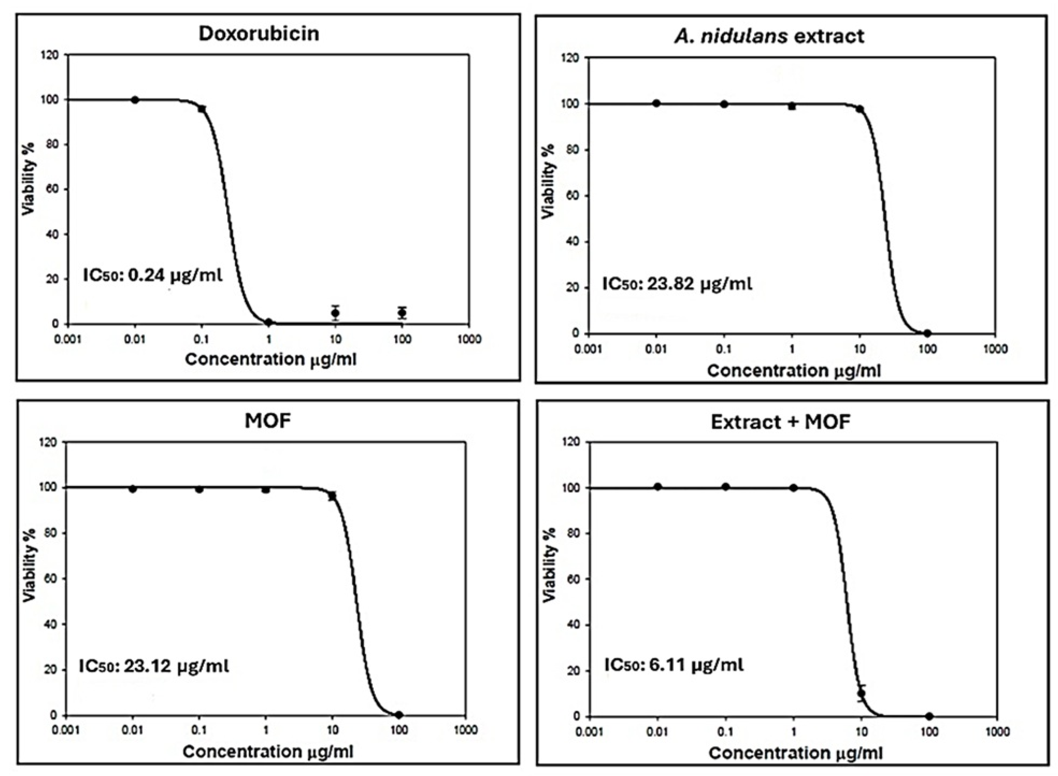
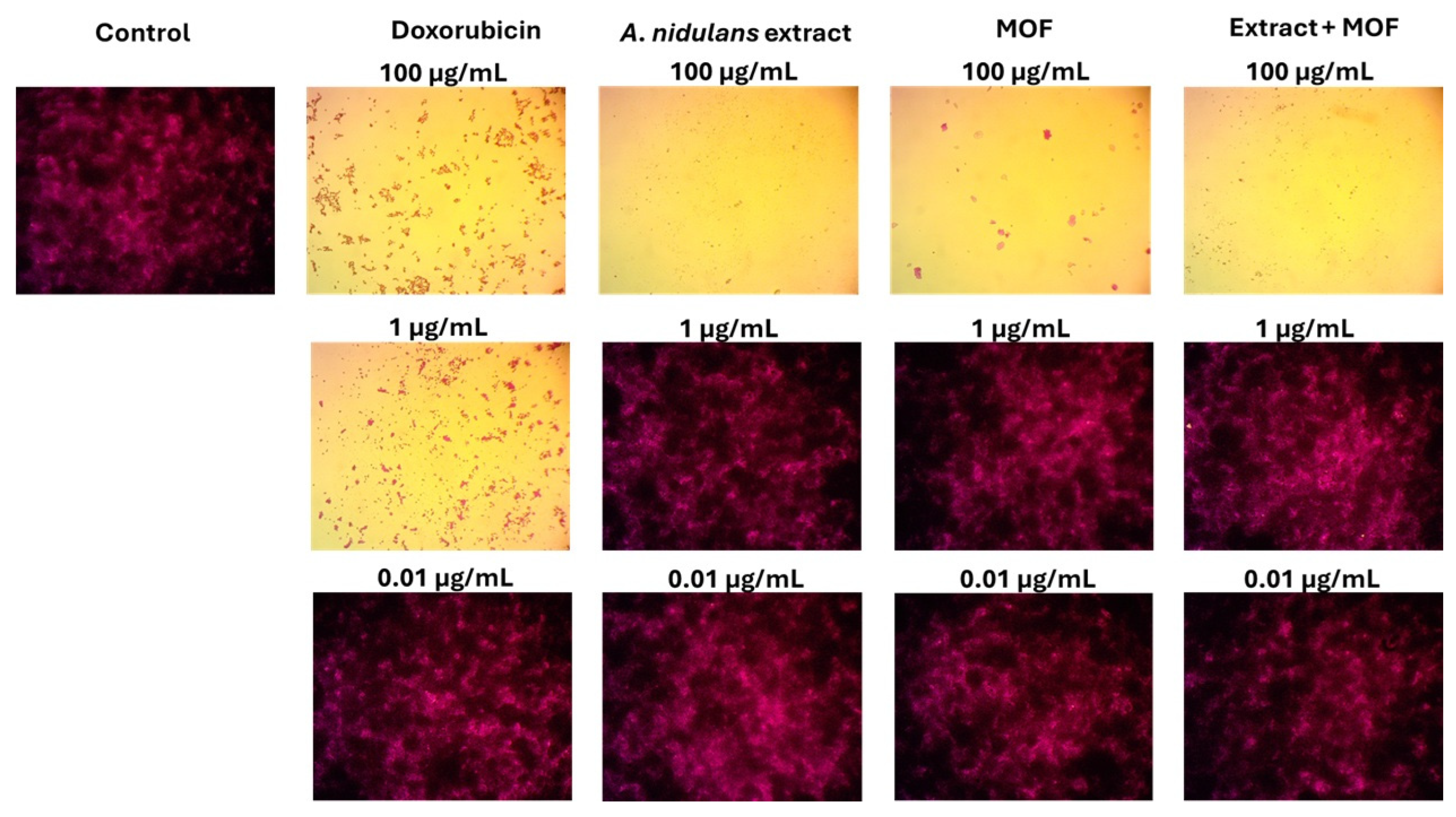
In the current study, the A. nidulans extract, ZIF-8, and ZIF-8@nidulans, in addition to standard Doxorubicin, were tested for their effect on the cell viability of human colon cancer (HCT-116) cell line using SRB assay, and the IC50 values were determined from dose–response curves of different concentrations. In comparison with doxorubicin (IC50 = 0.24 µg/mL), the cytotoxicity assay of A. nidulans extract, ZIF-8, and ZIF-8@nidulans indicated IC50 values of 23.82, 23.12, and 6.11 µg/mL, respectively, thus exhibiting potent cytotoxicity of MOF + extract against the HCT-116 cell line (Figure 8). At different tested concentrations, Figure 9 shows the effects of A. nidulans extract, MOF, and MOF + extract on the HCT-116 cell line compared with the control and doxorubicin treatment.

3. Experimental Work

3.1. Fungal Extract Preparation

Aspergillus nidulans AUMC 15444 (GenBank accession no. OR064351) was utilized to prepare fungal extract by fermentation on rice medium for one month at 30 °C [40]. After that, extraction by ethyl acetate (EtOAc) was carried out, defatted with n-hexane, and then the filtrate was evaporated to obtain a dry extract [41].

3.2. LC-ESI-MS/MS Analysis of A. nidulans Extract

The SCIEX Triple Quad 5500+ MS/MS system (Agilent Technologies, Waldbronn, Germany) is equipped with an electrospray ionization (ESI) negative mode for ion detection. The experimental condition and tentative identification of compounds were conducted using the reported protocol used by [42]. A blank control was included in the LC-MS/MS analysis, and the blank peaks were filtered and excluded using MSdial software version 4.92.

3.3. Antibacterial Investigation

The antibacterial efficacy of the samples was assessed by the well diffusion method [43], on Muller–Hinton agar medium, against Burkholderia cepacian ATCC 25416, Escherichia coli ATCC 8739, and Salmonella typhimurium ATCC 14028 strains at three concentrations (10, 5, 2.5%). Chloramphenicol (1%, 0.5%, and 0.25%) was used as a standard (positive control), while sterile distilled H2O and dimethyl sulfoxide were used as negative controls. The diameter of the inhibition zone around the well (6 mm) was considered positive activity.

3.4. Cytotoxicity Assay

The cancer cell lines were obtained from Nawah Scientific Inc. (Mokatam, Cairo, Egypt). HCT-116 (Human colon cancer) cells were cultured in RPMI medium amended with 100 units/mL penicillin, 100 mg/mL streptomycin, and 10% heat inactivated fetal bovine serum and incubated in a humidified 5% CO2 atmosphere at 37 °C. Different concentrations of the samples were tested to determine the IC50 in addition to Doxorubicin as a standard. The Sulforhodamine B (SRB) assay was employed to estimate cancer cell viability, and a BMG LABTECH FLUOstar Omega microplate reader (Ortenberg, Germany) was utilized to measure absorbance at 540 nm [44].

3.5. Synthesis of Pristine ZIF-8 and ZIF-8@nidulans Nanocomposites

Pristine ZIF-8 was prepared via a modified, eco-friendly room-temperature chemical co-precipitation method. In a typical procedure, Zn(NO3)2.6H2O and 2-methylimidazole were dissolved separately in deionized water at a predetermined molar ratio [45]. The 2-methylimidazole solution was rapidly poured into the zinc nitrate solution under vigorous magnetic stirring for 2 h. The resulting white precipitate was isolated via high-speed centrifugation, washed three times with fresh water/ethanol to remove unreacted precursors, and dried in a vacuum oven at 60 °C overnight. For the synthesis of the ZIF-8@nidulans nanocomposites, a physical–chemical encapsulation technique was adopted. Varied weight fractions (2%, 4%, and 6 wt.%) of the dried Aspergillus nidulans extract powder relative to the theoretical mass of ZiF-8 were pre-dissolved in the ligand solution prior to the addition of the zinc salt precursor. The subsequent crystallization, washing, and drying protocols were identical to those used for pristine ZIF-8, yielding light-colored composite powders labeled ZIF-8@2% nidulans, ZIF-8@4% nidulans, and ZIF-8@6% nidulans.

3.6. Statistical Analysis

Every experiment was run three times. The data were reported as the mean ± standard error (SE) and determined below the 0.05 level of significance using multiple comparison tests (Duncan) and analysis of variance (one-way ANOVA) using the SPSS program, version 16 (IBM, Armonk, NY, USA).

3.7. Material Characterization Techniques

The crystalline phases and structural configurations were investigated using an XRD LANScientific equipped with Cu Kα radiation (λ = 1.5406 Å) over a 2θ range of 5° to 35°. Surface morphologies and microstructural features were visualized using a FESEM operating at an accelerating voltage of 30.0 kV. Localized elemental distribution and chemical composition metrics were collected via an EDS elemental mapping system attached to the FESEM framework.

3.8. Electrochemical and Photoelectrochemical Measurements

Electrode Preparation: The working electrodes were prepared by dispersing 5 mg of the synthesized material (pristine ZIF-8 or ZIF-8@nidulans nanocomposite) in 1 mL of a mixture containing deionized water, isopropanol, and 5 wt.% Nafion® solution (3:1:0.05 v/v). The suspension was ultrasonicated for 30 min to ensure uniform dispersion. Then, 10 µL of the resulting ink was drop-cast onto a pre-cleaned FTO glass substrate (active area: 0.25 cm2) and dried at 60 °C for 2 h under ambient conditions. Prior to deposition, the FTO substrates were sequentially cleaned with acetone, ethanol, and deionized water in an ultrasonic bath for 15 min each, followed by drying under nitrogen flow. The mass loading was controlled to be approximately 0.2 mg cm−2 for all electrodes.
All electrochemical tests—including Cyclic Voltammetry (CV), linear sweep voltammetry (LSV), Electrochemical Impedance Spectroscopy (EIS), and transient photocurrent responses (i-t)—were recorded using a Corrtest Cs305 electrochemical workstation. A conventional three-electrode cell configuration was employed, containing the synthesized material deposited onto a conductive substrate (e.g., FTO glass or glassy carbon) as the working electrode, a Pt wire as the counter electrode, and a saturated calomel electrode (SCE) as the reference electrode, immersed in a suitable aqueous electrolyte. All potentials were converted and calibrated against the Reversible Hydrogen Electrode (RHE) scales via the standard Nernst relation:
Evs. RHE = Emeasured + Ereference+ 0.059pH
The transient photocurrent measurements (i-t) were carried out under chopped-light on/off cycles using a solar simulator source with a calibrated light intensity. EIS curves were recorded over a frequency span from 100 kHz to 0.1 Hz at an open-circuit potential under an amplitude of 5 mV. Newport 91160-1000 solar simulator equipped with a 1000 W Xenon lamp and an AM 1.5G filter (Irvine, CA, USA).

4. Conclusions

In summary, this study successfully demonstrates the design, characterization, and deployment of an innovative, dual-functional bio-hybrid platform based on a ZIF-8 metal–organic framework integrated with bioactive Aspergillus nidulans extract. Detailed structural and elemental analyses (XRD, FESEM, and EDS mapping) confirmed that the biological extract was uniformly incorporated throughout the MOF host without altering its underlying crystalline framework. Photoelectrochemical characterizations confirmed that the loading of the fungal extract strongly dictates the interfacial kinetic pathways and charge-transport rates. The ZIF-8@2% nidulans composite emerged as the optimal formulation, demonstrating excellent transient photocurrent stability, reduced charge-transfer resistance (Rct), and a minimized Tafel kinetic slope of 310 mV/dec, indicating that hydrogen generation proceeds via efficient Tafel recombination. This marked enhancement stems from a unique photobiochemical synergy, in which the fungal extract serves as an integrated proposed mechanistic hypothesis that suppresses charge recombination and accelerates electron injection into the ZIF-8 structure. Concurrently, the material exhibited strong antimicrobial properties, driven by the structural synergy between the components. Ultimately, this work offers an eco-friendly approach to developing high-efficiency, multi-functional nanomaterials from biological extracts, paving the way for advanced applications in solar energy harvesting and biomedical technologies.

Author Contributions

A.B.G.T.: Conceptualization; Data curation; Investigation; Visualization; Writing—review & editing. F.H.A.: Conceptualization; Funding acquisition; Supervision; Writing—original draft; Writing—review & editing. A.M.A.: Conceptualization; Investigation; Visualization; Supervision; Writing—original draft; Writing—review & editing. M.E.A.: Formal analysis; Methodology; Validation, Writing—original draft. M.H.A.H.: Data curation; Investigation; Supervision. A.M.A.H.: Formal analysis; Methodology; Validation; Writing—original draft. All authors have read and agreed to the published version of the manuscript.

Funding

The authors express their gratitude to Princess Nourah bint Abdulrahman University Researchers Supporting Project number (PNURSP2026R38), Princess Nourah bint Abdulrahman University, Riyadh, Saudi Arabia.

Data Availability Statement

The raw data supporting the conclusions of this article will be made available by the authors on request.

Acknowledgments

The authors express their gratitude to Princess Nourah bint Abdulrahman University Researchers Supporting Project number (PNURSP2026R38), Princess Nourah bint Abdulrahman University, Riyadh, Saudi Arabia.

Conflicts of Interest

The authors declare no conflict of interest.

References

  1. Jayabal, R. Towards a carbon-free society: Innovations in green energy for a sustainable future. Results Eng. 2024, 24, 103121. [Google Scholar] [CrossRef]
  2. Alreshidi, M.A.; Yadav, K.K.; Shoba, G.; Gacem, A.; Padmanabhan, S.; Ganesan, S.; Guganathan, L.; Bhutto, J.K.; Saravanan, P.; Fallatah, A.M. Hydrogen in transport: A review of opportunities, challenges, and sustainability concerns. RSC Adv. 2025, 15, 23874–23909. [Google Scholar] [CrossRef] [PubMed]
  3. Sarmah, M.K.; Singh, T.P.; Kalita, P.; Dewan, A. Sustainable hydrogen generation and storage—A review. RSC Adv. 2023, 13, 25253–25275. [Google Scholar] [PubMed]
  4. Ahmed, S.A.M.; Nagababu, P.; Rayalu, S. Advancements in solar-powered hydrogen production: A review of concentrated solar power electrolysis, photoelectrochemical, and PV-based electrolysis. Discov. Electrochem. 2025, 2, 68. [Google Scholar] [CrossRef]
  5. Hassan, Q.; Tabar, V.S.; Sameen, A.Z.; Salman, H.M.; Jaszczur, M. A review of green hydrogen production based on solar energy; techniques and methods. Energy Harvest. Syst. 2024, 11, 20220134. [Google Scholar]
  6. Ta, X.M.; Daiyan, R.; Nguyen, T.K.; Amal, R.; Tran-Phu, T.; Tricoli, A. Alternatives to water photooxidation for photoelectrochemical solar energy conversion and green H2 production. Adv. Energy Mater. 2022, 12, 2201358. [Google Scholar] [CrossRef]
  7. Hou, Y.; Zhuang, X.; Feng, X. Recent advances in earth-abundant heterogeneous electrocatalysts for photoelectrochemical water splitting. Small Methods 2017, 1, 1700090. [Google Scholar] [CrossRef]
  8. Zhao, Y.; Niu, Z.; Zhao, J.; Xue, L.; Fu, X.; Long, J. Recent advancements in photoelectrochemical water splitting for hydrogen production. Electrochem. Energy Rev. 2023, 6, 14. [Google Scholar] [CrossRef]
  9. Zhang, X.; Chen, A.; Zhong, M.; Zhang, Z.; Zhang, X.; Zhou, Z.; Bu, X.-H. Metal–organic frameworks (MOFs) and MOF-derived materials for energy storage and conversion. Electrochem. Energy Rev. 2019, 2, 29–104. [Google Scholar]
  10. Kong, L.; Zhong, M.; Shuang, W.; Xu, Y.; Bu, X.-H. Electrochemically active sites inside crystalline porous materials for energy storage and conversion. Chem. Soc. Rev. 2020, 49, 2378–2407. [Google Scholar] [CrossRef] [PubMed]
  11. Zhou, Q.; Yu, T.; Yang, R.; Guo, X.; Chen, Y. ZIF-8 and its derivative adsorbents for heavy metal removal in water: A review. ACS Omega 2025, 10, 47790–47801. [Google Scholar] [CrossRef] [PubMed]
  12. Gao, Y.; Qiao, Z.; Zhao, S.; Wang, Z.; Wang, J. In situ synthesis of polymer grafted ZIFs and application in mixed matrix membrane for CO2 separation. J. Mater. Chem. A 2018, 6, 3151–3161. [Google Scholar] [CrossRef]
  13. Behera, D.; Priyadarshini, P.; Parida, K. ZIF-8 metal–organic frameworks and their hybrid materials: Emerging photocatalysts for energy and environmental applications. Dalton Trans. 2025, 54, 2681–2708. [Google Scholar] [CrossRef] [PubMed]
  14. Wang, J.; Yuan, L.; Zhang, P.; Mao, J.; Fan, J.; Zhang, X.L. Advances in zeolitic-imidazolate-framework-based catalysts for photo-/electrocatalytic water splitting, CO2 reduction and N2 reduction applications. Nanoscale 2024, 16, 7323–7340. [Google Scholar] [CrossRef] [PubMed]
  15. Li, X.; Liang, H.; Liu, X.; Zhang, Y.; Liu, Z.; Fan, H. Zeolite imidazolate frameworks (ZIFs) derived nanomaterials and their hybrids for advanced secondary batteries and electrocatalysis. Chem. Rec. 2022, 22, e202200105. [Google Scholar] [CrossRef] [PubMed]
  16. Zhang, G.; Li, X.; Liu, Y.; Du, G.; Pang, H. Metal–organic framework derived micro-/nano-materials: Precise synthesis and clean energy applications. Inorg. Chem. Front. 2024, 11, 6275–6306. [Google Scholar] [CrossRef]
  17. Behera, B.; Singh, R.; Sharma, K.; Rai, A.; Singh, S.; Balan, B. A comprehensive review of advanced strategies to combat antimicrobial resistance. Arch. Microbiol. 2025, 207, 281. [Google Scholar] [CrossRef] [PubMed]
  18. Salam, M.A.; Al-Amin, M.Y.; Salam, M.T.; Pawar, J.S.; Akhter, N.; Rabaan, A.A.; Alqumber, M.A.A. Antimicrobial resistance: A growing serious threat for global public health. Healthcare 2023, 11, 1946. [Google Scholar] [CrossRef] [PubMed]
  19. Motaleb, K.Z.M.A.; Pranta, A.D.; Karim, F.-E.; Islam, M.R.; Islam, S.; Janutėnienė, J. Bio-waste as a resource for sustainable nanocomposites: Strategies and multifunctional applications. Funct. Compos. Struct. 2025, 7, 022002. [Google Scholar] [CrossRef]
  20. Pandey, M.; Nazar, R.; Elella, M.H.A.; Praharaj, S.; Makhado, E.; Rout, D.; Gilani, E.H. A comprehensive review of recent developments in biomedical materials based on graphene-modified bio-nanocomposites. BioNanoScience 2025, 15, 125. [Google Scholar]
  21. Ghazawi, K.F.; Fatani, S.A.; Mohamed, S.G.A.; Mohamed, G.A.; Ibrahim, S.R.M. Aspergillus nidulans—Natural metabolites powerhouse: Structures, biosynthesis, bioactivities, and biotechnological potential. Fermentation 2023, 9, 325. [Google Scholar] [CrossRef]
  22. Petrovic, S.M.; Barbinta-Patrascu, M.-E. Organic and biogenic nanocarriers as bio-friendly systems for bioactive compounds’ delivery: State-of-the art and challenges. Materials 2023, 16, 7550. [Google Scholar] [CrossRef] [PubMed]
  23. Van Tran, T.; Dang, H.H.; Nguyen, H.; Nguyen, N.T.T.; Nguyen, D.H.; Nguyen, T.T.T. Synthesis methods, structure, and recent trends of ZIF-8-based materials in the biomedical field. Nanoscale Adv. 2025, 7, 3941–3960. [Google Scholar] [CrossRef] [PubMed]
  24. Cui, C.-b.; Usukata, M.; Kakeya, H.; Onose, R.; Okada, G.; Takahashi, I.; Isono, K.; Osada, H. Acetophthalidin, a novel inhibitor of mammalian cell cycle, produced by a fungus isolated from a sea sediment. J. Antibiot. 1996, 49, 216–219. [Google Scholar] [CrossRef] [PubMed]
  25. Manniche, S.; Sprogøe, K.; Dalsgaard, P.W.; Christophersen, C.; Larsen, T.O. Karnatakafurans A and B: Two Dibenzofurans Isolated from the Fungus Aspergillus k arnatakaensis. J. Nat. Prod. 2004, 67, 2111–2112. [Google Scholar] [CrossRef] [PubMed]
  26. Gao, J.; León, F.; Radwan, M.M.; Dale, O.R.; Husni, A.S.; Manly, S.P.; Lupien, S.; Wang, X.; Hill, R.A.; Dugan, F.M. Benzyl derivatives with in vitro binding affinity for human opioid and cannabinoid receptors from the fungus Eurotium repens. J. Nat. Prod. 2011, 74, 1636–1639. [Google Scholar] [CrossRef] [PubMed]
  27. Miao, F.-P.; Liang, X.-R.; Liu, X.-H.; Ji, N.-Y. Aspewentins A–C, norditerpenes from a cryptic pathway in an algicolous strain of Aspergillus wentii. J. Nat. Prod. 2014, 77, 429–432. [Google Scholar] [CrossRef] [PubMed]
  28. Liu, Y.; Ding, L.; Zhang, Z.; Yan, X.; He, S. New antifungal tetrahydrofuran derivatives from a marine sponge-associated fungus Aspergillus sp. LS78. Fitoterapia 2020, 146, 104677. [Google Scholar] [CrossRef] [PubMed]
  29. Trisuwan, K.; Rukachaisirikul, V.; Kaewpet, M.; Phongpaichit, S.; Hutadilok-Towatana, N.; Preedanon, S.; Sakayaroj, J. Sesquiterpene and xanthone derivatives from the sea fan-derived fungus Aspergillus sydowii PSU-F154. J. Nat. Prod. 2011, 74, 1663–1667. [Google Scholar] [CrossRef] [PubMed]
  30. Hasegawa, Y.; Fukuda, T.; Hagimori, K.; Tomoda, H.; Ōmura, S. Tensyuic acids, new antibiotics produced by Aspergillus niger FKI-2342. Chem. Pharm. Bull. 2007, 55, 1338–1341. [Google Scholar] [CrossRef] [PubMed]
  31. Antia, B.S.; Aree, T.; Kasettrathat, C.; Wiyakrutta, S.; Ekpa, O.D.; Ekpe, U.J.; Mahidol, C.; Ruchirawat, S.; Kittakoop, P. Itaconic acid derivatives and diketopiperazine from the marine-derived fungus Aspergillus aculeatus CRI322-03. Phytochemistry 2011, 72, 816–820. [Google Scholar] [CrossRef] [PubMed]
  32. Cutler, H.G.; Lauren, D.R.; Wilkins, A.L.; Holland, P.T.; Hill, R.A.; Dugan, F.M. Ruakuric acid: A natural product from Aspergillus fumigatus. Phytochemistry 1996, 43, 209–214. [Google Scholar] [CrossRef] [PubMed]
  33. Xu, Y.; Wang, L.; Gong, Q.; Zhu, G.; Yuan, C.; Zuo, M.; Rao, Q.; Zhu, W.; Hao, X. Kojic acid derivatives and sesquiterpenes from the Aspergillus flavus GZWMJZ-288, a fungal endophyte of Garcinia multiflora. Nat. Prod. Commun. 2018, 13, 1934578X1801301102. [Google Scholar] [CrossRef]
  34. Rukachaisirikul, V.; Rungsaiwattana, N.; Klaiklay, S.; Pakawatchai, C.; Saithong, S.; Phongpaichit, S.; Borwornwiriyapan, K.; Sakayaroj, J. Indole-benzodiazepine-2, 5-dione derivatives from a soil fungus Aspergillus sp. PSU-RSPG185. Tetrahedron 2013, 69, 11116–11121. [Google Scholar] [CrossRef]
  35. Lin, Z.; Zhu, T.; Fang, Y.; Gu, Q.; Zhu, W. Polyketides from Penicillium sp. JP-1, an endophytic fungus associated with the mangrove plant Aegiceras corniculatum. Phytochemistry 2008, 69, 1273–1278. [Google Scholar] [CrossRef] [PubMed]
  36. Zhuravleva, O.I.; Afiyatullov, S.S.; Vishchuk, O.S.; Denisenko, V.A.; Slinkina, N.N.; Smetanina, O.F. Decumbenone C, a new cytotoxic decaline derivative from the marine fungus Aspergillus sulphureus KMM 4640. Arch. Pharmacal Res. 2012, 35, 1757–1762. [Google Scholar] [CrossRef] [PubMed]
  37. Ahuja, M.; Chiang, Y.-M.; Chang, S.-L.; Praseuth, M.B.; Entwistle, R.; Sanchez, J.F.; Lo, H.-C.; Yeh, H.-H.; Oakley, B.R.; Wang, C.C.C. Illuminating the diversity of aromatic polyketide synthases in Aspergillus nidulans. J. Am. Chem. Soc. 2012, 134, 8212–8221. [Google Scholar] [CrossRef] [PubMed]
  38. Wang, Y.-N.; Mou, Y.-H.; Dong, Y.; Wu, Y.; Liu, B.-Y.; Bai, J.; Yan, D.-J.; Zhang, L.; Feng, D.-Q.; Pei, Y.-H. Diphenyl ethers from a marine-derived Aspergillus sydowii. Mar. Drugs 2018, 16, 451. [Google Scholar] [CrossRef] [PubMed]
  39. Fujimoto, Y.; Miyagawa, H.; Tsurushima, T.; Irie, H.; Okamura, K.; Ueno, T. Structures of antafumicins A and B, novel antifungal substances produced by the fungus Aspergillus niger NH-401. Biosci. Biotechnol. Biochem. 1993, 57, 1222–1224. [Google Scholar] [CrossRef] [PubMed]
  40. Al Mousa, A.A.; Abouelela, M.E.; Al Ghamidi, N.S.; Abo-Dahab, Y.; Mohamed, H.; Abo-Dahab, N.F.; Hassane, A.M.A. Anti-staphylococcal, anti-Candida, and free-radical scavenging potential of soil fungal metabolites: A study supported by phenolic characterization and molecular docking analysis. Curr. Issues Mol. Biol. 2023, 46, 221–243. [Google Scholar] [CrossRef] [PubMed]
  41. Mohamed, H.; Hassane, A.; Atta, O.; Song, Y. Deep learning strategies for active secondary metabolites biosynthesis from fungi: Harnessing artificial manipulation and application. Biocatal. Agric. Biotechnol. 2021, 38, 102195. [Google Scholar] [CrossRef]
  42. Al Mousa, A.A.; Abouelela, M.E.; Hassane, A.M.A.; Al-Khattaf, F.S.; Hatamleh, A.A.; Alabdulhadi, H.S.; Dahmash, N.D.; Abo-Dahab, N.F. Cytotoxic potential of Alternaria tenuissima AUMC14342 mycoendophyte extract: A study combined with LC-MS/MS metabolic profiling and molecular docking simulation. Curr. Issues Mol. Biol. 2022, 44, 5067–5085. [Google Scholar] [CrossRef] [PubMed]
  43. Jahangirian, H.; Haron, M.D.J.; Shah, M.H.; Abdollahi, Y.; Rezayi, M.; Vafaei, N. Well diffusion method for evaluation of antibacterial activity of copper phenyl fatty hydroxamate synthesized from canola and palm kernel oils. Dig. J. Nanomater. Biostructures 2013, 8, 1263–1270. [Google Scholar]
  44. Skehan, P.; Storeng, R.; Scudiero, D.; Monks, A.; McMahon, J.; Vistica, D.; Warren, J.T.; Bokesch, H.; Kenney, S.; Boyd, M.R. New colorimetric cytotoxicity assay for anticancer-drug screening. JNCI J. Natl. Cancer Inst. 1990, 82, 1107–1112. [Google Scholar] [CrossRef] [PubMed]
  45. Sahu, K.P.; Singh, S.; Singh, A.K.; Azad, U.P.; Yadav, D.; Kumar, V.; Singh, S.K. Nanomaterials via ZIF-8: Preparations, catalytic and drug delivery applications. Chem. Eng. J. 2025, 508, 160663. [Google Scholar] [CrossRef]
Figure 1. X-ray diffraction (XRD) patterns of pristine ZIF-8 framework and ZIF-8@Aspergillus nidulans extract nanocomposites prepared with varying extract weight fractions (2%, 4%, and 6%).
Catalysts 16 00712 g001
Figure 2. Field-emission scanning electron microscopy (FESEM) micrographs of (a) pristine nanoscale pseudo-spherical ZIF-8 particles, (b) bulk-like aggregated pure Aspergillus nidulans extract, (c) ZIF-8@2% nidulans nanocomposite displaying anchored particulate-matrix framework (yellow dashed circle), and (d) ZIF-8@6% nidulans nanocomposite showing heavy particle encapsulation and aggregation (yellow dashed circle). All scale bars represent 500 nm.
Catalysts 16 00712 g002
Figure 3. Energy-dispersive X-ray spectroscopy (EDS) elemental mapping analysis of the ZiF-8@nidulans nanocomposite: (a) selected FESEM scanning area, (b) combined elemental overlay map, and individual elemental distribution channels for (c) carbon (C), (d) nitrogen (N), (e) oxygen (O), and (f) zinc (Zn). Note: Insert weight percentages (wt.%) inside the panel (b) legend based on raw data calibration.
Catalysts 16 00712 g003
Figure 4. Cyclic Voltammetry (CV) profiles recorded at progressive scan rates 10–100 mV s−1 in a three-electrode configuration for: (a) pristine ZIF-8, (b) pure Aspergillus nidulans extract, (c) ZIF-8@2% nidulans, (d) ZIF-8@4% nidulans, and (e) ZIF-8@6% nidulans nanocomposites.
Catalysts 16 00712 g004
Figure 5. Photoelectrochemical hydrogen evolution reaction (PEC-HER) performance metrics: (a) Linear sweep voltammetry (LSV) polarization curves for pristine materials and ZIF-8@nidulans nanocomposites evaluated under illumination, (b) corresponding Tafel kinetic plots (Potential vs. log J) derived from the polarization sweeps with linear fitting lines, (c) comparative bar chart displaying the required overpotential versus estimated Tafel slopes, and (d) multi-cycle transient photocurrent response (i-t) curves under chopped-light on/off illumination steps.
Catalysts 16 00712 g005
Figure 6. Electrochemical Impedance Spectroscopy (EIS) Nyquist plots showing the imaginary impedance (Z″) versus real impedance (Z′) profiles for pristine ZIF-8, pure Aspergillus nidulans extract, and the ZIF-8@nidulans nanocomposite series (2%, 4%, and 6%).
Catalysts 16 00712 g006
Figure 7. ESI-TIC negative mode chromatogram of A. nidulans ethyl acetate extract.
Catalysts 16 00712 g007
Figure 8. Curves of IC50 dose–response of cytotoxic activities of Doxorubicin, A. nidulans extract, ZIF-8, and ZIF-8@nidulans against the HCT-116 cell line.
Catalysts 16 00712 g008
Figure 9. Optical microscope images of cytotoxicity assay of Doxorubicin, A. nidulans extract, ZIF-8, and ZIF-8@nidulans against HCT-116 cell line, magnification power: X100.
Catalysts 16 00712 g009
Table 2. The antibacterial activity, in mm, of A. nidulans extract, ZIF-8, and ZIF-8@ nidulans against B. cepacian bacterial strain.
Test AgentConcentration
10%5%2.5%
A. nidulans extract13.33 ± 33 c 10.33 ± 33 c7.67 ± 33 c
MOF10.67 ± 33 d9.33 ± 33 c7.00 ± 00 c
MOF + Extract19.33 ± 33 b13.67 ± 33 b10.67 ± 33 b
Chloramphenicol31.67 ± 88 a20.67 ± 33 a12.33 ± 67 a
Data were presented as mean ± SE, and values accompanied by different letters are significant at p < 0.05.
Disclaimer/Publisher’s Note: The statements, opinions and data contained in all publications are solely those of the individual author(s) and contributor(s) and not of MDPI and/or the editor(s). MDPI and/or the editor(s) disclaim responsibility for any injury to people or property resulting from any ideas, methods, instructions or products referred to in the content.