


Abstract
Nanoparticles have attracted considerable interest for biomedical and catalytic applications due to their unique functional properties. This study aims to evaluate the structural, optical, photoelectric, photocatalytic, and wound-healing performance of cetyltrimethylammonium bromide (CTAB) and cellulose nanoparticles with complementary physicochemical characteristics. The nanoparticles were characterized using X-ray diffraction (XRD), scanning electron microscopy (SEM), Fourier transform infrared spectroscopy (FTIR), and Ultraviolet–Visible (UV–Vis) spectroscopy, while photoelectric properties were assessed through current–voltage (I–V) measurements. Photocatalytic activity was evaluated using methylene blue degradation under solar irradiation, and in vivo wound healing was examined using a rat excisional model over 13 days. Cellulose nanoparticles exhibited nearly double the photocurrent compared to CTAB, indicating enhanced charge transport efficiency. Photocatalytic results showed that cellulose achieved approximately ~70% degradation within 210 s, compared to ~50% for CTAB. In vivo findings revealed that cellulose achieved 82% wound closure, compared with 71% for CTAB, 67% for Betadine, and 35% for untreated controls, accompanied by improved tissue regeneration. Overall, cellulose nanoparticles exhibited better photoelectrochemical, photocatalytic, and wound-healing properties, whereas CTAB provided structural integrity and antimicrobial properties. These materials are therefore promising multifunctional nanomaterials for catalytic and biological applications.
1. Introduction
The process of wound healing is one of the most complex and multifaceted biological processes. It involves a series of coordinated responses at the micro and molecular levels. Upon tissue injury, the tissue’s response and reaction to the injury involve the five distinct and overlapping phases of the process. These phases include hemostasis, inflammation, proliferation, and remodeling [1]. Optimally healing tissue is possible by monitoring all of these phases [2]. Defects in healing tissue are common in the presence of infection, chronic disease, and/or large tissue loss. Defects in healing tissue amplify the importance of biological therapies to wound healing [3].
In recent times, nanotechnology-based biomaterials have gained importance as a possible means to enhance wound healing effectiveness as a result of the unique characteristics that they possess for controlling cell activity, disabling microorganisms, and advancing tissue engineering for the purpose of accelerating wound healing. The feasibility of such uses is underpinned by cutting-edge technologies capable of delivering materials of precise dimensionality for molecular-level intervention at the tissue development and remodeling cascade [4]. Among these advanced structures, a notable proportion of the research has centered on the development and application of biocompatible and biodegradable cellulose-based nanoparticles with good mechanical attributes. Such attributes contribute to the scaffold and wound care and healing components of a medical textile, which aids the tissue engineering of a wound. In addition, the ability to modify the mechanical attributes of these nanoparticles to concentrate therapeutic agents is of great benefit [5]. When these nanoparticles are properly constructed, they can improve the wound tissue bioavailability of therapeutic agents while reducing the bioavailability in surrounding tissues to limit systemic toxicity. For all these reasons, cellulose nanomaterials have received much attention for application in wound healing because of their biocompatibility, biodegradability, water retention properties, and cell proliferation capability [6].
Cetyltrimethylammonium bromide (CTAB) is an ammonium salt, a cationic quaternary surfactant, which finds wide application in nanomaterials synthesis and biomedical research owing to its antimicrobial effect and surface-stabilizing ability [7]. It was also the first of its type to be used. While most quaternary ammonium compounds have low interactivity with the microbial cell membranes, CTAB was first used due to its membrane-disruptive activity towards pathogenic microorganisms [8]. It is also used in the management of infections due to its physiological properties [9]. With respect to its application in nanoparticle systems, CTAB serves the dual purpose of biomechanical function and a protective anti-microbial shield to be used on a wound, and thus, its use in nanocarrier systems is likely to enhance stability and therapeutic efficacy [10]. At the same time, production of cellulose nanoparticles as renewable biopolymers is still coveted due to their biocompatibility, mechanical properties, and surface reactivity.
While there has been an increase in attention towards cellulose-based nanomaterials as well as CTAB-based nanomaterials independently, no extensive investigations have compared their structural, optical, photoelectrical, photoreactive, and healing capabilities on the same platform. In this regard, analyzing their interaction can lead us to comprehend more about the role that physicochemical properties play in the performance of such biomaterials. The high affinity towards biological structures, moisture retention capability, and cell growth stimulation are all due to the hydroxyl groups [11]. Besides being therapeutically valuable due to their non-toxic, biodegradable, and skin-penetrating strong antioxidants and healing properties, cellulose nanoparticles have other advantages [12].
In addition to the medical importance, cellulose and CTAB-based nanomaterials have gained interest due to the role played by such materials in photocatalysis of environmental pollutants, specifically organic dyes. Recent studies have also shown that CTAB and cellulose nanomaterials have great possibilities in photocatalytic dye decomposition and photoelectricity due to their adjustable surfaces and ability for charge transfer and absorption of light [13]. Photocatalytic action entails energy absorption through excitation of electrons from the valence band to the conduction band and creation of free radicals, which help oxidize the dyes [14]. Hydroxyl groups within cellulose increase dye adsorption by forming hydrogen bonds [15]. Similarly, being a cationic surfactant, CTAB may alter the surface charge distribution on the surface, hence increasing adsorption via electrostatic force, especially for anionic dye molecules [16]. Investigating the relationship between structure and photocatalytic dye degradation helps in understanding the multifunctionality of these nanomaterials. Therefore, this study aims to characterize and comparatively study CTAB and cellulose nanoparticles in terms of structural, optical, photophysical, photocatalytic, and in vitro wound healing properties. Furthermore, this study investigated the wound healing potential of CTAB and cellulose nanoparticles through the excision wound rat model
2. Results and Discussion
2.1. Characterization of CTAB and Cellulose Nanoparticles
2.1.1. X-Ray Diffraction (XRD)
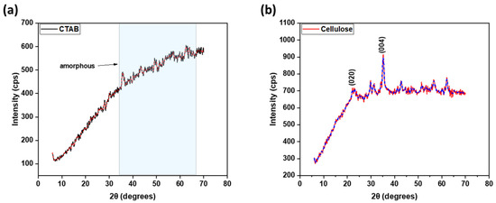
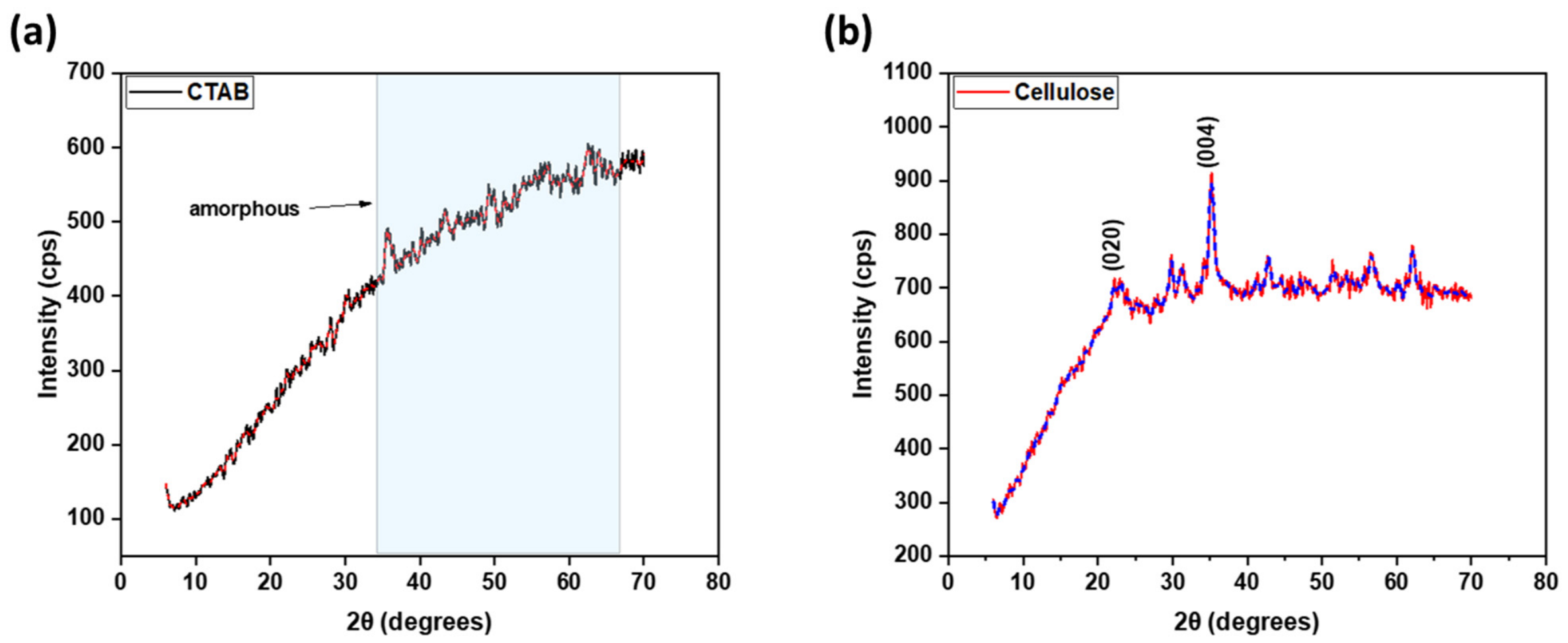
The XRD pattern for CTAB (Figure 1a) shows a wide diffuse halo around 2θ = 30–65° with no clear diffraction peaks, confirming its primarily amorphous character. This is consistent with previous accounts describing CTAB as a surfactant that is non-crystalline with no long-range structural order because of flexible alkyl chains and a disordered molecular packing [17]. In the synthesis of nanoparticles, this amorphous character is beneficial because CTAB is primarily a soft template and a stabilizing agent that promotes nucleation and prevents the agglomeration of particles by means of electrostatic interactions [18]. A broad halo diffraction pattern for similar amorphous CTAB materials has been observed in earlier research, which confirms that there is no long-range crystalline order.
Figure 1.
X-ray diffraction (XRD) patterns of (a) CTAB and (b) cellulose. The black (CTAB) and blue (Cellulose) curves represent the experimental XRD patterns, while the red line represents the fitted (calculated) diffraction pattern obtained by Rietveld refinement.
On the other hand, the cellulose (Figure 1b) has a semi-crystalline structure exhibiting a diffraction pattern peak at 2θ ≈ 22° and 34°, giving the (020) and (004) planes exhibiting the structure of cellulose I, as is shown in detail throughout the reports [19,20]. The existence of both crystalline and amorphous domains exemplifies the structural complexity of cellulose, as the crystalline portions provide mechanical strength, and the amorphous regions, which contain significant quantities of the hydroxyl groups, facilitate metal ion tethering and stabilization of nanoparticles [21]. These diffraction peaks indicate the semi-crystalline nature of cellulose I as found in the literature.
2.1.2. Scanning Electron Microscopy (SEM)
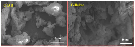
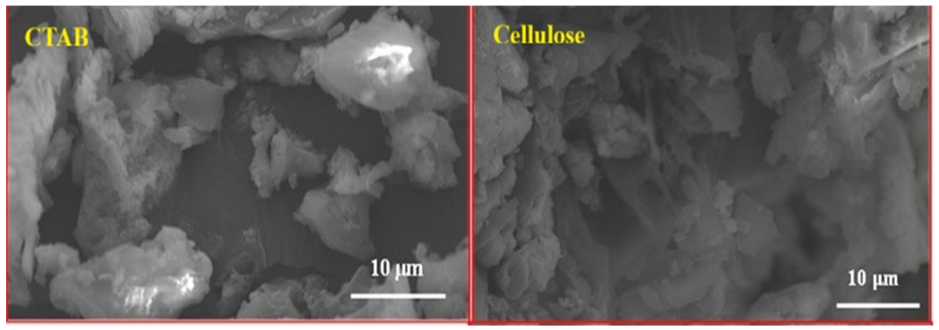
SEM and its applications, specifically regarding CTAB and cellulose nanoparticles, are illustrated in Figure 2. The rough surfaces and irregular shapes visible in the rough SEM images verified the predicted physical characteristics of the nanoparticles and the preparation techniques used to generate them.
Figure 2.
Scanning electron microscopy (SEM) micrographs of CTAB and cellulose nanoparticles.
CTAB nanoparticles are aggregates that are relatively separate, small, and porous [18]. SEM analysis revealed that there was a formation of an aggregated nanostructure with uneven particle size due to particle aggregation. Such morphology indicates partial stabilization of the nanoparticle surface through CTAB molecules, even though there is still some extent of agglomeration that occurs [22]. Irregular morphology and rough topography of CTAB nanoparticles may promote agglomeration and account for some of the antimicrobial and healing efficacy of CTAB nanoparticles through bilayer interactions [23]. Conversely, the cellulose nanoparticles are denser and layered, with a compact structure and relatively fewer large cavities. Cellulose structures were seen to appear larger and more compact when compared to CTAB due to possible fibrillar aggregation. Cellulose nanostructures also exhibit rough fibrillar morphology, indicative of considerable surface area, which may facilitate further functionalization, therapeutic loading, or act as a substrate for tissue incorporation [24]. This morphology might act as a bioactive wound healing scaffold to promote cell attachment, increase structural stability, and maintain the structural stability of the wound bed [25]. The agglomerated condition and roughened surface of the particles may improve targeted delivery and interaction of the particles with the wound tissue, thereby amplifying the optical, mechanical, and bioactive properties related to healing.
2.1.3. Fourier Transform Infrared Spectroscopy (FTIR)
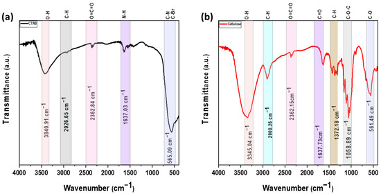
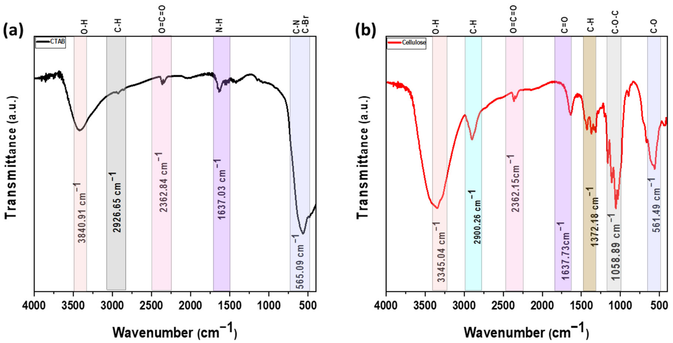
Fourier Transform Infrared Spectroscopy (FTIR) analysis evaluates the functional groups in CTAB and cellulose nanoparticles. This analysis confirms the composition of the molecules in each compound, as shown in Figure 3. For CTAB nanoparticles, there is a broad feature at 3840.91 cm−1 which corresponds to the O–H stretching vibrations associated with the moisture adsorbed on the surface of CTAB nanoparticles [26]. Peaks associated with the hydrophobic CTAB tail correspond to 2926.65 cm−1, being the C–H stretching of long alkyl chains of the cetyl group [27]. A weaker mid-range peak at 2362.84 cm−1 corresponds to the O=C=O stretching of atmospheric CO2, slightly present in the nanoparticles [28]. The strong and wide peak at 1637.03 cm−1 responds to the bending of the N-H and is possessed of the quaternary ammonium head group, reflecting the strong electrostatic association of CTAB with the surface of the nanoparticles [29]. The low-range peak at 565.09 cm−1 is of low frequency and is weak due to C–N and C–Br bond stretching, and indicates the presence of the counter ion of bromide, characteristic of CTAB molecules [30].
Figure 3.
FTIR spectra for (a) CTAB and (b) cellulose nanoparticles.
In the case of cellulose nanoparticles, the broad and intense band at 3345.04 cm−1 is due to O–H stretching vibrations from the intra- and intermolecular hydrogen bonds of the cellulose backbone [31]. A peak at 2900.26 cm−1, corresponding to the C–H stretching of the CH2 groups of the glucose units, emits the respective absorption band [32]. The small band at 2362.15 cm−1 is due to the O=C=O asymmetric stretching [33,34]. The band at 1637.73 cm−1 signifies the C=O stretching or the bending O–H of the adsorbed water [35]. The 1372.18 cm−1 and 1058.89 cm−1 peaks are due to the C–H bending and C–O–C asymmetric stretching, respectively, of the β-1,4-glycosidic linkages, thus confirming the polysaccharide structure of cellulose [36]. The weak band of 561.49 cm−1 corresponds to the C–O deformation vibrations [32].
2.2. Optical Properties Investigation
2.2.1. Transmission and Reflection Spectra of CTAB and Cellulose Thin Films
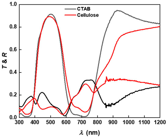
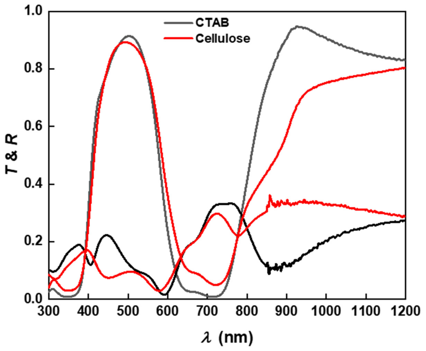
Figure 4 presents transmission and reflection spectra for the CTAB and cellulose thin films. The CTAB thin film has higher reflection and lower transmission, while the cellulose thin film has lower transmission. This means CTAB has the greatest light-scattering properties because CTAB films exhibit rougher and more aggregated morphologies and structures, which are more scattered [37]. Conversely, CTAB thin films have relatively higher transmission and lower reflection, indicating that they are more uniformly compact structures [38]. The optical films are uniform, structurally compact cellulose films, and rough CTAB exhibited the spectral features previously described. The differences in optical behavior can be attributed to variations in film morphology and structural organization. The CTAB film exhibited relatively higher reflection and lower transmission, which may be associated with increased light scattering arising from its rougher and more aggregated surface morphology. In contrast, the cellulose film showed lower reflection and higher transmission, indicating a more compact and homogeneous structure that facilitates light propagation through the film. These results are consistent with the SEM observations and demonstrate that the optical response of both films is strongly influenced by their microstructural characteristic [39].
Figure 4.
Transmission and reflection spectra of CTAB (black) and cellulose (Red) thin films.
2.2.2. Optical Absorption Spectra of CTAB and Cellulose Thin Films
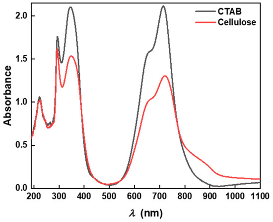
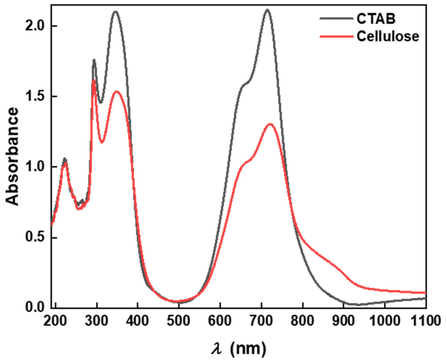
The optical absorption spectra of CTAB and cellulose thin films (Figure 5) indicate the presence of broad and well-defined bands in the visible region of the electromagnetic spectrum. The bands are associated with the nanoscale characteristics of the films and the molecular electronic transitions of the films. For both samples, the absorption spectra exhibited several peaks in the 350–500 nm range [40]. The CTAB film had a greater absorbance and sharper peaks, which suggests that the film had greater interactions with photons [41]. The differences in optical behavior can be attributed to variations in film morphology and structural organization. The CTAB film exhibited relatively higher reflection and lower transmission, which may be associated with increased light scattering arising from its rougher and more aggregated surface morphology. In contrast, the cellulose film showed lower reflection and higher transmission, indicating a more compact and homogeneous structure that facilitates light propagation through the film. These results are consistent with the SEM observations and suggest that the optical response of both films is strongly influenced by their microstructural characteristics. The enhanced transmission observed for cellulose may contribute to its improved photoresponsive performance, whereas the higher scattering behavior of CTAB is related to its less ordered morphology.
Figure 5.
Light absorption of CTAB and Cellulose thin films.
Regarding charge transfer capability, this may mean that CTAB responds to changes in solution concentration fast enough to form compact, ordered morphologies, which are structures with enhanced charge transfer capability [42]. The absorption spectra for the cellulose films, however, showed significantly more neutral and significantly more negative absorption spectra red shifts compared to those with more predominant, wider range, and energy surface absorption hydroxyl groups [43]. These are attributed to a higher surface in the optical bandgap [44]. The increase in negative absorption shift implies more light is being captured, which can facilitate charge carrier separation, further confirming its photoactive applications or use for healing [45]. Overall, the higher optical activity positive charge separation results differ due to higher crystallinity, surface morphology, and orientational differences in the molecular arrangement. The results are indicative of expected optical activity at the molecular arrangement form the basis for subsequent biological and photoelectrical activity evaluations of the films.
2.2.3. Optical Bandgap Energy (Eg) Analysis of CTAB and Cellulose Thin Films
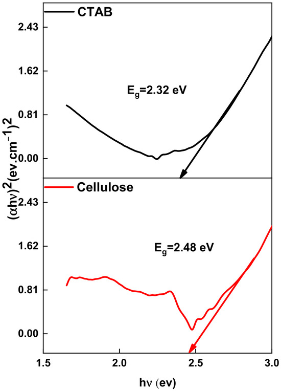
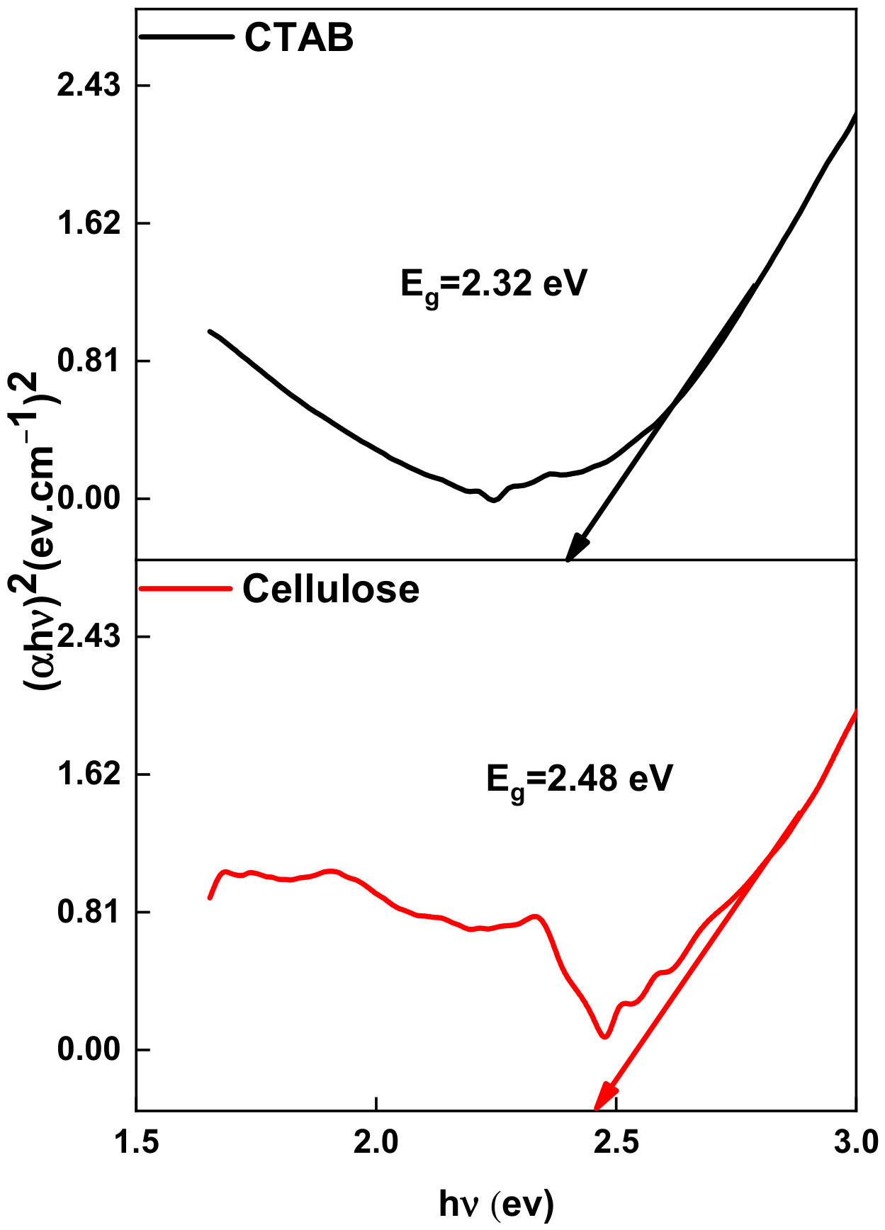
In Figure 6, Tauc plots illustrate the optical bandgap energies (Eg) of CTAB and cellulose thin films as derived from their absorption spectra. According to direct bandgap calculations, energies were derived from linear extrapolation of the (αhν)2 vs. photon energy (hν) graphs [46]. CTAB thin films demonstrated a higher bandgap energy value, which suggests lower mobility of charge carriers and electronic transitions in CTAB films. This observation also aligns with their packing and surfactant behavior [47,48]. In contrast, the cellulose thin films demonstrated a lower Eg, which suggests less energy was required for electron excitation, and light absorption was greater in the visible range [43]. The loss of energy due to the bandgap in the cellulose thin films is due to the localized states in the band structure and terminal hydroxyl groups, along with the conjugated carbon-oxygen bonds. This was discussed in detail by Srivastava et al. (2020), who explained that localized states diminish the degree to which the bandgap is pronounced [49]. Thus, thin cellulose films have better photoresistive properties than the CTAB films. The obtained optical bandgap values (2.32 eV for CTAB and 2.48 eV for cellulose) are consistent with previously reported values for organic and cellulose-based thin films. Similar bandgap values ranging from 2.3 to 3.0 eV have been reported depending on film preparation conditions, crystallinity, and molecular interactions, which likely have insulating characteristics to the hybrid nanocomposites [50]. The bandgap is the depth of the bandgap, which indicates better optical properties. The two materials, along with their structural and compositional differences, are illustrated in Figure 4, which portrays the absorption of the constituents [51].
Figure 6.
Energy gap calculations of CTAB and cellulose thin films. The arrow indicates the linear region selected for extrapolation to determine the optical band gap (Eg) using the Tauc plot.
2.2.4. Absorption Coefficient Analysis of CTAB and Cellulose Thin Films
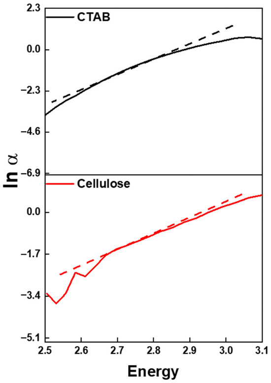
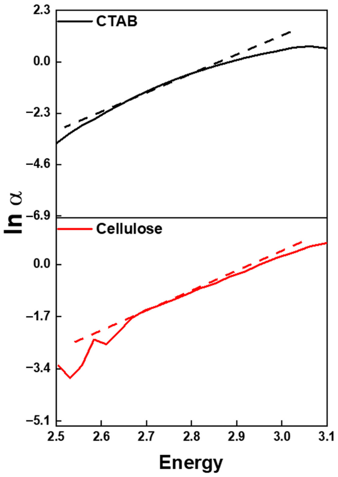
Displayed in Figure 7 are the fluctuations in the natural log of the absorption coefficient (ln α) against the photon energy for CTAB and cellulose thin films. ln α graphs show the storage absorption edge and the exponential nature of the localized gap band structure states. Between the pair of materials, the thin cellulose film is the more optically absorbed sample, as is evidenced by a more substantial drop in the slope, a more rapid decrease in slope, and more pronounced electronic transitions at the band edge.
Figure 7.
Ln α of CTAB and Cellulose thin films. The dotted lines represent the linear fitting of the exponential absorption edge used to determine the Urbach energy (Eu).
The higher intensity of localized tail states in this figure, for instance, may be responsible for increasing the frequency of electronic transitions that are photogenically induced, causing greater numbers of transitions, thus increasing, as illustrated in Figure 5, the lowering of the optical bandgap [52]. As for the CTAB thin film, it has a more gentle slope, and that slope is indicative of lower edge absorption due to a more ordered molecular arrangement and a lower defect density [53]. The continuous portion of a lnα graph is indicative of the existence of an exponential absorption tail, which is indicative of Urbach energy (Eu), along with associated structural disorder [54]. We can thus conclude that the cellulose film is characterized by a higher Eu value concomitant with a greater disorder, as well as lower absorption of light. In contrast, CTAB ensures an optically stable film, which can be advantageous for surface-modifying or insulating activities [47]. The value of Eu is directly proportional to the disorder within the film. This provides further evidence to support the results obtained from the bandgap and absorption studies, which were also influenced by the amount of disorder within the film. The composition and architecture of the films were the primary factors controlling the nature of optical transitions.
2.3. Photoelectric Properties of CTAB and Cellulose
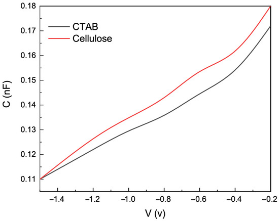
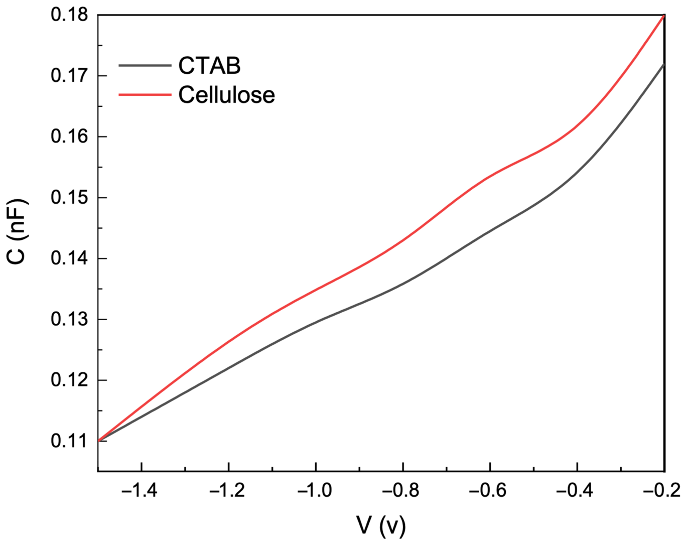
Optical properties and charge transport mechanisms for CTAB and cellulose thin films were described using capacitance–voltage (C–V) and current–voltage (I–V) measurements in the dark and light. The C–V profiles (Figure 8) suggest semiconducting behavior for both materials and the formation of a depletion layer at the film–electrode interface [55]. Cellulose had a greater capacitance than CTAB. This is attributed to the cellulose hydroxyl functional groups that allow for greater dielectric polarization and charge storage from dipolar charge trapping [56]. Cellulose will accumulate greater amounts of photogenerated carriers [57].
Figure 8.
Variation in capacitance with voltage for CTAB and cellulose thin films.
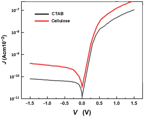
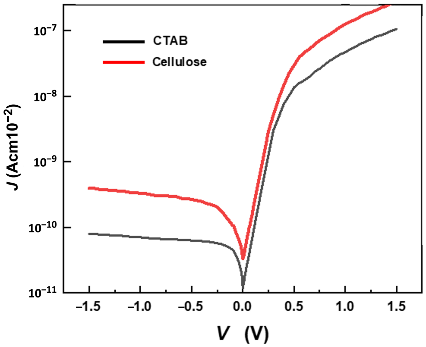
The light-responsive characteristics of the nanostructured films were further confirmed by the I–V curves (Figure 9). Current density increased considerably when the films were illuminated, especially for the cellulose film, which exhibited nearly double the photocurrent as CTAB [58]. This, as well as the comparison in photocurrent, corresponds to the absorption of light and the excitation of electrons across the band gap, as explained in Figure 6. Cellulose nanoparticles’ lower band gap enables charge generation to become more efficient during light absorption in the visible region, as well as during the circulation of charge. This is critical for effective photoelectric conversion [59]. On the other hand, CTAB had a slightly larger band gap, which reduced light absorption and the generation of photocurrent carriers. This is reflected in reduced photocurrent [60].
Figure 9.
The dark J–V characteristics of the CTAB and cellulose thin films heterostructure at room temperature.
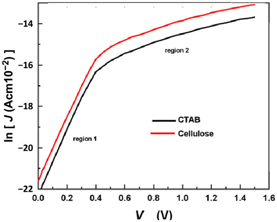
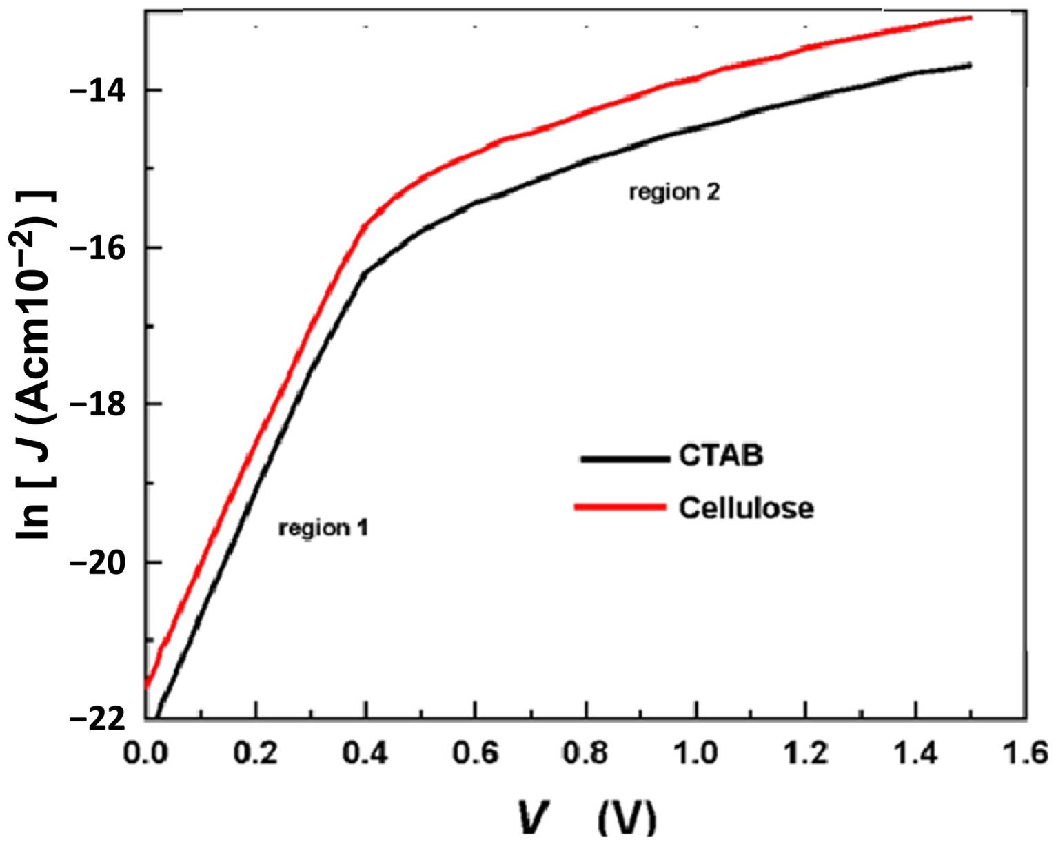
The ln(J) vs. V plots (Figure 10) demonstrate that both materials follow a thermionic emission conduction mechanism in which charge transport entails field-assisted carrier tunneling and carrier recombination. The linearity in the upper-voltage range indicates that photoexcited carriers much more readily surmount the barrier at the interface in the presence of light and, thus, become more mobile [61]. The greater slope for the cellulose suggests more energy-efficient charge transport, which agrees with the greater structural uniformity and better interfacial contact with the electrode [62].
Figure 10.
ln(JF) as a function of V for the heterostructure at room temperature.
Cellulose nanoparticles demonstrate superior performance in optoelectronics owing to their strong photoresponse, high capacitance, and enhanced charge transport [63]. Being bio-compatible and mechanically robust, cellulose nanostructures can also function as photoactive materials, tapping into light energy, and producing localized electric fields, which may promote cellular stimulation, enhance oxygen flow, and exert antimicrobial effects empirically observed in wound healing [50]. Thus, the photoelectric data substantiate the claim that optical excitation and charge transport efficacy directly influence the materials’ biological performance.
2.4. Photocatalytic Properties of CTAB and Cellulose
Methylene blue (MB) served as the test organic dye for the assessment of the photodegradation efficiency of CTAB and Cellulose nanoparticles due to their visible absorption spectrum and their known photodegradation mechanism. Photodegradation was carried out using solar light, and the photodegradation process was measured based on the decrease in the characteristic absorbance peak of MB at about 664 nm via UV–visible spectroscopy.
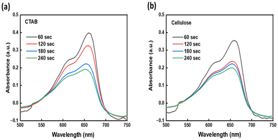
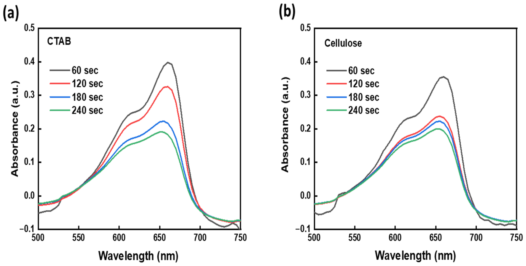
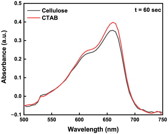
Photocatalytic degradation experiments were carried out using methylene blue (MB) aqueous solution (10 mg L−1) as a model organic pollutant. Prior to illumination, the suspension containing the photocatalyst and MB solution was magnetically stirred in the dark for 30 min to establish adsorption–desorption equilibrium. The photocatalytic reaction was performed in a glass beaker under natural solar irradiation. At selected irradiation intervals, aliquots were collected and centrifuged to remove the catalyst particles before UV–Vis analysis. The degradation process was monitored by measuring the decrease in the characteristic absorption peak of MB at 664 nm. The optical absorption behavior of the investigated materials was further analyzed and correlated with their photocatalytic activity. MB absorption spectra for varying exposure times from 0 to 210 s are shown in Figure 11. Both samples showed a decrease in absorption intensity with increasing exposure time, thus proving that photocatalytic degradation of the dye took place. Moreover, the faster reduction in the absorption of MB up to 210 s can be attributed to dye adsorption as well as degradation through the photodegradation process, owing to the surface hydroxyl groups and electrostatic interaction. Figure 12 shows the relative responses of CTAB and cellulose to 1 min irradiation.
Figure 11.
The photocatalytic properties of the synthesized (a) CTAB and (b) cellulose at different irradiation times up to 210 s.
Figure 12.
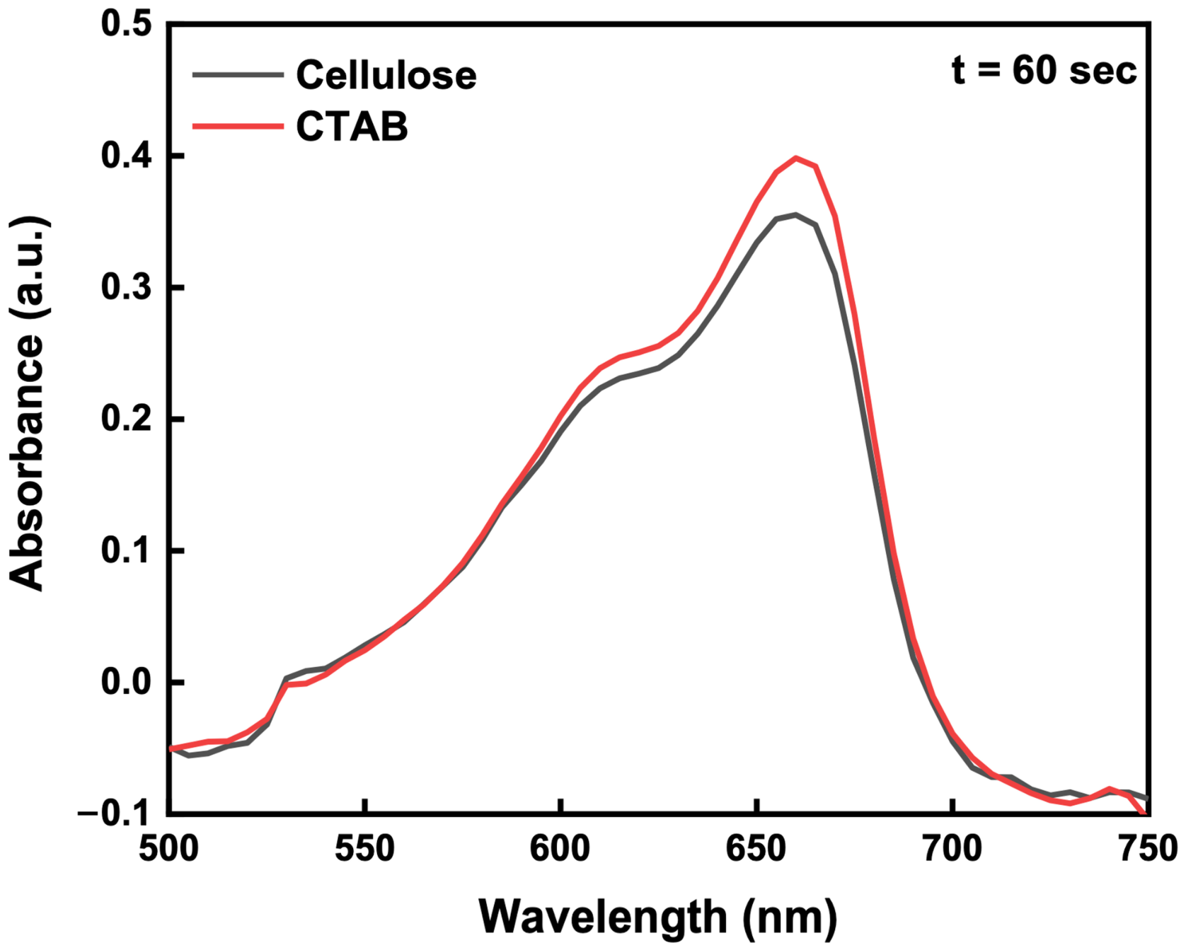
The photocatalytic properties of the synthesized CTAB and cellulose after 1 min irradiation.
The steady decline in the MB absorbance confirms that the photocatalytic process of activation of the nanomaterial leads to the formation of reactive particles that can participate in the oxidation process of the dye [64]. When exposed to light, electrons are promoted from the valence band to the conduction band, forming electrons and holes. These particles will then react with oxygen and water molecules to form reactive oxygen species, including •OH and O2•−, which play a key role in oxidation and dye decomposition [65]. The photodegradation process mainly includes the production of a photogenerated electron–hole pair under light illumination that results in the production of Reactive Oxygen Species (ROS) like hydroxyl radicals (–OH) and superoxide ions that further degrade methylene blue dye. Such types of degradation by ROS have similarly been found in recent photolytic studies [66].
Cellulose nanoparticles exhibited superior photocatalytic activity compared to CTAB. This enhanced performance can be directly correlated with their lower optical bandgap energy, as determined from Tauc analysis, which facilitates more efficient photon absorption and electron excitation under visible light [67]. Additionally, the semi-crystalline structure revealed by XRD and the abundance of surface hydroxyl groups confirmed by FTIR promote effective dye adsorption through hydrogen bonding and electrostatic interactions [68]. The layered and compact morphology observed in SEM further increases surface contact area, enhancing interfacial charge transfer and accelerating degradation kinetics [69].
On the other hand, CTAB exhibited comparatively low photocatalytic efficiency. The broadened band gap results in poor light absorption capacity and low charge carrier generation [26]. Nonetheless, the quaternary ammonium group with a positive charge increases the affinity of CTAB for the dye molecules due to electrostatic forces [70]. Although this factor helps in its photocatalytic activity, the amorphous nature and low charge mobility lead to poor electron–hole separation [71]. Therefore, the results show that photocatalysis performance is influenced by the presence of crystal structure, surface functional groups, band structure, and morphology. The relationship between the optical characteristics, charge transfer, and dye degradation indicates that the cellulose nanoparticle is used as a photocatalyst in response to visible light, whereas the CTAB mainly assists in charge control and adsorption. The photocatalytic behavior of the investigated materials can be interpreted in light of their structural and optical characteristics. Upon light irradiation, the absorbed photons promote charge generation within the material, facilitating the interaction of the surface with methylene blue molecules. The superior photocatalytic performance of cellulose compared with CTAB may be attributed to its lower optical bandgap, enhanced light absorption capability, and the presence of abundant surface hydroxyl groups, which promote dye adsorption and improve interfacial interactions. These findings demonstrate a clear relationship between the structural, optical, and photoelectrical properties of the investigated materials and their observed photocatalytic performance. Further studies involving kinetic analysis and catalyst stability assessments are required to achieve a more comprehensive understanding of their photocatalytic behavior.
2.5. Wound Healing Efficiency of Various Treatments over Time
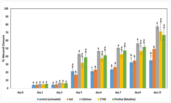
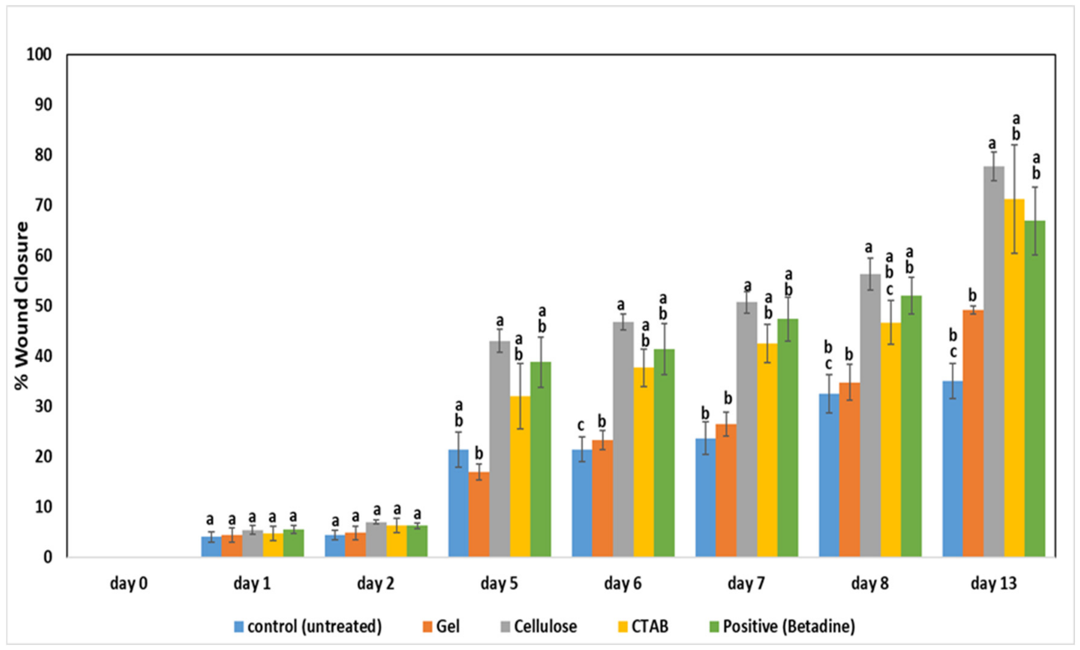
Wound closure proceeded in a treatment-dependent manner over 13 days, as shown in Figure 13. All groups had similar wound sizes on Day 0, demonstrating consistent wound induction. There was minimal closure (≈5–10%) in all groups during Days 1–2, with no statistically significant differences (p > 0.05). Significant divergence was observed by Day 5, with the cellulose group demonstrating the most closure (43%), followed by Betadine (38%) and CTAB (31%), while the gel and control groups showed minimal closure.
Figure 13.
Effect of various treatments on wound closure percentage over time. Data are presented as mean ± SD. Different lowercase letters indicate significant differences among treatment groups at the same time point (p < 0.05), while identical letters indicate no significant difference.
Beginning on Day 6, the wounds treated with cellulose demonstrated obtained repairs at an even faster rate, 64%, by Day 8, signaling granulation and collagen creation. CTAB induced steady healing (approximately 50% by Day 8), most likely due to the antimicrobial-related healing effect [72]. Gel and untreated controls fell behind due to being recovered less than 35% by Day 8. By Day 13, the most epithelialization was measured with cellulose at 82%, then CTAB at 71%, and then Betadine at 67%, while the controls only reached 35%.
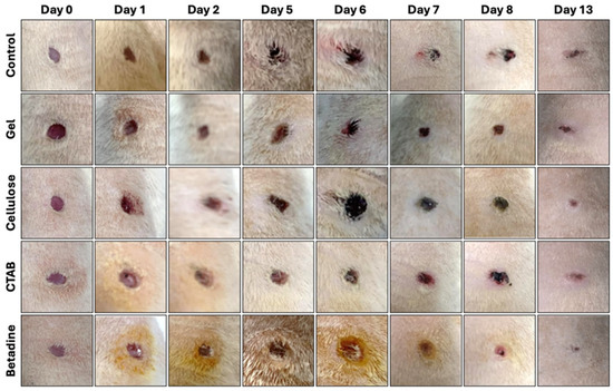
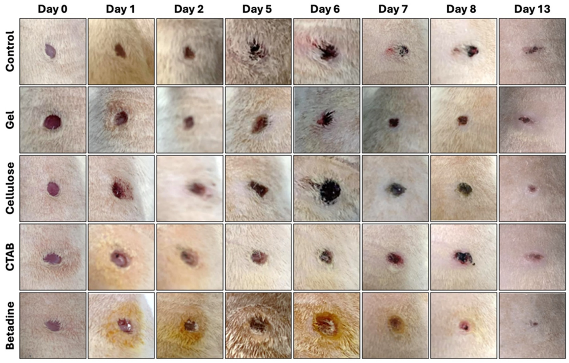
The findings of macroscopic observations (Figure 14) showed smoother and almost complete closure in wounds treated with cellulose in comparison to the control group, where closure was marked by persistent irregularities. The findings from cellulose-treated wounds are complemented by previous reports and explain cellulose’s ability to provide a moist environment, stimulate fibroblast proliferation, and improve collagen maturation [25,73]. Additionally, Marting Da Silva Filho et al. (2023) have shown that bacterial cellulose also improves the processes of angiogenesis and epithelial reformation in full-thickness wounds [72]. CTAB was likely to have improved the healing process of the wounds through the reduction of the microbes and the control of inflammation [74]. Other related studies have also reported on the high antibacterial efficiency of nanomaterials containing CTAB as a result of the positive charges of quaternary ammonium groups, which can help break down microorganisms’ cell membranes, leading to increased membrane permeability [75]. Such antibacterial effect can play an important role in lowering wound infections and facilitating the healing of wounds [76]. In addition to that, compared to other earlier reports, this research has provided additional insight into the multi-functional role of CTAB, as shown below. Moderate efficacy was shown by Betadine, which was most likely a result of the cytotoxic effects that the polyvinylpyrrolidone component exerted because of the prolonged exposure [77]. As for the gel, a lack of bioactive components resulted in minimal effects.
Figure 14.
Assessment of wound healing progression in rats.
Additionally, the rapid wound healing experienced in the cellulose and CTAB groups can be due in part to the structural properties revealed through the XRD analysis. The cellulose’s semi-crystalline structure and hydroxyl-rich amorphous areas promote cellular attachment and the formation of new tissues [78]. On the other hand, CTAB’s amorphous structure promotes stabilization of the bioactive interface. All of these properties together enhance the healing of wounds [79].
3. Materials and Methods
3.1. Biosynthesis and Thin Film Thickness Preparation of CTAB and Cellulose Nanoparticles
CTAB refers to cetyltrimethylammonium bromide (C16H33N(CH3)3Br), and the other chemicals used were microcrystalline cellulose ((C6H10O5)ₙ), sodium hydroxide (NaOH), and urea (CH4N2O), all obtained from Sigma-Aldrich. A cellulose thin-film solution was formed when cellulose was dissolved in a pre-cooled NaOH/urea aqueous system (7 wt % NaOH, 12 wt % urea) at −12 °C on a stirrer, until a clear and viscous solution was obtained (this process took approximately 1 h). CTAB was prepared separately by dissolving the required amount of CTAB in distilled water and making a 0.01 to 0.05 M solution. The prepared aqueous solution of CTAB was observed to be visually homogeneous without any noticeable precipitation before spin coating, signifying good short-term dispersion stability. Purity control was achieved through the use of analytical grade reagents, deionized water, and clean glass/quartz substrates. Also, the fixed concentrations of the reagents, the volumes of the solutions, the spin-coating speed, and coating time ensured consistency in the reproducibility of preparation and yield of the films. The solution of the prepared CTAB was homogeneous without precipitation before making the films, suggesting that the aqueous stability is good under the chosen experimental conditions. Glass and quartz substrates were ultrasonically cleaned by a 3-step process, which involves, in order, acetone, ethanol, and deionized water (this was followed to ensure thorough cleaning), and subsequently dried at 60 °C. Solutions of both cellulose and CTAB were spin-coated on the substrates at 2000 rpm for 30 s and dried at 70 °C for 2 h to yield transparent, smooth films. The thin films adhered well and were uniform. They were appropriate for studying their optical and photoelectric characteristics. The photocatalytic degradation experiments were performed using a 300 W Xenon arc lamp (gas-discharge lamp) (BAXIT, Shanghai, China) as the irradiation source. The lamp provides a broad spectral distribution that closely simulates natural solar irradiation, making it suitable for evaluating the photoresponsive behavior of the investigated materials. All measurements were conducted under identical experimental conditions to ensure a reliable comparison between CTAB and cellulose nanoparticles. The photocatalytic experiments were performed at pH ≈ 4 using an MB concentration of 15 mg L−1, a catalyst dosage of 1.0 g L−1, and an irradiation time of 210 s.” These conditions were kept constant throughout the experiments to enable a direct comparison between the photocatalytic performances of CTAB and cellulose nanoparticles
3.2. Characterization of the CTAB and Cellulose Nanoparticles
3.2.1. X-Ray Diffraction (XRD)
Using a Bruker D8 Advance diffractometer (Bruker Corporation, Karlsruhe, Germany) equipped with Cu Kα radiation (λ = 1.54060 Å), the recorded patterns of X-ray diffractometers for CTAB and Cellulose were recorded for a 2θ span of 10–80° at a scan rate of 0.02°/s. Diffraction patterns were analyzed qualitatively through the comparison of the positions of the obtained peaks with published literature values to assess the structural characteristics and degree of crystallinity. XRD data processing and plotting were accomplished with the Origin software (64-bit edition, 2018).
3.2.2. Optical Characterization
The optical characteristics of CTAB and cellulose nanoparticles were assessed using a V-670 Ultraviolet–Visible (UV–Vis) spectrophotometer (JASCO, Tokyo, Japan) in the range of 200–2500 nm at room temperature. Supernatants were discarded after the third wash, and each pellet was redispersed in distilled water until a uniform color was obtained. 400 µL of the suspension was transferred into a cuvette and filled to the mark with distilled water, and used for baseline correction.
3.2.3. Fourier Transform Infrared Spectroscopy (FTIR)
The determination of functional groups was conducted on a Nicolet iS5 FTIR-8400S spectrometer (Thermo Fisher Scientific, Waltham, MA, USA). The spectra were collected between 4000 and 400 cm−1 while recording under dioxygen at 40 °C. For each sample, 2 mg of the sample was combined with potassium bromide (KBr) to a dilution of 1:100, which was reduced to powder using a mortar and pestle. This powder was further ground into a fine powder. For measurement, the powder was pressed into a transparent disk at 13.79 MPa.
3.2.4. Scanning Electron Microscopy (SEM)
Using a Scanning Electron Microscope (SEM) (Thermo Fisher Scientific, Waltham, MA, USA), samples underwent a detailed morphological evaluation. Gold sputter coating was performed where needed to improve the sample’s electrical conductivity prior to mounting each sample on a metal stub with carbon adhesive tape. Using a JEOL JCM-6000PLUS microscope (JEOL Ltd., Tokyo, Japan).
The total sample collections had systematic imaging performed with the operational voltage of the microscope set to 10 kV. The images taken during this process were employed in the measurement of the differences in sizes the various sample surfaces exhibited, and in the characterization of the surfaces of the samples.
3.3. Photoelectrical Properties
The films prepared were studied regarding their photoelectrical behavior. Their electrical response was studied with and without light. A digital source meter was used to determine the current–voltage characteristics. These measurements assessed the charge transport and photo response of the CTAB and cellulose nanoparticle films to analyze their possible applications in biomedicine and electronics.
3.4. Photocatalytic Activity Assessment
Photocatalytic performance of the prepared CTAB and cellulose nanoparticles was investigated through the use of methylene blue (MB) dye as the target compound. An aqueous solution of MB was then prepared and added to the prepared nanoparticles. The mixture was kept in a stirred condition until a homogenous mixture was achieved.
The reaction mixture was subjected to irradiation for various periods of time (0–210 s). For each period of irradiation, samples were taken for the evaluation of their absorbance intensities through the use of the UV–Visible spectrophotometer. Photocatalytic decomposition was assessed through the observation of reduced absorption intensity corresponding to the absorbance peak of the MB dye. Comparative analysis between CTAB and cellulose was performed based on the rate of absorbance decrease and spectral changes observed during irradiation.
3.5. Preparation of CTAB and Cellulose-Loaded Topical Ointment
An ointment was made by blending 1 g of ultrasound gel with 1 mg of the synthesized nanoparticles, followed by sonication for adequate mixing and complete homogenization. In the next step, the gel prepared was placed in a small jar and preserved at room temperature.
3.6. Experimental Design and Treatment Procedure
The Sprague Dawley rats used for this study were twelve male rats weighing 200–250 g each, which were procured from the Research Center at the Faculty of Science, Beirut Arab University. They were kept in a well-ventilated room at a temperature of 25 ± 2 °C with free access to standard rodent chow and tap water and were placed on a light-dark cycle of 12/12 h. The rats were also given one week to acclimatize before the experiment. The study protocol was approved by the Institutional Review Board (IRB) of Beirut Arab University under protocol number (2023-A-0057-S-M-0559).
The rats were allocated into four groups of three rats: Group I (Control, untreated), Group II (CTAB-loaded ointment), Group III (Cellulose-loaded ointment), and Group IV (Positive control, Betadine gel ointment). Two bilateral full-thickness excision wounds were imposed on the dorsal region of each rat. Each rat’s left wound was treated with ultrasound gel alone (vehicle control). In contrast, the right wound received a treatment, which was monitored with daily wound closure monitoring of the wound from day 0 to day 13.
3.7. Excision Wound Model in Rats
For this study, an excision wound model was used on acclimated rats. Anesthesia was administered as an intraperitoneal injection according to the individual body weight of the test specimen. It should be noted that maximum attention was given to minimize the discomfort of the test specimens. The test specimens were first anesthetized, after which they were positioned prone.
The dorsal surface was then shaved and prepared with an alcohol-iodine solution. Subsequently, two full-thickness circular excision wounds, 4.00 mm in diameter, were performed on the dorsal thoracic area. For this, using a biopsy punch, a position 1 cm lateral to the vertebral column and 5 cm from the ear was selected [80].
These wounds were monitored for over 13 days. Treatments were applied daily from day 0 to day 13 and were only administered once daily. Wound area was measured daily using a digital caliper. The percentage of wound contraction is referred to as the extent of healing. Wound healing is expressed in terms of the percentage of contraction of the wound, and in this case, the healing process started as per the calculated formula. The duration of complete epithelialization (in days) was also noted, indicating that the wound was fully healed. The defined day is the day the scab detaches without exposing any raw surface.
Percentage (%) of wound contraction = 1 − AD/AO × 100,
AO is the wound area on zero days, and AD is the area on corresponding days (0, 1, 2, 5, 6, 7, 8, and 13 days).
According to the study design, after 13 days of treatment, the animals were euthanized while under general anesthesia on the 13th day. Subsequently, tissue samples from the previously mentioned area were collected from all groups for histopathological assessment.
3.8. Statistical Analysis
Statistical analysis was incorporated using SPSS version 27. A one-way ANOVA was carried out to assess information on wound closure. Where applicable, Tukey post hoc tests were conducted to confirm which groups were responsible for the differences. A p-value of 0.05 was the cutoff for statistical significance.
4. Conclusions
This research revealed that the nanoparticles made from CTAB and cellulose are multifunctional in nature and have the potential for use in phototherapy for wound healing. The cellulose nanoparticles showed better optical, photoelectric, photocatalytic, and wound healing efficiency than CTAB, leading to rapid wound healing, improved granulation tissue formation, and collagen production. On the other hand, CTAB added beneficial antimicrobial and surface-stabilizing characteristics that contributed to the healing process. The increased photocatalytic efficiency of cellulose nanocrystals was due to the lower band gap energy, effective charge transfer ability, and presence of abundant surface hydroxyl groups that facilitate the adsorption of dyes and formation of reactive oxygen species. The interconnection among the structure, optical properties, and the efficacy of dye decomposition reveals the multi-functional capability of cellulose-based nano-materials as efficient photoactive biomaterials, while CTAB only served to improve surface charge and antimicrobial properties. Overall, this work offers a comparative multifunctional assessment of the properties of CTAB and cellulose nanoparticles for their use in biomedicine and catalysis. Additional studies on long-term biocompatibility, ideal dosage conditions, and mechanisms of action are strongly advised for future biomedical applications.
Author Contributions
Conceptualization, N.Y.A., N.A.D., M.I.K. and T.A.H.; methodology, N.Y.A., N.A.D., M.I.K., S.A. and T.A.H. and M.I.K.; software, N.Y.A. and N.A.D.; validation, N.A.D., T.A.H., S.A.A. and M.I.K.; formal analysis, N.A.D., N.Y.A., T.A.H. and M.I.K.; investigation, N.Y.A., N.A.D., M.I.K. and T.A.H.; data curation, N.A.D.; N.Y.A., S.A. and T.A.H.; resources, N.Y.A. and N.A.D.; visualization, T.A.H., M.I.K., N.A.D. and S.A.A.; writing—original draft preparation, N.A.D.; writing—review and editing, T.A.H., N.Y.A., N.A.D., M.I.K. and M.M.A.; supervision, T.A.H. and M.I.K.; project administration, M.M.A. All authors have read and agreed to the published version of the manuscript.
Funding
This research received no external funding.
Data Availability Statement
Data from this study are all contained in the manuscript. Raw data will be supplied upon reasonable request from the corresponding author.
Acknowledgments
The biological experiments described in this study were performed at the Faculty of Science, Beirut Arab University.
Conflicts of Interest
The authors declare no conflicts of interest.
Abbreviations
The following abbreviations are used in this manuscript:
| AD | Wound area on corresponding days |
| AO | Initial wound area (Day 0) |
| ANOVA | Analysis of variance |
| CTAB | Cetyltrimethylammonium bromide |
| Eg | Optical bandgap energy |
| Eu | Urbach energy |
| FTIR | Fourier transform infrared spectroscopy |
| IRB | Institutional Review Board |
| I–V | Current–voltage |
| J–V | Current density–voltage |
| KBr | Potassium bromide |
| MB | Methylene blue |
| NaOH | Sodium hydroxide |
| SEM | Scanning electron microscopy |
| SPSS | Statistical Package for the Social Sciences |
| UV–Vis | Ultraviolet–visible spectroscopy |
| XRD | X-ray diffraction |
| C–V | Capacitance–voltage |
References
- Rodrigues, M.; Kosaric, N.; Bonham, C.A.; Gurtner, G.C. Wound Healing: A Cellular Perspective. Physiol. Rev. 2019, 99, 665–706. [Google Scholar] [CrossRef] [Scilit] [PubMed]
- Singh, P.; Ahn, S.; Kang, J.-P.; Veronika, S.; Huo, Y.; Singh, H.; Chokkaligam, M.; El-Agamy Farh, M.; Aceituno, V.C.; Kim, Y.J.; et al. In Vitro Anti-Inflammatory Activity of Spherical Silver Nanoparticles and Monodisperse Hexagonal Gold Nanoparticles by Fruit Extract of Prunus Serrulata: A Green Synthetic Approach. Artif. Cells Nanomed. Biotechnol. 2017, 46, 2022–2032. [Google Scholar] [CrossRef] [Scilit] [PubMed]
- Atala, A.; Irvine, D.J.; Moses, M.; Shaunak, S. Wound Healing Versus Regeneration: Role of the Tissue Environment in Regenerative Medicine. MRS Bull. 2010, 35, 597–606. [Google Scholar] [CrossRef] [Scilit] [PubMed]
- Blanco-Fernandez, B.; Castaño, O.; Mateos-Timoneda, M.Á.; Engel, E.; Pérez-Amodio, S. Nanotechnology Approaches in Chronic Wound Healing. Adv. Wound Care 2021, 10, 234–256. [Google Scholar] [CrossRef] [Scilit] [PubMed]
- Abdelhamid, H.N.; Mathew, A.P. Cellulose-Based Nanomaterials Advance Biomedicine: A Review. Int. J. Mol. Sci. 2022, 23, 5405. [Google Scholar] [CrossRef] [Scilit] [PubMed]
- Teixeira, M.A.; Paiva, M.C.; Amorim, M.T.P.; Felgueiras, H.P. Electrospun Nanocomposites Containing Cellulose and Its Derivatives Modified with Specialized Biomolecules for an Enhanced Wound Healing. Nanomaterials 2020, 10, 557. [Google Scholar] [CrossRef] [Scilit] [PubMed]
- Bai, Y.; Zhang, J.; Dong, S.; Li, J.; Zhang, R.; Pu, C.; Chen, G. Effect of Anion on the Corrosion Inhibition of Cationic Surfactants and a Mechanism Study. Desalin. Water Treat. 2020, 188, 130–139. [Google Scholar] [CrossRef] [Scilit]
- Carvalho, G.C.; Marena, G.D.; Karnopp, J.C.F.; Jorge, J.; Sábio, R.M.; Martines, M.A.U.; Bauab, T.M.; Chorilli, M. Cetyltrimethylammonium Bromide in the Synthesis of Mesoporous Silica Nanoparticles: General Aspects and in Vitro Toxicity. Adv. Colloid Interface Sci. 2022, 307, 102746. [Google Scholar] [CrossRef] [Scilit] [PubMed]
- Grigore, M.; Biscu, E.; Holban, A.; Gestal, M.; Grumezescu, A. Methods of Synthesis, Properties and Biomedical Applications of CuO Nanoparticles. Pharmaceuticals 2016, 9, 75. [Google Scholar] [CrossRef] [Scilit] [PubMed]
- Mallick, S.; Nag, M.; Lahiri, D.; Pandit, S.; Sarkar, T.; Pati, S.; Nirmal, N.P.; Edinur, H.A.; Kari, Z.A.; Ahmad Mohd Zain, M.R.; et al. Engineered Nanotechnology: An Effective Therapeutic Platform for the Chronic Cutaneous Wound. Nanomaterials 2022, 12, 778. [Google Scholar] [CrossRef] [Scilit] [PubMed]
- Shaghaleh, H.; Xu, X.; Wang, S. Current Progress in Production of Biopolymeric Materials Based on Cellulose, Cellulose Nanofibers, and Cellulose Derivatives. RSC Adv. 2018, 8, 825–842. [Google Scholar] [CrossRef] [Scilit] [PubMed]
- Bal-Öztürk, A.; Alarçin, E.; Yaşayan, G.; Avci-Adali, M.; Khosravi, A.; Zarepour, A.; Iravani, S.; Zarrabi, A. Innovative Approaches in Skin Therapy: Bionanocomposites for Skin Tissue Repair and Regeneration. Mater. Adv. 2024, 5, 4996–5024. [Google Scholar] [CrossRef] [Scilit]
- Mokhena, T.C.; John, M.J. Cellulose Nanomaterials: New Generation Materials for Solving Global Issues. Cellulose 2020, 27, 1149–1194. [Google Scholar] [CrossRef] [Scilit]
- Khan, I.; Saeed, K.; Zekker, I.; Zhang, B.; Hendi, A.H.; Ahmad, A.; Ahmad, S.; Zada, N.; Ahmad, H.; Shah, L.A.; et al. Review on Methylene Blue: Its Properties, Uses, Toxicity and Photodegradation. Water 2022, 14, 242. [Google Scholar] [CrossRef] [Scilit]
- Negi, A. Cationized Cellulose Materials: Enhancing Surface Adsorption Properties Towards Synthetic and Natural Dyes. Polymers 2024, 17, 36. [Google Scholar] [CrossRef] [Scilit] [PubMed]
- Chang, Y.; Dou, N.; Liu, M.; Jiang, M.; Men, J.; Cui, Y.; Li, R.; Zhu, Y. Efficient Removal of Anionic Dyes from Aqueous Solution Using CTAB and β-Cyclodextrin-Induced Dye Aggregation. J. Mol. Liq. 2020, 309, 113021. [Google Scholar] [CrossRef] [Scilit]
- Zhao, Q.; Wang, T.; Wang, J.; Zheng, L.; Jiang, T.; Cheng, G.; Wang, S. Fabrication of Mesoporous Hydroxycarbonate Apatite for Oral Delivery of Poorly Water-Soluble Drug Carvedilol. J. Non-Cryst. Solids 2012, 358, 229–235. [Google Scholar] [CrossRef] [Scilit]
- Khoeini, M.; Najafi, A.; Rastegar, H.; Amani, M. Improvement of Hollow Mesoporous Silica Nanoparticles Synthesis by Hard-Templating Method via CTAB Surfactant. Ceram. Int. 2019, 45, 12700–12707. [Google Scholar] [CrossRef] [Scilit]
- Nam, S.; French, A.D.; Condon, B.D.; Concha, M. Segal Crystallinity Index Revisited by the Simulation of X-Ray Diffraction Patterns of Cotton Cellulose Iβ and Cellulose II. Carbohydr. Polym. 2016, 135, 1–9. [Google Scholar] [CrossRef] [Scilit] [PubMed]
- Podgorbunskikh, E.; Kuskov, T.; Bukhtoyarov, V.; Lomovsky, O.; Bychkov, A. Recrystallization of Cellulose, Chitin and Starch in Their Individual and Native Forms. Polymers 2024, 16, 980. [Google Scholar] [CrossRef] [Scilit] [PubMed]
- Stanciu, M.-C.; Tanasă, F.; Teacă, C.-A. Crystallinity Changes in Modified Cellulose Substrates Evidenced by Spectral and X-Ray Diffraction Data. Polysaccharides 2025, 6, 30. [Google Scholar] [CrossRef] [Scilit]
- Moulik, S.P.; Chakraborty, I.; Rakshit, A.K. Role of Surface-active Materials (Amphiphiles and Surfactants) in the Formation of Nanocolloidal Dispersions, and Their Applications. J. Surfact. Deterg. 2022, 25, 703–727. [Google Scholar] [CrossRef] [Scilit]
- Akköse, A.P. A Study on the Synergistic Antibacterial Effects of CTAB-Capped Silver Nanoparticles and Antibiotics. Master’s Thesis, The Graduate School of Natural and Applied Sciences of Middle East Technical University, Ankara, Turkey, 2024. [Google Scholar]
- Karimian, A.; Parsian, H.; Majidinia, M.; Rahimi, M.; Mir, S.M.; Samadi Kafil, H.; Shafiei-Irannejad, V.; Kheyrollah, M.; Ostadi, H.; Yousefi, B. Nanocrystalline Cellulose: Preparation, Physicochemical Properties, and Applications in Drug Delivery Systems. Int. J. Biol. Macromol. 2019, 133, 850–859. [Google Scholar] [CrossRef] [Scilit] [PubMed]
- Abazari, M.F.; Gholizadeh, S.; Karizi, S.Z.; Birgani, N.H.; Abazari, D.; Paknia, S.; Derakhshankhah, H.; Allahyari, Z.; Amini, S.M.; Hamidi, M.; et al. Recent Advances in Cellulose-Based Structures as the Wound-Healing Biomaterials: A Clinically Oriented Review. Appl. Sci. 2021, 11, 7769. [Google Scholar] [CrossRef] [Scilit]
- Gaur, J.; Kumar, S.; Zineddine, M.; Kaur, H.; Pal, M.; Bala, K.; Kumar, V.; Lotey, G.S.; Musa, M.; El Outassi, O. CTAB-Crafted ZnO Nanostructures for Environmental Remediation and Pathogen Control. Sci. Rep. 2024, 14, 20561. [Google Scholar] [CrossRef] [Scilit] [PubMed]
- Gavrilko, T.; Gnatyuk, I.; Styopkin, V.; Shcherban, N.; Baran, J.; Drozd, M. FTIR and DSC Studies of Binary Mixtures of Long-Chain Aliphatic Compounds: Lauric Acid—Cetyl-Trimethylammonium Bromide. Ukr. J. Phys. 2018, 63, 413–424. [Google Scholar] [CrossRef] [Scilit]
- Su, G.; Yang, C.; Zhu, J.-J. Fabrication of Gold Nanorods with Tunable Longitudinal Surface Plasmon Resonance Peaks by Reductive Dopamine. Langmuir 2015, 31, 817–823. [Google Scholar] [CrossRef] [Scilit] [PubMed]
- Mandal, D.; Sarmah, J.K.; Gupta, J. NanoRevolution: Pioneering Applications of Nanotechnology in Type II Diabetes Care. Eng. Proc. 2023, 56, 56. [Google Scholar] [CrossRef] [Scilit]
- Nithiyanantham, U.; Ede, S.R.; Ozaydin, M.F.; Liang, H.; Kundu, S.; Rathishkumar, A. Low Temperature, Shape-Selective Formation of Sb2Te3 Nanomaterials and Their Thermoelectric Applications. RSC Adv. 2015, 5, 89621–89634. [Google Scholar] [CrossRef] [Scilit]
- Cichosz, S.; Masek, A. IR Study on Cellulose with the Varied Moisture Contents: Insight into the Supramolecular Structure. Materials 2020, 13, 4573. [Google Scholar] [CrossRef] [Scilit] [PubMed]
- Agrebi, F.; Ghorbel, N.; Bresson, S.; Abbas, O.; Kallel, A. Study of Nanocomposites Based on Cellulose Nanoparticles and Natural Rubber Latex by ATR/FTIR Spectroscopy: The Impact of Reinforcement. Polym. Compos. 2019, 40, 2076–2087. [Google Scholar] [CrossRef] [Scilit]
- Uddin, M.H.; Haque, M.M. Preparation and Characterization of Cellulose Nanoparticles from Agricultural Wastes and Their Application in Polymer Composites. Sch. Int. J. Chem. Mater. Sci. 2023, 6, 18–23. [Google Scholar] [CrossRef] [Scilit]
- Aljohani, M.M.; Khasim, S.; Albalawi, M.E.; Alfadhli, S.; Ali, M.M.; Waraky, A.; Darwich, N.; Khalil, M.I.; Hamdalla, T.A. Green Synthesis of Silver and Silver Oxide Nanoparticles from Cedrus Libani: Biological Activity and Influence of Film Thickness on Optical Properties. Ukr. J. Phys. Opt. 2026, 27, 01080–01101. [Google Scholar] [CrossRef] [Scilit]
- Munene, J.M.; Onyatta, J.O.; Yusuf, A.O. Characterization of Water Hyacinth Powder Using FTIR Spectroscopy and the Adsorption Behaviour of Pb2+, Cd2+, Zn2+, Ni2+ and Cr2+ in Aqueous Solution. AJACR 2020, 6, 47–55. [Google Scholar] [CrossRef] [Scilit]
- Librando, V.; Minniti, Z.; Lorusso, S. Ancient and Modern Paper Characterization by FTIR and Micro-Raman Spectroscopy. Conserv. Sci. Cult. Herit. 2011, 11, 249–268. [Google Scholar]
- Llombart, P.; Palafox, M.A.; MacDowell, L.G.; Noya, E.G. Structural Transitions and Bilayer Formation of CTAB Aggregates. Colloids Surf. A Physicochem. Eng. Asp. 2019, 580, 123730. [Google Scholar] [CrossRef] [Scilit]
- Drisko, G.L. Control over Nanomaterial Physical Properties with an Emphasis on Light-Matter Interactions. Material Chemistry. Available online: https://hal.science/tel-05164285v1/file/HDRisko_imprimante.pdf (accessed on 7 April 2026).
- Reimer, M.; Zollfrank, C. Cellulose for Light Manipulation: Methods, Applications, and Prospects. Adv. Energy Mater. 2021, 11, 2003866. [Google Scholar] [CrossRef] [Scilit]
- Wu, Y.; Zhu, W. Organic Sensitizers from D–π–A to D–A–π–A: Effect of the Internal Electron-Withdrawing Units on Molecular Absorption, Energy Levels and Photovoltaic Performances. Chem. Soc. Rev. 2013, 42, 2039–2058. [Google Scholar] [CrossRef] [Scilit] [PubMed]
- Halder, K.; Sabnam, K.; Sen, A.; Dasgupta, S. Tuning Properties of CTAB-HSA-AuNR Nanocomposite Antibacterial Thin Films. Chem. Phys. Impact 2024, 9, 100682. [Google Scholar] [CrossRef] [Scilit]
- Wellia, D.V.; Syafawi, A.; Putri, Y.E.; Muldarisnur. The Effect of Cetyltrimethylammonium Bromide (CTAB) Addition on Green Synthesis of Porous N-Doped TiO2 for Photoreduction of Heavy Metal Ion Cr(VI). RSC Adv. 2023, 13, 29645–29656. [Google Scholar] [CrossRef] [Scilit] [PubMed]
- Atta, A.; Abdelhamied, M.M.; Abdelreheem, A.M.; Berber, M.R. Flexible Methyl Cellulose/Polyaniline/Silver Composite Films with Enhanced Linear and Nonlinear Optical Properties. Polymers 2021, 13, 1225. [Google Scholar] [CrossRef] [Scilit] [PubMed]
- Khichar, K.K.; Dangi, S.B.; Dhayal, V.; Kumar, U.; Hashmi, S.Z.; Sadhu, V.; Choudhary, B.L.; Kumar, S.; Kaya, S.; Kuznetsov, A.E.; et al. Structural, Optical, and Surface Morphological Studies of Ethyl Cellulose/Graphene Oxide Nanocomposites. Polym. Compos. 2020, 41, 2792–2802. [Google Scholar] [CrossRef] [Scilit]
- Maleki, A.; He, J.; Bochani, S.; Nosrati, V.; Shahbazi, M.-A.; Guo, B. Multifunctional Photoactive Hydrogels for Wound Healing Acceleration. ACS Nano 2021, 15, 18895–18930. [Google Scholar] [CrossRef] [Scilit] [PubMed]
- Darwich, N.A.; Mezher, M.; Abdallah, A.M.; El-Sayed, A.F.; El Hajj, R.; Hamdalla, T.A.; Khalil, M.I. Biosynthesis; Characterization; and Antibacterial, Antioxidant, and Docking Potentials of Doped Silver Nanoparticles Synthesized from Pine Needle Leaf Extract. Processes 2024, 12, 2590. [Google Scholar] [CrossRef] [Scilit]
- Bao, Y.; Oluwafemi, A. Recent Advances in Surface Modified Gold Nanorods and Their Improved Sensing Performance. Chem. Commun. 2024, 60, 469–481. [Google Scholar] [CrossRef] [Scilit] [PubMed]
- Mondal, J.; Lamba, R.; Yukta, Y.; Yadav, R.; Kumar, R.; Pani, B.; Singh, B. Advancements in Semiconductor Quantum Dots: Expanding Frontiers in Optoelectronics, Analytical Sensing, Biomedicine, and Catalysis. J. Mater. Chem. C 2024, 12, 10330–10389. [Google Scholar] [CrossRef] [Scilit]
- Srivastava, D.; Kuklin, M.S.; Ahopelto, J.; Karttunen, A.J. Electronic Band Structures of Pristine and Chemically Modified Cellulose Allomorphs. Carbohydr. Polym. 2020, 243, 116440. [Google Scholar] [CrossRef] [Scilit] [PubMed]
- Vicente, A.T.; Araújo, A.; Mendes, M.J.; Nunes, D.; Oliveira, M.J.; Sanchez-Sobrado, O.; Ferreira, M.P.; Águas, H.; Fortunato, E.; Martins, R. Multifunctional Cellulose-Paper for Light Harvesting and Smart Sensing Applications. J. Mater. Chem. C 2018, 6, 3143–3181. [Google Scholar] [CrossRef] [Scilit]
- Niu, S.; Huyan, H.; Liu, Y.; Yeung, M.; Ye, K.; Blankemeier, L.; Orvis, T.; Sarkar, D.; Singh, D.J.; Kapadia, R.; et al. Bandgap Control via Structural and Chemical Tuning of Transition Metal Perovskite Chalcogenides. Adv. Mater. 2017, 29, 1604733. [Google Scholar] [CrossRef] [Scilit] [PubMed]
- Elsayed, K.A.; Mahmoud, K.H.; Haladu, S.A.; Magami, S.M.; Manda, A.A.; Kayed, T.S.; Baroot, A.; Khan, M.Y.; Cevik, E.; Drmosh, Q.A.; et al. Thermal, Dielectric and Optical Studies on Cellulose Acetate Butyrate-Gold Nanocomposite Films Prepared by Laser Ablation. J. Mater. Res. Technol. 2023, 23, 419–437. [Google Scholar] [CrossRef] [Scilit]
- Gibaud, A.; Bardeau, J.-F.; Dutreilh-Colas, M.; Bellour, M.; Balasubramanian, V.V.; Robert, A.; Mehdi, A.; Reyé, C.; Corriu, R.J. Influence of Functional Organic Groups on the Structure of CTAB Templated Organosilica Thin Films. J. Mater. Chem. 2004, 14, 1854–1860. [Google Scholar] [CrossRef] [Scilit]
- Darwich, N.A.; Mezher, M.; Abdallah, A.M.; Hachem, Z.; El-Sayed, A.F.; El Hajj, R.; Hamdalla, T.A.; Khalil, M.I. Silver and Yttrium-Doped Silver Nanoparticles From Pine Needle Leaf Extract: Synthesis, Characterization, Antioxidant, Antiuropathogenic Bacterial, and Docking Activities. Bioinorg. Chem. Appl. 2025, 2025, 1566870. [Google Scholar] [CrossRef] [Scilit]
- Vieira, D.H.; Nogueira, G.L.; Oliveira, R.F.D.; Gomes, H.L.; Alves, N. Metal-Electrolyte-Semiconductor Capacitors to Quantify Interface State Density in Printed ZnO for Low-Voltage UV Photodetectors. ACS Appl. Electron. Mater. 2025, 7, 8180–8190. [Google Scholar] [CrossRef] [Scilit]
- Sun, Z.; Liu, J.; Wei, H.; Guo, Q.; Bai, Y.; Zhao, S.; Wang, S.; Li, L.; Zhang, Y.; Tian, Y.; et al. Ultrahigh Energy Storage Capacity in Multilayer-Structured Cellulose-Based Dielectric Capacitors Caused by Interfacial Polarization-Coupled Schottky Barrier Height. J. Mater. Chem. A 2023, 11, 20089–20101. [Google Scholar] [CrossRef] [Scilit]
- Chai, Y.D.; Pang, Y.L.; Lim, S.; Chong, W.C.; Lai, C.W.; Abdullah, A.Z. Recent Progress on Tailoring the Biomass-Derived Cellulose Hybrid Composite Photocatalysts. Polymers 2022, 14, 5244. [Google Scholar] [CrossRef] [Scilit] [PubMed]
- Pasindu, V.; Yapa, P.; Dabare, S.; Munaweera, I. Multifunctional Transition Metal Oxide/Graphene Oxide Nanocomposites for Catalytic Dye Degradation, Renewable Energy, and Energy Storage Applications. RSC Adv. 2025, 15, 33162–33186. [Google Scholar] [CrossRef] [Scilit] [PubMed]
- Wang, X.; Yao, C.; Wang, F.; Li, Z. Cellulose-Based Nanomaterials for Energy Applications. Small 2017, 13, 1702240. [Google Scholar] [CrossRef] [Scilit] [PubMed]
- Rahman, K.H.; Kar, A.K. Influence of Catalyst Loading on Photocatalytic Degradation Efficiency of CTAB-Assisted TiO2 Photocatalyst towards Methylene Blue Dye Solution. Bull. Mater. Sci. 2022, 45, 18. [Google Scholar] [CrossRef] [Scilit]
- Merlin Livingston, L.M.; Harikrishnan, R.; Thandaiah Prabu, R.; Kumar, A. Comparative Analysis of Solar Cell Architecture Utilizing Thermionic Emission Based Carrier Transport versus Tunneling Transport. Phys. Scr. 2025, 100, 055920. [Google Scholar] [CrossRef] [Scilit]
- Teng, C.P.; Tan, M.Y.; Toh, J.P.W.; Lim, Q.F.; Wang, X.; Ponsford, D.; Lin, E.M.J.; Thitsartarn, W.; Tee, S.Y. Advances in Cellulose-Based Composites for Energy Applications. Materials 2023, 16, 3856. [Google Scholar] [CrossRef] [Scilit] [PubMed]
- Saleh, A.K.; El-Sayed, M.H.; El-Sakhawy, M.A.; Alshareef, S.A.; Omer, N.; Abdelaziz, M.A.; Jame, R.; Zheng, H.; Gao, M.; Du, H. Cellulose-based Conductive Materials for Bioelectronics. ChemSusChem 2025, 18, e202401762. [Google Scholar] [CrossRef] [Scilit] [PubMed]
- Kurniawan, T.A.; Mengting, Z.; Fu, D.; Yeap, S.K.; Othman, M.H.D.; Avtar, R.; Ouyang, T. Functionalizing TiO2 with Graphene Oxide for Enhancing Photocatalytic Degradation of Methylene Blue (MB) in Contaminated Wastewater. J. Environ. Manag. 2020, 270, 110871. [Google Scholar] [CrossRef] [Scilit] [PubMed]
- Rettig, I.D.; McCormick, T.M. Enrolling Reactive Oxygen Species in Photon-to-Chemical Energy Conversion: Fundamentals, Technological Advances, and Applications. Adv. Phys. X 2021, 6, 1950049. [Google Scholar] [CrossRef] [Scilit]
- Fu, J.; Liang, F.; Zhong, W.; Kuang, T.; Yin, Z.; Li, Y.; Huang, Z.; Liu, H.; Ma, D. Enhanced Catalytic Degradation Activity through Quenching Introduces Pd Doping in TiO2 Derived from NH2-MIL-125(Ti). Environ. Res. 2025, 285, 122387. [Google Scholar] [CrossRef] [Scilit] [PubMed]
- Subagyo, R.; Diakana, A.R.; Anindika, G.R.; Akhlus, S.; Juwono, H.; Zhang, L.; Arramel; Kusumawati, Y. Modification of Sugar Cane Bagasse with CTAB and ZnO for Methyl Orange and Methylene Blue Removal. ACS Omega 2024, 9, 25251–25264. [Google Scholar] [CrossRef] [Scilit] [PubMed]
- Khan, A.; Hong, R.; Chen, S.; Liu, T.; Kaya, S.; Chen, W.; Xu, D. Dual-Charged, Non-Swelling Amine/Carboxyl Functionalized Fe–Mn Mixed Oxide Composite for Simultaneous Removal of Anionic and Cationic Azo Dyes: Experimental and DFT Studies. Chin. J. Chem. Eng. 2026, 90, 214–231. [Google Scholar] [CrossRef] [Scilit]
- Manimaran, M.; Norizan, M.N.; Kassim, M.H.M.; Adam, M.R.; Abdullah, N.; Faiz Norrrahim, M.N. Surfaces and Interfaces Analysis on Different Carboxymethylation Reaction Time of Anionic Cellulose Nanoparticles Derived from Oil Palm Biomass. Nanotechnol. Rev. 2025, 14, 20250152. [Google Scholar] [CrossRef] [Scilit]
- Chamorro, A.F.; Lerma, T.A.; Palencia, M. CTAB Surfactant Promotes Rapid, Efficient, and Simultaneous Removal of Cationic and Anionic Dyes through Adsorption on Glycerol/Citrate Polyester. Water 2024, 16, 1860. [Google Scholar] [CrossRef] [Scilit]
- Balakrishnan, A.; Mathew, F.P.; Tom Joseph, M.; Varghese, M.M.; Aryamol, K.S.; Chinthala, M.; Rajamohan, N.; Weng, B. Engineered Cellulose-Supported Photocatalysts for Clean Energy and Environmental Remediation: Progress and Prospects. J. Mater. Chem. A 2026, 14, 5473–5519. [Google Scholar] [CrossRef] [Scilit]
- da Silva Filho, P.M.; Mariano, S.R.; Andrade, A.L.; Lopes, J.B.A.C.; de Azevedo Pinheiro, A.; de Azevedo, M.I.G.; de Medeiros, S.C.; de Vasconcelos, M.A.; da Cruz Fonseca, S.G.; Grangeiro, T.B.; et al. Antibacterial and Antifungal Action of CTAB-Containing Silica Nanoparticles against Human Pathogens. Int. J. Pharm. 2023, 641, 123074. [Google Scholar] [CrossRef] [Scilit] [PubMed]
- Tudoroiu, E.-E.; Dinu-Pîrvu, C.-E.; Albu Kaya, M.G.; Popa, L.; Anuța, V.; Prisada, R.M.; Ghica, M.V. An Overview of Cellulose Derivatives-Based Dressings for Wound-Healing Management. Pharmaceuticals 2021, 14, 1215. [Google Scholar] [CrossRef] [Scilit] [PubMed]
- Soliman, W.E.; Elsewedy, H.S.; Younis, N.S.; Shinu, P.; Elsawy, L.E.; Ramadan, H.A. Evaluating Antimicrobial Activity and Wound Healing Effect of Rod-Shaped Nanoparticles. Polymers 2022, 14, 2637. [Google Scholar] [CrossRef] [Scilit] [PubMed]
- Anandakumar, H. Nano-Antibacterial Materials as an Alternative Antimicrobial Strategy. JCIMS 2023, 1, 45–55. [Google Scholar] [CrossRef] [Scilit]
- Cui, Y.; Niu, Q.; Hu, Z.; He, H.; Wei, X.; Wang, M.; Chen, X.-L.; Wang, X. Multifunctional Nanozyme-Hydrogel System in Bacterial-Infected Wounds. Cell Biomater. 2025, 1, 100074. [Google Scholar] [CrossRef] [Scilit]
- Ortega-Llamas, L.; Quiñones-Vico, M.I.; García-Valdivia, M.; Fernández-González, A.; Ubago-Rodríguez, A.; Sanabria-de La Torre, R.; Arias-Santiago, S. Cytotoxicity and Wound Closure Evaluation in Skin Cell Lines after Treatment with Common Antiseptics for Clinical Use. Cells 2022, 11, 1395. [Google Scholar] [CrossRef] [Scilit] [PubMed]
- Ciolacu, D.E. Hydrogels from Renewable Resources: Advances in 3D Networks Based on Cellulose and Hemicellulose. Polymers 2025, 17, 2760. [Google Scholar] [CrossRef] [Scilit] [PubMed]
- Pamula, U.P.; Rao, T.N.; Prashanthi, Y.; Ganji, P. Synthesis and Characterization of ZnO@CTAB-SA Polymer Nanocomposites with Biological and Photocatalytic Activity. Front. Nanotechnol. 2025, 7, 1674700. [Google Scholar] [CrossRef] [Scilit]
- Mosmann, T. Rapid Colorimetric Assay for Cellular Growth and Survival: Application to Proliferation and Cytotoxicity Assays. J. Immunol. Methods 1983, 65, 55–63. [Google Scholar] [CrossRef] [Scilit] [PubMed]
Disclaimer/Publisher’s Note: The statements, opinions and data contained in all publications are solely those of the individual author(s) and contributor(s) and not of MDPI and/or the editor(s). MDPI and/or the editor(s) disclaim responsibility for any injury to people or property resulting from any ideas, methods, instructions or products referred to in the content. |
© 2026 by the authors. Licensee MDPI, Basel, Switzerland. This article is an open access article distributed under the terms and conditions of the Creative Commons Attribution (CC BY) license.