Abstract
This study explores the synthesis of copper oxide nanoparticles (CuO NPs) via green and conventional methods, with emphasis on their performance in hydrogen evolution reactions (HERs). CuO NPs synthesized using okra extract (CuOokra) and hydrazine hydrate (CuOhyd) were characterized using XRD, FTIR, SEM, HRTEM, and electrochemical techniques. Structural analysis revealed that CuOokra NPs have smaller crystallite sizes (39.8 nm) and higher defect densities than CuOhyd NPs (56.8 nm), while CuOhyd exhibited superior porosity and crystallinity. In HER studies, CuOhyd outperformed CuOokra, achieving a significantly lower overpotential (342.2 mV vs. 408.49 mV at 20 mA cm−2) and higher cathodic current density (15.9 vs. 11.3 mA cm−2 at −1.3 V). Electrochemical impedance spectroscopy (EIS) further confirmed the superior catalytic activity of CuOhyd NPs, showing minimal polarization resistance compared to CuOokra.
1. Introduction
The escalating global energy crisis has intensified the pursuit of green and sustainable energy solutions to mitigate environmental and economic challenges. Renewable energy sources such as wind, solar, and tidal power are attractive alternatives due to their environmental benefits and natural availability. However, their dependence on daily, seasonal, and regional fluctuations poses significant challenges to achieving consistent energy output and grid stability. To address these limitations, water electrolysis has emerged as a key strategy to overcome these challenges, enabling the efficient conversion of intermittent renewable energy into hydrogen, a storable, transportable, and versatile energy carrier, thereby enhancing the reliability and scalability of renewable energy systems [1,2]. This approach presents a promising pathway toward realizing zero-carbon energy systems, addressing the urgent global demand for clean and reliable power [3]. Electrochemical water splitting is an efficient and eco-friendly method for producing hydrogen and oxygen by applying a suitable voltage across electrodes [4]. The effectiveness of this process depends on the use of highly active and stable catalysts for the hydrogen and oxygen evolution reactions (HERs and OERs), which are critical for enhancing system performance and maximizing energy conversion efficiency.
Platinum (Pt), iridium (Ir), and ruthenium (Ru), along with their oxides, are widely recognized for their superior catalytic activity and durability, making them highly effective catalysts in water electrolysis. However, their high cost and limited availability remain significant barriers to widespread adoption [5,6]. To overcome these challenges, the development of non-noble, earth-abundant transition metal-based electrocatalysts has emerged as a critical research focus [7,8]. These materials offer greater cost-effectiveness and sustainable alternatives. Nevertheless, they face inherent challenges in achieving the optimal balance of catalytic efficiency, long-term durability, and resistance to harsh electrochemical environments. Advancing such catalysts is essential for enhancing water electrolysis technology and paving the way for large-scale green hydrogen production, a cornerstone of future zero-carbon energy systems.
The design of affordable transition metal (TM)-based nanomaterial catalysts for water splitting and environmental remediation has become an important and rapidly growing research area. Various materials have been studied to improve the adsorption and desorption characteristics of transition metals [9,10]. For the oxygen evolution reaction (OER), common choices include oxides, selenides, phosphides, and hydroxides, while materials like carbides, chalcogenides, nitrides, and alloys have shown promising performance in the hydrogen evolution reaction (HER) [11,12]. Among the various metal oxide nanoparticles (MO-NPs), materials such as ZnO, CeO2, Fe2O3, and CuO have received notable attention for their remarkable catalytic, optical, electrical, and mechanical properties. CuO nanoparticles are widely utilized across industries and healthcare sectors for their efficiency and multifunctional capabilities in catalytic applications [13,14].
Copper oxide (CuO), a p-type semiconductor with a narrow bandgap of 1.2–2.1 eV, was selected in this study due to its strong electrochemical activity, effective catalytic behavior, and excellent chemical stability, which make it highly suitable for electroanalytical chemistry and biological applications [15,16]. CuO nanoparticles (NPs) offer unique advantages, including high solar absorption efficiency, excellent thermal and electrical conductivity, non-toxicity, low cost, and natural abundance, which support their widespread use in renewable energy, environmental remediation, and healthcare. Additionally, their ability to exhibit weak ferromagnetic signals at the nanoscale, attributed to uncompensated Cu2+ surface spins, broadens their potential toward advanced technologies, such as spintronics [17]. Importantly, CuO NPs have demonstrated remarkable catalytic activity and long-term stability for the hydrogen evolution reaction (HER) in water splitting, particularly under alkaline conditions. Although HER activity is generally lower in alkaline media compared to acidic electrolytes [18,19], CuO’s combination of affordability, environmental compatibility, and multifunctional properties makes it an attractive and sustainable alternative to noble metal catalysts for large-scale green hydrogen production.
CuO nanoparticles are commonly synthesized using established techniques like hydrothermal [20], sonochemical [21], co-precipitation [22], sol–gel [23], and microwave-assisted processes [24]. However, these methods often involve toxic chemicals and are energy-intensive, raising environmental concerns. Green synthesis offers an eco-friendly alternative by employing natural extracts or biomolecules to serve as both reducing and stabilizing agents. This method typically utilizes abundant and renewable resources, like plants, algae, or microorganism extracts. As an example, M. Darroudi et al. [25] utilized okra fruit extract, abundant in polyphenolic compounds, like catechins and flavonoids, to achieve the green synthesis of CuO nanoparticles.
In this study, CuO NPs were synthesized via two distinct routes, a green method using okra extract and a conventional method using hydrazine hydrate, to examine how the synthesis pathway affects their structural, morphological, and electrochemical properties. The novelty lies in establishing a clear correlation between the unique characteristics introduced by bioactive-compound-mediated synthesis, such as smaller crystallite size, higher defect density, and altered pore structure, and their catalytic performance in the hydrogen evolution reaction (HER) under alkaline conditions. By directly contrasting an eco-friendly, plant-based approach with a traditional chemical route, this work provides new insights into the design of sustainable, cost-effective, and high-performance electrocatalysts. The prepared nanoparticles were thoroughly characterized using XRD, ATR-FTIR, nitrogen adsorption–desorption, HR-TEM, FESEM, and EDS analyses.
2. Analysis and Interpretation of Results
2.1. XRD
The XRD pattern of the CuO NPs synthesized through mobilization and complexation using okra extract or hydrazine with EDTA, followed by calcination at 400 °C, is shown in Figure 1. The diffraction peaks observed at d-spacings of 2.754 Å, 2.524 Å, 2.325 Å, 1.865 Å, 1.714 Å, 1.583 Å, 1.506 Å, 1.410 Å, 1.376 Å, and 1.305 Å correspond to the Miller indices (110), (11-1), (111), (20-2), (020), (202), (11-3), (31-1), (220), and (311), respectively. These reflections confirm the formation of a typical monoclinic crystal structure for all weight fractions of CuO NPs. Furthermore, the intense and sharp peaks confirm that CuO NPs are highly crystalline, aligning well with the standard data provided in JCPDS card 01-077-7717 [26,27]. The synthesized CuO nanoparticles display a face-centered cubic (FCC) lattice structure with a C12/C1 space group, which aligns with the typical monoclinic phase of CuO, as confirmed by the diffraction pattern.
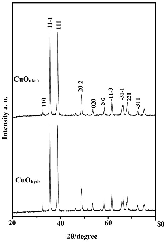
Figure 1.
XRD patterns of CuO NPs synthesized by okra or hydrazine.
The unit cell parameters obtained for CuO NPs synthesized using okra extract (CuOokra) and hydrazine and EDTA (CuOhyd), as shown in Table 1, closely align with the standard JCPDS card (01-077-7717) and experimental data from previous studies [28,29]. For CuOhyd, the lattice constants a = 4.686 Å, b = 3.427 Å, c = 5.133 Å, and β = 99.52° demonstrate a monoclinic structure with a calculated unit cell volume of 81.296 Å3. Similarly, for CuOokra, the values a = 4.690 Å, b = 3.427 Å, c = 5.133 Å, and β = 99.65° confirm a consistent monoclinic configuration with a slightly larger unit cell volume of 81.334 Å3. These results demonstrate the reliability of the synthesis methods in reproducing CuO NPs with minimal deviations from established structural parameters.

Table 1.
Surface texture and XRD data for obtaining structural parameters of the synthesized CuO nanoparticles prepared by two methods and a comparison with the literature data of the different samples.
The Williamson–Hall (W–H) plot (Equation (1)) [30] for the primary diffraction peaks (hkl) of the CuOhyd and CuOokra NPs provides insights into crystallite size and strain (Figure 2). The average crystallite size (D) and micro-strain (ε) values calculated from the linear fit of the W–H plots are D = 56.8 nm and ɛ = 1.097 10−3 for CuOhyd and D = 39.8 nm and ɛ = 0.831 10−3 for CuOokra. The results indicate that okra-extract-mediated synthesis produces a crystallite size that is 39.9% smaller compared with the hydrazine-based method. This reduction is likely influenced by the distinctive stabilizing and reducing nature of the okra extract. Additionally, the dislocation density (δ) calculated using Equation (1) was determined to be 0.310 10−5 nm−2 for CuOhyd and 0.630 10−5 nm−2 for CuOokra, highlighting a higher defect density in the latter. These differences in crystallite size, strain, and dislocation density play a critical role in defining the physical and chemical properties of the CuO NPs, particularly their suitability for hydrogen evolution reactions. Smaller crystallite sizes and higher strain can introduce more active sites and influence the electrical structure, potentially enhancing catalytic performance.
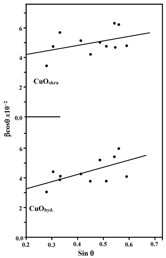
Figure 2.
Williamson–Hall plot of CuO NPs synthesized by okra or hydrazine.
2.2. ATR-FTIR Study
The ATR-FTIR spectra of CuO NPs in the 400–2000 cm−1 range (Figure 3) reveal key insights into their structural properties. For CuOhyd NPs, prominent absorption bands are observed at 418 and 472 cm−1, indicating Cu–O stretching vibrations and confirming the monoclinic phase of CuO, confirming the successful stabilization of this structure by hydrazine during synthesis. These findings align with previous studies [31,32], which emphasize the role of hydrazine as a reducing agent in achieving well-defined monoclinic structures.
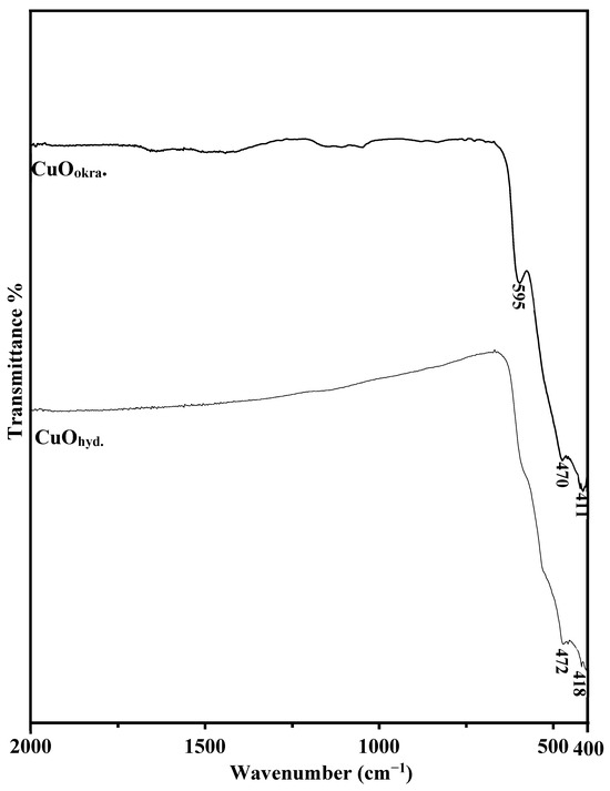
Figure 3.
FT-IR spectra of CuO nanoparticles synthesized using okra extract and hydrazine.
Conversely, CuOokra NPs exhibit additional spectral features. Along with the characteristic bands at 418 and 472 cm−1, a new band emerges at 595 cm−1. This band suggests the presence of structural defects or distortions within the lattice, likely introduced during the green synthesis process. These defects may be attributed to the bioactive compounds in the okra extract, which act as natural stabilizers and reducing agents, imparting unique structural defects. The observed structural defects, as indicated by the XRD results, suggest that they may significantly influence the physical and chemical behavior of the CuO nanoparticles. Structural defects can enhance surface reactivity and modify electronic properties, making the material potentially more suitable for catalytic and energy-related applications [33,34].
2.3. Morphological Analysis (FESEM and HRTEM)
The FESEM images in Figure 4 reveal distinct morphological differences between CuOokra and CuOhyd NPs. The okra-mediated synthesis yields nanoparticles with a predominantly spherical morphology and some degree of agglomeration, likely influenced by the natural bioactive compounds in the okra extract. These bio-compounds contribute to the controlled growth of CuO NPs, but their presence can also induce clustering due to interparticle forces and incomplete dispersion. In contrast, the hydrazine-assisted synthesis produces sponge-like porous morphology with irregularly shaped, agglomerated particles. The absence of natural stabilizers results in less control over nanoparticle growth, leading to highly porous nanostructures. This porosity is beneficial for applications requiring high surface area but reflects a compromise in uniformity and particle size control.
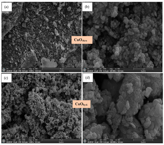
Figure 4.
Field-emission scanning electron microscopy (FESEM) images of CuO nanoparticles at low and high magnifications: (a,b) synthesized using okra extract and (c,d) prepared with hydrazine.
The analysis highlights the benefits of green synthesis methods, including a lower environmental impact and renewable materials, while producing spherical nanoparticles with various applications. In contrast, conventional methods offer structural features, like porosity for specific catalytic or adsorptive uses. These differences demonstrate how synthesis routes affect the properties and applications of CuO NPs.
Figure 5 shows HRTEM images of the synthesized CuO nanoparticles at different magnifications, providing a detailed view of their structural features. The nanoparticles display polydisperse and quasi-spherical agglomerated particles, consistent with the FESEM analysis. The CuOokra and CuOhyd NPs have average sizes of 56 nm and 25 nm, respectively, which correlate well with the XRD data. Although some agglomeration is observed, the lattice fringes observed at higher magnifications confirm the polycrystalline nature of the material, indicating the preservation of its crystalline structure.
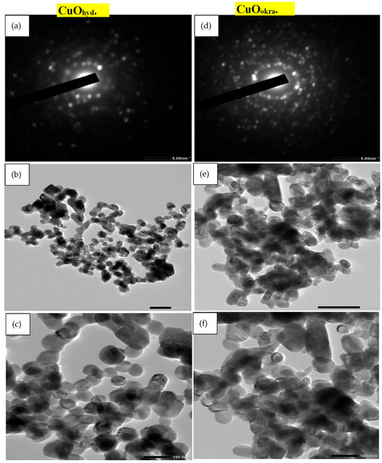
Figure 5.
High-resolution transmission electron microscopy (HRTEM) images at low and high magnifications with corresponding SAED patterns of CuO nanoparticles synthesized using hydrazine (a–c) and okra extract (d–f).
The particle size distribution of CuO nanoparticles was evaluated using HR-TEM images and is presented in Figure 6. The CuO nanoparticles prepared with okra extract exhibited an average particle size of approximately 19.75 nm with a size range of 11.21–29.60 nm, while those synthesized using hydrazine showed a smaller average particle size of 14.80 nm and a range of 7.71–21.16 nm. The relatively uniform distribution and reduced mean size observed for the hydrazine-derived nanoparticles indicate a more uniform nucleation and growth process compared to the okra-extract-derived sample. These results suggest that the choice of reducing agent plays a critical role in influencing crystallite growth and size uniformity of the resulting CuO nanoparticles.
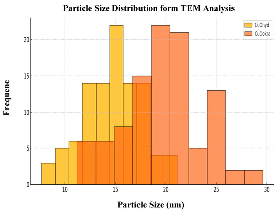
Figure 6.
Particle size distribution derived from HR-TEM analysis of CuO nanoparticles synthesized using okra extract and hydrazine.
The SAED analysis confirms that the synthesized CuO nanoparticles are crystalline. The presence of ring-shaped diffraction patterns in both CuOokra and CuOhyd samples indicates their polycrystalline structure. Key diffraction planes, such as (111) and (-311), align with XRD findings, validating the monoclinic phase of CuO. For CuOhyd, the high degree of crystallinity is a characteristic of the controlled nucleation and growth facilitated by the hydrazine. The absence of diffuse rings further supports the minimal presence of structural defects, reinforcing the effectiveness of the conventional synthesis method in producing structurally stable CuO NPs. In contrast, the SAED pattern of CuOokra shows diffraction rings with slightly diffused features in addition to the prominent rings of the monoclinic CuO phase. This observation suggests the presence of some structural imperfections, likely introduced during the green synthesis process. These defects could be attributed to the biochemical activity of the bioactive compounds in the okra extract, which influence nucleation dynamics and growth mechanisms. Although the crystallinity is lower compared to CuOhyd, the added structural distortions may contribute to the formation of more surface-active sites, which could improve the catalytic and adsorption performance of the nanoparticles.
The EDAX spectra in Figure 7 confirm the elemental composition of the CuO NPs, showing peaks corresponding to copper (Cu) and oxygen (O) without any noticeable impurities. The elemental analysis of CuOokra indicates atomic percentages of approximately 73% copper and 27% oxygen. The CuOhyd results in a Cu-rich composition with 86.04% Cu and 13.96% O. This difference can be attributed to the stronger reducing ability of hydrazine, which likely leads to a higher copper content compared to the bio-mediated reduction process in the okra extract. These variations in the Cu/O ratio are important, as they can influence the electronic structure and reactivity of the nanoparticles, potentially affecting their performance in applications such as catalysis and energy storage. Elemental mapping further corroborates these findings, showing a homogeneous distribution of Cu and O across the nanoparticle surfaces for both samples.
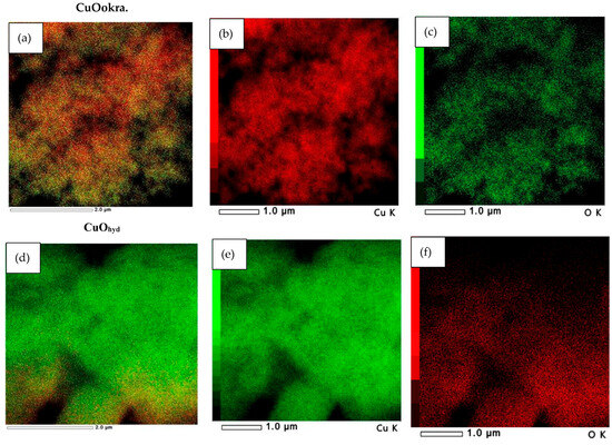
Figure 7.
Elemental mapping of Cu and O in CuO nanoparticles synthesized using okra extract (a–c) and hydrazine (d–f), confirming successful nanoparticle formation.
Figure 8 displays the elemental mapping of Cu and O in CuO nanoparticles synthesized using hydrazine and okra methods. The distinct signals for Cu and O confirm the successful formation and high purity of the NPs. The agglomerated structure and irregular shape may result from the green synthesis method; however, the elemental mapping indicates the presence of CuO.
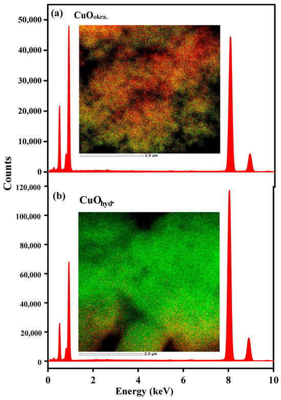
Figure 8.
Energy-dispersive X-ray spectroscopy (EDX) analysis of CuO nanoparticles synthesized using okra extract (a) and hydrazine (b), confirming the presence of Cu and O.
2.4. Surface Texturing
The structural properties of the CuO NPs prepared by two different methods were thoroughly analyzed using N2 adsorption–desorption isotherms and pore size distribution curves. Figure 9 illustrates that CuOhyd and CuOokra exhibit type IV isotherms with H3 hysteresis loops, characteristic of mesoporous materials. This indicates that the synthesized CuO NPs possess a well-defined pore structure. The isotherm of CuOhyd indicates higher adsorption at elevated relative pressures (P/P0 > 0.5), suggesting larger mesopores and a broader pore size distribution. The wider hysteresis loop signifies interconnected pores of varying sizes. The CuOokra isotherm demonstrates lower overall adsorption volumes but exhibits a sharper hysteresis loop. This suggests a more uniform pore size distribution, likely due to the stabilizing effects of bioactive compounds in the okra extract, which influence the growth and assembly of nanoparticles. The reduced pore volume may be attributed to the biogenic synthesis process, which tends to produce smaller, more compact structures.
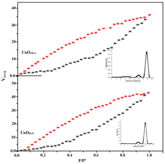
Figure 9.
N2 adsorption–desorption isotherms and pore volume distribution curves of CuO nanoparticles synthesized using okra extract and hydrazine.
The pore analysis reveals that CuOhyd has a smaller pore size of 18 nm, while CuOokra shows a larger pore size of 27 nm. CuOhyd also features a higher pore volume of 0.0801 cm3/g and a BET surface area of 46.21 m2/g compared to CuOokra, which has a pore volume of 0.0592 cm3/g and a surface area of 32.62 m2/g. Both materials have similar average pore sizes around 17.83 Å. The greater surface area and pore volume of CuOhyd enhance the exposure of active sites, making it more favorable for applications in heterogeneous catalysis, including hydrogen evolution and photocatalytic processes.
2.5. Hydrogen Evolution
2.5.1. Cathodic Polarization Analysis
Cathodic polarization studies were conducted in an alkaline medium to assess the HER activity of the synthesized copper oxide nanoparticles, revealing a reduced polarization response that reflects moderate electrocatalytic efficiency. The cathodic polarization curves for CuOokra and CuOhyd NPs, as well as for a bulk Cu electrode in 1.0 M KOH, are shown in Figure 10. The electrocatalytic activity of the CuOokra NPs and CuOhyd NPs towards the alkaline HER is significant. The onset potential for hydrogen evolution on the nanoparticle-modified electrodes is shifted slightly in the positive direction compared to bulk copper, indicating a reduced but still enhanced electrocatalytic response. Specifically, at a current density of 20 mA cm−2, the H2 evolution potential on the CuO NP electrodes exhibits a positive shift exceeding 200 mV relative to the bulk Cu electrode. This enhancement emphasizes the superior catalytic efficiency of the CuO NPs in facilitating HER under alkaline conditions. This observation highlights that CuOhyd NPs exhibit superior electrocatalytic activity toward the alkaline HER compared to other catalysts investigated. The CuOhyd catalyst achieves the lowest overpotential for H2 evolution, demonstrating its exceptional catalytic performance. The cathodic overpotential (η) is a critical parameter influencing the HER kinetics and the corresponding H2 evolution rate. Lower η values are highly desirable, as they indicate a more energy-efficient catalytic process. To quantify this performance, the overpotential at a current density of 20 mA cm−2 (ηp20) was measured for the CuO NP electrodes, as summarized in Table 2. Notably, the CuOhyd NP catalyst achieves a significantly lower ηp20 of 342.2 mV compared to the CuOokra NP electrode (ηp20 = 408.49 mV). This pronounced difference emphasizes the enhanced catalytic efficiency of the CuOhyd NPs, further strengthening their potential as a high-performance electrocatalyst for the alkaline HER. Copper oxide nanoparticles (CuO NPs) catalysts’ HER catalytic performance was favorably compared to previous studies (Table 3), indicating that these catalysts are either smaller or comparable to the materials reported in different media [35,36,37,38,39,40,41,42].
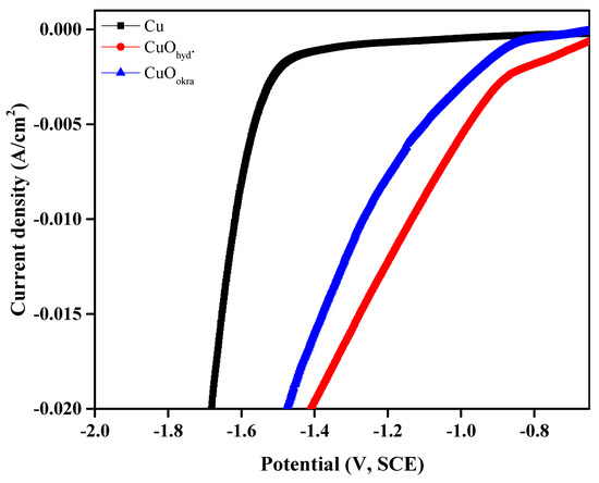
Figure 10.
Cathodic polarization measurements of CuO nanoparticles in a 1 M KOH solution at 25 °C.

Table 2.
Cathodic polarization parameters of CuO nanoparticles in alkaline media of a 1 M KOH solution at 25 °C.

Table 3.
Comparison of catalytic performance for the HER with previous studies.
At a given potential, the rate of hydrogen evolution is indicated by the measured current density, which serves as a useful, though slightly diminished, indicator of HER activity when polarization responses are lower [43,44]. The cathodic current densities (ic) extracted from the cathodic polarization curves at −1.3 V (SCE) for the CuOokra and CuOhyd NPs are 11.3 and 15.9 mA cm−2, respectively. These results demonstrate the superior electrocatalytic performance of the CuOhyd NPs. As expected, the cathodic current densities increased with more negative cathodic potentials, highlighting the robust catalytic activity of both electrodes under applied overpotentials. The remarkable activity of the CuOhyd NPs can be attributed to their unique sponge-like morphology and high porosity nanostructure, which provide an increased active surface area and enhanced electron transfer efficiency. Given their high performance, the CuOhyd NPs hold significant promise for practical applications, particularly in industrial-scale electrolytic hydrogen production under alkaline conditions.
2.5.2. Electrochemical Impedance Spectroscopy, EIS
EIS analysis was conducted to corroborate the findings from cathodic polarization measurements and further elucidate the HER kinetics on the synthesized CuO NPs. As a nondestructive steady-state technique, EIS is particularly suited for assessing the electrocatalytic performance of various electrodes during H2 evolution. The Nyquist plots presented in Figure 11a,b show distinct differences in HER performance between CuOokra and CuOhyd NPs. The characteristic behavior of the electrodes, reflecting porous and rough surface structures, is evident in the Nyquist plots [45,46]. The plots exhibit two semicircles: the high-frequency semicircle corresponds to the surface porosity, which reflects chemical, physical, or geometrical surface inhomogeneities [47], while the low-frequency semicircle is associated with the kinetics of the HER [48,49].
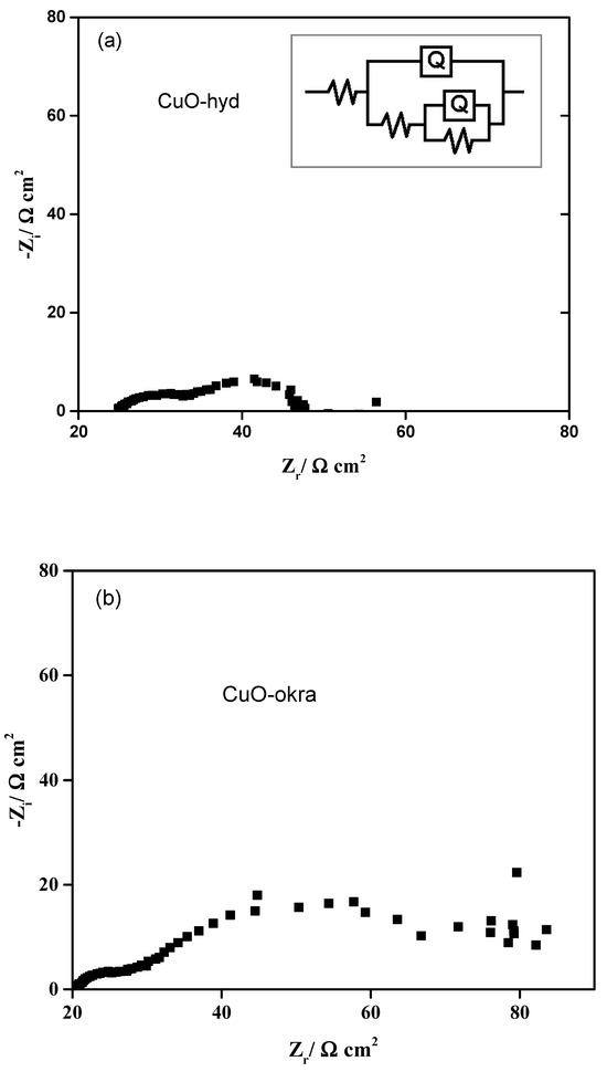
Figure 11.
Nyquist plots of CuO-hyd (a) and CuO-okra (b) nanoparticles in a 1 M KOH solution at 25 °C.
A comparative analysis of the Nyquist plots and the data summarized in Table 4 demonstrates that the CuOhyd NP electrodes possess the smallest semicircle diameter, particularly at the low-frequency region (Figure 11a). This observation confirms that the CuOhyd NPs exhibit the lowest resistance to H2 evolution, signifying their superior catalytic activity relative to the CuOokra NPs. To quantitatively evaluate the system, a two-time constant equivalent circuit model was employed to fit the experimental EIS data, as depicted in the inset of Figure 11a. The extracted fitting parameters, listed in Table 4, highlight the exceptionally low polarization resistance (Rp = R1 + R2) of the CuOhyd NP electrodes. The minimal Rp value further underscores the high electrocatalytic efficiency of the CuOhyd NPs, confirming their potential as a highly active catalyst for hydrogen evolution under alkaline conditions.

Table 4.
EIS parameters of CuO nanoparticles in 1.0 M KOH at 25 °C.
3. Experimental
3.1. Materials
Copper chloride dehydrates (CuCl2·2H2O; 99.0% Sigma-Aldrich, St. Louis, MO, USA), sodium hydroxide (NaOH; Merck, Darmstadt, Germany), ethylenediaminetetraacetic acid disodium salt dihydrate (EDTA, C2H4(NH2)2, Merck), and hydrazine hydrate (50–60% Sigma-Aldrich) were used. Fresh okra pods were sourced from a local fruit shop in Al Qurayyat, Saudi Arabia.
3.1.1. Extraction Process of Okra Fruit
Fresh okra pods were collected from a local store in Al Qurayyat, Saudi Arabia. The pods were carefully washed and sliced into small pieces. About 20 g of the chopped okra was combined with 50 mL of distilled water and heated with stirring at 60 °C for 2 h. After cooling, the mixture was filtered using filter paper, and the resulting extract was stored in the refrigerator for future use in experiments.
3.1.2. Green Synthesis of CuO Nanoparticles
CuO nanoparticles were synthesized by dissolving 6.04 g of CuCl2·2H2O in 50 mL of distilled H2O, followed by stirring at 25 °C for 30 min. Then, 50 mL of okra extract was added gradually to the solution, and the mixture was stirred continuously at 60 °C for 4 h, resulting in the formation of a green gel. The pH of the mixture was adjusted to 11 using 4 M NaOH, and the reaction was left undisturbed for 2 h to promote precipitation. Additional drops of NaOH were added to ensure complete precipitation. The sol–gel was then filtered, and the obtained precipitate was dried at 110 °C for 4 h and subsequently calcined at 400 °C for 3 h. The final product was designated as CuOokra.
3.1.3. Fabrication of CuO NPs Using Hydrazine Hydrate
CuO nanoparticles were prepared by dissolving 6.04 g of CuCl2·2H2O in 50 mL of distilled H2O and stirring the solution at 25 °C for 30 min. Then, 2 mL of a 0.1 M EDTA solution and 1.0 mL of hydrazine hydrate were added, and the mixture was stirred for an additional 4 h. The pH was adjusted to 11 using 4 M NaOH and left undisturbed for 2 h to ensure full precipitation. Additional drops of NaOH were added as needed to complete the process. The resulting sol–gel was filtered, and the precipitate was dried at 110 °C for 4 h, followed by calcination at 400 °C for 3 h. The final product was labeled CuOhyd.
3.2. Working Electrode Preparation and Electrochemical Measurement
Electrochemical tests were carried out using a potentiostat connected to electrochemistry software. The CuOhyd and CuOokra materials were used as working electrodes, which were prepared by mixing carbon black with an adhesive binder (PVDF) in equal amounts and applying the mixture onto a conductive glass substrate. The electrochemical setup included the working electrode, a saturated calomel electrode (SCE) as the reference, and a platinum wire as the counter electrode. The system was filled with 1.0 M KOH as the electrolyte. Measurements involved linear sweep voltammetry (LSV) at a sweep rate of 10 mV/s and electrochemical impedance spectroscopy (EIS) across a frequency range of 100 kHz to 0.1 Hz, with a small voltage perturbation of 0.01 V. These methods were used to evaluate the electrodes’ electrochemical performance and impedance behavior.
3.3. Structural and Morphological Characterization
The phase composition, purity, crystallinity, and crystallite size of the synthesized samples were analyzed at room temperature using an X-ray diffractometer. Scanning was performed at a rate of 2θ = 2.5°/min using Ni-filtered copper radiation (λ = 1.5404 Å) with an operating voltage of 30 kV and a current of 10 mA. The crystallite size (D) and micro-strain (ε) were estimated from the XRD patterns using the W–H approach [30] as follows:
In this equation, K is the shape factor with a typical value of 0.9, λ represents the wavelength of Cu Kα radiation (1.5406 Å), θ is the Bragg diffraction angle, and βexp corresponds to the full width at half maximum (FWHM), which includes contributions from both instrument broadening and sample-related effects.
The volume of the unit cell in a monoclinic structure was calculated using the following formula:
Here, a, b, and c represent the lattice constants of the monoclinic crystal structure, and β denotes the angle between the a and c axes.
Additionally, the dislocation density (δ) is determined using the following equation:
where D indicates the average dimension of the crystalline domains.
ATR-FTIR spectra were collected using a Fourier-transform infrared spectrophotometer. The samples were finely ground and mixed with potassium bromide (KBr) in a 1:100 ratio to form pellets. Spectral data were recorded at room temperature over a range of 4000 to 400 cm−1.
Scanning Electron Microscopy (SEM) was performed in high vacuum mode using a field emission system to closely examine the surface morphology and structural features of the samples.
Nitrogen adsorption–desorption isotherms were measured at 77 K to evaluate the surface characteristics of the samples. The specific surface area and pore size distribution were determined using the Brunauer–Emmett–Teller (BET) method, with an experimental error of ±0.5 m2/g for surface area measurements and ±0.2 nm for pore size determination.
High-resolution transmission electron microscopy (HR-TEM) along with energy-dispersive X-ray spectroscopy (EDS) elemental mapping was carried out at an accelerating voltage of 200 kV. The analysis provided detailed insight into the internal structure and elemental distribution of the samples.
4. Conclusions
This study highlights the significance of synthesis methods in tailoring the properties and functionalities of CuO NPs for advanced applications. CuO nanoparticles synthesized through the green method with okra extract (CuOokra) and the conventional method with hydrazine hydrate (CuOhyd) showed noticeable differences in structure, electrochemical behavior, and biological properties, with a generally lower polarization response observed across both samples. CuOhyd NPs demonstrated exceptional electrocatalytic performance for the hydrogen evolution reaction (HER) in alkaline media, with a low overpotential (342.2 mV at 20 mA cm−2), high current density (15.9 mA cm−2 at −1.3 V), and minimal polarization resistance, attributed to their superior porosity and crystallinity, whereas CuOokra NPs showcased enhanced sustainability.
The CuO NPs, spanning energy and healthcare sectors, emphasize their versatility and practical relevance. This work underscores the importance of green synthesis as a sustainable alternative to conventional methods, offering an eco-friendly pathway to high-performance nanomaterials. Such advancements pave the way for scalable, cost-effective solutions in renewable energy, addressing critical global challenges in sustainability and clean energy transitions.
Author Contributions
All authors have played pivotal roles in the research and development of this manuscript. E.K.Alenezy Performed the characterization analyses and contributed to drafting the manuscript. T.M.Salama: Meticulously performed data characterization and analysis, ensuring precision and reliability throughout the study. N.Hashem: Provided expert supervision, led discussions on the findings, and played a key role in drafting, revising, and refining the manuscript, ensuring its completion to the highest standard. I.O.Ali: provided in-depth contributions to result analysis, actively shaping the discussion, and was instrumental in drafting and polishing the manuscript. All authors have read and agreed to the published version of the manuscript.
Funding
This work was funded by the Deanship of Graduate Studies and Scientific Research at Jouf University under grant No. (DGSSR-2024-02-02205).
Data Availability Statement
The original contributions presented in this study are included in the article. Further inquiries can be directed to the corresponding authors.
Acknowledgments
The authors extend their appreciation to the Deanship of Graduate Studies and Scientific Research at Jouf University for funding this work under grant No. (DGSSR-2024-02-02205).
Conflicts of Interest
The authors declare no conflicts of interest.
References
- Ye, L.; Wen, Z. Self-supported three-dimensional Cu/Cu2O–CuO/rGO nanowire array electrodes for an efficient hydrogen evolution reaction. Chem. Commun. 2018, 54, 6388–6391. [Google Scholar] [CrossRef] [PubMed]
- Zhou, Q.; Li, T.-T.; Qian, J.; Hu, Y.; Guo, F.; Zheng, Y.-Q. Self-supported hierarchical CuOx@Co3O4 heterostructures as efficient bifunctional electrocatalysts for water splitting. J. Mater. Chem. A 2018, 6, 14431–14439. [Google Scholar] [CrossRef]
- Ifkovits, Z.P.; Evans, J.M.; Meier, M.C.; Papadantonakis, K.M.; Lewis, N.S. Decoupled electrochemical water-splitting systems: A review and perspective. Energy Environ. Sci. 2021, 14, 4740–4759. [Google Scholar] [CrossRef]
- Sirisomboonchai, S.; Li, X.; Kitiphatpiboon, N.; Channoo, R.; Li, S.; Ma, Y.; Kongparakul, S.; Samart, C.; Abudula, A.; Guan, G. Fabrication of CuOx nanowires@NiMnOx nanosheets core@shell-type electrocatalysts: Crucial roles of defect modification and valence states for overall water electrolysis. J. Mater. Chem. A 2020, 8, 16463–16476. [Google Scholar] [CrossRef]
- Li, L.; Wang, P.; Shao, Q.; Huang, X. Metallic nanostructures with low dimensionality for electrochemical water splitting. Chem. Soc. Rev. 2020, 49, 3072–3106. [Google Scholar] [CrossRef]
- Yin, K.; Chao, Y.; Lv, F.; Tao, L.; Zhang, W.; Lu, S.; Li, M.; Zhang, Q.; Gu, L.; Li, H.; et al. One Nanometer PtIr Nanowires as High-Efficiency Bifunctional Catalysts for Electrosynthesis of Ethanol into High Value-Added Multicarbon Compound Coupled with Hydrogen Production. J. Am. Chem. Soc. 2021, 143, 10822–10827. [Google Scholar] [CrossRef]
- Yuan, C.-Z.; Hui, K.S.; Yin, H.; Zhu, S.; Zhang, J.; Wu, X.-L.; Hong, X.; Zhou, W.; Fan, X.; Bin, F.; et al. Regulating Intrinsic Electronic Structures of Transition-Metal-Based Catalysts and the Potential Applications for Electrocatalytic Water Splitting. ACS Mater. Lett. 2021, 3, 752–780. [Google Scholar] [CrossRef]
- Li, W.; Wang, C.; Lu, X. Integrated transition metal and compounds with carbon nanomaterials for electrochemical water splitting. J. Mater. Chem. A 2021, 9, 3786–3827. [Google Scholar] [CrossRef]
- Gao, M.; Sheng, W.; Zhuang, Z.; Fang, Q.; Gu, S.; Jiang, J.; Yan, Y. Efficient Water Oxidation Using Nanostructured α-Nickel-Hydroxide as an Electrocatalyst. J. Am. Chem. Soc. 2014, 136, 7077–7084. [Google Scholar] [CrossRef]
- Hutchings, G.S.; Zhang, Y.; Li, J.; Yonemoto, B.T.; Zhou, X.; Zhu, K.; Jiao, F. In Situ Formation of Cobalt Oxide Nanocubanes as Efficient Oxygen Evolution Catalysts. J. Am. Chem. Soc. 2015, 137, 4223–4229. [Google Scholar] [CrossRef]
- Li, Y.; Wang, J.; Tian, X.; Ma, L.; Dai, C.; Yang, C.; Zhou, Z. Carbon doped molybdenum disulfide nanosheets stabilized on graphene for the hydrogen evolution reaction with high electrocatalytic ability. Nanoscale 2016, 8, 1676–1683. [Google Scholar] [CrossRef] [PubMed]
- Ma, R.; Zhou, Y.; Chen, Y.; Li, P.; Liu, Q.; Wang, J. Ultrafine Molybdenum Carbide Nanoparticles Composited with Carbon as a Highly Active Hydrogen-Evolution Electrocatalyst. Angew. Chem. Int. Ed. 2015, 54, 14723–14727. [Google Scholar] [CrossRef] [PubMed]
- Javid-Naderi, M.J.; Sabouri, Z.; Jalili, A.; Zarrinfar, H.; Samarghandian, S.; Darroudi, M. Green synthesis of copper oxide nanoparticles using okra (Abelmoschus esculentus) fruit extract and assessment of their cytotoxicity and photocatalytic applications. Environ. Technol. Innov. 2023, 32, 103300. [Google Scholar] [CrossRef]
- Negrescu, A.M.; Killian, M.S.; Raghu, S.N.V.; Schmuki, P.; Mazare, A.; Cimpean, A. Metal Oxide Nanoparticles: Review of Synthesis, Characterization and Biological Effects. J. Funct. Biomater. 2022, 13, 274. [Google Scholar] [CrossRef]
- Okoye, P.; Azi, S.; Qahtan, T.; Owolabi, T.; Saleh, T. Synthesis, properties, and applications of doped and undoped CuO and Cu2O nanomaterials. Mater. Today Chem. 2023, 30, 101513. [Google Scholar] [CrossRef]
- Dantas, A.P.; Raimundo, R.A.; Neto, P.F.; Lopes, C.M.; Santos, J.R.; Loureiro, F.J.; Pereira, T.O.; Morales, M.A.; Medeiros, E.S.; Macedo, D.A. Copper oxide nanofibers obtained by solution blow spinning as catalysts for oxygen evolution reaction. Ceram. Int. 2024, 50, 13034–13045. [Google Scholar] [CrossRef]
- Seehra, M.; Punnoose, A. Particle size dependence of exchange-bias and coercivity in CuO nanoparticles. Solid State Commun. 2003, 128, 299–302. [Google Scholar] [CrossRef]
- Vineesh, T.V.; Yarmiayev, V.; Zitoun, D. Tailoring the electrochemical hydrogen evolution activity of Cu3P through oxophilic surface modification. Electrochem. Commun. 2020, 113, 106691. [Google Scholar] [CrossRef]
- Rheinländer, P.J.; Herranz, J.; Durst, J.; Gasteiger, H.A. Kinetics of the Hydrogen Oxidation/Evolution Reaction on Polycrystalline Platinum in Alkaline Electrolyte Reaction Order with Respect to Hydrogen Pressure. J. Electrochem. Soc. 2014, 161, F1448–F1457. [Google Scholar] [CrossRef]
- Khandaker, J.I. Hydrothermal synthesis of CuO nanoparticles and a study on property variation with synthesis temperature. J. Appl. Fundam. Sci. 2020, 6, 52. [Google Scholar]
- Shui, A.; Zhu, W.; Xu, L.; Qin, D.; Wang, Y. Green sonochemical synthesis of cupric and cuprous oxides nanoparticles and their optical properties. Ceram. Int. 2013, 39, 8715–8722. [Google Scholar] [CrossRef]
- Rangel, W.M.; Santa, R.A.A.B.; Riella, H.G. A facile method for synthesis of nanostructured copper (II) oxide by coprecipitation. J. Mater. Res. Technol. 2020, 9, 994–1004. [Google Scholar] [CrossRef]
- Patel, M.; Mishra, S.; Verma, R.; Shikha, D. Synthesis of ZnO and CuO nanoparticles via Sol gel method and its characterization by using various technique. Discov. Mater. 2022, 2, 1. [Google Scholar] [CrossRef]
- Thakur, N.; Anu; Kumar, K.; Kumar, A. Effect of (Ag, Zn) co-doping on structural, optical and bactericidal properties of CuO nanoparticles synthesized by a microwave-assisted method. Dalton Trans. 2021, 50, 6188–6203. [Google Scholar] [CrossRef]
- Murugan, B.; Rahman, M.Z.; Fatimah, I.; Anita Lett, J.; Annaraj, J.; Kaus, N.H.M.; Al-Anber, M.A.; Sagadevan, S. Green synthesis of CuO nanoparticles for biological applications. Inorg. Chem. Commun. 2023, 155, 111088. [Google Scholar] [CrossRef]
- Moroda, M.D.; Deressa, T.L.; Tiwikrama, A.H.; Chala, T.F. Green synthesis of copper oxide nanoparticles using Rosmarinus officinalis leaf extract and evaluation of its antimicrobial activity. Next Mater. 2024, 7, 100337. [Google Scholar] [CrossRef]
- Pawar, S.M.; Patil, S.S.; Sonawane, K.D.; More, V.B.; Patil, P.S. Hydrothermally synthesized copper oxide nanoparticles: Rietveld analysis and antimicrobial studies. Surf. Interfaces 2024, 51, 104598. [Google Scholar] [CrossRef]
- Nzilu, D.M.; Madivoli, E.S.; Makhanu, D.S.; Wanakai, S.I.; Kiprono, G.K.; Kareru, P.G. Green synthesis of copper oxide nanoparticles and its efficiency in degradation of rifampicin antibiotic. Sci. Rep. 2023, 13, 14030. [Google Scholar] [CrossRef]
- Rehman, S.; Shad, N.A.; Sajid, M.M.; Ali, K.; Javed, Y.; Jamil, Y.; Sajjad, M.; Nawaz, A.; Sharma, S.K. Tuning Structural and Optical Properties of Copper Oxide Nanomaterials by Thermal Heating and Its Effect on Photocatalytic Degradation of Congo Red Dye. J. Chem. Chem. Eng. 2022, 41, 1549–1560. [Google Scholar]
- Williamson, G.K.; Smallman, R.E., III. Dislocation densities in some annealed and cold-worked metals from measurements on the X-ray debye-scherrer spectrum. Philos. Mag. 1956, 1, 34–46. [Google Scholar] [CrossRef]
- Berra, D.; Laouini, S.E.; Benhaoua, B.; Ouahrani, M.R.; Berrani, D.; Rahal, A. Green synthesis of copper oxide nanoparticles by Pheonix dactylifera L leaves extract. J. Nanomater. Biostructures 2018, 13, 1231–1238. [Google Scholar]
- Bin Mobarak, M.; Hossain, S.; Chowdhury, F.; Ahmed, S. Synthesis and characterization of CuO nanoparticles utilizing waste fish scale and exploitation of XRD peak profile analysis for approximating the structural parameters. Arab. J. Chem. 2022, 15, 104117. [Google Scholar] [CrossRef]
- Huang, G.; Zhu, Y. Synthesis and photoactivity enhancement of ZnWO4 photocatalysts doped with chlorine. CrystEngComm 2012, 14, 8076–8082. [Google Scholar] [CrossRef]
- Jaihindh, D.P.; Anand, P.; Chen, R.-S.; Yu, W.-Y.; Wong, M.-S.; Fu, Y.-P. Cl-doped CuO for electrochemical hydrogen evolution reaction and tetracycline photocatalytic degradation. J. Environ. Chem. Eng. 2023, 11, 109852. [Google Scholar] [CrossRef]
- Jiang, N.; Tang, Q.; Sheng, M.; You, B.; Jiang, D.; Sun, Y. Nickel sulfides for electrocatalytic hydrogen evolution under alkaline conditions: A case study of crystalline NiS, NiS2, and Ni3S2 nanoparticles. Catal. Sci. Technol. 2016, 6, 1077–1084. [Google Scholar] [CrossRef]
- Hanan, A.; Shu, D.; Aftab, U.; Cao, D.; Laghari, A.J.; Solangi, M.Y.; Abro, M.I.; Nafady, A.; Vigolo, B.; Tahira, A.; et al. Co2FeO4@rGO composite: Towards trifunctional water splitting in alkaline media. Int. J. Hydrogen Energy 2022, 47, 33919–33937. [Google Scholar] [CrossRef]
- Yang, S.; Wen, H.; Liu, Z.; Zhai, J.; Yu, Y.; Li, K.; Huang, Z.; Sun, D. Engineering double sulfur-vacancy in CoS1.097@MoS2 Core-shell heterojunctions for hydrogen evolution in a wide pH range. Inorg. Chem. 2023, 62, 17401–17408. [Google Scholar] [CrossRef] [PubMed]
- Feng, L.; Vrubel, H.; Bensimon, M.; Hu, X. Easily prepared dinickel phosphide (Ni2P) nanoparticles as an efficient and robust electrocatalyst for hydrogen evolution. Phys. Chem. Chem. Phys. 2014, 16, 5917–5921. [Google Scholar] [CrossRef] [PubMed]
- Man, H.-W.; Tsang, C.-S.; Li, M.-M.-J.; Mo, J.; Huang, B.; Lee, L.Y.S.; Leung, Y.-C.; Wong, K.-Y.; Tsang, S.C.E. Transition metal-doped nickel phosphide nanoparticles as electro-and photocatalysts for hydrogen generation reactions. Appl. Catal. B 2019, 242, 186–193. [Google Scholar] [CrossRef]
- Meshkian, R.; Dahlqvist, M.; Lu, J.; Wickman, B.; Halim, J.; Thornberg, J.; Tao, Q.; Li, S.; Intikhab, S.; Snyder, J. W-Based Atomic Laminates and Their 2D Derivative W1.33C MXene with Vacancy Ordering. Adv. Mater. 2018, 30, 1706409. [Google Scholar] [CrossRef]
- Hanan, A.; Ahmed, M.; Lakhan, M.N.; Shar, A.H.; Cao, D.; Asif, A.; Ali, A.; Gul, M. Novel rGO@Fe3O4 nanostructures: An active electrocatalyst for hydrogen evolution reaction in alkaline media. J. Indian Chem. Soc. 2022, 99, 100442. [Google Scholar] [CrossRef]
- Belhadj, H.; Messaoudi, Y.; Khelladi, M.R.; Azizi, A. A facile synthesis of metal ferrites (MFe2O4, M = Co, Ni, Zn, Cu) as effective electrocatalysts toward electrochemical hydrogen evolution reaction. Int. J. Hydrogen Energy 2022, 47, 20129–20137. [Google Scholar] [CrossRef]
- Birry, L.; Lasia, A. Studies of the Hydrogen Evolution Reaction on Raney Nickel—Molybdenum Electrodes. J. Appl. Electrochem. 2004, 34, 735–749. [Google Scholar] [CrossRef]
- Badawy, W.; Nady, H.; Negem, M. Cathodic hydrogen evolution in acidic solutions using electrodeposited nano-crystalline Ni–Co cathodes. Int. J. Hydrogen Energy 2014, 39, 10824–10832. [Google Scholar] [CrossRef]
- Krstajic, N.V.; Jovic, V.D.; Gajic-Krstajic, L.; Jovic, B.M.; Antozzi, A.L.; Martelli, G.N. Electrodeposition of Ni–Mo alloy coatings and their characterization as cathodes for hydrogen evolution in sodium hydroxide solution. Int. J. Hydrogen Energy 2008, 33, 3676–3687. [Google Scholar] [CrossRef]
- Hu, H.; Qiao, M.; Pei, Y.; Fan, K.; Li, H.; Zong, B. Kinetics of hydrogen evolution in alkali leaching of rapidly quenched Ni–Al alloy. Appl. Catal. A 2003, 252, 173–183. [Google Scholar] [CrossRef]
- Los, P.; Lasia, A.; Ménard, H.; Brossard, L. Impedance studies of porous lanthanum-phosphate-bonded nickel electrodes in concentrated sodium hydroxide solution. J. Electroanal. Chem. 1993, 360, 101–118. [Google Scholar] [CrossRef]
- Solmaz, R.; Kardaş, G. Hydrogen evolution and corrosion performance of NiZn coatings. Energy Convers. Manag. 2007, 48, 583–591. [Google Scholar] [CrossRef]
- Shervedani, R.K.; Mardam, A.R. Kinetics of hydrogen evolution reaction on nanocrystalline electrodeposited Ni62Fe35C3 cathode in alkaline solution by electrochemical impedance spectroscopy. Electrochim. Acta 2007, 53, 426–433. [Google Scholar] [CrossRef]
Disclaimer/Publisher’s Note: The statements, opinions and data contained in all publications are solely those of the individual author(s) and contributor(s) and not of MDPI and/or the editor(s). MDPI and/or the editor(s) disclaim responsibility for any injury to people or property resulting from any ideas, methods, instructions or products referred to in the content. |
© 2025 by the authors. Licensee MDPI, Basel, Switzerland. This article is an open access article distributed under the terms and conditions of the Creative Commons Attribution (CC BY) license (https://creativecommons.org/licenses/by/4.0/).