Article

Electrophoretic Deposition of Gold Nanoparticles on Highly Ordered Titanium Dioxide Nanotubes for Photocatalytic Application

1
Laboratoire de Photovoltaïque, Centre de Recherches et des Technologies de l′Energie, Technopole de Borj Cedria, BP 95, Hammam-Lif 2050, Tunisia
2
Institut de Disseny per a la Fabricació i Producció Automatitzada, Universitat Politècnica de València, 46022 València, Spain
3
College of Science, Imam Mohammad Ibn Saud Islamic University (IMSIU), P.O. Box 5701, Riyadh 11623, Saudi Arabia
4
Laboratory of Advanced Materials and Process Engineering, Physics Department, Faculty of Sciences, Ibn Tofail University, Kenitra 14000, Morocco
*
Author to whom correspondence should be addressed.
Catalysts 2025, 15(8), 781; https://doi.org/10.3390/catal15080781 (registering DOI)
Submission received: 30 May 2025 / Revised: 9 August 2025 / Accepted: 14 August 2025 / Published: 16 August 2025
(This article belongs to the Special Issue Photocatalysis towards a Sustainable Future)

Abstract

This work focused on the photocatalytic performance enhancement of titanium dioxide (TiO2) nanotubes decorated by gold nanoparticles. The surface of the nanotubes synthesized using the anodization technique was modified with subsequent deposition of gold nanoparticles (Au-NPs) via electrophoretic deposition. The impact of electrophoretically deposited gold nanoparticles (Au-NPs) on TiO2 nanotubes, with varying deposition times (5 min, 8 min and 12 min), was investigated in the degradation of amido black (AB) dye. The morphological analysis using scanning electron microscopy (SEM, TESCAN VEGA3, TESCAN Orsay Holding, Brno, Czech Republic) and transmission electron microscopy (TEM, JEM—100CX2, JEOL Japan). revealed a well-organized nanotubular structure of TiO2, with a wall thickness of 25 nm and an internal diameter of 75 nm. Optical study, including photoluminescence and diffuse reflectance spectroscopy, provided evidence of charge transfer between the Au-NPs and the TiO2-NTs. Furthermore, the photocatalytic measurements showed that the enhanced photocatalytic activity of the TiO2-NTs resulted from successful Au deposition onto their surface, surpassing that of the pure sample. This improvement is attributed to the higher work function of gold nanoparticles, which effectively promoted the separation of photogenerated electron–hole pairs. The sample Au-NPs/TiO2-NTs with a deposition time of 5 min exhibited the best photocatalytic efficiency, achieving an 85% degradation rate after 270 min under UV irradiation. Moreover, the enhancement obtained was also attributed to the plasmonic effect induced by Au-NPs. Kinetic investigations revealed that the photocatalytic reaction followed apparent first-order kinetics, highlighting the efficiency of Au-NPs/TiO2-NTs as a photocatalyst for dye degradation.

1. Introduction

Titanium dioxide (TiO2) has been extensively studied to be integrated into various applications, including photocatalytic degradation of pollutants in both water and gas phases, hydrogen production, energy storage and generation, gas detection, optical instrument fabrication, ceramics, and biomedical fields [1,2].
Titanium ranks among the most abundant metals globally, with TiO2 boasting advantageous characteristics including robust chemical stability, non-toxicity, and cost-effectiveness [3]. Many studies focused on TiO2 powders, presenting challenges for certain applications due to the complexity and costs associated with material recovery and reuse, particularly in processes like photocatalytic oxidation of organic pollutants in water. To overcome these issues, it is possible to immobilize the powder onto various substrates, such as glass, activated carbon, silica, or polymers [4].
Many research studies have been interested in the use of TiO2 nanotubes (TiO2-NTs), thanks to their large specific surface area, excellent mechanical adhesion, excellent electron percolation path, and simple synthesis process [5,6,7]. Recently, TiO2-NTs have been used specifically in the degradation of organic pollutants as photocatalysts. However, TiO2-NTs are far from being a perfect photocatalyst [2], due to their wide band gap limitation, leading to low efficiency [3] when irradiated with visible light. One effective approach to overcoming such an obstacle is to deposit noble metals [4], in particular gold, onto the surface of TiO2-NTs. Gold is sufficiently stable to resist corrosion under photocatalytic conditions, and it exhibits a characteristic surface plasmon band in the visible light region, thanks to the collective excitation of electrons [8,9].
The metallic decoration effectively reduces the recombination of photogenerated charges and expands the absorption spectrum of TiO2 into the visible region, attributed to localized surface plasmon resonance (LSPR) [10,11]. This phenomenon allows noble metallic nanoparticles to absorb and scatter light within the visible spectrum. The resonance characteristics depend not only on the metal type but also on other factors such as the shape, the size, and the surrounding environment of the synthesized nanoparticles [12]. The enhanced activity under visible light observed after modifying titania with plasmonic nanoparticles like silver [13], gold [14], and platinum [15] is attributed to their involvement in electron transfer mechanisms [16]. In fact, metallic nanoparticles absorb photons via their localized surface plasmon resonance (LSPR), facilitating electron transfer from the noble metal to the conduction band of titania, known as the LSPR sensitization effect [17].
In particular, gold nanoparticles have gained popularity recently due to their excellent chemical stability and photocatalytic performance [18,19,20]. Au-NPs dispersed on TiO2 nanotubes have shown promising results in degrading pollutants through the generation of reactive oxygen species (ROS) [21,22].
Organic molecules are oxidized either by electron-deficient metals as they return to their metallic state or by O2− [23,24]. Under UV irradiation, the metallic nanoparticles deposited on TiO2 enhance the transfer of photogenerated electrons, thus extending the lifetime of charge carriers involving the formation of a Schottky barrier [24].
To deposit Au-NPs, electrophoretic deposition (EPD) was employed. It is a cost-effective method for processing advanced ceramic materials and coatings used in both academia and industry thanks to its versatility with different materials and combinations, the simplicity of the equipment and a wide range of novel applications [25,26,27,28].
In this work, we used a two-step process to synthesize highly ordered vertical TiO2 nanotube arrays (TNAs) through anodic oxidation in an organic electrolyte. Subsequently, we deposited varying densities of Au nanoparticles on the TiO2 nanotubes using the electrophoresis method with different deposition times (5 min, 8 min, and 12 min). For the first time, the elaborated samples were used for the degradation of amido black dye to test their photocatalytic efficiency. The effect of the Au-NPs on the TiO2-NTs photocatalytic performance was discussed.

2. Results

2.1. Morphological Analysis

In order to explain the photocatalytic activity of the synthesized samples, a morphology study was carried out.

2.1.1. SEM Analysis

Highly ordered TiO2 nanotubes with a diameter around 100 nm fabricated vertically on a titanium substrate were obtained, as shown in Figure 1. We clearly noticed the decoration of the nanotubes with spherical gold nanoparticles, which are uniformly distributed on the surface, as evidenced in Figure 1b–d. The nanoparticles exhibit a size distribution ranging from approximately 20 to 30 nm, with the majority falling around 25 nm (Figure 1e), in addition to gold agglomerates whose sizes increase with the deposition time, varying between 100 and 200 nm.

2.1.2. EDX Analysis

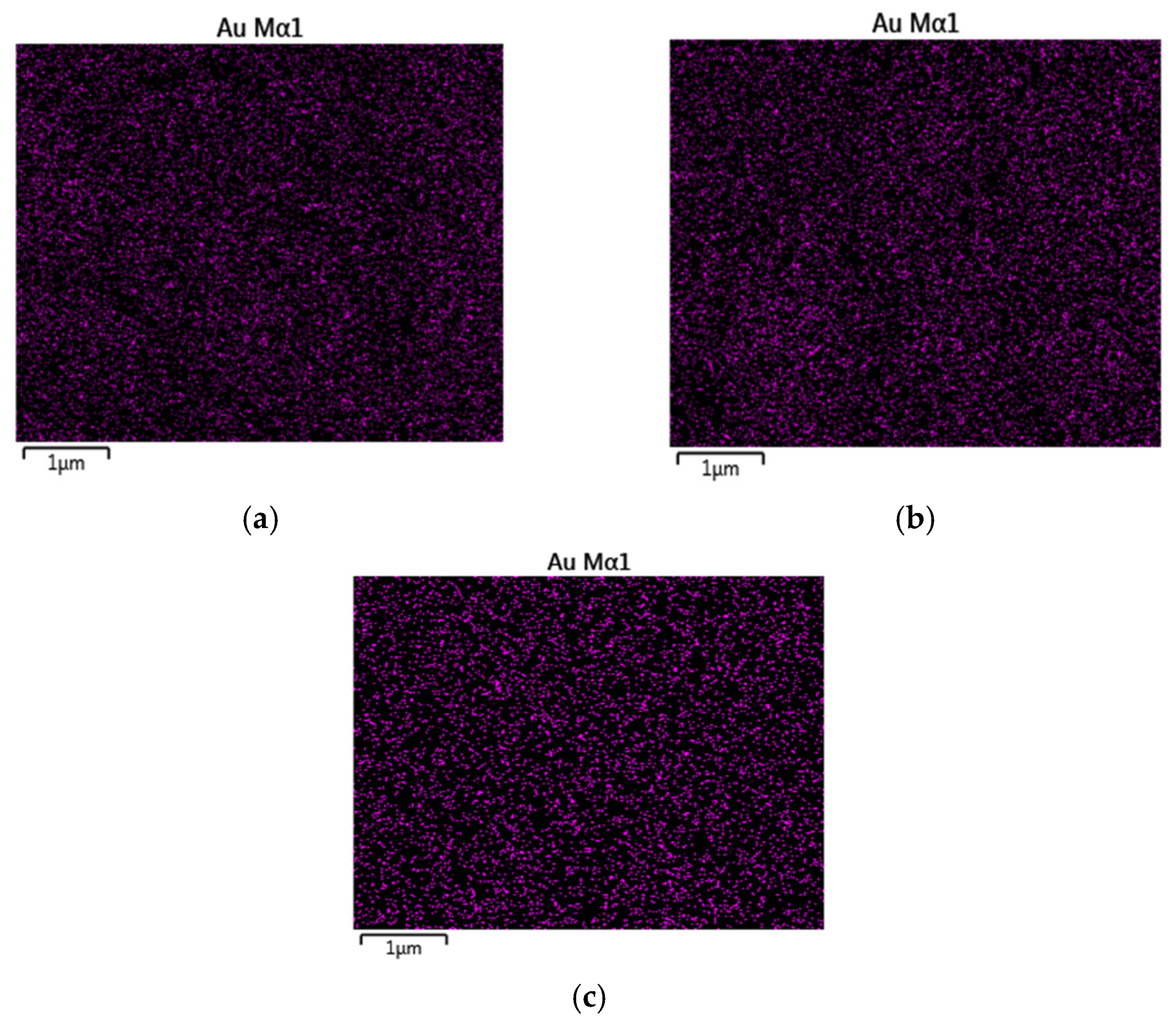
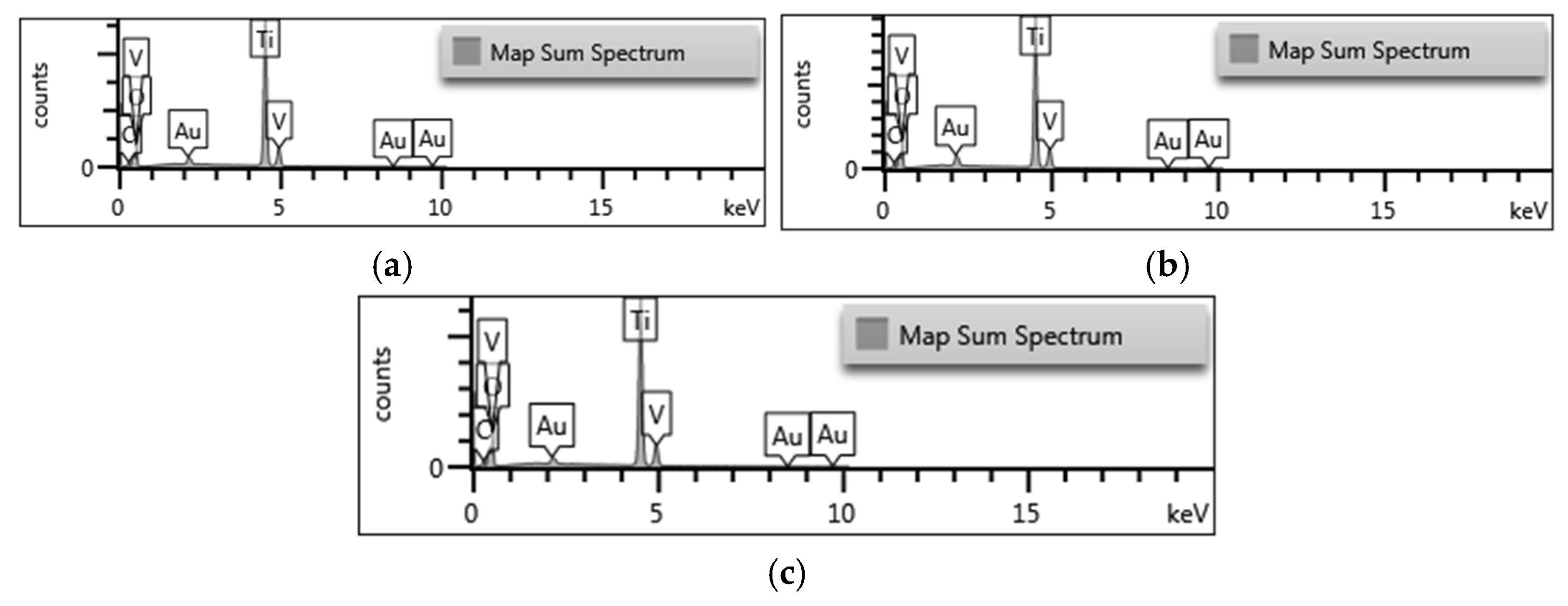
The EDX analysis reveals a progressive increase in gold (Au) (Figure 2 and Figure 3) nanoparticle deposition on TiO2 nanotubes with longer processing times. Quantitative data shows the Au atomic percentage rising from 0.13% (5 min) to 0.16% (8 min) and 0.17% (12 min), indicating enhanced but gradually diminishing returns with extended deposition. Corresponding elemental maps visually confirm this trend, transitioning from sparse Au distribution at 5 min to more concentrated nanoparticle coverage at longer durations. The marginal difference between 8- and 12 min samples suggests approaching saturation in Au loading efficiency (Table 1). These findings demonstrate deposition time’s critical role in controlling Au nanoparticle density on TiO2 substrates.

2.1.3. TEM Analysis

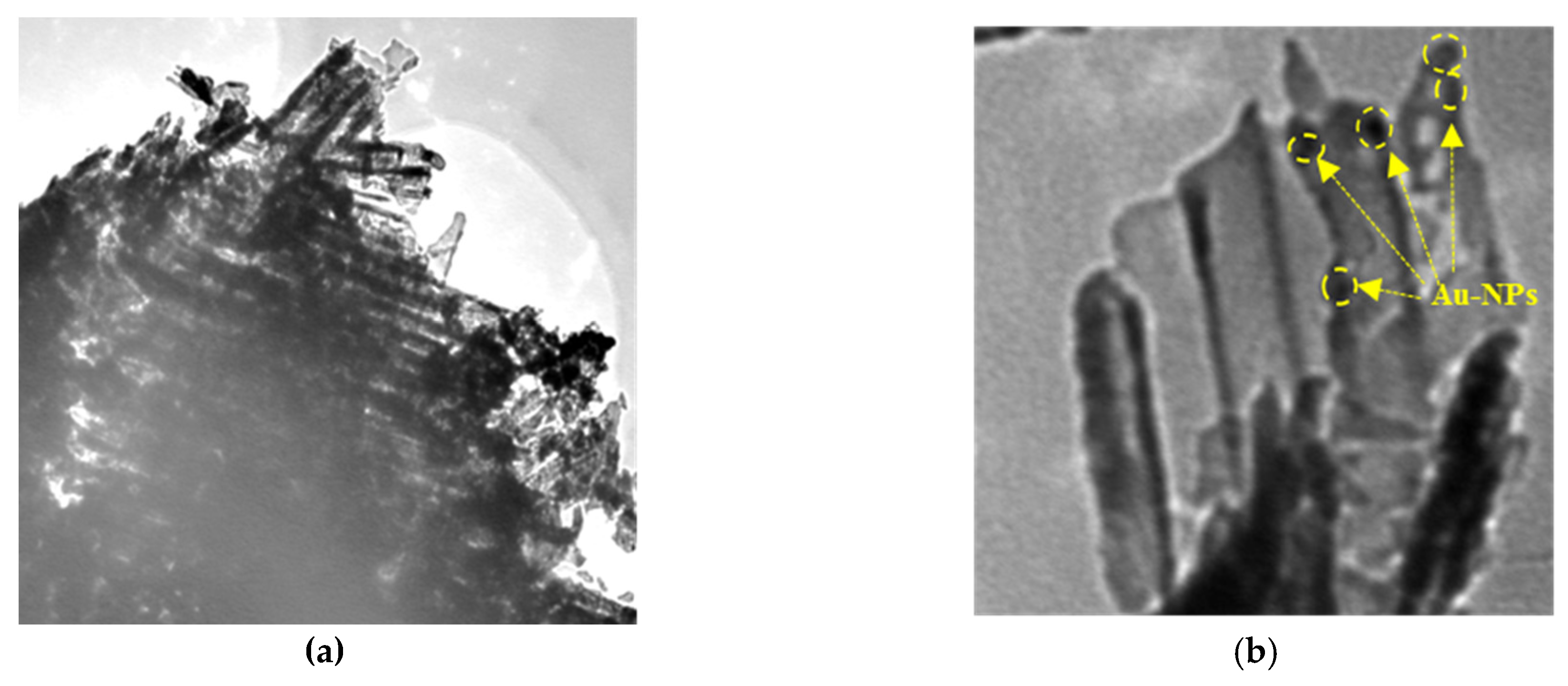
To investigate the growth mechanism of nanotubes, the distribution of gold nanoparticles, and the dimensions of the synthesized nanostructures, the transmission electron microscopy (TEM) was used. According to Figure 4, clearly visible nanotubes were observed, exhibiting an average diameter of approximately 100 nm and lengths of 15 µm.
Additionally, TEM confirms the existence of gold nanoparticles with sizes ranging between 10 and 30 nm (Figure 4). Depositing Au-NPs leads to structural changes, where the deposition covers the nanotube surface and its sidewalls.

2.2. Structural Analysis

XRD Analysis

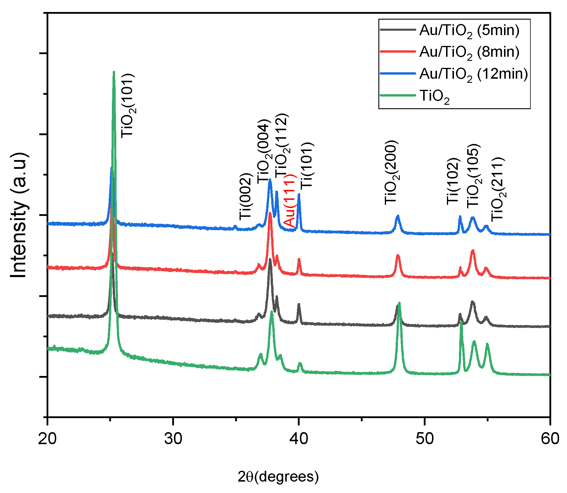
The analysis using X-ray diffraction permits the determination of the crystalline phase, the elemental lattice parameters, and the estimation of the size of the crystallites. Figure 5 shows the XRD patterns of pure TiO2 nanotubes and those decorated with Au-NPs at varying electrophoresis times. All samples underwent an annealing step at 400 °C. The XRD diffractograms showed the domination of anatase phase for all samples, with peaks at 2θ = 25.45°, 37.29°, 38.2°, 47.93°, 53.78°, and 54.93° corresponding to the lattice planes of (101), (004), (112), (200), (105), and (211), respectively. The diffraction peaks at 2θ = 40.28° and 52.85° are associated with the titanium substrate. The peak at the 2θ = 38.2° is indexed to the (111) face of the Au-NPs, according to JCPDS 65-2870 [29]. However, the peak associated with gold cannot be observed until after 12 min of deposition, due to the very low amount of Au incorporated, possibly below the sensitivity threshold of the XRD. The absence of relative peaks of the gold can be explained by the small quantity detected by EDX and the size of the dispersed nanoparticles, as shown by the TEM and EDX analyses (Figure 2, Figure 3 and Figure 4).
After assessing the XRD patterns in Figure 5 it seems that the Au loading has an influence on the crystal facets of TiO2. For bare nanotubes, the preferred orientation of anatase is (101), with the highest peak intensity. As the deposition of Au nanoparticles increases to 5 min, this peak deteriorates to reach ¼ the intensity of the pure nanotubes and continuously decreases with the further addition of Au nanoparticles. The intensity report between the plane peaks (004) and (101) peaks for the 5 min sample, but then slightly decreases and stabilizes, as shown in the table below, which suggests that the Au loading shifts the preferred orientation from (101) to (004) due to competitive interfacial energy minimization and strain mediated reorientation. Meanwhile, the intensity of (112) peak increases as well but decreases for the 8 min sample and then reaches a maximum for 8 min sample. The Au-NPs prefer binding to high energy facets like (004) and (112) [30,31] due to the thermodynamic tendency to minimize their overall energy [32,33]. Moreover, it seems that Au NPs induce compressive strain on TiO2, which is proven by the microstrain calculation, which increases with Au deposition. So, (004) and (112) planes clearly accommodate the strain better than (101), promoting their preferential growth. These facets have already proven their high photocatalytic activity compared to (101) [34] (Table 2).
The XRD patterns are analyzed to determine the crystallite size of the samples according to the Debye–Scherrer equation [35]:
D =   K   λ β   cos Θ
where K is a constant (0.9 for spherical crystallites), β (rad) is the line broadening at half the maximum intensity, λ (nm) is the X-ray wavelength, and ϴ (◦) is the Bragg angle.
The Wilson equation for microstrain determination is as follows [7]:
ε = β 4 tan θ  
The results presented in Table 3 below are related to the structural properties of TiO2 nanotubes before and after the Au nanoparticles deposition. It also seems that the anatase crystallite size increases with time due to increased Au-NPs quantity but then decreases for 12 min Au/TiO2-NTs. The increase in microstrain with Au loading further corroborates lattice distortions, thus leading to defect generation [36,37].

2.3. Optical Analysis

2.3.1. Surface Reflectivity

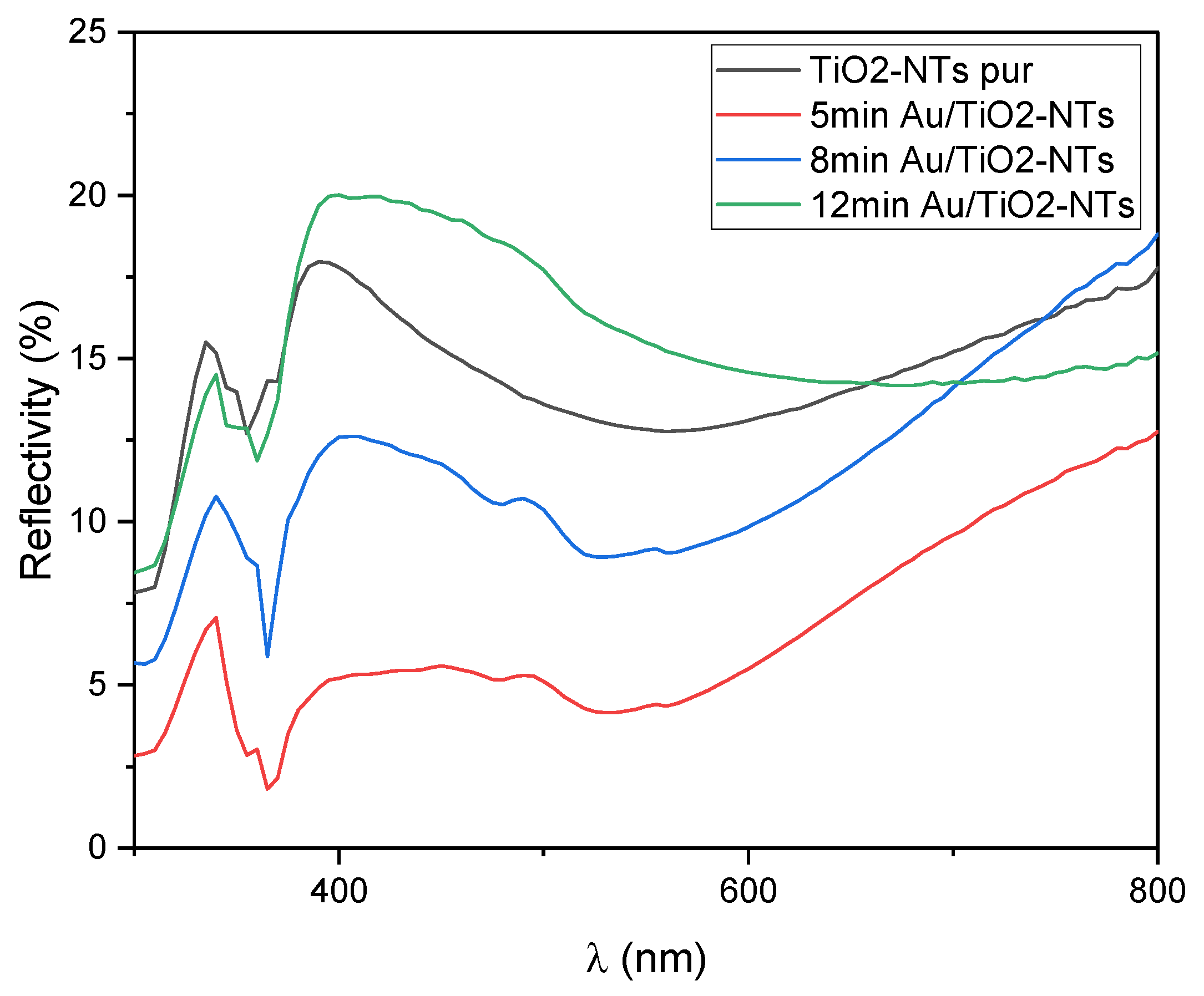
The presence of localized surface plasmon resonance (LSPR) in Au-NPs-decorated TiO2 nanotubes (NTs) directly influences their diffuse reflectivity spectra, which in turn affects the determined band gap energies [38]. Figure 6 demonstrates the altered diffuse reflectance upon Au-NPs decoration compared to pure TiO2-NTs, indicating the interaction of LSPR with the light scattering properties of the material. After major assessment, it is clear to state that the addition of Au nanoparticles influenced optical properties of TiO2-NTs, where the reflectivity diffuse drastically decreases after Au-NPs decoration during 5 min and 8 min compared to pure nanotubes. However, the sample decorated during 12 min exhibited an increase of around 390 nm corresponding to the band gap energy of anatase TiO2-NTs (3.2 eV) [39]. This is mainly due to the obtained dense and rough morphology of the sample after the aggregation of Au-NPs as shown in SEM images [40].
The bandgap for the pure and decorated NTs was determined by analyzing the reflectivity spectrum, employing the Kubelka–Munk function [41]:
F R = 1 R 2 2 R
where F (R) is the Kubelka–Munk function and R is reflection coefficient.
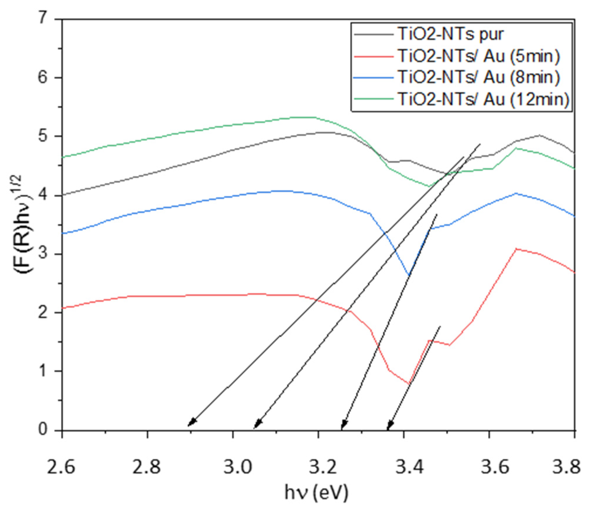
The optical bandgap was determined by analyzing Tauc plots, specifically the relationship between F R h ν 1 / 2 and h ν (photon energy).
The intersection of the tangent line extrapolated from the linear portion of the plot with the x-axis provided the bandgap energy E g = h ν , as illustrated in Figure 7.
Based on the data in Table 3, the reduction in the TiO2 band gap could be attributed to the defect state’s introduction by gold deposition (as seen at 12 min). The LSPR of Au-NPs could also contribute to enhanced light absorption in the visible region. This enhanced absorption, driven by the collective oscillation of electrons in the gold nanoparticles, could modify the shape of the diffuse reflectance spectrum and consequently impact the extrapolation of the band gap energy. Therefore, the determined band gap values, particularly the values obtained for 5 min and 8 min of Au-NP deposition, are not solely a result of defect states but are also modulated by the optical effects of the LSPR phenomenon influencing the overall light interaction with the Au-NPs/TiO2-NTs nanocomposite (Table 4).
This could be supported with Hajjaji et al. study where he evaluated the deposition of Pt-NPs on TiO2 nanotubes. The optical bandgap was reduced as the addition increased. He stated that it could be explained by the presence of induced defects that introduce energy levels in the bandgap of TiO2. This decrease results in an improvement in their response in the visible range and the photocatalytic performance [42].

2.3.2. Photoluminescence Spectroscopy

Photoluminescence spectroscopy was used to elucidate the influence of surface modifications on TiO2 nanotube (NT) charge carrier dynamics and their correlation with photocatalytic performance. This method, predicated on photo-induced electron excitation, facilitates the study of charge transfer and recombination processes.
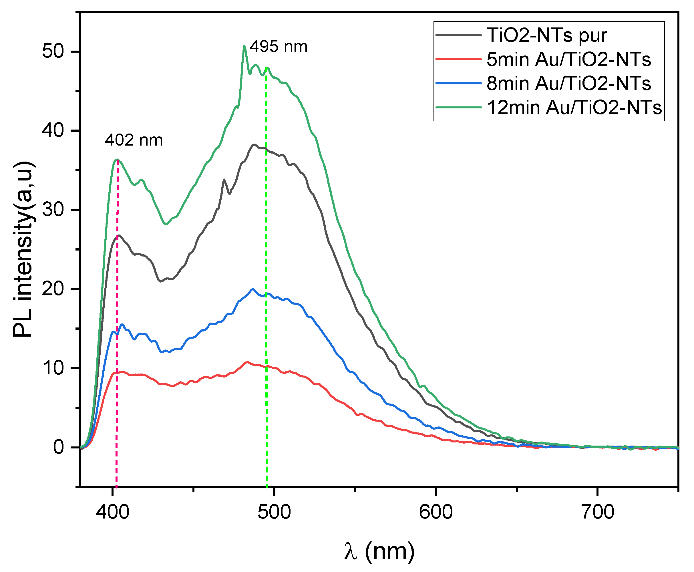
As depicted in Figure 8, the photoluminescence spectra of gold-modified TiO2-NTs exhibited higher intensities compared to the untreated samples, which is consistent with prior research [43]. The spectral features at 402 nm and 495 nm were attributed to band-to-band transitions in TiO2 and interfacial charge transfer between the semiconductor and gold nanoparticles, respectively. The reduced photoluminescence intensity observed for the 5 min deposition sample suggests the formation of a Schottky barrier, characterized by a gold nanoparticle work function exceeding that of TiO2-NTs.
This barrier promotes electron transfer from the semiconductor to the metal, thereby suppressing electron–hole recombination [44].
Conversely, the increased photoluminescence intensity for the 12 min deposition sample, where gold nanoparticles form agglomerates, indicates the presence of an ohmic contact. In this case, the lower work function of the gold agglomerates relative to TiO2-NTs facilitates charge injection from the metal into the semiconductor, leading to a larger electron–hole recombination rate.

2.4. Photocatalytic Application

The degradation of amido black under UV irradiation has been studied using the newly developed nanocomposites. The photocatalytic mechanism can be elucidated through the following equations [43]:
TiO 2 + h ν   e CB + h + VB
O 2 + e O 2 °
h + + H 2 O OH ° + H +
h + + OH OH °
R + h + R °
OH ° + R CO 2 + H 2 O
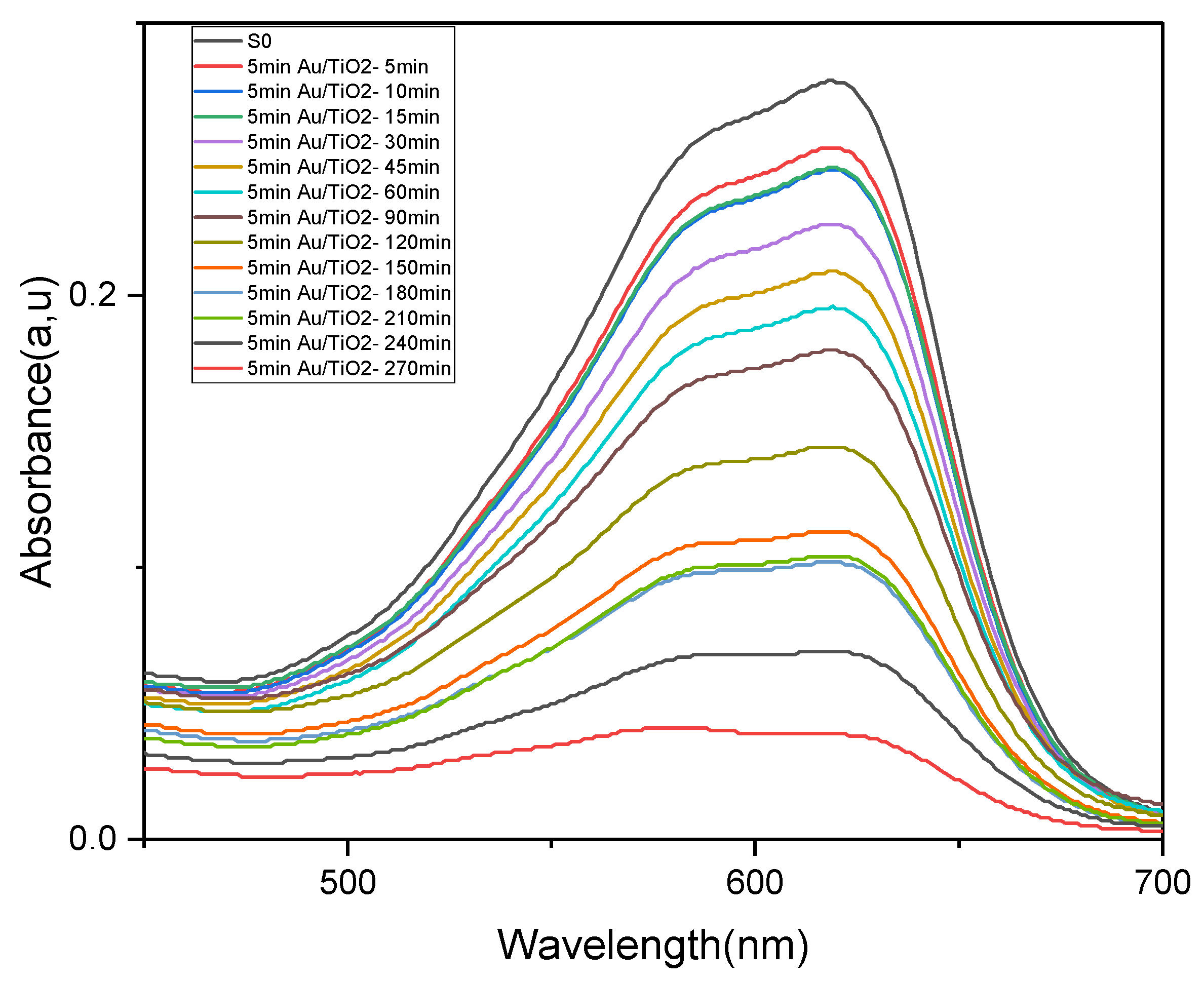
Figure 9 illustrates the absorption spectra of amido black using Au-NPs/TiO2-NTs 5 min deposition time. The decrease in absorption intensity at 618 nm wavelength occurs naturally over the course of irradiation. The degradation efficiency (D) is determined using the equation [45]:
D % = 100 I I initial 100
where II represents the intensity of peak 618 nm and Iinitial is the initial intensity of the amido black solution.
The degradation of amido black achieves approximately 86% within 270 min, as depicted in this table, whereas bare TiO2-NTs present only 78% of amido black degradation efficiency (Table 5).
The relationship between the concentration of amido black and its absorbance can be described using the Beer–Lambert law:
ln I incident I transmited = ε Cl = A
where ε represents the molar absorption coefficient, C is the molar concentration, l is the cuvette profounder, and A is the absorption coefficient. The linearity between concentration and absorbance can be obtained as follows:
A 0 A t = C 0 C t
where A0, A(t), C0, and C(t) represent the absorption and the concentration at t = 0 and t, respectively. By calculating the logarithm of Equation (12), the following relation is obtained:
ln A 0 A = Kt = ln C 0 C
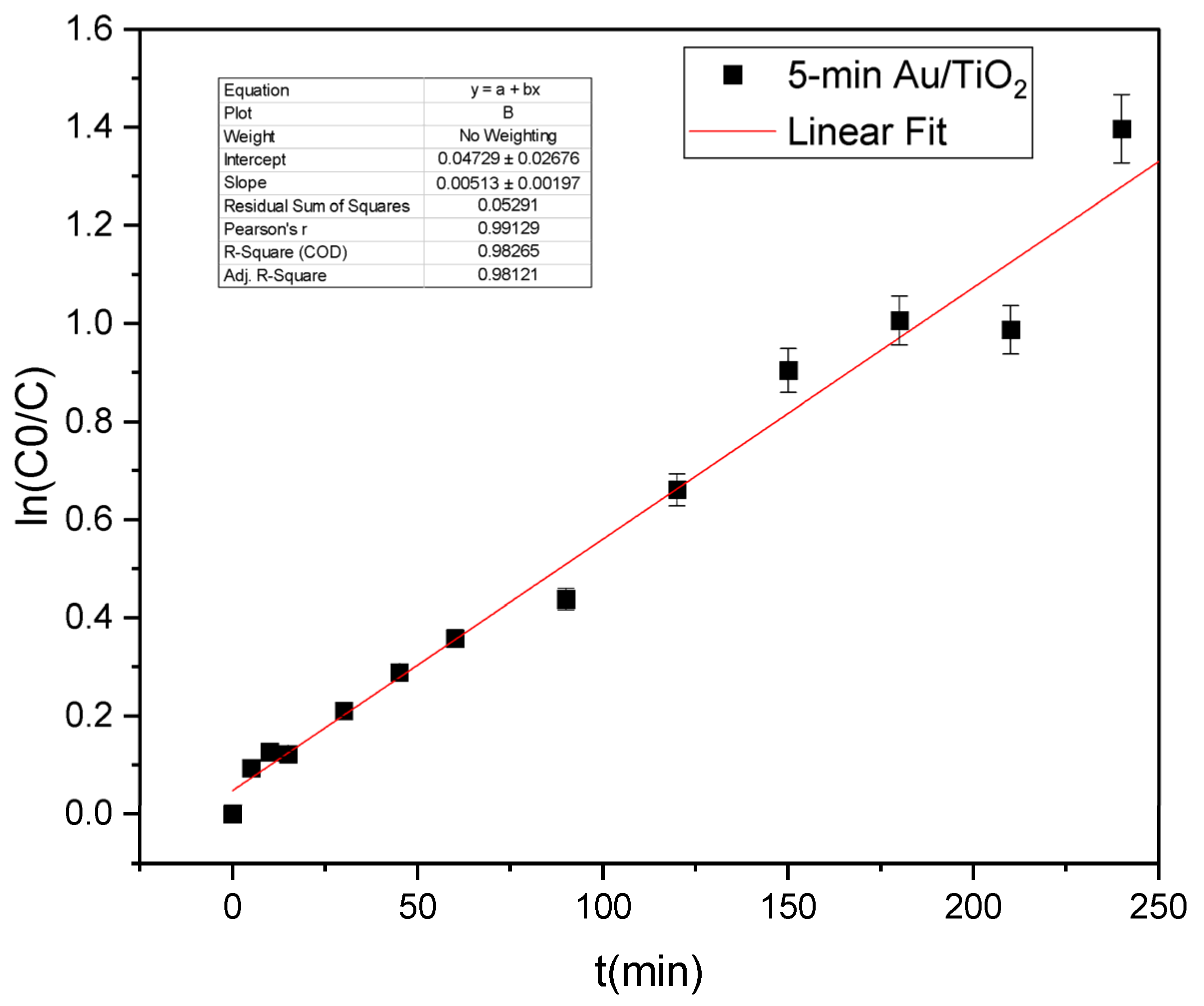
Plotting ln(A0/A) against t yields a linear relationship (Figure 10), where the slope represents the photodegradation reaction rate constant ‘k’. This parameter serves as a key indicator of the photocatalytic efficacy of the sample under investigation.
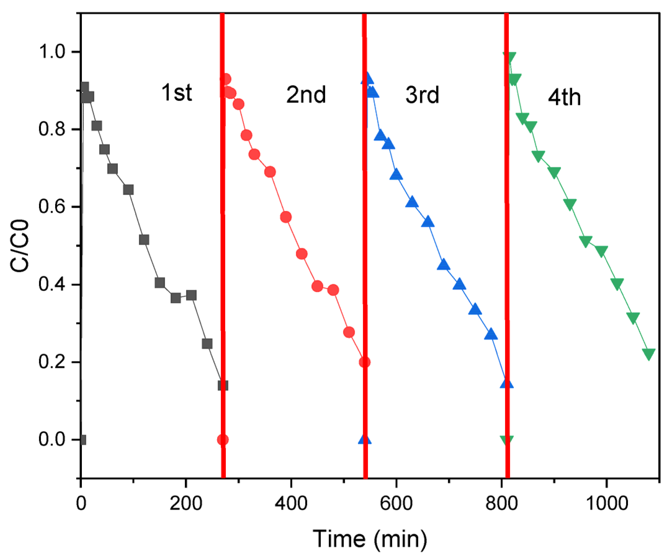
The photocatalytic performance for black amido degradation (Table 6) with 5 min Au/TiO2 nanotubes was repeated four times to determine the degree of reusability of the catalyst; these results (Figure 11) show the degradation efficiency decreased by only 9%.
The highest rate constant observed for the TiO2 sample decorated with Au-NPs for 5 min is approximately 0.005 min−1, which is nearly three times higher than that of the pure sample, indicating its superior activity as a nanocomposite. Compared to the 5 min sample, the other samples showed poor performances, which could be due to the observed agglomeration in the SEM images where the Au-NPs formed giant clusters blocking the opening of the nanotubes, thereby preventing the light from dispersing inside. However, poorly formed Schottky junctions between Au and TiO2 could also be responsible for the decreased photodegradation activity in the 8 min and 12 min samples. The substantial results can be attributed to the charge separation phenomena, which begins with the absorption of light by titanium dioxide nanotubes, leading to the photogeneration of electron–hole pairs; meanwhile, Au-NPs absorb UV light and generate localized surface plasmon resonances (LSPR). LSPR is a collective oscillation of conduction electrons in the metal nanoparticles, resulting in the enhancement of the local electromagnetic field around the nanoparticles.
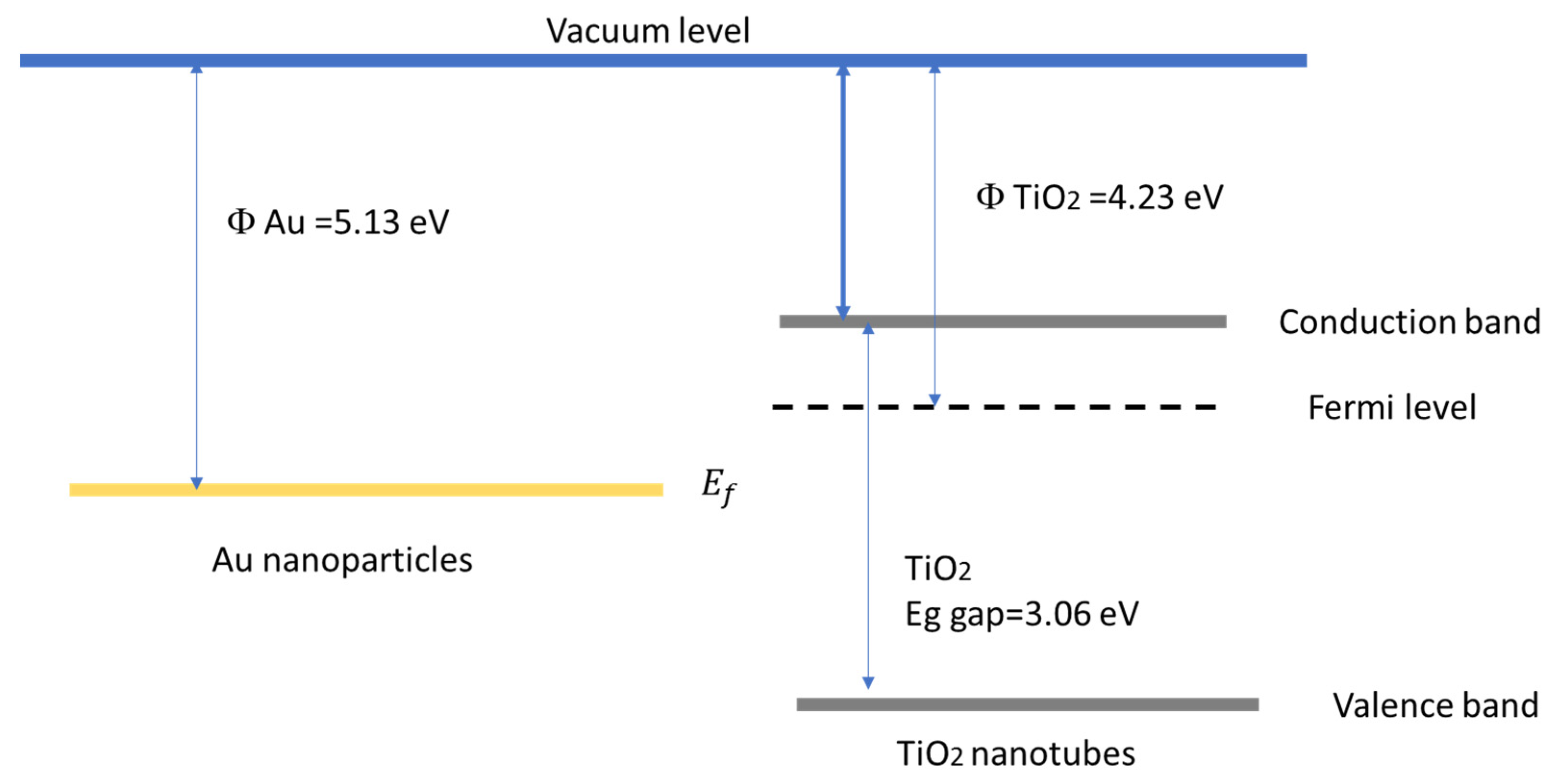
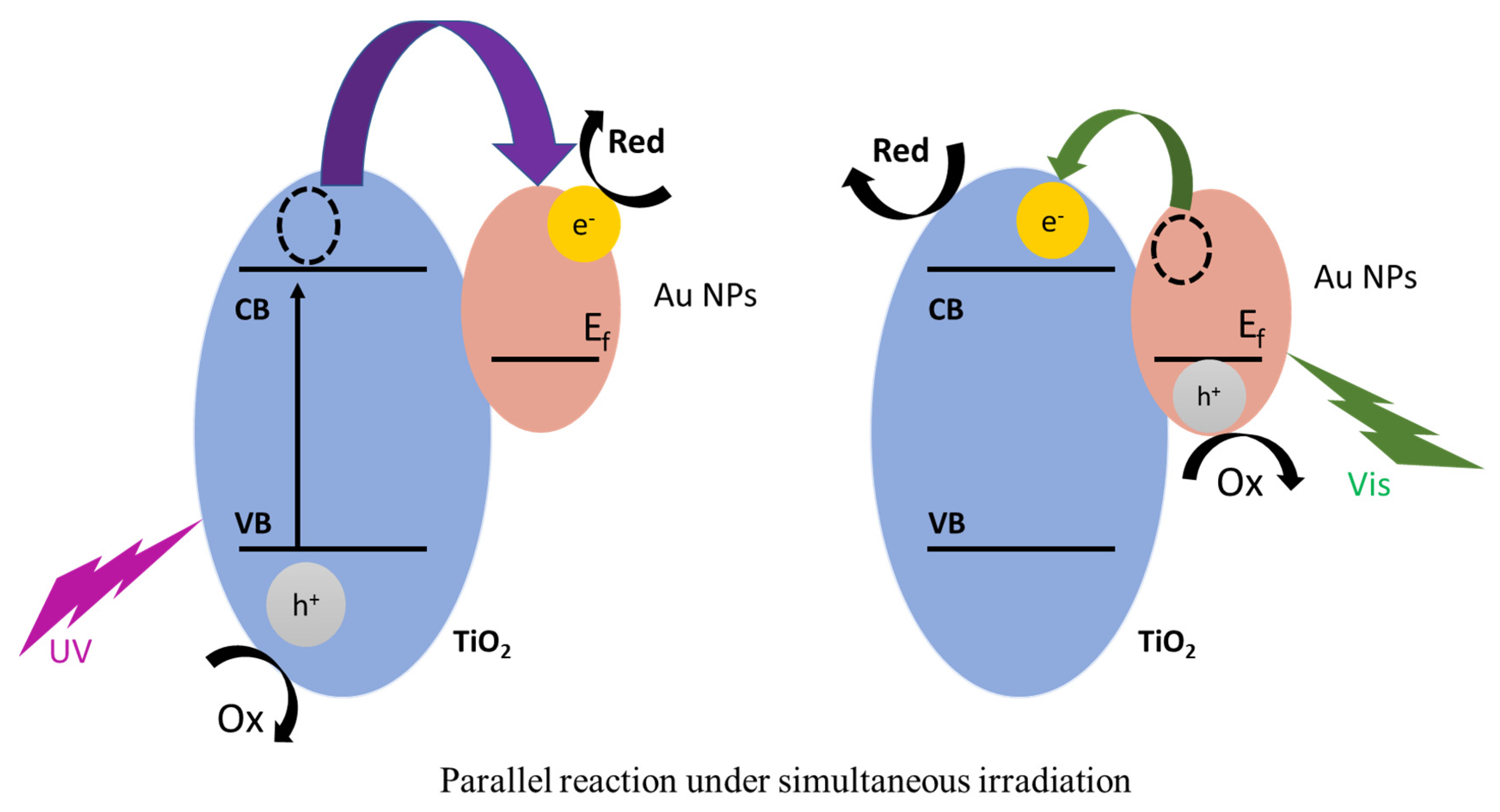
According to the literature, for spherically shaped Au nanoparticles with a size of approximately 25 nm and predominant (111) facets, the work function is ranged between 5.1 and 5.3 eV [48], which is lower than anatase TiO2 nanotubes with a work function of 4.2–4.5 eV (Figure 12) [49]. At the heterojunction where the Fermi levels are equilibrated, the electrons transfer from TiO2 (lower work function) to Au (higher work function) creating a Schottky barrier at the interface. The excitation of TiO2 (~3.2 eV) under UV irradiation photogenerates e in the conduction band and h+ in the valence band; furthermore, the Au/TiO2 interface where the Schottky barrier resembles an electron sink induces the transfer of photoexcited electron from the conduction band of TiO2 to Au-NPs, thereby reducing their recombination. The remaining h+ in the valence band participate in the oxidation reaction for the organic pollutant degradation (Figure 13) [50]. However, the Au-NPs generally exhibit localized surface plasmon resonance LSPR in the visible range light spectrum around 520–550 nm [51]; alas, its contribution to light absorption is minimal since it does not directly absorb UV light, therefore the enhancement of LSPR is low, but it can generate sufficient energetic charge carriers “hot electrons” to participate in redox reactions [52]. Since the used UV irradiation is about 256 nm in wavelength, which is more than enough to create hot electrons capable of injection and overcome the Schottky barrier, the hot electron yielding is very low, almost negligible since most of the photons are absorbed by TiO2 [53]. In this case, the loading of Au nanoparticles on the surface of TiO2 nanotubes for 5 min possibly has a synergetic effect, extending charge separation by trapping electrons and reducing e/h+ recombination, as confirmed by PL analysis; additionally, it generates ROS by increasing •O2 production with injected hot electrons (Figure 14), thus demonstrating the duality of photocatalysis performed by both species, due to the optimal amount of loading at the perfect size and shape, compared to the other samples where aggregation of Au NPs on the surface is observed, as seen in the SEM and TEM analysis. As reported previously by other studies, Ping She et al. investigated the size effect of Au NSs nanospheres loaded on ZnO NRs nanorods on the photocatalytic activity, with 40 nm Au NSs exhibiting optimal performance due to a balance between competing mechanisms [54]. Smaller Au NSs (<40 nm) enhances charge separation through favorable Fermi level alignment and greater interfacial contact, facilitating electron transfer from ZnO to Au, while larger Au NSs (>40 nm) strengthens plasmonic effects and light harvesting but reduces charge separation efficiency. The 40 nm Au NSs strike an ideal compromise, maximizing both charge separation and light utilization, which synergistically boosts the generation of reactive oxygen species (•O2 and •OH) for organic pollutant degradation. This size-dependent optimization highlights the importance of tailoring Au NS dimensions to harmonize electronic and optical properties in plasmonic–semiconductor photocatalysts [55].
However, these samples might demonstrate enhanced photocatalytic performance under visible light. This is because larger or aggregated Au nanoparticles often exhibit a red-shifted LSPR peak, which improves their absorption within the visible range and increases light-harvesting efficiency for visible light-driven reactions [56,57].
The direct contact between the nanotubes and gold nanoparticles facilitates the rapid transfer of electrons from TiO2 to Au-NPs, enhancing the efficiency of redox reactions, the electrons transferred to Au-NPs can participate in the reduction reaction of adsorbed oxygen to superoxide radicals ( O 2 °   , O2•-) whereas the holes left in the valence band of TiO2 can oxidize water to hydroxyl radicals (•OH) [58,59]. These are highly reactive species that can oxidize and degrade various organic pollutants.

3. Discussion

In this work, TiO2 nanotubes were successfully synthesized via electrochemical anodization and subsequently decorated with Au nanoparticles using different electrophoresis times. SEM and TEM analyses revealed a self-organized nanotubular structure of TiO2, measuring 15 μm in length and vertically aligned on a titanium substrate. The nanotubes exhibited a wall thickness of approximately 25 nm and a diameter of 75 nm. The presence of gold nanoparticles was confirmed through XRD characterization and EDX spectra. SEM imaging detected the formation of Au nanoparticle agglomerates specially for high deposition times (12 min). The decoration with Au nanoparticles resulted in a reduction in the optical bandgap from 3.1 eV to approximately 2.966 eV, thereby enhancing the absorption of TiO2 nanotubes in the visible spectrum.
The Au-NPs/TiO2-NTs prepared within 5 min exhibited superior photocatalytic activity in the degradation of amido black and having lower energy band gap than TiO2.
The enhanced photocatalytic activity observed in our study is attributed to the plasmonic effect of Au nanoparticles resulting in the charge separation phenomenon. In conclusion, Au-NPs/TiO2-NTs represent one of the most effective photocatalysts for dye degradation.

4. Materials and Methods

4.1. Anodization of Titanium Dioxide Nanotubes

Anodization of the titanium foil was carried out using an electrolyte mixture containing 100 mL ethylene glycol, 1 wt% ammonium fluoride, and 2 vol% water. All experiments were performed at room temperature using a commercially available titanium foil (2.5 cm × 1 cm × 1 mm, 99.7%) as the anode and a platinum wire as the cathode by applying DC voltage using an Aplab DC power (Mumbai, India) supply. Prior to anodizing, the surface was polished and cleaned to ensure orderly growth of the nanotubes. The samples are polished with SiC abrasive papers (Hammam Sousse, Tunisia) in grades 320 to 2200. The substrates were then ultrasonically cleaned and washed with acetone, ethanol, and distilled water, each for 10 min, then dried under a stream of N2 gas for a few seconds. The preparation of TiO2 nanotubes by the anodic oxidation method involves a two-stage anodization process. The first stage of the anodizing process was carried out at 60 V for 45 min. After 45 min, the grown titanium nanotubes were ultrasonically stripped in an ethanol solution for 15 min.
The peeled titanium nanotube layer now behaves as a template for titanium nanotube growth on the substrate permitting better structural formation and uniform growth. Next, the second anodization was carried out at 60 V at room temperature for 120 min using the same electrolyte mixture. Finally, the samples were washed with ethanol and were annealed for 3 h at 400 °C (heating and cooling rate of 5 °C/min) to ensure the anatase crystal structure [60,61].

4.2. Electrophoresis of Gold Nanoparticles on Titania Nanotubes

Gold nanoparticles (commercial gold nanoparticles with 5 nm particle size dispersed in PBS, Sigma-Aldrich) were deposited on TiO2 nanotubes using the electrophoresis technique. This involved the electrophoretic deposition of gold nanoparticle suspensions onto a TiO2 substrate in a glass cell at ambient temperature. The deposition vessel contained 10 mL of the gold suspension. Two meticulously polished titanium electrodes were positioned vertically within the deposition vessel at a distance of 10 mm. By applying a direct current (DC) voltage of 20 V [26], gold nanoparticles were deposited onto the titanium electrode substrate for different deposition times of 5, 8, and 12 min.

4.3. Characterization of Au-NPs/TiO2-NTs

The structural properties of pure and decorated TiO2 nanotubes samples were analyzed using an X-ray diffractometer (Philips X’PERTMPD, Eindhoven, The Netherlands) to in the 2θ range (20– 80°), with CuKα radiation (λ = 1.54060 Å). The optical properties were investigated by UV–Vis-IR and photoluminescence (PL) spectroscopies by means of a PerkinElmer Lambda 950 (Waltham, MA, USA) spectrophotometer between 200 and 1200 nm and Perkin Elmer LS55 (Waltham, MA, USA) equipped with a xenon lamp at an excitation wavelength of λ = 340 nm. The samples morphology and the elemental composition were studied using scanning electron microscopy (SEM, TESCAN VEGA3) coupled with energy-dispersive X-ray spectroscopy (EDS, TESCAN VEGA3, TESCAN Orsay Holding, Brno, Czech Republic). The grain size, shape, and polydispersity were assessed by transmission electron microscopy (JEM—100CX2, JEOL, Tokyo, Japan).

4.4. Photocatalytic Degradation of Amido Black Dye

In this section, we aim to investigate how altering the electrophoresis duration of Au-NPs deposition impacts the photocatalytic activity of TiO2. Therefore, an amido black solution was used as the inorganic pollutant to be tested. The dye, (500 mL, pioneer forensics LLC, Greeley, CO, USA) is usually used in crime scenes and has the chemical formula: 4-amino-5-hydroxy-3-[(4-nitrophenyl) azo]-6-(phenylazo)-2,7-naphthalenedisulphonic acid disodium salt. To perform photocatalytic tests, UV–visible spectroscopy served as the tool to monitor pollutant degradation over varying exposure durations. The measurements were performed in the (400–700 nm) range to avoid the pollutant’s negligible absorption beyond 900 nm. By analyzing the absorption spectra and deriving kinetic constants, the photocatalytic efficacy of TiO2 decorated with Au-NPs was studied [46].
The experimental procedure is outlined below: The specimens were submerged in a cuvette filled with 10 mL of an aqueous solution of amido black, with a concentration of 5.10–3 M, prepared using ultrapure water as the solvent. A germicidal lamp, with an electrical power rating of 16 W, emitting primarily in the ultraviolet range at a wavelength of 256 nm, was employed.
Before subjecting the samples to irradiation, the specimens underwent a 10 min period of darkness to establish an equilibrium in adsorption–desorption between the solution and TiO2 nanotubes surfaces. Subsequently, they were exposed to UV light for varying durations (5, 10, 15, 30, 45, 60, 90, 120, 150, 180, 210, 240, and 270 min). Prior to each irradiation period, the samples underwent a rinse with ultrapure water, followed by immersion in a cuvette filled with ultrapure water under UV light for 10 min to clean the surface.

Author Contributions

Conceptualization, B.M.S. and A.H.; methodology, A.B.; validation, A.G., R.B.Z., A.H., and A.B.; investigation, H.B., R.B.Z., and S.S.; writing—original draft preparation, H.B. and S.S.; writing—review and editing, L.K., A.H., A.G., B.M.S., and A.H.; supervision, A.B. and A.H.; project administration, L.K.; funding acquisition, L.K. All authors have read and agreed to the published version of the manuscript.

Funding

This work has been funded by the Conselleria d’Innovació, Universitats, Ciència i Societat Digital (Generalitat Valenciana) under Prometeus 2023 program, grant CIPROM2022/03.

Data Availability Statement

Data are contained within the article.

Conflicts of Interest

The authors declare no conflicts of interest.

References

  1. Macak, J.M.; Tsuchiya, H.; Ghicov, A.; Yasuda, K.; Hahn, R.; Bauer, S.; Schmuki, P. TiO2 nanotubes: Self-organized electrochemical formation, properties and applications. Curr. Opin. Solid State Mater. Sci. 2007, 11, 3–18. [Google Scholar] [CrossRef]
  2. Galstyan, V.; Macak, J.M.; Djenizian, T. Anodic TiO2 nanotubes: A promising material for energy conversion and storage. Appl. Mater. Today 2022, 29, 101613. [Google Scholar] [CrossRef]
  3. Carp, O.; Huisman, C.L.; Reller, A. Photoinduced reactivity of titanium dioxide. Prog. Solid State Chem. 2004, 32, 33–177. [Google Scholar] [CrossRef]
  4. Shan, A.Y.; Ghazi, T.I.M.; Rashid, S.A. Immobilisation of titanium dioxide onto supporting materials in heterogeneous photocatalysis: A review. Appl. Catal. A Gen. 2010, 389, 1–8. [Google Scholar] [CrossRef]
  5. Chauhan, A.; Verma, R.; Kumari, S.; Sharma, A.; Shandilya, P.; Li, X.; Batoo, K.M.; Imran, A.; Kulshrestha, S.; Kumar, R. Photocatalytic dye degradation and antimicrobial activities of Pure and Ag-doped ZnO using Cannabis sativa leaf extract. Sci. Rep. 2020, 10, 7881. [Google Scholar] [CrossRef]
  6. Peleyeju, M.G. Electrochemical and Solar Photoelectrocatalytic Oxidation of Selected Organic Compounds at Carbon-Semiconductor Based Electrodes. Ph.D. Thesis, University of Johannesburg, Johannesburg, South Africa, 2017. [Google Scholar]
  7. Sassi, S.; Trabelsi, K.; El Jery, A.; Abidi, M.; Hajjaji, A.; Khezami, L.; Karrech, A.; Gaidi, M.; Soucase, B.M.; Bessais, B. Synergistic effect of CuxOy-NPs/TiO2-NTs heterostructure on the photodegradation of amido black staining. Optik 2023, 272, 170234. [Google Scholar] [CrossRef]
  8. Wang, J.; Sun, S.; Ding, H.; Chen, W.; Liang, Y. Preparation of a composite photocatalyst with enhanced photocatalytic activity: Smaller TiO2 carried on SiO2 microsphere. Appl. Surf. Sci. 2019, 493, 146–156. [Google Scholar] [CrossRef]
  9. Baaloudj, O.; Nasrallah, N.; Kebir, M.; Khezami, L.; Amrane, A.; Assadi, A.A. A comparative study of ceramic nanoparticles synthesized for antibiotic removal: Catalysis characterization and photocatalytic performance modeling. Environ. Sci. Pollut. Res. 2021, 28, 13900–13912. [Google Scholar] [CrossRef]
  10. Hou, W.; Cronin, S.B. A review of surface plasmon resonance-enhanced photocatalysis. Adv. Funct. Mater. 2013, 23, 1612–1619. [Google Scholar] [CrossRef]
  11. Kunwar, S.; Sui, M.; Pandey, P.; Gu, Z.; Pandit, S.; Lee, J. Improved configuration and LSPR response of platinum nanoparticles via enhanced solid state dewetting of In-Pt bilayers. Sci. Rep. 2019, 9, 1329. [Google Scholar] [CrossRef] [PubMed]
  12. Xie, S.; Choi, S.-I.; Xia, X.; Xia, Y. Catalysis on faceted noble-metal nanocrystals: Both shape and size matter. Curr. Opin. Chem. Eng. 2013, 2, 142–150. [Google Scholar] [CrossRef]
  13. Trabelsi, K.; Hajjaji, A.; Gaidi, M.; Bessais, B.; El Khakani, M.A. Enhancing the photoelectrochemical response of TiO2 nanotubes through their nanodecoration by pulsed-laser-deposited Ag nanoparticles. J. Appl. Phys. 2017, 122, 064503. [Google Scholar] [CrossRef]
  14. Wu, L.-P.; Wang, W.-G.; Mo, D.; Duan, J.-L.; Li, X.-J. Effect of Au position on the photoelectrochemical and photocatalytic activity of TiO2 nanotubes under UV irradiation. Inorg. Nano-Met. Chem. 2024, 54, 1154–1162. [Google Scholar] [CrossRef]
  15. Hajjaji, M.; Missaoui, K.; Trabelsi, K.; Bouzaza, A.; Hajjaji, A.; Bessais, B.; Assadi, A. Platinum nanoparticles decorated TiO2 nanotubes for VOCs and bacteria removal in simulated real condition: Effect of the deposition method on the photocatalytic degradation process efficiency. J. Photochem. Photobiol. A Chem. 2025, 458, 115975. [Google Scholar] [CrossRef]
  16. Hajjaji, M.A.; Missaoui, K.; Trabelsi, K.; Bouzaza, A.; Bessais, B.; Hajjaji, A.; Assadi, A.A. Electrodeposited Platinum Nanoparticles on Highly Ordered Titanium Dioxide Nanotubes for Photocatalytic Application: Enhancement of Photocatalytic Degradation of Amido Black Dye. Catal. Lett. 2024, 154, 1242–1254. [Google Scholar] [CrossRef]
  17. Bera, S.; Lee, J.E.; Rawal, S.B.; Lee, W.I. Size-dependent plasmonic effects of Au and Au@ SiO2 nanoparticles in photocatalytic CO2 conversion reaction of Pt/TiO2. Appl. Catal. B Environ. 2016, 199, 55–63. [Google Scholar] [CrossRef]
  18. LEI, B.; XUE, J.; JIN, D.; NI, S.; SUN, H. Fabrication, annealing, and electrocatalytic properties of platinum nanoparticles supported on self-organized TiO2 nanotubes. Rare Met. 2008, 27, 445–450. [Google Scholar] [CrossRef]
  19. Moon, K.-S.; Choi, E.-J.; Bae, J.-M.; Park, Y.-B.; Oh, S. Visible light-enhanced antibacterial and osteogenic functionality of Au and Pt nanoparticles deposited on TiO2 nanotubes. Materials 2020, 13, 3721. [Google Scholar] [CrossRef]
  20. Tian, M.; Wu, G.; Chen, A. Unique electrochemical catalytic behavior of Pt nanoparticles deposited on TiO2 nanotubes. Acs Catal. 2012, 2, 425–432. [Google Scholar] [CrossRef]
  21. Yamaguchi, M.; Abe, H.; Ma, T.; Tadaki, D.; Hirano-Iwata, A.; Kanetaka, H.; Watanabe, Y.; Niwano, M. Bactericidal activity of TiO2 nanotube thin films on Si by photocatalytic generation of active oxygen species. Langmuir 2020, 36, 12668–12677. [Google Scholar] [CrossRef] [PubMed]
  22. Fornari, A.M.D.; de Araujo, M.B.; Duarte, C.B.; Machado, G.; Teixeira, S.R.; Weibel, D.E. Photocatalytic reforming of aqueous formaldehyde with hydrogen generation over TiO2 nanotubes loaded with Pt or Au nanoparticles. Int. J. Hydrog. Energy 2016, 41, 11599–11607. [Google Scholar] [CrossRef]
  23. Kowalska, E.; Mahaney, O.O.P.; Abe, R.; Ohtani, B. Visible-light-induced photocatalysis through surface plasmon excitation of gold on titania surfaces. Phys. Chem. Chem. Phys. 2010, 12, 2344–2355. [Google Scholar] [CrossRef]
  24. Zielińska-Jurek, A. Progress, Challenge, and Perspective of Bimetallic TiO2-Based Photocatalysts. J. Nanomater. 2014, 2014, 208920. [Google Scholar] [CrossRef]
  25. Lincho, J.; Mazierski, P.; Klimczuk, T.; Martins, R.C.; Gomes, J.; Zaleska-Medynska, A. TiO2 nanotubes modification by photodeposition with noble metals: Characterization, optimization, photocatalytic activity, and by-products analysis. J. Environ. Chem. Eng. 2024, 12, 112990. [Google Scholar] [CrossRef]
  26. Leordean, C.; Marta, B.; Gabudean, A.-M.; Focsan, M.; Botiz, I.; Astilean, S. Fabrication of highly active and cost effective SERS plasmonic substrates by electrophoretic deposition of gold nanoparticles on a DVD template. Appl. Surf. Sci. 2015, 349, 190–195. [Google Scholar] [CrossRef]
  27. Besra, L.; Liu, M. A review on fundamentals and applications of electrophoretic deposition (EPD). Prog. Mater. Sci. 2007, 52, 1–61. [Google Scholar] [CrossRef]
  28. Hosseingholilou, S.; Dorranian, D.; Ghoranneviss, M. Characterization of gold nanoparticle thin film prepared by electrophoretic deposition method. Gold Bull. 2020, 53, 1–10. [Google Scholar] [CrossRef]
  29. Lee, M.-S.; Hong, S.-C.; Kim, D. Fabrication of patterned gold electrodes with spin-coated-and-fired Au (1 1 1) film by the soft lithography. Appl. Surf. Sci. 2006, 252, 5019–5025. [Google Scholar] [CrossRef]
  30. Tian, F.H.; Wang, X.; Zhao, W.; Zhao, L.; Chu, T.; Yu, S. Adsorption of 2-propanol on anatase TiO2 (101) and (001) surfaces: A density functional theory study. Surf. Sci. 2013, 616, 76–84. [Google Scholar] [CrossRef]
  31. Tanner, A.J.; Robin, K.; Helen, H.F.; Geoff, T. Chemical Modification of Polaronic States in Anatase TiO2 (101). J. Phys. Chem. C 2021, 125, 14348–14355. [Google Scholar] [CrossRef]
  32. Zhang, Q.; Wang, H. Facet-dependent catalytic activities of Au nanoparticles enclosed by high-index facets. ACS Catal. 2014, 4, 4027–4033. [Google Scholar] [CrossRef]
  33. Xiao, C.; Lu, B.-A.; Xue, P.; Tian, N.; Zhou, Z.-Y.; Lin, X.; Lin, W.-F.; Sun, S.-G. High-index-facet-and high-surface-energy nanocrystals of metals and metal oxides as highly efficient catalysts. Joule 2020, 4, 2562–2598. [Google Scholar] [CrossRef]
  34. Ye, L.; Liang, Y. First principles study on band gap modulation of TiO2 (112) surface for enhancing optical properties. Phys. B Condens. Matter 2024, 674, 415579. [Google Scholar] [CrossRef]
  35. Chowdhury, R.I.; Hossen, M.A.; Mustafa, G.; Hussain, S.; Rahman, S.N.; Farhad, S.F.U.; Murata, K.; Tambo, T.; Islam, A.B.M.O. Characterization of chemically deposited cadmium sulfide thin films. Int. J. Mod. Phys. B 2010, 24, 5901–5911. [Google Scholar] [CrossRef]
  36. A, J.K.; Kekuda, D. Performance analysis of a DC magnetron sputtered Cu2O/TiO2 heterojunction photodetector for short-wavelength detection. Sens. Actuators A. Phys. 2025, 388, 116517. [Google Scholar]
  37. Shabalina, A.; Fakhrutdinova, E.; Chen, Y.-W.; Lapin, I. Preparation of gold-modified F, N-TiO2 visible light photocatalysts and their structural features comparative analysis. J. Sol-Gel Sci. Technol. 2015, 75, 617–624. [Google Scholar] [CrossRef]
  38. Khezami, L.; Lounissi, I.; Hajjaji, A.; Guesmi, A.; Assadi, A.A.; Bessais, B. Synthesis and characterization of TiO2 nanotubes (TiO2-NTs) decorated with platine nanoparticles (Pt-NPs): Photocatalytic performance for simultaneous removal of microorganisms and volatile organic compounds. Materials 2021, 14, 7341. [Google Scholar] [CrossRef] [PubMed]
  39. Sassi, S.; Bouich, A.; Hajjaji, A.; Khezami, L.; Bessais, B.; Soucase, B.M. Cu-Doped TiO2 Thin Films by Spin Coating: Investigation of Structural and Optical Properties. Inorganics 2024, 12, 188. [Google Scholar] [CrossRef]
  40. Sassi, S.; Bouich, A.; Bessais, B.; Khezami, L.; Soucase, B.M.; Hajjaji, A. Comparative Analysis of Anodized TiO2 Nanotubes and Hydrothermally Synthesized TiO2 Nanotubes: Morphological, Structural, and Photoelectrochemical Properties. Materials 2024, 17, 5182. [Google Scholar] [CrossRef] [PubMed]
  41. Yuwono, A.H.; Sofyan, N.; Kartini, I.; Ferdiansyah, A.; Pujianto, T.H. Nanocrystallinity enhancement of TiO2 nanotubes by post-hydrothermal treatment. Adv. Mater. Res. 2011, 277, 90–99. [Google Scholar] [CrossRef]
  42. Zare, M.; Mortezaali, A.; Shafiekhani, A. Photoelectrochemical determination of shallow and deep trap states of platinum-decorated TiO2 nanotube arrays for photocatalytic applications. J. Phys. Chem. C 2016, 120, 9017–9027. [Google Scholar] [CrossRef]
  43. Perillo, P.M.; Rodríguez, D.F. Anodization growth of self-organized ZrO2 nanotubes on zircaloy-4. Evaluation of the photocatalytic activity. Matéria 2015, 20, 627–635. [Google Scholar] [CrossRef]
  44. Saquib, M. TiO2-mediated photocatalytic degradation of a triphenylmethane dye (gentian violet), in aqueous suspensions. Dye. Pigment. 2003, 56, 37–49. [Google Scholar] [CrossRef]
  45. Lin, Z.; Wang, X.; Liu, J.; Tian, Z.; Dai, L.; He, B.; Han, C.; Wu, Y.; Zeng, Z.; Hu, Z. On the role of localized surface plasmon resonance in UV-Vis light irradiated Au/TiO2 photocatalysis systems: Pros and cons. Nanoscale 2015, 7, 4114–4123. [Google Scholar] [CrossRef] [PubMed]
  46. Trabelsi, K.; Jemai, S.; El Jery, A.; Sassi, S.; Guesmi, A.; Khezami, L.; Hajjaji, A.; Gaidi, M.; Bessais, B. Ag-NPs coating influence on TiO2-NTs photocatalytic performances on Amido Black staining. Res. Sq. 2022, 1–19. [Google Scholar] [CrossRef]
  47. Fu, C.; Li, M.; Li, H.; Li, C.; Wu, X.G.; Yang, B. of Au nanoparticle/TiO2 hybrid films for photoelectrocatalytic degradation of methyl orange. J. Alloys Compd. 2017, 692, 727–733. [Google Scholar] [CrossRef]
  48. Musa, I.; Ghabboun, J. Work function, electrostatic force microscopy, tunable photoluminescence of gold nanoparticles, and plasmonic interaction of gold nanoparticles/rhodamine 6G nanocomposite. Plasmonics 2024, 20, 2531–2540. [Google Scholar] [CrossRef]
  49. Surah, S.S.; Vishwakarma, M.; Kumar, R.; Nain, R.; Sirohi, S.; Kumar, G. Tuning the electronic band alignment properties of TiO2 nanotubes by boron doping. Results Phys. 2019, 12, 1725–1731. [Google Scholar] [CrossRef]
  50. Yu, Y.; Wen, W.; Qian, X.-Y.; Liu, J.-B.; Wu, J.-M. UV and visible light photocatalytic activity of Au/TiO2 nanoforests with Anatase/Rutile phase junctions and controlled Au locations. Sci. Rep. 2017, 7, 41253. [Google Scholar] [CrossRef]
  51. Koushki, E.; Mowlavi, A.A.; Hoseini, S.T. Application of Localized Surface Plasmon Resonance of Conjugated Gold Nanoparticles in Spectral Diagnosis of SARS-CoV-2: A Numerical Study. Plasmonics 2023, 18, 1847–1855. [Google Scholar] [CrossRef]
  52. Manuel, A.P.; Shankar, K. Hot electrons in TiO2–noble metal nano-heterojunctions: Fundamental science and applications in photocatalysis. Nanomaterials 2021, 11, 1249. [Google Scholar] [CrossRef]
  53. Kumar, A.; Choudhary, P.; Kumar, A.; Camargo, P.H.C.; Krishnan, V. Recent advances in plasmonic photocatalysis based on TiO2 and noble metal nanoparticles for energy conversion, environmental remediation, and organic synthesis. Small 2022, 18, 2101638. [Google Scholar] [CrossRef]
  54. Okuno, T.; Kawamura, G.; Muto, H.; Matsuda, A. Photocatalytic properties of Au-deposited mesoporous SiO2–TiO2 photocatalyst under simultaneous irradiation of UV and visible light. J. Solid State Chem. 2016, 235, 132–138. [Google Scholar] [CrossRef]
  55. She, P.; Xu, K.; Zeng, S.; He, Q.; Sun, H.; Liu, Z. Investigating the size effect of Au nanospheres on the photocatalytic activity of Au-modified ZnO nanorods. J. Colloid Interface Sci. 2017, 499, 76–82. [Google Scholar] [CrossRef] [PubMed]
  56. Lee, S.Y.; Kang, D.; Jeong, S.; Do, H.T.; Kim, J.H. Photocatalytic degradation of rhodamine B dye by TiO2 and gold nanoparticles supported on a floating porous polydimethylsiloxane sponge under ultraviolet and visible light irradiation. ACS Omega 2020, 5, 4233–4241. [Google Scholar] [CrossRef]
  57. Wu, L.; Li, F.; Xu, Y.; Zhang, J.W.; Zhang, D.; Li, G.; Li, H. Plasmon-induced photoelectrocatalytic activity of Au nanoparticles enhanced TiO2 nanotube arrays electrodes for environmental remediation. Appl. Catal. B Environ. 2015, 164, 217–224. [Google Scholar] [CrossRef]
  58. Gomes, J.; Lincho, J.; Domingues, E.; Quinta-Ferreira, R.M.; Martins, R.C. N–TiO2 photocatalysts: A review of their characteristics and capacity for emerging contaminants removal. Water 2019, 11, 373. [Google Scholar] [CrossRef]
  59. Hajjaji, A.; Jemai, S.; Rebhi, A.; Trabelsi, K.; Gaidi, M.; Alhazaa, A.; Al-Gawati, M.; El Khakani, M.; Bessais, B. Enhancement of photocatalytic and photoelectrochemical properties of TiO2 nanotubes sensitized by SILAR-Deposited PbS nanoparticles. J. Mater. 2020, 6, 62–69. [Google Scholar] [CrossRef]
  60. Abidi, M.; Hajjaji, A.; Bouzaza, A.; Trablesi, K.; Makhlouf, H.; Rtimi, S.; Assadi, A.; Bessais, B. Simultaneous removal of bacteria and volatile organic compounds on Cu2O-NPs decorated TiO2 nanotubes: Competition effect and kinetic studies. J. Photochem. Photobiol. A Chem. 2020, 400, 112722. [Google Scholar] [CrossRef]
  61. Ali, I.; Alharbi, O.M.L.; Alothman, Z.A.; Badjah, A.Y. Kinetics, thermodynamics, and modeling of amido black dye photodegradation in water using Co/TiO2 nanoparticles. Photochem. Photobiol. 2018, 94, 935–941. [Google Scholar] [CrossRef]
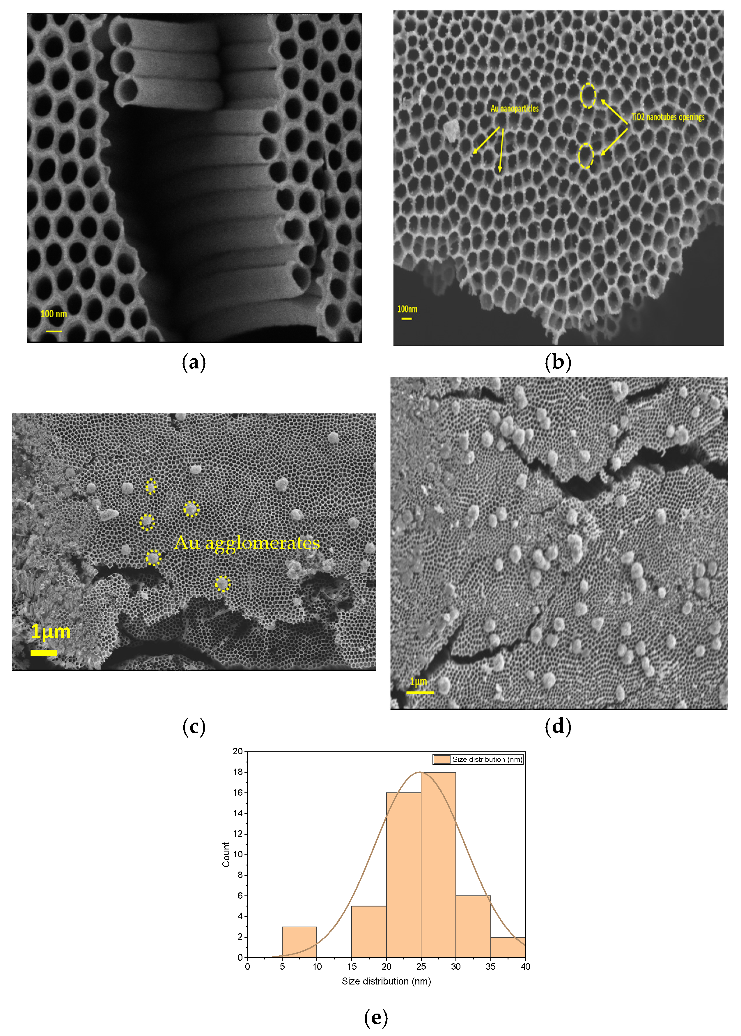
Figure 1. SEM images of (a) TiO2 nanotubes and TiO2 nanotubes decorated with Au nanoparticles during different times: (b) 5 min (Scalebar = 100 nm); (c) 8 min (Scalebar = 1 μm); (d) 12 min (Scalebar = 1 μm); and (e) size distribution of Au spherical nanoparticles.
Catalysts 15 00781 g001
Figure 2. EDX mapping of gold nanoparticles with different deposition times: (a) 5 min; (b) 8 min; and (c) 12 min, respectively.
Catalysts 15 00781 g002
Figure 3. EDX results of TiO2 nanotubes decorated with Au nanoparticles: (a) 5 min; (b) 8 min; and (c) 12 min.
Catalysts 15 00781 g003
Figure 4. TEM images of (a) TiO2 nanotube and (b) TiO2 nanotubes decorated with Au-NPs during 5 min.
Catalysts 15 00781 g004
Figure 5. X-ray diffraction patterns of pure TiO2-NTs decorated with Au-NPs.
Catalysts 15 00781 g005
Figure 6. The diffuse reflectance spectrum of pure and decorated TiO2 nanotubes.
Catalysts 15 00781 g006
Figure 7. Gap energy determination for different samples.
Catalysts 15 00781 g007
Figure 8. Photoluminescence spectra of pure and decorated TiO2-NTs.
Catalysts 15 00781 g008
Figure 9. Absorption spectra of amido black at different UV irradiation time in presence Au-NPs (5 min)/TiO2-NTs.
Catalysts 15 00781 g009
Figure 10. The logarithm plots of Au-NPs (5 min)/TiO2-NTs.
Catalysts 15 00781 g010
Figure 11. Reusability of the 5 min Au/TiO2 nanotubes for four photocatalytic cycles.
Catalysts 15 00781 g011
Figure 12. Schematic illustration of TiO2-NTs and Au-NPs before heterojunction.
Catalysts 15 00781 g012
Figure 13. Schematic illustration of TiO2 modified Au-NPs, photocatalytic mechanism under UV light irradiation.
Catalysts 15 00781 g013
Figure 14. Schematic illustration of possible charge behavior in Au/TiO2 under simultaneous irradiation of UV and Vis light [54].
Catalysts 15 00781 g014
Table 1. EDX analysis for TiO2 nanotubes decorated with Au nanoparticles at different times.
Atomic %
SamplesTitanium (Ti)Oxygen (O)Gold (Au)
5 min Au/TiO2-NTs62.5132.610.13
8 min Au/TiO2-NTs59.0334.610.16
12 min Au/TiO2-NTs58.3138.130.17
Table 2. XRD peaks intensity report of anatase TiO2 nanotubes.
TiO2-NTs5 Min
Au/TiO2-NTs
8 Min
Au/TiO2-NTs
12 Min
Au/TiO2-NTs
I(004)/I(101)0.210.910.830.82
I(112)/I(101)0.0640.420.290.64
Table 3. Crystallite size of pure and decorated TiO2 NTs at different deposition time.
Samples2θ (°)FWHM (rad)D (nm)Microstrain (%) × 10−3
Bare TiO2-NTs25.1560.002267622.5
5 min Au/TiO2-NTs25.1540.00218964.22.4
8 min Au/TiO2-NTs25.1560.00217164.72.4
12 min Au/TiO2-NTs25.1420.00247756.72.8
Table 4. Bandgap values of pure and decorated TiO2 NTs at different deposition time.
SamplesTiO2-NTs TiO2 NTs/Au
(5 min)
TiO2 NTs/Au
(8 min)
TiO2 NTs/Au
(12 min)
Band gap (eV)3.063.363.252.89
Table 5. The degradation efficiency of amido black at different UV irradiation time in presence Au-NPs (5 min)/TiO2-NTs.
Time(s)51530456090120150180210240270
D%8.8611.4718.9925.0930.1035.4848.3859.4963.4462.7275.2786.02
Table 6. Comparison of catalysts for the photodegradation of amido black AB.
CatalystConditionsPhotodegradation
Efficiency (%)
Photodegradation Time (min)Ref
Pt-NPs/TiO2-NTs[Black Amido] = 5 mg L−1
[catalyst] = thin film (2.5 × 2.5 cm)
UV lamp = 15 W (256 nm)
9790[16]
Ag-NPs/TiO2-NTs[Black Amido] = 5 mg L−1
[catalyst] = thin film (2.5 × 2.5 cm2)
UV lamp = 15 W (256 nm)
96.4270[46]
Au-NPs/TiO2-NTs[Methyl Orange] = 5.2 mg L−1
[catalyst] = thin film (Ti wires Ø = 1.5 mm and L = 6 cm)
UV lamp = 350 W (365 nm)
72.5200[47]
Au-NPs/TiO2-NTs[Black Amido] = 5 mg L−1
[catalyst] = thin film (2.5 × 1 cm2)
UV lamp = 15 W (256 nm)
86270This work
Disclaimer/Publisher’s Note: The statements, opinions and data contained in all publications are solely those of the individual author(s) and contributor(s) and not of MDPI and/or the editor(s). MDPI and/or the editor(s) disclaim responsibility for any injury to people or property resulting from any ideas, methods, instructions or products referred to in the content.