Article

Plasmon-Driven Catalytic Inhibition of pATP Oxidation as a Mechanism for Indirect Fe²⁺ Detection on a SERS-Active Platform

by
Alexandru-Milentie Hada
1,
Mihail-Mihnea Moruz
1,2,
Alexandru Holca
1,2,
Simion Astilean
1,2,
Marc Lamy de la Chapelle
1,3,* and
Monica Focsan
1,2,*
1
Nanobiophotonics and Laser Microspectroscopy Centre, Interdisciplinary Research Institute on Bio-Nano-Sciences, Babes-Bolyai University, Treboniu Laurian Street, 400271 Cluj-Napoca, Romania
2
Biomolecular Physics Department, Faculty of Physics, Babes-Bolyai University, Mihail Kogalniceanu Street, 400084 Cluj-Napoca, Romania
3
Institut des Molécules et Matériaux du Mans (IMMM–UMR6283), Université du Mans, Olivier Messiaen Avenue, 72085 Le Mans, France
*
Authors to whom correspondence should be addressed.
Catalysts 2025, 15(7), 667; https://doi.org/10.3390/catal15070667
Submission received: 23 May 2025 / Revised: 26 June 2025 / Accepted: 7 July 2025 / Published: 8 July 2025

Abstract

The detection of Fe2+ in environmental water sources is critical due to its biological relevance and potential toxicity at elevated levels. Herein, we report a plasmon-driven catalytic sensing nanoplatform based on p-aminothiophenol (pATP)-functionalized silver nanoparticles (AgNPs) for the selective and sensitive detection of Fe2+. The nanoplatform exploits the inhibition of the plasmon-driven catalytic conversion of pATP to 4,4-dimercaptoazobenzene (DMAB), monitored via surface-enhanced Raman scattering (SERS) spectroscopy. The catalytic efficiency was quantified by the intensity ratio between the formed DMAB-specific Raman band and the common aromatic ring vibration band of pATP and DMAB. This ratio decreased proportionally with increasing Fe2+ concentration over a range of 100 µM to 1.5 mM, with a calculated limit of detection of 39.7 µM. High selectivity was demonstrated against common metal ions, and excellent recovery rates (96.6–99.4%) were obtained in real water samples. Mechanistic insights, supported by chronopotentiometric measurements under light irradiation, revealed a competitive oxidation pathway in which Fe2+ preferentially consumes plasmon-generated hot holes over pATP. This mechanism clarifies the observed catalytic inhibition and supports the design of redox-responsive SERS sensors. The platform offers a rapid, low-cost, and portable solution for Fe2+ monitoring and holds promise for broader applications in detecting other redox-active analytes in complex environmental matrices.

1. Introduction

Plasmonic nanostructures, particularly those based on noble metals such as silver (Ag) and gold (Au), have emerged as highly versatile platforms for initiating and investigating light-driven chemical transformations at metal–solution interfaces. Upon irradiation with visible light, these materials support localized surface plasmon resonance (LSPR), a collective oscillation of conduction band electrons that leads to intense local electromagnetic field enhancement at the nanoparticle (NP) surface [1]. The decay of these plasmons through non-radiative pathways results in the generation of energetic charge carriers, commonly referred to as “hot electrons” and “hot holes” [2]. These species are capable of initiating or accelerating chemical reactions by transiently occupying molecular orbitals of adsorbed species, providing a unique approach to catalysis that is fundamentally distinct from thermal or classical electrochemical mechanisms [3].
The concept of plasmon-driven catalysis has attracted widespread interest due to its ability to lower activation barriers, enable room-temperature reactions, and provide spatial and temporal control through light modulation [4,5,6,7,8]. A variety of experimental model systems have been proposed to study these light-induced catalytic phenomena, with one of the most established being the oxidative coupling of para-aminothiophenol (pATP) to 4,4-dimercaptoazobenzene (DMAB) on the surface of plasmonic NPs [9,10,11,12,13,14,15]. This reaction has become a benchmark in the field of plasmonic catalysis for several reasons. First, both the reactant and product are Raman-active molecules with well-defined and distinct vibrational signatures, allowing for direct, real-time monitoring via surface-enhanced Raman spectroscopy (SERS) [14]. Second, the reaction is surface-mediated and strongly dependent on the optical and electronic properties of the NP substrate [16], making it an excellent probe for evaluating plasmonic activity.
The conversion of pATP to DMAB is generally understood to occur via a light-induced oxidation mechanism, where hot holes generated by plasmon decay are involved in removing electrons from pATP, promoting its coupling into the azo-bonded dimer. The presence of oxygen and/or surface-bound reactive species can modulate this pathway [17], further indicating its redox-sensitive nature. Recent studies have suggested that the rate and efficiency of this transformation are highly dependent on NP morphology [18], ligand environment [19], and excitation wavelength [20]. In addition, because hot holes are a finite resource generated during plasmon decay, any competitive processes at the NP surface, such as the presence of other oxidizable species, can significantly influence the outcome of the pATP-to-DMAB reaction. Based on this sensitivity to the surrounding chemical environment, plasmon-driven catalytic reactions, such as the pATP-to-DMAB transformation, have been increasingly explored as the foundation for developing responsive sensing platforms [21,22]. In particular, catalysis-based SERS sensors enable the indirect detection of analytes by monitoring changes in the catalytic conversion efficiency of surface-bound probe molecules. Unlike conventional SERS sensors that rely on the direct spectral signature of the analyte itself [23,24,25], this indirect approach utilizes the modulation of a well-characterized plasmon-induced reaction as the readout. Any species that interferes with, competes in, or promotes the catalytic pathway can influence the reaction dynamics, leading to quantifiable changes in the resulting SERS signal.
Among redox-active species of environmental and biological relevance, Fe2+ is of particular significance due to its dual role as an essential micronutrient and a potential contaminant at elevated concentrations [26]. Iron in its divalent form is chemically unstable in aqueous environments, undergoing oxidation to Fe3+ [27], especially in the presence of dissolved oxygen [28]. This oxidation is thermodynamically favorable and can occur readily under ambient conditions. Given its prevalence in both natural and industrial water systems, reliable detection of Fe2+ is crucial for ensuring water quality and public health [29]. Conventional detection techniques, including colorimetric assays [30], atomic absorption spectroscopy [31], and electrochemical analysis [32], offer good sensitivity but often require costly instrumentation or sample pre-treatment or are not easily adapted for field deployment. While a previous study [22] employed plasmon-driven catalysis for Fe2+ detection via the p-nitrothiophenol (pNBT)-to-DMAB reduction on AuNPs, no mechanistic explanation was provided for the observed catalytic behavior, highlighting the need for deeper insight into such systems.
Therefore, in this work, we report a catalysis-based sensing platform using pATP-functionalized AgNPs for the selective detection of Fe2+ ions. The system exploits the inhibition of the plasmon-driven pATP-to-DMAB conversion as the sensing mechanism, monitored through characteristic SERS band intensities and quantified via catalytic efficiency. We demonstrate that this approach enables sensitive and selective detection of Fe2+ in both controlled and real water samples. Importantly, a key novelty of this study lies in the mechanistic investigation of the sensing process, where electrochemical measurements reveal a competitive oxidation pathway between Fe2+ and pATP at the nanoparticle surface. This mechanistic insight not only clarifies the origin of the observed catalytic inhibition but also provides a conceptual framework for the design of future catalysis-based SERS sensors. By understanding how redox-active species interact with plasmon-generated charge carriers, this work advances the broader development of plasmonic sensing platforms tailored for diverse environmental and analytical applications.

2. Results and Discussion

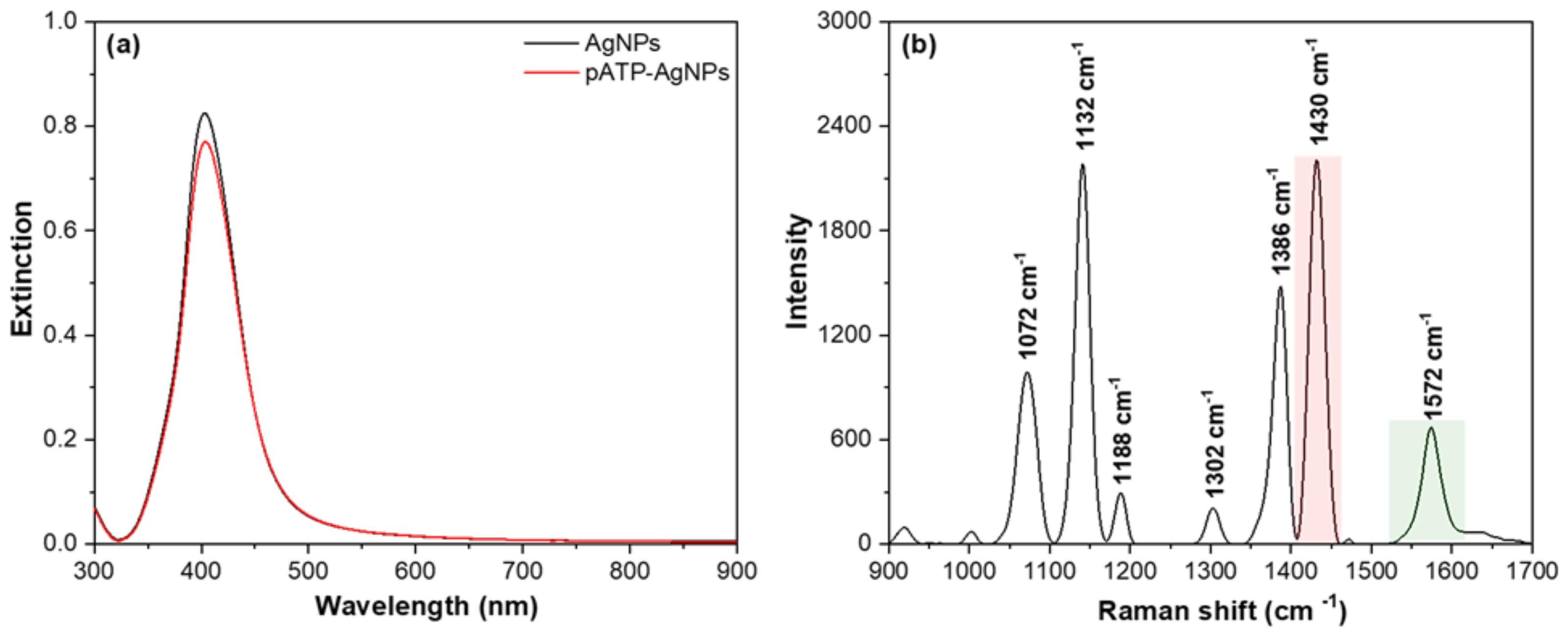
2.1. Synthesis of AgNPs and Functionalization with pATP

The synthesized AgNPs exhibit a single, well-defined LSPR band centered at 402 nm, with a full width at half maximum (FWHM) of 55 nm (Figure 1a—black line), suggesting a spherical morphology and good monodispersity. The hydrodynamic size of the AgNPs was measured to be 51 ± 4 nm (Figure S1a). Moreover, to confirm the morphology of the AgNPs, scanning electron microscopy (SEM) measurements were performed, and a representative image is shown in Figure S2a. The NPs exhibit a spherical shape with an average diameter of 45 ± 3 nm, based on the analysis of over 100 individual particles. This average size is in good agreement with the values obtained from DLS measurements, considering the standard deviation and the fact that hydrodynamic diameter also accounts for the surface-bound molecules and the solvation layer surrounding the NPs. Additionally, the measured zeta potential of −53 mV confirms the strong electrostatic stabilization of the colloidal suspension (Figure S1b), indicating excellent chemical stability over time. Next, the colloidal solution was incubated with pATP (40 µM) for 1 h, followed by purification, and the extinction spectrum is presented in Figure 1a with a red line.
Following functionalization with pATP, the colloidal AgNP solution exhibited no observable changes in the LSPR profile, demonstrating that the NPs maintained their stability and structural integrity. Interestingly, no significant shift in the LSPR band was observed after pATP functionalization. This behavior is attributed to the sub-monolayer surface coverage of pATP on the AgNPs, which was intentionally optimized to preserve catalytic activity. A densely packed monolayer would likely inhibit the plasmon-driven dimerization process by restricting the spatial proximity and reorientation of adjacent pATP molecules required for coupling into DMAB [20]. The limited surface loading ensures sufficient surface accessibility and catalytic efficiency, while inducing minimal changes to the local dielectric environment, thus explaining the absence of a detectable LSPR shift in the UV-Vis spectra. To further investigate the adsorption of pATP onto the AgNPs surface, zeta potential measurements were performed before and after functionalization (Figure S1b). The bare AgNPs exhibited a zeta potential of −53 mV, consistent with their citrate-stabilized negative surface charge. After incubation with pATP and purification, the zeta potential shifted to −46 mV, indicating a less negative surface. This shift is attributed to the presence of amine groups (–NH2) from pATP molecules adsorbed on the NP surface, which partially neutralize the negative charge. These results support the successful functionalization of AgNPs with pATP, even in the absence of an observable LSPR shift. Moreover, to evaluate potential changes in NP morphology and size after functionalization, SEM measurements were also performed post-conjugation with pATP. As shown in the representative images (Figure S2b), the AgNPs retained their spherical shape and monodispersity after functionalization. A statistical analysis performed on more than 100 NPs revealed an average diameter of 45 ± 4 nm, which is in close agreement with the pre-functionalization average size of 45 ± 3 nm. These results confirm that the pATP adsorption process did not induce aggregation or morphological alteration of the nanoparticles, preserving their structural integrity for plasmon-driven catalysis. Furthermore, SERS measurements were performed using a portable Raman spectrometer with 532 nm laser excitation. The SERS spectrum presented in Figure 1b reveals the fingerprint SERS signal of pATP. Notably, the strong peaks observed at 1072 cm−1 and 1572 cm−1 correspond to the C–S stretching vibration and the aromatic ring C–C stretching, respectively [14]. Additional peaks at 1188 cm−1 and 1302 cm−1 are attributed to C–H bending and C–N stretching vibrations, respectively [19]. Furthermore, the presence of distinct peaks associated with DMAB indicates that a catalytic transformation of pATP occurred during laser irradiation. Specifically, the in-plane C–H bending vibration at 1132 cm−1 and the symmetric and asymmetric N=N stretching vibrations at 1386 cm−1 and 1430 cm−1 [33], respectively, confirm the plasmon-driven catalysis conversion of pATP to DMAB. To quantify the catalytic conversion of pATP to DMAB, the catalytic efficiency was evaluated by calculating the intensity ratio between the DMAB-specific band at 1430 cm−1 (IDMAB) and the common aromatic ring C–C stretching band at 1572 cm−1 (Icommon). This ratio, denoted as
k = I D M A B I c o m m o n
reflects the extent of the plasmon-driven reaction. For the freshly prepared pATP-functionalized AgNP sample, the initial k value, k0, was determined to be 3.3. This parameter will be used as a reference in the following Fe2+ detection experiments to monitor changes in catalytic efficiency.

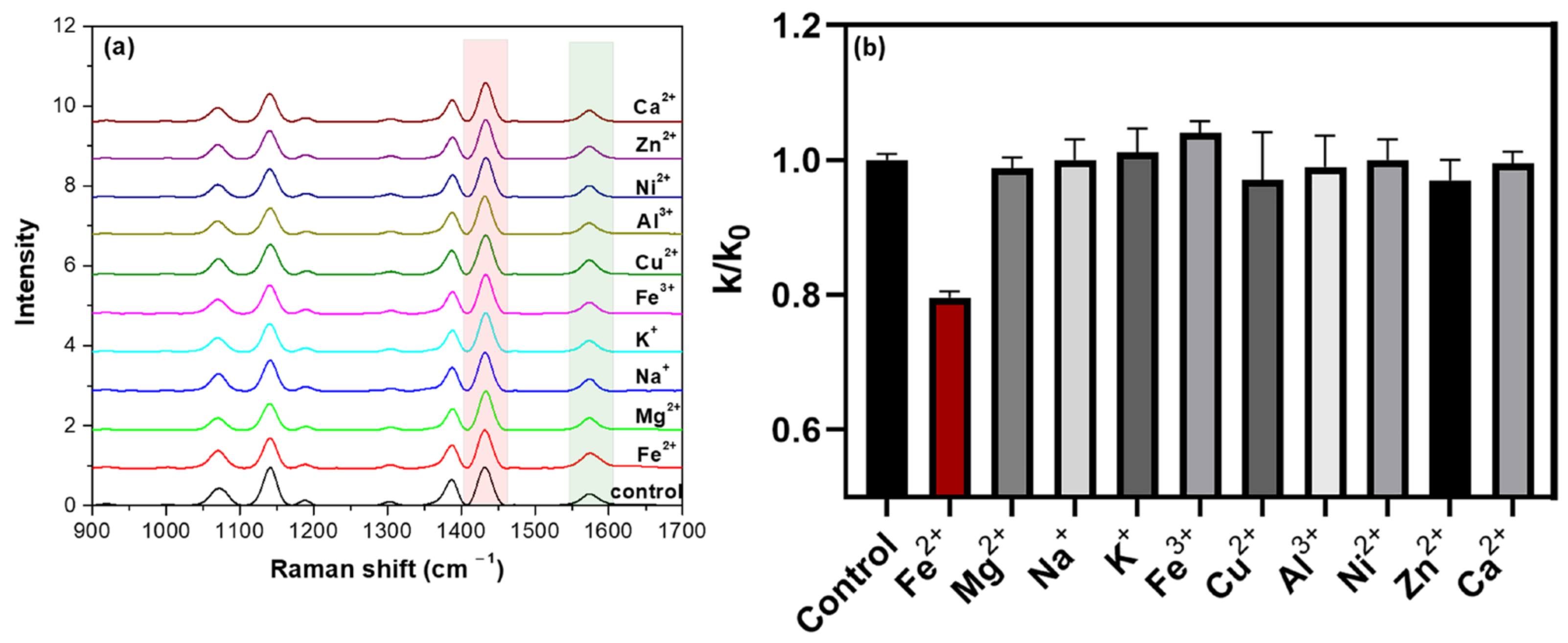
2.2. Detection of Fe2+ Using the Catalysis-Based Sensing Platform

The plasmon-driven catalytic conversion of pATP to DMAB on the surface of AgNPs can be influenced by the surrounding chemical environment. Certain ions or molecules can inhibit or enhance this catalytic process by competing for surface sites or altering local reaction conditions. By exploiting this sensitivity, pATP-functionalized AgNPs can serve as an effective sensing platform (Figure 2a, control) for ion detection. Therefore, to investigate whether specific ions can interfere with the plasmon-driven catalytic conversion of pATP to DMAB, a selectivity assay was performed. The pATP-functionalized AgNP colloidal solution was incubated for 3 min with various metal ions commonly found in aqueous environments, including Mg2+, Na+, K+, Cu2+, Al3+, Ni2+, Zn2+, Fe3+, Ca2+, and Fe2+. After incubation, SERS spectra were recorded for each sample using a portable Raman spectrometer with 532 nm laser excitation (Figure 2a).
The catalytic efficiency (k) was calculated for each condition and normalized to the control value (k0) obtained in the absence of added ions. The resulting k/k0 ratios are shown in Figure 2b. For all ions tested except Fe2+, the catalytic efficiency remained comparable to the control, indicating no significant interference with the catalytic reaction. In contrast, the presence of Fe2+ led to a pronounced decrease in catalytic efficiency, suggesting a strong inhibitory effect. These results demonstrate a clear selectivity of the pATP-AgNP system for Fe2+ ions. The underlying mechanism responsible for this inhibition will be discussed later in this section.
To determine whether the observed catalytic inhibition is dependent on the Fe2+ concentration, a sensitivity assay was conducted. The pATP-functionalized AgNP colloidal solution was incubated for 3 min with varying concentrations of Fe2+, ranging from 100 µM to 5 mM. After incubation, SERS spectra were recorded (Figure 3a) for each Fe2+ concentration. A gradual decrease in the intensity of the DMAB-specific band at 1430 cm−1 compared to the common one at 1572 cm−1 was observed with increasing Fe2+ concentration, indicating a stepwise inhibition of the catalytic conversion of pATP. Simultaneously, a shift in the common aromatic ring’s C–C stretching band was noted: in the absence of Fe2+, this band appears at 1572 cm−1, but at higher Fe2+ concentrations, it progressively shifts to 1582 cm−1. This shift suggests a reorientation of the aromatic ring and indicates that the vibrational signal at 1582 cm−1 is increasingly dominated by unconverted pATP molecules rather than DMAB [20], further supporting the inhibition of the catalytic process. To quantify the extent of this inhibition, the catalytic efficiency (k) was calculated for each sample. These values were normalized to the control efficiency (k0) and plotted as k/k0 versus Fe2+ concentration (Figure 3b).
The resulting calibration plot revealed a linear relationship between k/k0 and Fe2+ concentration in the range of 100 µM to 1.5 mM, with an excellent correlation coefficient (R2 = 0.997). The limit of detection (LOD) was determined to be 39.7 µM. This detection limit is particularly relevant for environmental monitoring, as the World Health Organization recommends a maximum Fe2+ concentration of 35 µM in drinking water [29]. Although the calculated LOD (39.7 µM) slightly exceeds the World Health Organization guideline value for total iron in drinking water (35 µM), the sensor remains suitable for practical applications. It can function as a threshold-based tool to rapidly indicate whether Fe²⁺ levels approach or exceed safe limits. Moreover, Fe2+ is the more bioavailable and reactive iron species, particularly under reducing conditions that are common in groundwater or aging infrastructure, making its selective detection highly relevant. To further improve sensitivity, simple preconcentration strategies such as evaporation or membrane filtration can be applied. Additionally, a prespike method using known Fe2+ additions can enhance detection reliability near the threshold, supporting the use of this platform in semi-quantitative or screening workflows. Thus, the proposed catalytic sensing platform offers a practical tool for identifying Fe2+-contaminated water. Any inhibition of the catalytic activity (k/k0) can serve as a warning that the water may be unsafe for consumption from a Fe2+ detection perspective. Additionally, the relative standard deviation (RSD) for three independent measurements at a Fe2+ concentration of 500 µM was calculated to be as low as 0.76%, demonstrating the high precision and reproducibility of the sensing platform.
To understand the origin of the catalytic inhibition observed in the presence of Fe2+, it is important to consider the redox behavior of this ion. Fe2+ is known to be chemically unstable in aqueous environments, undergoing oxidation to Fe3+ over time. In parallel, the catalytic conversion of pATP to DMAB is a plasmon-driven process, where photoexcited AgNPs relax through the generation of energetic charge carriers, particularly hot holes, which facilitate the oxidation of pATP on the NP surface. However, when Fe2+ ions are introduced into the colloidal solution, a marked inhibition of the catalytic reaction is observed. This suggests that the hot holes generated upon plasmon excitation are being consumed by a competing process: the oxidation of Fe2+ to Fe3+. Therefore, a competitive oxidation mechanism is proposed, in which both Fe2+ and pATP compete for the available hot holes at the NP surface. As the concentration of Fe2+ increases, a greater fraction of these energetic carriers is diverted toward Fe2+ oxidation, resulting in a progressive decrease in catalytic efficiency for pATP conversion. Specifically, since oxidation occurs at the NP surface, the higher the concentration of Fe2+ ions in solution, the greater their density becomes on the NP surface, and thus the more pronounced the capture of hot holes. To support this hypothesis, chronopotentiometric measurements were performed. The experiments involved irradiating the samples with a 530 nm LED on a working electrode while recording the open-circuit potential over time in the absence of any externally applied current. These measurements provide insights into the relative oxidation potentials and help confirm whether Fe2+ oxidation is thermodynamically favored over pATP oxidation under plasmonic excitation. The corresponding data are presented in Figure 4a–c.
Figure 4a compares the potential changes over time under continuous irradiation of ultrapure water and the colloidal solution of AgNPs. In the case of pure water, no significant change in potential is observed over the 300 s irradiation period, confirming the lack of photochemical activity. By contrast, the AgNPs solution displays a gradual negative shift in open-circuit potential, reaching approximately −0.095 V. This shift is attributed not to a redox process but to the aggregation of the NPs, a phenomenon known to influence the electrochemical environment at the electrode surface [34]. Next, Figure 4b examines the redox behavior of Fe2+ in two conditions: as a free aqueous solution (red line) and in the presence of AgNPs (black line). In water alone, Fe2+ undergoes slow oxidation, reflected by a modest potential change of +0.003 V after 300 s. The potential evolves from the initial negative oxygen-dominated state toward a more positive potential as Fe²⁺ oxidizes, which occurs simultaneously with the oxygen depletion at the surface of the working electrode. However, when Fe2+ is incubated with AgNPs, a significantly larger potential increase (+0.063 V) is observed under the same conditions. This positive shift not only opposes the aggregation-driven decrease seen with AgNPs alone but also indicates that plasmonic excitation enhances the oxidation of Fe2+ to Fe3+, supporting the occurrence of a plasmon-driven catalysis process. Furthermore, Figure 4c presents a comparison of three systems: pATP-functionalized AgNPs (black line), Fe2+ with AgNPs (red line), and Fe2+ with pATP-functionalized AgNPs (blue line). For the pATP-AgNP system, the potential decreases to −0.0486 V under irradiation. Although previous SERS analysis confirmed that pATP undergoes oxidation on the surface of AgNPs under 532 nm excitation, the observed negative shift in potential is primarily attributed to NP aggregation. However, this shift is less pronounced than the one observed for unmodified AgNPs (−0.095 V), suggesting that either the aggregation is partially suppressed by the presence of pATP on the surface of the NPs or the oxidation of pATP partially offsets the potential decrease. As pATP oxidation consumes hot holes (which would tend to make the potential more positive), it could counteract the negative shift caused by aggregation, leading to a less negative overall potential. In the Fe2+–AgNP system, we already noted a +0.063 V potential, demonstrating the plasmon-assisted oxidation of Fe2+. The positive shift indicates a net consumption of electrons (or generation of holes) on the electrode surface, consistent with an oxidation reaction. Remarkably, when Fe2+ is introduced to pATP-functionalized AgNPs, the potential shift increases further to +0.093 V. This enhanced shift confirms that Fe2+ oxidation is strongly favored, even in the presence of pATP, further supporting the hypothesis that hot holes generated during plasmon decay are preferentially consumed by Fe2+. These results collectively support the hypothesis of a competitive oxidation mechanism, in which Fe2+ outcompetes pATP for the hot holes generated at the AgNP surface during plasmon excitation. The higher potential difference observed for Fe2+, compared to that of pATP, explains the progressive inhibition of pATP-to-DMAB conversion as Fe2+ concentration increases. Thus, Fe2+ preferentially undergoes oxidation by plasmon-generated hot holes, limiting their participation in the pATP-to-DMAB conversion. This competitive oxidation pathway explains the observed catalytic inhibition and forms the mechanistic basis of the sensor’s selectivity. Moreover, to further rationalize the high selectivity of the sensing platform toward Fe2+, it is essential to also consider the electronic structure and redox properties of the tested metal ions in relation to the plasmon-generated charge carriers involved in the catalytic process. The oxidation potential of Fe2+ aligns well with the energy level of hot holes generated upon plasmonic excitation of AgNPs. From an electronic configuration perspective, Fe2+ possesses partially filled 3d6 orbitals that can readily participate in redox processes. In contrast, ions such as Mg2+, Na+, and K+ have closed-shell configurations (2p6), corresponding to noble gas stability, and are thus less susceptible to further oxidation. While Ni2+ is also redox-active, its oxidation typically requires a higher energy input compared to Fe2+. Conversely, Cu2+ tends to undergo reduction and acts preferentially as an oxidizing agent in redox systems. These distinctions highlight Fe2+ as a particularly suitable candidate for interaction with plasmon-generated hot holes, providing a mechanistic basis for the high selectivity observed in the sensing platform.
Overall, given this competitive oxidation mechanism, the sensing strategy demonstrated in this work can be extended to the detection of other redox-active species. Specifically, any analyte capable of undergoing oxidation and possessing an oxidation potential lower (more favorable) than that of pATP could similarly compete for plasmon-generated hot holes. Under such conditions, the inhibition of the pATP-to-DMAB catalytic conversion would serve as an indirect but sensitive indicator of the presence of the target species, enabling the development of a broader class of plasmon-enhanced catalytic sensors.

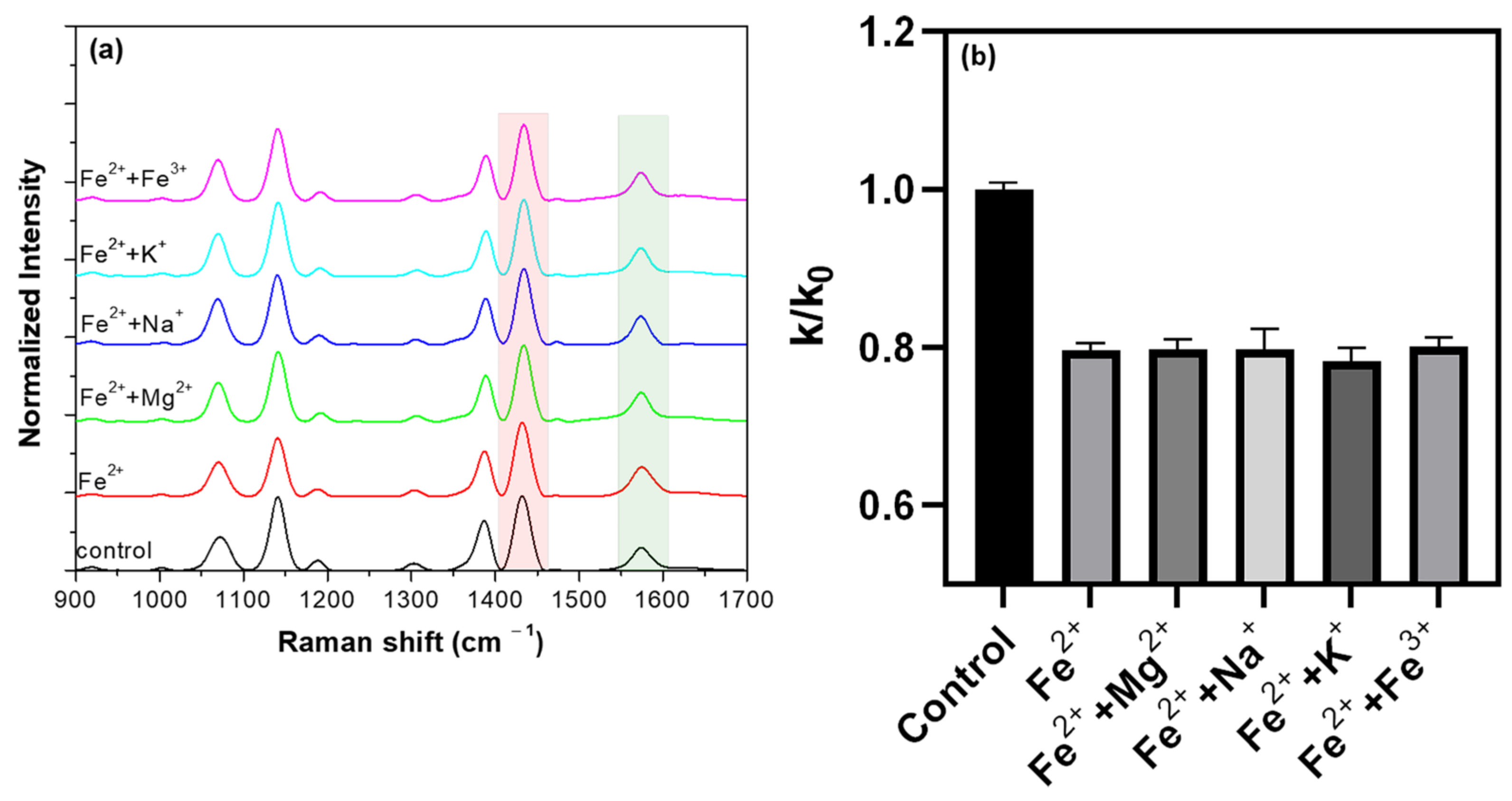
2.3. Competitivity and Real Water Samples Assays for the Detection of Fe2+

Next, to evaluate the ability of the sensing platform to selectively detect Fe2+ in the presence of other metal ions, a competitivity assay was conducted. The pATP-functionalized AgNP colloidal solution was incubated with mixtures containing Fe2+ (500 µM) and one of the most commonly found metal ions in water (Mg2+, Na+, K+, or Fe3+), each at a final concentration of 500 µM. After 3 min of incubation, SERS spectra were acquired under 532 nm laser excitation (Figure 5a).
The catalytic efficiency (k) for each sample was determined and normalized to the control value (k0). The resulting k/k0 ratios (Figure 5b) were then used to calculate the corresponding Fe2+ concentrations based on the calibration curve established in Figure 3b. As summarized in Table S1, the calculated Fe2+ recoveries ranged from 95.8% to 103.0% in the presence of the tested interfering ions. Moreover, to evaluate the sensor’s performance under more realistic conditions, where Mg2+, Na+, K+, or Fe3+ ions are more abundant compared to Fe2+, we conducted a second competitive assay (Figure S3) in which 200 µM Fe2+ was detected in the presence of 1 mM of potentially interfering ions (Na+, K+, Mg2+, and Fe3+). The calculated recoveries ranged between 98% and 101% (Table 1), indicating that the presence of excess common ions does not significantly interfere with Fe2+ detection. These results confirm not only the high selectivity of the sensor toward Fe2+ but also its excellent precision in complex ionic environments, further supporting its applicability in real-world water analysis.
Finally, to evaluate the performance of the sensing platform in complex matrices, a real water sample was analyzed. The pATP-functionalized AgNP colloidal solution was incubated with tap water, and the catalytic efficiency was assessed via SERS measurements. No detectable Fe2+ concentration was observed in the unspiked sample. Subsequently, the tap water was spiked with Fe2+ at two concentrations (0.5 mM and 1 mM) and incubated with the sensing colloidal solution under the same conditions (Table 2).
The relative catalytic efficiencies (k/k0) were extracted and used to calculate the Fe2+ concentrations using the previously established calibration curve. The calculated recoveries ranged from 96.6% to 99.4%, confirming the high precision and accuracy of the sensor in a real water matrix. It is noteworthy that the Fe2+ oxidation in the presence of dissolved oxygen might be a real problem for the accuracy of the proposed sensor. However, it can be stabilized for reliable detection through simple preservation strategies such as acidification. Specifically, immediate acidification of water samples significantly slows down oxidation kinetics by inhibiting electron transfer to O2. This widely used approach allows for accurate Fe2+ determination even in oxygenated environments, supporting the practical applicability of the sensor for real sample analysis. Therefore, these findings demonstrate that the developed catalytic sensing platform is suitable for environmental applications, offering a rapid, low-cost, and portable method for detecting hazardous levels of Fe2+ in real water samples.

3. Materials and Methods

3.1. Materials

Ascorbic acid, sodium citrate, silver nitrate (AgNO3), 4-aminothiophenol (pATP), iron (II) chloride (FeCl2), magnesium chloride (MgCl2), sodium chloride (NaCl), potassium chloride (KCl), iron (III) chloride (FeCl3), copper chloride (CuCl2), aluminum chloride (AlCl3), nickel chloride (NiCl2), calcium chloride (CaCl2), and zinc chloride (ZnCl2) were acquired from Sigma-Aldrich (Saint Louis, MO, USA). All experiments were conducted using analytical-grade reagents. Solutions were prepared with ultrapure water (resistivity ≥18 MΩ·cm) obtained from a Milli-Q purification system (Millipore, Merck, MA, USA).

3.2. Synthesis of AgNPs

A modified citrate-based method was employed for the synthesis of AgNPs [35]. Briefly, 0.6 mL of ascorbic acid (10 mM) was added to 47 mL of ultrapure water, and the mixture was heated to a boil under magnetic stirring. In parallel, 1.25 mL of ultrapure water was combined with 1.5 mL of trisodium citrate (10 mg/mL) and 290 µL of AgNO3 (10 mg/mL), followed by stirring for 5 min. Upon reaching the boiling point, the citrate–AgNO3 mixture was rapidly introduced into the ascorbic acid solution and stirred for 30 min under continued heating, followed by an additional 30 min with heat turned off. A visible color change from colorless to bright yellow was observed, indicating nanoparticle formation. The resulting colloidal suspension was purified by centrifugation at 12,000 rpm for 10 min, resuspended in ultrapure water, and stored at 4 °C until further use.

3.3. Functionalization of AgNPs with pATP

The purified colloidal AgNPs solution was functionalized with pATP by incubating 500 µL of NPs with 10 µL of pATP (40 µM) for 1 h at room temperature. Following incubation, the samples were purified by centrifugation at 4000 rpm for 30 min and resuspended in ultrapure water at a pH of 8 for further use (hereafter denoted as pATP-functionalized AgNPs).

3.4. Sensitivity Assay

To evaluate the Fe2+ sensing performance of the pATP-functionalized AgNPs, 150 µL of the NP suspension was mixed with Fe2+ solutions (150 µL) at concentrations ranging from 100 µM to 5 mM (100 µM, 200 µM, 300 µM, 500 µM, 750 µM, 1000 µM, 1500 µM, 2000 µM, and 5000 µM). The mixtures were incubated for 3 min at room temperature. The catalytic activity of the system, specifically the transformation of pATP to DMAB, was then assessed using a portable Raman spectrometer equipped with a 532 nm excitation source. The inhibition of the catalytic reaction caused by Fe2+ was quantified and used to generate a calibration curve. The limit of detection (LOD) was determined using the following equation [36]:
L O D = 3.3 × S y S
where Sy represents the standard deviation of response, and S is the slope of the calibration curve. All measurements were conducted in triplicate to ensure reproducibility.

3.5. Selectivity Assay

The selectivity of the pATP-functionalized AgNPs toward Fe2+ was assessed by comparing their catalytic performance in the presence of various metal ions. To this end, 150 µL of the pATP-AgNP colloidal suspension was incubated for 3 min at room temperature with 150 µL of aqueous solutions containing different metal chlorides, each at a concentration of 1 mM (Mg2+, Na+, K+, Cu2+, Al3+, Ni2+, Zn2+, Ca2+), except for Fe2+ and Fe3+, which were tested at 0.5 mM. The catalytic conversion of pATP to DMAB was monitored using a Raman spectrometer (Ocean Insights, Orlando, FL, USA) with 532 nm excitation. All assays were conducted in triplicate to ensure consistency and reliability.

3.6. Competitivity Assay

To investigate the potential interference of coexisting metal ions on Fe2+ detection, a competitivity assay was conducted by incubating pATP-functionalized AgNPs with a mixed-ion solution containing Fe2+ (0.5 mM) and other commonly encountered metal ions in natural waters (Mg2+, Na+, K+, and Fe3+), each at the same final concentration of 0.5 mM. After 3 min of incubation at room temperature, the catalytic inhibition of the pATP-to-DMAB conversion on the NP surface was analyzed using the Ocean Insights Raman spectrometer with 532 nm excitation. The recovered Fe2+ concentration was determined by referencing the catalytic efficiency against the established calibration curve. The second competitivity assay was performed by incubating pATP-functionalized AgNP colloidal solution with mixtures containing Fe2+ (200 µM) and one of the most commonly found metal ions in water (Mg2+, Na+, K+, or Fe3+), each at a final concentration of 1000 µM. All measurements were repeated in triplicate to ensure reproducibility.

3.7. Real Sample Assay

To validate the applicability of the catalytic sensing platform in real-world conditions, tap water was selected as a representative sample. Initial analysis confirmed the absence of detectable Fe2+ ion concentrations. The sample was subsequently spiked with Fe2+ at two concentrations (0.5 mM and 1 mM), and the system’s detection capability was assessed using the previously established calibration curve. A standard spike-and-recovery approach was employed to evaluate the accuracy and reliability of the sensor in complex matrices. All measurements were repeated in triplicate to ensure reproducibility.

3.8. Equipment

UV-Vis-NIR absorption spectra were recorded using a JASCO V-670 spectrophotometer (Tokyo, Japan) with a 2 mm quartz cuvette (Helma, Germany). Data acquisition and analysis were performed using Spectra Manager 2.15.01 software (JASCO). Surface-enhanced Raman scattering (SERS) measurements were carried out using a QEPro-Raman-532 spectrometer (Ocean Insight, USA) equipped with a 532 nm laser diode. The detection range was 900–1700 cm−1, with an optical resolution of 14 cm−1. The integration time was set at 5 s. The measurements were performed in the same quartz cuvette from Helma used for UV-Vis measurements. A 300 µL sample was used for each measurement. Chronopotentiometry experiments were performed using a PalmSens 4 potentiostat (PalmSens BV, Houten, The Netherlands) controlled via PSTrace 5.11.1006 software. Measurements were conducted under open-circuit conditions (i = 0) using a screen-printed electrode (SPE) consisting of a gold working electrode (WE), a gold counter electrode (CE), and an Ag/AgCl reference electrode (RE). A 530 nm LED light source from Thorlabs (M530L4—Newton, NJ, USA) was used to irradiate the samples during the measurements. The hydrodynamic diameter of the AgNPs was measured by Dynamic Light Scattering (DLS) using a Nano ZS90 Zetasizer (Malvern Panalytical Ltd., Worcestershire, UK) equipped with a 5 mW He–Ne laser at 633 nm. The zeta potential of the NPs was determined using the same instrument operating at a 90° scattering angle configuration. To confirm the morphology and size of the AgNPs, scanning electron microscopy (SEM) measurements were carried out using a ZEISS GeminiSEM 360 series Field Emission SEM (Oberkochen, Germay), equipped with an in-lens secondary electron detector. The images were acquired at an accelerating voltage of 15 kV, a beam current of 1.3 nA, and a working distance of 2.0–2.2 mm.

4. Conclusions

In conclusion, we developed a plasmon-driven catalytic sensing platform based on pATP-functionalized AgNPs, capable of selectively detecting Fe2+ ions through inhibition of the catalytic conversion of pATP to DMAB. This system combines the high sensitivity of surface-enhanced Raman spectroscopy with the chemical responsiveness of a plasmon-driven redox process, enabling fast, low-cost, and portable detection in both controlled and real-world aqueous environments. The sensor demonstrated excellent selectivity toward Fe2+, with negligible interference from other common metal ions, and showed high reproducibility and accurate recovery in tap water samples.
One of the key advantages of this approach lies in its mechanism: by relying on the competitive consumption of plasmon-generated hot holes, the platform offers the potential for broader application in detecting any redox-active analyte with a more favorable oxidation potential than pATP. However, this same feature also introduces a limitation: in complex samples containing multiple oxidizable species, selectivity may be compromised due to overlapping reactivity. The presence of several redox-active compounds may lead to signal suppression or convolution, requiring further optimization or additional separation steps.
Despite this limitation, the strategy presented here offers a promising route toward modular, tunable, and efficient sensing platforms, especially in environmental monitoring, where the detection of specific contaminants such as Fe2+ is essential. Catalysis-based sensing platforms, such as the one demonstrated in this work, show great promise for the development of next-generation analytical tools due to their inherent signal amplification, responsiveness to molecular reactivity, and compatibility with portable detection systems.

Supplementary Materials

The following supporting information can be downloaded at: https://www.mdpi.com/article/10.3390/catal15070667/s1, Figure S1. (a) The hydrodynamic size of the AgNPs. (b) The zeta potential of AgNPs before and after functionalization with pATP. Figure S2. Representative SEM image of AgNPs (a) before and (b) after functionalization with pATP. Figure S3. The SERS spectra of the pATP-AgNPs in the presence of a mixture containing Fe2⁺ (200 μM) and one of the most commonly found metal ions in water (Mg2⁺, Na⁺, K⁺, or Fe³⁺), each at a final concentration of 1000 μM. Table S1. Recovery tests for Fe2+ (500 μM) in the presence of different metal ions (500 μM), based on the analysis of relative catalytic efficiency (k/k₀) using the established calibration curve.

Author Contributions

Conceptualization, A.-M.H., M.L.d.l.C., and M.F.; methodology, A.-M.H., M.-M.M., and A.H.; validation, A.-M.H., S.A., M.L.d.l.C., and M.F.; investigation, A.-M.H., M.-M.M., A.H., and S.A.; writing—original draft preparation, A.-M.H.; writing—review and editing, S.A., M.L.d.l.C., and M.F.; visualization, A.-M.H. and M.-M.M.; supervision, M.L.d.l.C. and M.F.; project administration, M.F.; funding acquisition, M.L.d.l.C. All authors have read and agreed to the published version of the manuscript.

Funding

This work was supported by the project “Plasmon mediated biology: Exploitation of plasmonics to investigate and enhance biological processes and application to biomedical issues (acronym: BioPlasmonics)” funded by the European Union—NextgenerationEU and the Romanian Government, under the National Recovery and Resilience Plan for Romania, contract number 760037/23.05.2023, code PNRR-C9-I8-CF-199/28.11.2022, through the Romanian Ministry of Research, Innovation and Digitalization, within Component 9, Investment I8. Mihnea Moruz acknowledges the financial support from the Special Scholarship for Scientific Activity grant awarded by STAR-UBB (Babes-Bolyai University), contract number 35801/28.11.2024.

Data Availability Statement

The data presented in this study are available upon request from the corresponding author.

Acknowledgments

Special thanks to Serban Grecu for performing the SEM measurements. During the preparation of this work, the authors used ChatGPT 4o (released on 13 May 2024) to improve the language and readability of the manuscript. After using this tool, the authors reviewed and edited the content as needed and take full responsibility for the publication.

Conflicts of Interest

The authors declare no conflicts of interest.

References

  1. Garcia, M.A. Surface Plasmons in Metallic Nanoparticles: Fundamentals and Applications. J. Phys. D Appl. Phys. 2011, 44, 283001. [Google Scholar] [CrossRef]
  2. Rossi, T.P.; Erhart, P.; Kuisma, M. Hot-Carrier Generation in Plasmonic Nanoparticles: The Importance of Atomic Structure. ACS Nano 2020, 14, 9963–9971. [Google Scholar] [CrossRef] [PubMed]
  3. Zhang, Z.; Zhang, C.; Zheng, H.; Xu, H. Plasmon-Driven Catalysis on Molecules and Nanomaterials. Acc. Chem. Res. 2019, 52, 2506–2515. [Google Scholar] [CrossRef] [PubMed]
  4. Gu, X.; Wang, H.; Camden, J.P. Utilizing Light-Triggered Plasmon-Driven Catalysis Reactions as a Template for Molecular Delivery and Release. Chem. Sci. 2017, 8, 5902–5908. [Google Scholar] [CrossRef]
  5. Tamaki, K.; Verma, P.; Yoshii, T.; Shimojitosho, T.; Kuwahara, Y.; Mori, K.; Yamashita, H. Design of Au Nanorods-Based Plasmonic Catalyst in Combination with Nanohybrid Pd-rGO Layer for Boosting CO2 Hydrogenation to Formic Acid under Visible Light Irradiation. Catal. Today 2023, 411–412, 113795. [Google Scholar] [CrossRef]
  6. Zhang, Z.; Xu, P.; Yang, X.; Liang, W.; Sun, M. Surface Plasmon-Driven Photocatalysis in Ambient, Aqueous and High-Vacuum Monitored by SERS and TERS. J. Photochem. Photobiol. C: Photochem. Rev. 2016, 27, 100–112. [Google Scholar] [CrossRef]
  7. Verma, R.; Sharma, G.; Polshettiwar, V. The Paradox of Thermal vs. Non-Thermal Effects in Plasmonic Photocatalysis. Nat. Commun. 2024, 15, 7974. [Google Scholar] [CrossRef]
  8. Kim, Y.; Dumett Torres, D.; Jain, P.K. Activation Energies of Plasmonic Catalysts. Nano Lett. 2016, 16, 3399–3407. [Google Scholar] [CrossRef]
  9. Yao, X.; Ehtesabi, S.; Höppener, C.; Deckert-Gaudig, T.; Schneidewind, H.; Kupfer, S.; Gräfe, S.; Deckert, V. Mechanism of Plasmon-Induced Catalysis of Thiolates and the Impact of Reaction Conditions. J. Am. Chem. Soc. 2024, 146, 3031–3042. [Google Scholar] [CrossRef]
  10. Ren, X.; Cao, E.; Lin, W.; Song, Y.; Liang, W.; Wang, J. Recent Advances in Surface Plasmon-Driven Catalytic Reactions. RSC Adv. 2017, 7, 31189–31203. [Google Scholar] [CrossRef]
  11. Long, Y.; Liping, M.; Liu, Y.; Song, P.; Xia, L. Conversion of PATP to DMAB based on Ag+-induced catalytic oxidation. J. Raman Spect. 2020, 51, 838–843. [Google Scholar] [CrossRef]
  12. Huang, Y.; Dong, B. pH Dependent Plasmon-Driven Surface-Catalysis Reactions of p,P′-Dimercaptoazobenzene Produced from Para-Aminothiophenol and 4-Nitrobenzenethiol. Sci. China Chem. 2012, 55, 2567–2572. [Google Scholar] [CrossRef]
  13. Canpean, V.; Iosin, M.; Astilean, S. Disentangling SERS Signals from Two Molecular Species: A New Evidence for the Production of p,P′-Dimercaptoazobenzene by Catalytic Coupling Reaction of p-Aminothiophenol on Metallic Nanostructures. Chem. Phys. Lett. 2010, 500, 277–282. [Google Scholar] [CrossRef]
  14. Sun, M.; Xu, H. A Novel Application of Plasmonics: Plasmon-Driven Surface-Catalyzed Reactions. Small 2012, 8, 2777–2786. [Google Scholar] [CrossRef]
  15. Gabudean, A.M.; Biro, D.; Astilean, S. Localized Surface Plasmon Resonance (LSPR) and Surface-Enhanced Raman Scattering (SERS) Studies of 4-Aminothiophenol Adsorption on Gold Nanorods. J. Mol. Struct. 2011, 993, 420–424. [Google Scholar] [CrossRef]
  16. Wang, J.L.; Ando, R.A.; Camargo, P.H.C. Investigating the Plasmon-Mediated Catalytic Activity of AgAu Nanoparticles as a Function of Composition: Are Two Metals Better than One? ACS Catal. 2014, 4, 3815–3819. [Google Scholar] [CrossRef]
  17. Zhong, H.; Chen, J.; Chen, J.; Tao, R.; Jiang, J.; Hu, Y.; Xu, J.; Zhang, T.; Liao, J. Plasmon Catalytic PATP Coupling Reaction on Ag-NPs/Graphite Studied via in Situ Electrochemical Surface-Enhanced Raman Spectroscopy. Phys. Chem. Chem. Phys. 2020, 22, 23482–23490. [Google Scholar] [CrossRef]
  18. Xu, J.-F.; Liu, G.-K. Laser-Induced Chemical Transformation of PATP Adsorbed on Ag Nanoparticles by Surface-Enhanced Raman Spectroscopy—A Study of the Effects from Surface Morphology of Substrate and Surface Coverage of PATP. Spectrochim. Acta Part A Mol. Biomol. Spectrosc. 2015, 138, 873–877. [Google Scholar] [CrossRef]
  19. Liu, Y.; Yang, D.; Zhao, Y.; Yang, Y.; Wu, S.; Wang, J.; Xia, L.; Song, P. Solvent-Controlled Plasmon-Assisted Surface Catalysis Reaction of 4-Aminothiophenol Dimerizing to p,p’-Dimercaptoazobenzene on Ag Nanoparticles. Heliyon 2019, 5, e01545. [Google Scholar] [CrossRef]
  20. Zhou, B.; Zhong, J.; Tang, X.; Liu, J.; Shen, J.; Wang, C.; Ou, W.; Wang, H.; Liu, L.; Pan, J.; et al. In Situ Surface-Enhanced Raman Spectroscopy Monitoring of Molecular Reorientation in Plasmon-Mediated Chemical Reactions. J. Catal. 2022, 413, 527–533. [Google Scholar] [CrossRef]
  21. Jiang, Y.; Cao, J.; Hu, S.; Cheng, T.; Wang, H.; Guo, X.; Ying, Y.; Liu, X.; Wang, F.; Wen, Y.; et al. Internal Standard Optimization Advances Sensitivity and Robustness of Ratiometric Detection Method. Analyst 2024, 149, 2806–2811. [Google Scholar] [CrossRef] [PubMed]
  22. Sun, Y.; Xu, G.; Wang, Y.; Song, P.; Zhang, Y.; Xia, L. Surface Plasmon-Assisted Catalytic Reduction of p-Nitrothiophenol for the Detection of Fe2+ by Surface-Enhanced Raman Spectroscopy. Anal. Biochem. 2023, 680, 115314. [Google Scholar] [CrossRef] [PubMed]
  23. Beeram, R.; Vepa, K.R.; Soma, V.R. Recent Trends in SERS-Based Plasmonic Sensors for Disease Diagnostics, Biomolecules Detection, and Machine Learning Techniques. Biosensors 2023, 13, 328. [Google Scholar] [CrossRef]
  24. Zhou, Y.; Lu, Y.; Liu, Y.; Hu, X.; Chen, H. Current Strategies of Plasmonic Nanoparticles Assisted Surface-Enhanced Raman Scattering toward Biosensor Studies. Biosens. Bioelectron. 2023, 228, 115231. [Google Scholar] [CrossRef]
  25. Choi, J.; Kim, J.-H.; Oh, J.-W.; Nam, J.-M. Surface-Enhanced Raman Scattering-Based Detection of Hazardous Chemicals in Various Phases and Matrices with Plasmonic Nanostructures. Nanoscale 2019, 11, 20379–20391. [Google Scholar] [CrossRef]
  26. Hada, A.-M.; Zetes, M.; Focsan, M.; Astilean, S.; Craciun, A.-M. Photoluminescent Histidine-Stabilized Gold Nanoclusters as Efficient Sensors for Fast and Easy Visual Detection of Fe Ions in Water Using Paper-Based Portable Platform. IJMS 2022, 23, 12410. [Google Scholar] [CrossRef]
  27. Tremaine, P.R.; LeBlanc, J.C. The Solubility of Magnetite and the Hydrolysis and Oxidation of Fe2+ in Water to 300 °C. J. Solut. Chem. 1980, 9, 415–442. [Google Scholar] [CrossRef]
  28. Mundra, S.; Tits, J.; Wieland, E.; Angst, U.M. Aerobic and Anaerobic Oxidation of Ferrous Ions in Near-Neutral Solutions. Chemosphere 2023, 335, 138955. [Google Scholar] [CrossRef]
  29. World Health Organization. In Guidelines for Drinking-Water Quality: Fourth Edition Incorporating First Addendum, 4th ed.; World Health Organization: Geneva, Switzerland, 2017; ISBN 978-92-4-154995-0.
  30. Laschuk, N.O.; Ebralidze, I.I.; Quaranta, S.; Kerr, S.T.W.; Egan, J.G.; Gillis, S.; Gaspari, F.; Latini, A.; Zenkina, O.V. Rational Design of a Material for Rapid Colorimetric Fe2+ Detection. Mater. Des. 2016, 107, 18–25. [Google Scholar] [CrossRef]
  31. Yaman, M.; Kaya, G. Speciation of Iron (II) and (III) by Using Solvent Extraction and Flame Atomic Absorption Spectrometry. Anal. Chim. Acta 2005, 540, 77–81. [Google Scholar] [CrossRef]
  32. Neethipathi, D.K.; Beniwal, A.; Ganguly, P.; Bass, A.; Scott, M.; Dahiya, R. Electrochemical Detection of Fe2+ Ions in Water Using 2-Dimensional g-C3N4 Modified Glassy Carbon Electrode-Based Sensor. In Proceedings of the 2023 IEEE Applied Sensing Conference (APSCON), Bengaluru, India, 23–25 January 2023; pp. 1–3. [Google Scholar]
  33. Huang, Y.-F.; Wu, D.-Y.; Zhu, H.-P.; Zhao, L.-B.; Liu, G.-K.; Ren, B.; Tian, Z.-Q. Surface-Enhanced Raman Spectroscopic Study of p-Aminothiophenol. Phys. Chem. Chem. Phys. 2012, 14, 8485. [Google Scholar] [CrossRef] [PubMed]
  34. Neves, M.M.P.S.; Nouws, H.P.A.; Delerue-Matos, C.; Martín-Yerga, D. Electrochemical Detection and Characterization of Nanoparticles: A Potential Tool for Environmental Purposes. Curr. Opin. Electrochem. 2020, 22, 58–64. [Google Scholar] [CrossRef]
  35. Li, H.; Xia, H.; Wang, D.; Tao, X. Simple Synthesis of Monodisperse, Quasi-Spherical, Citrate-Stabilized Silver Nanocrystals in Water. Langmuir 2013, 29, 5074–5079. [Google Scholar] [CrossRef] [PubMed]
  36. Desimoni, E.; Brunetti, B. About Estimating the Limit of Detection by the Signal to Noise Approach. Pharm. Anal. Acta 2015, 6, 4. [Google Scholar] [CrossRef]
Figure 1. (a) The extinction spectra of AgNPs before and after functionalization with pATP. (b) The SERS spectrum of pATP-AgNPs under 532 nm excitation. Key vibrational bands used to quantify the catalytic efficiency are highlighted: the DMAB-specific peak at 1430 cm−1 (red rectangle) and the common aromatic C–C stretching band at 1572 cm−1 (green rectangle).
Catalysts 15 00667 g001
Figure 2. (a) The SERS spectra of pATP-AgNPs in the presence of water (no metal ions added—as control) and different metal ions commonly found in water (Fe2+, Mg2+, Na+, K+, Fe3+, Cu2+, Al3+, Ni2+, Zn2+, Ca2+). Key vibrational bands used to quantify the catalytic efficiency are highlighted: the DMAB-specific peak at 1430 cm−1 (red rectangle) and the common aromatic C–C stretching band at 1572 cm−1 (green rectangle). (b) The extracted normalized catalytic efficiency (k/k0) for each colloidal solution in the presence of water (no metal ions added, control) and different metal ions commonly found in water.
Catalysts 15 00667 g002
Figure 3. (a) The SERS spectra of the colloidal solutions of pATP-AgNPs in the presence of different concentrations of Fe2+ ranging from 100 µM to 5 mM. Key vibrational bands used to quantify the catalytic efficiency are highlighted: the DMAB-specific peak at 1430 cm−1 (red rectangle) and the common aromatic C–C stretching band at 1572 cm−1 (green rectangle). (b) The plot of k/k0 as a function of Fe2+ concentration.
Catalysts 15 00667 g003
Figure 4. Chronopotentiometric measurements under 530 nm LED irradiation for various sample configurations using a screen-printed electrode in open-circuit conditions. Potential evolution in time under LED irradiation (a) of ultrapure water (red line) and AgNP colloidal solution (black line); (b) of Fe2+ in aqueous solution alone (red line) and in the presence of AgNPs (black line); and (c) of pATP-functionalized AgNPs (black line), Fe2+ with AgNPs (red line), and Fe2+ with pATP-functionalized AgNPs (blue line).
Catalysts 15 00667 g004
Figure 5. (a) The SERS spectra of the pATP-AgNPs in the presence of a mixture containing Fe2+ (500 µM) and one of the most commonly found metal ions in water (Mg2+, Na+, K+, or Fe3+), each at a final concentration of 500 µM. Key vibrational bands used to quantify the catalytic efficiency are highlighted: the DMAB-specific peak at 1430 cm−1 (red rectangle) and the common aromatic C–C stretching band at 1572 cm−1 (green rectangle). (b) The relative catalytic efficiency (k/k0) for each sample.
Catalysts 15 00667 g005
Table 1. Recovery tests for Fe2+ (200 µM) in the presence of different metal ions (1000 µM), based on the analysis of relative catalytic efficiency (k/k0) using the established calibration curve.
Added IonsAdded (mM)Found (mM)Recovered (%)
Fe2+ + Mg2+0.20.198 ± 0.01399.00 ± 6.50
Fe2+ + Na+0.202 ± 0.008101.00 ± 4.00
Fe2+ + K+0.199 ± 0.01899.50 ± 9.00
Fe2+ + Fe3+0.197 ± 0.01198.50 ± 5.50
Table 2. Recovery tests for Fe2+ spiked in real tap water samples, calculated based on relative catalytic efficiency (k/k0) and the corresponding calibration curve.
Added (mM)Found (mM)Recovered (%)
0Non-detactable-
0.50.497 ± 0.01299.41 ± 2.40
10.966 ± 0.01896.64 ± 1.80
Disclaimer/Publisher’s Note: The statements, opinions and data contained in all publications are solely those of the individual author(s) and contributor(s) and not of MDPI and/or the editor(s). MDPI and/or the editor(s) disclaim responsibility for any injury to people or property resulting from any ideas, methods, instructions or products referred to in the content.

