Article

Biosynthesis of Fe3O4 Nanoparticles Using Egg Albumin: Antifungal, Dielectric Analysis and Photocatalytic Activity

1
Interdisciplinary Nanotechnology Centre, Zakir Husain College of Engineering and Technology, Aligarh Muslim University, Aligarh 202002, India
2
Department of Biology, College of Science, Imam Mohammad Ibn Saud Islamic University, Riyadh 11623, Saudi Arabia
3
Department of Plant Protection, Aligarh Muslim University, Aligarh 202002, India
4
Department of Chemical Engineering, College of Engineering and Computer Sciences, Jazan University, Jazan 45142, Saudi Arabia
5
Department of Pharmaceutical Chemistry and Pharmacognosy, College of Pharmacy, Jazan University, Jazan 45142, Saudi Arabia
6
Organic and Medicinal Chemistry Research Lab., Department of Chemistry, Faculty of Science, University of Tabuk, Tabuk 71491, Saudi Arabia
7
Department of Biology, College of Science, Jazan University, Jazan 45142, Saudi Arabia
8
Department of Physical Science, Chemistry Division, College of Science, Jazan University, P.O. Box. 114, Jazan 45142, Saudi Arabia
9
Nanotechnology Research Unit, Jazan University, P.O. Box 114, Jazan 45142, Saudi Arabia
*
Authors to whom correspondence should be addressed.
Catalysts 2025, 15(6), 505; https://doi.org/10.3390/catal15060505
Submission received: 12 April 2025 / Revised: 5 May 2025 / Accepted: 14 May 2025 / Published: 22 May 2025
(This article belongs to the Special Issue Catalytic Materials for Hazardous Wastewater Treatment)

Abstract

:
The use of chemical pesticides has led to adverse effects on human health and the environment, prompting the exploration of alternative solutions. This study successfully biosynthesized iron oxide nanoparticles (Fe3O4 NPs) using chicken egg albumin, which served as reducing and capping agents, and evaluated their antifungal efficacy against Macrophomina phaseolina. The fungicidal potential of Fe3O4 NPs was assessed in vitro, demonstrating enhanced inhibition of M. phaseolina’s radial growth with increasing concentrations from 100 ppm to 300 ppm. Dielectric properties were also studied, revealing advantageous current conduction processes and conductive network development with temperature variation, which is particularly beneficial in the low-frequency range. At a fixed pH, dielectric studies showed increased mobile carrier movement and polarization with rising temperature at a fixed frequency. The photocatalytic activity of Fe3O4 NPs was assessed for the degradation of methylene blue (MB), an organic dye, under solar irradiation. In this study, Fe3O4 NPs photocatalysts achieved 89% (MB) degradation within 75 min. This research underscores the potential of using chicken egg albumin for the biosynthesis of Fe3O4 NPs. It offers a promising alternative for plant disease control and highlights their suitability for integration into eco-friendly plant protection strategies.

1. Introduction

Fast population growth and the effects of climate change provide serious problems for agriculture. Yield losses brought on by various plant diseases and pests are another factor contributing to the difficulty in meeting food demands. Fungi, which cause more than 25% of plant illnesses, are one of the main factors limiting yield [1]. Nanotechnology, which involves the manipulation of matter at the atomic and molecular levels within the nanoscale range (typically 1–100 nm), has brought transformative advancements across multiple domains, including electronics, healthcare, energy, and environmental science [2,3,4,5,6]. At the heart of nanotechnology lies nanomaterials, characterized by their distinctive physical, chemical, and mechanical properties that differ from bulk materials due to their enhanced surface-to-volume ratio, quantum effects, and precisely engineered nanostructures [7,8,9,10,11].
To fully harness the potential of nanotechnology in plant disease protection and management, it is imperative to investigate the effects and effectiveness of nanosized particles on microorganisms and their usage in the synthesis of fungicides and pesticides [12,13]. Nanotechnology has gradually benefited the construction of antimicrobial materials to control harmful organisms that impact people, animals, and crops [14]. The challenges created by growing environmental stressors and food needs in agriculture have created a viable solution. It provides a range of strategies to raise crop yields, such as applying nano pesticides to keep plants safe from diseases and nano fertilizers to increase soil quality [15,16]. By encapsulating substances at the nanoscale, nanomaterials can release molecules gradually, minimizing environmental impact and lowering the amount needed. Macrophomina phaseolina (Tassi) is a most destructive fungal plant pathogen [17]. It is distributed globally and is particularly problematic in arid climates, infecting over 500 plant species worldwide [18]. The root-rot fungus is an emerging disease of green gram caused by M. phaseolina and is responsible for significant losses in production [19].
In addition to being a sustainable and biocompatible capping and reducing agent, egg albumin’s biochemical diversity—which includes carboxyl, hydroxyl, and amino functional groups—is essential for stabilizing nanoparticles. Strong coordination with metal ions is made possible by these functional groups, which direct the development and nucleation procedure while inhibiting aggregation [20,21]. Because of this, Fe3O4 nanoparticles produced using egg albumin usually have a homogeneous size distribution, good colloidal stability, and improved biological compatibility, which makes this method especially advantageous for the production of environmentally benign nanomaterials.
According to Bachheti et al. [22], Fe3O4 NPs are a safe and effective antifungal agent with many uses in the medical, agricultural, and environmental fields. According to Parveen et al. [23], Fe3O4 NPs produced in an environmentally friendly manner with tannic acid demonstrated antimycotic action against Aspergillus niger, Cladosporium herbarium, Alternari aalternata, Penicillium chrysogenum, and Trichothecium roseum. Euphorbia helioscopia leaf extract biogenic Fe3O4 NPs were spherical, with a particle size range of 7–10 nm, and they were effective against the C. herbarium strain of fungal infection [24]. This work highlights several significant benefits over alternative synthesis techniques and capping agents, indicating the potential of employing egg albumin for environmentally friendly Fe3O4 NPs synthesis. As a reducing and capping agent, egg albumin provides a biocompatible, affordable, and sustainable substitute. Even though co-precipitation is a well-established chemical synthesis method with magnetic solid characteristics, it frequently uses toxic chemicals, negatively affecting the environment and reducing biocompatibility [25]. Egg albumin-synthesized Fe3O4 NPs are superior in terms of biocompatibility, stability in biological media, and environmental impact. These qualities combine to make egg albumin a viable option for biosynthesis, adhering to environmentally conscious ideals while preserving affordability and usefulness in real-world uses. Chinnadurai et al. [26] present a semi-green method for fabricating Fe3O4 NPs utilizing Channa straitus as part of a green strategy. They also claim that the synthesis of Fe3O4 NPs consists of three steps: initiation, development, and termination in mucus-stabilized NPs.
The optical properties of the prepared Fe3O4 NPs significantly influence their biological activities, particularly in applications such as imaging, photothermal therapy, and drug delivery. High optical transmittance and specific absorption characteristics enable these NPs to interact effectively with light, which can be harnessed for therapeutic and diagnostic purposes. Moreover, the surface plasmon resonance (SPR) effect in NPs can enhance cellular uptake and bioavailability, thereby improving the efficacy of drug delivery systems. Overall, the optical properties of Fe3O4 NPs play a crucial role in their interaction with biological tissues and cells, enhancing their functionality and effectiveness in various biomedical applications. Despite the promising potential of synthesizing Fe3O4 NPs using egg albumin, several research gaps remain. The optimization of synthesis parameters, such as the concentration of albumin, reaction temperature, pH, and time, needs thorough investigation to achieve consistent and uniform NPs production. Additionally, the exact mechanisms by which egg albumin facilitates the reduction and capping processes at the molecular level are not fully understood and require detailed study. The long-term stability of these NPs under various environmental conditions is another area that lacks comprehensive data. Finally, the scalability and economic feasibility of this biosynthesis method for large-scale production have yet to be evaluated, along with its environmental impact and potential for commercialization. Addressing these gaps will enhance the applicability and sustainability of this biosynthesis approach. The innovative synthesis of Fe3O4 NPs using chicken egg albumin represents a significant advancement in green nanotechnology. The albumin reduces iron salts to Fe3O4 NPs while simultaneously capping the NPs, preventing agglomeration and ensuring uniform size distribution. Furthermore, using albumin contributes to the sustainability of the synthesis process by utilizing a natural, biodegradable material. Water quality, marine life, and the food chain are all negatively impacted by organic contaminants. The textile industry uses a lot of organic chemicals and dyes, which are emitted as effluent throughout the dyeing and finishing operations. Over time, these organic dyes contaminate water sources due to their low biodegradability. This contamination poses a threat to ecosystems and human health [27]. However, lately, promising nanomaterials with high surface area, including Fe3O4, ZnO, and TiO2, have been enhanced and achieved effectiveness in the photodegradation of organic contaminants at the lab level. Additionally, ZnO NPs are used as a nanocatalyst to remove 2-Chlorophenol and on the degradation of lignin [28], TiO2 is used on the photocatalytic degradation of tebuconazole (TEB), Congo red, and cefotaxime [29,30], and Fe3O4 NPs and doped Fe3O4 are used on the photocatalytic degradation of carbol fuchsin dye [31], furacilin [32], and doxorubicin hydrochloride drugs [33]. Egg albumin provides a biocompatible, economical, and sustainable substitute for conventional co-precipitation, sol-gel, or hydrothermal processes, which frequently call for hazardous chemicals (such as ammonium hydroxide, surfactants, or stabilizers), high temperatures, and intricate reaction setups. Albumin is a naturally occurring protein-based bio template and stabilizing agent that inhibits aggregation, improves particle dispersion, and regulates shape without producing harmful by-products. Compared to alternative green synthesis methods employing plant extracts, microbes, or polysaccharides, albumin delivers constant functionalization due to its well-defined amino acid composition and intrinsic capacity to bind metal ions. Additionally, albumin-derived Fe3O4 NPs frequently display increased magnetic characteristics and stability, making them attractive for medicinal and environmental applications. The manuscript’s claim of originality and excellence would be reinforced with a thorough discussion that includes comparable data on particle size, crystallinity, magnetic characteristics, and efficiency in target applications. This innovative approach underscores the potential of utilizing natural biomolecules in nanomaterial synthesis, paving the way for eco-friendly and sustainable advancements in various applications, including biomedicine and agriculture. This work explores the possibility of using albumin extract as a sustainable and environmentally friendly way to synthesize antifungal Fe3O4 NPs. As a result, it is simple to utilize and obtain as a reducing agent in the NPs making process. This framework utilized chicken egg albumin for the biosynthesis of Fe3O4 NPs, serving as a substitute for harmful reducing or capping chemicals. We analyzed synthesized Fe3O4 NPs using XRD, FTIR, SEM, TEM, and UV-Vis NIR spectrophotometry, assessed their antifungal efficacy against M. phaseolina, and conducted dielectric analysis. The degrading potential of the synthesized Fe3O4 NPs for the removal of MB dye from water in the presence of sunlight was also evaluated.

2. Results and Discussion

2.1. X-Ray Diffraction (XRD)

Using the Scherrer formula as in Equation (1), the average crystalline size (D) of Fe3O4 NPs was determined from the XRD pattern [34].
D = k λ β c o s θ
The wavelength (CuKα) is utilized, where k represents the shape factor (0.9), θ is the diffraction angle, and β is the full width at half maximum (FWHM). The sample’s phase structure was determined using XRD. The XRD pattern was detected at 30.16°, 35.50°, 43.17°, 53.50°, 57.29°,62.80°,71.39°, and 74.28° corresponding to cubic Fe3O4 planes (220), (311), (400), (422), (511), (440), (620), and (533), respectively, were compared with JCPDS file 65–3107, and no impurity phase were detected, as shown in Figure 1a. The crystallite size of Fe3O4 NPs corresponding to the maximum intensity peak was determined using the (311) plane. The crystallite size was calculated using the Scherrer formula, yielding a value of 20 nm. The structural parameters, such as lattice constant (a): 8.395 Å, lattice strain (ε): 0.0023, and dislocation density (δ): 3.56 × 10¹⁵ lines/m2 for the prepared Fe3O4 NPs were calculated using equations based on XRD data (Table 1). The optimal values were selected based on the smallest particle size (20.6 nm) and highest crystallinity (sharp XRD peaks). The data suggest that at pH 8, 85 °C, 0.1 M iron salt concentration, 90 min reaction time, and calcination at 500 °C for 4 h, the NPs exhibit the best properties.

2.2. Fourier Transform Infrared Spectroscopy (FTIR)

The functional group of the bio-fabricated Fe3O4 NPs was determined using FTIR. Figure 2b depicts the FTIR spectra of Fe3O4 NPs. The FTIR spectrum of Fe3O4 NPs corresponds with prior findings, as expected. O-H stretching vibration is responsible for the broad and strong band visible at 3390 cm−1. The bands at 884 cm−1, 1329 cm−1, and 1571 cm−1 correspond to the C-O stretching vibration, the C-H deformation vibration, and the C=C stretching vibration, respectively [35]. Compared to earlier literature on Fe3O4 NPs, a strong absorption band developed in the FTIR spectra of the Fe3O4 NPs at around 569 cm−1, matching the stretching mode of Fe-O [36]. The FTIR spectra of the Fe3O4 NPs showed the development of a prominent absorption band at about 580 cm−1, which matched the Fe-O stretching mode.

2.3. Scanning Electron Microscopy (SEM)

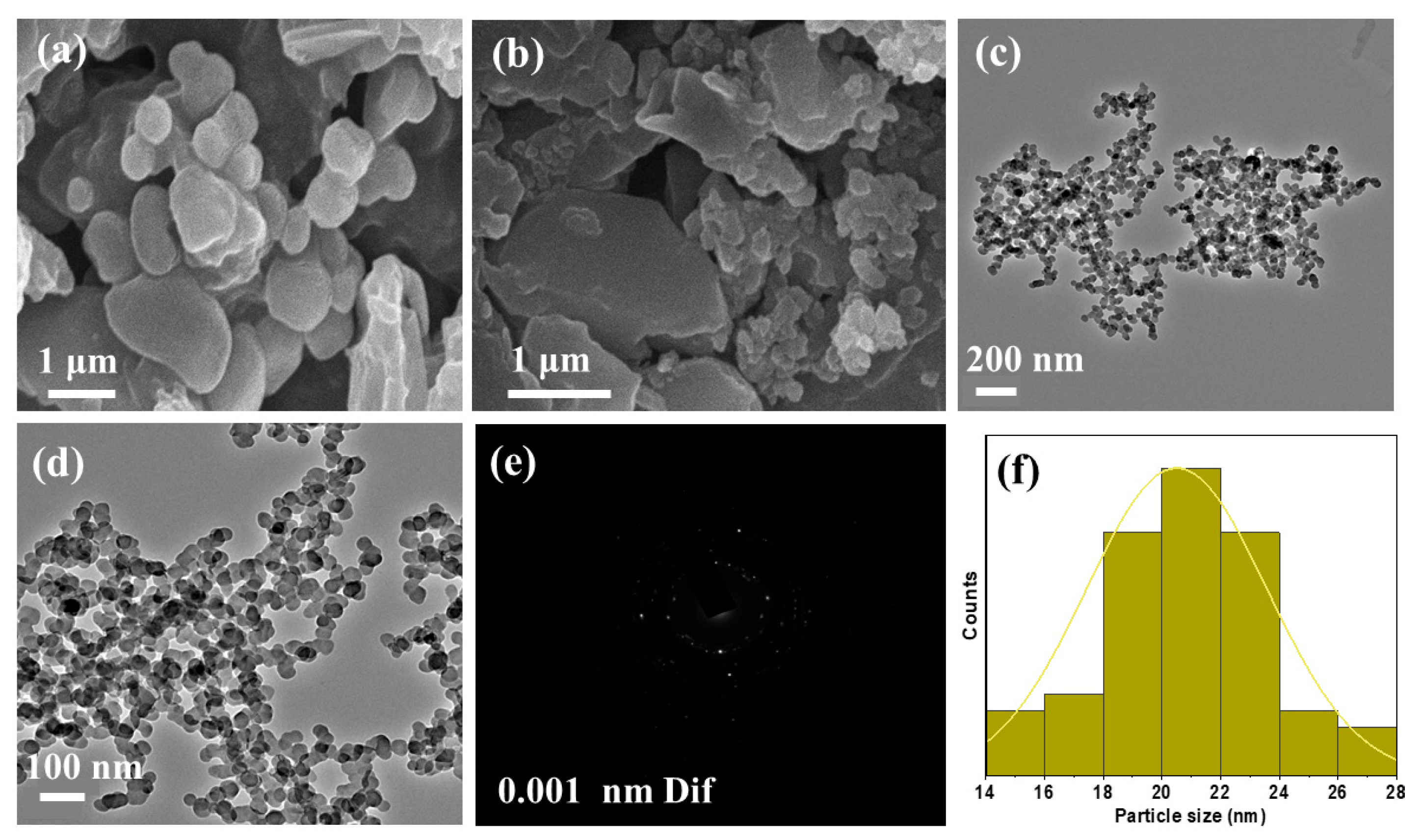
SEM techniques were used to investigate the microstructure and surface morphology of Fe3O4 NPs. Figure 2a,b show the scanned images under SEM, which show highly uniform and smooth particle dispersion with negligible agglomeration. The size of the Fe3O4 NPs is consistent with the TEM pictures, which show spherical particles. However, particle aggregation may be seen in the micrographs. Inter-granular gaps are another feature that can be seen, indicating that the grains are more connected. Figure 2a,b demonstrates the results, which reveal less agglomerated formations with spherical particles. The non-agglomerated SEM images showed distinct and stable particles.

2.4. Transmission Electron Microscopy (TEM)

The TEM picture of the Fe3O4 NPs in Figure 2c,d revealed that the nanomaterial had a particle size distribution of 20.60 nm. ImageJ software calculated particle size based on the TEM images in Figure 2c,d. The nanoscale production of Fe3O4 NPs was validated through TEM images. Figure 2c,d depicts a narrow distribution of Fe3O4 NPs, with an average particle size of approximately 20.60 nm determined using Image J software. Figure 2e reveals that the SAED pattern has concentric rings, indicating polycrystalline. Figure 2f illustrates a broader distribution of Fe3O4 NPs. The homogeneous distribution of Fe3O4 NPs was predicted to improve catalytic activity. The well-dispersed average particle size using a TEM micrograph was calculated and found to be 20.60 nm.

2.5. UV-Visible Spectroscopy

UV-visible spectroscopy is among the most effective techniques for investigating the optical properties of NPs dissolved in a solvent. In the 300–800 nm region, the optical absorption of bio-transformed Fe3O4 NPs was examined. The correlation between k and incident photon energy is shown using Tauc’s equation (Equations (2) and (3)) [37].
k = A ( E E g ) n  
f ( R ) = k s = A ( E E g ) n
In the above equation, ‘n’ represents a constant that may vary based on the type of electronic transition, where ‘n’ equals 1/2 for an allowed direct transition and ‘n’ equals 2 for a permissible indirect transition.   E g is the band gap, photon energy, and A is a constant that depends on the properties of the NPs. Figure 3a displays the broad absorbance spectra for Fe3O4 NPs, measured between 300 and 800 nm. The broad spectra were observed between 340 and 460 nm due to the presence of biological entities present in the egg albumin. Tauc’s figure was used to compute the bandgap; as shown in Figure 4b, the bandgap (Eg) is obtained by extrapolating the linear region on the x-axis. The bandgap energy of 3.01 eV for the phyto-fabricated semiconducting Fe3O4 NPs was determined from the Tauc plot (Figure 3b), an (αhν)2 vs. energy (hν) graph that was drawn from UV–Visible absorption data of Fe3O4 NPs. As synthesized Fe3O4 NPs by chicken egg albumin’s biological components caused the broad spectra between 340 and 460 nm, confirming metal oxide NPs’ absorbance.

2.6. Dielectric Study of Fe3O4 NPs

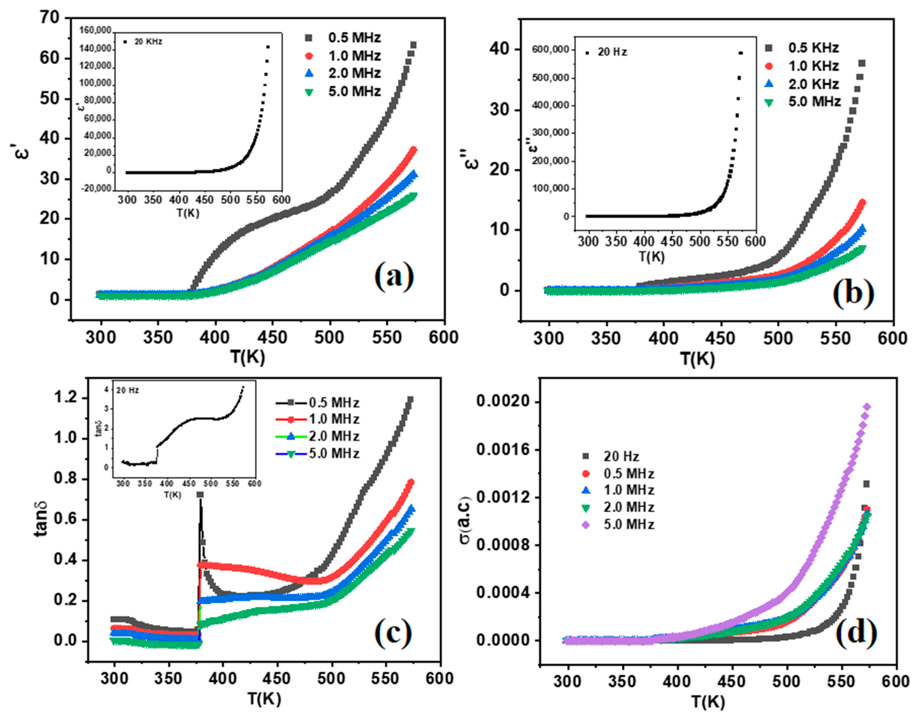
Table 2 presents the examination and tabulation of conductivity [σ (AC)], the real part of dielectric permittivity (ε′), the imaginary part of dielectric permittivity (ε″), and loss tangent (tanδ) as functions of temperature (T in Kelvin) at selected frequencies of 20 Hz, 0.5 MHz, 1.0 MHz, 2.0 MHz, and 5.0 MHz. The temperature and frequency-independent approaches to the intrinsic static dielectric constant are shown in σ (AC), ε′, ε″, and tanδ below 425 K [38]. It showed better dielectric properties ranging from 550–573 K. Furthermore, when T(K) > 573 K grows; Figure 4 illustrates that no discernible dielectric relaxation peaks were found in the loss tangent (tan δ), and the actual component of the dielectric permittivity with temperature-dependent ε’ (T) in Kelvin increases in a defined step. The relaxation mechanism is believed to be hidden by ionic conduction [39]. The relaxation peak in the tan δ curve can only be seen when the electrode polarization effect is taken out of the picture. Frequency-dependent dielectric relaxation in the Fe3O4 NPs is caused by charge carriers building across sample regions with varying conductivities, such as conducting grains and insulating grain boundaries, at high temperatures. Maxwell–Wagner describes this dielectric relaxation’s extrinsic cause [40].
A prominent method for characterizing the charge carrier/ion dynamical dynamics is to study frequency-dependent conductivity. The relation (σ (AC) = ε0ε’ωtan δ) was used to calculate the σ (AC) values to investigate the effects of particle size on the alternating current conductivity of Fe3O4 NPs [41]. Figure 4d displays the frequency-dependent conductivity σ (AC) for each Fe3O4 NPs in the 300 K–573 K range. Temperature-dependent AC conductivity increases because heat generates charge carriers. To provide further insight, it can be observed that the alternating current (AC) conductivity adheres to Jonscher’s power law, expressed as (σ (AC) = dc + Afn)), where σ (AC) represents the total conductivity, dc is the extrapolated direct current (dc) conductivity, n signifies the temperature, and f represents the frequency. A and F stand for material-specific temperature-dependent constant and the temperature, respectively. Figure 4 displays the temperature dependence of the fabricated Fe3O4 NPs. As shown in Figure 4, Fe3O4 NPs' dielectric constants increase sharply as temperature increases in the low-frequency range, but they tend to increase first and then become stable at higher frequencies [42]. Then, with further increase, it tends to decrease after reaching relaxation. This phenomenon is attributed to varied responses of different polarization mechanisms across various frequency ranges and is also accountable for polarization relaxation [43].
The three primary polarization mechanisms of ferroelectric materials are turning-direction polarization, space charge polarization, and displacement polarization. Of them, displacement polarization has the shortest response time (~10−13 s), allowing the polarization process to develop within the frequency range being monitored fully. However, the time required for inducing turning-direction and space charge polarization is significantly longer than displacement polarization, estimated to be around 10−5 s. As a result, this relaxation polarization can only be developed at very low frequencies. In the low-frequency range, field stress or thermal motion can prompt electrons to accumulate at the grain boundaries if the resistance of the boundaries is sufficiently high, resulting in polarization. Conversely, as the frequency increases, the pile-up effect diminishes as electrons consistently alter their direction of motion, reducing polarization. The space charge polarization resulting from non-uniform dielectric structure and other flaws like porosity, grain structure, and impurities is another component affecting lower frequencies permittivity. A space charge layer is established when a few free charges persist at the boundary surface’s center, counterbalancing each other. This allows for changes to the space field, the same as improving the dielectric characteristics. At relatively high frequencies, the creation of turning-direction and space charge polarizations is limited, except for displacement polarization. As a result, the dielectric constant’s dispersion decreases, and it exhibits a frequency-independent response. Put simply, the presence of dispersion in the dielectric constant can be attributed to the polarization mechanisms operating at different frequencies. Space charge accumulation along with substantial interfacial polarization around grain boundaries, which becomes more prevalent at low frequencies, represents the cause of the noticeably elevated permittivity seen at 20 Hz. The intrinsic heterogeneity as well as defect sites for bio-synthesized FeO4 nanoparticles additionally affect this behavior, which is frequently observed in nanostructured metal oxides [36,44,45].

2.7. Factors Affecting Biosynthesis of Fe3O4 NPs

Temperature, reactant concentration, reaction time, and pH are key variables affecting the biosynthesis of Fe3O4 NPs employing phytochemicals. Each parameter strongly determines the form, size, and general characteristics of the synthesized NPs. A detailed explanation of how several variables affect the synthesis process may be found below.

2.7.1. pH

The pH of the reaction mixture influences the ionization state of the metal ions and phytochemicals, as well as the reduction process and the stability of the NPs. Under low acidic conditions, insufficient capping leads to an inadequate reduction of iron ions and the agglomeration of NPs. Under simple conditions, enhanced reduction and stabilization result in the production of smaller, more uniform NPs. Precipitation can happen quickly at highly high pH values, producing larger particles. For the most effective reduction and stability during the synthesis of Fe3O4 NPs, a pH between 8 and 10 is basic [46].

2.7.2. Temperature

Temperature affects both the kinetics of the reduction reaction and the rate at which the NPs grow. Longer growth times produce larger NPs since low temperatures slow the reaction rate. High temperatures quicken the nucleation process and enhance the reaction rate, resulting in smaller NPs due to limited development. Generally, moderate temperatures (between 60 and 80 °C) control the size and homogeneity of the NPs and the reduction rate [47].

2.7.3. Concentration of Reactants

The concentration of the biological extract and the iron salt precursor dictates the availability of reactants for the reduction process and the ensuing production of NPs. High concentrations of iron salts have the potential to produce rapid nucleation and agglomeration, resulting in more widespread and less uniform NPs. On the other hand, low iron salt content may result in inadequate reduction and subpar NPs production. When determining the optimal ratio of phytochemical extract to iron salt, the plant extract should be at an equally good ratio, with typical values for iron salt being 0.1–0.5 M [48].

2.7.4. Reaction Time

The reaction time determines the length of the reduction and growth processes, which affect the size and crystallinity of the NPs. The quick reaction time could result in partial reduction and smaller, possibly unstable NPs. Reaction times that are too long can cause aggregation, even while they allow for complete reduction and development, which produces well-formed and stable NPs. Depending on the specific biological extract and other variables, two to twenty-four hours are often needed for effective reactions [49].

2.8. Photocatalytic Study of Fe3O4 NPs Under Solar Light

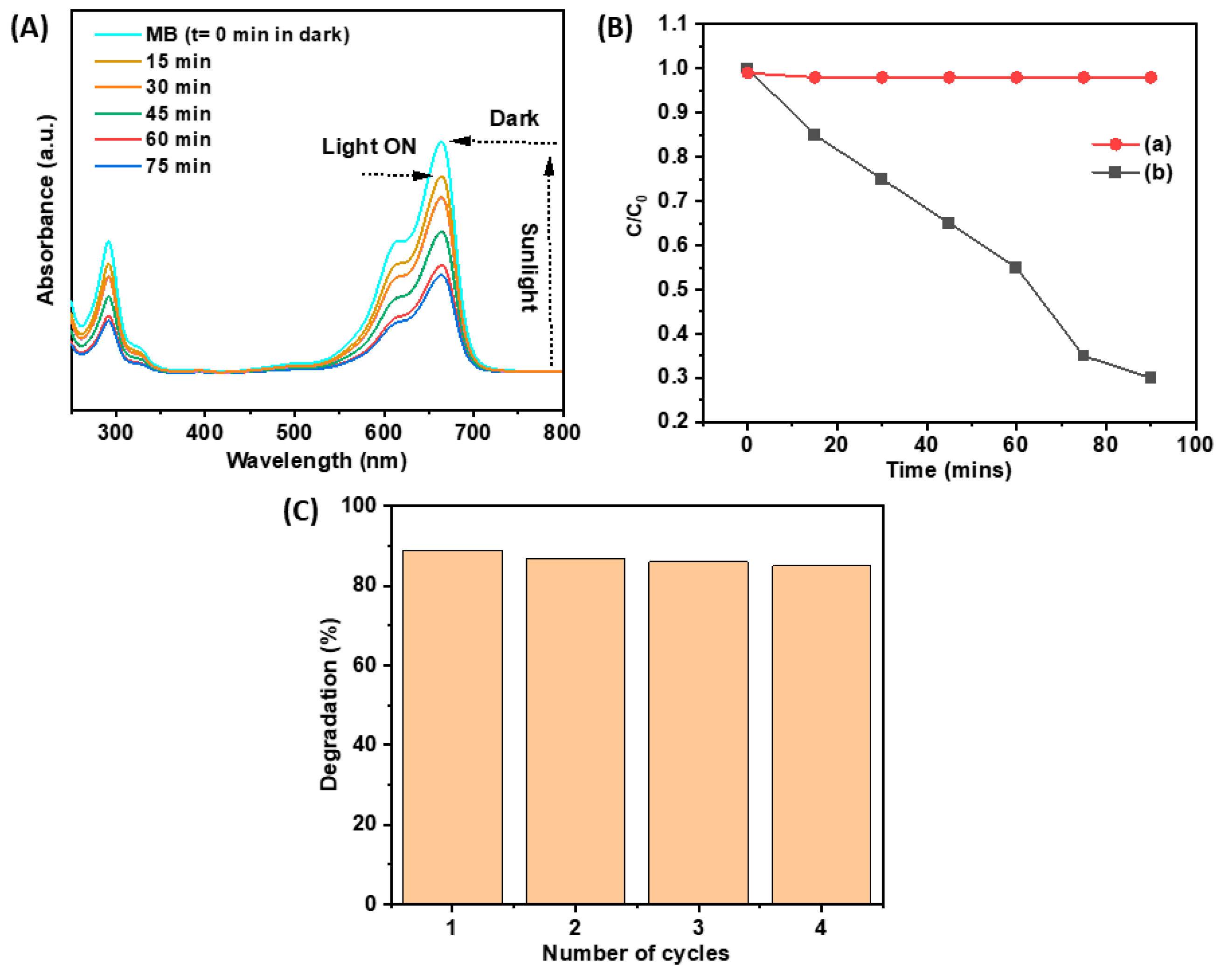
The photodegradation of MB was investigated utilizing Fe3O4 NPs by exposing the MB solution to sun irradiation. The reaction kinetics were analyzed to ascertain the order of the photocatalytic degradation events, revealing that the dye degradation adhered to first-order kinetics [38]. The rate constant revealed that the degradation rate of MB was 0.012 s−1 using solar light irradiation. The photocatalytic degradation efficiency of the organic dyes was calculated using the following formula, as mentioned earlier.
The MB absorbance remained relatively constant, signifying the absence of degradation activity in the absence of a photocatalyst, as evidenced during photodegradation under solar irradiation in control conditions. The solution subjected to solar radiation exhibited a reduction in absorbance, as illustrated in Figure 5A, from time t = 0 min to 75 min. The photocatalytic efficacy of MB without the presence of the photocatalyst (Fe3O4 NPs) under natural sunlight remained constant, as illustrated in Figure 5B. The efficacy of the photocatalytic activity of biosynthesized Fe3O4 NPs was evaluated through the degradation of methylene blue dye in an aqueous solution under natural sunshine. Following four successive cycles, the photocatalytic degradation of MB exhibited stability at 89%, 87%, 86%, and 85%, respectively, as depicted in Figure 5C, over a 75-min exposure to natural sunshine. This study indicates that the Fe3O4 NPs derived from albumin extract exhibit exceptional photocatalytic capabilities. The photocatalysis performance comparison of our Fe3O4 with other iron oxide-based nanomaterials is presented in Table 3.

2.9. In Vitro Assessment of Fe3O4 NPs Against M. phaseolina

The efficacy of Fe3O4 NPs was examined at three different concentrations, viz., 100, 200, and 300 ppm, to determine the inhibition zones against M. phaseolina using the poison food technique (Table 4). Fe3O4 NPs extract was coated with stabilizing agents, viz., silica, preventing them from aggregating and settling. The Fe3O4 NPs showed significant ability to inhibit the fungal growth of M. phaseolina at concentrations of 200 to 300 ppm, with an inhibition zone of 34–52%, respectively, over the respective control (p ≤ 0.05; Table 4). However, M. phaseolina was found to be most sensitive to Fe3O4 NPs, resulting in a growth inhibition zone of 52% at a concentration of 300 ppm over the respective control (p ≤ 0.05; Table 4). The next concentration was 200 ppm, which efficiently suppressed the radial growth of M. phaseolina mycelium by 34% inhibition zones over the respective control (p ≤ 0.05; Table 4). The lowest reduction of radial growth of M. phaseolina mycelium was recorded with a concentration of 100 ppm, with 22% inhibition zones over the respective control (p ≤ 0.05; Table 4). The experiment showed that increasing the concentration of Fe3O4 NPs increased the percent inhibition zone, which caused greater suppression of radial growth of M. phaseolina.
Degree of freedom (DF) refers to the number of independent values that can vary in an analysis without violating constraints. The degrees of freedom depend on the number of groups and the total sample size. The antifungal activity of Fe3O4 NPs was found to be effective and significantly inhibited the radial growth of the M. phaseolina. Hence, Fe3O4 NPs can be regarded as highly effective antifungal agents against many phytopathogenic fungi [23,57]. NPs’ mediated toxicity in fungi occurs through various mechanisms, including causing cell wall damage/membrane damage, gene regulation, DNA interactions, ion release, damage to hyphae and spores, effect on biofilm formation, reactive oxygen species (ROS) impact on mitochondria, gene regulation, and protein levels and ROS generation, resulting in a cascade of damage including lipid peroxidation and mitochondrial impairment [58,59]. Exposure to NPs promotes modifications in the fungal cell wall, encompassing surface contraction, cellular aggregation, pit and pore development, and overall deformation [60,61]. Fe₃O₄ nanoparticles can inhibit fungal growth by causing DNA damage, protein denaturation, and disrupting the cell wall and membrane integrity, and also induce lipid peroxidation through the generation of reactive oxygen species, cause ribosomal disassembly, and deactivate essential enzymes [62]. However, Fe3O4 NPs can bind to fungal cell walls, affecting their structure and permeability [63]. The magnetic characteristics of Fe₃O₄ enable targeted dispersion and contact with fungal cells, while their nanoscale size allows for deeper penetration and enhanced surface reactivity [64]. Moreover, Fe₃O₄ nanoparticles can cause perforations in cellular structures, damage mitochondria, release cytochrome-c into the cytosol, and elevate metacaspase levels, leading to programmed cell death [65,66].
The albumin protein from egg white plays a crucial role in the biofabrication of Fe3O4 NPs by acting as both a reducing and capping agent. The functional groups in albumin, such as hydroxyl, amino, and carboxyl groups, facilitate the reduction of iron salts to iron ions, leading to the formation of Fe3O4 NPs. Additionally, albumin’s protein structure provides numerous binding sites that cap the NPs, preventing aggregation and ensuring uniform size distribution. This capping action stabilizes the NPs in colloidal solutions, enhancing their biocompatibility and making them suitable for biomedical applications. The biocompatible nature of albumin allows for further functionalization, enabling the conjugation of drugs or targeting agents to the NPs. Moreover, using albumin in the synthesis process is environmentally friendly as it eliminates the need for toxic chemicals, making the process sustainable and safe. This biosynthesis method underscores the potential of natural biomolecules in fabricating eco-friendly and functional nanomaterials for advanced applications.
Optimizing the yield of Fe3O4 NPs using chicken egg albumin entails methodically adjusting crucial synthesis parameters like temperature, pH, reactant concentration, and reaction duration. The final nanomaterial was obtained by calcining the nanomaterial at 500 °C. As an example, different pH levels (6, 7, 8, 9, 10), temperatures (30 °C, 50 °C, 85 °C, 100 °C), iron salt concentrations (0.1 M, 0.2 M, 0.3 M, 0.4 M), reaction times (1.5, 4, 8, 24 h), calcination temperatures (300 °C, 400 °C, 500 °C, and 600 °C), and calcination times (3, 4, 5, and 6 h) were tested to optimize these conditions. TEM, SEM, FTIR, XRD, and UV-Vis spectroscopy were among the methods used to assess the yield and characteristics of the fabricated Fe3O4 NPs. The best yield and NPs quality have been achieved at 0.1 M iron (II) nitrate hexahydrate, pH 8, 85 °C, 1.5 h of reaction time, and 4 h of calcination at 500 °C. Optimization can make the biosynthesis technique practical, scalable, and ecologically benign. This optimization ensures that the biosynthesis method is efficient, environmentally friendly, and scalable for practical applications.
Iron ions (Fe3+ or Fe2+) are reduced by albumin extract to zero-valent iron (Fe0) by electron donation, which starts the fabrication of NPs (Fe3+ + reducing agent → Fe0). The albumin extract functional groups, like amino groups, hydroxyl, and carboxyl, help to stabilize the NPs by creating a shield that stops oxidation and guarantees that the particles are evenly distributed and stable (Fe (NO3)2·6H2O+ albumin extract → capped and stabilized Fe3O4 NPs) [67]. Ultimately, the NPs oxidize to fabricate magnetite (Fe3O4) in the presence of oxygen or under controlled heating (Fe0 + O2→ Fe3O4 or 3Fe2+ + 4H2O + O2→ Fe3O4 + 8H+). By producing biocompatible Fe3O4 NPs without hazardous chemicals, this economical and environmentally beneficial process complies with green chemistry principles. The research on bioengineered Fe3O4 NPs emphasizes their noteworthy prospects in photocatalytic and biological domains because of their distinct physicochemical characteristics and eco-friendly manufacturing techniques.
According to Ammar et al. [68], fungicide activity against M. phaseolina, Rhizocotonia solani, Fusarium sporotrichioides, F. proliferatum, F. solani, and F. oxysporum was marginally increased by zinc NPs. Chitosan NPs (@0.1%) significantly reduced the development of Alternaria alternata, M. phaseolina, and R. solani by 82%, 88%, and 34%, respectively [69]. At concentrations between 0.03 and 0.15%, chitosan NPs inhibited the growth of F. verticillioides by 20–60%, reaching complete inhibition at 0.21%. Similarly, A. alternata showed a MIC of 0.24%, and M. phaseolina reached a MIC of 0.26% [70]. The findings in this study are consistent with earlier investigations that revealed the development of the biogenic Fe3O4 NPs from Eichhornia crassipes leaf extract was indicated by a peak in the UV spectral data at 379 nm [71]. According to Shahid et al. [72], Fe2O3 NPs were present in Apis mellifera honey because of a distinct and strong peak at 350 nm. According to earlier reports, the alkyl halides were identified by distinctive transmission bands found at 540.36 and 574.83 cm1 [73]. According to FTIR studies, all of the egg albumin’s phenols, alcohols, amines, and alkanes work together to reduce, stabilize, and cap the biogenic Fe3O4 NPs. Additionally, recent research has shown that iron oxide-based NPs range in size from 10 nm to 100 nm [74,75]. A TEM revealed that the size variation of the iron NPs, which have a thick surface layer and are nearly spherical, ranges from 16.2 to 18.55 nm [76]. The biomolecules of Laurus nobilis leaf extract used in the biosynthesis process were identified as the source of the matrix-like structure in which the biogenic Fe3O4 NPs were seen to be embedded [47]. The average size of the biogenic Fe3O4 NPs was 46.3 nm, and they had a spherical shape [77].
The egg albumin methodology is environmentally benign and has little influence on the environment, unlike chemical synthesis methods that necessitate severe conditions and dangerous substances. Microbial synthesis is natural and friendly, but scalability is difficult due to lengthy reaction periods and intricate biological processes. Like other green and chemical synthesis processes, egg albumin’s inherent functional groups contribute to its high functionalization potential. Fe3O4 NPs produced with egg albumin have significant magnetic characteristics and are stable in biological conditions, making them appropriate for medical applications. The innovative aspect of this study is biosynthesis, which results in improved structural and functional features, even though Fe3O4 NPs made with egg albumin have been extensively reported for various applications. The synthesis is entirely dependent on temperature phenomena, and the innovative aspect of this study is biosynthesis, which results in improved structural and functional features. Superior antifungal, dielectric, and photocatalytic performance results from the exact egg albumin-mediated synthesis that produces controlled particle size, improved crystallinity, and increased surface area. Furthermore, the research presents Fe3O4 nanostructures that utilize synergistic effects to enhance dielectric, antifungal, and photocatalytic capabilities. For the first time, the removal of pathogens, dielectric, and photocatalytic properties of Fe3O4 NPs produced by egg albumin have been investigated, providing a viable and affordable path for pathogen removal and high-performance energy applications.

3. Materials and Methods

3.1. Materials

Fresh chicken eggs were used to extract 70 mL of albumin, analytically pure chemicals such as 0.1 M iron (II) nitrate hexahydrate (Fe (NO3)2·6H2O) (98% pure, Fisher Scientific, Mumbai, India), which were then utilized in the synthesis to Fe3O4 NPs using double-distilled water (DDW). After manually separating the egg white (albumin) from the yolk, it was filtered using a double-layer muslin fabric to remove contaminants; it was utilized without additional purification. The protein, which had an estimated purity of above 90%, was employed right away to guarantee freshness and preserve functionality. Further, potato dextrose agar (PDA) (Sigma-Aldrich, St. Louis, MO, USA) was taken in 250 mL conical flasks and autoclaved at 15 kg/cm2 pressure for 15–20 min at 121 °C. After autoclaving, these conical flasks were placed under a laminar airflow chamber for a few minutes to cool down and then mixed with a solution containing 2.5 mg/L of chloramphenicol (Sigma-Aldrich, USA) (½ PDA + c) [78]. The target organic pollutant, MB (C16H18ClN3S. H2O), was provided by Biochem, Germany, and had a molecular weight of 319.85 g/mol and a purity of 98%.

3.2. Biosynthesis Synthesis of Fe3O4 NPs

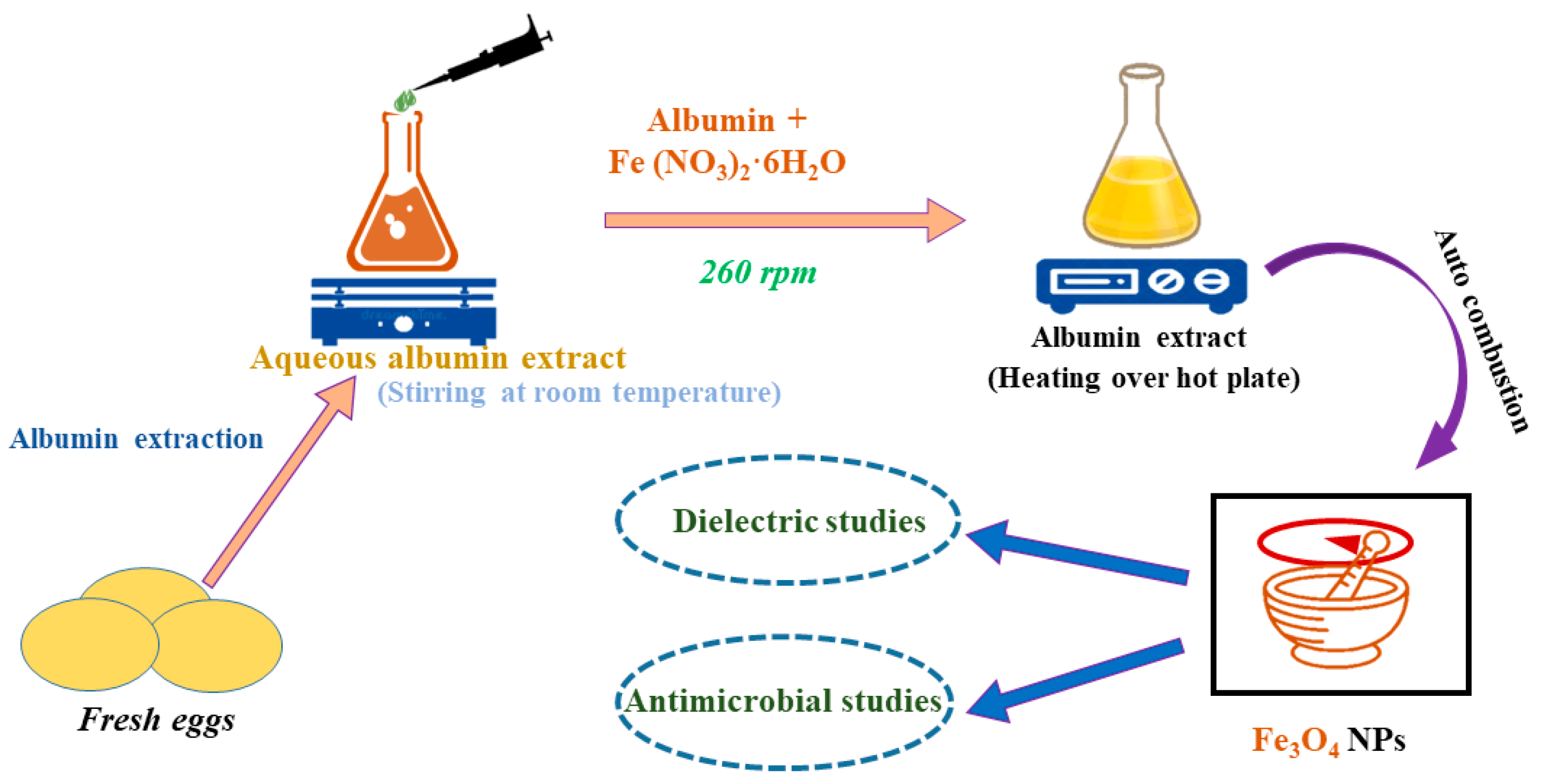
Here, we used a sol-gel-assisted biosynthesis approach to synthesize Fe3O4 NPs via a biological route that was eco-friendly, economical, non-toxic, and, most notably, produced the highest possible yield of the product (Figure 6). Raw eggs were used to extract albumin, which was then employed as a naturally reducing agent. Additionally, it served as a capping and stabilizing agent, preventing NPs' aggregation and assisting in maintaining their size, shape, and surface morphology. To synthesize NPs using chicken eggs, we followed previously reported work [79]. The synthesis of Fe3O4 NPs was performed using 0.1 M of iron (II) nitrate hexahydrate (Fe (NO3)2·6H2O) (98% pure, Fisher Scientific, Mumbai, India), 70 mL fresh chicken egg albumin, and DDW. First of all, in 100 mL DDW, 0.1 M of iron (II) nitrate hexahydrate was added, and 70 mL of freshly obtained albumin was added slowly and stirred for 90 min with a gradual increase in temperature up to 85 °C, and a precipitate was obtained. The precipitate was calcined at 500 °C for 4 h. The obtained material was dried at 85 °C for 24 h in a hot air oven (SSK Enterprises, Mumbai, India).

3.3. Characterization of Fe3O4 NPs

The unit cell dimension and crystallinity phase were ascertained using a Rigaku Miniflex advance diffractometer (Neu-Isenburg, Germany) (Cu anode material, 15 mA current, 30 kV voltages, wavelength (Cu) 1.541838 Å). Optics: In the analysis of Fe3O4 NPs at the angle 2θ, beta filtering with graphite, an automated divergence slit, and a monochromator are employed. The 2θ range for Fe3O4 NPs spans 5.0° to 80.0° with increments of 0.04°. A scanning electron microscope investigated surface morphology and element identification (Model No. JSM 6510 LV, make: JEOL, Japan). The surface morphology, size, crystalline structure, and shape of NPs were investigated through transmission electron microscopy (TEM) and selected area electron diffraction (SAED) patterns. Fe3O4 NPs powder samples were suspended in ethanol before precipitating on the grid. Subsequently, the TEM (make—JEOL, Japan; model-JEM 2100) positioned the sample for scanning. Particle size distribution analysis was performed using Image J Launcher software version (1.4.3.6.7) produced in Tokyo, Japan, incorporating broken symmetry functionalities.
The spectrum of Fe3O4 NPs was recorded using Fourier transform infrared (FTIR) spectroscopy, covering wave numbers from 400 to 4000 cm−1 (Make—Bruker, Model: Tensor 35). This analysis aimed to scrutinize the functional bio-molecules. Each sample was prepared by compressing 200 mg of KBr, a non-absorbing medium serving as the background under high pressure. A Cary 5000 (Make: Agilent, Country: Santa Clara, California, U.S.) spectrometer with 1 cm quartz cells was used to record the UV–vis spectra at room temperature in the range of 300–800 nm. Key Sight Agilent E4990A was used to analyze the sample from 20 Hz to 5 MHz in the 300 K to 573 K temperature range at selected frequencies. An X-ray diffractometer was employed to scrutinize the crystal structure and lattice properties of Fe3O4 NPs.

3.4. Temperature-Dependent Dielectric Studies of Fe3O4 NPs

Fe3O4 NPs are semiconductors with a broad bandgap of 3.01 eV that exclusively absorb light in the ultraviolet region. The semiconductor Fe3O4 NPs were made in circular pellets with a 13 mm diameter and a thickness of 2.65 mm by coating with the silver paste on both sides and sandwiching the insulating material between them. The Fe3O4 powder was compressed in a hydraulic press (Athena Technology, Mumbai, India) at 5 tons of pressure for two minutes to produce uniformly dense pellets [33,80]. To guarantee consistent conductivity and good electrode contact, high-purity silver paste was added to both surfaces with a fine brush to make a uniform thickness of about 20 µm. It was then dried for 30 min at 80 °C.
The sample was scanned over 20 Hz to 5 MHz in the 300 K to 573 K temperature range at different selected frequencies. Fe3O4 NPs are a highly effective contender for dielectrics because they are used in microwaves. Energy storage devices greatly benefit from the dielectric characteristics of metal oxide NPs. The study examined the relationship between temperature and the dielectric permittivity of real (ε′), imaginary (ε″), dissipation factor (tanδ), and conductivity σ (a.c). The complex permittivity, which is represented by Equation (4), is crucial to the study.
ε ( ω ) = ε ( ω ) i ε ( ω )
In this case, the real part of entity ε*(ω), represented by ε′(ω), indicates the material’s ability to store energy, while the imaginary part ε″(ω) indicates the energy loss within the metal oxide NPs in the form of heat. Equation (5) provides an experimental measurement of the real component of dielectric permittivity.
ε = C p d ε 0 A
where
A is the round pellet’s cross-sectional area;
d is its thickness;
Cp is its capacitance. The permittivity of air is represented by ε0 = 8.854 × 10⁻¹² F/m.

3.5. Photocatalytic Study

The photodegradation of MB was investigated utilizing Fe3O4 NPs by exposing the MB solution to sun irradiation. The reaction kinetics were analyzed to ascertain the order of the photocatalytic degradation events, revealing that the dye degradation adhered to first-order kinetics, as expressed by the following Equation (6) [81].
C t = C 0 e k t
C 0 is the starting dye concentration in ppm,   C t is a concentration at time t in minutes, k is the rate constant in min−1, and t is time, as shown in Equation (7).
l n C t C 0 = k t
The rate constant calculated using Equations (6) and (7) revealed that the degradation rate of MB was 0.012 s−1 using solar light irradiation. The photocatalytic degradation efficiency of the organic dyes was calculated using the following formula, as mentioned in Equation (8):
D e g r a d a t i o n   ( % ) = ( A 0 A t A 0 × 100 )
where A 0 is the absorbance of the dye solution before photo-irradiation, and A t is the absorbance of solutions in suspension after photo-irradiation for a specific time t (0 min to 75 min).

3.6. In Vitro Assessment of Fe3O4 NPs Against Pathogenic Fungus and Growth Inhibition

The antifungal activity of Fe3O4 NPs at concentrations of 100, 200, and 300 ppm against the pathogenic fungus M. phaseolina was assessed using the poison food technique used by Falck [82] under in vitro conditions. The Fe3O4 NPs concentration was prepared in DDW to get the desired concentration of NPs after mixing the DMSO solution (Sigma-Aldrich, USA) in the PDA media in equal amounts before pouring in sterilized Petri plates. Each concentration of Fe3O4 NPs was added to 20–25 mL of the potato dextrose agar (PDA) medium. The control plate (without Fe3O4 NPs) was maintained with a PDA medium. Subsequently, approximately 20–25 mL of the warmed molten PDA medium containing each test concentration of Fe3O4 NPs was poured into each sterilized Petri plate under a laminar airflow chamber. The plates with and without Fe3O4 NPs were inoculated by placing a 5 mm mycelial disc cut with a cork borer of M. phaseolina into the center and incubating for 28 ± 2 °C in the biochemical oxygen demand (BOD) incubator (Labtronix, Noida, India). Three replicates of each concentration and control plate were maintained. The colony diameter of all cultured plates was measured seven days post-inoculation, coinciding with the control plates achieving complete mycelial development of the pathogen. The percentage of mycelial growth inhibition of the pathogen was determined using the methodology provided by Vincent (Equation (9)) [83].
Mycelial   growth   inhibition   ( % ) = C T C × 100
where
C = radial growth of fungus in control;
T = radial growth of fungus in treatment.

3.7. Statistical Analysis

The collected data, encompassing several features under examination, was subjected to statistical analysis using R software (version 2.14.1). The evaluated factors that indicate significant differences (p = 0.05) were identified using Duncan’s Multiple Range Test (DMRT). An ANOVA was performed using OPSTAT [78].

4. Conclusions

A thorough comparison with other biosynthesis methods and well-established chemical processes highlights the uniqueness and benefits of using egg albumin for Fe3O4 NPs. In an aqueous medium at normal temperature, egg albumin has an acceptable reaction time, benign reaction conditions, good yield and reproducibility, and high synthesis efficiency. Fe3O4 NP synthesis using egg albumin is a greener alternative to chemical production. Egg albumin, an abundant and biodegradable biomaterial, reduces, caps, and improves NP biocompatibility, facilitating synthesis. Researchers could modify synthesis parameters to increase Fe3O4 NP yield and uniformity. These NPs could be functionalized with biomolecules for targeted water remediation, antifungal action, and medicine. The biosynthesis of Fe3O4 NPs via sol-gel-assisted was safe, cost-effective, and yielded the most. X-ray diffraction revealed this sample’s good crystallinity and Fe3O4 NPs' cubic structure. FTIR functional group estimation showed Fe-O, indicating metal oxide formation. SEM indicated low aggregation and well-distributed NPs. Dielectric studies reveal that temperature and frequency increase mobile carrier movement and polarization to a limit, and the drop in M. phaseolina radial growth was inhibited and antifungal by Fe3O4 NPs. The dielectric properties and promising antifungal activity against M. phaseolina make these NPs suitable for agricultural and biomedical uses. This makes Fe3O4 NPs effective antifungals for sustainable agriculture. The photocatalytic activity of Fe3O4 NPs was assessed for the degradation of methylene blue (MB) under solar irradiation. In this study, Fe3O4 NPs photocatalysts achieved 89% (MB) degradation within 75 min. Finally, this biosynthesis technique must be scaled and made economically viable before commercialization. These suggestions will improve sustainable and versatile nanomaterial development, furthering science and conservation.

Author Contributions

Conceptualization, A.R., I.A. and A.A.; Methodology, A.R. and I.A.; Validation, A.A., S.K.A., S.M.K. and M.I.; Formal Analysis, G.K., H.P., S.M. and B.R.P.; Investigation, A.R., I.A., A.A., M.I. and S.M.K.; Resources, A.R., I.A. and A.A.; Data Curation, A.A.A., H.A.R., B.R.P., S.K.A. and G.K.; Writing—Original Draft Preparation, A.R., I.A. and A.A.; Writing—Review and Editing, A.R., I.A., A.A., S.M.K. and M.I.; Visualization, A.R., I.A., A.A., H.P. and S.M.; Supervision, A.A. and S.K.A.; Project Administration, S.K.A.; Funding Acquisition, A.A. and S.K.A. All authors have read and agreed to the published version of the manuscript.

Funding

This research was funded by the Deanship of Scientific Research at Imam Mohammad Ibn Saud Islamic University (IMSIU) grant number IMSIU-DDRSP2502.

Data Availability Statement

Data will be made available from authors upon reasonable request.

Acknowledgments

This work was supported and funded by the Deanship of Scientific Research at Imam Mohammad Ibn Saud Islamic University (IMSIU) (grant number IMSIU-DDRSP2502).

Conflicts of Interest

The authors reported no potential conflicts of interest.

References

  1. Malhi, G.S.; Kaur, M.; Kaushik, P. Impact of Climate Change on Agriculture and Its Mitigation Strategies: A Review. Sustainability 2021, 13, 1318. [Google Scholar] [CrossRef]
  2. Ahmed, S.; Ansari, A.; De, B.; Mukherjee, S.; Negi, D.S.; Ranjan, P. An electrochemical bio-electronic tongue based on borophene/PPy@ITO hybrid for selective caffeine identification. Analyst 2025, 150, 962–974. [Google Scholar] [CrossRef] [PubMed]
  3. Ansari, A.; Ahmed, S.; Mashniwi, M.H.; Shinde, S.M.; Khan, A.; Ranjan, P.; Negi, D.S. Recent advancements in photodetection utilizing inorganic, organic low-dimensional materials and their hybrids. J. Mater. Sci. 2025, 60, 2711–2743. [Google Scholar] [CrossRef]
  4. Ali, S.K.; Hakami, O.; Zelai, T.; Imran, M.; Manqari, H.M.A.; Alamri, A.A.; Ghazwani, N.A.; Ahmed, S. Pioneering photocatalysts: WSe2/ZIF-9 Z-scheme nanocomposites for efficient hexavalent chromium removal. Inorg. Chem. Commun. 2025, 172, 113733. [Google Scholar] [CrossRef]
  5. Ahmed, S.; Ansari, A.; Li, Z.; Mazumdar, H.; Siddiqui, M.A.; Khan, A.; Ranjan, P.; Kaushik, A.; Vinu, A.; Kumar, P. Point-of-Care Health Diagnostics and Food Quality Monitoring by Molecularly Imprinted Polymers-Based Histamine Sensors. Adv. Sens. Res. 2025, 4, 2400132. [Google Scholar] [CrossRef]
  6. Ansari, A.; Ahmed, S.; Rehman, B.; Ali, S.K.; Azooz, R.E.; Hassan, K.F.; Khan, A.; Ranjan, P.; Negi, D.S. A synergistic effect of ZnO low dimensional rods/PEDOT:PSS hybrid structure for UV radiation detection. Appl. Phys. A 2024, 130, 692. [Google Scholar] [CrossRef]
  7. Ahmed, S.; Ansari, A.; Ali, S.K.; Patil, B.R.; Riyaz, F.; Khan, A.; Ranjan, P. Pioneering Sensing Technologies Using Borophene-Based Composite/Hybrid Electrochemical Biosensors for Health Monitoring: A Perspective. Anal. Sens. 2024, 4, e202400034. [Google Scholar] [CrossRef]
  8. Ansari, A.; Ahmed, S.; Siddiqui, M.A.; Khan, A.; Tailor, S.; Kumar, P.; Ranjan, P.; Negi, D.S. PEDOT:PSS, TiO2 nanoparticles, and carbon quantum dot composites as ultraviolet sensors. ACS Appl. Nano Mater. 2024, 7, 9789–9799. [Google Scholar] [CrossRef]
  9. Ahmed, S.; Ansari, A.; Bishwanathan, S.; Siddiqui, M.A.; Tailor, S.; Gupta, P.K.; Negi, D.S.; Ranjan, P. Electronic tongue based on ZnO/ITO@glass for electrochemical monitoring of spiciness levels. Langmuir 2024, 40, 4434–4446. [Google Scholar] [CrossRef]
  10. Ansari, A.; Ahmed, S.; Siddiqui, M.A.; Khan, A.; Banerjee, A.; Negi, D.S.; Ranjan, P. Investigation of complex hybrids in lithium salt under ultraviolet energy source. J. Mater. Sci. Mater. Electron. 2024, 35, 108. [Google Scholar] [CrossRef]
  11. Ahmed, S.; Ansari, A.; Siddiqui, M.A.; Imran, M.; Kumari, B.; Khan, A.; Ranjan, P. Electrochemical and optical-based systems for SARS-COV-2 and various pathogens assessment. Adv. Nat. Sci. Nanosci. Nanotechnol. 2023, 14, 033001. [Google Scholar] [CrossRef]
  12. Verma, S.K.; Das, A.K.; Patel, M.K.; Shah, A.; Kumar, V.; Gantait, S. Engineered nanomaterials for plant growth and development: A perspective analysis. Sci. Total Environ. 2018, 630, 1413–1435. [Google Scholar] [CrossRef] [PubMed]
  13. Mohseniazar, M.; Barin, M.; Zarredar, H.; Alizadeh, S.; Shanehbandi, D. Potential of Microalgae and Lactobacilli in biosynthesis of silver nanoparticles. BioImpacts 2011, 1, 149–152. [Google Scholar] [CrossRef]
  14. Khan, A.U.; Khan, M.; Khan, M.M. Selected nanotechnology and nanostructure for drug delivery, nanomedicine and cure. Bioprocess Biosyst. Eng. 2020, 43, 1339–1357. [Google Scholar] [CrossRef]
  15. Behl, T.; Kaur, A.I.; Sehgal, S.; Singh, S.; Sharma, N.; Bhatia, S.; Al-Harrasi, A.; Bungau, S. The dichotomy of nanotechnology as the cutting edge of agriculture: Nano-farming as an asset versus nanotoxicity. Chemosphere 2022, 288, 132533. [Google Scholar] [CrossRef]
  16. Agrawal, S.; Kumar, V.; Kumar, S.; Shahi, S.K. Plant development and crop protection using phyto nanotechnology: A new window for sustainable agriculture. Chemosphere 2022, 299, 134465. [Google Scholar] [CrossRef]
  17. Kumar, S.; Vishnoi, V.K.; Kumar, P.; Dubey, R.C. Survival of Macrophomina phaseolina in plant tissues and soil. In Macrophomina Phaseolina. Ecobiology, Pathology and Management; Kumar, P., Dubey, R.C., Eds.; Academic Press: Cambridge, MA, USA, 2023; pp. 205–224. [Google Scholar] [CrossRef]
  18. Marquez, N.; Giachero, M.L.; Declerck, S.; Ducasse, D.A. Macrophomina phaseolina: General characteristics of pathogenicity and methods of control. Front. Plant Sci. 2021, 12, 634397. [Google Scholar] [CrossRef]
  19. Kaur, S.; Dhillon, G.S.; Brar, S.K.; Vallad, G.E.; Chand, R.; Chauhan, V.B. Emerging phytopathogen Macrophomina phaseolina: Biology, economic importance and current diagnostic trends. Crit. Rev. Microbiol. 2012, 38, 136–151. [Google Scholar] [CrossRef] [PubMed]
  20. Moshafi, M.H.; Ranjbar, M.; Ilbeigi, G. Biotemplate of albumen for synthesized iron oxide quantum dots nanoparticles (QDNPs) and investigation of antibacterial effect against pathogenic microbial strains. Int. J. Nanomed. 2019, 14, 3273–3282. [Google Scholar] [CrossRef]
  21. Sun, J.; Wang, Y.; Zhang, Y.; Xu, C.; Chen, H. Egg albumin-assisted hydrothermal synthesis of Co3O4 quasi-cubes as superior electrode material for supercapacitors with excellent performances. Nanoscale Res. Lett. 2019, 14, 340. [Google Scholar] [CrossRef]
  22. Bachheti, R.K.; Konwarh, R.; Gupta, V.; Husen, A.; Joshi, A. Green synthesis of iron oxide nanoparticles: Cutting edge technology and multifaceted applications. In Nanomaterials and Plant Potential; Husen, A., Iqbal, M., Eds.; Springer: Cham, Switzerland, 2019; pp. 239–259. [Google Scholar] [CrossRef]
  23. Parveen, S.; Wani, A.H.; Shah, M.A.; Devi, H.S.; Bhat, M.Y.; Koka, J.A. Preparation, characterization and antifungal activity of iron oxide nanoparticles. Microb. Pathog. 2018, 115, 287–292. [Google Scholar] [CrossRef] [PubMed]
  24. Henam, S.D.; Ahmad, F.; Shah, M.A.; Parveen, S.; Wani, A.H. Microwave Synthesis of Nanoparticles and Their Antifungal Activities. Spectrochim. Acta Part A Mol. Biomol. Spectrosc. 2019, 213, 337–341. [Google Scholar] [CrossRef]
  25. Hariani, P.L.; Faizal, M.; Setiabudidaya, D. Synthesis and properties of Fe3O4 nanoparticles by co-precipitation method to removal procion dye. Int. J. Environ. Sci. Dev. 2013, 4, 336. [Google Scholar] [CrossRef]
  26. Chinnadurai, G.; Subramanian, R.; Selvi, P. Fish mucus stabilized iron oxide nanoparticles: Fabrication, DNA damage and bactericidal activity. Inorg. Nano-Met. Chem. 2021, 51, 550–559. [Google Scholar] [CrossRef]
  27. Islam, T.; Repon, M.R.; Islam, T.; Sarwar, Z.; Rahman, M.M. Impact of textile dyes on health and ecosystem: A review of structure, causes, and potential solutions. Environ. Sci. Pollut. Res. 2023, 30, 9207–9242. [Google Scholar] [CrossRef]
  28. Slimani, S.; Talone, A.; Abdolrahimi, M.; Imperatori, P.; Barucca, G.; Fiorani, D.; Peddis, D. Morpho-structural and magnetic properties of CoFe2O4/SiO2 nanocomposites: The effect of the molecular coating. J. Phys. Chem. C 2023, 127, 8840–8849. [Google Scholar] [CrossRef]
  29. Frolova, L.; Sukhyy, K. Study of the photocatalytic degradation of furacilin from aqueous solutions using the biochar/Fe3O4 nanocomposite. Appl. Nanosci. 2023, 13, 6895–6904. [Google Scholar] [CrossRef]
  30. Koli, R.R.; Phadatare, M.R.; Sinha, B.B.; Sakate, D.M.; Ghule, A.V.; Ghodake, G.S.; Deshpande, N.G.; Fulari, V.J. Gram bean extract-mediated synthesis of Fe3O4 nanoparticles for tuning the magneto-structural properties that influence the hyperthermia performance. J. Taiwan Inst. Chem. Eng. 2019, 95, 357–368. [Google Scholar] [CrossRef]
  31. Cai, W.; Weng, X.; Chen, Z. Highly efficient removal of antibiotic rifampicin from aqueous solution using green synthesis of recyclable nano-Fe3O4. Environ. Pollut. 2019, 247, 839–846. [Google Scholar] [CrossRef]
  32. Shi, X.; Zhang, X.; Gao, W.; Zhang, Y.; He, D. Removal of microplastics from water by magnetic nano-Fe3O4. Sci. Total Environ. 2022, 802, 149838. [Google Scholar] [CrossRef]
  33. Rashid, T.; Raza, A.; Saleh, H.A.M.; Khan, S.; Rahaman, S.; Pandey, K.; AlDamen, M.A.; Sama, F.; Ahmad, A.; Shahid, M.; et al. Green Synthesis of Ni/Fe3O4/rGO Nanocomposites for Desulfurization of Fuel. ACS Appl. Nano Mater. 2023, 6, 18905–18917. [Google Scholar] [CrossRef]
  34. Shehu, Z.; Nyakairu, G.W.A.; Tebandeke, E.; Odume, O.N. Circular Economy Approach for Treatment of Water-Containing Diclofenac Using Recyclable Magnetic Fe3O4 Nanoparticles: A Case Study of Real Water Sample from Lake Victoria. J. Pharm. Res. Int. 2023, 35, 66–81. [Google Scholar] [CrossRef]
  35. Falck, R. Wachtumgesetze, wachstumLaktorehnund temperature wertderholzersterenden. Myceture 1907, 32, 38–39. [Google Scholar]
  36. Sharma, A.; Sahu, G.; Mahapatra, S.P. Synthesis, characterization, and dielectric studies of magnetic iron oxide nanoparticles. Ferroelectrics 2024, 618, 11–24. [Google Scholar] [CrossRef]
  37. Sitorus, F.S.; Tumbelaka, R.M.; Istiqomah, N.I.; Nuswantari, A.; Suharyadi, E. Dielectrics and Optical Properties of Green-Synthesized Fe3O4 Nanoparticles. Key Eng. Mater. 2023, 941, 173–181. [Google Scholar] [CrossRef]
  38. Wang, H.; Li, H. Fe3O4 microplate filled PEI matrix composite with remarkable nonlinear conductive characteristics, dielectric property, and low percolation threshold. Heliyon 2023, 9, e22514. [Google Scholar] [CrossRef]
  39. Ding, E.; Bai, B.; Wang, X.; Zhang, W.; Liu, W.; Zhang, L. Enhanced microwave absorption performance of geopolymer composites by incorporating hollow Fe3O4 nanospheres. Int. J. Appl. Ceram. Technol. 2024, 21, 806–817. [Google Scholar] [CrossRef]
  40. Sadat, M.E.; Bud’ko, S.L.; Ewing, R.C.; Xu, H.; Pauletti, G.M.; Mast, D.B.; Shi, D. Effect of dipole interactions on blocking temperature and relaxation dynamics of super paramagnetic iron oxide (Fe3O4) nanoparticle systems. Materials 2023, 16, 496. [Google Scholar] [CrossRef]
  41. Hayat, K.; Khan, K.; Bibi, F.; Sfina, N.; Elhadi, M.; Safeen, K.; Abdullaev, S.; Rahman, N.; Shah, S.K. Investigation of size-dependent electrical, dielectric, and magnetic properties of iron oxide nanostructures. Mater. Chem. Phys. 2024, 315, 128882. [Google Scholar] [CrossRef]
  42. Laid, T.M.; Abdelhamid, K.; Eddine, L.S.; Abderrhmane, B. Optimizing the biosynthesis parameters of iron oxide nanoparticles using central composite design. J. Mol. Struct. 2021, 1229, 129497. [Google Scholar] [CrossRef]
  43. Kaushal, P.; Verma, N.; Kaur, K.; Sidhu, A.K. Green synthesis: An eco-friendly route for the synthesis of iron oxide nanoparticles. Front. Nanotechnol. 2021, 3, 655062. [Google Scholar] [CrossRef]
  44. Radon, A.; Łukowiec, D.; Kremzer, M.; Mikuła, J.; Włodarczyk, P. Electrical conduction mechanism and dielectric properties of spherical shaped Fe3O4 nanoparticles synthesized by co-precipitation method. Materials 2018, 11, 735. [Google Scholar] [CrossRef] [PubMed]
  45. Afzia, M.; Khan, R.A.; Ismail, B.; Zaki, M.E.A.; Althagafi, T.M.; Alanazi, A.A.; Khan, A.U. Correlation between magnetic and dielectric response of CoFe2O4: Li1+/Zn2+ nanopowders having improved structural and morphological properties. Molecules 2023, 28, 2824. [Google Scholar] [CrossRef]
  46. Abdullah, J.A.A.; Eddine, L.S.; Abderrhmane, B.; Alonso-González, M.; Guerrero, A.; Romero, A. Green synthesis and characterization of iron oxide nanoparticles by Pheonix dactylifera leaf extract and evaluation of their antioxidant activity. Sustain. Chem. Pharm. 2020, 17, 100280. [Google Scholar] [CrossRef]
  47. Demirezen, D.A.; Yılmaz, Ş.; Yılmaz, D.D.; Yıldız, Y.Ş. Green synthesis of iron oxide nanoparticles using Ceratonia siliqua L. aqueous extract: Improvement of colloidal stability by optimizing synthesis parameters, and evaluation of antibacterial activity against Gram-positive and Gram-negative bacteria. Int. J. Mat. Res. 2022, 113, 849–861. [Google Scholar] [CrossRef]
  48. Sidkey, N. Biosynthesis, characterization and antimicrobial activity of iron oxide nanoparticles synthesized by fungi. Al-Azhar J. Pharm. Sci. 2020, 62, 164–179. [Google Scholar] [CrossRef]
  49. Slavin, Y.N.; Bach, H. Mechanisms of antifungal properties of metal nanoparticles. Nanomaterials 2022, 12, 4470. [Google Scholar] [CrossRef]
  50. Zhang, Q.; Yu, L.; Xu, C.; Zhao, J.; Pan, H.; Chen, M.; Xu, Q.; Diao, G. Preparation of highly efficient and magnetically recyclable Fe3O4@C@Ru nanocomposite for the photocatalytic degradation of methylene blue in visible light. Appl. Surf. Sci. 2019, 483, 241–251. [Google Scholar] [CrossRef]
  51. Nuengmatcha, P.; Porrawatkul, P.; Chanthai, S.; Sricharoen, P.; Limchoowong, N. Enhanced photocatalytic degradation of methylene blue using Fe2O3/graphene/CuO nanocomposites under visible light. J. Environ. Chem. Eng. 2019, 7, 103438. [Google Scholar] [CrossRef]
  52. Dagher, S.; Soliman, A.; Ziout, A.; Tit, N.; Hilal-Alnaqbi, A.; Khashan, S.; Alnaimat, F.; Abu Qudeiri, J. Photocatalytic removal of methylene blue using titania-and silica-coated magnetic nanoparticles. Mater. Res. Exp. 2018, 5, 065518. [Google Scholar] [CrossRef]
  53. Imran, M.; Alam, M.M.; Hussain, S.; Ali, M.A.; Shkir, M.; Mohammad, A.; Ahamad, T.; Kaushik, A.; Irshad, K. Highly photocatalytic active r-GO/Fe3O4 nanocomposites development for enhanced photocatalysis application, A facile low-cost preparation and characterization. Ceram. Int. 2021, 47, 31973–31982. [Google Scholar] [CrossRef]
  54. Tseng, W.J.; Chuang, Y.C.; Chen, Y.A. Mesoporous Fe3O4@Ag@TiO2 nanocomposite particles for magnetically recyclable photocatalysis and bactericide. Adv. Powder Technol. 2018, 29, 664–671. [Google Scholar] [CrossRef]
  55. Hussain, S.; Alam, M.M.; Imran, M.; Ali, M.S.; Ahamad, T.; Haidyrah, A.S.; Alotaibi, S.M.A.R.; Naik, M.U.D.; Shariq, M. A facile low-cost scheme for highly photoactive Fe3O4-MWCNTs nanocomposite material for degradation of methylene blue. Alex. Eng. J. 2022, 61, 9107–9117. [Google Scholar] [CrossRef]
  56. Li, Y.; Zhang, P.; Li, M.; Shakoor, N.; Adeel, M.; Zhou, P.; Guo, M.; Jiang, Y.; Zhao, W.; Lou, B.; et al. Application and mechanisms of metal-based nanoparticles in the control of bacterial and fungal crop diseases. Pest Manag. Sci. 2023, 79, 21–36. [Google Scholar] [CrossRef]
  57. Athie-García, M.S.; Piñón-Castillo, H.A.; Muñoz-Castellanos, L.N.; Ulloa-Ogaz, A.L.; Martínez-Varela, P.I.; Quintero-Ramos, A.; Duran, R.; Murillo-Ramirez, J.G.; Orrantia-Borunda, E. Cell wall damage and oxidative stress in Candida albicans ATCC10231 and Aspergillusniger caused by palladium nanoparticles. Toxicology 2018, 48, 111–120. [Google Scholar] [CrossRef]
  58. Babele, P.K.; Thakre, P.K.; Kumawat, R.; Tomar, R.S. Zinc oxide nanoparticles induce toxicity by affecting cell wall integrity pathway, mitochondrial function and lipid homeostasis in Saccharomyces cerevisiae. Chemosphere 2018, 213, 65–75. [Google Scholar] [CrossRef]
  59. Kim, S.W.; Kim, K.S.; Lamsal, K.; Kim, Y.J.; Kim, S.B.; Jung, M.Y.; Sim, S.J.; Kim, H.S.; Chang, S.J.; Kim, J.K.; et al. An in vitro study of the antifungal effect of silver nanoparticles on oak wilt pathogen Raffaelea sp. J. Microbiol. Biotechnol. 2009, 19, 760–764. [Google Scholar] [PubMed]
  60. Ouda, S.M. Antifungal activity of silver and copper nanoparticles on two plant pathogens, Alternariaalternata and Botrytis cinerea. Res. J. Microbiol. 2014, 9, 34. [Google Scholar] [CrossRef]
  61. Prucek, R.; Tuček, J.; Kilianová, M.; Panáček, A.; Kvítek, L.; Filip, J.; Kolář, M.; Tománková, K.; Zbořil, R. The targeted antibacterial and antifungal properties of magnetic anocomposite of iron oxide and silver nanoparticles. Biomaterials 2011, 32, 4704–4713. [Google Scholar] [CrossRef]
  62. Azadi, S.; Amani, A.M.; Jangjou, A.; Vaez, A.; Zareshahrabadi, Z.; Zare, A.; Kasaee, S.R.; Kamyab, H.; Chelliapan, S.; Mosleh-Shirazi, S. Fe3O4@SiO2/Schiff-base/Zn (II) nanocomposite functioning as a versatile antimicrobial agent against bacterial and fungal pathogens. Sci. Rep. 2025, 15, 5694. [Google Scholar] [CrossRef]
  63. Abbas, H.S.; Krishnan, A. Magnetic nanosystems as a therapeutic tool to combat pathogenic fungi. Adv. Pharm. Bull. 2020, 10, 512. [Google Scholar] [CrossRef] [PubMed]
  64. Peng, Q.; Huo, D.; Li, H.; Zhang, B.; Li, Y.; Liang, A.; Wang, H.; Yu, Q.; Li, M. ROS-independent toxicity of Fe3O4 nanoparticles to yeast cells: Involvement of mitochondrial dysfunction. Chem. Biol. Interact. 2018, 287, 20–26. [Google Scholar] [CrossRef]
  65. Kumar, U.; Singh, P.K.; Madheshiya, P.; Kumar, I. Role of Nanoparticles in Plant Disease Management. In Nanotechnology-based Sustainable Agriculture; John Wiley & Sons: Hoboken, NJ, USA, 2025; pp. 347–376. [Google Scholar] [CrossRef]
  66. Sadat, M.; Patel, R.; Sookoor, J.; Bud’Ko, S.L.; Ewing, R.C.; Zhang, J.; Xu, H.; Wang, Y.; Pauletti, G.M.; Mast, D.B. Effect of spatial confinement on magnetic hyperthermia via dipolar interactions in Fe3O4 nanoparticles for biomedical applications. Mater. Sci. Eng. C 2014, 42, 52–63. [Google Scholar] [CrossRef] [PubMed]
  67. Ammar, H.A.; Alghazaly, M.S.; Assem, Y.; Abou Zeid, A.A. Bioengineering and optimization of PEGylated zinc nanoparticles by simple green method using Monascuspurpureus, and their powerful antifungal activity against the most famous plant pathogenic fungi, causing food spoilage. Environ. Nanotechnol. Monit. Manag. 2021, 16, 100543. [Google Scholar] [CrossRef]
  68. Saharan, V.; Mehrotra, A.; Khatik, R.; Rawal, P.; Sharma, S.S.; Pal, A. Synthesis of chitosan-based nanoparticles and their in vitro evaluation against phytopathogenic fungi. Int. J. Biol. Macromol. 2013, 62, 677–683. [Google Scholar] [CrossRef]
  69. Iqbal, N.; Shoaib, A.; Fatima, Q.; Farah, M.A.; Raja, V. Synthesis and antifungal efficacy of chitosan nanoparticles against notorious mycotoxigenic phytopathogens. Plant Stress 2024, 14, 100614. [Google Scholar] [CrossRef]
  70. Jagathesan, G.; Rajiv, P. Biosynthesis and Characterization of Iron Oxide Nanoparticles Using Eichhornia Crassipes Leaf Extract and Assessing Their Antibacterial Activity. Biocatal. Agric. Biotechnol. 2018, 13, 90–94. [Google Scholar] [CrossRef]
  71. Shahid, H.; Shah, A.A.; Shah Bukhari, S.N.U.; Naqvi, A.Z.; Arooj, I.; Javeed, M.; Aslam, M.; Chandio, A.D.; Farooq, M.; Gilani, S.J.; et al. Synthesis, Characterization, and Biological Properties of Iron Oxide Nanoparticles Synthesized from Apis mellifera Honey. Molecules 2023, 28, 6504. [Google Scholar] [CrossRef]
  72. Donga, S.; Bhadu, G.R.; Chanda, S. Antimicrobial, Antioxidant and Anticancer Activities of Gold Nanoparticles Green Synthesized Using Mangifera indica Seed Aqueous Extract. Artif. Cells Nanomed. Biotechnol. 2020, 48, 1315–1325. [Google Scholar] [CrossRef]
  73. Miri, A.; Najafzadeh, H.; Darroudi, M.; Miri, M.J.; Kouhbanani, M.A.J.; Sarani, M. Iron oxide nanoparticles: Biosynthesis, magnetic behavior, cytotoxic effect. Chem. Open 2021, 10, 327–333. [Google Scholar] [CrossRef]
  74. Salama, A.; Abedin, R.; Elwakeel, K. Influences of greenly synthesized iron oxide nanoparticles on the bioremediation of dairy effluent using selected microbial isolates. Int. J. Environ. Sci. Technol. 2022, 19, 7019–7030. [Google Scholar] [CrossRef]
  75. Tsilo, P.H.; Basson, A.K.; Ntombela, Z.G.; Dlamini, N.G.; Pullabhotla, V.S.R.R. Synthesis and Characterization of Iron Nanoparticles from a Bioflocculant Produced by Pichiakudriavzevii Isolated from Kombucha Tea SCOBY. Bioengineering 2024, 11, 1091. [Google Scholar] [CrossRef]
  76. Yassin, M.T.; Al-Otibi, F.O.; Al-Askar, A.A.; Alharbi, R.I. Green Synthesis, Characterization, and Antifungal Efficiency of Biogenic Iron Oxide Nanoparticles. Appl. Sci. 2023, 13, 9942. [Google Scholar] [CrossRef]
  77. Mutalib, M.A.; Aziz, F.; Jamaludin, N.A.; Yahya, N.; Ismail, A.F.; Mohamed, M.A.; Yusop, M.Z.M.; Salleh, W.N.W.; Jaafar, J.; Yusof, N. Enhancement in photocatalytic degradation of methylene blue by LaFeO3-GO integrated photocatalyst-adsorbents under visible light irradiation. Korean J. Chem. Eng. 2018, 35, 548–556. [Google Scholar] [CrossRef]
  78. Raza, A.; Shoeb, M.; Mashkoor, F.; Rahaman, S.; Mobin, M.; Jeong, C.; Yusuf Ansari, M.; Ahmad, A. Phoenix dactylifera Mediated Green Synthesis of Mn Doped Nanoparticles and its Adsorption Performance for Methyl Orange dye Removal: A Comparative Study Mater. Chem. Phys. 2022, 286, 126173. [Google Scholar] [CrossRef]
  79. Donta, P. Structural and temperature dependent dielectric behaviour of BxFe(3−x)O4 nanoferrite particles. Micro Nano Lett. 2024, 19, e12194. [Google Scholar] [CrossRef]
  80. Siragam, S.; Dubey, R.S.; Pappula, L. Synthesis and investigation of dielectric properties of nanoceramic composite material for microwave applications. Micro Nano Lett. 2020, 15, 1156–1161. [Google Scholar] [CrossRef]
  81. Vincent, J.M. Distortion of fungal hyphae in the presence of certain inhibitors. Nature 1947, 159, 850. [Google Scholar] [CrossRef]
  82. Sheoran, O.P.; Tonk, D.S.; Kaushik, L.S.; Hasija, R.C.; Pannu, R.S. Statistical Software Package for Agricultural Research Workers. In Recent Advances in Information Theory, Statistics & Computer Applications; Hooda, D.S., Hasija, R.C., Eds.; Department of Mathematics Statistics, CCS HAU: Hisar, India, 1998; pp. 139–143. [Google Scholar]
  83. Majid, F.; Bashir, M.; Bibi, I.; Ayub, M.; Khan, B.S.; Somaily, H.H.; Al-Mijalli, S.H.; Nazir, A.; Iqbal, S.; Iqbal, M. Green synthesis of magnetic Fe3O4 nanoflakes using vegetables extracts and their magnetic, structural and antibacterial properties evaluation. Z. Für Phys. Chem. 2023, 237, 1345–1360. [Google Scholar] [CrossRef]
Figure 1. (a) XRD pattern of Fe3O4 NPs synthesized by biological route; (b) FTIR spectra of Fe3O4 NPs confirmed the formation of metal oxide bon.
Catalysts 15 00505 g001
Figure 2. SEM images of Fe3O4 NPs synthesized by biological route at (a) 1 μm, (b) 1 μm; TEM images of Fe3O4 NPs synthesized by biological route; (c) at 200 nm scale; (d) at 100 nm scale; (e) SAED pattern of Fe3O4 NPs confirmed the formation polycrystalline nanomaterial, and (f) TEM histogram average particle size of 20.60 nm was calculated using Image J software.
Catalysts 15 00505 g002
Figure 3. (a) Fe3O4 NPs absorbance peak at 398 nm, (b) Band gap of 3.01 eV using Tauc plot.
Catalysts 15 00505 g003
Figure 4. (a) Real part of dielectric permittivity of Fe3O4 NPs, (b) imaginary part of dielectric permittivity of Fe3O4 NPs, (c) tangent loss of Fe3O4 NPs, (d) AC conductivity of Fe3O4 NPs.
Catalysts 15 00505 g004
Figure 5. Photocatalytic degradation of MB using Fe3O4 NPs from time t = 0 min to 75 min (A), C/ C 0 versus time plots 5 (B) and the degradation percentage efficiency up to 4 cycles (C).
Catalysts 15 00505 g005
Figure 6. Albumin extract-based Fe3O4 NPs synthesized by eco-friendly biological route.
Catalysts 15 00505 g006
Table 1. Presenting experimental results for optimizing the synthesis conditions of Fe3O4 NPs.
ParameterTested ValuesParticle Size (nm)Crystallinity
(XRD FWHM)
Optimum Condition
pH6, 7, 8, 940.2, 28.5, 20.6, 25.3Broad, Medium, Sharp, BroadpH 8
Temperature (°C)30, 50, 85, 10035.6, 30.4, 20.6, 22.8Broad, Medium, Sharp, Sharp85 °C
Iron Salt Conc. (M)0.1, 0.2, 0.3, 0.420.6, 50.3,35.8, 18.4Sharp, Broad, Medium, Broad0.1 M
Reaction Time (hrs)1.5, 4, 8, 2420.6, 45.2, 30.8, 22.5Sharp, Medium, Sharp, Broad1.5 h
Calcination Temp (°C)300, 400, 500, 60035.2, 28.4, 20.6, 22.3Broad, Medium, Sharp, Sharp500 °C
Calcination Time (hrs)3, 4, 5, 632.4,20.6, 28.6, 21.8Broad, Sharp, Medium, Sharp4 h
Table 2. Observed values of real part dielectric permittivity (ε′), the imaginary part of dielectric constant (ε″), tangent loss (tanδ), and conductivity (σ (AC) within the temperature range of 300–573 K.
Frequencyε′ε″Tanδσ (AC) (S·m−1)
20 Hz145,000600,0004.000.0065
0.50 MHz67381.200.0024
1.0 MHz37140.800.0016
2.0 MHz31100.600.0021
5.0 MHz25070.400.0027
Table 3. Photocatalysis performance comparison of our Fe3O4 with other iron oxide-based nanomaterials.
NanomaterialMB Photodegradation (%)Time (min)K
(min−1)
Reference
Fe3O489750.0133This work
Fe3O4@C@Ru92.701400.0176[50]
Fe2O3/graphene/CuO94.27400.0725[51]
Fe3O4@SiO2971400.0474[52]
rGO-Fe3O498.30800.0464[53]
Fe3O4@Ag@TiO279.901200.0120[54]
Fe3O4/MWCNT98.40600.7398[55]
GO-LaFeO391.201500.0137[56]
Table 4. Efficacy of Fe3O4 NPs on the colonization of M. phaseolinain in vitro.
TreatmentConcentrations
(ppm)
Inhibition Zone (%) of M. phaseolina After Seven Days
Fe3O4 NPs30051.5 a
20034.3 b
10021.5 c
Control0 d
ANOVADF3
Sum of Squares4238
Mean Squares1412.66
F-Calculated209.98
Significance0.0000
Values a,b,c,d followed by same letter within a column are not significantly different according to Duncan’s multiple-range test (p ≤ 0.05).
Disclaimer/Publisher’s Note: The statements, opinions and data contained in all publications are solely those of the individual author(s) and contributor(s) and not of MDPI and/or the editor(s). MDPI and/or the editor(s) disclaim responsibility for any injury to people or property resulting from any ideas, methods, instructions or products referred to in the content.