Article

Enhanced Production, Purification, and Characterization of α-Glucosidase from NTG-Mutagenized Aspergillus niger for Industrial Applications

1
NingXia Academy of Metrology & Quality Inspection, Yinchuan 750000, China
2
School of Food and Tourism, Shanghai Urban Construction Vocational College, Shanghai 201415, China
3
School of Life Sciences, East China Normal University, Shanghai 200241, China
*
Author to whom correspondence should be addressed.
These authors contributed equally to this work.
Catalysts 2025, 15(5), 450; https://doi.org/10.3390/catal15050450
Submission received: 29 March 2025 / Revised: 29 April 2025 / Accepted: 2 May 2025 / Published: 5 May 2025
(This article belongs to the Special Issue State-of-the-Art Enzyme Engineering and Biocatalysis in China)

Abstract

:
α-Glucosidase is an essential enzyme widely used in the food and biotechnology industries for the production of isomaltose oligosaccharides (IMOs). In this study, Aspergillus niger ASP004 was subjected to two rounds of nitrosoguanidine (NTG) mutagenesis, yielding a high-producing strain, NTG-II-3-64, with a 3.7-fold increase in enzyme activity under optimized fermentation conditions. The enzyme was purified through ultrafiltration, ethanol precipitation, DEAE ion-exchange, and gel filtration chromatography, resulting in an electrophoretically pure heterodimeric protein with a native molecular mass of approximately 230 kDa, composed of two subunits. The purified α-glucosidase exhibited optimal activity at 60 °C and pH 4.5, maintaining over 90% activity within a pH range of 3.0–6.0. Kinetic analysis using p-nitrophenyl-α-D-glucopyranoside as the substrate showed a Km of 0.17 mM and a Vmax of 18.7 µmol min−1 mg−1. Additionally, the enzyme displayed transglucosidase activity, converting maltose into isomaltose oligosaccharides (IMOs), including isomaltose, panose, and isomaltriose. These findings highlight the effectiveness of NTG mutagenesis for enhancing α-glucosidase production and support its industrial application.

Graphical Abstract

1. Introduction

α-Glucosidase (EC.3.2.1.20), also known as α-D-glucosidase or transfer glucosidase, is a member of the glucoside hydrolase family [1]. It catalyzes the hydrolysis of α-glucosidic bonds in substrates containing these bonds, releasing α-D-glucose from the non-reducing end [2,3]. Additionally, α-glucosidase can transfer glucose residues to other carbohydrate substrates, forming α-1,6-glucoside bonds and generating non-fermentable oligo-isomaltose [4]. Its industrial significance lies in its ability to hydrolyze substrates like maltose and starch for the production of isomaltose oligosaccharides (IMOs) [1,5].
IMOs mainly consist of oligosaccharides like isomaltose, panose, and isomaltotriose, linked by α-1,6-glycosidic bonds [6]. These compounds exhibit resistance to digestion, low sweetness, high thermal and acid stability, and excellent moisture retention. Additionally, they offer various health benefits, including promoting probiotic growth, preventing dental caries, and enhancing immunity [7,8,9]. In the food industry, IMOs are typically synthesized from starch using α-glucosidase as a key enzyme. However, the high production cost of IMOs is partly due to the expense of α-glucosidase, making the enhancement of enzyme productivity essential for cost reduction.
Most studies on α-glucosidase focus on microorganisms, with fewer reports from plants and animals [10]. Several α-glucosidase-producing strains have been isolated for their hydrolytic properties, including Bacillus subtilis HTG [11], Xanthophyllomyces dendrorhous [12], and Cellvibrio japonicus [13]. Despite their promise, these strains face limitations, such as the difficulty of detecting hydrolytic zones in culture media.
Genetic engineering has enabled the heterologous expression of α-glucosidase in various systems, such as Aspergillus niger in A. nidulans [14], Emericella nidulans [15] and Pichia pastoris [16]. Additionally, thermostable α-glucosidases from Geobacillus sp. and Thermus thermophilus have been expressed in Escherichia coli [17], and Pichia pastoris has also been used to express α-glucosidase from A. neoniger [18,19]. However, due to regulatory and safety concerns, these engineered strains are not yet applicable in the food industry.
α-Glucosidase from A. niger exhibits broad substrate specificity [20]. A recent report highlighted the production of α-glucosidase from A. niger strain AE-TGU by Amano Enzyme Inc [21]. However, the yield remains low and requires further optimization. Mutation breeding is a widely used and effective approach in microbial strain improvement, with many high-yield strains successfully applied in industrial production [16]. Physical and combined mutation methods have been commonly employed for A. niger mutagenesis [22,23,24], yet reports on chemical mutagenesis using nitrosoguanidine (NTG) are limited. Thus, exploring NTG mutagenesis could prove beneficial for developing high-yield strains.
α-Glucosidase from various microorganisms exhibits diverse properties, such as molecular mass, which typically ranges from 40 to 150 kDa [25]. The isoelectric point of α-glucosidase from A. niger generally falls between 3.0 and 5.0, with an optimal pH range of 3.5 to 6.0 [26]. McCleary et al. [27] reported that α-glucosidase from A. niger has a molecular mass of 116 kDa, an isoelectric point of 5.0, and an optimal pH of 4.0 to 4.5. Kato et al. [28] found that α-glucosidase from A. nidulans has an optimal pH of 5.5 and an optimal temperature of 45 °C. Additionally, Sheu et al. [29] described α-glucosidase from A. carbonarious with a molecular mass of 328 kDa, an isoelectric point of 5.0, an optimal pH range of 4.2 to 5.0, and an optimal temperature of 60 °C. These variations highlight the need for further exploration of the enzymatic properties of α-glucosidase to support its effective industrial application.
Nitrosoguanidine (NTG) has been a pivotal chemical mutagen in microbial genetics since its introduction in the 1960s. Renowned for its potent mutagenic activity, NTG induces point mutations by alkylating guanine bases in DNA, leading to mispairing during replication. Early studies, such as those by Botstein and Jones [30], demonstrated that NTG-induced mutations in Escherichia coli are nonrandomly distributed, with a propensity to affect regions near the replication fork. This characteristic was further exploited by Oeschger and Berlyn [31], who developed a co-mutagenesis technique enabling localized mutagenesis within specific chromosomal regions, facilitating targeted genetic studies. The advantages of NTG mutagenesis include its high efficiency in generating point mutations and its ability to produce a broad spectrum of mutants without the need for sophisticated equipment. However, its potent mutagenic nature also poses significant disadvantages. NTG is highly toxic and carcinogenic, necessitating stringent safety protocols during handling.
This study aimed to enhance α-glucosidase activity in A. niger through NTG mutagenesis. The enzyme was subsequently purified, and its biochemical properties were characterized. Its catalytic performance in converting IMOs was also evaluated. These findings support the broader application of α-glucosidase in the food industry, particularly in the development and processing of functional oligosaccharides.

2. Results and Discussion

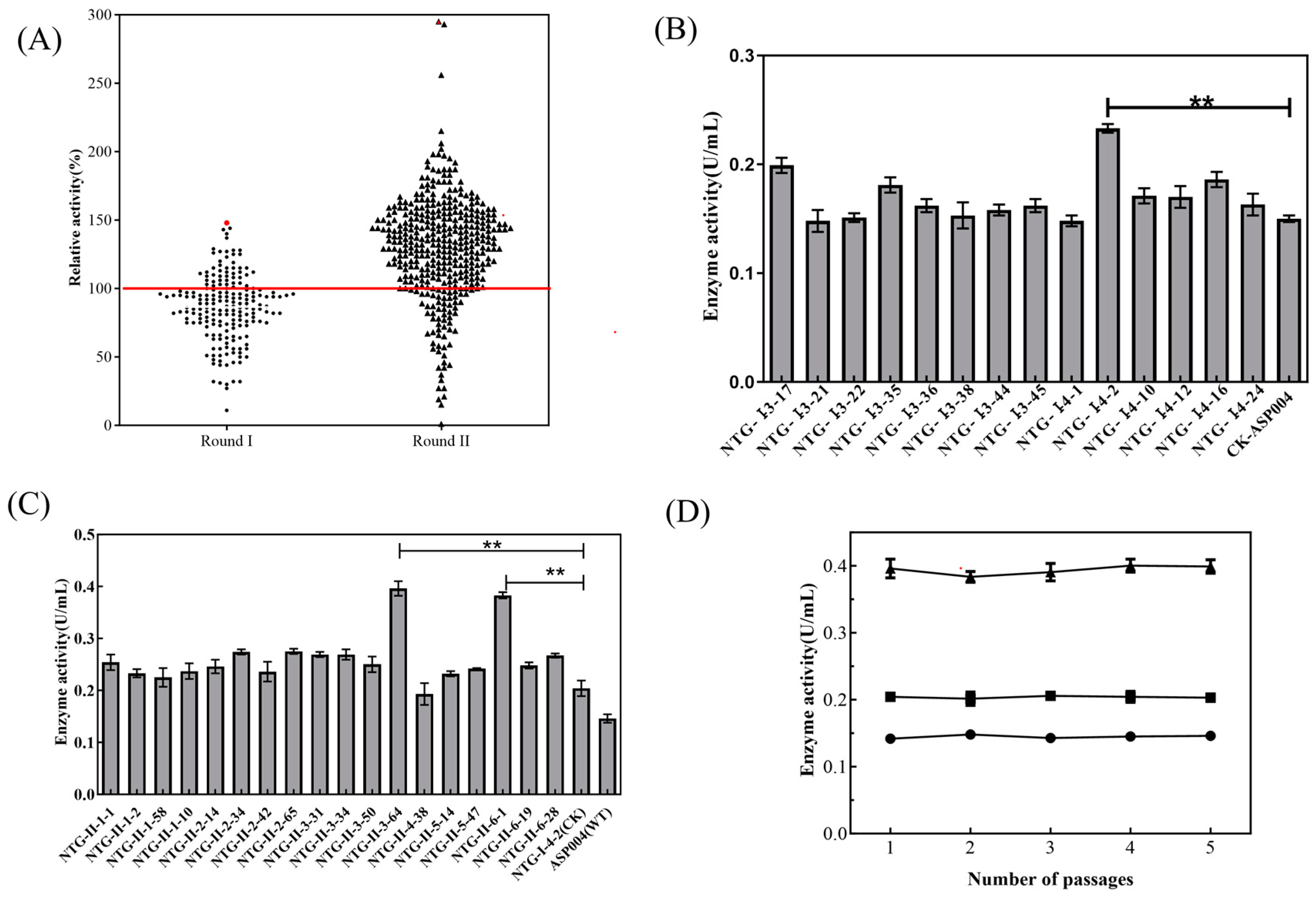
2.1. Mutagenesis and Screening of High-Yielding Strain

A. niger ASP004 underwent two rounds of mutation breeding, with the initial screening results shown in Figure 1A. In the first round, 235 strains were screened, of which 6.0% exhibited more than a 20% increase in enzyme activity. In the second round, 382 strains were screened, with 4.7% demonstrating a similar enhancement in enzyme activity.
Mutant strain NTG-I-4-2, selected from the first round of secondary screening, exhibited an enzyme activity of 0.233 ± 0.004 U·mL−1 (Figure 1B), representing a 55% increase compared to the wild-type strain ASP004 (0.15 ± 0.003 U·mL−1). Following further mutagenesis of NTG-I-4-2 using NTG and subsequent rounds of initial screening and secondary screening, the high-yielding strain NTG-II-3-64 was obtained (Figure 1C). This strain demonstrated an enzyme activity of 0.396 ± 0.016 U·mL−1, which was 70% higher than that of NTG-I-4-2.
As shown in Figure 1D, the enzyme activity of the high-yielding strain NTG-II-3-64 remained consistently high and stable over five generations, with a coefficient of variation of 1.7%.
α-Glucosidase has been reported to be synthesized by various Aspergillus species, including A. niger [20], A. carbonarious [29], A. awamori [32], and A. nidulans [28]. These findings suggest that multiple Aspergillus species are capable of producing α-glucosidase. In A. niger, targeted mutagenesis of the Asn694 residue in α-glucosidase has been shown to alter the enzyme’s hydrolytic and transglucosylation activities, thereby improving its suitability for industrial applications [33].

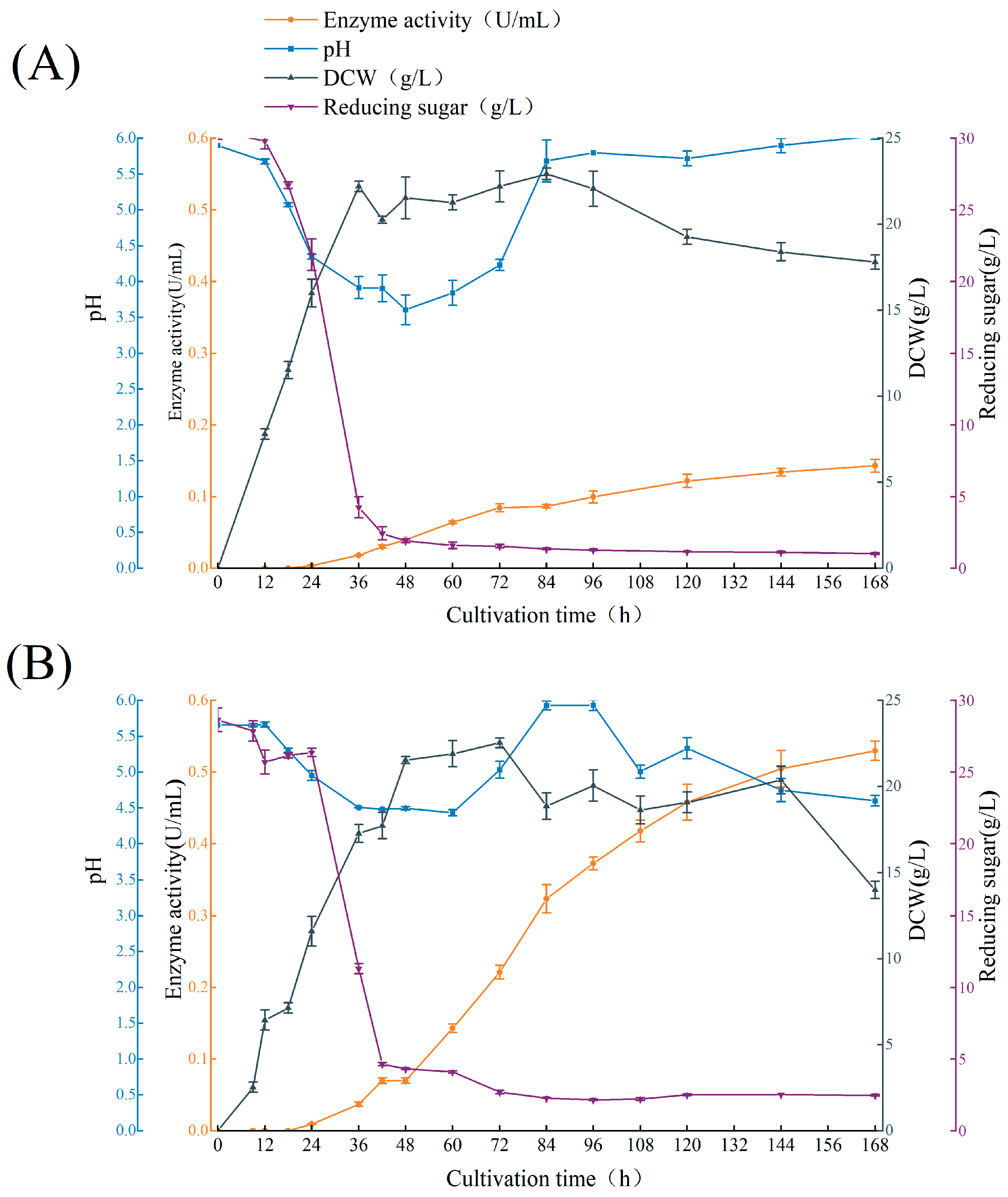
2.2. Batch Culture Profile of A. niger ASP004 and NTG-II-3-64

As shown in Figure 2, the biomass accumulation and reducing sugar concentrations of A. niger ASP004 and NTG-II-3-64 followed similar trends throughout the fermentation process. Biomass increased rapidly within 0–36 h for ASP004 and 0–48 h for NTG-II-3-64. The reducing sugar content in the fermentation broth decreased sharply from 0 to 42 h, corresponding to the period of rapid biomass growth. After 48 h, the reducing sugar content declined at a slower rate.
As shown in Figure 2A, α-glucosidase production by A. niger ASP004 initiated during the stable growth phase and increased rapidly between 24 and 72 h. After 72 h, the growth rate of enzyme activity slowed, but continued to rise, reaching 0.143 ± 0.01 U·mL−1 at 168 h. These findings suggest an incomplete coupling between α-glucosidase production and A. niger ASP004 growth. The pH of the fermentation broth initially decreased, reaching 3.6 at 48 h, likely due to the consumption of reducing sugars and the production of organic acids. After 48 h, the pH gradually increased to 5.8 by 96 h and remained stable until the end of fermentation. The fermentation profile of A. niger ASP004 in this study was consistent with the cultivation time course of A. niger CCRC 31494 reported by Chen et al. [34].
As shown in Figure 2B, α-glucosidase activity of A. niger NTG-II-3-64 increased rapidly after 48 h, coinciding with the stable growth phase. Although the growth rate slowed after 120 h, enzyme activity continued to rise until 168 h, reaching 0.530 ± 0.02 U·mL−1, which is 3.7 times higher than that of A. niger ASP004.
Throughout the cultivation process, the pH of the fermentation broth fluctuated between 4.5 and 6.0. Since pH variations can significantly impact enzyme production, careful pH regulation should be considered in future experiments.

2.3. Isolation and Purification of α-Glucosidase

α-Glucosidase was isolated and purified from the 120 h fermentation broth of the high-yielding strain NTG-II-3-64 by ultrafiltration and ethanol precipitation. The enzyme activity and protein concentration at each purification step are shown in Table 1. Following purification, the specific activity reached 0.18 U·mg−1, representing a 4.15-fold increase in purity with a 55% enzyme activity yield, effectively completing the primary separation of the crude enzyme.
The isoelectric point of α-glucosidase from A. niger typically ranges from 3.0 to 5.0, making anion-exchange chromatography an effective purification method. As shown in Figure 3, increasing the NaCl concentration of the elution buffer (0–0.6 M) resulted in four distinct A280 absorption peaks. Enzyme activity and protein concentrations of the collected fractions (Table 2) revealed that peak 1 exhibited the highest α-glucosidase activity (0.485 U·mL−1), with peaks 2, 3, and 4 showing significantly lower activity. Most of the enzyme activity was concentrated in peak 1.
Gel filtration chromatography using a HiPrep 16/60 Sephacryl S-200 HR column (Figure 3C) showed that the third A280 absorption peak had the highest absorbance and α-glucosidase activity. Fractions from each peak were collected and analyzed for enzyme activity and protein concentration (Table 3). The highest α-glucosidase activity (0.23 U·mL−1) was observed in peak 3, whereas peaks 1 and 2 showed minimal activity, suggesting a lower concentration of α-glucosidase.
Compared to the crude enzyme from the original fermentation supernatant, the α-glucosidase purified in the final step of gel filtration chromatography was enhanced by 22.22-fold, with a specific activity of 1.0 U·mg−1 and an activity yield of 15.28% (Table 1).
Native-PAGE and SDS-PAGE analysis of α-glucosidases at different purification steps are shown in Figure 4. Crude extracts (Lane 2) showed multiple contaminants, while gel filtration (Lanes 5–6) produced a homogeneous preparation. The peak 1 of gel filtration chromatography (Figure 4B, Lane 6) revealed that the α-glucosidase produced by A. niger strain NTG-II-3-64 is a two-subunit enzyme. Similarly, Kato et al. [28] reported that α-glucosidase from A. nidulans was a heterodimeric protein comprising 74 and 55 kDa subunits. In contrast, α-glucosidase from the thermophilic fungus Malbranchea cinnamomea was reported as a monomer with molecular mas of 65.7 kDa [35]. Native-PAGE showed a single band about at 230 kDa, while SDS-PAGE revealed two bands, corresponding to subunits with molecular mass above and below 130 kDa, due to denaturation and depolymerization of the enzyme. The purified enzyme (Lane 6) aligns closely with the commercial α-glucosidase (Lane 7) in band position, validating correct molecular mass and purity. The molecular masses of α-glucosidase estimated by SDS-PAGE were 75 kDa for A. niger ITV-01 [36] and 145 kDa for A. neoniger [8], indicating that the molecular weights of α-glucosidase vary among different sources.

2.4. Enzymatic Properties of α-Glucosidase

2.4.1. Effect of Temperature and pH on α-Glucosidase Activity

The optimum reaction temperature of α-glucosidase produced by NTG-II-3-64 was 60 °C (Figure 5A). For thermal stability determination, the enzyme was incubated at 35 °C to 80 °C for 60 min, then rapidly cooled in an ice-water bath. With the increase in temperature, the thermostability of α-glucosidase decreased. The residual enzyme activity was 62% and 43% at 50 °C and 60 °C, respectively. Only 7% of the remaining enzyme activity was retained at 70 °C, and no residual enzyme activity was detected at 75 °C and 80 °C, because the high-temperature heating led to the irreversible changes in the structure of α-glucosidase, which resulted in the inactivation of the enzyme (Figure 5B).
Kita et al. reported that the optimal reaction temperature for α-glucosidase produced by A. niger was found to be 55 °C, with enzyme activity remaining above 90% within the 0–55 °C range; however, enzyme activity was lost at 70 °C [37]. The maximum activity of α-glucosidase from A. niger ITV-01 occurred at 80 °C [36], which is higher than that observed in this study. Through immobilization, chemical modification, and the use of additive techniques, the α-glucosidase from the thermophilic archaebacterium Thermococcus strain AN1 maintained good activity at 130 °C [38].
The effects of pH on the activity and stability of α-glucosidase are shown in Figure 5C,D. For pH stability determination, the enzyme was incubated in buffer solutions ranging from pH 3.0 to 8.0 at 4 °C for 24 h. The enzyme exhibited optimal activity at pH 4.5, and remained relatively stable in an acidic environment (pH 3.0–6.0), retaining over 90% of its activity. However, enzyme activity declined sharply at pH levels above 6.
α-glucosidase from the thermophilic fungus M. cinnamomea exhibited optimal activity at pH 6.0 and 50 °C [35], highlighting the distinct characteristics of α-glucosidase from different sources.

2.4.2. Effect of Metal Ions and EDTA on α-Glucosidase Activity

The purified α-glucosidase was incubated with different metal ions and EDTA for 1 h at 25 °C, and the relative residual enzyme activity was measured, as shown in Table 4. Most metal ions at 1 mmol·L−1, including Fe3+ (76.1% residual activity) and Cu2+ (80.1%), significantly inhibited the enzyme, while others like Zn2+, Mg2+, and Mn2+ caused milder inhibition. Notably, Ca2+ enhanced activity (109%), suggesting a potential regulatory role, whereas NaCl had little effect (98.2%). EDTA, tested at 20–100 mmol·L−1, showed no consistent inhibition, with slight activation at 20 mM (110%), indicating that α-glucosidase does not strictly depend on metal cofactors.
Zhang et al. [39] reported that Ca2+ enhanced the activity of α-glucosidase produced by A. niger, while Cu2+ and Fe2+ inhibited the enzyme. In contrast, disodium EDTA had no significant effect on enzyme activity. Similarly, Chen et al. [40] studied recombinant α-glucosidase produced by a P. pastoris host and found that the addition of 1 mM of Cu2+, Ca2+, Fe3+, or other metal ions did not influence the recombinant enzyme’s activity. These findings align with the results of the present study, highlighting the inhibitory effects of specific metal ions on α-glucosidase activity.

2.4.3. Effect of Surfactants on α-Glucosidase Activity

The effects of various surfactants on α-glucosidase activity are shown in Table 5. Non-ionic surfactants, including Tween 20, Tween40, Tween80, Span 80, Triton X-100, and PEG 2000, all enhanced enzyme activity at 1% concentration, with Span 80 showing the greatest activation (131% residual activity). In contrast, the ionic surfactant SDS strongly inhibited the enzyme, reducing activity to just 50%. These results demonstrate that non-ionic surfactants can significantly boost α-glucosidase performance, likely by stabilizing the enzyme or improving substrate accessibility, while ionic surfactants like SDS disrupt enzyme function, probably through denaturation.
Sheu et al. [29] investigated the α-glucosidase isolated and purified from the fermentation broth of A. carbonarious and found that 5 mmol·L−1 SDS reduced enzyme activity to 88% of the control. Yoon and Robyt [41] studied the effects of non-ionic surfactants on various starch hydrolases and observed that surfactants such as PEG 2000, PEG 1000, Triton X-100, and PEG 1500 significantly enhanced enzyme activity. However, the impact of non-ionic surfactants on α-glucosidase activity has not been reported in the literature.

2.4.4. Substrate Specificity of α-Glucosidase

The substrate specificity of α-glucosidase is shown in Table 6. Maltose was the preferred substrate for α-glucosidase, with the highest enzyme activity set at 100%. The enzyme also exhibited activity toward α-methylglucose, soluble starch, sucrose, isomaltose, isomaltriose, and panose, while showing no activity against lactose and 4-nitrophenyl-β-glucopyranoside. These results indicate that α-glucosidase is a bond-specific enzyme with broad substrate compatibility. It primarily cleaves α-1,4-glycosidic bonds and can also act on α-1,2- and α-1,6-glycosidic bonds, but it does not hydrolyze β-1,4-glycosidic bonds, consistent with previous reports [14].
Kita et al. [37] investigated the substrate specificity of α-glucosidase from A. niger and found that it could hydrolyze oligosaccharides, including maltose, maltotriose, maltotetraose, maltopentose, and isomaltose, as well as soluble starch. In addition to cleaving α-1,4-glycosidic bonds, the enzyme could also act on α-1,2- and α-1,6-glycosidic bonds. Notably, maltose, maltotetraose, maltopentose, and phenyl-α-maltoside were identified as its preferred substrates. These findings are consistent with the results of the present study.

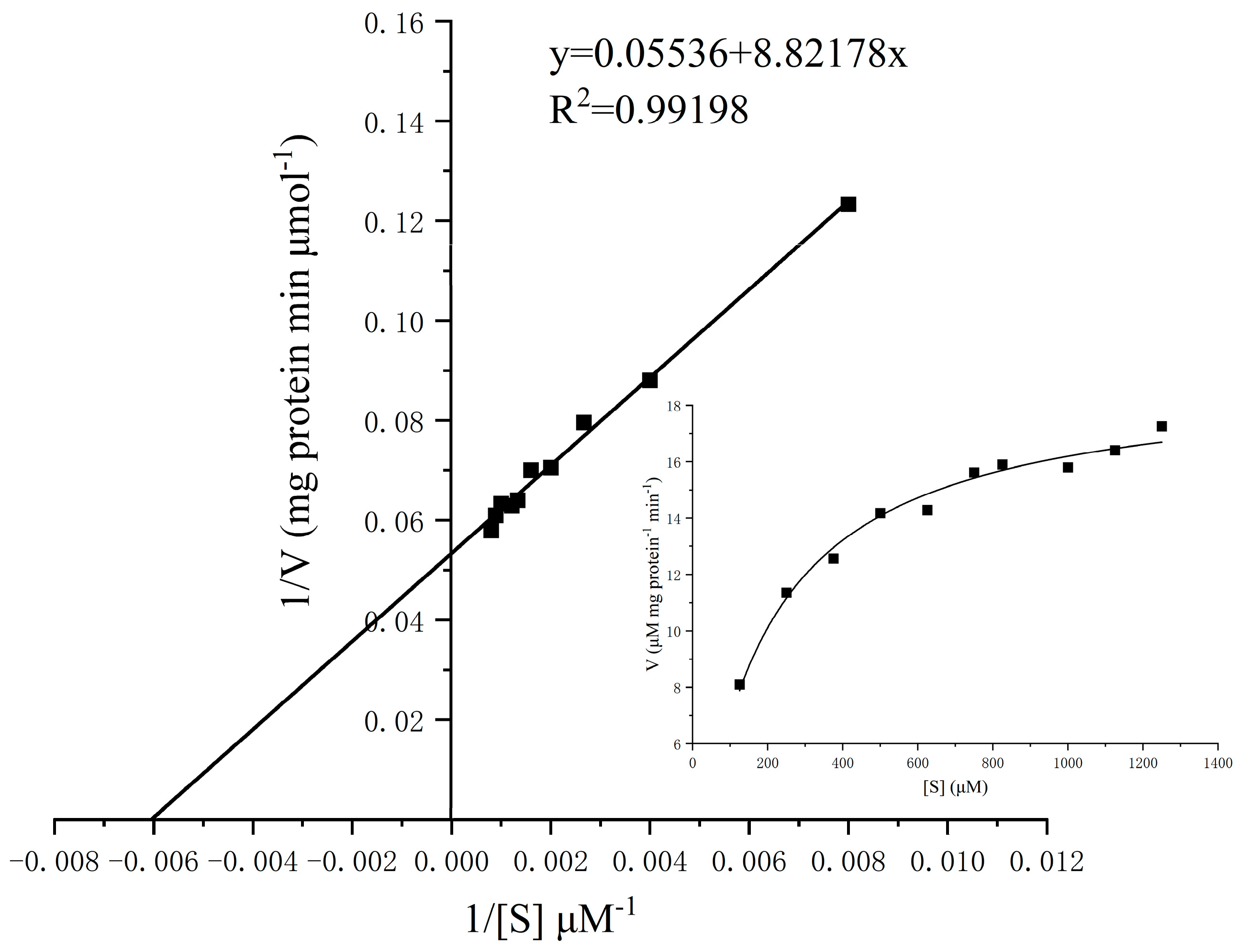
2.4.5. Kinetics Analysis of α-Glucosidase

The initial reaction rate of purified α-glucosidase at varying pNPG concentrations was measured, and the kinetic curves are presented in Figure 6. The enzyme exhibited a Km of 0.17 mM and a Vmax of 18.7 µmol min−1 mg−1 at 50 °C and pH 5.5. Previous reports indicated that the Km values of α-glucosidase from A. niger GN-3 for pNPG were 0.62 mM [15], while that from another A. niger strain was 0.7 mM [27]. In contrast, the α-glucosidase from Thermoascus aurantiacus showed a much lower Km of 0.07 µM and a higher Vmax of 318.0 µmol min−1 mg−1 [42], indicating a significantly stronger substrate affinity and catalytic efficiency.

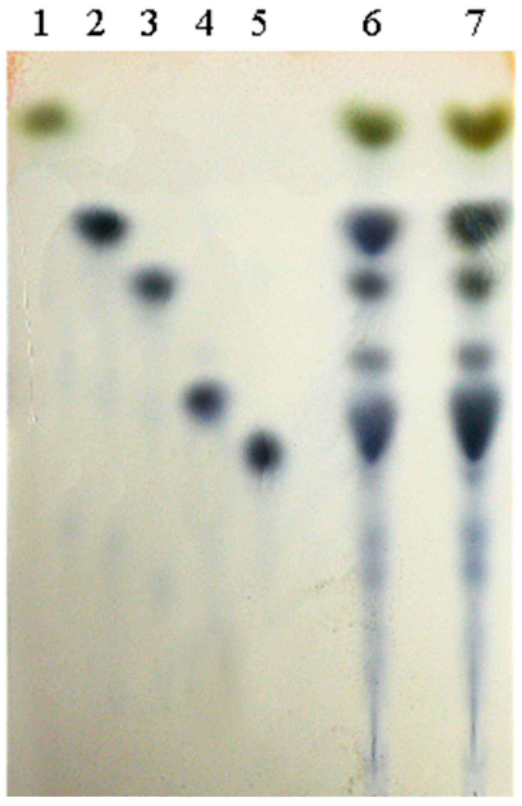
2.4.6. Detection of α-Glucosidase Transglycosidase Products

TLC analysis of α-glucosidase transglucosylation products (Figure 7) showed that the purified α-glucosidase converts maltose into isomaltose, panose, and isomaltriose (Lane 6), similar to the commercial enzyme (Lane 7). This confirms that the α-glucosidase from strain NTG-II-3-64 exhibits transglucosidase activity.

3. Materials and Methods

3.1. Strains, Chemicals and Medium

The A. niger ASP004 strain, producing α-glucosidase, was maintained in our laboratory. Glucose, maltose, isomaltose, panose, and isomaltriose were purchased from Sinopharm Chemical Reagents (Shanghai, China). Nitrosoguamidine (NTG), 4-nitrophenyl-α-D-glucopyranoside (pNPG), and methyl-α-D-glucopyranoside (α-MG) were purchased from Sigma-Aldrich Co., Ltd. (St. Louis, MO, USA). Protein markers and the malt extract were purchased from Thermo Fisher Scientific Inc. (Waltham, MA, USA). Yeast extract was provided from Angel Yeast Co., Ltd. (Yichang, Hubei, China). Commercial α-glucosidase was supplied from Amano (Nagoya, Japan).
Potato Dextrose Agar was purchased from Sinopharm Chemical Reagents (Shanghai, China). The α-MG–Bengal red medium contained (g·L−1) 20 α-MG, 3 NaNO3, 1 K2HPO4·3H2O, 0.5 KCl, 0.5 MgSO4·7H2O, 0.01 FeSO4·7H2O,18 Agar, and 66 mg Bengal red, with a pH of 7.2. The liquid fermentation medium was composed of (g·L−1) 40 malt extract, 20 yeast extract, 5 K2HPO4·3H2O and 0.5 MgSO4·7H2O, adjusted to a pH of 5.9. The optimal enzyme-producing fermentation medium for strain NTG-II-3-64 consisted of (g·L−1) 40 malt extract powder, 3 yeast peptone, 2.5 tratone X-100, 0.5 MgSO4·7H2O, and 5 K2HPO4·3H2O, with a pH of 5.9.

3.2. α-Glucosidase Activity Determination

α-glucosidase catalyzes the hydrolysis of 4-nitrophenyl-α-D-glucopyranoside (pNPG) to release p-nitrophenol (pNP), which exhibits a yellow color under alkaline conditions. The amount of pNP produced can be quantified using a colorimetric method to determine the enzyme activity level [43].
The detection method was as follows: 0.02 mL of 5 mM pNPG substrate solution and 0.07 mL of 0.1 M acetoacetate–sodium acetate buffer (pH 5.5) were added to a 96-well plate, followed by incubation at 50 °C for 3 min. Then, 0.01 mL of appropriately diluted crude enzyme solution was added, and the reaction continued at 50 °C for 15 min. To stop the reaction and initiate color development, 0.1 mL 1 M Na2CO3 solution was added. Absorbance was measured at 405 nm using a Synergy HT microplate reader (BioTek, Winooski, VT, USA). The control group followed the same steps without enzyme addition. One unit of α-glucosidase activity was defined as the amount of enzyme that hydrolyzed the substrate to release 1 μmol of pNP per minute.

3.3. Mutagenesis and Screening of High-Yield Strain of α-Glucosidase

A. niger ASP004 was activated on a fresh PDA slant and incubated at 30 °C for 60 h. The spores were scraped, suspended in normal saline, and adjusted to 106 spores mL−1. For the mutagenesis, 1 mL of NTG solution (prepared in 0.05 M trimaleic acid buffer, pH 6.0) was added to the spore suspension, while the control group was treated with buffer without NTG. The mixture, wrapped in tin foil to prevent light exposure, was incubated at 30 °C and 200 rpm. After incubation, the mixture was centrifuged at 4000× g for 5 min to collect the cells. The pellet was washed three times with normal saline, resuspended, and spread onto an α-MG screening plate for further analysis.
The screening process for high-yielding α-glucosidase strains followed the process outlined in Figure 8. After mutagenesis, the strains were diluted, spread onto α-MG–Bengal red plates, and incubated at 30 °C for 3 d. Rapidly growing mutants in α-MG–Bengal red plates were selected and transferred to fresh PDA plates. Spore suspensions (107 spores·mL−1) were prepared after 60 h of incubation at 30 °C. A 2% inoculum was added to 250 mL shake flasks containing 50 mL of liquid fermentation medium and cultured at 30 °C and 200 rpm for 4 d. Control strains were similarly inoculated for comparison. Strains with a ≥20% increase in enzyme activity were re-screened using the same procedure. The coefficient of variation (CV) was calculated; strains with a CV below 5% and significantly higher enzyme activity were considered as high-yielding strains. The selected strain was preserved on a PDA slant and designated as the starting strain for subsequent experiments.

3.4. Batch Culture Profile of High-Yield Strain of α-Glucosidase

High-yield α-glucosidase strains with confirmed genetic stability were transferred to fresh PDA slants and incubated at 30 °C for 3 d. After incubation, spores were scraped using 0.1% Tween 80 to prepare a spore suspension with a concentration of approximately 107 spores·mL−1. A 2% inoculum of the suspension was added to a liquid fermentation medium and cultured at 30 °C and 200 rpm. Samples were collected at specific time points (0, 9, 12, 18, 24, 36, 42, 48, 60, 72, 84, 96, 108, and 168 h) for the determination of enzyme activity, biomass, pH, and reducing sugar content.
Biomass determination: Biomass was measured using a gravimetric method. Pre-weighed filter paper was dried at 80 °C to a constant weight. The fermentation broth was filtered through the paper, and retained cells were washed three times with distilled water. After drying overnight at 80 °C, the final weight was recorded. Biomass was calculated by subtracting the initial filter paper weight from the final weight [23].
Reducing sugar content determination: The reducing sugar content was measured using the DNS method. In a 25 mL test tube, 2.0 mL of diluted sample and 1.5 mL of DNS reagent were mixed and incubated at 100 °C for 5 min. After rapid cooling, the volume was adjusted to 25 mL with distilled water. A control with 2.0 mL of distilled water instead of the sample was used for baseline correction. Absorbance was measured at 520 nm using a spectrophotometer, and reducing sugar content was determined using a glucose standard curve.

3.5. Method of Isolation and Purification of α-Glucosidase

After 6 d of fermentation, the broth was centrifuged at 4 °C and 10,000× g to remove cells and debris. The supernatant was filtered through a 0.22 µm Millipore Express PES hydrophilic membrane (filter diam. 90 mm, Millipore Sigma, Darmstadt, Germany) to obtain the crude enzyme solution. It was then concentrated five-fold using a 30 kDa cutoff hollow fiber column (0.5 mm Fiber, GE Healthcare, Chicago, IL, USA) while maintained in an ice-water bath. Next, five volumes of pre-cooled 95% ethanol were added, and the mixture was incubated at 4 °C for 12 h. The precipitate was collected by centrifugation at 4 °C and 10,000× g for 15 min, dissolved in 10 mM phosphate buffer (pH 6), and equilibrated overnight to ensure complete dissolution. It was then dialyzed against 10 mM phosphate buffer (pH 6) overnight at 4 °C.
For purification, the dialyzed solution was concentrated using a 30 kDa cutoff centrifugal concentrator (Amicon® Ultra Centrifugal Filter, Millipore Sigma, Darmstadt, Germany), then filtered through a 0.22 µm membrane. The filtrate was loaded onto a 1 mL DEAE Sepharose Fast Flow anion-exchange column (GE Healthcare, Chicago, IL, USA) pre-equilibrated with 10 mM phosphate buffer (pH 6). Elution was carried out using a linear NaCl gradient (0 to 0.6 M) at 0.8 mL·min−1 over 50 column volumes. Fractions with the highest enzyme activity were pooled and dialyzed against 10 mM phosphate buffer (pH 6) for 4 h, followed by a buffer change and continued dialysis overnight. The enzyme solution was then concentrated using a 30 kDa cutoff centrifugal concentrator.
Further purification was performed using a Sephacryl S-200 High-Resolution gel filtration column (GE Healthcare, Chicago, IL, USA). A 1 mL sample was applied, and elution proceeded at 0.5 mL·min−1. Active fractions were collected, yielding the purified enzyme. At each purification step, enzyme activity and protein concentration were measured, and purity and molecular characteristics were assessed using SDS-PAGE and Native-PAGE. The enzymatic properties of the purified α-glucosidase were then analyzed.

3.6. Enzyme Property Determination

3.6.1. Effect of Temperature and pH on Enzyme Activity

The activity of the purified α-glucosidase was measured at temperatures ranging from 35 °C to 80 °C, with the highest activity set as 100% to generate a temperature–activity curve. For thermal stability, the enzyme was incubated at 35 °C to 80 °C for 60 min, then rapidly cooled in an ice-water bath. The activity of an unincubated sample was set as 100%, and the relative residual activity at each temperature was assessed.
For pH-dependent activity analysis, 0.1 M buffers with pH 3.0 to 8.0 were used: citrate–sodium citrate (pH 3.0–4.0), aceto-sodium acetate (pH 4.5–6.0), and phosphate (pH 6.5–8.0). The purified enzyme was incubated in each buffer at 50 °C, and activity was measured, with the highest set as 100% to plot a pH–activity curve. For pH stability determination, the enzyme was incubated in buffer solutions ranging from pH 3.0 to 8.0 at 4 °C for 24 h, and residual activity was measured to determine relative stability across pH levels.

3.6.2. Effect of Metal Ions and EDTA on Enzyme Activity

The effect of metal ions on purified α-glucosidase activity was assessed by incubating the enzyme with a 1 mM final concentration of various metal ions at 25 °C for 1 h. The control sample, without metal ions, was set as 100% activity, and relative residual activity was measured. To evaluate EDTA’s impact, the enzyme was incubated with disodium EDTA at final concentrations of 10 to 100 mM under the same conditions. The untreated control sample was defined as 100% activity, and relative residual activity was determined.

3.6.3. Effect of Surfactants on Enzyme Activity

The effect of various surfactants on the activity of purified α-glucosidase was evaluated by mixing the enzyme with Tween 20, Tween 40, Tween 80, Span-80, Triton X-100, and PEG 2000, each at an initial concentration of 2%, resulting in a final surfactant concentration of 1%. The mixtures were incubated at room temperature (25 °C) for 1 h. The enzyme activity of the control sample, without any surfactant, was set as 100%, and the relative residual enzyme activity was determined.

3.6.4. Substrate Specificity

To evaluate the substrate specificity of α-glucosidase, 2% solutions of α-methylglucose, maltose, sucrose, lactose, soluble starch, isomaltose, panose, isomaltotriose, and 4-nitrophenyl-β-glucopyranoside (pNPG) were used as substrates. The relative enzyme activity of α-glucosidase was determined using each substrate, with the highest activity set as 100%.

3.6.5. Enzymatic Kinetic Analysis

To evaluate the kinetic properties of the purified α-glucosidase, pNPG solutions were prepared at concentrations of 1.25, 2.5, 3.75, 5.0, 6.25, 7.5, 8.25, 10.0, 11.2, 12.5, 13.75, and 15 mM. The enzyme’s reaction rates were measured at each substrate concentration to assess its activity.

3.6.6. Detection of α-Glucosidase Transglycosidase Products by Thin-Layer Chromatography (TLC)

To prepare the glucosidase products for TLC, in a 10 mL tube, mix 1 mL of 0.1 mol/L acetate buffer (pH 5.5), 0.5 mL of 30% (v/v) maltose, and 0.5 mL each of purified and commercial α-glucosidase (0.4 U/mL). Incubate at 50 °C for 1 h, heat at 100 °C for 5 min to inactivate enzymes, and then centrifuge at 10,000× g for 5 min. Filter the supernatant through a 0.22 μm membrane for TLC analysis.
A 1 µL sample was applied to a 100 × 200 mm GF254 silica gel plate (Qingdao Haiyang Chemical, Qingdao, Shandong, China), including glucose, maltose, isomaltose, panose, isomaltotriose, standard isomaltose, and the glucosidase products of purified and commercial. The plate was developed three times in a solvent system of n-butanol/glacial acetic acid/water (2:1:1, v/v/v) for 10 h, then air-dried. The color reagent was prepared by mixing diphenylamine (4 g/100 mL acetone), aniline (4 mL/100 mL acetone), and 20 mL of 85% phosphoric acid. The plate was dipped in the reagent, removed immediately, air-dried, and heated at 80 °C for 1 h to visualize the chromatographic bands.

3.7. Statistical Analysis

Statistical significance was evaluated using a t-test with SPSS version 26 (IBM, Armonk, NY, USA). Results were presented as means ± standard deviation (SD) from a minimum of three experiments.

4. Conclusions

In this study, a high-yielding strain, NTG-II-3-64, was obtained from A. niger ASP004 through two rounds of NTG mutation breeding, resulting in a 3.7-fold increase under optimized conditions. The crude α-glucosidase solution from NTG-II-3-64 underwent ultrafiltration concentration, ethanol precipitation, ion-exchange chromatography, and gel filtration chromatography, yielding electrophoretically pure α-glucosidase. The purified enzyme exhibited optimal activity at 60 °C and pH 4.5, with stability maintained in acidic environments (pH 3.0–6.0), retaining over 90% activity. Ca2+, Tween 20, Tween 40, Tween 80, Span 80, Triton X-100, PEG 2000, and 20 mM EDTA enhanced enzyme activity, while K+, Mg2+, Zn2+, Fe3+, Cu2+, and SDS inhibited it. Additionally, α-glucosidase from NTG-II-3-64 exhibited transglucosidase activity, converting maltose into isomalto oligosaccharides (IMOs). These findings offer valuable insights for the industrial production and application of α-glucosidase.

Author Contributions

B.Y.: experimentation, data processing, manuscript writing, funding acquisition; Q.L.: experimentation, data processing, manuscript writing; J.X.: data processing; G.F.: data processing; Z.C.: conceptualization, supervision; H.G.: conceptualization, methodology, data curation, revision, funding acquisition. All authors have read and agreed to the published version of the manuscript.

Funding

Funding was provided by the Ningxia Association for Science and Technology Youth Talent Support Program NingXia Kexie (2024) No.6; the Natural Science Foundation of Shanghai Municipality (22ZR141200) (Shanghai, China); the Ningxia Provincial Natural Science Foundation (2023AAC03735); and the Ningxia technology-benefiting-people program (2024CMG03049). The authors thank the Instruments Sharing Platform of School of Life Sciences, East China Normal University, for technical service.

Data Availability Statement

The data that support the findings of this study are available from the corresponding author upon reasonable request.

Conflicts of Interest

The authors declare that they have no conflicts of interest in this work.

References

  1. Zhou, B.; Huang, N.; Zeng, W.; Zhang, H.; Chen, G.; Liang, Z. Development of a strategy for the screening of α-glucosidase-producing microorganisms. J. Microbiol. 2020, 58, 163–172. [Google Scholar] [CrossRef] [PubMed]
  2. Nimpiboon, P.; Nakapong, S.; Pichyangkura, R.; Ito, K.; Pongsawasdi, P. Synthesis of a novel prebiotic trisaccharide by a type I α-glucosidase from B. licheniformis strain TH4-2. Process Biochem. 2011, 46, 448–457. [Google Scholar] [CrossRef]
  3. Yamamoto, K.; Miyake, H.; Kusunoki, M.; Osaki, S. Steric hindrance by 2 amino acid residues determines the substrate specificity of isomaltase from Saccharomyces cerevisiae. J. Biosci. Bioeng. 2011, 112, 545–550. [Google Scholar] [CrossRef]
  4. Okuyama, M.; Saburi, W.; Mori, H.; Kimura, A. α-Glucosidases and α-1, 4-glucan lyases: Structures, functions, and physiological actions. Cell. Mol. Life Sci. 2016, 73, 2727–2751. [Google Scholar] [CrossRef]
  5. Meissner, H.; Liebl, W. Thermotoga maritima maltosyltransferase, a novel type of maltodextrin glycosyltransferase acting on starch and malto-oligosaccharides. Eur. J. Biochem. 1998, 258, 1050–1058. [Google Scholar] [CrossRef]
  6. Basu, A.; Mutturi, S.; Prapulla, S.G. Modeling of enzymatic production of isomaltooligosaccharides: A mechanistic approach. Catal. Sci. Technol. 2015, 5, 2945–2958. [Google Scholar] [CrossRef]
  7. Um, H.E.; Park, B.R.; Kim, Y.M.; Lee, B.H. Slow digestion properties of long-sized isomaltooligosaccharides synthesized by a transglucosidase from Thermoanaerobacter thermocopriae. Food Chem. 2023, 417, 135892. [Google Scholar] [CrossRef] [PubMed]
  8. Kumar, S.; Basu, A.; Anu-Appaiah, K.A.; Gnanesh Kumar, B.S.; Mutturi, S. Identification and characterization of novel transglycosylating α-glucosidase from Aspergillus neoniger. J. Appl. Microbiol. 2020, 129, 1644–1656. [Google Scholar] [CrossRef]
  9. Song, Y.B.; Lamothe, L.M.; Rodriguez, N.E.; Rose, D.R.; Lee, B.H. New insights suggest isomaltooligosaccharides are slowly digestible carbohydrates, rather than dietary fibers, at constitutive mammalian α-glucosidase levels. Food Chem. 2022, 383, 132456. [Google Scholar] [CrossRef]
  10. Song, K.M.; Okuyama, M.; Kobayashi, K.; Mori, H.; Kimura, A. Characterization of a glycoside hydrolase family 31 α-glucosidase involved in starch utilization in Podospora anserine. Biosci. Biotechnol. Biochem. 2013, 77, 2117–2124. [Google Scholar] [CrossRef]
  11. Krohn, B.M.; Lindsay, J.A. Purification and characterization of a thermostable α-glucosidase from a Bacillus subtilis high-temperature growth transformant. Current Microbiol. 1991, 22, 273–278. [Google Scholar] [CrossRef]
  12. Marín, D.; Linde, D.; Fernández-Lobato, M. Purification and biochemical characterization of an α-glucosidase from Xanthophyllomyces dendrorhous. Yeast 2006, 23, 117–125. [Google Scholar] [CrossRef] [PubMed]
  13. Larsbrink, J.; Izumi, A.; Hemsworth, G.R.; Davies, G.J.; Brumer, H. Structural enzymology of Cellvibrio japonicus Agd31B protein reveals α-transglucosylase activity in glycoside hydrolase family 31. J. Biol. Chem. 2012, 287, 43288–43299. [Google Scholar] [CrossRef]
  14. Nakamura, A.; Nishimura, I.; Yokoyama, A.; Lee, D.G.; Hidaka, M.; Masaki, H.; Kimura, A.; Chiba, S.; Uozumi, T. Cloning and sequencing of an α-glucosidase gene from Aspergillus niger and its expression in A. nidulans. J. Biotechnol. 1997, 53, 75–84. [Google Scholar] [CrossRef] [PubMed]
  15. Ogawa, M.; Nishio, T.; Minoura, K.; Uozumi, T.; Wada, M.; Hashimoto, N.; Kawachi, R.; Oku, T. Recombinant α-Glucosidase from Aspergillus niger. Overexpression by Emericella nidulans, Purification and Characterization. J. Appl. Glycosci. 2006, 53, 13–16. [Google Scholar] [CrossRef]
  16. Zhao, N.; Xu, Y.; Wang, K.; Zheng, S. Synthesis of isomalto-oligosaccharides by Pichia pastoris displaying the Aspergillus niger α-glucosidase. J. Agric. Food Chem. 2017, 65, 9468–9474. [Google Scholar] [CrossRef]
  17. Zhang, F.; Wang, W.; Bah, F.B.; Song, C.; Zhou, Y.; Ji, L.; Yuan, Y. Heterologous expression of a thermostable α-glucosidase from Geobacillus sp. strain HTA-462 by Escherichia coli and its potential application for isomaltose–oligosaccharide synthesis. Molecules 2019, 24, 1413. [Google Scholar]
  18. Kumar, S.; Mutturi, S. Expression of a novel α-glucosidase from Aspergillus neoniger in Pichia pastoris and its efficient recovery for synthesis of isomaltooligosaccharides. Enzym. Microb. Technol. 2020, 141, 109653. [Google Scholar] [CrossRef] [PubMed]
  19. Tissopi, T.; Kumar, S.; Sadhu, A.; Mutturi, S. Surface display of novel transglycosylating α-glucosidase from Aspergillus neoniger on Pichia pastoris for synthesis of isomaltooligosaccharides. Biochem. Eng. J. 2022, 181, 108400. [Google Scholar] [CrossRef]
  20. Kimura, A.; Takenishi, S.; Takata, M.; Tsujisaka, Y.; Chiba, S. Properties and Substrate Specificity of a-Glucosidase from Aspergillus niger GRM 3. J. Appl. Glycosci. 1997, 44, 303–312. [Google Scholar]
  21. EFSA Panel on Food Contact Materials, Enzymes and Processing Aids (CEP); Silano, V.; Barat Baviera, J.M.; Bolognesi, C.; Cocconcelli, P.S.; Crebelli, R.; Gott, D.M.; Grob, K.; Lambré, C.; Lampi, E.; et al. Safety evaluation of the food enzyme α-glucosidase from the Aspergillus niger strain AE-TGU. EFSA J. 2022, 20, e07240. [Google Scholar] [PubMed]
  22. Li, W.; Chen, G.; Gu, L.; Zeng, W.; Liang, Z. Genome Shuffling of Aspergillus niger for Improving Transglycosylation Activity. Appl. Biochem. Biotechnol. 2014, 172, 50–61. [Google Scholar] [CrossRef] [PubMed]
  23. Chen, G.G.; Li, W.; Zhang, Y.K.; Qin, Y.L.; Wu, K.Y.; Liang, Z.Q. A high-throughput method for screening of Aspergillus niger mutants with high transglycosylation activity by detecting non-fermentable reducing sugar. World J. Microbiol. Biotechnol. 2011, 27, 1519–1523. [Google Scholar] [CrossRef] [PubMed]
  24. Kang, S.W.; Ko, E.H.; Lee, J.S.; Kim, S.W. Over-production of β-glucosidase by Aspergillus niger mutant from lignocellulosic biomass. Biotechnol. Lett. 1999, 21, 647–650. [Google Scholar] [CrossRef]
  25. Wang, S.; Wang, Q.; Zhou, Y.; Lu, Z.; Zhang, G.; Ma, Y. A new GH13 α-glucosidase from alkaliphilic Bacillus pseudofirmus 703 with both exo-α-l, 4-glucosidase and oligo-l, 6-glucosidase activities toward amylopectin. Int. J. Biol. Macromol. 2017, 101, 973–982. [Google Scholar] [CrossRef]
  26. Krohn, B.M.; Lindsay, J.A. Purification, characterization, and comparison of an α-glucosidase (endo-oligo-1, 4-glucosidase) from the mesophile Bacillus subtilis and the obligate thermophile Bacillus caldolyticus. Curr. Microbiol. 1991, 22, 133–140. [Google Scholar] [CrossRef]
  27. Mccleary, B.V.; Gibson, T.S.; Sheehan, H.; Casey, A.; Horgan, L.; O’Flaherty, J. Purification, properties, and industrial significance of transglucosidase from Aspergillus niger. Carbohydr. Res. 1989, 185, 147–162. [Google Scholar] [CrossRef]
  28. Kato, N.; Suyama, S.; Shirokane, M.; Kato, M.; Kobayashi, T.; Tsukagoshi, N. Novel α-glucosidase from Aspergillus nidulans with strong transglycosylation activity. Appl. Environ. Microbiol. 2002, 68, 1250–1256. [Google Scholar] [CrossRef]
  29. Sheu, D.C.; Duan, K.J.; Lin, C.T. Purification and characterization of α-glucosidase from Aspergillus carbonarious. Biotechnol. Tech. 1994, 8, 515–520. [Google Scholar] [CrossRef]
  30. Botstein, D.; Jones, E.W. Nonrandom Mutagenesis of the Escherichia coli Genome by Nitrosoguanidine. J. Bacteriol. 1969, 98, 847–848. [Google Scholar] [CrossRef]
  31. Oeschger, M.P.; Berlyn, M.K.B. A simple procedure for localized mutagenesis using nitrosoguanidine. Mol. Gen. Genet. MGG 1974, 134, 77–83. [Google Scholar] [CrossRef]
  32. Anindyawati, T.; Ann, Y.G.; Ito, K.; Iizuka, M.; Minamiura, N. Two kinds of novel a-glucosidases from Aspergillus awamori KT-11: Their purifications, properties and specificities. J. Ferment. Bioeng. 1998, 85, 465–469. [Google Scholar] [CrossRef]
  33. Ma, M.; Okuyama, M.; Sato, M.; Tagami, T.; Klahan, P.; Kumagai, Y.; Mori, H.; Kimura, A. Effects of mutation of Asn694 in Aspergillus niger α-glucosidase on hydrolysis and transglucosylation. Appl. Microbiol. Biotechnol. 2017, 101, 6399–6408. [Google Scholar] [CrossRef] [PubMed]
  34. Chen, W.C.; Lee, S.L.; Tung, P.Y. Effects of initial maltose concentration, agitation and aeration rates on the production of glucosyltransferase by Aspergillus niger CCRC 31494. Biotechnol. Lett. 1996, 18, 489–494. [Google Scholar] [CrossRef]
  35. Yan, Q.J.; Han, P.; Yang, S.Q.; Jiang, Z.Q. Purification and characterization of a novel α-glucosidase from Malbranchea cinnamomea. Biotechnol. Lett. 2015, 37, 1279–1286. [Google Scholar] [CrossRef] [PubMed]
  36. del Moral, S.; Barradas-Dermitz, D.M.; Aguilar-Uscanga, M.G. Production and biochemical characterization of α-glucosidase from Aspergillus niger ITV-01 isolated from sugar cane bagasse. 3 Biotech 2018, 8, 7. [Google Scholar] [CrossRef]
  37. Kita, A.; Matsui, H.; Somoto, A.; Takata, M.; Chiba, S. Substrate Specificity and Subsite Affinities of Crystalline α-glucosidase from Aspergillus niger. Agric. Biol. Chem. 1991, 55, 2327–2335. [Google Scholar] [CrossRef]
  38. Piller, K.; Daniel, R.M.; Petach, H.H. Properties and stabilization of an extracellular α-glucosidase from the extremely thermophilic archaebacteria Thermococcus strain AN 1: Enzyme activity at 130 °C. Biochim. Biophys. Acta 1996, 1292, 197–205. [Google Scholar] [CrossRef]
  39. Zhang, Y.K.; Li, W.; Wu, K.Y.; Chen, G.G.; Liang, Z.Q. Purification and characterization of an intracellular α-glucosidase with high transglycosylation activity from Aspergillus niger M-1. Prep. Biochem. Biotechnol. 2011, 41, 201–217. [Google Scholar] [CrossRef]
  40. Chen, D.L.; Tong, X.; Chen, S.W.; Chen, S.; Wu, D.; Fang, S.G.; Wu, J.; Chen, J. Heterologous expression and biochemical characterization of α-glucosidase from Aspergillus niger by Pichia pastroris. J. Agric. Food Chem. 2010, 58, 4819–4824. [Google Scholar] [CrossRef]
  41. Yoon, S.H.; Robyt, J.F. Activation and stabilization of 10 starch-degrading enzymes by Triton X-100, polyethylene glycols, and polyvinyl alcohols. Enzym. Microb. Technol. 2005, 37, 556–562. [Google Scholar] [CrossRef]
  42. Carvalho, A.F.A.; Boscolo, M.; da Silva, R.; Ferreira, H.; Gomes, E. Purification and characterization of the α-glucosidase produced by thermophilic fungus Thermoascus aurantiacus CBMAI 756. J. Microbiol. 2010, 48, 452–459. [Google Scholar] [CrossRef] [PubMed]
  43. Kong, W.; Wu, D.; Hu, N.; Li, N.; Dai, C.; Chen, X.; Suo, Y.; Li, G.; Wu, Y. Robust hybrid enzyme nanoreactor mediated plasmonic sensing strategy for ultrasensitive screening of anti-diabetic drug. Biosens. Bioelectron. 2018, 99, 653–659. [Google Scholar] [CrossRef] [PubMed]
Figure 1. Screening results of NTG mutagenesis. (A) Initial screening of ASP004. Note: The red line indicates the yield of the wild-type A. niger. (B) First round of secondary screening; (C) second round of secondary screening; (D) genetic stability of three strains. Note: -●- ASP004; -■- NTG-I-4-2; -▲- NTG-II-3-64. ** p < 0.01. The red dots and red triangles indicate the α-glucosidase high-yielding strains identified through screening.
Catalysts 15 00450 g001
Figure 2. Fermentation profile of A. niger ASP004 and the high-yielding strain NTG-II-3-64. (A) Enzyme activity, pH, biomass and reducing sugar content of A. niger ASP004 at different time intervals in fermentation medium; (B) enzyme activity, pH, biomass and reducing sugar content of high-yielding strain NTG-II-3-64 at different time intervals in optimized fermentation medium. DCW stands for dry cell weight.
Catalysts 15 00450 g002
Figure 3. Elution profile of α-glucosidase on a DEAE fast-flow column. (A) Ion-exchange chromatography of α-glucosidase; (B) amplified view of absorption peak 1 at A280 from (A); (C) elution profile of α-glucosidase on a Sephacryl S-200 column. The numbers 1, 2, 3, and 4 represent distinct absorption peaks.
Catalysts 15 00450 g003
Figure 4. (A) Native-PAGE and (B) SDS-PAGE analysis of α-glucosidases at different purification steps. Lane 1: marker; Lane 2: crude enzyme solution; Lane 3: ultrafiltration; Lane 4: ethanol precipitation, Lane 5: peak 1 of ion-exchange chromatography; Lane 6: peak 3 of gel filtration chromatography; Lane 7: commercial enzyme (Amano, Nagoya, Japan).
Catalysts 15 00450 g004
Figure 5. Effects of temperature and pH on the activity and stability of α-glucosidase. (A) Optimal temperature; (B) thermal stability; (C) optimal pH; (D) pH stability.
Catalysts 15 00450 g005
Figure 6. Kinetic analysis of α-glucosidase activity on pNPG.
Catalysts 15 00450 g006
Figure 7. Detection of transglycosylation activity of α-glucosidases by TLC. Lane 1: glucose; Lane 2: maltose; Lane 3: isomaltose; Lane 4: panose; Lane 5: isomaltotriose; Lane 6: transglycosylation products of purified α-glucosidases; Lane 7: transglycosylation products of commercial α-glucosidases.
Catalysts 15 00450 g007
Figure 8. Screening process for high-yield strains of α-glucosidase.
Catalysts 15 00450 g008
Table 1. Results of purification of α-glucosidase from A. niger NTG-II-3-64.
StepsTotal Protein (mg)Total Enzyme Activity (U)Specific Enzyme Activity (U·mg−1)Yield
(%)
Purification Fold
Crude enzyme375.3416.820.0451001
Ultrafiltration112.0110.130.09060.232.00
Ethanol precipitation49.759.240.18654.934.13
DEAE—fast flow5.584.850.86928.8319.31
Sephacryl S-2002.572.571.015.2822.22
Table 2. Purification results of α-glucosidases using ion-exchange chromatography.
Peak 1Peak 2Peak 3Peak 4
Enzyme activity (U·mL−1)0.4850.0580.0280.052
Total enzyme activity (U)2.180.180.090.47
Total protein (mg)2.511.481.9610.08
Specific enzyme activity (U·mg−1)0.870.120.040.05
Table 3. Purification results of α-glucosidases using gel filtration purification.
Peak 1Peak 2Peak 3
Enzyme activity (U·mL−1)00.0020.23
Total enzyme activity (U)0.0030.0062.57
Total protein (mg)0.0060.0452.57
Specific enzyme activity (U·mg−1)00.131.0
Table 4. Effect of metal ions and EDTA on α-glucosidase activity.
CompoundConcentration (mmol·L−1)Residual Activity (%)
Control0100
KCl185.8 ± 2.3 **
MgCl2188.9 ± 3.2 **
ZnCl2185.8 ± 2.4 **
Mncl2190.9 ± 1.2 *
NaCl198.2 ± 1.7
LiCl193.4 ± 4.4
CaCl21109 ± 2 *
CoCl2191.6 ± 2.8
FeCl3176.1 ± 3.2 **
CuCl2180.1 ± 2.9 **
EDTA20110 ± 2 *
40102 ± 5.13
6093 ± 4.35 *
8098.6 ± 5.03
10099.5 ± 5.5
Values are expressed as mean ± SD (n = 3). * p < 0.05, ** p < 0.01 compared to control.
Table 5. Effects of surfactants on α-glucosidases activity.
SurfactantsConcentration (%)Residual Activity (%)
Control0100
Tween 201.0116 ± 2.2 **
Tween 401.0117 ± 4.8 *
Tween 801.0120 ± 2.3 **
Span 801.0131 ± 0.7 ***
Triton X-1001.0122 ± 4.3 **
SDS1.050 ± 0.8 ***
PEG 20001.0127 ± 0.67 ***
Values are expressed as mean ± SD (n = 3). * p < 0.05, ** p < 0.01, *** p < 0.001 compared to control.
Table 6. Substrate specificity of α-glucosidase.
SubstrateRelative Activity (%)
maltose100
α-D-glucopyranoside16.8 ± 0.19 ***
sucrose60.5 ± 1.3 **
lactose0
soluble starch59.1 ± 1.2 **
isomaltose38.2 ± 2.3 ***
panose39.4 ± 2.8 ***
isomaltotriose38.8 ± 1.2 ***
4-Nitrophenyl β-D-glucuronide0
Values are expressed as mean ± SD (n = 3). ** p < 0.01, *** p < 0.001 compared to maltose.
Disclaimer/Publisher’s Note: The statements, opinions and data contained in all publications are solely those of the individual author(s) and contributor(s) and not of MDPI and/or the editor(s). MDPI and/or the editor(s) disclaim responsibility for any injury to people or property resulting from any ideas, methods, instructions or products referred to in the content.

