Article

Mechanochemically Modified TiO2 Photocatalysts: Combination of Visible-Light Excitability and Antibacterial Effect

Research Group of Environmental and Inorganic Photochemistry, Center for Natural Sciences, Faculty of Engineering, University of Pannonia, P.O. Box 158, H-8201 Veszprém, Hungary
*
Author to whom correspondence should be addressed.
Catalysts 2025, 15(4), 316; https://doi.org/10.3390/catal15040316
Submission received: 3 March 2025 / Revised: 19 March 2025 / Accepted: 21 March 2025 / Published: 26 March 2025
(This article belongs to the Special Issue Green Chemistry and Catalysis)

Abstract

:
The goal of this work was to prepare modified titanium dioxide catalysts applicable for self-cleaning and disinfecting surfaces, possessing both antibacterial and photocatalytic activity in the visible-light region, via green and affordable synthesis. For this purpose, silverization was chosen due to its antibacterial and electron-capturing effects, and to achieve efficient visible-light excitation, urea was used as a precursor for nitrogen doping. Mechanochemical activation with grinding, as an environmentally friendly process, was applied for the catalyst modification under various conditions, such as the amounts of the modifying substances, the milling time, the ratio of the weights of the material to be ground, and the grinding balls. The photocatalytic activity in the UV and visible range was tested in suspensions with oxalic acid and coumarin as model compounds. The antibacterial effect was measured by the bioluminescence of Vibrio fischeri bacteria. The highest photocatalytic activity in the visible range was observed with the nitrogen-doped titanium dioxide (N-TiO2) prepared with 10% urea. Silveration of N-TiO2 (up to 0.2%) decreased photocatalytic activity while improving the antibacterial efficiency. To maximize both effects, mechanical mixtures of the separately modified catalysts (N-TiO2 and Ag-TiO2) were also examined in different ratios. The 1:1 mixture provided the optimum combination.

1. Introduction

Epidemics in recent years have made it necessary to ensure effective disinfection of various surfaces. A practical solution could be achieved by multifunctional surface coatings using modified titanium dioxide capable of both thermic and photocatalytic self-disinfection [1]. However, TiO2 can only be excited by UV light, utilizing just a small proportion of solar radiation or indoor light. TiO2 has four polymorph variations: rutile, anatase, brookite, and high-pressure phase TiO2 II. Thermodynamically, the rutile is the most stable, anatase and brookite transform into rutile around 500–700 °C [2], and TiO2 II can be prepared from the other polymorphs at high temperatures under high pressure or with shock wave techniques [3,4]. The excitation threshold energies of the most studied anatase and rutile are 3.23 and 3.02 eV, respectively. Besides the band-gap energies, the photoactivity of a catalyst also depends largely on the surface properties [5,6]. Di Paola et al. synthesized nanocrystalline TiO2 powders (with various ratios of anatase, brookite, and rutile) by thermohydrolysis from TiCl4 in aqueous chloride solutions [7]. For the photocatalytic degradation of 4-nitrophenol, the highest activity was experienced with those samples that contained all three polymorphs. Preparation of TiO2 was also realized by various other methods, e.g., hydrothermal [8] and solvothermal [9,10] techniques, as well as simple precipitation [11,12]. While the previous methods provided mostly anatase phase, the latter one produced anatase–rutile mixtures, in which higher calcination temperature increased the rutile ratio.
The photocatalytic activity of TiO2 can be increased by extending the lifetime of the photogenerated electron–hole pair. In addition, its excitation range can be extended to longer wavelengths by decreasing the band gap. These goals can be reached by surface modification, i.e., deposition of precious (Au, Ag, Pt, etc.) or transition (Cr, Co, Ni, etc.) metals [13,14], or by doping with transition metal ions [15] or non-metal elements (C, N, P, etc. [16]) into the crystal lattice.
To achieve antibacterial efficiency and visible light-excitability with the same catalyst, the modification of TiO2 with Ag and N offers a feasible solution, which can be realized in several ways: sol–gel process, chemical or photochemical deposition, precipitation-forming reduction, hydrothermal method, etc. [17,18,19]. For the preparation of TiO2 modified with silver and/or nitrogen, various Ti-, Ag-, and N-precursors were used [18,20,21,22,23,24,25,26,27,28,29]. When both silver deposition and nitrogen doping of TiO2 were applied, the efficiency of phenol degradation under solar light was higher than any of those observed by one of these modifications but without synergistic effect [30]. Our research group achieved encouraging results in the synthesis of visible light-excitable catalysts, producing various nitrogen- and silver-modified TiO2 catalysts by sol–gel, precipitation reduction, and photoreduction processes [31,32].
In the case of scaling up, the listed production methods are less favorable due to their solvent requirements or complexity. Mechanochemical modification of photocatalysts with grinding is an environmentally friendly process with various advantages, e.g., easy to carry out in the solid phase without the addition of any solvents and the formation of by-products [33,34]. Among the different equipment available for grinding, planetary ball mills have the best shear-to-impact ratio and are therefore considered the best choice for mechanochemical modification of TiO2. It leads to different phase transitions depending on the phase composition of the starting material [35,36,37]. Since milling alone did not cause any dramatic change in the spectral response of TiO2, mechanochemical doping with various elements, such as sulfur [38], carbon [39], and nitrogen [39,40,41,42], was also studied. Mechanochemically doped N-TiO2 showed considerable photocatalytic efficiencies under visible light in the oxidation of nitrogen oxide [43], p-nitrophenol, and methyl orange [44].
N-TiO2 nanoparticles synthesized by the sol–gel method displayed antibacterial activities against Escherichia coli in water under visible-light irradiation [27]. However, to produce a catalyst that has effective antibacterial properties not only in the light but also in the dark, we need a doping element that generates reactive oxidative species (ROS, e.g., HO, H2O2, and O2•−) from oxygen without irradiation. Silver nanoparticles and compounds containing silver fulfill this requirement and are very widely used as antibacterial agents [45,46,47,48]. Hence, silver-modified TiO2 nanoparticles proved antibacterial activity against E. coli and Staphylococcus aureus [20]. Accordingly, many scientific works focused on the modification of TiO2 by silver [49,50,51,52].
In addition to the cultivation of these bacteria, the bioluminescence of Vibrio fischeri deep-sea bacteria has been successfully used to study antibacterial activity [53,54,55,56]. TiO2 nanoparticles proved to be nontoxic to this test organism [57].
Although mechanochemical doping is an easy and green way of chemical modification of catalysts, only a very few publications have reported the synthesis of Ag-modified TiO2 via the ball-milling process. Aysin et al. found that the silver was not incorporated into the lattice of TiO2, but it remained on the surface of the particles [58].
Based on the results described above, our aim was to prepare modified titanium-dioxide catalysts, using mechanochemical treatment of the initial TiO2. Nitrogen and silver modifiers were applied to provide both photocatalytic activity in the visible-light region and antibacterial effect, making the catalyst suitable to produce self-cleaning and disinfecting surfaces. Our results indicated that the best solution to achieve this goal is the physical mixture of the separately nitrogen- and silver-modified catalysts in a 1:1 ratio.

2. Results and Discussion

To properly present our results, it is necessary to introduce the applied system of designation of our samples. The code of different mechanochemically treated titanium dioxide (TD) is built according to the following principle: the first is the modifying element and its concentration, and after the addition of TD, the duration of treatment in minutes can be found. In cases where the ratio of material to ball weight is different from 1:10, the ratio is also indicated in the code. For example, a photocatalyst prepared with 10 wt% urea, 60 min of ball milling, and a material-to-ball ratio of 1:5 received the code N10-TD-60-1:5. All prepared samples with preparation conditions are summarized in Supplementary Materials Table S1.

2.1. Mechanochemical Treatment of TiO2

Before any doping of the photocatalyst, mechanochemically treated titanium oxide (TD) was characterized. The phase compositions of planetary ball-milled TD are shown in Figure 1. The initial TD consists of anatase and rutile in an 88.6-to-11.4 percentage ratio, but after 30 and 60 min of grinding, the anatase ratio significantly decreased, and the amount of rutile and brookite phases gradually increased. The observed phase transitions agree with previous results [36], although the brookite phase was found only in the case of wet grinding. The specific surface area of TD and TD-60 samples was also measured, and it decreased from 50.0 m2/g to 37.3 m2/g, which can be attributed to the agglomeration of particles induced by the high-energy milling of the originally nanosized catalyst.
The photocatalytic activity of TD-30 and TD-60 was tested with two model compounds. The excitation of titanium dioxide results in an electron–hole pair separated from each other in the conduction and valence bands, respectively. The molecules adsorbed on the surface of the catalyst can react either with the hole (hvb+) or with the electron (ecb). In these possible reactions, different kinds of ROS, such as hydroxyl radical, peroxide radical, and excited oxygen atoms, can be formed (Equations (1)–(6) for aqueous solution).
TiO2(hvb+) + H2Oads → TiO2 + HOads + H+
TiO2(hvb+) + HOads → TiO2 + HOads
TiO2(ecb) + O2 ads → O2•−ads
TiO2(ecb) + O2•−ads → O22−ads
O22−ads → 2Oads
Oads + hvb+ → O*
One of our model compounds, coumarin, has been successfully used as a hydroxyl radical scavenger [59]. It is very advantageous that although different hydroxycoumarins are produced in the scavenging reaction, only 7-hydroxycoumarine has a measurable emission with a known quantum yield, making it possible to follow the reaction. Our other model compound, oxalic acid, can react not only with the hydroxyl radical but also with the excited oxygen atoms and with the photogenerated holes, as shown in Equations (7)–(11). This means that the decomposition rate of oxalic acid is not only dependent on the amount of hydroxyl radical formed upon the excitation of the catalyst but other ROS open routes to degradation [13].
HC2O4 + HO→ HO + COOH + CO2
COOH + O2 → HO2 + CO2
O* + H(COO)2 → O = C(OH)O + CO2
O = C(OH)O + H+ → CO2 + H2O
HC2O4 + TiO2(hvb+) → TiO2 + COOH + CO2
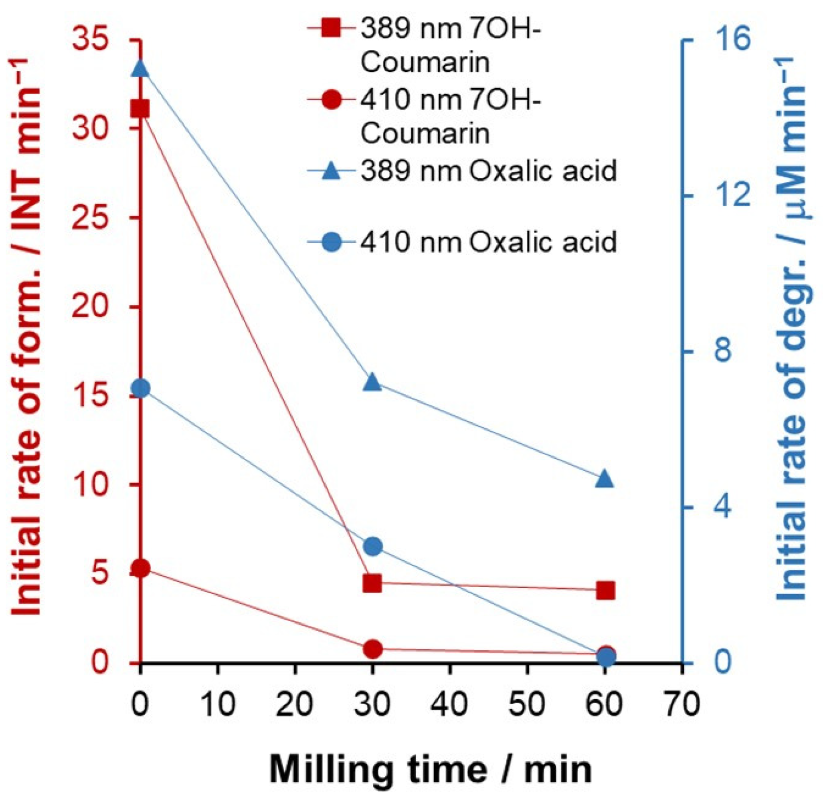
First, the photoactivity of TD, TD-30, and TD-60 photocatalysts was tested with the solution of model compounds upon irradiation with visible light (λmax = 453 nm), but the change in measured quantities, such as the formation of 7-hydroxycoumarin and concentration of oxalic acid, was comparable to the measurement error. By using lamps with shorter-wavelength maxima (λmax = 410 nm or λmax = 389 nm), the photocatalytic performance of catalysts could be measured. As can be seen in Figure 2, the activity of the catalysts decreased with increasing milling time for both wavelengths and both model compounds. Uzunova-Bujnova and coworkers had the same finding for mechanochemically treated TiO2, and they collected many possible reasons for this phenomenon [60]. The decrease in the amount of high-Fermi-level anatase with milling time is one factor. Another problem is that the dehydroxylation of surfaces by high-energy impact leads to the formation of Ti3+ ions, which act as charge-carrier recombination centers. Finally, crystal defects such as hole traps are created, which can be the main factor in reducing the degradation rate.

2.2. Modification with Nitrogen

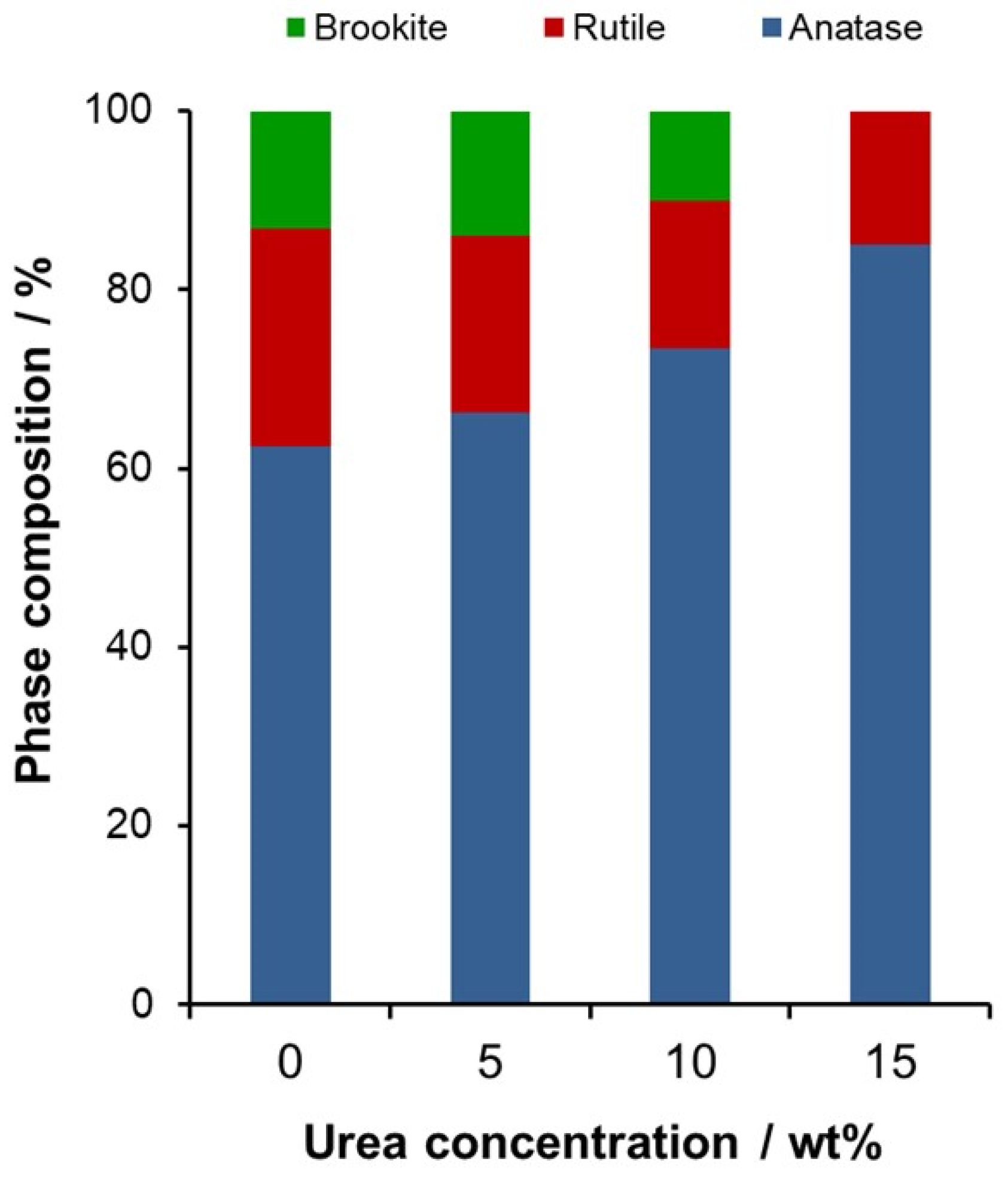
To achieve visible-light excitability, urea was used as a nitrogen precursor additive during the mechanochemical treatment. First, the effect of the nitrogen content was studied by adding urea in different initial weight percentages (wt%), while the other modification parameters (powder-to-ball weight ratio—1:10; milling time—60 min) were fixed. As shown in Section 2.1, a significant change in the phase composition of the commercial TiO2 was observed due to the 60-min milling. In comparison, when the urea dose was 5 wt%, 3.9% less anatase was converted to rutile and brookite. By further increasing the amount of urea, it was found that the nitrogen source prevented the formation of brookite and the conversion of anatase to rutile (Figure 3), in accordance with the results of Yin et al. [41].
The reason for this phenomenon is that the decomposition of urea and its chemical reactions with titanium dioxide take place at the same time as the phase transformation, leaving less mechanochemical energy for the later process. The crystallite size altered slightly (around 7%, the error of the measurement) and not tendentially compared to the size of the starting material (Supplementary Table S1).
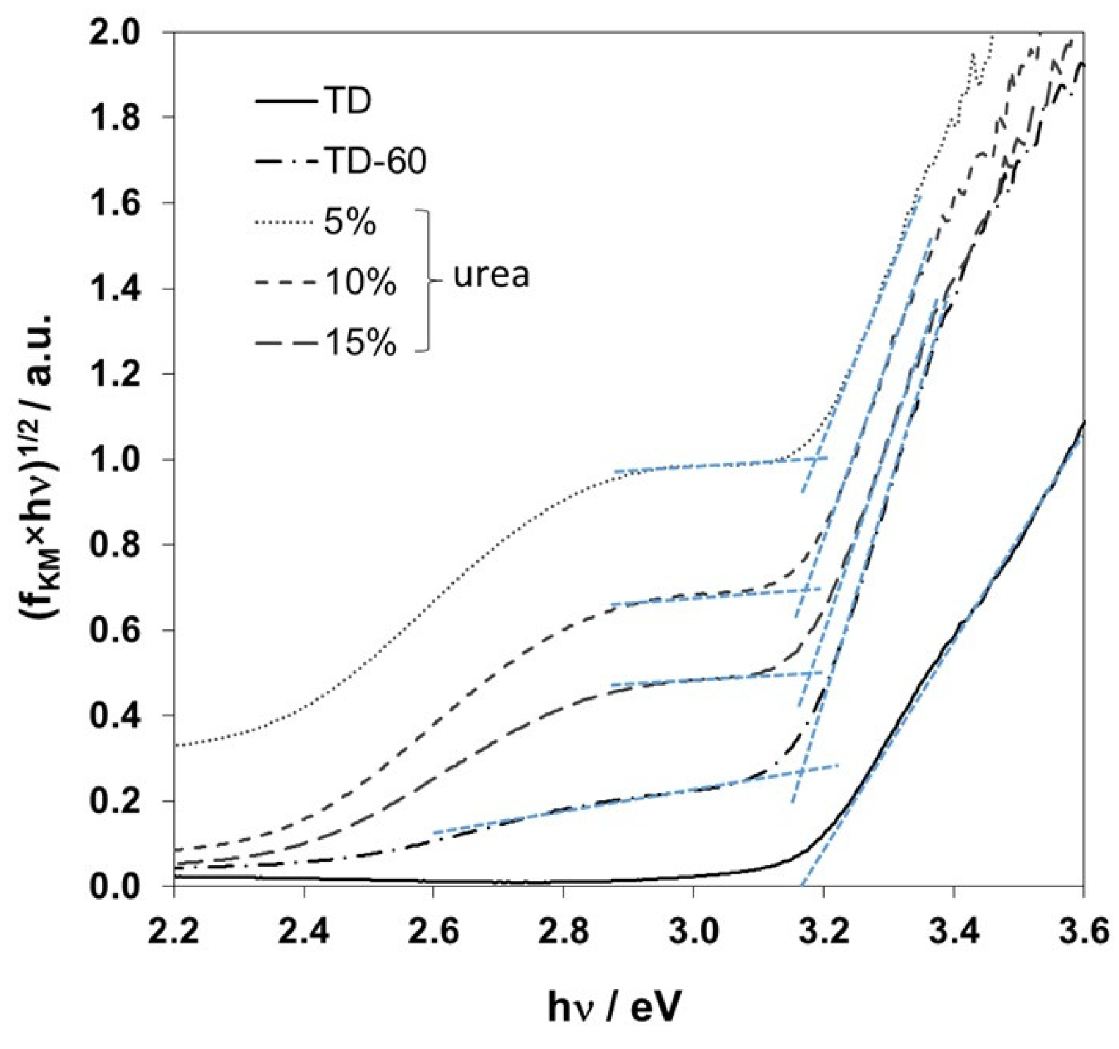
The color of the catalyst changed from the initial white to pale yellow with the addition of urea. To compare the optical properties, the diffuse reflectance of the prepared samples was measured using an integrating sphere attachment on a luminescence spectrometer. The reflectance data were then used to obtain the Kubelka–Munk function (fKM), and from the Tauc plot, the band-gap energies were estimated (Figure 4 and Supplementary Table S1). The band-gap energies obtained practically agreed within a very narrow range (3.16–3.18 eV). This observation is in line with the results published by Makuła et al. [61]. They developed a baseline method for a reliable application of the Tauc plots to determine the band gap of modified semiconductors, particularly titanium dioxide. According to their observation, the band gap of the photocatalyst remains the same even after modification if the amount of the modifier is orders of magnitude lower than that of the initial semiconductor (TD in this case). Nevertheless, modifications can cause the formation of bands with lower potential, allowing the use of visible light, as our results also show.
Due to the aim of the modification with nitrogen, the photocatalytic activity of the produced catalyst was tested only in the visible range (λem = 453 nm), using coumarin as a model compound. From the change in the emission intensities during the irradiation time, the rate of formation of oxidative HO radicals was also determined.
Although there was a remarkable change in the phase composition of the commercial TiO2 due to 60 min milling, it did not influence its activity (Figure 5a). With the addition of 5 wt% urea, the formation rate became six times faster (0.2534 INT min−1) than that of TD-60 (0.0423 INT min−1) and became even faster (0.2987 INT min−1) when 10 wt% urea was used. Since the photocatalytic activity could not be improved by further increasing the urea content (15 wt%), 10 wt% urea was used as the optimum amount in the following experiments (Figure 5b).
The ideal weight ratio of the catalyst and grinding balls was investigated with the other conditions fixed: 10 wt% urea addition and 60 min milling (Supplementary Table S1). Logically, the 1:10 ratio exerted more mechanical energy than the 1:5 ratio, which affected the phase composition and promoted the formation of brookite (Figure 6).
In line with the results of Di Paola et al. [7], when all three polymorphs were present in the catalyst, the 1:10 ratio was found to be better than the 1:5 ratio; the corresponding formation rates were 0.2987 INT min−1 and 0.2029 INT min−1.
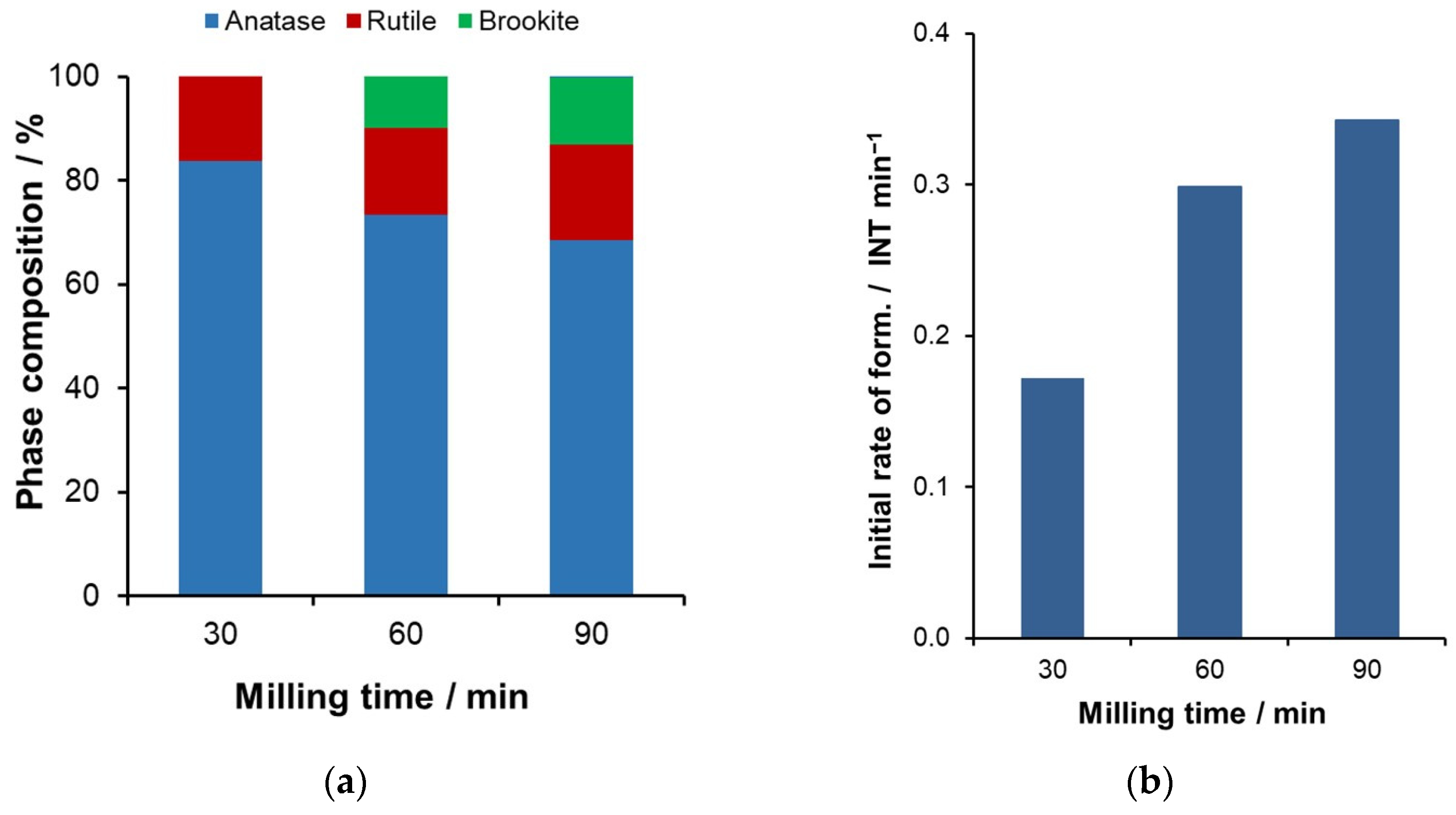
Finally, the milling time was studied with the previous optimal parameters (10 wt% urea, 1:10 powder-to-ball weight ratio). Figure 7a shows that 30 min of milling was not sufficient for the formation of brookite, but after 60 min, the brookite concentration was 10%, which increased by 3% with the treatment time of 90 min.
The initial formation rate followed the same trend (Figure 7b), showing the importance of brookite in the photocatalytic activity. For time and energy efficiency, the 60 min milling was selected for further experiments because the difference in brookite concentration (3%) and formation rate (15%) between the 60- and 90-min treatments was small. Another interesting phenomenon was observed when comparing the results with those obtained by milling pure TiO2 (Section 2.1). After 30 and 60 min of milling, the brookite concentration was 11% and 13.2% with the bare catalyst, while there was none and 10% with the addition of 10 wt% urea. These results suggest that several factors affect the formation of brookite: it is inhibited by the nitrogen content and promoted by higher grinding energy (longer grinding).
The photoactivity of three different batches was also tested (Supplementary Figure S1) to show that the same activity could be easily reproduced compared to other modification methods (e.g., sol–gel).

2.3. Modification with Silver

The main objective of the silver doping was to prepare an antibacterial catalyst, so the results obtained from experiments with different milling times and silver concentrations were evaluated on the basis of toxicity, but the photoactivity was also tested. It is well documented that Ag-TiO2 synthesized by different methods can have better photoactivity than bare photocatalyst, but our main goal with silver doping was to ensure the antibacterial property of the catalyst. Since the photoactivity under a visible lamp (λmax = 410 nm) was very poor for all silver-doped samples, the photocatalytic experiments were carried out under a UV lamp (λmax = 389 nm). The results obtained with coumarin as a model compound were complemented by the measurements of the photocatalytic decomposition of oxalic acid. In our previous work, we have shown that the degradation of oxalic acid can be enhanced by silver doping of TiO2 [13].
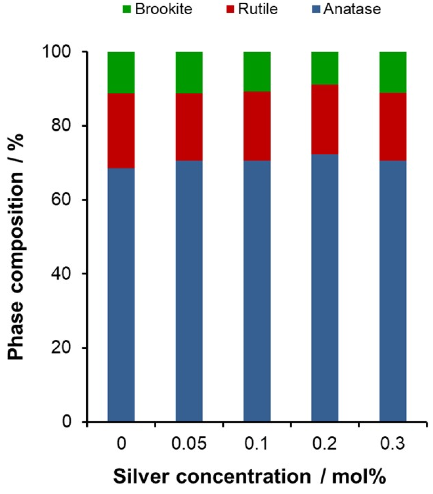
In the optimization process, the initial silver concentration was changed from 0.05 to 0.3 mol%, and the milling time was fixed at 30 min. The XRD results of these samples, presented in Figure 8, show that practically no change in the phase composition of the samples can be detected compared to the TD-30 sample, indicating that the energy consumption of doping with this small amount of silver does not extract significant energy from the phase transformation of TiO2 during the mechanochemical treatment. The same conclusion can be drawn from the energy gaps (Supplementary Table S1) because their values calculated from the Tauc plot are identical within the error of measurement. In the visible region of the diffuse reflectance spectra, some increase in the baseline can be observed as a consequence of Ag doping, especially for Ag0.3-TD-30 samples.
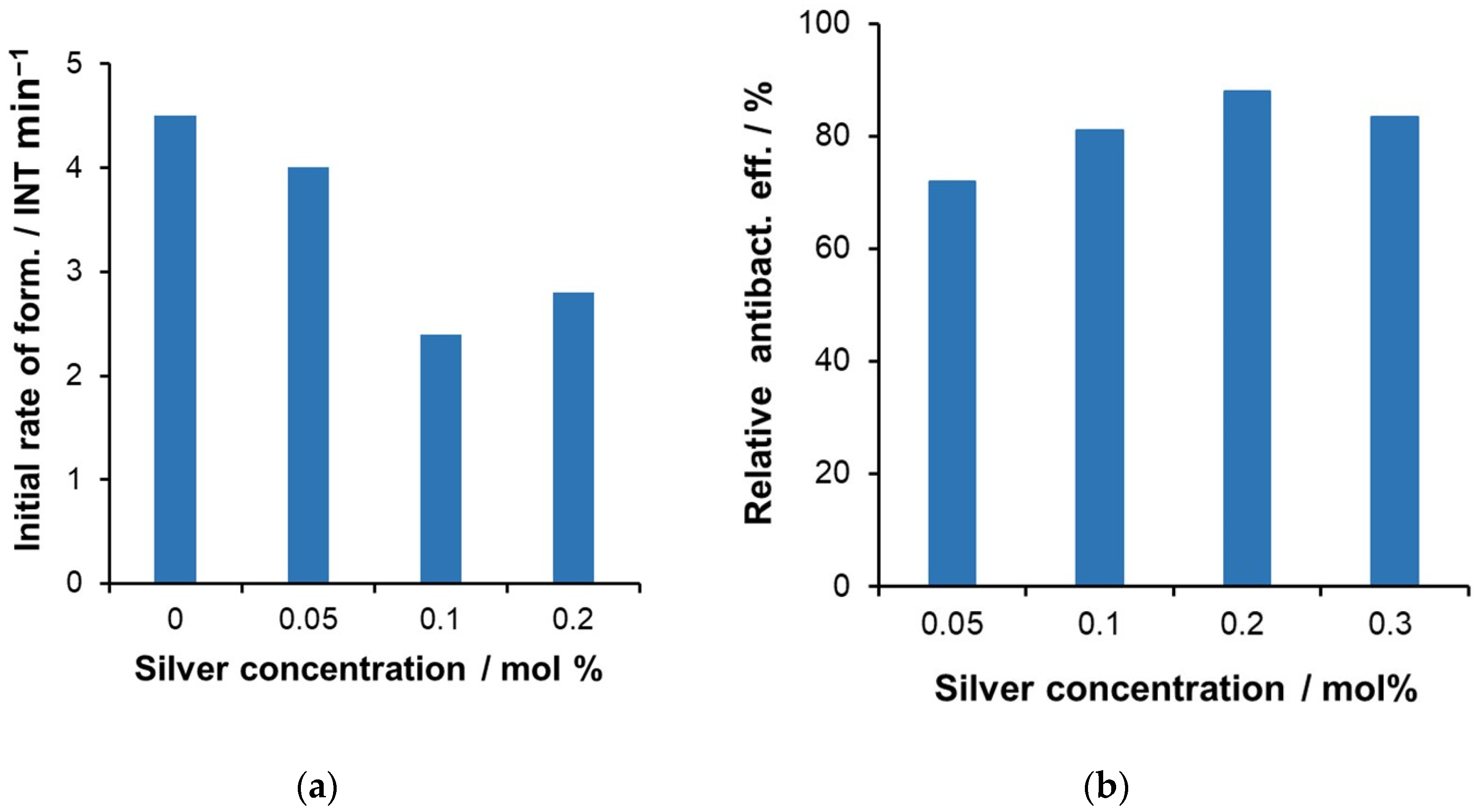
The photoactivity data with coumarin in UV and the toxicity results in the dark with Vibrio fischeri bacteria are shown in Figure 9a and Figure 9b, respectively. The rate of the formation of 7-hydroxycoumarin decreased by approximately 50% when 0.1 mol% silver was introduced into the semiconductor, and this value increased only slightly when the silver content was elevated to 0.2 mol%. The decreased photoactivity suggests that the efficiency of hydroxyl radical formation decreased with the loading of silver.
In contrast, the decomposition rates of oxalic acid shown in Supplementary Figure S2 were better for silver-modified catalysts than for TD-30, although the rate was decreased by increasing the silver concentration. From Equations (7)–(11), we can see that the degradation of oxalic acid can take place in several ways with the help of different ROS formed by the excitation of the photocatalyst. For silver photodeposited TiO2, it was found that the Ag particles on the surface of the catalyst function as an electron trap and thus enhance the electron–hole separation. The reaction of trapped electrons with adsorbed oxygen (Equation (3)) leads to the formation of superoxide radical anion, which eventually generates excited oxygen atoms capable of decomposing oxalic acid (Equations (4)–(6)). This reaction is considered to be the key step in the degradation of oxalic acid [13]. Otherwise, oxalic acid, as an electron donor capable of reacting directly with the hole (Equation (11)), makes it possible to degrade without hydroxyl radicals. Thus, the comparison of the photocatalytic activities obtained for coumarin and oxalic acid shows that silver modification of the catalyst probably decreases the rate of hydroxyl radical formation and enhances the formation of other reactive species.
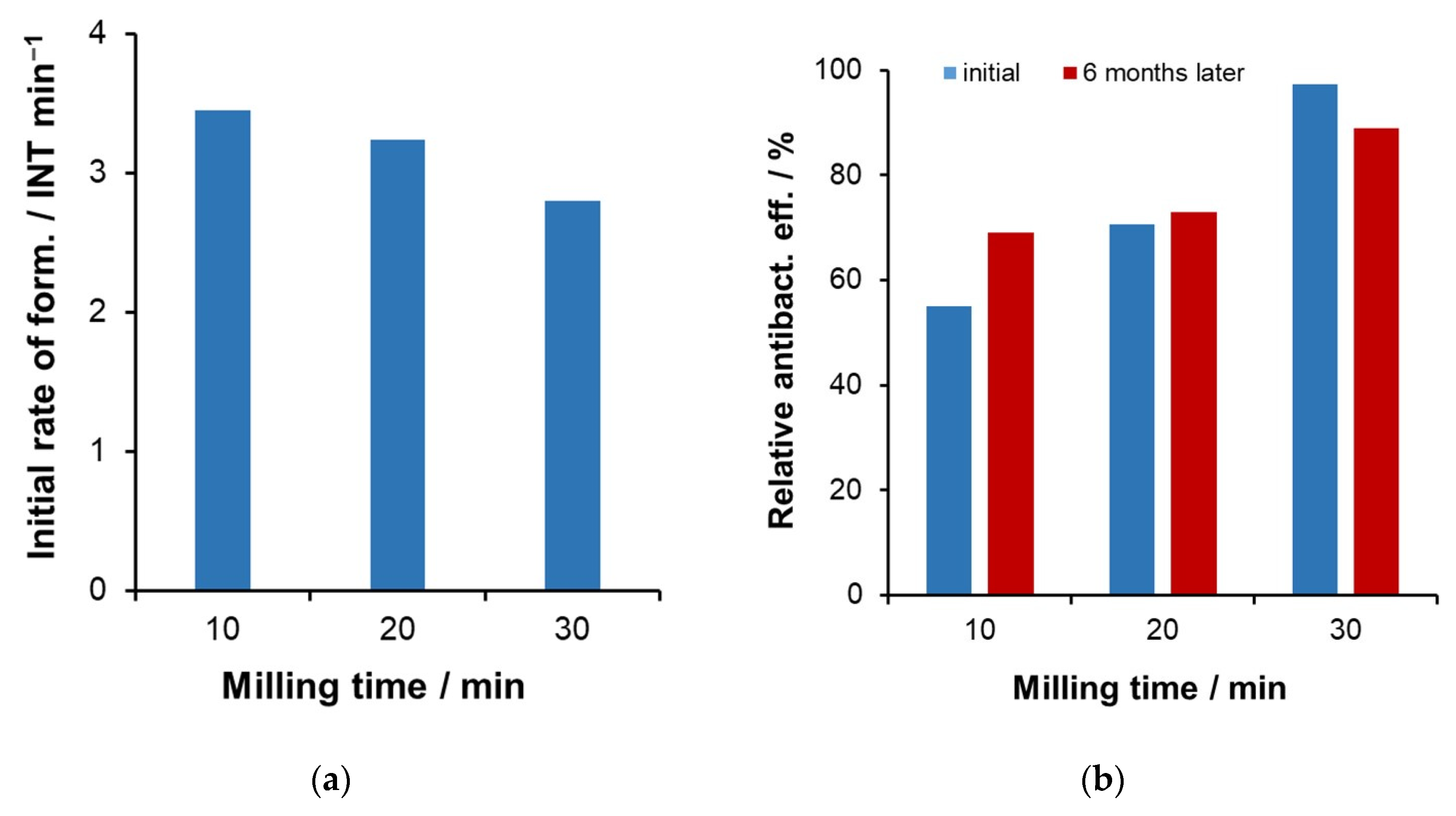
Although the photoactivity of the silverized catalysts was not outstanding, the antibacterial efficiency measured by Vibrio fischeri bacteria was superior, as shown in Figure 9b. The best performance was achieved with 0.2 mol% theoretical silver content, but lower silver concentrations also showed significant efficiencies. For further optimization, the concentration of 0.2 mol% silver was selected, and the milling time was systematically changed. Increasing the milling time from 10 to 30 min significantly reduces the photoactivity for both model compounds. The change in the initial rate of 7-hydroxycoumarin formation shown in Figure 10a makes it clear that the negative effects discussed above for the mechanochemical treatment of bare TiO2 also apply to the silver-modified catalysts. The decrease in the initial degradation rate for oxalic acid was even more pronounced, as illustrated in Supplementary Figure S2b. The initial rate, as measured for samples subjected to 10 min of milling diminished to less than half of its initial value after 30 min of milling.
However, the main question to be addressed was to find the optimal milling time for the bactericidal effect. The results presented in Figure 10b show that despite the poor photoactivity, the catalyst ground for 30 min has the best antibacterial response. It is worth mentioning that a catalyst ground for 60 min was also prepared, but the antibacterial efficiency was lower than that of Ag0.2-TD-30. Our samples were stored in air, and it was important to know how stable the antibacterial efficacy is in terms of technical application. The experiments were repeated six months later, using the same test with Vibrio fischeri bacteria as before, immediately after the preparation of the catalysts. The reproducibility of the previous results was very convincing; as shown in Figure 10b, the values are the same within the measurement error.

2.4. Combination of Photocatalytic and Antibacterial Activities

As seen in the preliminary experiments, titanium dioxide alone showed negligible activity at 453 nm irradiation; however, as a result of the modification with nitrogen, significant hydroxyl radical generation was detected at this wavelength. Since 10% of the various amounts of urea used for nitrogen doping resulted in the highest rate of hydroxyl radical generation (Figure 5), this was used for further studies. Although increasing the milling time increased the photocatalytic efficiency, a milling time of 1 h was considered sufficient because the further increase in efficiency was much smaller with respect to a 50% longer milling time (Figure 7b).
However, the photoactivity of the silverized catalyst under UV irradiation showed that increasing the silver concentration decreased the activity, although not in a completely monotonic way (Figure 9a). Consistent with this observation, increasing the milling time also reduced the activity (Figure 10a). In contrast, increasing the milling time (up to 30 min) significantly enhanced the antibacterial effect (longer milling did not improve the activity), and the effect of silver concentration was maximal at 0.2%.
Therefore, it was expected that the combination of nitrogen doping with silverization, in order to obtain both beneficial features in one catalyst, would result in photocatalytic and antibacterial efficiencies lower than the maximum values observed in the case of individually modified catalysts. To test this, a bifunctional catalyst was prepared. The reaction conditions used were based on those previously found to be optimal: 0.2 mol% silver and 10 wt% urea concentration, 60 min milling (for nitrogen doping), and 1:10 powder-to-ball weight ratio. As expected, both the rate of hydroxyl radical formation and the antibacterial activity (Figure 11) were inferior to those of the individually modified catalysts. This result showed that one catalyst cannot be expected to perform both functions with maximum efficiency.
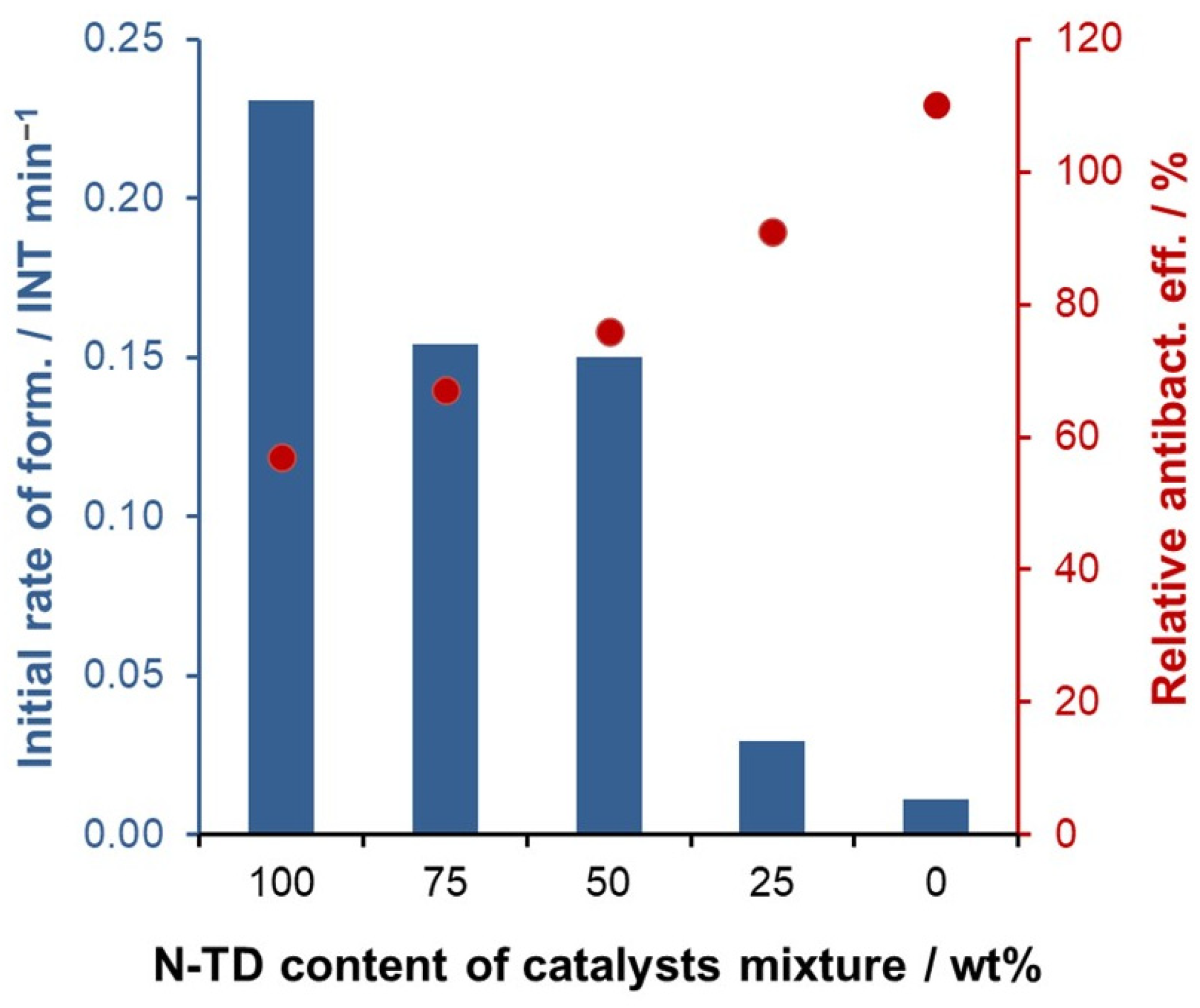
Therefore, it was concluded that another combination method, the physical mixing of two separately modified catalysts, would be worthwhile. To take advantage of both beneficial effects, the mechanical blending of N10-TD-60 and Ag0.2-TD-30 in different ratios was investigated. The resulting catalysts were mixed in proportions of 25, 50, and 75%, and the results were compared with those obtained with 100 and 0% compositions. As shown in Figure 12, as the silvered catalyst ratio increases, the antibacterial efficiency increases almost linearly, while the rate of hydroxyl radical generation is only slightly different for the 75% and 50% nitrogenized catalyst mixtures, and it decreases dramatically for the 25% mixture. Therefore, the 50–50% mixture was found to be the best.
Comparing the initial hydroxyl radical generation rate of Ag0.2-N10-TD-60 (0.0784 INT min−1) and that of the ideal mechanical mixture (0.1501 INT min−1), the latter one was almost two times faster. In addition, the antibacterial efficiency was about 76%, also higher than that of the doubly modified catalyst. These results clearly prove that this method provides a more advantageous solution for the preparation of a bifunctional photocatalyst. At the same time, half the amount of modifier is used compared to the previous method, thus reducing the reagent amounts and costs. Therefore, this method is worth using for the production of larger quantities under greener conditions.

3. Materials and Methods

3.1. Materials

The following materials were used in the experiments: Degussa P25 TiO2 (now Evonik AEROXIDE® P25 TiO2, Evonik, Wesseling, Germany), consisting of 11.4% rutile and 88.6% anatase (specific surface area: 50 m2 g−1); oxalic acid (Acros Organics, Shanghai, China); coumarin (Carlo Erba Reagent, Cornaredo, Italy); urea (Scharlab Hungary Kft., Debrecen, Hungary); silver nitrate (Forr-Lab Kft., Budapest, Hungary); barium sulfate, potassium permanganate, and sulfuric acid (Reanal, Budapest, Hungary); and freeze-dried Vibrio fischeri bacteria (Hach Lange GmbH, Berlin, Germany).
High-purity water, used as a solvent in this study, was double-distilled and then purified with a Milli-Q system (Merck Millipore, Burlington, MA, USA).

3.2. Mechanochemical Modification of the Catalysts

A FRITSCH Pulverisette 6 planetary monoball mill (Fritsch GmbH, Idar-Oberstein, Germany) was used for the mechanochemical treatment. Both the grinding jar (internal volume: 500 cm3) and the balls (average weight, 2.984 g; diameter, 10 mm) were made of corundum. The mill was operated at 400 rpm for different desired durations. In the preparation of the modified catalysts, the titanium-dioxide powder was suspended in the solution of the selected nitrogen and/or silver source, and then dried at 60 °C before being introduced into the reaction vessel. As a final step, to remove residual reagents and by-products from the final product, the samples were calcined at 400 °C for 1 h in a Nabertherm (Lilienthal, Germany) muffle furnace.

3.3. Photocatalytic Experiments

The photocatalytic experiments were carried out in a laboratory-scale 2-necked Duran glass reactor (100 cm3) [31,62]. A magnetic stirrer at the bottom of the reactor and continuous air bubbling at a flow rate of 10 dm3 h−1 ensured the homogeneous distribution of the model compound and catalyst. Two different light sources were used for irradiation: a 60 W UV LED (10 cm from the reactor), which emitted most of its energy between 360 and 400 nm (λmax = 389 nm), and its photon flux (I0) was 4.68 × 10⁻⁷ mol photon cm−2 min−1. The other setup consisted of two 7 W visible LEDs (reactor-lamp distance: 4 cm on both sides) with an emission maximum at 453 nm (I0 = 5.54 × 10⁻⁷ mol photon cm−2 min−1). Photon flux was measured in both cases by tris(oxalato)ferrate(III) chemical actinometry. To ensure that the LEDs were operating at maximum power, they were turned on 15 min before the experiments, and the light intensity was checked at the end. The reactor did not have an outer glass jacket for thermoregulation, but the heat emitted by the LED was practically negligible (3–4 °C after 240 min). In our previous work, such a small increase in temperature had no appreciable effect on the rate of photocatalytic degradation. The reason for this apparent temperature independence is that the changes in the reaction rate constant(s) and the adsorption constant as a function of temperature are opposite. For homogenization and to reach the adsorption–desorption equilibrium, the suspension was stirred in the reactor for 20 min before irradiation. The adsorption of the model compounds on the catalysts was 3–5%. Our results, in all cases, are averaged data of three replicate experiments, and the standard deviation of the repeated experiments was around 3–5%.

3.4. Analytical Procedures

3.4.1. Characterization of the Catalysts

The crystalline-phase composition of the products was determined by X-ray diffraction analysis (XRD), using the Rietveld method. The XRD patterns were obtained on a Philips PW 3710 powder diffractometer (Philips Analytical, Almelo, The Netherlands) with a Cu-Kα radiation source (λ = 1.5405 Å). Diffuse reflectance spectra (DRS) of the catalysts were recorded on a luminescence spectrometer (LS 50-B, PerkinElmer, Waltham, MA, USA) equipped with an integrating sphere attachment, using BaSO4 as a reference. The reflectance data were then used to obtain the Kubelka–Munk function, and the band-gap energies were calculated from the Tauc plot [63]. The specific surface area of some samples was measured by nitrogen adsorption/desorption isotherms using a Micromeritics ASAP 2000-type instrument (Micromeritics Instrument Corporation, Norcross, GA, USA) on samples (m ≈ 1.0 g) previously outgassed in vacuum at 160 °C. The surface areas of the samples were determined by the BET (Brunauer–Emmett–Teller) method from the corresponding nitrogen adsorption isotherms.

3.4.2. Measurements of Photocatalytic Activity

Two model compounds were selected to study the photocatalytic activity. Coumarin—a widely used hydroxyl radical scavenger [59,64,65]—was chosen for several reasons. HO radicals are the most commonly generated oxidative species during semiconductor excitation, and their disinfecting effect is related to their oxidizing properties. Their oxidation potential is very high (2.18 eV at pH = 7 [66]), and they react non-selectively with cellular components [67]. Hydroxyl radicals generated by photolysis of hydrogen peroxide have been shown to effectively kill Streptococcus mutans bacteria during biofilm treatment [68]. Using the same method in combination with sonolysis of water, hydroxyl radicals have also been generated and shown significant bactericidal activity by oxidatively damaging the DNA of Staphylococcus aureus bacteria [69]. The strong antibacterial activity of hydroxyl radicals has also been demonstrated in other bacteria, such as Aggregatibacter actinomycetemcomitans [70].
The other test compound chosen was oxalic acid. Its degradation mechanism is relatively simple: it is finally transformed into carbon dioxide and water without the formation of any stable intermediate [13]. Both hydroxyl radicals and superoxide ions play an important role in this process. In addition, oxalic acid is an excellent electron donor, so it can also react directly with the photoproduced holes, too.
For the analysis of the photocatalytic activity, 5 cm3 samples were taken, and the solid phase was removed by filtration through Millipore Millex-LCR PTFE 0.45 μm filters (Merck Millipore, Burlington, MA, USA). During the measurement with coumarin, the absorption spectral changes in the irradiated reaction mixtures were followed with a Perkin Elmer Lambda 25 spectrophotometer (PerkinElmer), while the emission spectra were recorded with a Perkin Elmer LS 50B spectrofluorometer (PerkinElmer) (ℓ = 1 cm). Using oxalic acid as a model compound, the pH of the aqueous phase of the reaction mixture was determined with an SP 10T electrode connected to a Consort C561 apparatus (Consort, Birmingham, UK), and the concentration was followed by classical permanganometry, using the aliquot of the clear liquid sample.

3.4.3. Determination of Antibacterial Effect

To investigate the antibacterial effect of the catalyst, the biological activity of Vibrio fischeri deep-sea bacteria was monitored by luminescence. The tests were performed using the LUMISTOX kit (Fisher Scientific, Loughborough, UK), which is sufficient for 20 measurements. The kit contains 20 doses of luminescent bacteria, 250 cm3 of reconstitution solution, and 50 cm3 of 7.5 (m/m)% NaCl solution, which was diluted to 2 (m/m)% for the measurements. The kit was stored frozen until required.
Measurements were performed using a ToxAlert 100 device (Merck, Darmstadt, Germany), which indicates the intensity of bioluminescence in relative light units (RLU).
The frozen Vibrio fischeri bacteria and the reconstitution solution had to be thawed prior to measurement. The thawing time varied depending on the ambient temperature. To revive the bacteria, 2 cm3 of the 12.5 cm3 reconstitution solution was measured onto the freeze-dried bacterial strain and allowed to stand at 15 °C for 10–15 min. The bacterial suspension was then washed into a 15 cm3 glass tube with the remaining reconstitution solution and diluted with another 1 cm3 of 2% NaCl solution. The final mixture was then incubated at 15 °C for 80 min. The lifetime of the “revived” bacteria was reported by the manufacturer to be 3 h at 15 °C. After the incubation period, the bacterial suspension was homogenized again, and then 500–500 μL of the suspension per minute was added to the sample holders (20 pieces) containing the catalyst powder immobilized in a polymer coating on a suitable support. A similarly immobilized reference sample was used for comparison. It had been officially accepted as an effective antibacterial coating after measurements with various bacteria in an accredited institute. In the case of blind test, the bacterial suspension was stored in a glass vial. Dispensing took 23 min. Then, 200 μL samples were taken at specified intervals from the sample containers stored away from light. At each sampling point, the ambient temperature was recorded, and the intensity (RLU) values were measured by the instrument. By comparing the results measured in the presence of the catalyst with the result of the reference sample, information about the antibacterial effect of our samples was obtained. The effect of the reference sample was taken to be 100%.

4. Conclusions

Modification of TiO2 with silver and/or nitrogen was realized to gain a photocatalyst which is both excitable by visible light and of antibacterial activity. Mechanochemical activation with grinding as an environmentally friendly process was utilized for the modification, optimizing the treating conditions, such as the amounts of the modifying substances, the milling time, and the material-to-ball ratio.
The photocatalytic activity, tested by oxalic acid and coumarin as model compounds, was gradually decreased upon silverization, regarding the titanium dioxide with and without nitrogen modification. However, the antibacterial effect measured by bioluminescent Vibrio fischeri bacteria was enhanced by increasing the silver content in both cases. The highest photocatalytic activity in the visible range was reached with the nitrogen-doped titanium dioxide prepared with 10% urea. Simultaneous modification with nitrogen and silver resulted in considerably moderated photoactivities. Hence, to optimize both functions, mechanical blending of the separately modified catalysts (N10-TD-60 and Ag0.2-TD-30N-TiO2) was also examined in various ratios. The 50–50% mixture provided the best results; both its photoactivity in the visible range and its antibacterial effect significantly exceeded those of the doubly modified catalyst. In addition, the mixing method requires less modifier; therefore, it offers a reasonable to produce larger quantities under environmentally friendly conditions.

Supplementary Materials

The following supporting information can be downloaded at https://www.mdpi.com/article/10.3390/catal15040316/s1, Table S1: Sample code with modification parameters, phase composition, crystallite size, and optical band gap of the catalysts studied; Figure S1: Photocatalytic activity of the three different batches of a nitrogen-doped catalyst (10 wt% urea, 1:10 powder-to-ball weight ratio, 60 min milling); Figure S2: Initial rate of oxalic acid degradation with silver-modified catalyst ground (a) with different concentrations of silver (milling time 30 min) and (b) with different milling times (concentration of silver 0.2 mol%). The initial concentrations of the irradiated solutions: cox = 0.875 mM and ccat = 1 g/L.

Author Contributions

Conceptualization, O.F., M.K. and E.S.-B.; methodology, O.F., M.K., L.F. and E.S.-B.; software, O.F., M.K., L.F. and E.S.-B.; validation, O.F., L.F. and M.K.; formal analysis, O.F., M.K., L.F. and E.S.-B.; investigation O.F., M.K., E.S.-B. and P.C.-K.; resources, O.H.; data curation, O.F., M.K., L.F. and P.C.-K.; writing—original draft preparation, O.F., M.K., E.S.-B., P.C.-K. and O.H.; writing—review and editing, O.H.; visualization, O.F., M.K. and E.S.-B.; supervision, E.S.-B. and O.H.; project administration, O.H.; funding acquisition, O.H. All authors have read and agreed to the published version of the manuscript.

Funding

This work was supported by the Ministry for Innovation and Technology of Hungary from the National Research, Development, and Innovation Fund, financed under the 2021 Thematic Excellence Program funding scheme (grant number TKP2021-NKTA-21).

Data Availability Statement

The data presented in this study are available upon request from the corresponding author. The data are not publicly available due to privacy.

Acknowledgments

The authors gratefully acknowledge Éva Makó, Tatjána Juzsakova, and Edina Ring-Nyári’s competent assistance in the XRD and specific surface area measurements, as well as in sample preparation, respectively.

Conflicts of Interest

The authors declare no conflicts of interest.

References

  1. Bregnocchi, A.; Jafari, R.; Momen, G. Design Strategies for Antiviral Coatings and Surfaces: A Review. Appl. Surf. Sci. Adv. 2022, 8, 100224. [Google Scholar] [CrossRef]
  2. Lee, A.-C.; Lin, R.-H.; Yang, C.-Y.; Lin, M.-H.; Wang, W.-Y. Preparations and Characterization of Novel Photocatalysts with Mesoporous Titanium Dioxide (TiO2) via a Sol–Gel Method. Mater. Chem. Phys. 2008, 109, 275–280. [Google Scholar] [CrossRef]
  3. Beh Gin-Colin, S.; Girot, T.; Le Caek, G.; Mocellin, A. Kinetics and Mechanisms of Phase Transformations Induced by Ball-Milling in Anatase TiO2. J. Solid State Chem. 2000, 149, 41–48. [Google Scholar]
  4. Huberty, J.; Xu, H. Kinetics Study on Phase Transformation from Titania Polymorph Brookite to Rutile. J. Solid State Chem. 2008, 181, 508–514. [Google Scholar] [CrossRef]
  5. Luttrell, T.; Halpegamage, S.; Tao, J.; Kramer, A.; Sutter, E.; Batzill, M. Why Is Anatase a Better Photocatalyst than Rutile?—Model Studies on Epitaxial TiO2 Films. Sci. Sci. Rep. 2014, 4, 4043. [Google Scholar] [CrossRef]
  6. Ryu, J.; Choi, W. Substrate-Specific Photocatalytic Activities of TiO2 and Multiactivity Test for Water Treatment Application. Environ. Sci. Technol. 2008, 42, 294–300. [Google Scholar] [CrossRef]
  7. Di Paola, A.; Cufalo, G.; Addamo, M.; Bellardita, M.; Campostrini, R.; Ischia, M.; Ceccato, R.; Palmisano, L. Photocatalytic Activity of Nanocrystalline TiO2 (Brookite, Rutile and Brookite-Based) Powders Prepared by Thermohydrolysis of TiCl4 in Aqueous Chloride Solutions. Colloids Surf. A Physicochem. Eng. Asp. 2008, 317, 366–376. [Google Scholar] [CrossRef]
  8. Luo, W.; Taleb, A. Large-Scale Synthesis Route of TiO2 Nanomaterials with Controlled Morphologies Using Hydrothermal Method and TiO2 Aggregates as Precursor. Nanomaterials 2021, 11, 365. [Google Scholar] [CrossRef]
  9. Vajedi, F.S.; Dehghani, H. Synthesis of Titanium Dioxide Nanostructures by Solvothermal Method and Their Application in Preparation of Nanocomposite Based on Graphene. J. Mater. Sci. 2016, 51, 1845–1854. [Google Scholar] [CrossRef]
  10. Domingues, L.A.C.S.; Carriello, G.M.; Pegoraro, G.M.; Mambrini, G.P. Synthesis of TiO2 Nanoparticles by the Solvothermal Method and Application in the Catalysis of Esterification Reactions. An. Acad. Bras. Cienc. 2024, 96, e20240096. [Google Scholar] [CrossRef]
  11. Buraso, W.; Lachom, V.; Siriya, P.; Laokul, P. Synthesis of TiO2 Nanoparticles via a Simple Precipitation Method and Photocatalytic Performance. Mater. Res. Express 2018, 5, 115003. [Google Scholar] [CrossRef]
  12. Wang, Z.; Liu, S.; Cao, X.; Wu, S.; Liu, C.; Li, G.; Jiang, W.; Wang, H.; Wang, N.; Ding, W. Preparation and Characterization of TiO2 Nanoparticles by Two Different Precipitation Methods. Ceram. Int. 2020, 46, 15333–15341. [Google Scholar] [CrossRef]
  13. Szabó-Bárdos, E.; Czili, H.; Horváth, A. Photocatalytic Oxidation of Oxalic Acid Enhanced by Silver Deposition on a TiO2 Surface. J. Photochem. Photobiol. A Chem. 2003, 154, 195–201. [Google Scholar] [CrossRef]
  14. Ojani, R.; Raoof, J.B.; Safshekan, S. Photoinduced Deposition of Palladium Nanoparticles on TiO2 Nanotube Electrode and Investigation of Its Capability for Formaldehyde Oxidation. Electrochim. Acta 2014, 138, 468–475. [Google Scholar] [CrossRef]
  15. Amorós-Pérez, A.; Cano-Casanova, L.; Castillo-Deltell, A.; Lillo-Ródenas, M.Á.; Román-Martínez, M.d.C. TiO2 Modification with Transition Metallic Species (Cr, Co, Ni, and Cu) for Photocatalytic Abatement of Acetic Acid in Liquid Phase and Propene in Gas Phase. Materials 2018, 12, 40. [Google Scholar] [CrossRef]
  16. Akhter, P.; Arshad, A.; Saleem, A.; Hussain, M. Recent Development in Non-Metal-Doped Titanium Dioxide Photocatalysts for Different Dyes Degradation and the Study of Their Strategic Factors: A Review. Catalysts 2022, 12, 1331. [Google Scholar] [CrossRef]
  17. Mäki-Arvela, P.; Murzin, D.Y. Effect of Catalyst Synthesis Parameters on the Metal Particle Size. Appl. Catal. A Gen. 2013, 451, 251–281. [Google Scholar] [CrossRef]
  18. Bakar, S.A.; Ribeiro, C. Nitrogen-Doped Titanium Dioxide: An Overview of Material Design and Dimensionality Effect over Modern Applications. J. Photochem. Photobiol. C Photochem. Rev. 2016, 27, 1–29. [Google Scholar] [CrossRef]
  19. Jiang, D.; Otitoju, T.A.; Ouyang, Y.; Shoparwe, N.F.; Wang, S.; Zhang, A.; Li, S. A Review on Metal Ions Modified TiO2 for Photocatalytic Degradation of Organic Pollutants. Catalysts 2021, 11, 1039. [Google Scholar] [CrossRef]
  20. Durango-Giraldo, G.; Cardona, A.; Zapata, J.F.; Santa, J.F.; Buitrago-Sierra, R. Titanium Dioxide Modified with Silver by Two Methods for Bactericidal Applications. Heliyon 2019, 5, e01608. [Google Scholar] [CrossRef]
  21. Xiong, Z.; Ma, J.; Ng, W.J.; Waite, T.D.; Zhao, X.S. Silver-Modified Mesoporous TiO2 Photocatalyst for Water Purification. Water Res. 2011, 45, 2095–2103. [Google Scholar] [CrossRef] [PubMed]
  22. Liu, H.; Dong, X.; Liu, T.; Duan, C.; Zhu, Z. In-Situ Fabrication of Silver-Modified TiO2 Microspheres with Enhanced Visible Photocatalytic Activity. Mater. Lett. 2013, 110, 111–113. [Google Scholar] [CrossRef]
  23. Liu, H.; Dong, X.; Liu, T.; Zhu, Z. In-Situ Fabrication of Silver-Modified TiO2 Microspheres for Enhanced Visible Light Driven Photocatalytic Activities. Sol. Energy Mater. Sol. Cells 2015, 132, 86–93. [Google Scholar] [CrossRef]
  24. Liu, H.; Dong, X.; Duan, C.; Su, X.; Zhu, Z. Silver-Modified TiO2 Nanorods with Enhanced Photocatalytic Activity in Visible Light Region. Ceram Int. 2013, 39, 8789–8795. [Google Scholar] [CrossRef]
  25. Vamathevan, V.; Amal, R.; Beydoun, D.; Low, G.; McEvoy, S. Photocatalytic Oxidation of Organics in Water Using Pure and Silver-Modified Titanium Dioxide Particles. J. Photochem. Photobiol. A Chem. 2002, 148, 233–245. [Google Scholar] [CrossRef]
  26. Chaker, H.; Chérif-Aouali, L.; Khaoulani, S.; Bengueddach, A.; Fourmentin, S. Photocatalytic Degradation of Methyl Orange and Real Wastewater by Silver Doped Mesoporous TiO2 Catalysts. J. Photochem. Photobiol. A Chem. 2016, 318, 142–149. [Google Scholar] [CrossRef]
  27. He, P.; Tao, J.; Huang, X.; Xue, J. Preparation and Photocatalytic Antibacterial Property of Nitrogen Doped TiO2 Nanoparticles. J. Solgel. Sci. Technol. 2013, 68, 213–218. [Google Scholar] [CrossRef]
  28. Liu, Y.; He, J.; Sun, Y.; Hu, J.; Li, C.; Xue, G.; Ognier, S. A Comparison of N-doped TiO2 Photocatalysts Preparation Methods and Studies on Their Catalytic Activity. J. Chem. Technol. Biotechnol. 2013, 88, 1815–1821. [Google Scholar] [CrossRef]
  29. Wang, X.; Lim, T.-T. Effect of Hexamethylenetetramine on the Visible-Light Photocatalytic Activity of C–N Codoped TiO2 for Bisphenol A Degradation: Evaluation of Photocatalytic Mechanism and Solution Toxicity. Appl. Catal. A Gen. 2011, 399, 233–241. [Google Scholar] [CrossRef]
  30. Devi, L.G.; Nagaraj, B.; Rajashekhar, K.E. Synergistic Effect of Ag Deposition and Nitrogen Doping in TiO2 for the Degradation of Phenol under Solar Irradiation in Presence of Electron Acceptor. Chem. Eng. J. 2012, 181–182, 259–266. [Google Scholar] [CrossRef]
  31. Wafi, A.; Szabó-Bárdos, E.; Horváth, O.; Pósfai, M.; Makó, É.; Juzsakova, T.; Fónagy, O. The Photocatalytic and Antibacterial Performance of Nitrogen-Doped TiO2: Surface-Structure Dependence and Silver-Deposition Effect. Nanomaterials 2020, 10, 2261. [Google Scholar] [CrossRef]
  32. Kocsis, G.; Szabó-Bárdos, E.; Fónagy, O.; Farsang, E.; Juzsakova, T.; Jakab, M.; Pekker, P.; Kovács, M.; Horváth, O. Characterization of Various Titanium-Dioxide-Based Catalysts Regarding Photocatalytic Mineralization of Carbamazepine Also Combined with Ozonation. Molecules 2022, 27, 8041. [Google Scholar] [CrossRef] [PubMed]
  33. Ghosh, M.; Mondal, M.; Mandal, S.; Roy, A.; Chakrabarty, S.; Chakrabarti, G.; Pradhan, S.K. Enhanced Photocatalytic and Antibacterial Activities of Mechanosynthesized TiO2–Ag Nanocomposite in Wastewater Treatment. J. Mol. Struct. 2020, 1211, 128076. [Google Scholar] [CrossRef]
  34. Ellouzi, I.; Bouddouch, A.; Bakiz, B.; Benlhachemi, A.; Abou Oualid, H. Glucose-Assisted Ball Milling Preparation of Silver-Doped Biphasic TiO2 for Efficient Photodegradation of Rhodamine B: Effect of Silver-Dopant Loading. Chem. Phys. Lett. 2021, 770, 138456. [Google Scholar] [CrossRef]
  35. Šubrt, J.; Criado, J.M.; Szatmáry, L.; Diánez-Millán, M.J.; Murafa, N.; Pérez-Maqueda, L.A.; Brezová, V. Mechanochemical Synthesis of Visible Light Sensitive Titanium Dioxide Photocatalyst. Int. J. Photoenergy 2011, 2011, 156941. [Google Scholar] [CrossRef]
  36. Dulian, P.; Buras, M.; Żukowski, W. Modyfication of Photocatalytic Properties of Titanium Dioxide by Mechanochemical Method. Pol. J. Chem. Technol. 2016, 18, 68–71. [Google Scholar] [CrossRef]
  37. Valeeva, A.A.; Sushnikova, A.A.; Rempel, A.A. Phase Composition Tuning by High-Energy Ball Milling of Titanium Dioxide Powders. Inorg Chem. Commun. 2024, 159, 111727. [Google Scholar] [CrossRef]
  38. Jalalah, M.; Faisal, M.; Bouzid, H.; Ismail, A.A.; Al-Sayari, S.A. Dielectric and Photocatalytic Properties of Sulfur Doped TiO2 Nanoparticles Prepared by Ball Milling. Mater. Res. Bull. 2013, 48, 3351–3356. [Google Scholar] [CrossRef]
  39. Yin, S.; Komatsu, M.; Zhang, Q.; Saito, F.; Sato, T. Synthesis of Visible-Light Responsive Nitrogen/Carbon Doped Titania Photocatalyst by Mechanochemical Doping. J. Mater. Sci. 2007, 42, 2399–2404. [Google Scholar] [CrossRef]
  40. Yin, S.; Yamaki, H.; Komatsu, M.; Zhang, Q.; Wang, J.; Tang, Q.; Saito, F.; Sato, T. Preparation of Nitrogen-Doped Titania with High Visible Light Induced Photocatalytic Activity by Mechanochemical Reaction of Titania and Hexamethylenetetramine. J. Mater. Chem. 2003, 13, 2996. [Google Scholar] [CrossRef]
  41. Yin, S.; Yamaki, H.; Komatsu, M.; Zhang, Q.; Wang, J.; Tang, Q.; Saito, F.; Sato, T. Synthesis of Visible-Light Reactive TiO2−N Photocatalyst by Mechanochemical Doping. Solid State Sci. 2005, 7, 1479–1485. [Google Scholar] [CrossRef]
  42. Nosaka, Y.; Matsushita, M.; Nishino, J.; Nosaka, A. Nitrogen-Doped Titanium Dioxide Photocatalysts for Visible Response Prepared by Using Organic Compounds. Sci. Technol. Adv. Mater. 2005, 6, 143–148. [Google Scholar] [CrossRef]
  43. Yin, S.; Yamaki, H.; Zhang, Q.; Komatsu, M.; Wang, J.; Tang, Q.; Saito, F.; Sato, T. Mechanochemical Synthesis of Nitrogen-Doped Titania and Its Visible Light Induced NO Destruction Ability. Solid State Ion. 2004, 172, 205–209. [Google Scholar] [CrossRef]
  44. Tang, Y.-C.; Huang, X.-H.; Yu, H.-Q.; Tang, L.-H. Nitrogen-Doped Photocatalyst Prepared by Mechanochemical Method: Doping Mechanisms and Visible Photoactivity of Pollutant Degradation. Int. J. Photoenergy 2012, 2012, 960726. [Google Scholar] [CrossRef]
  45. Hamad, A.; Khashan, K.S.; Hadi, A. Silver Nanoparticles and Silver Ions as Potential Antibacterial Agents. J. Inorg. Organomet. Polym. Mater. 2020, 30, 4811–4828. [Google Scholar] [CrossRef]
  46. Chernousova, S.; Epple, M. Silver as Antibacterial Agent: Ion, Nanoparticle, and Metal. Angew. Chem. Int. Ed. 2013, 52, 1636–1653. [Google Scholar] [CrossRef]
  47. Chen, M.; Yan, L.; He, H.; Chang, Q.; Yu, Y.; Qu, J. Catalytic Sterilization of Escherichia Coli K 12 on Ag/Al2O3 Surface. J. Inorg. Biochem. 2007, 101, 817–823. [Google Scholar] [CrossRef]
  48. Chang, Q.; He, H.; Ma, Z. Efficient Disinfection of Escherichia Coli in Water by Silver Loaded Alumina. J. Inorg. Biochem. 2008, 102, 1736–1742. [Google Scholar] [CrossRef]
  49. Pham, T.-D.; Lee, B.-K. Feasibility of Silver Doped TiO2/Glass Fiber Photocatalyst under Visible Irradiation as an Indoor Air Germicide. Int. J. Environ. Res. Public Health 2014, 11, 3271–3288. [Google Scholar] [CrossRef]
  50. Pulido Melián, E.; González Díaz, O.; Doña Rodríguez, J.M.; Colón, G.; Navío, J.A.; Macías, M.; Pérez Peña, J. Effect of Deposition of Silver on Structural Characteristics and Photoactivity of TiO2-Based Photocatalysts. Appl. Catal. B 2012, 127, 112–120. [Google Scholar] [CrossRef]
  51. Madadi, M.; Ghorbanpour, M.; Feizi, A. Antibacterial and Photocatalytic Activity of Anatase Phase Ag-doped TiO2 Nanoparticles. Micro. Nano Lett. 2018, 13, 1590–1593. [Google Scholar] [CrossRef]
  52. Ali, T.; Ahmed, A.; Alam, U.; Uddin, I.; Tripathi, P.; Muneer, M. Enhanced Photocatalytic and Antibacterial Activities of Ag-Doped TiO2 Nanoparticles under Visible Light. Mater. Chem. Phys. 2018, 212, 325–335. [Google Scholar] [CrossRef]
  53. Backhaus, T.; Grimme, L.H. The Toxicity of Antibiotic Agents to the Luminescent Bacterium Vibrio Fischeri. Chemosphere 1999, 38, 3291–3301. [Google Scholar] [CrossRef]
  54. Hernando, M.D.; De Vettori, S.; Martínez Bueno, M.J.; Fernández-Alba, A.R. Toxicity Evaluation with Vibrio Fischeri Test of Organic Chemicals Used in Aquaculture. Chemosphere 2007, 68, 724–730. [Google Scholar] [CrossRef]
  55. Vihodceva, S.; Šutka, A.; Sihtmäe, M.; Rosenberg, M.; Otsus, M.; Kurvet, I.; Smits, K.; Bikse, L.; Kahru, A.; Kasemets, K. Antibacterial Activity of Positively and Negatively Charged Hematite (α-Fe2O3) Nanoparticles to Escherichia Coli, Staphylococcus Aureus and Vibrio Fischeri. Nanomaterials 2021, 11, 652. [Google Scholar] [CrossRef] [PubMed]
  56. Gmurek, M.; Horn, H.; Majewsky, M. Phototransformation of Sulfamethoxazole under Simulated Sunlight: Transformation Products and Their Antibacterial Activity toward Vibrio Fischeri. Sci. Total Environ. 2015, 538, 58–63. [Google Scholar] [CrossRef] [PubMed]
  57. Heinlaan, M.; Ivask, A.; Blinova, I.; Dubourguier, H.-C.; Kahru, A. Toxicity of Nanosized and Bulk ZnO, CuO and TiO2 to Bacteria Vibrio Fischeri and Crustaceans Daphnia Magna and Thamnocephalus Platyurus. Chemosphere 2008, 71, 1308–1316. [Google Scholar] [CrossRef] [PubMed]
  58. Aysin, B.; Ozturk, A.; Park, J. Silver-Loaded TiO2 Powders Prepared through Mechanical Ball Milling. Ceram. Int. 2013, 39, 7119–7126. [Google Scholar] [CrossRef]
  59. Czili, H.; Horváth, A. Applicability of Coumarin for Detecting and Measuring Hydroxyl Radicals Generated by Photoexcitation of TiO2 Nanoparticles. Appl. Catal. B 2008, 81, 295–302. [Google Scholar] [CrossRef]
  60. Uzunova-Bujnova, M.; Dimitrov, D.; Radev, D.; Bojinova, A.; Todorovsky, D. Effect of the Mechanoactivation on the Structure, Sorption and Photocatalytic Properties of Titanium Dioxide. Mater. Chem. Phys. 2008, 110, 291–298. [Google Scholar] [CrossRef]
  61. Makuła, P.; Pacia, M.; Macyk, W. How To Correctly Determine the Band Gap Energy of Modified Semiconductor Photocatalysts Based on UV–Vis Spectra. J. Phys. Chem. Lett. 2018, 9, 6814–6817. [Google Scholar] [CrossRef] [PubMed]
  62. Szabó-Bárdos, E.; Cafuta, A.; Hegedűs, P.; Fónagy, O.; Kiss, G.; Babić, S.; Škorić, I.; Horváth, O. Photolytic and Photocatalytic Degradation of Nitrofurantoin and Its Photohydrolytic Products. J. Photochem. Photobiol. A Chem. 2020, 386, 112093. [Google Scholar] [CrossRef]
  63. Beranek, R.; Kisch, H. Tuning the Optical and Photoelectrochemical Properties of Surface-Modified TiO2. Photochem. Photobiol. Sci. 2008, 7, 40–48. [Google Scholar] [CrossRef]
  64. Leandri, V.; Gardner, J.M.; Jonsson, M. Coumarin as a Quantitative Probe for Hydroxyl Radical Formation in Heterogeneous Photocatalysis. J. Phys. Chem. C 2019, 123, 6667–6674. [Google Scholar] [CrossRef]
  65. Náfrádi, M.; Farkas, L.; Alapi, T.; Hernádi, K.; Kovács, K.; Wojnárovits, L.; Takács, E. Application of Coumarin and Coumarin-3-Carboxylic Acid for the Determination of Hydroxyl Radicals during Different Advanced Oxidation Processes. Radiat. Phys. Chem. 2020, 170, 108610. [Google Scholar] [CrossRef]
  66. Fernández-Castro, P.; Vallejo, M.; San Román, M.F.; Ortiz, I. Insight on the Fundamentals of Advanced Oxidation Processes. Role and Review of the Determination Methods of Reactive Oxygen Species. J. Chem. Technol. Biotechnol. 2015, 90, 796–820. [Google Scholar] [CrossRef]
  67. Wu, G.; Wang, J.; Wan, Q.; Cao, S.; Huang, T.; Lu, J.; Ma, J.; Wen, G. Kinetics and Mechanism of Sulfate Radical-and Hydroxyl Radical-Induced Disinfection of Bacteria and Fungal Spores by Transition Metal Ions-Activated Peroxymonosulfate. Water Res. 2023, 243, 120378. [Google Scholar] [CrossRef]
  68. Nakamura, K.; Shirato, M.; Kanno, T.; Örtengren, U.; Lingström, P.; Niwano, Y. Antimicrobial Activity of Hydroxyl Radicals Generated by Hydrogen Peroxide Photolysis against Streptococcus Mutans Biofilm. Int. J. Antimicrob. Agents 2016, 48, 373–380. [Google Scholar] [CrossRef]
  69. Sheng, H.; Nakamura, K.; Kanno, T.; Sasaki, K.; Niwano, Y. Bactericidal Effect of Photolysis of H2O2 in Combination with Sonolysis of Water via Hydroxyl Radical Generation. PLoS ONE 2015, 10, e0132445. [Google Scholar] [CrossRef]
  70. Tenkumo, T.; Ishiyama, K.; Prymak, O.; Nakamura, K.; Shirato, M.; Ogawa, T.; Miyashita, M.; Takahashi, M.; Epple, M.; Kanno, T.; et al. Bactericidal Activity and Recovery Effect of Hydroxyl Radicals Generated by Ultraviolet Irradiation and Silver Ion Application on an Infected Titanium Surface. Sci. Rep. 2020, 10, 8553. [Google Scholar] [CrossRef]
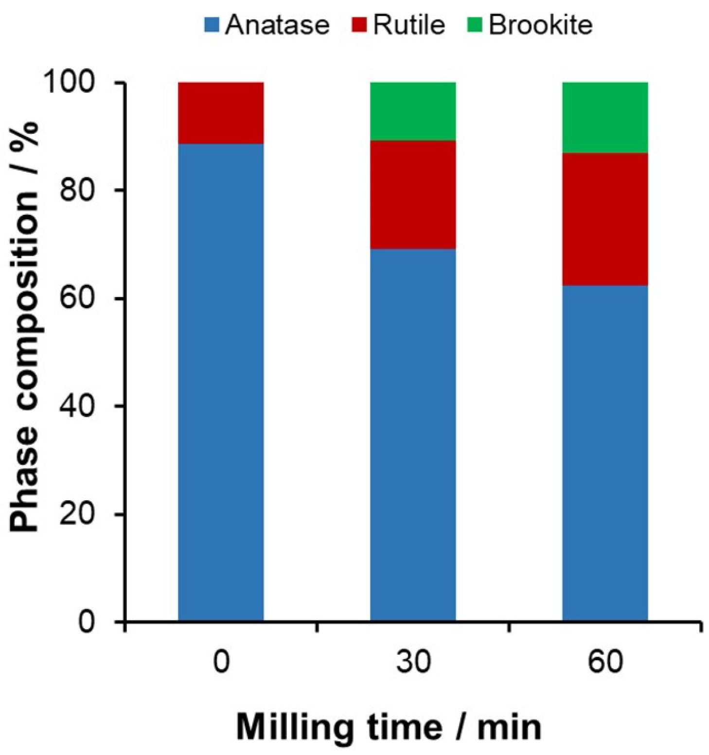
Figure 1. Phase composition obtained from XRD results for Degussa P25 (TD) ground at different milling times. The grinding process was carried out with a material-to-ball ratio of 1:10 in a grinding jar, at 400 rpm.
Catalysts 15 00316 g001
Figure 2. The initial rates of 7-hydroxycoumarine formation (left axis) and that of oxalic acid degradation (right axis) determined in the suspension of Degussa P25 TiO2 ground at different durations (samples: TD, TD-30, and TD-60). The initial concentrations of the irradiated solutions: 1 g/L for the catalysts and ccoum = 0.1 mM for coumarin or cox = 0.875 mM for oxalic acid.
Catalysts 15 00316 g002
Figure 3. Phase composition of the nitrogen-modified catalysts at various amounts of urea used for nitrogen doping (60 min milling time).
Catalysts 15 00316 g003
Figure 4. Tauc plots obtained from the diffuse reflectance spectra of the initial (TD) and treated samples.
Catalysts 15 00316 g004
Figure 5. (a) Change in the luminescence intensity of 7-hydroxycoumarin formed. (b) Initial rate of HO radical formation of nitrogen-modified catalysts (λexc = 332 nm, c(coumarin)0 = 0.1 mM).
Catalysts 15 00316 g005
Figure 6. Change in the phase composition (a) and photocatalytic activity (b) in the case of different powder-to-ball weight ratios (10 wt% urea, 60 min milling time).
Catalysts 15 00316 g006
Figure 7. Change in the phase composition (a) and photocatalytic activity (b) in the case of different milling times (10 wt% urea, 1:10 powder-to-ball weight ratio).
Catalysts 15 00316 g007
Figure 8. Phase composition obtained from XRD results of samples modified with different amounts of silver. The 30-min grinding process was performed with a material-to-ball ratio of 1:10 in a grinding jar, at 400 rpm.
Catalysts 15 00316 g008
Figure 9. Photocatalytic and antibacterial efficiencies of silver-modified catalysts. (a) Initial rate of the 7-hydroxycoumarin formation (initial concentration of the irradiated solutions ccat = 1g/L and ccoum = 0.1 mM). (b) Antibacterial efficiency measured by Vibrio fischeri bacteria relative to the reference sample.
Catalysts 15 00316 g009
Figure 10. Photocatalytic and antibacterial efficiencies of silver-modified catalyst ground for different durations. The theoretical silver content of the catalyst was 0.2 mol%. (a) Initial rate of the 7-hydroxycoumarin formation (initial concentration of the irradiated solutions ccat = 1 g/L and ccoum = 0.1 mM). (b) Antibacterial efficiency measured by Vibrio fischeri bacteria after sample preparation and 6 months later, relative to the reference sample.
Catalysts 15 00316 g010
Figure 11. The initial rate of the 7-hydroxycoumarin formation and the relative antibacterial effect in the case of nitrogen-doped (N10-TD-60, abbreviated as N-TD), silverized (Ag0.2-TD-30 abbreviated as Ag-TD), and both nitrogen-doped and silverized (Ag0.2-N10-TD-60 abbreviated as Ag-N-TD) catalysts (λirr = 453 nm).
Catalysts 15 00316 g011
Figure 12. The initial rate of the 7-hydroxycoumarin formation and the relative antibacterial effect in the case of different mechanical mixtures of N10-TD-60 (abbreviated as N-TD) and Ag0.2-TD-30 catalysts (λirr = 453 nm).
Catalysts 15 00316 g012
Disclaimer/Publisher’s Note: The statements, opinions and data contained in all publications are solely those of the individual author(s) and contributor(s) and not of MDPI and/or the editor(s). MDPI and/or the editor(s) disclaim responsibility for any injury to people or property resulting from any ideas, methods, instructions or products referred to in the content.