Article

Enantioselectivity Enhancement of a Geobacillus thermoleovorans CCR11 Lipase by Rational Design

by
Aaron-Salvador Bustos-Baena
1,
Rodolfo Quintana-Castro
2,
María Guadalupe Sánchez-Otero
2,
Graciela Espinosa-Luna
1,
María Remedios Mendoza-López
3,
Carolina Peña-Montes
1 and
Rosa María Oliart-Ros
1,*
1
Unidad de Investigación y Desarrollo en Alimentos, Tecnológico Nacional de México Campus Veracruz, Miguel A. de Quevedo 2779, Veracruz CP 91897, Veracruz, Mexico
2
Facultad de Bioanálisis, Universidad Veracruzana, Iturbide s/n Esquina Carmen Serdán, Veracruz CP 91700, Veracruz, Mexico
3
Instituto de Química Aplicada, Universidad Veracruzana, Luis Castelazo Ayala s/n, Xalapa CP 91190, Veracruz, Mexico
*
Author to whom correspondence should be addressed.
Catalysts 2025, 15(2), 168; https://doi.org/10.3390/catal15020168
Submission received: 7 January 2025 / Revised: 30 January 2025 / Accepted: 8 February 2025 / Published: 12 February 2025
(This article belongs to the Special Issue New Trends in Industrial Biocatalysis, 2nd Edition)

Abstract

:
Lipases are enzymes that catalyze the hydrolysis of carboxylic esters at a lipid–water interface and are able to catalyze reactions such as alcoholysis, esterification, transesterification, and enantioselective synthesis in organic media. They are important biocatalysts for biotechnological and industrial applications—such as in the food and flavor industry—and in the production of biopharmaceuticals, biofuels, biopolymers, and detergents. A desirable property of lipases is stereoselectivity for the production of chemicals with high optical purity. In this work, we report the improvement of the enantioselective capabilities of the Geobacillus thermoleovorans CCR11 lipase. By means of a rational design and bioinformatic approaches, six amino acids of the catalytic cavity of the lipase LipTioCCR11 were substituted resulting in an increase in the optimum temperature of the enzyme and in the resistance to the presence of organic solvents in hydrolytic reactions, and in the promotion of the enantioselective recognition of R isomers of carboxylic acids with importance for the pharmaceutical and food industries.

1. Introduction

Lipases (acyl glycerol hydrolases EC 3.1.1.3) are ubiquitous enzymes that catalyze the hydrolysis of carboxylic esters at a lipid–water interface and have the ability to catalyze reactions such as alcoholysis, esterification, transesterification, and enantioselective synthesis in organic media. These catalytic properties have made them the subject of interest and of outmost importance in various biotechnological and industrial applications, such as in the food and flavor industry, and in the production of biopharmaceuticals, biofuels, biopolymers, and detergents [1,2]. For example, the regiospecificity of lipases is one of the most important advantages in the modification of oils and fats for the production of high-value-added products, such as structured lipids (human milk or cocoa butter substitutes) [3,4].
Lipases have a characteristic globular structure composed of β-sheets surrounded by α-helices, forming a conserved α/β-hydrolase domain. The catalytic cavity of lipases, also known as the catalytic cleft, contains a catalytic site and various binding sites that play essential roles in the catalytic reactions and in determining enantioselectivity. The catalytic site is composed of a catalytic triad of serine (Ser), aspartic or glutamic acid (Asp/Glu), and histidine (His) residues, and a region known as the oxyanion hole that stabilizes the substrate transition state, ensuring correct formation of the final product. The catalytic cavity binding sites are classified as: the acyl-binding pocket, a non-polar region that in hydrolytic reactions has a high affinity for the fatty acids on triglycerides and in synthetic reactions facilitates the attachment of the substrate’s carboxylic group; a mainly hydrophobic surface known as the hydrophobic cavity that interacts with large substituents of secondary alcohols; and the hydrophilic cavity that guides nucleophilic groups, such as hydroxyls, toward the catalytic serine [5]. The arrangement and size of these regions within the catalytic cavity largely determine the preference of lipases for a specific enantiomer. Mutations that modify the hydrophobicity or expand the volume of the catalytic cavity can significantly alter the substrate orientation, thus optimizing the enantioselectivity of the enzyme and enhancing its application in chiral synthesis [1].
An important characteristic of lipases is their catalytic promiscuity—that is, the ability to perform a broad range of reactions or transformations under non-ordinary conditions acting on non-natural substrates, in organic solvents, or in anhydrous media. Stereoselectivity is a desirable property of lipases for the production of chemicals with high optical purity [6]. In particular, enantioselectivity allows the synthesis of chirally pure enantiomers applicable as building blocks in the pharmaceutical, chemical and agrochemical industries [7,8,9,10]. Enantiomeric purity is a key factor in the efficacy of drugs and agrochemicals, where only one of the enantiomers has therapeutic or biological activity. In the pharmaceutical industry, enantioselective lipases are fundamental for the synthesis of chiral intermediates and active pharmaceutical ingredients, and for the kinetic resolution of drug precursors for the synthesis of the enantiomers with the desired pharmacological activity [10,11]. Examples of drugs synthesized with the intervention of lipolytic enzymes are paclitaxel side chain, crizotinib, pregabalin, doxazosin, ezetimibe, and naproxen [7]. With the emergence of pandemic-level communicable diseases as well as the constant increase in the prevalence of chronic-degenerative illnesses, a need to efficiently synthesize chiral molecules that meet the need for new medications has arisen; since the traditional approach of enantiomeric resolution is complicated and often it is not sustainable, the development of enantioselective biocatalysts and reactions using lipases is of outmost importance [12,13]. In the agrochemical industry, enantioselective lipases find applications in the production of enantiomerically pure herbicides, pesticides and pheromones, that improves the efficacy of the products, minimizes toxicity risks and reduces their environmental impact [14,15]. The synthesis of other value-added products, such as flavors, fragrances, and chiral polymers, has benefited chemical, biotechnological, and food industries as well.
However, lipolytic enzymes do not always possess the required enantioselectivity for some substrates, and only a few have found their way to commercialization and industrial application. In consequence, methods to engineer existing enzymes have been developed, considering that stereoselectivity is the compositional result of multiple molecular factors such as flexibility, stability, and capping; consequently, the selection of the particular amino acids to be modified is of the greatest importance, since modifying one particular factor might influence other catalytic properties [16]. These methodologies have made it possible not only to improve the enantioselective capabilities, but also to confer these properties to otherwise non enantioselective lipases or to adapt them to specific applications [17,18]. A remarkable example is Lipase B from Candida antarctica (CAL-B), which is one of the most studied enzymes. It has been used for the resolution of a variety of alcohols and carboxylic acids, including the production of chiral intermediates for drugs such as ibuprofen and naproxen [17,19]; its enantioselectivity for chiral carboxylates was improved by modifying five amino acids located at the catalytic cavity resulting also in an increased activity [20,21,22]. Another example is lipase A from Candida antarctica (CAL-A), a thermostable enzyme with the particular capacity to resolve tertiary alcohols [23]; its enantioselectivity for p-nitrophenyl derivatives of the phenylbutanoic acid and for an ibuprofen ester was also improved by protein engineering [23,24]. Finally, the Pseudomonas aeruginosa lipase (PAL)—which is used in the synthesis of enantiomerically pure esters for fragrance and flavor production [25]—has also been subjected to protein engineering studies, mainly with directed evolution, for the enantioselective hydrolysis of (RS)-p-nitrophenyl-2-methyldecanoate and (RS)-2-methyldecanoate [26,27].
In that respect, computational methods have demonstrated their utility for activity prediction and assessment, such as rational design, which has become an important strategy for modifying enzymes, especially those to be used in industrial applications in which reaction conditions usually involve extremes of temperature and/or pH, high pressures and the presence of organic solvents [28,29]. In rational design implementation, mutations are introduced in enzyme gene sequences based on the relationships between enzyme structures and functions. One strategy for defining the mutations to be introduced is the multiple sequence alignment where sequence modifications are made in conserved regions of homologous enzymes previously related to the desired properties, such as increased activity or enantioselectivity [30]. After that, the impact of the introduced modifications on enzymatic capabilities might be predicted by computational methods such as molecular docking and verified experimentally [31].
Initial attempts to apply rational design to lipases and improve its catalytic properties faced various limitations due to the lack of detailed structural information and advanced computational tools to predict how mutations at key points in the structure would affect enzyme performance. Consequently, approaches were limited to empirical modifications in critical regions of the active site, leading to limited improvements and to the need for extensive experimental screens to identify effective mutants [32,33]. The lipases CAL-A and CAL-B from Candida antarctica and Lip1 from Candida rugosa were the subject of studies exploring their potential in kinetic resolution reactions, with modest results due to the narrowness of the catalytic cavity and difficulty in accommodating bulky or chiral substrates [34]. After the development of computational tools, a better prediction of the impact of mutations on enzyme structure and dynamics was attained, leading to more targeted strategies. In the case of CAL-A, mutation of specific residues such as L367G was achieved by computational analyses that identified this residue as a critical hotspot. This mutation achieved a tenfold increase in activity towards tertiary alcohols while maintaining enantioselectivity above 90% [35]. Later, the enlargement of the catalytic cavity volume by the point mutations V278S and S429G, which allowed optimization of the access of bulky substrates to the catalytic cavity so higher conversion rates and high levels of enantioselectivity in esterification reactions were achieved [36]. The enantioselectivity towards sec-alcohols of Lipase B from Candida antarctica (CAL-B) was increased by the replacement of four amino acids of the acyl-binding pocket based on the sequence alignment with PLP lipase from Pseudozyme brasiliensis [37]. In that respect, Maldonado et al. (2021) [5] identified key sites within lipases’ molecules where mutations could be introduced to improve enantioselectivity against chiral carboxylic acids and chiral alcohols: (1) the acyl-binding pocket, (2) the hydrophobic cavity, (3) the hydrophilic cavity, and (4) the oxyanion hole.
Geobacillus thermoleovorans CCR11 is a thermoalkalophilic bacterium isolated by our research group from the thermal pool of El Carrizal, Veracruz, Mexico. This bacterium produces various lipolytic enzymes of biotechnological interest [38,39,40]; in particular, the lipase LipMatCCR11 is a promising biocatalyst since it has demonstrated a high biocatalytic activity in transesterification reactions and in the synthesis of aromatic esters and amines in hexane and xylene as reaction media reaching conversion percentages comparable with those of CAL-B [41,42].
The purpose of our investigation was to improve the enantioselective capabilities of the Geobacillus thermoleovorans CCR11 LipMatCCR11 lipase by a rational design strategy followed by its confirmation by bioinformatic and experimental methodologies.

2. Results and Discussion

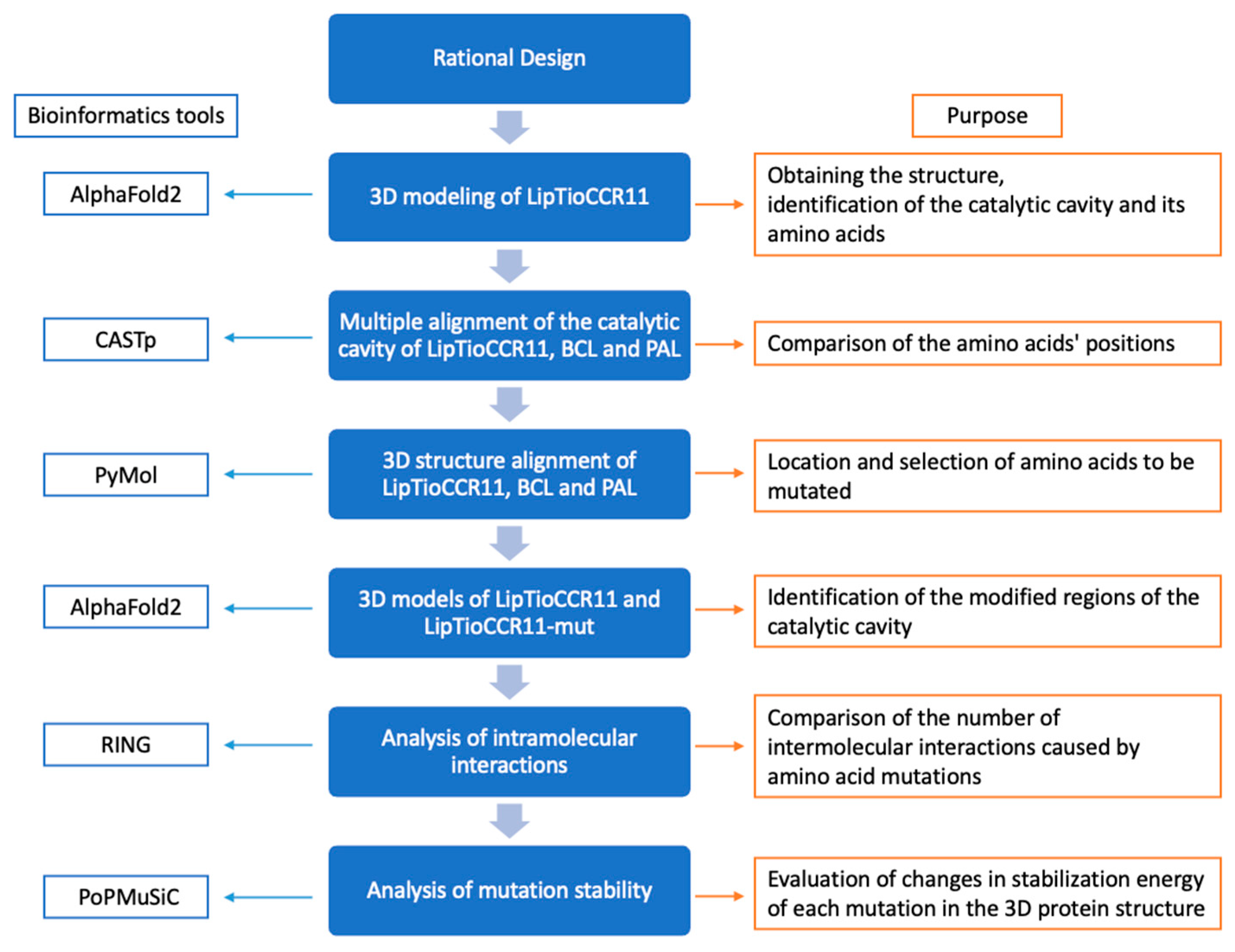
2.1. Rational Design of LipTioCCR11 Lipase to Increase Its Enantioselectivity

The rational design of LipTioCCR11 (previously LipMatCCR11) was accomplished by a multiple alignment strategy, with the lipases BCL from Burkholderia cepacia and PAL from Pseudomonas aeruginosa as models; these enzymes were chosen due to their structural similarity with LipTioCCR11 lipase and because they possess key residues for enantioselectivity, which is a desirable property in lipases for its application in the synthesis of optically active compounds that are used as building blocks for the pharmaceutical, chemical, and agrochemical industries [43]. The amino acids of the catalytic cavities of the three enzymes were aligned to choose those to be mutated. LipTioCCR11-mut was constructed by substituting the following six amino acids: F156M (at the oxyanion hole), L310F, F320L and I459V (at the hydrophobic cavity) and V311A, M313F (at the acyl-binding pocket). These residues’ positions have been identified as important for the enantioselectivity of lipases against chiral carboxylic acids [5]. As indicated in the Materials and Methods section, the criteria for amino acid substitution were to increase enzyme catalytic cavity volume and flexibility so large hydrophobic amino acids that were located in key positions for enantioselectivity in LipTioCCR11 were substituted with smaller ones present in PAL and/or BCL, as shown in Table 1, except for the acyl-binding pocket and the hydrophobic cavity, where the substituents were aromatic amino acids to promote new interactions with the substrate.

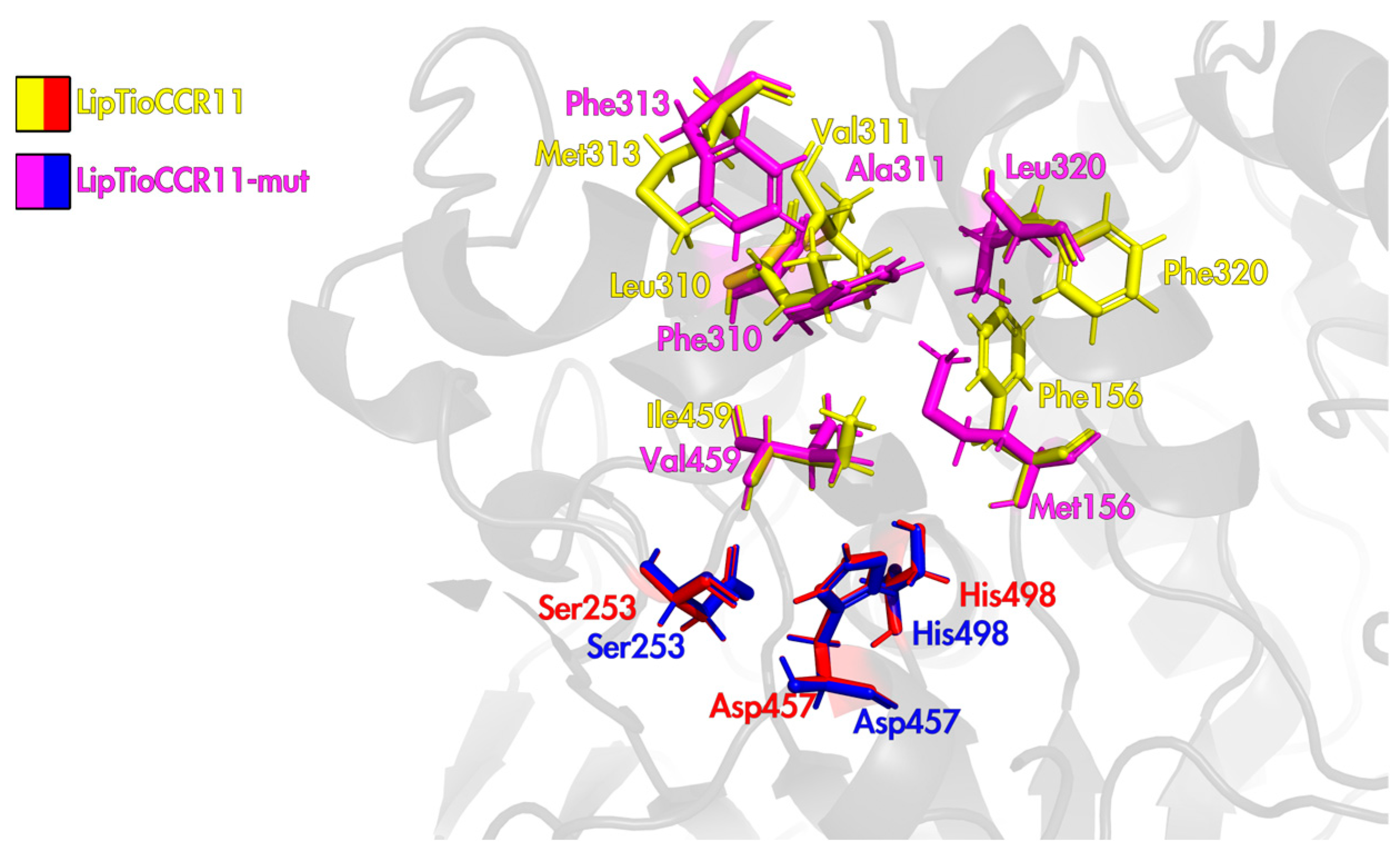
2.2. Three-Dimensional Structure of LipTioCCR11 Lipase

The 3D model of LipTioCCR11 was obtained by AlphaFold2 with a pLDDT value of 91 according to the IDDT-Cα metric [44]. AI platforms like AlphaFold allow obtaining reliable three-dimensional structures in a short time that are built based on structures elucidated by experimental methods [45,46,47]. As shown in Figure 1, LipTioCCR11 possesses the canonical structure of lipases, consisting of an α/β-fold structure with 12 α-helices and 11 β-strands. The catalytic serine (Ser253) is located at the nucleophilic elbow between the β4-strand and the α5-helix, and the other two residues of the catalytic triad, Asp321 and His362, are located within the catalytic cavity. It can be observed that the thioredoxin domain does not spatially interfere with the lipase domain. By means of the CASTp server (http://sts.bioe.uic.edu/castp/background.html; accessed on 17 January 2024) it was determined that the catalytic cavity is composed of 43 residues (46% hydrophobic, 28% of intermediate polarity, 10% hydrophilic, and 5% of neutral character). The hydrophobicity of lipase catalytic cavity plays a crucial role in the interaction with hydrophobic substrates and in catalysis [48]. The calculated active site cavity volume is 358 Å3.
The flexibility of the LipTioCCR11 3D model was determined using the CABS-flex 2.0 server through RMSF (Root Mean Square Fluctuation) values [49]. The catalytic triad residues, Ser253, Asp457, and His498, displayed RMSF values of 0.448 Å, 0.538 Å, and 0.485 Å, respectively, at 37 °C, indicating a high degree of rigidity that enables the enzyme to selectively recognize and clamp the substrate.
The intramolecular interactions of the 3D model were analyzed using the RING server (https://ring.biocomputingup.it/submit; accessed on 24 January 2024) [50], which calculates the type of interactions based on geometric parameters and restrictions. LipTioCCR11 was found to have 433 hydrogen bonds, 33 π—π interactions, one π cation, 13 ionic bonds, and 389 van der Waals forces. A disulfide bond belonging to thioredoxin between cysteine 420 and cysteine 423 was observed. All those electrostatic interactions are important for lipolytic activity and enzyme stability in aqueous solutions and in non-aqueous environments, where lipases carry out synthetic reactions [51,52].

2.3. Three-Dimensional Structure LipTioCCR11-Mut Lipase

The 3D model of LipTioCCR11-mut was obtained by AlphaFold2 with a pLDDT value of 94 according to the IDDT-Cα metric. The α/β-fold canonical structure of the enzyme was maintained, while the substitutions of the six amino acids in LipTioCCR11-mut increased the flexibility of the catalytic site by expanding its range of motion. At 37 °C, the flexibility value of Ser253 increased from 0.448 Å in LipTioCCR11 to 0.520 Å in LipTioCCR11-mut; the flexibility of Asp457 increased from 0.538 Å to 0.626 Å, and the flexibility of His498 decreased from 0.485 Å to 0.414 Å. These modifications resulted in an increased flexibility of the catalytic site and an increased volume of the catalytic cavity, from 358 Å3 in LipTioCCR11 to 406 Å3, mainly due to the absence of the aromatic ring of Phe when substituting Phe156Met at the oxyanion hole, and the presence of lower molecular weight residues when changing Val311Ala at the acyl-binding pocket, and Phe320Leu and Ile459V at the hydrophobic cavity (Figure 2). The number of intramolecular interactions was also increased: 532 hydrogen bonds, 36 π–π interactions, 2 π cation interactions, 16 ionic bonds, and 442 van der Waals forces were observed, which meant that compared to LipTioCCR11, the mutant enzyme was 7% stiffer.
The computational determination of the thermodynamic variations of proteins is fundamental in predicting the impact of point mutations on the protein’s structural stability. By means of PoPMuSiC, the folding free energy (ΔΔG) of each mutation was calculated, where a negative ΔΔG value indicates an energy release and in consequence, an increased protein stability, while a positive ΔΔG suggests destabilization [53]. As can be seen in Table 2, the ΔΔG values caused by the substitution of the residues Phe156Met, Leu310Phe, Val311Ala, Met313Phe, Phe320Leu, and Ile459Val were <0, indicating the increase in the enzyme’s stability.

2.4. Cloning and Expression of LipTioCCR11 and LipTioCCR11-Mut Lipases

The gene that encodes the lipase LipMatCCR11 was transferred from the pET3b to the pHTP8 vector and was expressed in E. coli BL21(DE3). The resulting recombinant lipase (LipTioCCR11) is composed of 520 residues and has a molecular weight of 57 kDa (43 kDa of the lipase plus 14 kDa of the 6xHis + thioredoxin tag). LipTioCCR11 was purified by immobilized metal ions affinity chromatography (IMAC) with a 90% yield, a 3.15 purification factor, and a final concentration of 3.4 mg/mL. As observed in Figure 3A, SDS-PAGE and zymography demonstrated the purity and activity of the LipTioCCR11 lipase. In comparison with the LipMatCCR11 lipase [39], the expression of the gene in the pHTP8 vector increased the concentration (800-fold) and the specific activity (3.2 Kat/mg vs. 3.58 mkat/mg of LipMatCCR11) due to the increased solubility conferred by the thioredoxin fusion protein [54].
The lipTioCCR11-mut gene was cloned in the pHTP8 vector and expressed in E. coli BL21(DE3) as well. It was purified by immobilized metal ions affinity chromatography (IMAC) with a purification yield of 90%, a purification factor of 3.81, a final concentration of 10 mg/mL, and a specific activity of 0.95 Kat/mg. As observed in Figure 3B, it has a molecular weight of 57 kDa and is an active enzyme, as shown in the zymography.

2.5. Biochemical Characteristics of LipTioCCR11 and LipTioCCR11-Mut Activity

The characteristics of the enzymatic activity of both lipases were determined. As shown in Table 3, LipTioCCR11 showed the highest specific activity (3.2 Kat/mg) at 40 °C, pH 8, over a substrate of 16 carbons (p-nitrophenyl palmitate). The enzyme’s activity was not significantly affected by calcium ions but showed an 80–90% inhibition in the presence of copper, sodium, manganese, and mercury. The enzyme maintained 84% of its activity in the presence of hexane, however, butanol and acetone inhibited it up to 90%. A 100% reduction in activity was observed in the presence of PMSF and SDS, and the addition of EDTA and β-mercaptoethanol provoked a 75% decrease in activity. During storage at room temperature, the enzyme’s activity decreased by 35% after 10 days and 42% after 30 days. At 4 °C, a 24% decrease was observed after 25 days of storage. LipTioCCR11 turned out to be a much more active enzyme than its predecessor LipMatCCR11 due to the thioredoxin label, which not only aided in solubility but also conferred greater stability and shelf life.
On the other side, LipTioCCR11-mut showed its maximum specific activity (0.95 Kat/mg) at 60 °C, pH 8, over a 12-carbon substrate (p-nitrophenyl laurate). The activity of the mutant enzyme increased in the presence of some metal ions (130% with barium, 123% with magnesium, and 118% with potassium) but decreased by up to 55% with sodium and mercury ions. Activity increase was also observed when incubated with organic solvents (180% in heptane, 150% in octanol, 120% in hexane and 123% in ethanol), but butanol and acetone inhibited 90% of the enzyme activity. A 100% reduction in activity was observed in the presence of PMSF and SDS but increased to 110% in the presence of β-mercaptoethanol (Table 3). During storage at room temperature, activity showed a 30% reduction after 10 days and a 35% reduction after 30 days. During storage at 4 °C the activity showed a reduction of 45% after 10 days and 62% after 30 days. The catalytic activity of the mutant lipase was 70% reduced compared to LipTioCCR11 due to the decrease in structural flexibility [55]. However, the increase in intramolecular interactions conferred rigidity and robustness to the enzyme as to have a higher optimum temperature (60 °C vs. 40 °C) and the ability to withstand harsh conditions as the presence of organic solvents. Both features increase the possibilities of the enzyme of being used in biotechnological applications that require high process temperatures and the presence of solvents to improve the solubility of the reactants.

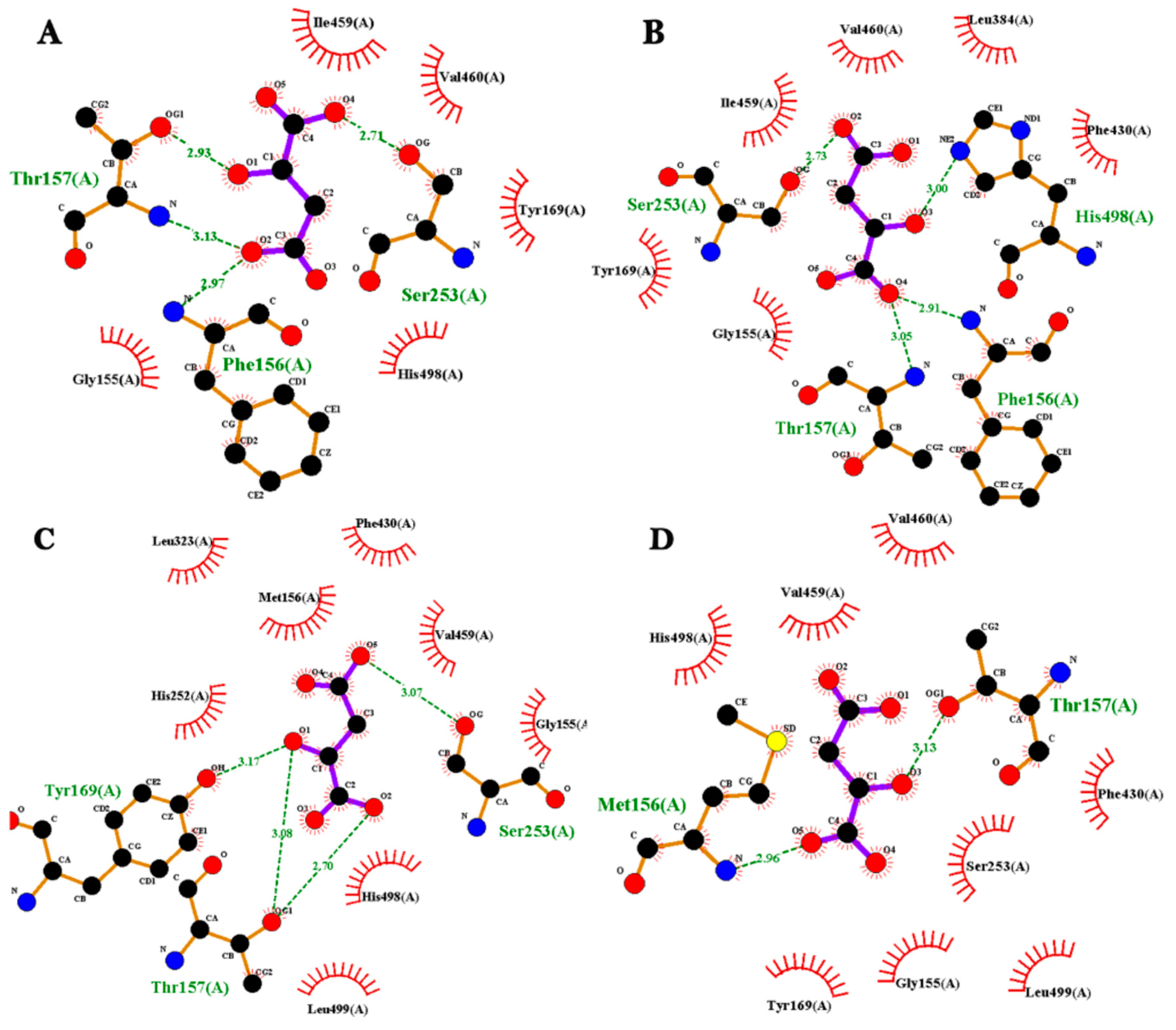
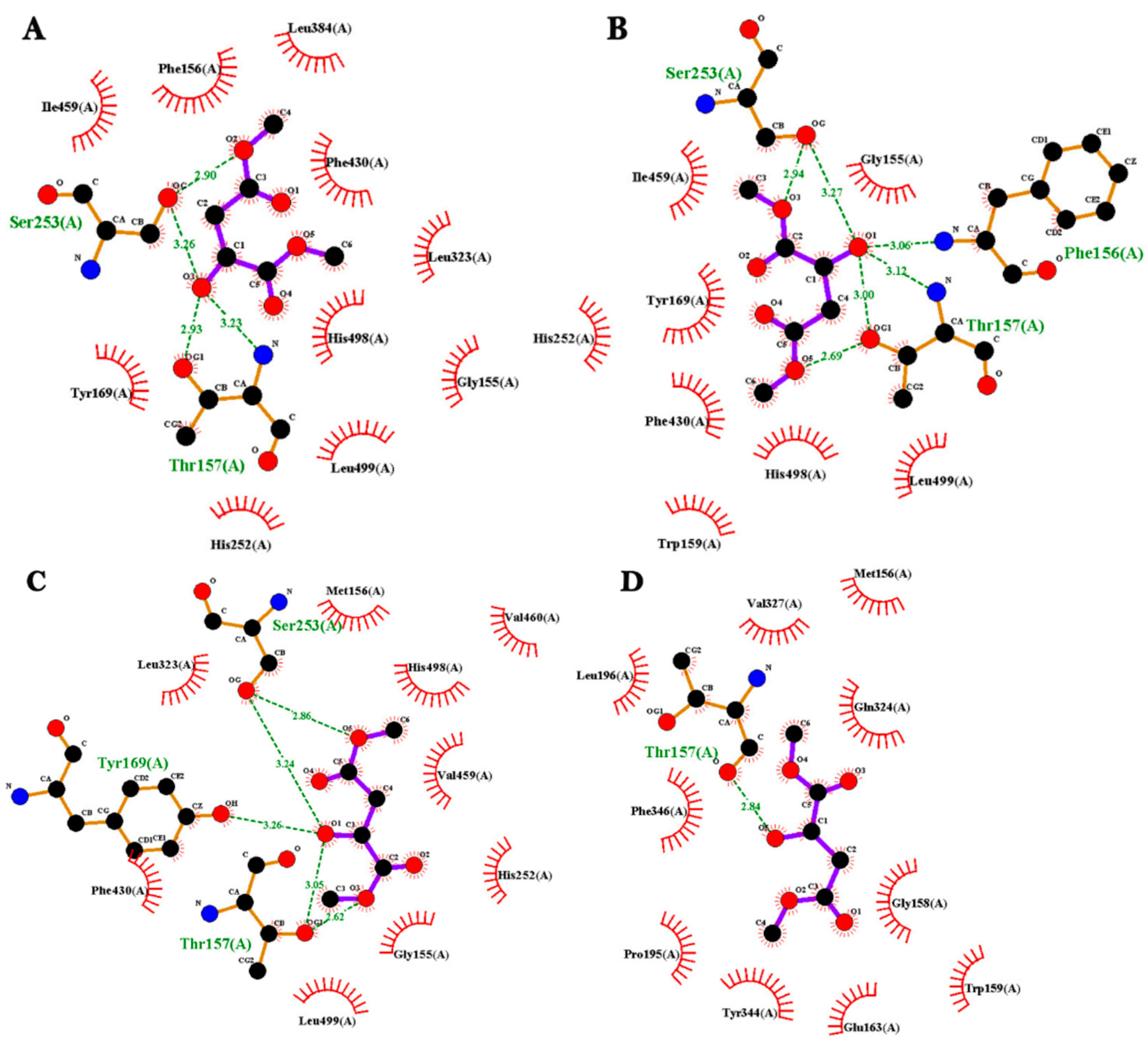
2.6. Molecular Docking Analysis

Molecular docking analysis was performed in order to verify whether the substituted residues in LipTioCCR11-mut were favoring the enantioselective recognition of D-malic acid, L-malic acid, (R)-dimethyl malate, and (S)-dimethyl malate. These substrates were chosen because they have potential applications in biotechnology for the environmentally friendly synthesis of various drug precursors, intermediates in chemical synthesis, biopolymers, agricultural formulations; in the food industry; and in medical studies related to metabolic diseases [41,55,56].
The comparison of LipTioCCR11 and LipTioCCR11-mut enzymes by coupling the two isomers of malic acid and the two enantiomers of dimethyl malate was made using the same parameters and force fields for the protein and substrate structures. Special attention was paid to the catalytic triad composed of the Ser, His, and Asp residues found in the catalytic site and to the impact of the substituted residues in each of their respective locations (F156M: oxyanion hole; L310F, F320L, I459V: hydrophobic cavity; V311A and M313F: acyl binding pocket).
The docking analysis of LipTioCCR11 and D-malic acid indicated an adequate interaction between the amino acids in the enzyme catalytic cavity and the substrate (Figure 4). The catalytic Ser253 interacts with the hydroxyl group of the substrate forming a hydrogen bond, with a distance of 2.71 Å and an affinity energy of −4.7 Kcal/mol (Figure 4A). In addition, the catalytic His498 is in close proximity, forming hydrophobic interactions with the substrate. A hydrogen bond is formed between Thr157 and the hydroxyl group of the chiral carbon of the substrate with a distance of 2.93 Å and with a hydroxyl group of C3 of D-malic acid (3.17 Å). The Phe156 (the oxyanion hole) forms a hydrogen bond with the hydroxyl group of C3 of the substrate, with a distance of 2.97 Å, confirming the importance of this position in the stabilization of the transition state, by facilitating the insertion and positioning of the substrate within the active site. The amino acids Gly155, Tyr169, Ile459 and Val460 of LipTioCCR11 catalytic cavity establish four hydrophobic interactions with D-malic acid. With respect with L-malic acid isomer, a hydrogen bond between the catalytic Ser253 and one of the hydroxyls groups of the ester bond of the substrate is observed with a favorable distance of 2.73 Å and a binding energy of −4.8 Kcal/mol. A hydrogen bond of the catalytic His498 is formed with the hydroxide of the chiral carbon with a distance of 3 Å. Here we again observe the participation of the oxyanion hole (Phe156) and of Thr157 establishing hydrogen bonds with the hydroxyl group of C4 of the substrate with a distance of 2.91 and 3.05 Å and the amino acids Gly155, Tyr169, Leu384, Phe430, Ile459 (hydrophobic cavity), and Val460 establishing hydrophobic interactions with the substrate (Figure 4B). Participation in both couplings of the oxyanion hole (Phe156) is emphasized, being a relevant position for the stabilization and positioning of the substrate, without steric hindrance. The Isoleucine 459 is part of the hydrophobic cavity and provides a favorable environment for substrate binding, helping to maintain the location and stability of the substrate, and favoring the possibility of the reaction. Consequently, it is concluded that LipTioCCR11 does not show enantioselective preference of the D- and L-isomers of malic acid.
In contrast, the mutated enzyme LipTioCCR11-mut showed a remarkable recognition of the D-malic acid isomer over L-malic acid. The catalytic Ser253 interacts with the hydroxyl group of the D-malic acid substrate forming a hydrogen bond, with a distance of 3.07 Å and an affinity energy of −3.9 Kcal/mol. The substitution of F320L and I459V residues in the hydrophobic cavity promoted the increase of its volume, providing a favorable electrostatic environment, less steric impediments and the generation of new interactions with D-malic acid. Also, the substitution at the oxyanion hole of Phe156 by a methionine promoted an internal rearrangement, generating the adequate environment and space for Thr157 to serve as a key residue for the establishment of a hydrogen bond between the hydroxyl group of the chiral carbon of D-malic acid, with a distance of 3.08 Å. This same amino acid forms another hydrogen bond with a hydroxyl group of the asymmetric carbon (C2) of the substrate (3.17 Å). The aromatic ring of Tyr169 forms a hydrogen bond with the hydroxyl group of the chiral carbon of D-malic acid (3.17 Å), acting as a stabilizing mechanism in conjunction with the hydrophobic interactions of the amino acids His252, Leu323, Phe156Met, Phe430, Ile459Val, His498, and Leu499 with D-malic acid (Figure 4C). In the region of the acyl-binding pocket and the hydrophobic cavity the addition of two aromatic rings by changing Met313Phe and Leu310Phe, respectively, resulted in an increase in the interaction sites between the substrate and the enzyme favoring a better coupling of the preferred D-malic acid enantiomer.
On the contrary, the replacement of the six amino acids in LipTioCCR11-mut caused the L-malic acid not to be recognized by the catalytic Ser253. Steric hindrance was generated for L-malic acid, limiting its access to the active site, due to the establishment of a hydrogen bridge between the Phe156Met and the hydroxyl group of the substrate (2.96 Å), and between Thr157 and the hydroxyl group of the chiral carbon of L-malic acid (3.13 Å); hydrophobic-type interactions of the amino acids Gly155, Tyr169, Phe430, Ile459Val, Val460, His498, and Leu499 were also observed (Figure 4D). These results indicate an enantioselective preference for the D-isomer of malic acid.
The docking analysis with the enantiomers of (RS)-dimethyl malate provides an approach to the recognition and possible affinity with the products of the reaction, resulting in information on the enantioselectivity of the lipases (Figure 5). Diverse interactions were observed between LipTioCCR11 with (R)-dimethyl malate, such as the formation of a hydrogen bond between the catalytic Ser253 and the hydroxyl group of the chiral carbon of (R)-dimethyl malate with a distance of 3.26 Å, and an interaction of the same type with the methyl group with a distance of 2.9 Å and an energy of −4.6 Kcal/mol. These distances are in the allowed ranges for hydrogen bond interactions. The catalytic His498 also has a hydrophobic proximity and interaction with the substrate as well as multiple hydrophobic interactions of residues Gly155, Phe156, Tyr169, His252, Leu323, Leu384, Phe430, Ile459, and Leu499 (Figure 5A), indicating an affinity for (R)-dimethyl malate. With the isomer (S)-dimethyl malate, LipTioCCR11 forms two hydrogen bonds between the catalytic Ser253 and the methyl (2.94 Å) and hydroxyl groups of the chiral carbon (3.27 Å). In addition, the oxyanion hole (Phe156) interacts with the chiral carbon of the (S)-dimethyl malate and, with the help of Thr157 stabilizes it, forming hydrogen bonds (3.06 Å and 3.12 Å, respectively) with an energy of −4.7 Kcal/mol. It is possible that these amino acids fulfill a stabilizing function for the subsequent recognition of the catalytic triad. Hydrophobic interactions of residues Gly155, Trp159, Tyr169, His252, Phe430, Ile459, His498, and Leu499 are also involved in this coupling (Figure 5B). All these interactions indicate that LipTioCCR11 recognizes the enantiomers of (RS)-dimethyl malate, and in consequence, does not show enantioselectivity for those two molecules.
The docking analysis of LipTioCCR11-mut with I-dimethyl malate revealed that the catalytic Ser256 interacts in two ways with the carbonyl ester, the first by a hydrogen bond at a distance of 2.86 Å and the second with the chiral hydroxyl group at a distance of 3.24 Å with an energy of −3.6 Kcal/mol. The presence of Phe156Met with a stabilizing interaction of hydrophobic character is observed in close proximity, Tyr169 maintains a hydrogen bond with the chiral hydroxyl group of the substrate (3.26 Å), joined by Thr157 directed towards the same region with a distance of 3.05 Å and another bond in the direction of methyl group (2.62 Å). Associated with hydrophobic interactions within the cavity are residues Gly155, His252, Leu323, Phe430, Val459, catalytic His498, and Leu499 (Figure 5C). The substitutions allowed a better space and environment for the R enantiomer to be properly accommodated. On the contrary, steric hindrances were generated so that the (S)-dimethyl malate isomer was kept away from the catalytic site; only a hydrogen bond is observed between the amino acid Thr157 and the hydroxyl group of the chiral carbon, with a distance of 2.84 Å and an energy of −3.1 Kcal/mol, but no interaction with the catalytic serine is observed, so the substitution of the six amino acids has a direct effect on substrate preference, promoting enantioselectivity for the isomI(R)-dimethyl malate. Hydrophobic interactions controlled by residues Met156, Gly158, Trp159, Glu163, Pro195, Leu196, Gln324, Val327, Tyr344, and Phe346 are also observed (Figure 5D).

2.7. Analysis of the Enantioselective Synthesis of (R)- and (S)-Dimethyl Malate

The synthesis of (R)- and (S)-dimethyl malate was carried out as indicated in the Materials and Methods section, using LipTioCCR11 and LipTioCCR11-mut enzymes, either D-malic acid or L-malic acid as substrates, methanol as the methyl donor, and methanol or acetonitrile as reaction medium at 25 °C or 40 °C for 24 h. The commercial enzyme lipase B from Candida antarctica (CAL-B, Novozym 435) was used as a positive control due to its demonstrated capability to perform enantiomeric reactions with a high enantiomeric excess (ee > 90%) [57,58,59,60]. The product of the synthesis reaction was identified by gas chromatography-mass spectrometry, as indicated in the Materials and Methods section, resulting in (RS)-dimethyl malate as shown in Figure 6.
When LipTioCCR11 was used as catalyst for the synthesis of (RS)-dimethyl malate in methanol at 25 °C, an ee of 59% for the (R)-isomer was observed, suggesting a limited enantioselectivity of LipTioCCR11 for the resolution of (DL)-malic acid (Table 4). This behavior was expected since in the docking analysis an interaction between the key amino acids of the catalytic cavity of LipTioCCR11 and both D- and L-malic acid were observed, indicating that the enzyme does not show an enantioselective preference of the D- and L-isomers of malic acid.
On the contrary, when LipTioCCR11-mut was used as catalyst, the ee increased up to 94.5% for the (R)-isomer, demonstrating the effectiveness of the amino acid substitutions introduced to LipTioCCR11-mut to obtain an enantioselective enzyme. In the docking analysis a remarkable recognition of the D-malic acid isomer over L-malic acid was observed since the catalytic Ser253 of the enzyme did not interact with any region of L-malic acid molecule. The enzyme CAL-B also performed an enantioselective reaction but with a preference for the synthesis of the (S)-isomer, with an ee of 83.5% (Table 4). The improvement in the enantioselective synthesis of (R)-dimethyl malate by LipTioCCR11-mut could be attributed to the increase in the volume of the catalytic cavity (from 358 Å3 in LipTioCCR11 to 406 Å3 in LipTioCCR11-mut) as has been proposed by Song et al. (2023) who reported that by having larger catalytic cavities, the enantioselectivity increases due to the spatial arrangement of the substrate and its better control by the hydrophobic regions [29].
The increase in reaction temperature (40 °C) modified the synthetic activity of LipTioCCR11 increasing the area under the curve (AUC) of the chromatographic peak by 2.3 times, from 54 ± 1.8 AU at 25 °C to 124 ± 0.9 AU at 40 °C, but the enantioselectivity was not affected, observing an ee of 53.5% for the (R)-isomer (Table 4). An increased temperature can improve the enantiomeric synthesis activity of lipases by increasing substrate solubility, favoring active site flexibility, accelerating catalytic reactions and reducing steric constraints due to internal cavity rearrangements [61]. On the other side, the increase in temperature did not affect the synthetic activity of LipTioCCR11-mut probably due to the rigidity of the catalytic site but decreased the enantioselectivity by 10% (ee of 83.5%) for the (R)-isomer, as observed also with CAL-B for the (S)-isomer (Table 4).
It has been reported that the log P of the solvent used as reaction medium significantly impacts the enantioselectivity of lipase-catalyzed reactions by modifying diverse intermolecular interactions between the substrate and the enzyme cavity; as log P changes, hydrophobic interactions are altered, which affect the affinity of the enzyme for the substrate, favoring more hydrophobic substrates in solvents with higher log P [31,57,62]. When using acetonitrile (LogP= −0.34) as the reaction medium at 25 °C, a significant increase in LipTioCCR11 synthetic activity was observed (AUC = 320 ± 0.6 AU vs. 37 ± 2.3 AU at 25 °C in methanol (LogP= −0.74), as was a slight change in the substrate preference for the (S)-isomer (ee of 54.5%) in comparison with the reaction in methanol. A reduction of the enantiomeric excess to 80.5%, with no change in synthetic activity (AUC = 909 ± 0.7 AU) for the (R)-isomer was observed for LipTioCCR11-mut (Table 5), and for CAL-B with an ee of 51% (AUC = 1030 ± 1.2 AU) for the (R)-isomer.
When the reactions were undertaken at 40 °C in acetonitrile, the synthetic activity of LipTioCCR11 was reduced (AUC = 124 ± 0.9 AU vs. 271 ± 1.6 AU at 25 °C), but the enantioselectivity was conserved with an ee of 55.5% for the (R)-isomer. With LipTioCCR11-mut a slight increase in synthetic activity (AUC = 1033 ± 1.6 AU) and enantioselectivity was observed, resulting in an ee of 82.5% for the (R)-isomer. The synthetic activity of CAL-B enzyme was reduced, and the enantiopreference was changed, with an ee of 67.5% for the (S)-isomer (Table 5).
Maldonado et al. (2021) [5] highlighted the importance of identifying key mutation sites for the control of enantioselectivity in enzymes, emphasizing how even subtle amino acid modifications close to the catalytic site can drastically change enzyme efficiency. Mutations that expanded the catalytic cavity from 358 Å3 to 406 Å3 in LipTioCCR11-mut resulted in a significant improvement in enantioselectivity, increasing the enantiomeric excess (ee) from 59% to 94.5% for the ®-isomer during dimethyl malate synthesis. This emphasizes how important specific amino acids are in the catalytic cavity since, as being part of the catalytic mechanism, they can play a key role in interacting with the substrate, allowing its orientation and spatial disposition, so it is properly accommodated in the catalytic cavity. Their disposition not only guides the accommodation of the substrate but also determines precisely which of the enantiomers will be favored during the reaction [5]. That is particularly important for the synthesis of enantiomerically pure compounds applicable in diverse industries, such as the pharmaceutical, agrochemical, foods and fine chemicals, where the activity, efficacy, or toxicity of the chemicals are of the utmost importance.

3. Materials and Methods

3.1. Bacterial Strain, Plasmids and Culture Medium

Plasmid used for cloning and expression of LipTioCCR11 and LipTioCCR11-mut was pHTP8 (NZYEasy Cloning and Expression kit VIII, Lisboa, Portugal), which confers kanamycin resistance and adds two fusion tags at the amino-terminal end of the protein: a 6xHis tag that facilitates purification, and a thioredoxin tag that increases the solubility of the recombinant protein. Strain used for transformation and expression was E. coli BL21(DE3) (PROMEGA, Madison, WI, USA).
LB medium (tryptone10 g/L, yeast extract 5 g/L, sodium chloride 10 g/L, and kanamycin 50 µg/mL, pH 7.5; and agar 15 g/L for solid medium) was used for bacterial growth and recombinant enzymes expression. Culture conditions were 100 rpm at 37 °C.

3.2. Rational Design of LipTioCCR11 Amino Acid Sequence

In order to improve the enantioselectivity of LipTioCCR11 lipase a rational design approach was used (Figure 7). First, we created a 3D model of LipTioCCR11 in AlphaFold2 with a pLDDT score of 91 (https://colab.research.google.com/github/sokrypton/ColabFold/blob/main/AlphaFold2.ipynb; accessed on 17 January 2024) [63] and identified the amino acids that compose the catalytic cavity of the enzyme, divided into the oxyanion hole, the hydrophobic cavity, the hydrophilic cavity and the acyl-binding pocket, using the CasTp platform (http://sts.bioe.uic.edu/castp/index.html?3trg; accessed on 19 January 2024).
Then, a multiple alignment was made with the amino acids that compose the catalytic cavity of LipTioCCR11 and two reported enantioselective lipolytic enzymes: the BCL lipase from Burkholderia epaciana (PDB: 1oil) and the PAL lipase from Pseudomonas aeruginosa (PDB: 1ex9) in order to select the target amino acids for mutation. Maldonado et al. [5] identified as important in the control of enantioselectivity against chiral carboxylic acids the following amino acids: in BCL L17, P113, H114, S117, F119, A120, F122, V123, L163, L167, V266, and V267 and in PAL M16, P108, H109, S112, T114, A115, F117, L118, L159, L162, L231, and L232. The three-dimensional structures of the selected lipases were aligned by PyMol as well, and the conserved and substrate-interacting regions were determined. The criteria for amino acid substitution were to increase enzyme catalytic cavity volume and flexibility so large hydrophobic amino acids that were located in key positions for enantioselectivity in LipTioCCR11 were substituted with smaller ones present in PAL and/or BCL, except for the acyl-binding pocket and the hydrophobic cavity, where the substituents were aromatic amino acids to promote new interactions with the substrate.
Once the substitutions of the selected amino acids in LipTioCCR11 lipase were made in silico, three-dimensional models of the native (LipTioCCR11) and modified enzyme (LipTioCCR11-mut) were created in AlphaFold2 (pLDDT > 90) [63]. The 3D structures of LipTioCCR11 and LipTioCCR11-mut were superimposed to observe the correctness of the substitutions and to verify that the amino acids corresponding to the catalytic site remained in the same coordinates. Models were analyzed at the RING server (Residue Interaction Network Generator) http://old.protein.bio.unipd.it/ring/?fbclid=IwAR2EX0sGJO4KKLco13ukb-R_4o7PTmjIiVYd3KlkjtMArFZbufbujtuGaXU; accessed on 24 January 2024) to evaluate all possible non-covalent interactions at the atomic level considering the following distance values: H-bond 3.5 Å, ionic bond 4 Å, cation-π interaction 5 Å, π-π interaction 6.5 Å, disulfide bond 2.5 Å, Van der Waals interaction 0.5 Å.
After the analysis with the RING server, we used the PopMuSiC (https://soft.dezyme.com/query/create/pop; accessed on 24 January 2024) to calculate the energetic changes (ΔΔG) caused by the amino acids’ substitutions allowing us to identify the mutations that may stabilize or destabilize the enzyme. A negative ΔΔG value indicates an increased protein stability, while a positive ΔΔG suggests destabilization [53].

3.3. Construction of Expression Systems for LipTioCCR11 and LipTioCCR11-Mut

The mutated LipTioCCR11 lipase gene (lipTioCCR11-mut) was synthesized and inserted into the pGEM-Kan vector by Macrogen (Geumcheon-gu, Seoul, Republic of Korea). Both lipTioCCR11-mut and lipMatCCR11 genes, the latter contained in the pET-28b vector [39], were amplified by PCR using the primers AaF 5′-TCA GCA AGG GCT GAG G ATG GCA GTT TCA CGC and AaR 3′-TCA GCG GAA GCT GAG G TTA TAC TGC TCG GCA AGT GCA AGT C. The reaction conditions used were: 1 cycle (94 °C for 7 min), 30 cycles (95 °C 1 min, 58.5 °C 1.5 min, and 73 °C 2 min), and a final cycle of 73 °C 10 min. Then, both genes were inserted into the pHTP8 vector following the NZYEasy Cloning and Expression System instructions. The lipTioCCR11-pHTP8 and lipTioCCR11-mut-pHTP8 constructs were used to transform E. coli BL21(DE3) and the transformants were screened on LB medium agar plates (10 g/L tryptone, 5 g/L yeast extract, 5 g/L NaCl, and 15 g/L agar) for kanamycin resistance.

3.4. Expression and Collection of LipTioCCR11 and LipTioCCR11-Mut Lipases

E. coli BL21(DE3) strains transformed with lipTioCCR11-pHTP8 and lipTioCCR11-mut-pHTP8 constructions were grown overnight (0.8 D.O.) at 37 °C, 200 rpm in LB medium containing 100 mg/mL kanamycin. Then, IPTG (0.7 mM) was added, and induction proceeded for 3 h at 25 °C and 100 rpm. After induction time, cells were collected by centrifugation (13,000× g, 20 min, 4 °C) and then suspended in lysate/binding buffer (0.05 M sodium phosphate, 0.3 M NaCl, 0.005 M imidazole, pH 8), placed on ice, and disaggregated by sonication (three 25 s pulses with 35 s pauses between pulses) in an ultrasonic processor (Ultrasonic Processor VEX130PB, Thermo Fisher Scientific, CDMX, Mexico). The homogenate was centrifuged (13,000× g, 10 min, 4 °C) and the supernatant was filtered through a 0.22 µm membrane (MCE-Millipore, Burlington, MA, USA) under sterile conditions. Protein concentration, SDS-PAGE profile and lipolytic activity were determined.

3.4.1. Purification of LipTioCCR11 and LipTioCCR11-Mut Lipases

LipTioCCR11 and LipTioCCR11-mut lipases were purified by immobilized metal ions affinity chromatography (IMAC) following supplier’s instructions (Profinity IMAC, BioRad S.A, CDMX, Mexico). The obtained fractions were subjected to protein quantification, SDS-PAGE and lipolytic activity quantification.

3.4.2. Protein Quantification

Protein concentration was measured following Bradford methodology [64]. Briefly, 245 µL of Bradford reagent (Bradford Reagent, Merck S.A. de C.V., Naucalpan, Mexico) were added to 5 µL of sample, and absorbance was read at a wavelength of 470 nm on a Bio Rad 550 plate reader (BioRad S.A, CDMX, Mexico). A standard curve was prepared with 0–1 mg/mL of bovine serum albumin (BSA, 9048-46-8, Merck S.A. de C.V., Naucalpan, Mexico).

3.4.3. Quantification of Lipolytic Activity

The quantitative determination of lipolytic activity was accomplished by the spectrophotometric assay described by Nawani [65] using p-nitrophenyl-laurate (pNPL) as substrate. The assay conditions used were previously determined [39]. The reaction mixture consisted of 0.1 mL enzyme solution, 0.8 mL 0.05 M phosphate buffer (pH 8), and 0.1 mL 0.01 M pNPL in ethanol. A blank tube containing all the ingredients except for the enzyme solution was included and was substituted by 0.05 M phosphate buffer (pH 8). The hydrolytic reaction was carried out at 40 °C for 20 min, and 150 rpm, after which 0.25 mL of 0.1 M Na2CO3 was added. The mixture was centrifuged (16,000× g, 15 min, 25 °C) and the absorbance at 410 nm was determined. Enzyme activity is expressed in katal (kat), defined as the catalytic activity responsible for the transformation of one mole of substrate per second. Results were interpolated in a standard curve prepared with 0–215 µM of pNPL.

3.4.4. Polyacrylamide Gel Electrophoresis (SDS-PAGE)

Proteins were analyzed by SDS-PAGE using 12% polyacrylamide vertical gels as described by Laemmli [66,67]. Prior to electrophoresis 20 µL of samples (5–10 mg/mL of protein) were mixed with 10 µL of sample buffer (2.5 mL of 10% SDS; 0.2 mL of β-mercaptoethanol; 0.5 mL of 0.05% bromophenol blue) and incubated at 92 °C for 2 min. Gels were analyzed for lipolytic activity by zymography and stained for protein detection with Coomasie Brilliant Blue (R)-250 (Sigma-Aldrich, Merck S.A. de C.V., Naucalpan, Mexico).

3.4.5. Zymography

The lipolytic activity of the protein bands of SDS-PAGE gels was determined using the technique reported by Prim et al. (2003) [66]. After electrophoresis, the gel was incubated at room temperature in a renaturing solution (0.05 M sodium phosphate buffer, pH 8, with 2.5% Triton X-100) for 30 min under 150 rpm agitation. Then, the solution was removed, and the gel was washed twice with 0.05 M sodium phosphate buffer, pH 8, for 20 min. After the second wash the gel was covered for 3 min with a solution of 25 mM methylumberiferyl butyrate (dissolved in monomethylether ethylene glycol) in 10 mL of sodium phosphate buffer, pH 8. Lipolytic activity was observed as a brilliant band under UV transillumination (Gel Doc XR+ System, BioRad S.A, CDMX, Mexico).

3.5. Characterization of LipTioCCR11 and LipTioCCR11-Mut Lipases

Effect of temperature on enzymes’ activity and stability. The optimal temperature for lipolytic activity was determined by the spectrophotometric assay using p-nitrophenyl laurate as substrate at different temperatures (30 to 90 °C) at pH 7.5. Temperature stability was determined by incubating enzymes for 30 min at 30–70 °C and measuring residual activity by the spectrophotometric assay.
Effect of pH on enzymes’ activity and stability. Optimal pH was determined by the spectrophotometric assay with p-nitrophenyl laurate as substrate at different pHs (5–10) at the previously determined optimum temperature. The buffers used were 0.05 M sodium acetate buffer (pH 5), 0.05 M potassium phosphate buffer (pH 6–7), 0.05 M HEPES buffer (pH 8), 0.05 M CHES buffer (pH 9), and 0.05 M glycylglycine buffer (pH 10). The effect of pH on enzyme stability was determined by incubating enzymes for 20 min at 40 °C in the above-mentioned solutions (pH 5–10) and measuring residual activity by the spectrophotometric assay at the optimal temperature and pH determined previously.
Substrate specificity. It was determined by the spectrophotometric assay using different p-nitrophenyl esters (C2, C4, C5, C8, C12, C14, C16, C18) as substrates at the optimal temperature and pH determined previously.
Effect of detergents on enzymes’ activity. The effect of detergents was measured by incubating enzymes for 20 min at the optimal temperature and pH in 0.05 M CHES buffer containing 1% of Triton X-100, Tween 20, Tween 80 or SDS (Sigma-Aldrich, Merck S.A. de C.V., Naucalpan, Mexico). An incubation without detergent was used to calculate the residual activity, which was measured by the spectrophotometric assay at the optimal temperature and pH determined previously.
Effect of organic solvents, metal ions and inhibitors on enzyme activity. The effect of organic solvents on enzyme activity was measured by the spectrophotometric assay after pre-incubation with 1:1 v/v of methanol, ethanol, butanol, terbutanol, 1-propanol, 2-propanol, octanol, acetone, and hexane. The effect of inhibitors was determined after incubation for with EDTA (1 mM), β-mercaptoethanol (1% v/v), PMSF (1 mM). For metal ions, activity was determined after incubation in an aqueous solution of 1 mM CaCl2, KCl, MnCl2, NaCl, BaCl2, HgCl2, LiCl, MgCl2, SrCl2, and CuCl2 in 0.05 M sodium phosphate buffer, pH 8. In all cases, samples were incubated for 1 h at 30 °C and 150 rpm in a Thermomixer C-Eppendorf. An incubation without organic solvents, metal ions and inhibitors was used to calculate the residual activity, that was measured by the spectrophotometric assay at the optimal temperature and pH determined previously.

3.6. Molecular Docking Studies

The substrates used for molecular docking analysis were D-malic acid, L-malic acid, dimethyl (R)-malate, and dimethyl (S)-malate, whose structures were built in Avogadro v1.2.0 and minimized with the MMFF94 force field [68]. D-malic acid and L-malic acid were the substrates, and dimethyl (R)-malate and dimethyl (S)-malate were the products in the experimental reactions.
Molecular docking was performed with the AutoDock Vina program coupled to Chimera (https://www.cgl.ucsf.edu/chimera/docs/ContributedSoftware/vina/vina.html; accessed on 29 January 2024). The model preparation consisted in the removal of water molecules, repairing of truncated side chains, adding missing hydrogens, and assigning partial charges [69,70]. It was performed with a step number of 100, a step size of 0.02 Å, a conjugate gradient of 10, and an update interval of 10. The AMBER (ff14SB) force field [71] was selected for the standard protein residues and AM1-BCC for the other. Charges were assigned to substrates using the AMBER (ff14SB) force field and Gasteiger [72], which is a faster method based on atom types and connectivity.

3.7. Analysis of the Enantioselective Production of (RS)-Dimethyl Malate

The enantioselectivity of LipTioCCR11 and LipTioCCR11-mut lipases was analyzed by assaying the synthesis of (R)- or (S)-dimethyl malate using (D)-malic acid and (L)-malic acid as substrates (MERCK, Rahway, NJ, USA); methanol (JT Baker, Phillipsburg, NJ, USA) as the methyl donor; and one of two solvents as the reaction medium, methanol (JT Baker) or acetonitrile (JT Baker). The enzymes were prepared by lyophilization at their respective optimum pH. The concentration of the substrates was 10 mM, the amount of enzyme was 15 mg, the volume of the reaction medium was 3 mL, the reaction temperatures were 25 °C and 40 °C, and the reaction time was 24 h. The presence of (R)- or (S)-dimethyl malate was identified by gas chromatography, performed with an HP-6890 GC with FID and a 7683 autosampler, operated with ChemStation G1701CA software LTS 01.11 (Agilent Technologies Mexico, CDMX, Mexico) and equipped with an HP-Innowax (60 m × 0.25 mm i.d., 0.25 µm film thickness). N2 was used as the carrier gas. Vaporization chamber and detector temperatures were set at 240 °C. The column temperature was maintained at 190 °C for 5 min and then increased at 10 °C/min to 220 °C and held for 3 min. The product concentration was determined by the peak area using the external standard method. The identity of the products was verified by gas chromatography and mass spectrometry (GC-MS). An Agilent Technologies gas chromatograph Model 6890N (Net Work GC System; Agilent Technologies Mexico, CDMX, Mexico) with a DB-5, 5% -phenyl-methylpolysiloxane column (60 m, 0.25 mm id, 0.25 mm film thickness) was used. The initial temperature was 150 °C, which was maintained for 5 min. The temperature was then raised to 210 °C using a heating ramp of 30 °C/min. From 210 °C, the temperature was ramped to 213 °C at a rate of 1 °C/min. Helium was used as carrier gas at a flow rate of 1 mL/min, the injector temperature was 250 °C, split injection, with a split ratio of 20:1. For mass spectrometry, an Agilent Technologies model 5975 inert XL mass spectrometer was used. Mass spectra were obtained by electron impact ionization at 70 eV. For identification, the mass spectra obtained for each compound were compared with a database (HP Chemstation-NIST 05 mass spectra search program, version 2.0d; Agilent Technologies Mexico, CDMX, Mexico).

3.8. Statistical Analysis

Data in the analysis of the enantioselective synthesis of (R)- and (S)-dimethyl malate section are given as mean ± standard deviation. Statistical significance was calculated by a Student’s t-test. The differences were considered significant at p < 0.05.

4. Conclusions

The change of six amino acids of the catalytic cavity of LipTioCCR11 lipase that were selected by rational design resulted in a lipase (LipTioCCR11-mut) with improved capabilities in hydrolytic and synthetic reactions. The hydrolytic reactions benefited by an increase in optimal temperature, up to 60 °C, and in the stability when exposed to organic solvents, making the LipTioCCR11-mut lipase a more robust enzyme. On the other side, the enzyme acquired the capability to perform enantioselective synthetic reactions, as demonstrated by the synthesis of (R)-dimethyl malate, a compound that has biotechnological importance due to its versatility as an intermediate in the synthesis of chiral building blocks used in pharmaceutical industry for the synthesis of drugs that require high enantiomerical purity, such as cardiovascular drugs and antiviral agents, for the production of precursors for biodegradable polymeric materials and in the food industry for the synthesis of flavors, fragrances, and nutraceuticals.
Only a few enantioselective lipases have been described and commercialized to date: Candida antarctica lipase B (CAL-B, Novozym 435), Amano PS lipase from Pseudomonas cepacia and Amano-P lipase from Pseudomonas fluorescens. The possibility of obtaining novel enantioselective enzymes by rational design represents a good alternative to expand the offer of biocatalysts with characteristics that meet the needs of biotechnological industries, such as LipMatCCR11-mut, designed in the present investigation. In addition, this work’s findings provide information regarding the impact of changing amino acids that conform the catalytic cavity over the spatial conformation and the substrate selectivity of thermoalkaliphilic lipases of the Geobacillus genus, on which information is scarce.

Author Contributions

Conceptualization, R.M.O.-R. and R.Q.-C.; design and methodology, A.-S.B.-B.; validation, R.Q.-C. and M.G.S.-O.; formal analysis, C.P.-M. and R.M.O.-R.; investigation, A.-S.B.-B. and G.E.-L.; acquisition, analysis, or interpretation of data, M.R.M.-L. and A.-S.B.-B.; data curation, A.-S.B.-B. and M.G.S.-O.; funding acquisition, R.M.O.-R. and C.P.-M.; writing—original draft preparation, A.-S.B.-B. and R.M.O.-R.; writing—review and editing, R.M.O.-R., M.G.S.-O. and C.P.-M.; supervision, G.E.-L., M.G.S.-O. and R.Q.-C.; project administration, R.M.O.-R. and C.P.-M. All authors have read and agreed to the published version of the manuscript.

Funding

This study was supported by the project 18073.23-P from the Tecnológico Nacional de México.

Data Availability Statement

Data are contained within the article.

Acknowledgments

Aaron-Salvador Bustos-Baena acknowledges his PhD scholarship from the National Council for Humanities, Science, and Technology (Conahcyt-México).

Conflicts of Interest

The authors declare no conflicts of interest.

References

  1. Casas-Godoy, L.; Gasteazoro, F.; Duquesne, S.; Bordes, F.; Marty, A.; Sandoval, G. Lipases: An overview. In Methods in Molecular Biology; Humana Press: New York, NY, USA, 2018; Volume 1835, pp. 3–38. [Google Scholar] [CrossRef]
  2. Treichel, H.; de Oliveira, D.; Mazutti, M.A.; Di Luccio, M.; Oliveira, J.V. A Review on microbial lipases production. Food Bioprocess Technol. 2010, 3, 182–196. [Google Scholar] [CrossRef]
  3. Xu, X. Production of specific-structured triacylglycerols by lipase-catalyzed reactions: A review. Eur. J. Lip. Sci. Technol. 2000, 102, 287–303. [Google Scholar] [CrossRef]
  4. Park, J.-Y.; Park, K.-M. Lipase and Its Unique Selectivity: A Mini-Review. J. Chem. 2022, 2022, 7609019. [Google Scholar] [CrossRef]
  5. Maldonado, M.R.; Alnoch, R.C.; de Almeida, J.M.; dos Santos, L.A.; Andretta, A.T.; Ropaín, R.d.P.C.; de Souza, E.M.; Mitchell, D.A.; Krieger, N. Key mutation sites for improvement of the enantioselectivity of lipases through protein engineering. Biochem. Eng. J. 2021, 172, 108047. [Google Scholar] [CrossRef]
  6. Chen, H.; Meng, X.; Xu, X.; Liu, W.; Li, S. The molecular basis for lipase stereoselectivity. Appl. Microbiol. Biotechnol. 2018, 102, 3487–3495. [Google Scholar] [CrossRef]
  7. Carvalho, A.C.L.d.M.; Fonseca, T.D.S.; de Mattos, M.C.; Oliveira, M.D.C.F.d.; de Lemos, T.L.G.; Molinari, F.; Romano, D.; Serra, I. Recent advances in lipase-mediated preparation of pharmaceuticals and their intermediates. Int. J. Mol. Sci. 2015, 16, 29682–29716. [Google Scholar] [CrossRef]
  8. Bhardwaj, K.K.; Gupta, R. Synthesis of chirally pure enantiomers by lipase. J. Oleo Sci. 2017, 66, 1073–1084. [Google Scholar] [CrossRef]
  9. Ferraccioli, R. Progress on the stereoselective synthesis of chiral molecules based on metal-catalyzed dynamic kinetic resolu-tion of alcohols with lipases. Symmetry 2021, 13, 1744. [Google Scholar] [CrossRef]
  10. Soto, M.; Sanz-Machín, I.; Rodríguez-Solla, H.; Gotor-Fernández, V. Chemoenzymatic stereoselective synthesis of trans-flavan-4-ols via lipase-catalyzed kinetic resolutions. Catalysts 2021, 11, 1296. [Google Scholar] [CrossRef]
  11. Watts, O.F.B.; Berreur, J.; Collins, B.S.L.; Clayden, J. Biocatalytic enantioselective synthesis of atropisomers. Acc. Chem. Res. 2022, 55, 3362–3375. [Google Scholar] [CrossRef]
  12. Ferrario, V.; Ebert, C.; Nitti, P.; Pitacco, G.; Gardossi, L. Modelling and predicting enzyme enantioselectivity: The aid of computational methods for the rational use of lipase B from Candida antarctica. Curr. Biotechnol. 2015, 4, 87–99. [Google Scholar] [CrossRef]
  13. Yang, H.; Yu, H.; Stolarzewicz, I.A.; Tang, W. Enantioselective transformations in the synthesis of therapeutic agents. Chem. Rev. 2023, 123, 9397–9446. [Google Scholar] [CrossRef] [PubMed]
  14. Da Costa, S.P.; Schuenck-Rodrigues, R.A. Lipases in the Enantioselective Biocatalysis Process to Obtain Enantiomerically Pure Racemic Products. J. Phys. Chem. Res. 2022, 4, 146. [Google Scholar]
  15. Vivek, K.; Sandhia, G.S.; Subramaniyan, S.J.B.A. Extremophilic lipases for industrial applications: A general review. Biotechnol. Adv. 2022, 60, 108002. [Google Scholar] [CrossRef]
  16. Choi, Y.; Park, J.-Y.; Chang, P.-S. Integral Stereoselectivity of lipase based on the chromatographic resolution of enantiomeric/regioisomeric diacylglycerols. J. Agric. Food Chem. 2021, 69, 325–331. [Google Scholar] [CrossRef]
  17. Cen, Y.; Li, D.; Xu, J.; Wu, Q.; Wu, Q.; Lin, X. Highly Focused Library-Based Engineering of Candida antarctica Lipase B with (S)-Selectivity Towards sec-Alcohols. Adv. Synth. Catal. 2019, 361, 126–134. [Google Scholar] [CrossRef]
  18. Li, D.; Chen, X.; Chen, Z.; Lin, X.; Xu, J.; Wu, Q. Directed evolution of lipase A from Bacillus subtilis for the preparation of enantiocomplementary sec-alcohols. Green Synth. Catal. 2021, 2, 290–294. [Google Scholar] [CrossRef]
  19. McLoughlin, E.C.; Twamley, B.; O’Boyle, N.M. Candida antarctica Lipase B mediated kinetic resolution: A sustainable method for chiral synthesis of antiproliferative β-lactams. Eur. J. Med. Chem. 2024, 276, 116692. [Google Scholar] [CrossRef]
  20. Wu, Q.; Soni, P.; Reetz, M.T. Laboratory evolution of enantiocomplementary Candida antarctica lipase B mutants with broad substrate scope. J. Am. Chem. Soc. 2013, 135, 1872–1881. [Google Scholar] [CrossRef]
  21. Qin, Z.; Wang, Y.; Li, S.; Wu, Q.; Zhu, D. Enhancing the enantioselectivity of Candida antarctica lipase B for secondary alcohols by engineering the entrance to the binding pocket. J. Mol. Catal. B Enzym. 2013, 85–86, 100–106. [Google Scholar]
  22. Shen, J.-W.; Qi, J.-M.; Zhang, X.-J.; Liu, Z.-Q.; Zheng, Y.-G. Significantly increased catalytic activity of Candida antarctica lipase B for the resolution of cis-(±)-dimethyl 1-acetylpiperidine-2,3-dicarboxylate. Catal. Sci. Technol. 2018, 8, 4718–4725. [Google Scholar] [CrossRef]
  23. Engström, K.; Nyhlén, J.; Sandström, A.G.; Bäckvall, J.-E. Directed evolution of an enantioselective lipase with broad substrate scope for hydrolysis of α-substituted esters. J. Am. Chem. Soc. 2010, 132, 7038–7042. [Google Scholar] [CrossRef] [PubMed]
  24. Sandström, A.G.; Wikmark, Y.; Engström, K.; Nyhlén, J.; Bäckvall, J.-E. Combinatorial reshaping of the Candida antarctica lipase A substrate pocket for enantioselectivity using an extremely condensed library. Proc. Natl. Acad. Sci. USA 2012, 109, 78–83. [Google Scholar] [CrossRef]
  25. Reetz, M.T. Witnessing the birth of directed evolution of stereoselective enzymes as catalysts in organic chemistry. Adv. Synth. Catal. 2022, 364, 3326–3335. [Google Scholar] [CrossRef]
  26. Reetz, M.T.; Wilensek, S.; Zha, D.; Jaeger, K.-E. Directed evolution of an enantioselective enzyme through combinatorial multiple-cassette mutagenesis. Angew. Chem. Int. Ed. Engl. 2001, 40, 3589–3591. [Google Scholar] [CrossRef]
  27. Prasad, S.; Bocola, M.; Reetz, M.T. Revisiting the lipase from Pseudomonas aeruginosa: Directed evolution of substrate ac-ceptance and enantioselectivity using iterative saturation mutagenesis. ChemPhysChem 2011, 12, 1550–1557. [Google Scholar] [CrossRef]
  28. Tian, M.; Yang, L.; Lv, P.; Wang, Z.; Fu, J.; Miao, C.; Li, Z.; Li, L.; Liu, T.; Du, W.; et al. Improvement of methanol tolerance and catalytic activity of Rhizomucor miehei lipase for one-step synthesis of biodiesel by semi-rational design. Bioresour. Technol. 2022, 348, 126769. [Google Scholar] [CrossRef]
  29. Song, Z.; Zhang, Q.; Wu, W.; Pu, Z.; Yu, H. Rational design of enzyme activity and enantioselectivity. Front. Bioeng. Biotechnol. 2023, 11, 1129149. [Google Scholar] [CrossRef]
  30. Sternke, M.; Tripp, K.W.; Barrick, D. Consensus sequence design as a general strategy to create hyperstable, biologically active proteins. Proc. Natl. Acad. Sci. USA 2019, 116, 11275–11284. [Google Scholar] [CrossRef]
  31. Zhang, Y.; Feng, T.; Ni, X.; Xia, J.; Suo, H.; Yan, L.; Zou, B. Immobilized lipase based on SBA-15 adsorption and gel embedding for catalytic synthesis of isoamyl acetate. Food Biosci. 2024, 60, 104427. [Google Scholar] [CrossRef]
  32. Kazlauskas, R.J.; Bornscheuer, U.T. Finding better protein engineering strategies. Nat. Chem. Biol. 2009, 5, 526–529. [Google Scholar] [CrossRef] [PubMed]
  33. Palomo, J.M. Semisynthetic Enzymes by Protein–Peptide Site-Directed Covalent Conjugation: Methods and Applications. In Methods in Enzymology; Academic Press: Cambridge, MA, USA, 2017; Volume 590, pp. 305–316. [Google Scholar]
  34. Monteiro, R.R.; Virgen-Ortiz, J.J.; Berenguer-Murcia, Á.; da Rocha, T.N.; dos Santos, J.C.; Alcántara, A.R.; Fernandez-Lafuente, R. Biotechnological relevance of the lipase A from Candida antarctica. Catal. Today 2021, 362, 141–154. [Google Scholar] [CrossRef]
  35. Löfgren, J.; Görbe, T.; Oschmann, M.; Svedendahl Humble, M.; Bäckvall, J.E. Transesterification of a tertiary alcohol by en-gineered Candida antarctica Lipase A. ChemBioChem 2019, 20, 1438–1443. [Google Scholar] [CrossRef] [PubMed]
  36. Wagner, K.; Hummel, A.; Yang, J.; Horino, S.; Kanomata, K.; Akai, S.; Gröger, H. Protein engineering of lipase a from Candida antarctica to improve esterification of tertiary alcohols. ChemBioChem 2024, 25, e202400082. [Google Scholar] [CrossRef] [PubMed]
  37. Yi, S.; Park, S. Enhancing enantioselectivity of Candida antarctica lipase B towards chiral sec-alcohols bearing small substituents through hijacking sequence of A homolog. Tetrahedron Lett. 2021, 75, 153186. [Google Scholar] [CrossRef]
  38. Castro-Ochoa, L.D.; Rodríguez-Gómez, C.; Valerio-Alfaro, G.; Ros, R.O. Screening, purification and characterization of the thermoalkalophilic lipase produced by Bacillus thermoleovorans CCR11. Enzym. Microb. Technol. 2005, 37, 648–654. [Google Scholar] [CrossRef]
  39. Quintana-Castro, R.; Díaz, P.; Valerio-Alfaro, G.; García, H.S.; Oliart-Ros, R. Gene cloning, expression and characterization of the Geobacillus thermoleovorans CCR11 thermoalkaliphilic lipase. Mol. Biotechnol. 2009, 42, 75–83. [Google Scholar] [CrossRef]
  40. Espinosa-Luna, G.; Sánchez-Otero, M.G.; Quintana-Castro, R.; Matus-Toledo, R.E.; Oliart-Ros, R.M. Gene Cloning and Characterization of the Geobacillus thermoleovorans CCR11 Carboxylesterase CaesCCR11, a New Member of Family XV. Mol. Biotechnol. 2016, 58, 37–46. [Google Scholar] [CrossRef]
  41. Sánchez-Otero, M.G.; Quintana-Castro, R.; Mora-González, P.C.; Márquez-Molina, O.; Valerio-Alfaro, G.; Oliart-Ros, R. Enzymatic reactions and synthesis of n-butyl caproate: Esterification, transesterification and aminolysis using a recombinant lipase from Geobacillus thermoleovorans CCR11. Environ. Technol. 2010, 31, 1101–1106. [Google Scholar] [CrossRef]
  42. Badillo-Zeferino, G.; Ruiz-López, I.; Oliart-Ros, R.; Sánchez-Otero, M. Improved expression and immobilization of Geobacillus thermoleovorans CCR11 thermostable recombinant lipase. Biotechnol. Appl. Biochem. 2017, 64, 62–69. [Google Scholar] [CrossRef]
  43. Ema, T. Rational strategies for highly enantioselective lipase-catalyzed kinetic resolutions of very bulky chiral compounds: Substrate design and high-temperature biocatalysis. Tetrahedron Asymmetry 2004, 15, 2765–2770. [Google Scholar] [CrossRef]
  44. Mariani, V.; Biasini, M.; Barbato, A.; Schwede, T. lDDT: A local superposition-free score for comparing protein structures and models using distance difference tests. Bioinformatics 2013, 29, 2722–2728. [Google Scholar] [CrossRef] [PubMed]
  45. Melnyk, I.; Lozano, A.; Das, P.; Chenthamarakshan, V. Alphafold distillation for improved inverse protein folding. arXiv 2022, arXiv:2210.03488. [Google Scholar] [CrossRef]
  46. Nealon, J.O.; Philomina, L.S.; McGuffin, L.J. Predictive and experimental approaches for elucidating protein–protein interactions and quaternary structures. Int. J. Mol. Sci. 2017, 18, 2623. [Google Scholar] [CrossRef]
  47. Burley, S.K.; Berman, H.M. Open-access data: A cornerstone for artificial intelligence approaches to protein structure prediction. Structure 2021, 29, 515–520. [Google Scholar] [CrossRef]
  48. Shin, H.-D.; Kim, J.-H.; Kim, T.-K.; Kim, S.-H.; Lee, Y.-H. Esterification of hydrophobic substrates by lipase in the cyclodextrin induced emulsion reaction system. Enzym. Microb. Technol. 2002, 30, 835–842. [Google Scholar] [CrossRef]
  49. Kurcinski, M.; Oleniecki, T.; Ciemny, M.P.; Kuriata, A.; Kolinski, A.; Kmiecik, S. CABS-flex standalone: A simulation environment for fast modeling of protein flexibility. Bioinformatics 2018, 35, 694–695. [Google Scholar] [CrossRef]
  50. Clementel, D.; Del Conte, A.; Monzon, A.M.; Camagni, G.F.; Minervini, G.; Piovesan, D.; E Tosatto, S.C. RING 3.0: Fast generation of probabilistic residue interaction networks from structural ensembles. Nucleic Acids Res. 2022, 50, W651–W656. [Google Scholar] [CrossRef]
  51. Vahidi, S.H.; Monhemi, H.; Sabzevar, B.H.; Eftekhari, M. Electrostatic interactions of enzymes in non-aqueous conditions: Insights from molecular dynamics simulations. J. Biomol. Struct. Dyn. 2023, 43, 291–304. [Google Scholar] [CrossRef]
  52. Seiler, M.; Stock, S.; Drews, A. pH-dependent electrostatic interactions between enzymes and nanoparticles in pickering emulsions-influence on activity and droplet size. J. Biotechnol. 2024, 382, 28–36. [Google Scholar] [CrossRef]
  53. Dehouck, Y.; Grosfils, A.; Folch, B.; Gilis, D.; Bogaerts, P.; Rooman, M. Fast and accurate predictions of protein stability changes upon mutations using statistical potentials and neural networks: PoPMuSiC-2.0. Bioinformatics 2009, 25, 2537–2543. [Google Scholar] [CrossRef] [PubMed]
  54. McNiff, M.; Haynes, E.; Dixit, N.; Gao, F.; Laurence, J. Thioredoxin fusion construct enables high-yield production of soluble, active matrix metalloproteinase-8 (MMP-8) in Escherichia coli. Protein Expr. Purif. 2016, 122, 64–71. [Google Scholar] [CrossRef] [PubMed]
  55. Kamal, Z.; Mohammad, T.A.S.; Krishnamoorthy, G.; Rao, N.M. Role of active site rigidity in activity: MD simulation and fluorescence study on a lipase mutant. PLoS ONE 2012, 7, e35188. [Google Scholar] [CrossRef] [PubMed]
  56. Poveda, J. Beneficial effects of microbial volatile organic compounds (MVOCs) in plants. Appl. Soil Ecol. 2021, 168, 104118. [Google Scholar] [CrossRef]
  57. Wang, Q.; Al Makishah, N.H.; Li, Q.; Li, Y.; Liu, W.; Sun, X.; Wen, Z.; Yang, S. Developing clostridia as cell factories for short-and medium-chain ester production. Front. Bioeng. Biotechnol. 2021, 9, 661694. [Google Scholar] [CrossRef]
  58. Toledo, M.; José, C.; Briand, L. Esterificación enzimática de antiinflamatorios no esteroideos con glicerol. Jorn. En Cienc. Apl. "Dr. Jorge Ronco" 2020, 2. Available online: https://revistas.unlp.edu.ar/CienciasAplicadas/article/view/9574 (accessed on 17 January 2024).
  59. Martín-Matute, B.; Edin, M.; Bäckvall, J. Highly Efficient Synthesis of Enantiopure Diacetylated C2-Symmetric Diols by Ruthenium- and Enzyme-Catalyzed Dynamic Kinetic Asymmetric Transformation (DYKAT). Chem. A Eur. J. 2006, 12, 6053–6061. [Google Scholar] [CrossRef]
  60. Ottosson, J.; Fransson, L.; Hult, K. Substrate entropy in enzyme enantioselectivity: An experimental and molecular modeling study of a lipase. Protein Sci. 2002, 11, 1462–1471. [Google Scholar] [CrossRef]
  61. Kumar, R.; Goomber, S.; Kaur, J. Engineering lipases for temperature adaptation: Structure function correlation. Biochim. Biophys. Acta (BBA) Proteins Proteom. 2019, 1867, 140261. [Google Scholar] [CrossRef]
  62. Overbeeke, P.L.A.; Koops, B.C.; Verheij, H.M.; Slotboom, A.J.; Egmond, M.R.; Jongejan, J.A.; Heijnen, J.J. Activity and enantioselectivity of modified lipases in organic solvents. Biocatal. Biotransform. 2000, 18, 59–77. [Google Scholar] [CrossRef]
  63. Jumper, J.; Evans, R.; Pritzel, A.; Green, T.; Figurnov, M.; Ronneberger, O.; Tunyasuvunakool, K.; Bates, R.; Žídek, A.; Potapenko, A.; et al. Applying and improving AlphaFold at CASP14. Proteins Struct. Funct. Bioinform. 2021, 89, 1711–1721. [Google Scholar] [CrossRef] [PubMed]
  64. Bradford, M.M. A rapid and sensitive method for the quantitation of microgram quantities of protein utilizing the principle of protein-dye binding. Anal. Biochem. 1976, 72, 248–254. [Google Scholar] [CrossRef] [PubMed]
  65. Nawani, N.; Dosanjh, N.; Kaur, J. A novel thermostable lipase from a thermophilic Bacillus sp.: Characterization and esterification studies. Biotechnol. Lett. 1998, 20, 997–1000. [Google Scholar] [CrossRef]
  66. Prim, N.; Sánchez, M.; Ruiz, C.; Pastor, F.J.; Diaz, P. Use of methylumbeliferyl-derivative substrates for lipase activity characterization. J. Mol. Catal. B Enzym. 2003, 22, 339–346. [Google Scholar] [CrossRef]
  67. Laemmli, U.K. Cleavage of structural proteins during the assembly of the head of bacteriophage T4. Nature 1970, 227, 680–685. [Google Scholar] [CrossRef]
  68. Reges, M.; Marinho, M.M.; Marinho, E.S. In silico characterization of hypoglycemic agent Phenformin using classical force field MMFF94. Int. J. Rec. Res. Rev. 2018, 11, 36–43. [Google Scholar]
  69. Chen, S.; Li, B.; Chen, L.; Jiang, H. Uncovering the mechanism of resveratrol in the treatment of diabetic kidney disease based on network pharmacology, molecular docking, and experimental validation. J. Transl. Med. 2023, 21, 380. [Google Scholar] [CrossRef]
  70. Abadi, M.H.J.N.; Abyaneh, F.A.; Zare, N.; Zamani, J.; Abdoli, A.; Aslanbeigi, F.; Hamblin, M.R.; Tarrahimofrad, H.; Rahimi, M.; Hashemian, S.M.; et al. In silico design and immunoinformatics analysis of a chimeric vaccine construct based on Salmonella pathogenesis factors. Microb. Pathog. 2023, 180, 106130. [Google Scholar] [CrossRef]
  71. Chiacchio, M.A.; Legnani, L.; Fassi, E.M.A.; Roda, G.; Grazioso, G. Development of AMBER Parameters for Molecular Simulations of Selected Boron-Based Covalent Ligands. Molecules 2023, 28, 2866. [Google Scholar] [CrossRef]
  72. Callil-Soares, P.H.; Biasi, L.C.K.; Filho, P.d.A.P. Effect of preprocessing and simulation parameters on the performance of molecular docking studies. J. Mol. Model. 2023, 29, 251. [Google Scholar] [CrossRef]
Figure 1. 3D model of LipTioCCR11 obtained with AlphaFol2 (pLDDT value 91). The lipase domain is colored in blue, histidine tag in orange, and thioredoxin fusion protein domain in green.
Catalysts 15 00168 g001
Figure 2. Three-dimensional model of LipTioCCR11 and LipTioCCR11-mut catalytic cavity obtained with AlphaFold2 (pLDDT value 94). Sticks show the substituted amino acids. The residues Phe156, Leu310, Val311, Met313, Phe320, and Ile459 of LipTioCCR11 are highlighted in yellow. The residues Met156, Phe310, Ala311, Phe313, Leu313, and Val459 of LipTioCCR11-mut are highlighted in magenta. Catalytic triad residues (Ser253, Asp457, and His498) are highlighted in red in LipTioCCR11 and in blue in LipTioCCR11-mut.
Catalysts 15 00168 g002
Figure 3. Protein profile in SDS-PAGE and zymography of purified LipTioCCR11 and LipTioCCR11-mut lipases. (A) M: protein molecular weight markers (Dual Xtra, Bio-Rad S.A, CDMX, Mexico); Lane 1: purified LipTioCCR11 lipase, molecular weight of 57 kDa; Lane 2: lipolytic activity in the presence of MUF butyrate observed at 325 nm. (B) M: protein molecular weight markers (Dual Xtra, Bio-Rad); Lane 1: purified LipTioCCR11-mut lipase, molecular weight of 57 kDa; Lane 2: lipolytic activity in the presence of MUF butyrate observed at 325 nm.
Catalysts 15 00168 g003
Figure 4. (A) Interactions of LipTioCCR11 and D-malic acid; (B) interactions of LipTioCCR11 and L-malic acid; (C) interactions of LipTioCCR11-mut and D-malic acid; (D) interactions of LipTioCCR11-mut and L-malic acid.
Catalysts 15 00168 g004
Figure 5. (A) Interactions of LipTioCCR11 and (R)-dimethyl malate; (B) interactions of LipTioCCR11 and (S)-dimethyl malate; (C) interactions of LipTioCCR11-mut and (R)-dimethyl malate; (D) interactions of LipTioCCR11-mut and (S)-dimethyl malate.
Catalysts 15 00168 g005
Figure 6. GC-MS chromatographic profile confirming the identity of the (RS)-dimethyl malate molecule.
Catalysts 15 00168 g006
Figure 7. Workflow chart of the rational design used to improve the enantioselectivity of LipTioCCR11 lipase.
Catalysts 15 00168 g007
Table 1. Key amino acids for the enantioselectivity of the Burkholderia cepacia lipase (BCL) and of the Pseudomonas aeruginosa lipase (PAL) towards chiral carboxylic acids [5]. The corresponding positions in LipTioCCR11 are shown and marked in bold the ones that were modified.
LipasesAmino Acids
PALMet16Phe108His109Ser112Thr114Ala115Phe117Leu231Leu232
BCLLeu17Phe113His114Ser117Phe119Ala120Phe122Val266Val267
LipTioCCR11Phe156MetPhe304His305Ser308Leu310PheVal311AlaMet313PhePhe320LeuIle459Val
Table 2. Evaluation of changes in folding free energy (ΔΔG) produced by six amino acids’ substitutions in LiptioCCR11-mut assessed by PoPMuSiC. A negative ΔΔG indicates an increased protein stability.
MutationΔΔG (kcal/mol)
Phe156Met−0.89
Leu310Phe−0.66
Val311Ala−0.13
Met313Phe−0.86
Phe320Leu−1.06
Ile459Val−0.36
Table 3. Biochemical characteristics of LipTioCCR11 and LipTioCCR11-mut activity.
Biochemical CharacteristicLipTioCCR11LipTioCCR11-Mut
Optimal pH88
Optimal temperature (°C)4060
Substrate preferenceC16C12
Specific activity3.2 kat/mg0.95 kat/mg
Effect of inhibitors and detergents (relative activity ± standard deviation, %)None: 100
Triton-X100: 26.7 ± 2.3
Tween-20: 41.6 ± 1.6
Tween-80: 23.3 ± 1.9
EDTA: 66.5 ± 0.9
β-mercaptoethanol: 54.8 ± 0.7
SDS: 0
PMSF: 0
None: 100
Triton-X100: 54.4 ± 0.7
Tween-20: 86.8 ± 1.5
Tween-80: 81.3 ± 2.1
EDTA: 59.5 ± 1.3
β-mercaptoethanol: 109.4 ± 1.8
SDS: 0
PMSF: 0
Effect of metal ions (relative activity ± standard deviation, %)None: 100
K+1: 79.7 ± 2.6
Na+1: 24.8 ± 0.6
Ca+2: 99.3 ± 2.3
Ba+2: 65.7 ± 2.7
Mn+2: 13.8 ± 1.1
Li+1: 37.8 ± 1.6
Hg+2: 6.3 ± 0.8
Sr+2: 39.3 ± 0.8
Cu+2: 25.5 ± 0.6
Mg+2: 55.1 ± 1.7
None: 100
K+1: 115.3 ± 1.8
Na+1: 55.4 ± 0.5
Ca+2: 109.9 ± 2
Ba+2: 132.9 ± 2.3
Mn+2: 103.6 ± 1.4
Li+1: 101.7 ± 1.9
Hg+2: 52.1 ± 1
Sr+2: 114.8 ± 1.5
Cu+2: 103.8 ± 1.3
Mg+2: 125.7 ± 1.6
Effect of exposure to organic solvents (relative activity ± standard deviation, %)None: 100
Methanol: 37.2 ± 2.1
Ethanol: 54.5 ± 0.3
Acetone: 7.1 ± 0.6
2-propanol: 18.8 ± 1.4
Acetonitrile: 76.9 ± 0.7
Tertbutanol: 33.4 ± 1.2
Butanol: 6.4 ± 0.8
Octanol: 53.1 ± 1.7
Hexane: 86.9 ± 2.3
Heptane: 32.3 ± 2
None: 100
Methanol: 103.7 ± 1.1
Ethanol: 128.2 ± 0.9
Acetone: 61.3 ± 2.1
2-propanol: 82.7 ± 2.3
Acetonitrile: 83.8 ± 0.4
Tertbutanol: 77.5 ± 1.8
Butanol: 25.6 ± 1.1
Octanol: 154.3 ± 1.7
Hexane: 118.4 ± 1.3
Heptane: 182 ± 1
Table 4. Analysis of the enantioselective synthesis of (R)- and (S)-dimethyl malate in methanol at 25 °C and 40 °C.
EnzymeMethanol 25 °C
RAUC (AU)SAUC (AU)ee%
LipTioCCR1154 ± 1.837 ± 2.3 *59 R
LipTioCCR11-mut990 ± 0.758 ± 2.1 *94.5 R
CAL-B197 ± 1.4935 ± 2.2 *83.5 S
Methanol 40 °C
LipTioCCR11124 ± 0.9107 ± 0.9 *53.5 R
LipTioCCR11-mut975 ± 0.7181 ± 1.3 *84 R
CAL-B505 ± 0.55950 ± 0.45 *65.5 S
AUC: area under the curve; AU: arbitrary units; ee: enantiomeric excess. R: (R)-dimethyl malate; S: (S)-dimethyl malate. Results are expressed and mean ± SD. Statistical significance was calculated by a Student’s t-test. * indicates statistical difference (p < 0.05).
Table 5. Analysis of the enantioselective synthesis of (R)- and (S)-dimethyl malate in acetonitrile at 25 °C and 40 °C.
EnzymeAcetonitrile 25 °C
RAUC (AU)SAUC (AU)ee%
LipTioCCR11271 ± 1.6320 ± 0.654.5 S
LipTioCCR11-mut909 ± 2.1322 ± 1.680.5 R
CAL-B1030 ± 1.21001 ± 1.451 R
Acetonitrile 40 °C
LipTioCCR11124 ± 0.9107 ± 0.955.5 R
LipTioCCR11-mut1033 ± 1.6220 ± 2.282.5 R
CAL-B406 ± 2.3840 ± 1.867.5 S
S: (S)-dimethyl malate, R: (R)-dimethyl malate.
Disclaimer/Publisher’s Note: The statements, opinions and data contained in all publications are solely those of the individual author(s) and contributor(s) and not of MDPI and/or the editor(s). MDPI and/or the editor(s) disclaim responsibility for any injury to people or property resulting from any ideas, methods, instructions or products referred to in the content.

