Article

Molybdenum Telluride-Promoted BiOCl Photocatalysts for the Degradation of Sulfamethoxazole Under Solar Irradiation: Kinetics, Mechanism, and Transformation Products

by
Alexandra A. Ioannidi
1,
Konstantinos Kouvelis
1,
Gkizem Ntourmous
1,
Athanasia Petala
2,
Dionissios Mantzavinos
1,
Maria Antonopoulou
3,* and
Zacharias Frontistis
4,*
1
Department of Chemical Engineering, University of Patras, Caratheodory 1, University Campus, 26504 Patras, Greece
2
Department of Environment, Ionian University, 29100 Zakynthos, Greece
3
Department of Sustainable Agriculture, University of Patras, 30131 Agrinio, Greece
4
Department of Chemical Engineering, University of Western Macedonia, 50132 Kozani, Greece
*
Authors to whom correspondence should be addressed.
Catalysts 2025, 15(1), 59; https://doi.org/10.3390/catal15010059
Submission received: 30 November 2024 / Revised: 31 December 2024 / Accepted: 7 January 2025 / Published: 10 January 2025
(This article belongs to the Special Issue Recent Advances in Photocatalytic Wastewater Treatment)

Abstract

:
This work examines the solar photocatalytic degradation of the antibiotic sulfamethoxazole (SMX) using molybdenum telluride (MoTe2)-promoted bismuth oxychloride (BiOCl). Different loadings of molybdenum telluride in the 0–1% range on BiOCl were synthesized and evaluated. Although the presence of MoTe2 did not alter either the adsorption capacity or the energy gap of BiOCl, the synthesized photocatalyst demonstrated higher photocatalytic activity due to the enhanced separation of photogenerated pairs. The 0.5MoTe2/BiOCl photocatalyst achieved a kinetic constant nearly 2.8 times higher than that of pure BiOCl, leading to the elimination of 500 μg/L SMX within 90 min. The system’s performance was enhanced under neutral to acidic conditions and lower SMX concentrations. Based on experiments with radical scavengers, photogenerated holes appeared to be the dominant species, with the contribution of reactive species following the order h + > O 2 / e > 1 O 2 > H O . Interestingly, in different water matrices, photocatalytic activity was not diminished and even increased by 20%, likely because of the action of photogenerated holes and the selectivity of secondary generated radicals. The photocatalyst retained > 90% of its activity after three sequential experiments. Finally, four transformation products from SMX photodegradation were identified via UHPLC-TOF-MS, and a degradation pathway is proposed.

1. Introduction

Pharmaceutical (PhAC) consumption has shown an increasing trend in recent decades, driven by the need to treat both age-related and chronic diseases as well as changes in clinical practice. This trend has raised concerns, considering the fact that PhACs have been detected in low concentrations (ng/L) in aqueous media and have been recognized as a potential environmental threat to living organisms [1]. To date, PhACs have been identified globally in surface waters [2] including freshwater ecosystems [3], estuaries [4], marine environments [5], groundwater [6], and drinking water [7], highlighting the inadequacy of conventional wastewater treatment methods to eliminate them and the emerging need for the development of more efficient technologies. In this context, advanced oxidation processes (AOPs) have gained great attention, showing very promising results as a sustainable approach to address water pollution challenges [8]. AOPs are characterized by the generation of highly reactive species, particularly hydroxyl radicals (OH), which possess strong oxidizing power and can degrade a wide variety of contaminants into harmless byproducts, such as water and carbon dioxide [9]. Among them, heterogeneous photocatalysis is free from the need to add oxidizing agents, as radical species are generated by photon absorption after the activation of the photocatalyst, usually a semiconductor, from sun irradiation [10,11]. The photogenerated radical species (electrons and holes) usually recombine in the bulk of the photocatalyst, losing their energy in the form of heat. However, under specific conditions, they remain separated and migrate to the photocatalyst surface, where they can participate in redox reactions with adsorbed species (e.g., water, oxygen, or organic pollutants) [12,13]. As a result, photocatalysis is considered a “green” technology for water remediation, resulting in an enormous number of publications on the topic in recent years [14]. In specific, a great amount of research efforts have been devoted to the removal of PhACs from aqueous media through photocatalytic reactions, showing promising results [15].
However, despite the fact that the photocatalytic mechanism seems quite simple, achieving high yields is a multifactorial function highly dependent on the semiconductor/photocatalyst characteristics. The redox potential of conduction and valence bands in correlation with the oxidation potential of the organic component to be treated, the curvature at the liquid–solid interface of the photocatalyst energy bands, and the energy band gap value are the most critical [16]. Additional challenges in photocatalytic materials development include cost reduction and environmental safety [17]. In this context, the construction of novel photocatalysts with superior activity, stability, and selectivity is probably the most crucial step to increase photocatalytic efficiency.
Among the various photocatalytic materials studied to date, bismuth-based photocatalysts such as BiVO4 [18], Bi2O3 [19], BiOBr [20,21], and BiOCl stand out because of their desirable features, including low cost, low toxicity, high stability, and promising photocatalytic performance. More specifically, because of their distinct electronic and hierarchical structure, BiOCl and BiOCl-based photocatalysts have generated significant interest within the scientific community [22]. Specifically, BiOCl has a layered structure belonging to the tetragonal crystal system, creating an internal electric field that promotes effective charge separation of photoinduced electron–hole pairs, while the ionic bonds that prevail in its structure make it chemically stable. Different synthetic strategies have been proposed to determine the absorption properties and morphological characteristics of BiOCl, such as the hydrolysis method [23,24], the hydro-/solvothermal methods [25], sol–gel [26], electrospinning [27], the solid reaction method [28], and so on, resulting in BiOCl photocatalysts rich in oxygen vacancies and active sites with desired electronic properties. One step further, the formation of heterojunctions can facilitate charge transfer and minimize undesirable electron–hole pair recombination thus significantly enhancing photocatalytic efficiency. Yu et al. synthesized Bi-modified Nb-doped BiOCl microflowers and examined their efficiency towards tetracycline degradation under visible light irradiation [29]. They concluded that the (Nb/Bi 1:7)/BiOCl sample exhibited the highest photocatalytic activity, resulting in 100% tetracycline degradation in a very short time (15 min). Wang et al. [30] proposed a BiOCl/TiO2 series of photocatalysts that could efficiently degrade a mix of antibiotics (tetracycline and ofloxacin) in aqueous media. In addition, PVP-induced Bi2S3/BiOCl microflowers with open hollow structures were proposed by Liu et al. for ciprofloxacin degradation in water, achieving more than 70% removal after 120 min under visible light irradiation [31].
Molybdenum ditelluride (MoTe2) belongs to the group of two-dimensional (2D) transition metal dichalcogenides (TMDs), which are known for their unique electronic, optical, and catalytic properties [32]. In general, TMDs are described by the formula MX2, with M symbolizing a transition metal (Mo, V, Nb, etc.) and X symbolizing a chalcogen (S, Te, Se), and have attracted great interest in photocatalysis mainly due to their tunable bandgap [33]. For example, L. Li et al. synthesized and investigated the photocatalytic properties of WS2/MoSe2 composite materials, finding that they had semiconducting characteristics with a direct bandgap of 1.65 eV and suitable redox potentials for water splitting [34]. MoSe2 was also combined with BiVO4, showing high tetracycline (91.9%) removal from water under sunlight irradiation and hydrogen production in a photoelectrocatalytic system [35]. Yao et al. prepared BiOCl modified with MoSe2 quantum dots, showing that the composite samples exhibited higher efficiency towards rhodamine B degradation than pristine BiOCl [36]. In contrast to the above-mentioned formulations, MoTe2 formed by stacked layers of Mo and Te has been only slightly investigated in the field of photocatalysis [37]. In each layer of MoTe2, molybdenum atoms are covalently bonded to tellurium atoms, while the layers themselves are held together by weak van der Waals forces, enabling the exfoliation of bulk MoTe2 into layered structures. In addition, MoTe2 is thermodynamically stable in both the semiconducting and semimetallic phases under ambient conditions, with a minimal energy difference between the two, allowing phase transitions to occur [32]. Its fast charge transport, adjustable bandgap, excellent carrier mobility, high sensitivity, optical responsiveness, large surface-to-volume ratio, and reactive edge sites contribute to its utility in applications such as energy, catalysis, sensors, photodetectors, and transistors [38,39,40]. In contrast, in the field of photocatalysis, there are only a few studies, dealing mainly with reductive reactions such as hydrogen production through water splitting [41] and CO2 reduction [42] rather than the decomposition of persistent micropollutants, as investigated in this work. Moreover, the performance of the catalytic system in environmentally relevant matrices remains unexplored.
Under this perspective, the present study seeks to leverage the unique properties of both components, proposing a MoTe2/BiOCl heterostructure as an innovative material for the degradation of sulfamethoxazole (SMX), a representative antibiotic, in aqueous media under simulated solar irradiation. By integrating the complementary properties of MoTe2 and BiOCl, the heterostructure is expected to be characterized by reduced photogenerated species recombination rate and enhanced optical response. Therefore, this work focuses on the investigation of the photodegradation efficiency and the examination of the effect of operating parameters, with particular emphasis on the effect of water matrices. Finally, the investigation of the possible generation of transformation products of SMX provides additional information for an integrated assessment of the proposed system. As far as we know, this is the first time the proposed system has been systematically examined for the photocatalytic degradation of sulfamethoxazole, with its transformation products identified. Moreover, the study comprehensively evaluates the system under a wide variety of conditions, including environmentally relevant matrices, highlighting its robustness and potential for practical applications.

2. Results

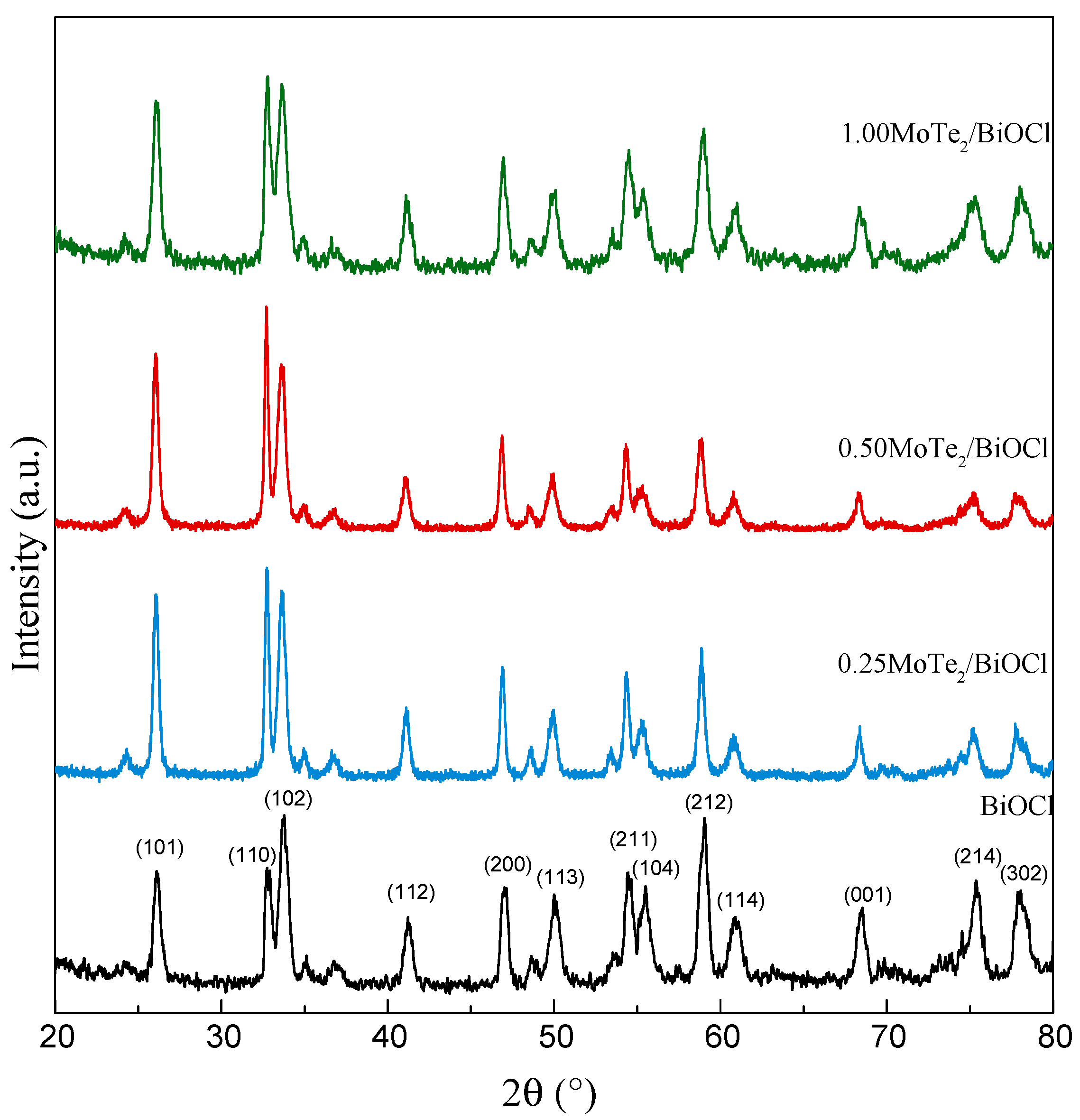
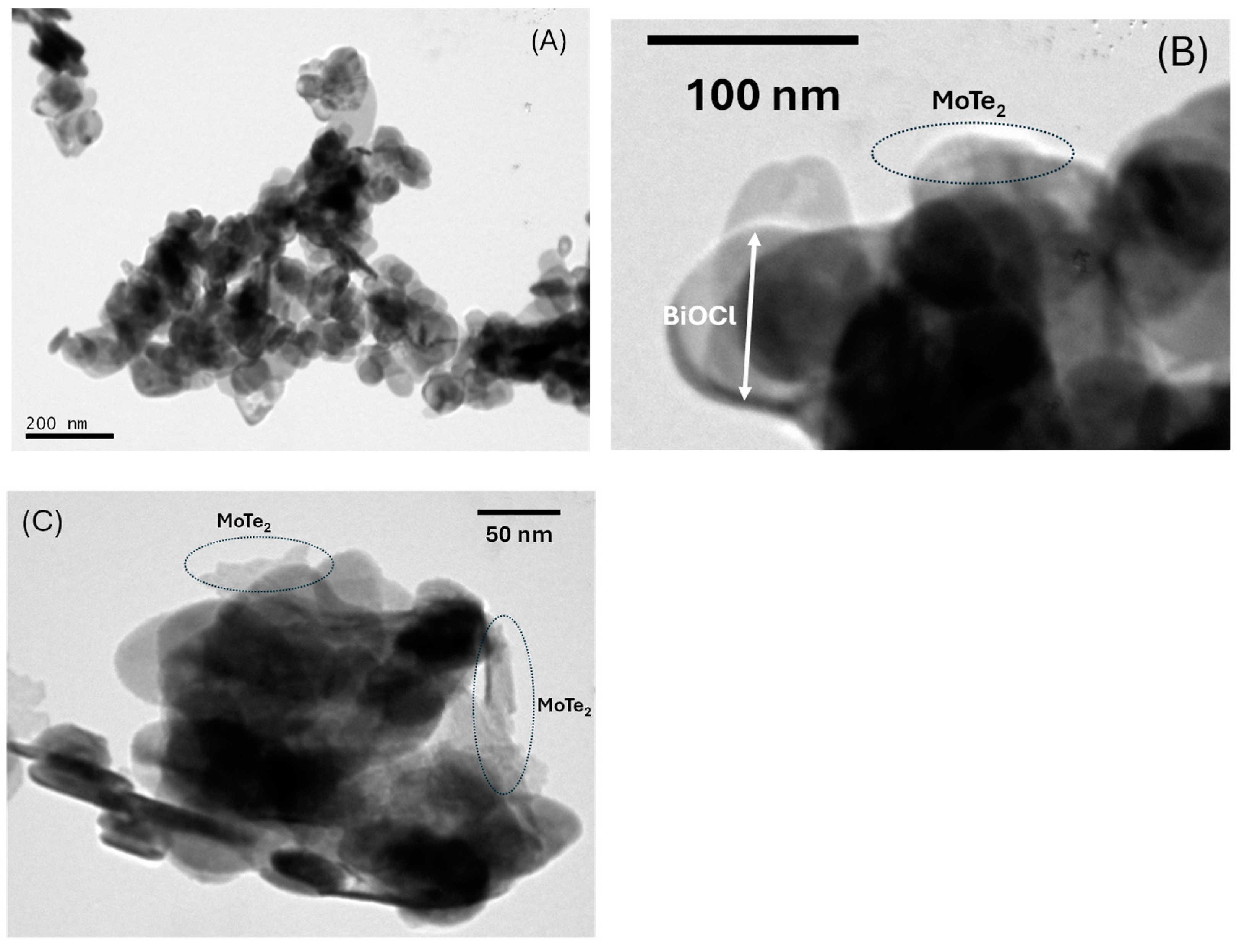
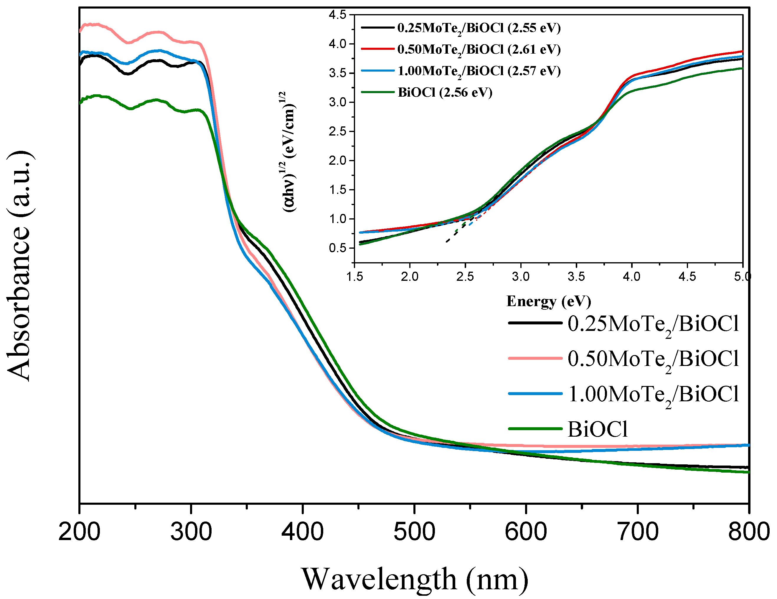
2.1. Physicochemical and Optical Characterization

The specific surface area (SSA) of the photocatalysts was calculated using the Brunauer–Emmett–Teller (BET) method, based on nitrogen adsorption isotherms recorded at liquid nitrogen temperature (77 K). Pure BiOCl, as well as molybdenum telluride-modified composites, exhibited a specific surface area of 16 ± 2 m2 g−1, in good accordance with values reported in previous studies [43]. The addition of MoTe2 had no discernible effect on the SSA values because of its low content and uniform dispersion within the BiOCl structure, which preserved the material’s structural and surface characteristics.
The bulk characteristics of the photocatalysts were determined using X-ray diffraction (XRD) analysis. The XRD patterns were recorded using a Cu Ka radiation source (λ = 1.54 Å) over a diffraction angle of 20–80°, and the results are presented in Figure 1. All observed diffraction peaks corresponded to the tetragonal structure of BiOCl, as indexed JCPDS No 6-249, confirming the retention of the crystalline framework of BiOCl across all samples. The low content of MoTe2 (≤1%) in the composites accounted for the absence of its characteristic diffraction peaks, as the signals were either below the detection limit of XRD or overlapped by the dominant BiOCl reflections. Additionally, as XRD primarily provides bulk structural information rather than surface-specific insights, the characteristic peaks of MoTe2 were undetectable, presumably because of its highly homogeneous dispersion on the BiOCl surface. However, it was observed that with the addition of MoTe2, these peaks gradually become sharper, indicating that the primary crystallite size increased. Indeed, the primary crystallite size estimated by the Scherrer equation using the (101) plane increased from 16 nm for pure BiOCl to 19 nm for the 0.50MoTe2/BiOCl sample.
Figure 2 shows characteristic TEM images of 0.50MoTe2/BiOCl. It is observed that BiOCl consists of well-shaped nanoplates with diameters ranging from 42 to 125 nm and an average thickness equal to ~13 nm. MoTe2 was detected on the surface of BiOCl in the form of nanoparticles of irregular size characterized by lower crystallinity.
The optical properties of the pristine sample and the modified photocatalysts were thoroughly investigated using diffuse reflectance spectroscopy (DRS), and the results are summarized in Figure 3. The absorbance spectrum revealed an absorption edge at approximately 470 nm, corresponding to the onset of electronic transition in the materials.
The band gap energy (Eg) of the photocatalysts was determined, employing the Tauc method, by plotting (αhν)1/2 vs. hν, assuming an indirect electronic transition [44]. Specifically, Eg was calculated by extrapolating both the linear region of the absorption edge and the initial offset region, identifying the intersection point as the band gap energy (inset graph).
For all samples, the calculated Eg was approximately 2.55 eV, indicating a minimal variation between the pristine BiOCl and the MoTe2-modified photocatalysts. These findings suggest that BiOCl retained its intrinsic electronic properties, with the addition of MoTe2 having a negligible effect on the bulk electronic structure, in good agreement with similar studies [45].

2.2. Photocatalytic Results

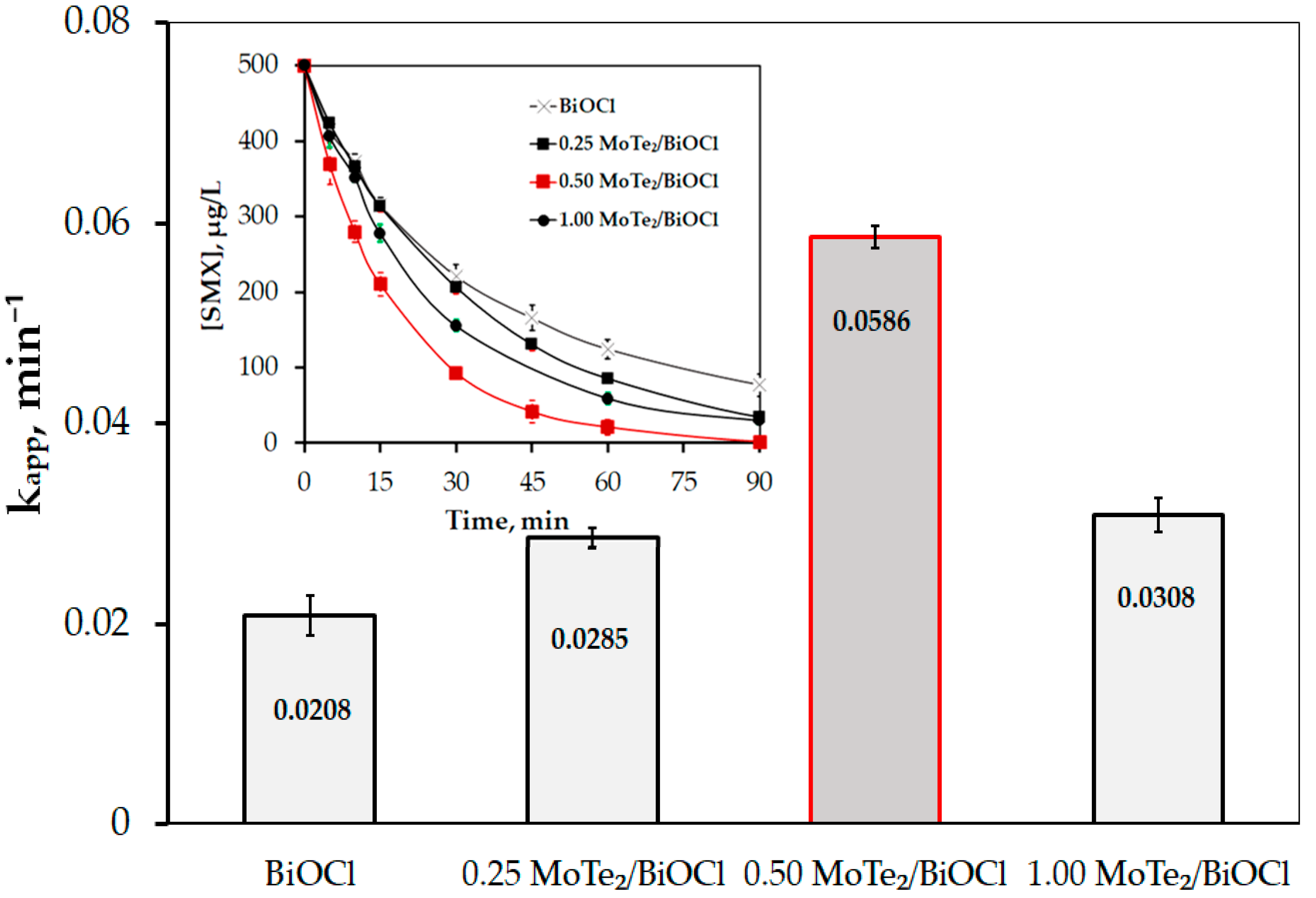
2.2.1. Photocatalytic Performance of MoTe2/BiOCl

To evaluate the photocatalytic activity of BiOCl, and that of the enhanced MoTe2/BiOCl samples (0.25–1% wt. MoTe2), photocatalytic experiments for SMX oxidation were conducted under simulated solar light with a radiation intensity of 7.3 × 10⁻⁷ einstein/(L.s). The solution was stirred in the dark for 15 min to allow the SMX to reach adsorption/desorption equilibrium on the photocatalyst surface. All photocatalysts demonstrated low adsorption capacity for SMX, with adsorption not exceeding 10% of the initial SMX concentration (500 μg/L) in any case.
As shown in Figure 4, the presence of MoTe2 as a cocatalyst with BiOCl enhanced the photocatalytic performance of the pure BiOCl for the SMX degradation, likely because of the effective separation of photogenerated electron–hole pairs (h⁺-e) [46].
Notably, as the MoTe2 loading increased from 0 to 0.25 wt.% and then to 0.5 wt.%, the apparent rate constant (kₐₚₚ) increased 1.37-fold and 2.82-fold, respectively, compared with the kₐₚₚ of pure BiOCl. Similarly, SMX removal improved from 376 μg/L to 415 μg/L and 480 μg/L after 60 min of reaction. However, further increasing the MoTe2 loading resulted in decreases in SMX removal (441 μg/L at 60 min) and kₐₚₚ, though both remained higher than those of pure BiOCl.
An optimal loading is commonly observed in photocatalysis, beyond which degradation decreases because of the blockage of active catalyst sites and the increased recombination of photogenerated electron–hole pairs [47,48].
For instance, Ma et al. [47] reported that the optimal loading of the carbon materials (C60) on BiOCl for the photocatalytic degradation of phenol was 1.0 wt.%, while Yan et al. [49] found 15 wt.% biochar in BiOCl to be the optimal loading. In others studies, He et al. [50] and Ioannidi et al. [45] reported optimal loadings of 0.75 wt.% BiOCl in BiVO₄ and 0.5 wt.% MoB in BiOCl, respectively.
To investigate the enhanced photocatalytic activity of the MoTe2/BiOCl heterojunctions, photoluminescence (PL) spectroscopy was performed. Figure 5 depicts the PL spectra of pure BiOCl, 0.25 MoTe2/BiOCl, 0.50 MoTe2/BiOCl, and 1.00 MoTe2/BiOCl. The luminescence intensities of the samples increased in the following order: 0.50 MoTe2/BiOCl < 0.25 MoTe2/BiOCl < 1.00 MoTe2/BiOCl < BiOCl. Pure BiOCl exhibited the highest PL intensity, indicating a higher recombination rate of electron–hole pairs compared with the composite materials. In contrast, the PL peak intensities of the MoTe2/BiOCl heterojunctions were lower, suggesting that the addition of MoTe2 effectively suppressed the recombination of electron–hole pairs, thus enhancing their photocatalytic efficiency. The fact that 0.50 MoTe2/BiOCl exhibited the lowest PL intensity was consistent with its status as the best-performing sample.

2.2.2. Effect of Initial Concentration of 0.5 MoTe2/BiOCl and SMX

Figure 6A illustrates the computed apparent rate constants (kapp) and SMX removal after 45 min of reaction for various initial concentrations of 0.5 MoTe2/BiOCl. Interestingly, although kapp increased with rising catalyst concentration from 0 to 1000 mg/L, this increase was not significant for 250 and 500 mg/L of 0.5 MoTe2/BiOCl, while for 1000 mg/L of 0.5MoTe2/BiOCl, the kapp increased only 1.15-fold compared with that for 500 mg/L 0.5 MoTe2/BiOCl.
Regarding SMX photolysis, it achieved only 21% SMX removal at 45 min (Figure 6A and Figure S2). However, with the addition of 100 mg/L of 0.5 MoTe2/BiOCl, SMX removal significantly improved, reaching 83% after 45 min photocatalytic reaction (Figure 6A and Figure S2). Further increases in 0.5MoTe2/BiOCl concentration resulted in smaller improvements, achieving 91%, 92%, and 96% for 250 mg/L, 500 mg/L, and 1000 mg/L 0.5MoTe2/BiOCl, respectively (Figure 6A and Figure S2).
It is widely recognized that increasing the catalyst concentration boosts the degradation rate because of the higher availability of active sites for the reaction. This trend persists until the degradation rate reaches its peak. At this point, the catalyst concentration provides enough active sites to absorb all available photons. Beyond this level, the degradation rate stabilizes [51,52]. This optimal concentration appears to be beyond the range of catalyst concentrations tested in this study but is likely close, given the minimal increase in SMX degradation observed after 250 mg/L of 0.5 MoTe2/BiOCl.
Considering that nearly complete SMX removal was achieved within 45 min using 500 mg/L 0.5 MoTe2/BiOCl, along with the cost of the catalyst and the impact of real water matrices on process efficiency, all subsequent experiments were conducted with 500 mg/L of the photocatalyst.
Next, several experiments were conducted with different initial concentrations of SMX under simulated solar illumination. As Figure 6B demonstrates, as the SMX concentration increased, the kapp value declined, indicating that the reaction did not follow true first-order kinetics but rather pseudo-first-order kinetics. This phenomenon is well-known in AOPs and is explained by the fact that, under specific experimental conditions, reactive species are produced at a constant rate. For low concentrations of pollutants, these reactive species are in excess. However, as the pollutant concentration increases, the reactive species likely become the limiting factor.
Additionally, higher concentrations of pollutants result in increased production of transformation products, leading to competition between the transformation products and pollutant molecules for the active sites of the catalysts and the reactive species [43,53,54]. Nevertheless, for the proposed photocatalytic system, as the SMX concentration increased from 250 μg/L to 500 μg/L and 1000 μg/L, the kapp value was reduced only 1.17-fold and 1.59-fold, respectively, while remaining within the same order of magnitude. This is an important finding, since, as shown in Figure 6C, the proposed photocatalytic system is capable of removing nearly all the added SMX. Specifically, 250 μg/L, 479 μg/L, and 919 μg/L of SMX were removed within 60 min when the initial concentrations were 250 μg/L, 500 μg/L, and 1000 μg/L, respectively, which is highly satisfactory considering the low concentrations at which SMX is often detected in environmentally relevant matrices.

2.2.3. Effect of Initial pH and Evaluation of Reactive Species Contribution Using Scavengers

Continuously, the impact of solution pH on SMX degradation and adsorption was investigated, and the results are shown in Figure 7A. Acidic and alkaline conditions were adjusted by adding H2SO4 and NaOH, respectively. The pH values were monitored during both adsorption and photodegradation of SMX. The final pHs of the adsorption experiments were found to be 3.2, 5, and 8 for initial pH values of 3.2, 5.5, and 9.1, respectively, while the final pHs for the photocatalytic degradation of SMX were 3, 4.5, and 5 for the same initial pH values. The pH reduction observed when the initial pH was 9.1 was likely due to the formation of acidic transformation products rather than the acidic character of BiOCl (pzc ≈ 2) [55].
It is worth noting that pH did not practically affect SMX adsorption, which was negligible in all cases. Furthermore, the solution pH had only a minor impact on SMX photodegradation, with a slight reduction observed under alkaline conditions. This was likely due to the weaker activity of the photogenerated reactive species as the solution pH increased [56]. However, in all cases, more than 85% of 500 μg/L SMX was successfully removed after 45 min of photodegradation.
Interestingly, the performance of treated BiOCl varied depending on the cocatalyst used. For instance, pure BiOCl showed low pH sensitivity, as in this study, but exhibited lower photocatalytic efficiency for SMX oxidation [55]. In contrast, the photocatalyst MoB/BiOCl demonstrated strong pH sensitivity for losartan photooxidation under solar light, achieving very high efficiency in acidic environments but low efficiency in alkaline conditions, as its kapp decreased from 0.9529 min⁻1 to 0.0541 min⁻1 [45].
In subsequent experiments, the roles of H O , h + , and 1 O 2 and O 2 were examined by using 2 mM tert-butanol (t-BuOH) [57], potassium iodide (KI) [58], and sodium azide (NaN3) [59], respectively, as radical scavengers, and the results are depicted in Figure 7B. The addition of NaN3 and t-BuOH slightly reduced the degradation rate of SMX, while the presence of KI caused a higher inhibition of SMX removal but did not completely inhibit it. This suggests that, besides the photogenerated holes, other reactive species such as O 2 contributed to the photodegradation of SMX. To confirm the critical role of photogenerated electrons in the in the examined photocatalytic system, and thus the indirect formation of O 2 , an experiment was performed in the presence of N2 gas [60]. Additionally, an experiment in the presence of 2 mM p-benzoquinone (p-BQ) as an O 2 scavenger was carried out [61]. The results of these experiments are demonstrated in Figure 7B. It was observed that both N2 gas and p-BQ inhibited the SMX degradation, with the latter causing even greater inhibition than KI. This could be attributed to the fact that BQ reacts not only with superoxide radicals but with conduction band electrons in the semiconductor, leading to competition between these processes and the formation of 1,4-hydroquinone [61]. Consequently, BQ competes with H O , O 2 , and electrons for reactions, which can notably hinder the photodegradation of the SMX even more.
Specifically, the kapp values were 0.0417 min−1, 0.0335 min−1, 0.0169 min−1, 0.0063 min−1, and 0.0050 min−1 in the presence of t-BuOH, NaN3, N2 gas, KI, and p-BQ, respectively, while the kapp value without any scavenger was 0.0586 min−1. To further elucidate the role of H O , an experiment was conducted at 4 mM t-BuOH (Figure S3). Although a stronger obstruction of SMX photocatalytic degradation was observed (80% SMX removal at 90 min) compared with 2 mM t-BuOH (96% SMX removal at 90 min), it was still less than the inhibition observed with N2 gas, KI, and p-BQ.
According to the literature, MoTe2 has a direct energy gap equal to 1.01–1.5 eV [41,62,63], and its valence band (VB) is located around 0.7–0.8 eV [64]. Gagné et al. [65] reported that the redox potential of SMX was 0.9 eV. Additionally, the bandgap of BiOCl depended on its preparation method, and for the preparation method used in this work, the band gap value was approximately 2.6 eV, with a VB of ~ 2.5 [66]. Although we did not have access to UPS to experimentally determine the positions of the valence and conduction bands, the EVB and ECB were estimated using the Mulliken electronegativity theory. The ECB was determined to be −0.35 and 0.736 eV, and the EVB, 1.15 and 3.33, for MoTe2 and BiOCl, respectively (as shown in Scheme 1).
Taking into account the above information, along with the experimental results in the presence of scavengers, a possible Z scheme photocatalytic mechanism for SMX degradation using 0.5MoTe2/BiOCl is depicted in Scheme 1. Specifically, MoTe2 acts as an electron trap, enhancing the separation of photoinduced charge carriers through electron cascading from BiOCl. The photogenerated h + may react with H O or H 2 O to produce H O or directly oxidize SMX, while the electrons in the CB of BiOCl transfer to the VB of MoTe2. The role of MoTe2 as an electron trap/acceptor has also been stated by Ali et al. [62], who studied the efficiency of Te−MoTe2−MoS2/ZnO photocatalysts for H2 production.
The more negative conduction band (CB) potential of MoTe2 relative to O 2 / O 2 (−0.33 eV, pH = 7) allows electrons to reduce O 2 , producing O 2 . Then, O 2 may react directly with SMX or/and with h + in the VB of BiOCl to form 1 O 2 . The potential contribution of reactive oxygen species follows the order h + > O 2 / e >  1 O 2 > H O .
All the degradation pathways mentioned above appear to play a significant role in the decomposition of SMX, as suggested by the transformation pathway outlined in Section 2.3.
It is interesting that the dominant reactive species vary depending on the modification of BiOCl. For instance, in our previous work [45], which examined the photocatalytic degradation of losartan using the photocatalyst 0.5 MoB/BiOCl, it was found that the dominant reactive species was singlet oxygen, followed by O 2 and h + . In contrast, Shen et al. [67], highlighted the significant role of e⁻ in the photocatalytic removal of NO using the Mn₃O₄/BiOCl photocatalyst. In another study, Pan et al. [15] reported that for the photocatalytic degradation of phenol under UV light, the reactive species contribution followed the order h + > O 2 > H O in the presence of CdS/BiOCl and O 2 > h + > H O with CdS/CQDs/BiOCl.

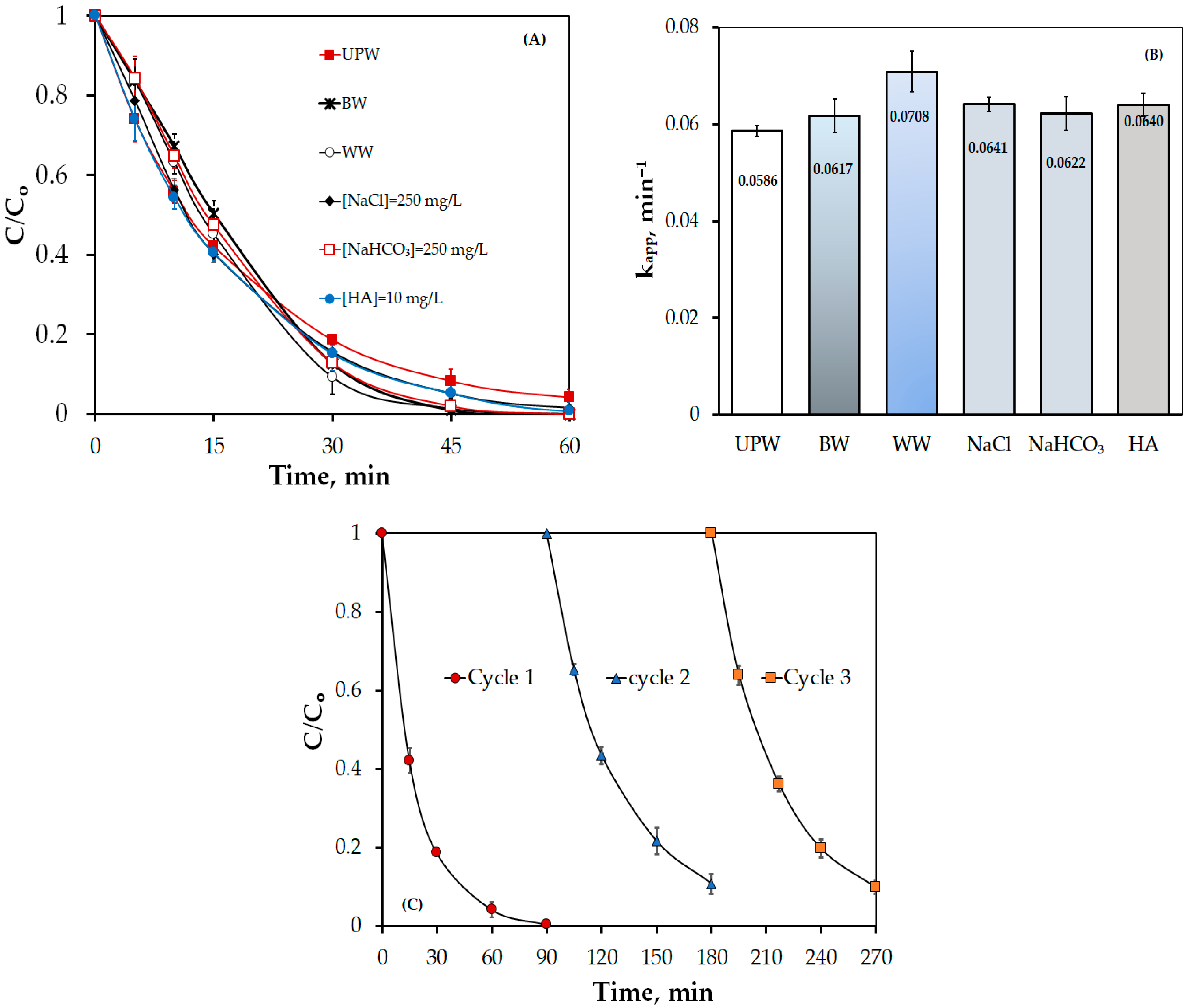
2.2.4. Effect of Water Matrix

Subsequently, the photocatalytic performance of 0.5MoTe2/BiOCl for SMX degradation in various water matrices was investigated, and the results are presented in Figure 8A,B. Table 1 provides the physicochemical characteristics of the bottled water (BW) and wastewater used in this study.
Surprisingly, the degradation rate of SMX remained constant or increased in all synthetic and real water matrices, achieving complete removal within less than 60 min. WW unexpectedly exhibited the highest kapp. This behavior can possibly be attributed to the presence of bicarbonate ( H C O 3 ) and C l , which generally have diverse roles. In some cases, these inorganic ions negatively impact performance processes [45,48,68], while in others, their presence enhances the degradation process [45,48,52,69,70,71,72].
This positive effect is likely due to the formation of more selective reactive species (e.g., C O 3 , C l ), even though these species have lower redox potentials than H O ( C O 3 / H C O 3 E° = 1.78 eV vs. NHE and C l / C l E° = 2.43 eV vs. NHE) (reactions 1–7) [73,74,75,76].
H C O 3 + h + C O 3 + H +
H C O 3 + H O C O 3 + H 2 O
H C O 3 + O 2 C O 3 2 + H O 2
C l + h + C l
C l + H O H O C l
C l + H O H O + C l
C l + C l C l 2
The beneficial effects of HA are primarily linked to its role as a sensitizer through charge transfer interactions [77]. Additional factors highlighted in the literature include the production of reactive oxygen species such as O 2 via HA photolysis [78,79] and the sequestration phenomenon, which reduces the escape of pollutant molecules into the bulk solution, increasing their chances of reacting with the active species generated on the photocatalyst’s surface under solar light because of HA adsorption [80].
Thus, the high efficiency observed in BW and WW resulted from a synergistic effect of various reactive species formed by inorganic and/or organic loadings, in combination with the photogenerated holes and O 2 , which, according to Figure 7B, were the dominant reactive species.
Table 2 presents a comparison of SMX photocatalytic degradation with other related studies, while Table 3 evaluates the efficiency of 0.5MoTe2/BiOCl compared with other BiOCl-based photocatalysts for the photocatalytic degradation of various pollutants. As shown in Table 2 and Table 3, although many photocatalysts demonstrated high photocatalytic efficiency in SMX removal and the degradation of other pollutants in UPW, their performance was comparable to that of 0.5MoTe2/BiOCl. This is noteworthy considering that the Xe lamps used in those studies typically operate at three to five times higher power than the 100 W Xe lamp used in this work, resulting in significantly greater irradiation intensity. Additionally, as highlighted in Table 2 and Table 3, many studies have fallen behind in addressing the effect of aqueous matrices. Moreover, studies that have examined photocatalytic performance in real water matrices often report significant inhibition.
Thus, the superior performance of 0.5MoTe2/BiOCl compared with other photocatalysts lies in its high efficiency in real water matrices. This advantage is attributed to the effective separation of holes and electrons (Figure 5), as well as the dominance of holes and superoxide oxygen that are more selective than hydroxyl radicals, resulting in enhanced photocatalytic efficiency.

2.2.5. Reuse of Photocatalysts

Τhe reusability of 0.5MoTe2/BiOCl was evaluated for the photodegradation of SMX in UPW through a series of three consecutive tests. In each experiment, 500 mg/L of 0.5 MoTe2/BiOCl was added to 60 mL of a 500 μg/L SMX solution, and the mixture was irradiated for 90 min. SMX conversion was monitored at 15, 30, 60, and 90 min intervals. After each test, the catalyst was recovered through vacuum filtration and dried at 50 °C overnight to remove any residual moisture. It was observed that, following a slight decrease in SMX removal after the first cycle, the photocatalytic performance remained stable in the second and third cycles (with 90% SMX removal), as shown in Figure 8C. This demonstrates the high stability of the catalyst and supports its potential for use in practical applications under realistic conditions.

2.3. Proposed Transformation Pathways

High-resolution mass spectrometry (HRMS) with a TOF analyzer operated in negative ionization mode was used for the identification of the TBPs. The structural assignment of the TBPs was based on the high-resolution accurate mass data (ion molecular formula and m/z [Μ-H]) depicted in Table 4. Based on the four identified TBPs, the photocatalytic transformation of SMX was found to include two main routes, as depicted in Figure 9.
The first route proceeds through the oxidation of the amino group of and the formation of the nitro-derivative of SMX (TBP1). Its formation can relate to the formation of 1O2 and the direct oxidation of the N atom of the amino group [96]. Nitro-SMX is a common TBP that has been generated during the degradation of SMX using various AOPs [96,97,98,99,100]. The second route includes the oxidation of the methyl attached to the isoxazole ring to a carbonyl group giving rise to the formation of TBP2. This TBP was also identified during the photocatalytic degradation of SMX using tungsten-modified TiO2 as a photocatalyst [100]. This route potentially involves the initial hydroxyl radical attack on the methyl group and the subsequent formation of the aldehyde structure. Further oxidation of the TBP2 followed by decarboxylation reactions can also lead to possible dealkylation [101]. Thereafter, and through various reactions by hydroxyl radicals and/or photogenerated holes, the opening of the isoxazole ring takes place, with the subsequent formation of two TBPs with lower molecular weights (TBP3 and TBP4). At 240 min, all the identified TBPs are completely eliminated. Subsequent application of heterogeneous photocatalysis can lead to the formation of aliphatic compounds such as organic acids (carboxylic or sulfonic acids), ketones, amines, and aldehydes before complete mineralization.

3. Materials and Methods

3.1. Chemical Reagents

For the photocatalyst synthesis: bismuth ΙΙΙ nitrate pentahydrate (Bi(NO3)3 ∙ H2O CAS: 10035-06-0), potassium chloride (KCl CAS: 7447-40-7), thiourea powder (NH2CSNH2 CAS: 62-56-6), acetic acid solution ≥ 99% (CH3CO2H CAS: 64-19-7), and crystal molybdenum telluride (MoTe2 CAS: 12058-20-7) were purchased from Sigma-Aldrich (Saint Louis, MO, USA). Sulfamethoxazole (C10H11N3O3S CAS: 723-46-6) was also obtained from Sigma-Aldrich.
For the model water systems: sodium bicarbonate (NaHCO3 CAS: 144-55-8), humic acid (C187H186O89N9S, CAS: 68131-04-4), and sodium chloride (NaCl CAS: 7647-14-5) were also supplied from Sigma-Aldrich and used without further purification.
For the trapping experiments: sodium azide (NaN3 CAS: 26628-22-8), potassium iodide (KI CAS: 7681-11-0), and tert-Butyl alcohol ((CH3)3COH CAS: 75-65-0) were sourced from Sigma-Aldrich.

3.2. Photocatalysts Preparation Procedure and Characterization

Pure BiOCl was synthesized following a specific procedure detailed elsewhere [102]. For the preparation of the modified photocatalysts, appropriate amounts of commercially obtained MoTe2 were incorporated into the BiOCl precursor solution.
The specific surface area of the photocatalysts was evaluated through nitrogen adsorption/desorption isοtherms using the Brunauer–Emmett–Teller (BET) method. The measurements were performed using a Micromeritics Gemini III 2375 analyzer (Norcross, GA, USA). The XRD analysis was conducted using a Brucker D8 advance diffractοmeter (Billerica, ΜA, USA) that utilized a Cu Ka radiation source (λ = 1.5406 Å). Phase identification was carried out using the Crystallographica software (Crystallographica Search-Match 3.1) in comparison with the standard JCPDS card data. The primary crystallite size was estimated with the Scherrer equation:
d = 0.9 λ B cos θ
where λ: X-ray wavelength corresponding to Cu Ka radiation (0.15406 nm), θ: diffraction angle, and B: the line broadening (in radians) at half of its maximum. To evaluate the optical properties of the photocatalysts, diffuse reflectance spectroscopy (DRS) was performed using a Varian Cary 3E spectrophotometer (Ρalο Altο, CA, USA) equipped with an integrating sphere, with BaSO4 serving as the reference material. A Gatan model 782 ES500 W Erlangshen CCD camera (Gatan Model 782 ES500 W, Pleasanton, CA, USA) was used for TEM analysis. To assess the increased photocatalytic activity of the as-prepared photocatalysts, photoluminescence (PL) spectroscopy was utilized using a Cary Eclipse Fluorescence Spectrometer (Santa Clara, CA, USA).

3.3. Analytical Determination

For the determination of SMX concentration, high-performance liquid chromatography was employed with a photodiode array (PDA) detector (Waters Alliance 2695, Miliford, MA, USA). The analysis was performed using a Kinetex XB-C18 column (dimension: 2.6 mm internal diameter × 50 mm length, Milford, MA, USA) maintained at a controlled temperature of 45 °C using an oven. The mobile phase was composed of a 70:30 ratio of 0.1% (v/v) H3PO4 and acetonitrile (ACN) flowing at a rate of 0.150 mL/min under isocratic conditions. All samples were filtered prior to injection.

3.4. Photocatalytic Tests

In a typical photocatalytic test, a preweighed amount of the photocatalyst (typically 60 mg) was added to 120 mL of 0.5 mg/L SMX solution (unless stated otherwise) and the suspension was kept in the dark for 15 min to achieve adsorption/desorption equilibrium and minimize any mass transfer limitations. Subsequently, the solution was exposed to simulated solar radiation applied by a solar simulator (Oriel, mode LCS-100) equipped with a 100 W xenon lamp. According to chemical actinometry, the intensity of the incident radiation to the reactor was 7.3 × 10⁻⁷ einstein/(L.s). At predetermined time intervals, 1.2 mL samples were withdrawn and filtered using PVDF membranes housed in polypropylene overmolds (0.22 μm, Whatman, Maidstone, UK).

3.5. Identification of Transformation By-Products (TBPs)

The TBPs generated during the process were identified using a high-resolution mass spectrometer (Bruker micrOTOF Focus II) coupled to a Dionex (Thermo Scientific, Waltham, MA, USA) Ultimate 3000 UHPLC. The TOF analyzer was operated in negative ionization mode with the following ESI-source parameters: dry gas flow rate at 8 L min−1 (nitrogen), nebulizer pressure at 2.4 bar, capillary voltage at 4200 V, and dry temperature at 200 °C. To ensure mass accuracy, ±5 ppm sodium formate was used as calibrant of the TOF analyzer. SMX and its TBPs were separated on an AcclaimTM RSLC 120 C18 (2.2 μm, 2.1 × 100 mm) column (Thermo Scientific) protected by an AcclaimTM 120 C18 guard cartridge (5 μm, 3.0 × 10 mm) from Thermo Scientific, and the injection volume was 10 μL.
A gradient elution (90/10 (0 min), 10/90 (15 min), 90/10 (17 min), and 90/10 (18 min)) was performed using water with 0.01% formic acid (A) and acetonitrile (B), and the flow rate was set at 0.15 mL min−1.

4. Conclusions

The development of catalytic materials with high potential to harness abundant solar light for the treatment of persistent micropollutants has long been an important goal for the research community. This work focused on examining the possible enhancement of the photocatalytic activity of BiOCl in the presence of MoTe2, a well-known 2D material that remains largely unexplored in studies related to photocatalytic degradation of persistent pollutants. Particular attention was given to evaluating the system under a wide variety of operating conditions, with an emphasis on environmentally relevant matrices, where the true challenge of micropollutant removal lies. Additionally, the study aimed to identify the possible transformation products derived from the photodegradation of sulfamethoxazole, providing deeper insights into the degradation mechanisms and potential environmental implications.
It was revealed that the presence of the 2D material did not significantly affect either the specific surface area or the energy gap, or, therefore, the absorbance of the synthesized material in the solar spectrum. A significant enhancement in photocatalytic activity (2.8 times greater than the pure BiOCl) was observed for the 0.5MoTe2/BiOCl sample, possibly due to enhanced separation of the photogenerated carriers. The system demonstrated satisfactory performance even at the high μg/L range of SMX concentration, and the degradation was enhanced at neutral and acidic pH. Unlike most photocatalytic studies, the presence of inorganic ions such as chlorides and bicarbonates or even humic acid did not decrease the degradation rate, while encouraging results were also obtained in the experiments performed in real matrices (bottled water and secondary effluent). This behavior was associated with the specific nature and selectivity of the reactive species that are involved in the SMX photodecomposition. This observation highlights the need for future research regarding the evaluation of activity observed in real matrices, where different interactions between reactive species, water matrix constituents, and target pollutants can occur. Finally, four transformation products were produced during SMX photodegradation, and future work must also examine their toxicity, as well as the toxicity of the treated effluent.

Supplementary Materials

The following supporting information can be downloaded at: https://www.mdpi.com/article/10.3390/catal15010059/s1, Figure S1: The absorption spectrum of SMX in an aqueous solution with a concentration of 15 mg/L; Figure S2: Effect of 0.5 ΜοΤe2/BiOCl concentration on 500 μg/L SMX degradation under solar simulated light in UPW and inherent pH; Figure S3: Effect of t-BuOH on 500 μg/L SMX degradation with 500 mg/L 0.5 ΜοΤe2/BiOCl under solar simulated light in UPW and inherent pH.

Author Contributions

Conceptualization, A.A.I., Z.F. and D.M.; methodology, A.A.I., A.P., M.A., Z.F. and D.M.; formal analysis, A.A.I., G.N., K.K., M.A. and A.P.; investigation, A.A.I., G.N., K.K., M.A. and A.P.; resources, Z.F., M.A. and D.M.; data curation, A.A.I., G.N., M.A. and A.P.; writing—original draft preparation, A.A.I., K.K., Z.F., M.A. and A.P.; writing—review and editing, A.A.I., A.P., M.A., Z.F. and D.M.; visualization, A.A.I., G.N., M.A. and A.P.; supervision, D.M.; funding acquisition, D.M. All authors have read and agreed to the published version of the manuscript.

Funding

This research received no external funding.

Data Availability Statement

Data are contained within the article.

Conflicts of Interest

The authors declare no conflicts of interest.

References

  1. Priya, A.K.; Gnanasekaran, L.; Rajendran, S.; Qin, J.; Vasseghian, Y. Occurrences and Removal of Pharmaceutical and Personal Care Products from Aquatic Systems Using Advanced Treatment—A Review. Environ. Res. 2022, 204, 112298. [Google Scholar] [CrossRef]
  2. Deo, R.P. Pharmaceuticals in the Surface Water of the USA: A Review. Curr. Environ. Health Rep. 2014, 1, 113–122. [Google Scholar] [CrossRef]
  3. Vatovec, C.; Kolodinsky, J.; Callas, P.; Hart, C.; Gallagher, K. Pharmaceutical Pollution Sources and Solutions: Survey of Human and Veterinary Medication Purchasing, Use, and Disposal. J. Environ. Manag. 2021, 285, 112106. [Google Scholar] [CrossRef] [PubMed]
  4. Letsinger, S.; Kay, P.; Rodríguez-Mozaz, S.; Villagrassa, M.; Barceló, D.; Rotchell, J.M. Spatial and Temporal Occurrence of Pharmaceuticals in UK Estuaries. Sci. Total Environ. 2019, 678, 74–84. [Google Scholar] [CrossRef]
  5. Ojemaye, C.Y.; Petrik, L. Pharmaceuticals in the Marine Environment: A Review. Environ. Rev. 2018, 27, 151–165. [Google Scholar] [CrossRef]
  6. Zainab, S.M.; Junaid, M.; Xu, N.; Malik, R.N. Antibiotics and Antibiotic Resistant Genes (ARGs) in Groundwater: A Global Review on Dissemination, Sources, Interactions, Environmental and Human Health Risks. Water Res. 2020, 187, 116455. [Google Scholar] [CrossRef]
  7. Glassmeyer, S.T.; Furlong, E.T.; Kolpin, D.W.; Batt, A.L.; Benson, R.; Boone, J.S.; Conerly, O.; Donohue, M.J.; King, D.N.; Kostich, M.S.; et al. Nationwide Reconnaissance of Contaminants of Emerging Concern in Source and Treated Drinking Waters of the United States. Sci. Total Environ. 2017, 581–582, 909–922. [Google Scholar] [CrossRef] [PubMed]
  8. Lama, G.; Meijide, J.; Sanromán, A.; Pazos, M. Heterogeneous Advanced Oxidation Processes: Current Approaches for Wastewater Treatment. Catalysts 2022, 12, 344. [Google Scholar] [CrossRef]
  9. Li, Z.; Wang, J.; Chang, J.; Fu, B.; Wang, H. Insight into Advanced Oxidation Processes for the Degradation of Fluoroquinolone Antibiotics: Removal, Mechanism, and Influencing Factors. Sci. Total Environ. 2023, 857, 159172. [Google Scholar] [CrossRef] [PubMed]
  10. Antoniadou, M.; Falara, P.P.; Likodimos, V. Photocatalytic Degradation of Pharmaceuticals and Organic Contaminants of Emerging Concern Using Nanotubular Structures. Curr. Opin. Green Sustain. Chem. 2021, 29, 100470. [Google Scholar] [CrossRef]
  11. Loeb, S.K.; Alvarez, P.J.J.; Brame, J.A.; Cates, E.L.; Choi, W.; Crittenden, J.; Dionysiou, D.D.; Li, Q.; Li-Puma, G.; Quan, X.; et al. The Technology Horizon for Photocatalytic Water Treatment: Sunrise or Sunset? Environ. Sci. Technol. 2019, 53, 2937–2947. [Google Scholar] [CrossRef]
  12. Stylidi, M.; Kondarides, D.I.; Verykios, X.E. Pathways of Solar Light-Induced Photocatalytic Degradation of Azo Dyes in Aqueous TiO2 Suspensions. Appl. Catal. B Environ. 2003, 40, 271–286. [Google Scholar] [CrossRef]
  13. Daskalaki, V.M.; Kondarides, D.I. Efficient Production of Hydrogen by Photo-Induced Reforming of Glycerol at Ambient Conditions. Catal. Today 2009, 144, 75–80. [Google Scholar] [CrossRef]
  14. Iervolino, G.; Zammit, I.; Vaiano, V.; Rizzo, L. Limitations and Prospects for Wastewater Treatment by UV and Visible-Light-Active Heterogeneous Photocatalysis: A Critical Review. Top. Curr. Chem. 2019, 378, 7. [Google Scholar] [CrossRef] [PubMed]
  15. Roslan, N.N.; Lau, H.L.; Suhaimi, N.A.A.; Shahri, N.N.M.; Verinda, S.B.; Nur, M.; Lim, J.-W.; Usman, A. Recent Advances in Advanced Oxidation Processes for Degrading Pharmaceuticals in Wastewater—A Review. Catalysts 2024, 14, 189. [Google Scholar] [CrossRef]
  16. Serra-Pérez, E.; Dražić, G.; Takashima, M.; Ohtani, B.; Kovačič, S.; Žerjav, G.; Tušar, N.N. Influence of the Surface Structure of the TiO2 Support on the Properties of the Au/TiO2 Photocatalyst for Water Treatment under Visible Light. Catal. Today 2024, 437, 114764. [Google Scholar] [CrossRef]
  17. Chen, P.; Zhang, P.; Cui, Y.; Fu, X.; Wang, Y. Recent Progress in Copper-Based Inorganic Nanostructure Photocatalysts: Properties, Synthesis and Photocatalysis Applications. Mater. Today Sustain. 2023, 21, 100276. [Google Scholar] [CrossRef]
  18. Monfort, O.; Pop, L.-C.; Sfaelou, S.; Plecenik, T.; Roch, T.; Dracopoulos, V.; Stathatos, E.; Plesch, G.; Lianos, P. Photoelectrocatalytic Hydrogen Production by Water Splitting Using BiVO4 Photoanodes. Chem. Eng. J. 2016, 286, 91–97. [Google Scholar] [CrossRef]
  19. Sajjad, S.; Leghari, S.A.K.; Zhang, J. Nonstoichiometric Bi2O3: Efficient Visible Light Photocatalyst. RSC Adv. 2013, 3, 1363–1367. [Google Scholar] [CrossRef]
  20. Zhang, K.; Zhang, Y.; Zhang, D.; Liu, C.; Zhou, X.; Yang, H.; Qu, J.; He, D. Efficient Photocatalytic Water Disinfection by a Novel BP/BiOBr S-Scheme Heterojunction Photocatalyst. Chem. Eng. J. 2023, 468, 143581. [Google Scholar] [CrossRef]
  21. Song, S.; Xing, Z.; Zhao, H.; Li, Z.; Zhou, W. Recent Advances in Bismuth-Based Photocatalysts: Environment and Energy Applications. Green Energy Environ. 2022, 8, 1232. [Google Scholar] [CrossRef]
  22. Xu, Z.; Zhang, C.; Zhang, Y.; Gu, Y.; An, Y. BiOCl-Based Photocatalysts: Synthesis Methods, Structure, Property, Application, and Perspective. Inorg. Chem. Commun. 2022, 138, 109277. [Google Scholar] [CrossRef]
  23. Dellinger, T.M.; Braun, P. V BiOCl Nanoparticles Synthesized in Lyotropic Liquid Crystal Nanoreactors. Scr. Mater. 2001, 44, 1893–1897. [Google Scholar] [CrossRef]
  24. Ye, P.; Xie, J.; He, Y.; Zhang, L.; Wu, T.; Wu, Y. Hydrolytic Synthesis of Flowerlike BiOCl and Its Photocatalytic Performance under Visible Light. Mater. Lett. 2013, 108, 168–171. [Google Scholar] [CrossRef]
  25. Cao, S.; Guo, C.; Lv, Y.; Guo, Y.; Liu, Q. A Novel BiOCl Film with Flowerlike Hierarchical Structures and Its Optical Properties. Nanotechnology 2009, 20, 275702. [Google Scholar] [CrossRef] [PubMed]
  26. Qu, X.; Zhao, X.; Liu, M.; Gao, Z.; Yang, D.; Shi, L.; Tang, Y.; Song, H. BiOCl/TiO2 Composite Photocatalysts Synthesized by the Sol–Gel Method for Enhanced Visible-Light Catalytic Activity toward Methyl Orange. J. Mater. Res. 2020, 35, 3067–3078. [Google Scholar] [CrossRef]
  27. Wang, C.; Shao, C.; Liu, Y.; Zhang, L. Photocatalytic Properties BiOCl and Bi2O3 Nanofibers Prepared by Electrospinning. Scr. Mater. 2008, 59, 332–335. [Google Scholar] [CrossRef]
  28. Ye, L.; Deng, Y.; Wang, L.; Xie, H.; Su, F. Bismuth-Based Photocatalysts for Solar Photocatalytic Carbon Dioxide Conversion. ChemSusChem 2019, 12, 3671–3701. [Google Scholar] [CrossRef] [PubMed]
  29. Zhong, Y.; Liu, Y.; Wu, S.; Zhu, Y.; Chen, H.; Yu, X.; Zhang, Y. Facile Fabrication of BiOI/BiOCl Immobilized Films with Improved Visible Light Photocatalytic Performance. Front. Chem. 2018, 6, 58. [Google Scholar] [CrossRef]
  30. Wang, Q.; Li, P.; Zhang, Z.; Jiang, C.; Zuojiao, K.; Liu, J.; Wang, Y. Kinetics and Mechanism Insights into the Photodegradation of Tetracycline Hydrochloride and Ofloxacin Mixed Antibiotics with the Flower-like BiOCl/TiO2 Heterojunction. J. Photochem. Photobiol. A Chem. 2019, 378, 114–124. [Google Scholar] [CrossRef]
  31. Liu, W.; Wang, S.; Zhao, Y.; Sun, C.; Xu, H.; Zhao, J. PVP-Induced Bi2S3/BiOCl Photocatalyst with Open Hollow Structures for the Removal of Ciprofloxacin under Visible-Light Irradiation. J. Alloys Compd. 2021, 861, 157995. [Google Scholar] [CrossRef]
  32. Shinde, P.V.; Hussain, M.; Moretti, E.; Vomiero, A. Advances in Two-Dimensional Molybdenum Ditelluride (MoTe2): A Comprehensive Review of Properties, Preparation Methods, and Applications. SusMat 2024, 4, e236. [Google Scholar] [CrossRef]
  33. Yu, S.; Wu, X.; Wang, Y.; Guo, X.; Tong, L. 2D Materials for Optical Modulation: Challenges and Opportunities. Adv. Mater. 2017, 29, 1606128. [Google Scholar] [CrossRef]
  34. Li, L.; Yang, H.; Yang, P. WS2/MoSe2 van der Waals Heterojunctions Applied to Photocatalysts for Overall Water Splitting. J. Colloid Interface Sci. 2023, 650, 1312–1318. [Google Scholar] [CrossRef]
  35. Singla, S.; Singh, P.; Basu, S.; Devi, P. BiVO4/MoSe2 Photocatalyst for the Photocatalytic Abatement of Tetracycline and Photoelectrocatalytic Water Splitting. Mater. Chem. Phys. 2023, 295, 127111. [Google Scholar] [CrossRef]
  36. Yao, C.; Wu, D.; Yuan, C.; Xue, X.; Liu, L.; Zhang, X. Design, Preparation, and Photocatalytic Performance of MoSe2 Quantum Dots Modified BiOCl Composite Photocatalysts. Opt. Mater. 2024, 147, 114745. [Google Scholar] [CrossRef]
  37. Lin, Y.-F.; Xu, Y.; Wang, S.-T.; Li, S.-L.; Yamamoto, M.; Aparecido-Ferreira, A.; Li, W.; Sun, H.; Nakaharai, S.; Jian, W.-B.; et al. Ambipolar MoTe2 Transistors and Their Applications in Logic Circuits. Adv. Mater. 2014, 26, 3263–3269. [Google Scholar] [CrossRef] [PubMed]
  38. Lei, Y.; Lin, Q.; Xiao, S.; Li, J.; Fang, H. Optically Active Telecom Defects in MoTe2 Fewlayers at Room Temperature. Nanomaterials 2023, 13, 1501. [Google Scholar] [CrossRef] [PubMed]
  39. Rhodes, D.A.; Jindal, A.; Yuan, N.F.Q.; Jung, Y.; Antony, A.; Wang, H.; Kim, B.; Chiu, Y.-C.; Taniguchi, T.; Watanabe, K.; et al. Enhanced Superconductivity in Monolayer Td-MoTe2. Nano Lett. 2021, 21, 2505–2511. [Google Scholar] [CrossRef]
  40. Octon, T.J.; Nagareddy, V.K.; Russo, S.; Craciun, M.F.; Wright, C.D. Fast High-Responsivity Few-Layer MoTe2 Photodetectors. Adv. Opt. Mater. 2016, 4, 1750–1754. [Google Scholar] [CrossRef]
  41. Wang, B.; Wang, X.; Wang, P.; Kuang, A.; Zhou, T.; Yuan, H.; Chen, H. Bilayer MoTe2/XS2 (X = Hf,Sn,Zr) Heterostructures with Efficient Carrier Separation and Light Absorption for Photocatalytic Water Splitting into Hydrogen. Appl. Surf. Sci. 2021, 544, 148842. [Google Scholar] [CrossRef]
  42. Yuan, Z.; Xiang, Y.; Jian, X.; Zhang, H.; Liu, M.; Cao, R.; Hu, Y.; Gao, X. Zn0.5Cd0.5S/MoTe2 Z-Scheme Heterojunction with Space-Separated Oxidation-Reduction Catalytic Sites for Photocatalytic CO2 Reduction. Sep. Purif. Technol. 2025, 359, 129836. [Google Scholar] [CrossRef]
  43. Kouvelis, K.; Karavaka, E.E.; Panagiotaras, D.; Papoulis, D.; Frontistis, Z.; Petala, A. Photocatalytic Degradation of Losartan with BiOCl/Sepiolite Nanocomposites. Catalysts 2024, 14, 433. [Google Scholar] [CrossRef]
  44. Liu, J.; Wang, H.; Chang, M.-J.; Sun, M.; Zhang, C.-M.; Yang, L.-Q.; Du, H.-L.; Luo, Z.-M. Facile Synthesis of BiOCl with Extremely Superior Visible Light Photocatalytic Activity Synergistically Enhanced by Co Doping and Oxygen Vacancies. Sep. Purif. Technol. 2022, 301, 121953. [Google Scholar] [CrossRef]
  45. Ioannidi, A.A.; Giannakopoulos, S.; Petala, A.; Frontistis, Z.; Mantzavinos, D. Fabrication of a Novel MoB/BiOCl Photocatalyst for Losartan and Escherichia coli Removal. Catal. Today 2024, 430, 114510. [Google Scholar] [CrossRef]
  46. Wu, T.; Li, J.; Chang, M.; Song, Y.; Sun, Q.; Wang, F.; Zou, H.; Shi, Z. Photoluminescence Properties and Photocatalytic Activities of SiO2@TiO2:Sm3+ Nanomaterials. J. Phys. Chem. Solids 2021, 149, 109775. [Google Scholar] [CrossRef]
  47. Ma, D.; Zhong, J.; Peng, R.; Li, J.; Duan, R. Effective Photoinduced Charge Separation and Photocatalytic Activity of Hierarchical Microsphere-like C60/BiOCl. Appl. Surf. Sci. 2019, 465, 249–258. [Google Scholar] [CrossRef]
  48. Ioannidi, A.; Petala, A.; Frontistis, Z. Copper Phosphide Promoted BiVO4 Photocatalysts for the Degradation of Sulfamethoxazole in Aqueous Media. J. Environ. Chem. Eng. 2020, 8, 104340. [Google Scholar] [CrossRef]
  49. Yan, Y.; Tang, X.; Ma, C.; Huang, H.; Yu, K.; Liu, Y.; Lu, Z.; Li, C.; Zhu, Z.; Huo, P. A 2D Mesoporous Photocatalyst Constructed by the Modification of Biochar on BiOCl Ultrathin Nanosheets for Enhancing the TC-HCl Degradation Activity. New J. Chem. 2020, 44, 79–86. [Google Scholar] [CrossRef]
  50. He, Z.; Shi, Y.; Gao, C.; Wen, L.; Chen, J.; Song, S. BiOCl/BiVO4 p–n Heterojunction with Enhanced Photocatalytic Activity under Visible-Light Irradiation. J. Phys. Chem. C 2014, 118, 389–398. [Google Scholar] [CrossRef]
  51. Abellán, M.N.; Giménez, J.; Esplugas, S. Photocatalytic Degradation of Antibiotics: The Case of Sulfamethoxazole and Trimethoprim. Catal. Today 2009, 144, 131–136. [Google Scholar] [CrossRef]
  52. Kanigaridou, Y.; Petala, A.; Frontistis, Z.; Antonopoulou, M.; Solakidou, M.; Konstantinou, I.; Deligiannakis, Y.; Mantzavinos, D.; Kondarides, D.I. Solar Photocatalytic Degradation of Bisphenol A with CuOx/BiVO4: Insights into the Unexpectedly Favorable Effect of Bicarbonates. Chem. Eng. J. 2017, 318, 39–49. [Google Scholar] [CrossRef]
  53. Outsiou, A.; Frontistis, Z.; Ribeiro, R.S.; Antonopoulou, M.; Konstantinou, I.K.; Silva, A.M.T.; Faria, J.L.; Gomes, H.T.; Mantzavinos, D. Activation of sodium persulfate by magnetic carbon xerogels (CX/CoFe) for the oxidation of bisphenol A: Process variables effects, matrix effects and reaction pathways. Water Res. 2017, 124, 97–107. [Google Scholar] [CrossRef] [PubMed]
  54. Ioannidou, E.; Ioannidi, A.; Frontistis, Z.; Antonopoulou, M.; Tselios, C.; Tsikritzis, D.; Konstantinou, I.; Kennou, S.; Kondarides, D.I.; Mantzavinos, D. Correlating the Properties of Hydrogenated Titania to Reaction Kinetics and Mechanism for the Photocatalytic Degradation of Bisphenol A under Solar Irradiation. Appl. Catal. B Environ. 2016, 188, 65–76. [Google Scholar] [CrossRef]
  55. Petala, A.; Arvaniti, O.S.; Travlou, G.; Mantzavinos, D.; Frontistis, Z. Solar Light Induced Photocatalytic Removal of Sulfamethoxazole from Water and Wastewater Using BiOCl Photocatalyst. J. Environ. Sci. Health Part A 2021, 56, 963–972. [Google Scholar] [CrossRef]
  56. Oturan, M.A.; Aaron, J.-J. Advanced Oxidation Processes in Water/Wastewater Treatment: Principles and Applications. A Review. Crit. Rev. Environ. Sci. Technol. 2014, 44, 2577–2641. [Google Scholar] [CrossRef]
  57. Andrews, N.L.P.; Fan, J.Z.; Forward, R.L.; Chen, M.C.; Loock, H.-P. Determination of the Thermal, Oxidative and Photochemical Degradation Rates of Scintillator Liquid by Fluorescence EEM Spectroscopy. Phys. Chem. Chem. Phys. 2017, 19, 73–81. [Google Scholar] [CrossRef] [PubMed]
  58. Trandafilović, L.V.; Jovanović, D.J.; Zhang, X.; Ptasińska, S.; Dramićanin, M.D. Enhanced Photocatalytic Degradation of Methylene Blue and Methyl Orange by ZnO:Eu Nanoparticles. Appl. Catal. B Environ. 2017, 203, 740–752. [Google Scholar] [CrossRef]
  59. Bancirova, M. Sodium Azide as a Specific Quencher of Singlet Oxygen during Chemiluminescent Detection by Luminol and Cypridina Luciferin Analogues. Luminescence 2011, 26, 685–688. [Google Scholar] [CrossRef]
  60. Lee, J.; Kok, S.H.W.; Ng, B.-J.; Kong, X.Y.; Putri, L.K.; Chai, S.-P.; Tan, L.-L. Hole Scavenger-Free Nitrogen Photofixation in Pure Water with Non-Metal B-Doped Carbon Nitride: Implicative Importance of B Species for N2 Activation. J. Environ. Chem. Eng. 2023, 11, 109511. [Google Scholar] [CrossRef]
  61. Puga, F.; Navío, J.A.; Hidalgo, M.C. A Critical View about Use of Scavengers for Reactive Species in Heterogeneous Photocatalysis. Appl. Catal. A Gen. 2024, 685, 119879. [Google Scholar] [CrossRef]
  62. Ali, S.A.; Majumdar, S.; Chowdhury, P.K.; Alshehri, S.M.; Ahmad, T. Ultrafast Charge Transfer Dynamics in Multifaceted Quaternary Te–MoTe2–MoS2/ZnO S-Scheme Heterostructured Nanocatalysts for Efficient Green Hydrogen Energy. ACS Appl. Energy Mater. 2024, 7, 7325–7337. [Google Scholar] [CrossRef]
  63. Lan, Y.; Xia, L.-X.; Huang, T.; Xu, W.; Huang, G.-F.; Hu, W.; Huang, W.-Q. Strain and Electric Field Controllable Schottky Barriers and Contact Types in Graphene-MoTe2 van Der Waals Heterostructure. Nanoscale Res. Lett. 2020, 15, 180. [Google Scholar] [CrossRef]
  64. Jones, L.A.H.; Xing, Z.; Swallow, J.E.N.; Shiel, H.; Featherstone, T.J.; Smiles, M.J.; Fleck, N.; Thakur, P.K.; Lee, T.-L.; Hardwick, L.J.; et al. Band Alignments, Electronic Structure, and Core-Level Spectra of Bulk Molybdenum Dichalcogenides (MoS2, MoSe2, and MoTe2). J. Phys. Chem. C 2022, 126, 21022–21033. [Google Scholar] [CrossRef] [PubMed]
  65. Gagné, F.; Blaise, C.; André, C. Occurrence of Pharmaceutical Products in a Municipal Effluent and Toxicity to Rainbow Trout (Oncorhynchus mykiss) Hepatocytes. Ecotoxicol. Environ. Saf. 2006, 64, 329–336. [Google Scholar] [CrossRef]
  66. Hou, J.; Dai, D.; Wei, R.; Wu, X.; Wang, X.; Tahir, M.; Zou, J.-J. Narrowing the Band Gap of BiOCl for the Hydroxyl Radical Generation of Photocatalysis under Visible Light. ACS Sustain. Chem. Eng. 2019, 7, 16569–16576. [Google Scholar] [CrossRef]
  67. Shen, T.; Shi, X.; Guo, J.; Li, J.; Yuan, S. Photocatalytic Removal of NO by Light-Driven Mn3O4/BiOCl Heterojunction Photocatalyst: Optimization and Mechanism. Chem. Eng. J. 2021, 408, 128014. [Google Scholar] [CrossRef]
  68. Alexopoulou, C.; Petala, A.; Frontistis, Z.; Drivas, C.; Kennou, S.; Kondarides, D.I.; Mantzavinos, D. Copper Phosphide and Persulfate Salt: A Novel Catalytic System for the Degradation of Aqueous Phase Micro-Contaminants. Appl. Catal. B Environ. 2019, 244, 178–187. [Google Scholar] [CrossRef]
  69. Choi, J.; Kim, H.; Lee, K.; Chen, N.; Kim, M.S.; Seo, J.; Lee, D.; Cho, H.; Kim, H.; Lee, J.; et al. Bicarbonate-Enhanced Generation of Hydroxyl Radical by Visible Light-Induced Photocatalysis of H2O2 over WO3: Alteration of Electron Transfer Mechanism. Chem. Eng. J. 2022, 432, 134401. [Google Scholar] [CrossRef]
  70. Tomara, T.; Frontistis, Z.; Petala, A.; Mantzavinos, D. Photocatalytic Performance of Ag2O towards Sulfamethoxazole Degradation in Environmental Samples. J. Environ. Chem. Eng. 2019, 7, 103177. [Google Scholar] [CrossRef]
  71. Petala, A.; Mantzavinos, D.; Frontistis, Z. Impact of water matrix on the photocatalytic removal of pharmaceuticals by visible light active materials. Curr. Opin. Green Sustain. Chem. 2021, 28, 100445. [Google Scholar] [CrossRef]
  72. Wu, J.-H.; Yu, H.-Q. Confronting the Mysteries of Oxidative Reactive Species in Advanced Oxidation Processes: An Elephant in the Room. Environ. Sci. Technol. 2024, 58, 18496–18507. [Google Scholar] [CrossRef] [PubMed]
  73. Raizada, P.; Sudhaik, A.; Patial, S.; Hasija, V.; Parwaz Khan, A.A.; Singh, P.; Gautam, S.; Kaur, M.; Nguyen, V.-H. Engineering Nanostructures of CuO-Based Photocatalysts for Water Treatment: Current Progress and Future Challenges. Arab. J. Chem. 2020, 13, 8424–8457. [Google Scholar] [CrossRef]
  74. Djaballah, M.L.; Belghit, A.; Dehane, A.; Merouani, S.; Hamdaoui, O.; Ashokkumar, M. Radicals (OH, Cl, ClO and Cl2) Concentration Profiles in the Intensified Degradation of Reactive Green 12 by UV/Chlorine Process: Chemical Kinetic Analysis Using a Validated Model. J. Photochem. Photobiol. A Chem. 2023, 439, 114557. [Google Scholar] [CrossRef]
  75. Jawad, A.; Lang, J.; Liao, Z.; Khan, A.; Ifthikar, J.; Lv, Z.; Long, S.; Chen, Z.; Chen, Z. Activation of Persulfate by CuOx@Co-LDH: A Novel Heterogeneous System for Contaminant Degradation with Broad PH Window and Controlled Leaching. Chem. Eng. J. 2018, 335, 548–559. [Google Scholar] [CrossRef]
  76. Shen, Z.; Zhang, Y.; Zhou, C.; Bai, J.; Chen, S.; Li, J.; Wang, J.; Guan, X.; Rahim, M.; Zhou, B. Exhaustive Denitrification via Chlorine Oxide Radical Reactions for Urea Based on a Novel Photoelectrochemical Cell. Water Res. 2020, 170, 115357. [Google Scholar] [CrossRef]
  77. Repousi, V.; Petala, A.; Frontistis, Z.; Antonopoulou, M.; Konstantinou, I.; Kondarides, D.I.; Mantzavinos, D. Photocatalytic Degradation of Bisphenol A over Rh/TiO2 Suspensions in Different Water Matrices. Catal. Today 2017, 284, 59–66. [Google Scholar] [CrossRef]
  78. Petala, A.; Noe, A.; Frontistis, Z.; Drivas, C.; Kennou, S.; Mantzavinos, D.; Kondarides, D.I. Synthesis and Characterization of CoOx/BiVO4 Photocatalysts for the Degradation of Propyl Paraben. J. Hazard. Mater. 2019, 372, 52–60. [Google Scholar] [CrossRef] [PubMed]
  79. Xu, H.; Cooper, W.J.; Jung, J.; Song, W. Photosensitized Degradation of Amoxicillin in Natural Organic Matter Isolate Solutions. Water Res. 2011, 45, 632–638. [Google Scholar] [CrossRef]
  80. Enriquez, R.; Pichat, P. Interactions of Humic Acid, Quinoline, and TiO2 in Water in Relation to Quinoline Photocatalytic Removal. Langmuir 2001, 17, 6132–6137. [Google Scholar] [CrossRef]
  81. Kumar, A.; Kumar, A.; Sharma, G.; Al-Muhtaseb, A.H.; Naushad, M.; Ghfar, A.A.; Stadler, F.J. Quaternary Magnetic BiOCl/g-C3N4/Cu2O/Fe3O4 Nano-Junction for Visible Light and Solar Powered Degradation of Sulfamethoxazole from Aqueous Environment. Chem. Eng. J. 2018, 334, 462–478. [Google Scholar] [CrossRef]
  82. Wang, K.; Yu, X.; Liu, Z.; Zhang, T.; Ma, Y.; Niu, J.; Yao, B. Interface Engineering of 0D/2D Cu2O/BiOBr Z-Scheme Heterojunction for Efficient Degradation of Sulfamethoxazole: Mechanism, Degradation Pathway, and DFT Calculation. Chemosphere 2024, 346, 140596. [Google Scholar] [CrossRef] [PubMed]
  83. Wang, J.; Yu, Z.; Zhao, T.; He, N.; Tan, Q.; Song, Y.; Chen, Y. The Degradation of Sulfamethoxazole Using a Heterojunction Photocatalytic Membrane Composed of Z-Scheme LaFeO3/Ag3PO4@GO: Assessment of Activity and Degradation Mechanisms. Colloids Surfaces A Physicochem. Eng. Asp. 2025, 705, 135733. [Google Scholar] [CrossRef]
  84. Shen, T.; Zeng, D.; Liu, Z.; Hu, Y.; Tian, Y.; Song, J.; Yang, T.; Guan, R.; Zhou, C. S-Scheme Bi/Bi2WO6/TiO2 Nanofiber Photocatalyst for Efficient Degradation of Sulfamethoxazole. Sep. Purif. Technol. 2024, 351, 128130. [Google Scholar] [CrossRef]
  85. Reddy, C.V.; Kakarla, R.R.; Cheolho, B.; Shim, J.; Aminabhavi, T.M. Heterostructured 2D/2D ZnIn2S4/g-C3N4 Nanohybrids for Photocatalytic Degradation of Antibiotic Sulfamethoxazole and Photoelectrochemical Properties. Environ. Res. 2023, 225, 115585. [Google Scholar] [CrossRef]
  86. Le-Duy, N.; Hoang, L.-A.T.; Nguyen, T.D.; Lee, T. Pd Nanoparticles Decorated BiVO4 Pine Architectures for Photocatalytic Degradation of Sulfamethoxazole. Chemosphere 2023, 321, 138118. [Google Scholar] [CrossRef] [PubMed]
  87. Zhang, B.; Fang, C.; Ning, J.; Dai, R.; Liu, Y.; Wu, Q.; Zhang, F.; Zhang, W.; Dou, S.; Liu, X. Unusual Aliovalent Cd Doped γ-Bi2MoO6 Nanomaterial for Efficient Photocatalytic Degradation of Sulfamethoxazole and Rhodamine B under Visible Light Irradiation. Carbon Neutralization 2023, 2, 646–660. [Google Scholar] [CrossRef]
  88. Zhang, J.; Gou, S.; Yang, Z.; Li, C.; Wang, W. Photocatalytic Degradation of Sulfamethoxazole over S-Scheme Fe2O3/g-C3N4 Photocatalyst under Visible Light. Water Cycle 2024, 5, 1–8. [Google Scholar] [CrossRef]
  89. Huang, Y.; Chen, F.; Guan, Z.; Luo, Y.; Zhou, L.; Lu, Y.; Tian, B.; Zhang, J. S-Scheme BiOCl/MoSe2 Heterostructure with Enhanced Photocatalytic Activity for Dyes and Antibiotics Degradation under Sunlight Irradiation. Sensors 2022, 22, 3344. [Google Scholar] [CrossRef]
  90. Guo, M.; Zhou, Z.; Yan, S.; Zhou, P.; Miao, F.; Liang, S.; Wang, J.; Cui, X. Bi2WO6–BiOCl Heterostructure with Enhanced Photocatalytic Activity for Efficient Degradation of Oxytetracycline. Sci. Rep. 2020, 10, 18401. [Google Scholar] [CrossRef]
  91. Sun, M.; Zhao, Q.; Du, C.; Liu, Z. Enhanced Visible Light Photocatalytic Activity in BiOCl/SnO2: Heterojunction of Two Wide Band-Gap Semiconductors. RSC Adv. 2015, 5, 22740–22752. [Google Scholar] [CrossRef]
  92. Grilla, E.; Kagialari, M.N.; Petala, A.; Frontistis, Z.; Mantzavinos, D. Photocatalytic Degradation of Valsartan by MoS2/BiOCl Heterojunctions. Catalysts 2021, 11, 650. [Google Scholar] [CrossRef]
  93. Ding, J.; Su, G.; Zhou, Y.; Yin, H.; Wang, S.; Wang, J.; Zhang, W. Construction of Bi/BiOI/BiOCl Z-Scheme Photocatalyst with Enhanced Tetracycline Removal under Visible Light. Environ. Pollut. 2024, 341, 122942. [Google Scholar] [CrossRef] [PubMed]
  94. Li, H.; Li, L.; Tang, Y.; Zhang, X.; Ji, S.; Luo, L.; Jiang, F. Photoinduced RhB-Sensitized Effect on a Novel AgI/BiOCl/Biochar Photocatalyst to Boost Its Photocatalytic Performance for 17α-Ethinyl Estradiol Degradation. Sep. Purif. Technol. 2024, 332, 125774. [Google Scholar] [CrossRef]
  95. Zhou, J.; Tang, H.; Yao, C.; Song, W.; Liu, C.; Song, W.; Zhang, Z. 2D/2D Ni-MOF/BiOCl S-Scheme Heterojunction with Boosted Charge Transfer and Photocatalytic Degradation of Tetracycline. Sustain. Mater. Technol. 2024, 39, e00825. [Google Scholar] [CrossRef]
  96. Wang, R.; You, H.; Li, Z.; Zhao, J.; Li, M.; Zhu, J.; Zhang, G.; Wang, X.; Leng, H.; Qi, S.; et al. Non-Radical Mediated Reduced Graphene Oxide/Polypyrrole Catalytic Ceramic Membrane-PDS System for Source Control of SMX. Chem. Eng. J. 2024, 479, 147769. [Google Scholar] [CrossRef]
  97. Mahdi Ahmed, M.; Barbati, S.; Doumenq, P.; Chiron, S. Sulfate Radical Anion Oxidation of Diclofenac and Sulfamethoxazole for Water Decontamination. Chem. Eng. J. 2012, 197, 440–447. [Google Scholar] [CrossRef]
  98. Guo, W.-Q.; Yin, R.-L.; Zhou, X.-J.; Du, J.-S.; Cao, H.-O.; Yang, S.-S.; Ren, N.-Q. Sulfamethoxazole Degradation by Ultrasound/Ozone Oxidation Process in Water: Kinetics, Mechanisms, and Pathways. Ultrason. Sonochem. 2015, 22, 182–187. [Google Scholar] [CrossRef]
  99. Ji, Y.; Fan, Y.; Liu, K.; Kong, D.; Lu, J. Thermo Activated Persulfate Oxidation of Antibiotic Sulfamethoxazole and Structurally Related Compounds. Water Res. 2015, 87, 1–9. [Google Scholar] [CrossRef] [PubMed]
  100. Ioannidou, E.; Frontistis, Z.; Antonopoulou, M.; Venieri, D.; Konstantinou, I.; Kondarides, D.I.; Mantzavinos, D. Solar Photocatalytic Degradation of Sulfamethoxazole over Tungsten—Modified TiO2. Chem. Eng. J. 2017, 318, 143–152. [Google Scholar] [CrossRef]
  101. Antonopoulou, Μ.; Konstantinou, I. Photocatalytic Degradation and Mineralization of Tramadol Pharmaceutical in Aqueous TiO2 Suspensions: Evaluation of Kinetics, Mechanisms and Ecotoxicity. Appl. Catal. A Gen. 2016, 515, 136–143. [Google Scholar] [CrossRef]
  102. Kouvelis, K.; Ioannidi, A.A.; Petala, A.; Souliotis, M.; Frontistis, Z. Photocatalytic Degradation of Losartan with Bismuth Oxychloride: Batch and Pilot Scale Demonstration. Catalysts 2023, 13, 1175. [Google Scholar] [CrossRef]
Figure 1. XRD patterns of the synthesized BiOCl-based photocatalysts.
Catalysts 15 00059 g001
Figure 2. TEM images of 0.50 wt.% MoTe2/BiOCl at magnifications of (A) 200 nm, (B) 100 nm, and (C) 50 nm.
Catalysts 15 00059 g002
Figure 3. UV-Vis DRS of the pristine and modified photocatalysts. Inset: Tauc plot for the determination of the band gap (Eg).
Catalysts 15 00059 g003
Figure 4. Photocatalytic evaluation of BiOCl and 0.25–1.00 MoTe2/BiOCl on SMX degradation in UPW under simulated solar light (R2 > 0.99); Inset: concentration-time profiles. Experimental conditions: [photocatalyst] = 500 mg/L, [SMX] = 500 μg/L.
Catalysts 15 00059 g004
Figure 5. Photoluminescence spectra of pure BiOCl, 0.25 MoTe2/BiOCl, 0.50 MoTe2/BiOCl, and 1.00 MoTe2/BiOCl. λexcitation = 290 nm.
Catalysts 15 00059 g005
Figure 6. (A) Effect of 0.5 MoTe2/BiOCl concentration on 500 μg/L SMX degradation in UPW under simulated solar irradiation. (B) Apparent rate constants at several SMX concentrations (R2 > 0.99). (C) SMX concentration removal during its photocatalytic degradation at various initial SMX concentrations. Experimental conditions: [0.5MoTe2/BiOCl] = 500 mg/L in UPW under simulated solar irradiation.
Catalysts 15 00059 g006
Figure 7. (A) Effect of initial pH on the photocatalytic degradation and adsorption of SMX. (B) Effect of 2 mM of reactive species scavengers on SMX photodegradation. Experimental conditions: [0.5MoTe2/BiOCl] = 500 mg/L, [SMX] = 500 μg/L in UPW under solar simulated irradiation.
Catalysts 15 00059 g007
Scheme 1. Proposed photocatalytic Z scheme mechanism of the 0.5 MoTe2/BiOCl heterostructure for SMX degradation under solar simulated irradiation.
Catalysts 15 00059 sch001
Figure 8. Effect of water matrices (A) on the photocatalytic degradation of SMX with 0.5 MoTe2/BiOCl and (B) on the apparent rate constants. (C) Reusability of 0.5 MoTe2/BiOCl in UPW. Experimental conditions: [0.5MoTe2/BiOCl] = 500 mg/L, [SMX] = 500 μg/L and inherent pH.
Catalysts 15 00059 g008
Figure 9. Proposed photocatalytic transformation of SMX.
Catalysts 15 00059 g009
Table 1. Physicochemical characteristics of the water matrices used in this work.
ParameterWastewater (WW)Bottled Water (BW)
pH8.57.4
Conductivity (20 °C), [μS/cm]934513
Total dissolved solids (TDS), [mg/L]654312
Total suspended solids (TSS), [mg/L]22-
Total hardness (CaCO3), [mg/L]287260
Chemical oxygen demand, [mg/L]48.5-
Total organic carbon, [mg/L]4.7-
Chlorides (Cl), [mg/L]2629.9
Bicarbonates (HCO3), [mg/L]278263.1
Sulfates (SO42−), [mg/L]62.329
Phosphates (PO4) [mg/L]14.9-
Nitrates (NO3), [mg/L]2.310.1
Bromides (Br), [mg/L]165.6-
Ca+2, [mg/L]11292.7
K+, [mg/L]15.40.65
Na+, [mg/L]76.35.5
Mg2+, [mg/L]-7.1
Table 2. Photocatalytic removal of SMX by different photocatalysts.
Photocatalysts[Catalyst], mg/L[SMX],
μg/L
Type of IrradiationRemovalRef.
BiOCl/g-C3N4/Cu2O/Fe3O420025,325Visible light (800 W Xe lamp)100% at 60 min in UPW[81]
Cu2O/BiOBr100020,000Solar light (250 W Xe lamp)90.7% at 30 min in UPW[82]
LaFeO3/Ag3PO4@GO-20,000300 W Xe lamp80.4% at 60 min in UPW[83]
Bi/Bi2WO6/TiO230020,000300 W Xe lamp (λ > 300 nm)96% at 60 min in UPW
86% at 60 min in 10 mg/L HA
[84]
ZnIn2S4/g-C3N420015,000Visible light89.4% at 120 min in UPW[85]
2.4% wt.
Pd/BiVO4
50010,000Visible light (300 W Xe lamp)100% at 200 min in UPW[86]
8% Cd doped γ-Bi2MoO610005000500 W long-arc xenon lamp100% at 210 min in UPW[87]
11% wt. Fe2O3/g-C3N430010,000Visible light (350 W Xe lamp)40% at 40 min in UPW at pH 9[88]
0.5% wt. MoTe2/BiOCl500500Solar light (100 W Xe lamp)96% at 90 min in UPW and
100% at 60 min in WW
This study
Table 3. Efficiency comparison of several photocatalysts based on BiOCl.
Photocatalysts[Photocatalysts], mg/LCompound, μg/LType of IrradiationRemovalRef.
BiOCl/MoSe2−30% wt.10001 SD,
20,000
Solar light (300 W Xe lamp)100% at 120 min in UPW[89]
1% wt. Bi2WO6–BiOCl1000Phenol,
20,000
Solar light (500 W Xe lamp)93% at 5 h in UPW[90]
0.5% wt. BiOCl/SnO25002 RhB,
4790
Visible light (500 W Xe lamp)80% at 10 h in UPW[91]
MoB/BiOCl5003 LOS
500
Solar light (100 W xe lamp)100% at 7.5 min in UPW
20% at 90 min in WW
[45]
0.25% wt. MoS2/BiOCl10004 VLS,
500
Solar light (100 W Xe lamp)10% at 120 min in WW[92]
Bi/BiOI/BiOCl10005 TC,
30,000
Visible light (300 W Xe lamp)80% at 60 min in UPW[93]
AgI/BiOCl/biochar5006 EE2,
3000
Visible light
(500 W Xe lamp)
98.6% at 12 min in UPW[94]
10% wt. Ni-MOF/BiOCl25005 TC,
10,000
32 W UV lamp60% at 180 min in UPW[95]
0.5 MoTe2/BiOCl500SMX,
500
Solar light (100 W Xe lamp)96% at 90 min in UPW and
100% at 60 min in WW
This study
1 Sulfadiazine: SD, 2 rhodamine B: RhB, 3 losartan: LOS, 4 valsartan: VLS, 5 tetracycline: TC, 6 17α-ethinyl estradiol: EE2.
Table 4. High-resolution accurate mass data of SMX and its TBPs.
SMX/TBP CodeIon Molecular Formulam/z [Μ-H]Δ (ppm)RDBE
SMXC10H10N3O3S252.0455−2.67.5
TBP 1C10H8N3O5S282.01871.28.5
TBP 2C10H8N3O4S266.0241−0.68.5
TBP 3C7H6N3O2S196.01841.46.5
TBP 4C7H4N3O4S225.9931−1.47.5
Disclaimer/Publisher’s Note: The statements, opinions and data contained in all publications are solely those of the individual author(s) and contributor(s) and not of MDPI and/or the editor(s). MDPI and/or the editor(s) disclaim responsibility for any injury to people or property resulting from any ideas, methods, instructions or products referred to in the content.

