Article

Immobilization and Kinetic Properties of ß-N-Acetylhexosaminidase from Penicillium oxalicum

Faculty of Chemical and Food Technology, Institute of Biotechnology, Slovak University of Technology, Radlinského 9, 81237 Bratislava, Slovakia
*
Author to whom correspondence should be addressed.
These authors contributed equally to this work.
Catalysts 2024, 14(10), 725; https://doi.org/10.3390/catal14100725
Submission received: 11 September 2024 / Revised: 11 October 2024 / Accepted: 15 October 2024 / Published: 16 October 2024
(This article belongs to the Section Biocatalysis)

Abstract

:
The application of immobilized enzymes often plays a key role in successfully implementing an economically feasible biocatalytic process at an industrial scale. Designing an immobilized biocatalyst involves solving several tasks, from the selection of the carrier and immobilization method to the characterization of the kinetic properties of the immobilized enzyme. In this study, we focused on the kinetic properties of free and immobilized ß-N-acetylhexosaminidase (Hex), a promising enzyme for application in the field of biotechnology, especially for the synthesis of bioactive carbohydrates. Hex was immobilized via covalent binding in methacrylate particles. The effect of immobilizing Hex from Penicillium oxalicum into porous particles on kinetic properties was investigated, and mathematical and experimental modeling showed that the kinetic behavior of the enzyme was significantly influenced by diffusion in the particles. Along with the study on kinetics, a simple method was developed to investigate the reversible inhibition of the immobilized enzyme in a continuous-flow system. The method is suitable for application in cases where a chromogenic substrate is used, and here it was applied to demonstrate the inhibitory effects of N-acetyl-glucosaminyl thiazoline (NAG-thiazoline) and O-(2-Acetamido-2-deoxy-D-glucopyranosylidene)amino N-phenyl carbamate ((Z)-PugNAc) on Hex.

1. Introduction

Enzymes play a key role in industrial biocatalytic processes and often constitute a significant share of the total production cost. One way to substantially decrease this cost is by immobilizing enzymes, allowing their effective separation from the reaction mixture and repeated use. Immobilizing enzymes on solid supports limits their mobility in liquid media while maintaining biocatalytic activity, providing a heterogeneous biocatalytic system [1]. Biocatalytic activity can be preserved by selecting a suitable immobilization method with a negligible effect on the functional groups of the catalytic site of the enzymes and minimal changes in their tertiary structure [2].
The enzyme ß-N-acetylhexosaminidase (Hex) is an exo-glycosidase that catalyzes the cleavage of N-terminally linked acetylated ß-glucosamine (GlcNAc) and ß-galactosamine (GalNAc) residues, known collectively as N-acetylhexosamines. Depending on their host organism, these residues can act as key structural and signaling components. In bacteria and fungi, GlcNAc serves as a cell wall component, specifically peptidoglycan and chitin, respectively. In animal cells, GlcNAc is an important component of the extracellular matrix [3]. Hex participates in the production of human milk oligosaccharides, such as lacto-N-triose II or lacto-N-neotetraose [4,5,6], various bioactive carbohydrates, and GalNAc-terminated oligosaccharides [7]. It can also be used as a tool in the chemo-enzymatic production of pure anomeric glycosides, such as 4-nitrophenyl-2-acetamido-2-deoxy-α-D-galactopyranoside and various α-substituted GlcNAc derivatives [8]. Thus, as Hex can be applied in different ways, its immobilization is gaining more attention.
The first study on immobilized Hex was published in 1977 [9]. In that study, Hex from Canavalia ensiformis was chemically bound to Sepharose 4B and used in a continuous system for the Hex enzyme assay using a UV-Vis spectrophotometric detector. Since then, only a few studies on immobilized Hex have been published. For example, chemically-crosslinked Hex from Turbo cornutus was immobilized using bovine serum albumin and glutaraldehyde [10]. In another study, Hex was co-immobilized with chitinase on a tannin–chitosan carrier for the continuous production of GlcNAc from chitin oligosaccharides [11]. Some researchers also immobilized Hex on agarose beads, but they used a recombinant Hex stripped of hydrolytic activity for the transglycolytic reaction to produce lactose-N-triose II, a human milk oligosaccharide [12]. In a study, Hex from Penicillium oxalicum was immobilized by entrapment in PVA lens-shaped particles known as Lentikats®. This strategy exhibited excellent storage stability, with only a 10% loss of activity after 18 months of storage at 4 °C [13].
This study focused on the kinetic characterization of Hex from P. oxalicum CCF 1959 immobilized by covalent linking to methacrylate particles to improve its efficacy and applicability in the field of biocatalysis, where Hex is considered a useful tool. The kinetic properties of free and immobilized Hex and the distribution of the enzyme in the carrier particles were investigated. Along with the study on kinetics, a simple method was developed to investigate the reversible inhibition of the immobilized enzyme in a continuous flow system.

2. Results and Discussion

2.1. Kinetic Properties of Hex

The kinetics of free Hex were investigated by evaluating the progress of the hydrolysis of 4NPGlcNAc at various initial concentrations. The set of experimental progress curves depicted in Figure 1 was used for testing the kinetic models specified in Section 3.6.1. The results of the non-linear regression for the respective models are shown in Table 1. Based on the residual sum of squares (RSS), the Michaelis–Menten model with uncompetitive substrate and non-competitive product inhibition was found to be the best model. The results of data fitting by this model are shown in Figure 1. The Km value for this model was 0.4105 ± 0.0004, which was similar to the values reported in other studies, where Km was found to range from 0.13 to 0.8 mM for Hex from different sources [14,15,16,17,18] and the same substrate (4NPGlcNAc).
The experimental data were best-described by the non-competitive product inhibition model, which is consistent with the findings of another study that reported GlcNAc as a strong non-competitive inhibitor with a Kip value of 9 mM [14]. In our study, GlcNAc exhibited significantly higher inhibitory potency, with a Kip value of 1.3611 ± 0.0014 mM.

2.2. Kinetic and Mass Transfer Effects in Particles with Immobilized Enzyme

As per the manufacturer’s information, the pore size of Purolite® Amino C2 methacrylate particles used for the enzyme immobilization ranged from 30 to 180 nm. Fungal hexosaminidases are heterotetramers consisting of two catalytic units, each approximately 65 kDa [14,19]. These units are non-covalently linked to two propeptides, which are 10 to 15 kDa, depending on the fungal source [14,19]. A single crystal structure of the fungal hexosaminidase from Aspergillus oryzae has been identified, showing a diameter of approximately 10 nm [19]. Based on these data, we assumed that the enzyme was immobilized in the pores of the carrier particles. Therefore, we expected mass transfer limitations to influence the kinetics of the enzymatic reaction.
To minimize the influence of particle mass transfer on the kinetic behavior of the immobilized enzyme, kinetic measurements were first performed with crushed immobilized enzyme particles. The experimental results (summarized in Table 2) showed that the Km of immobilized Hex was generally higher than that of free Hex.
Along with conformational changes and the steric effects of immobilization on the enzyme, these differences can result from mass transfer limitations in the porous structure of the carrier, which were probably present, even in crushed particles. The RSS models of substrate and product inhibition yielded better results than simple Michaelis–Menten kinetics. A model involving uncompetitive substrate inhibition and non-competitive product inhibition fitted the data with a slightly lower RSS, but the inhibitory effect was less pronounced on the immobilized enzyme than that on the free enzyme. This finding was supported by the graphical comparison of the results (Figure 2), according to which, the Michaelis–Menten equation could adequately describe the experimental data.
The results of Km evaluation for the free enzyme, enzyme immobilized in crushed and intact particles with kinetic data fitted using a simple Michaelis–Menten equation, and enzyme immobilized in intact particles with kinetic data described by the mathematical model including mass transfer effects are summarized in Table 3. The difference between the Km values for intact and crushed particles with immobilized Hex suggested that the kinetic behavior of the immobilized enzyme was influenced by mass transfer limitations. Therefore, the values of the parameters obtained by directly fitting the data with intact particles using the Michaelis–Menten equation can only be considered to be apparent.
To consider mass transfer limitations in intact particles, the experimental data were described by mathematical models represented by Equations (10)–(12). The resulting value of Km is presented in the bottom line of Table 4.
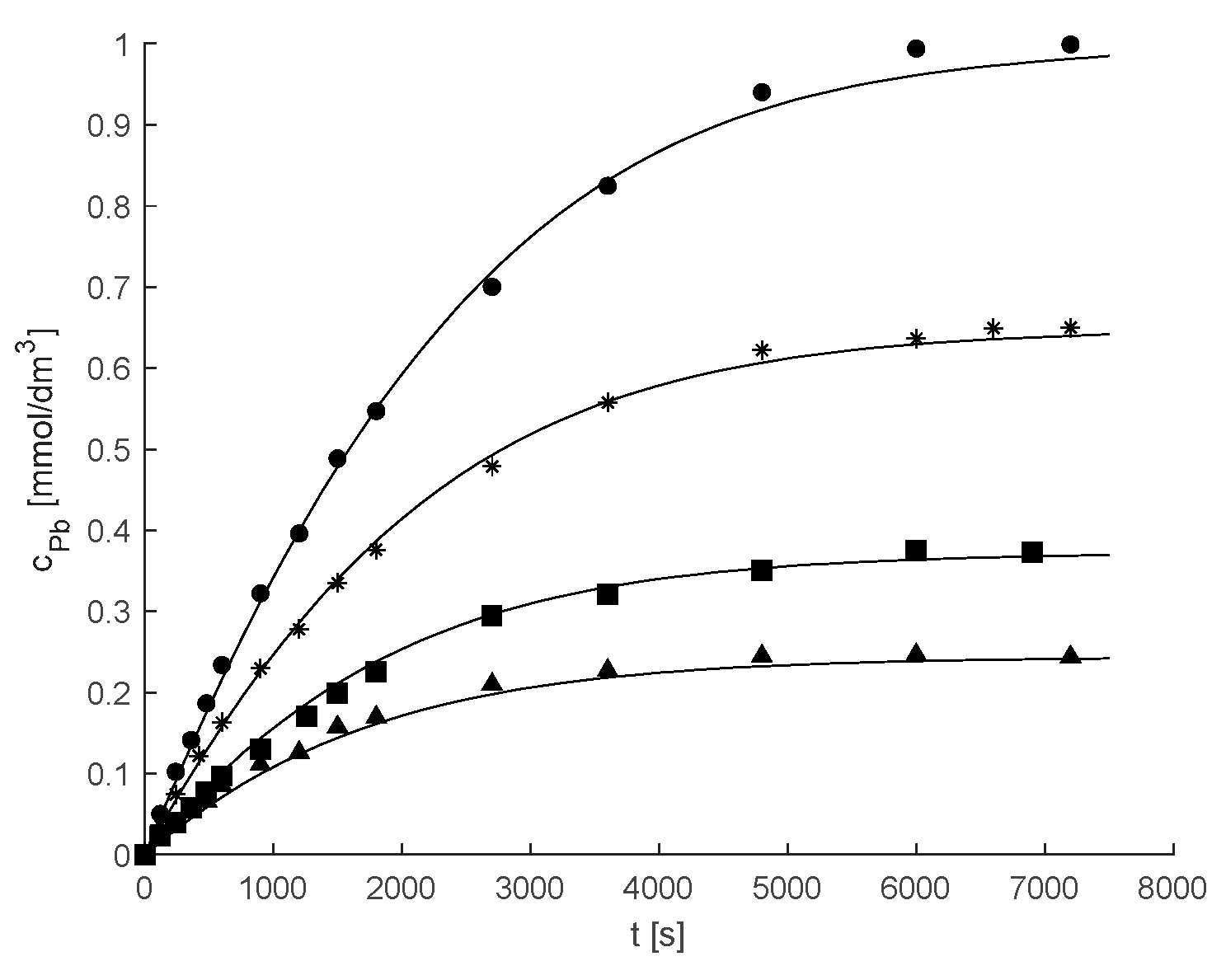
The results obtained (Figure 3) showed a good agreement between the experimental and calculated values.
The calculations shown in Figure 3 were based on the assumption that Hex was homogeneously distributed in the support particle. However, in a similar immobilization matrix based on poly (methyl methacrylate) with immobilized lipase (Novozyme 435), the enzyme was found to be localized in an external shell of the spherical particle with a thickness of 80–100 μm [20]. To investigate this possible heterogeneous distribution of the enzyme, Equation (10) was combined with the condition that the stoichiometric coefficients in the particle centrum up to r = rx were equal to zero, corresponding to a reaction-free zone; from rx to R, the stoichiometric coefficients were substituted with the values corresponding to the zone undergoing the enzymatic reaction. The effect of the size of the enzyme-free zone on the quality of the mathematical description of the kinetic data shown in Figure 3 was tested by repeating kinetic parameter optimization by non-linear regression for different values of rx. The results of mathematical modeling are summarized in Figure 4 in terms of the dependence of RSS on rx.
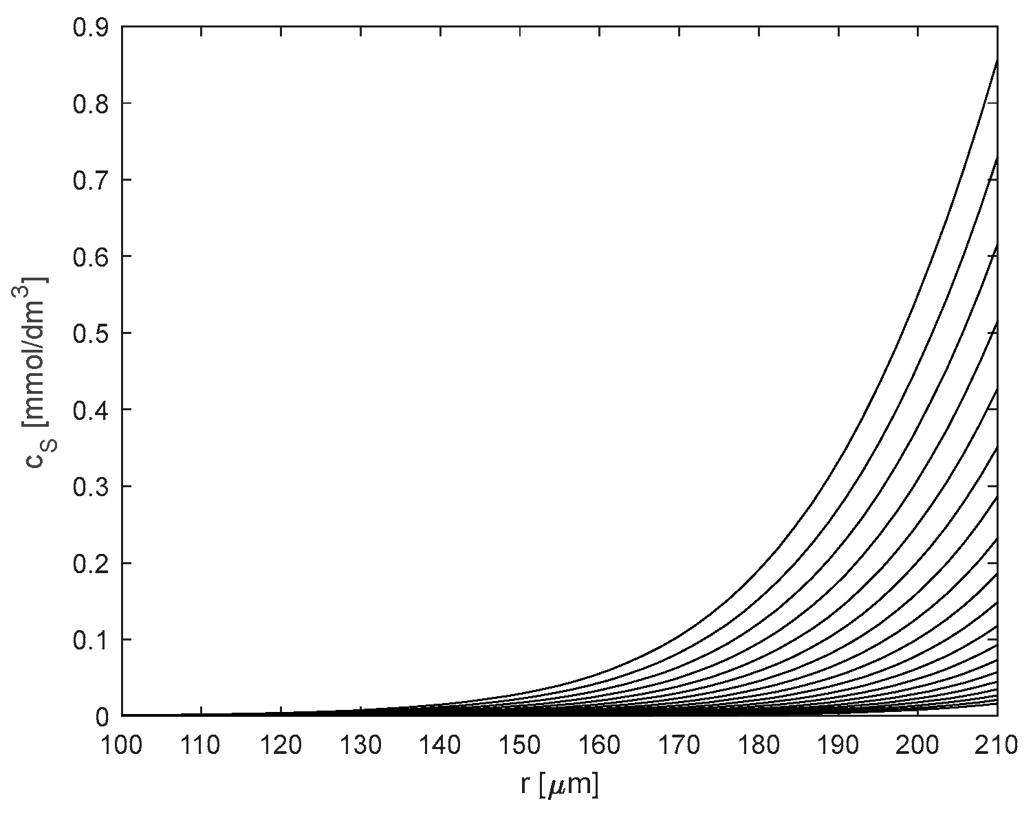
The minimum RSS values were obtained when rx was 0–110 μm; when rx was above this value, the RSS values increased with the rx value. This implied that the enzymatic reaction occurred in the zone from 110 μm up to 210 μm (the value of particle radius). This observation was confirmed by calculating the substrate concentration profiles in the particle during the reaction. The substrate concentration profiles for rx = 0 (homogeneous enzyme distribution) from the initial period of the reaction (top line) up to almost the end of the reaction (bottom line) are shown in Figure 5. This result indicated that mass transfer limitations were responsible for keeping the boundary of the reaction zone at approximately 120 μm from the center of the particle, as no substrate was present below this value.
The experimental kinetic results could not confirm or reject the hypothesis of a heterogeneous distribution of the enzyme in the particles, because the enzyme activity could not be detected in the zone with zero substrate concentration.
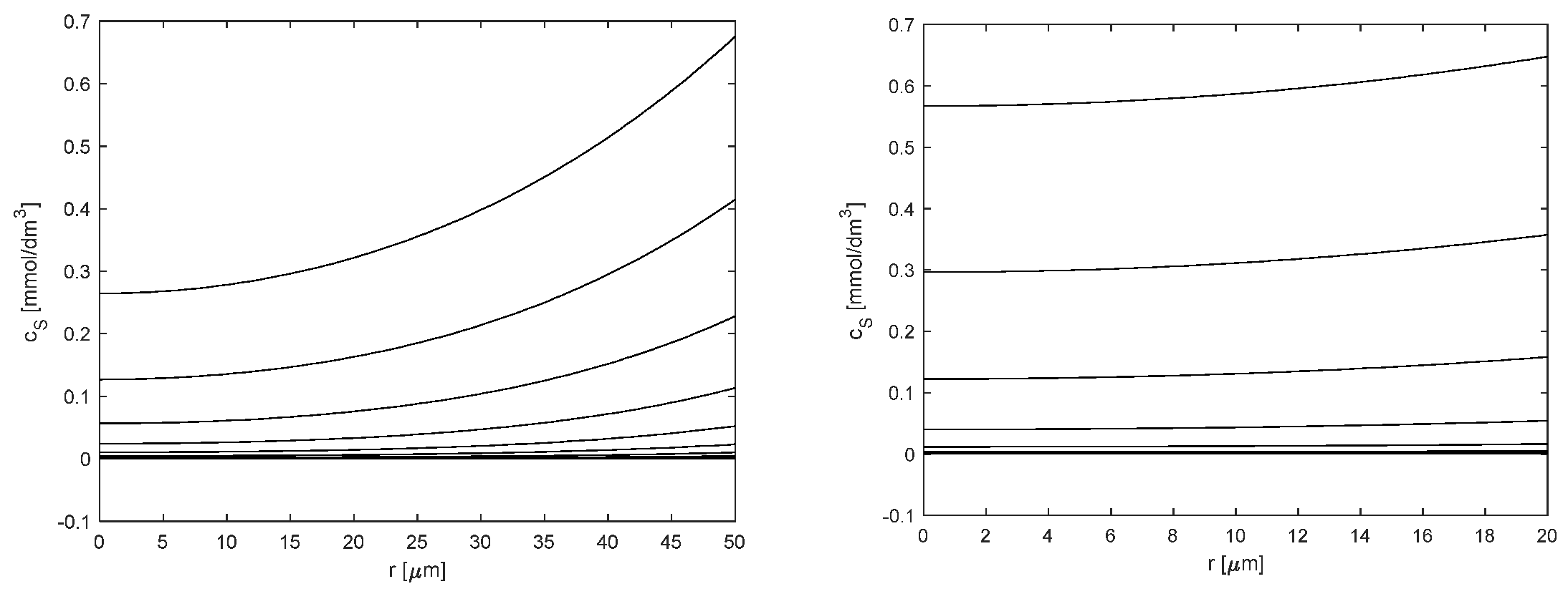
The data presented in Table 4 showed a slight difference between the Km value determined using crushed particles (0.5249 mM) and that calculated for intact particles, taking into account mass transfer effects (0.3493 mM). The two values should be the same in the case of negligible mass transfer limitations in the crushed particles. However, as shown in Figure 6, the substrate concentration profiles calculated for 50 μm or even 20 μm particles indicated that mass transfer limitations could also play a role in small particles. Therefore, assuming a high activity of the immobilized enzyme, particle crushing could not completely suppress mass transfer limitations.

2.3. Inhibition Study of Hex

Along with the inhibitory effect of the reaction substrate and product, the effect of inhibitors on the immobilized enzyme activity was tested using a continuous flow system assembled from PLC equipment. The combination of small flow reactors with standard analytical equipment has been widely applied to characterize immobilized enzymes using, e.g., flow microcalorimetry [21], capillary electrophoresis [22], amperometry [23], and HPLC equipment with offline [24] or online [25] substrate or product analysis, but this approach has not been applied to Hex. The advantage of using an HPLC device is that minimal modifications to the chosen system are sufficient to measure the activity of the immobilized enzyme. We selected PUGNAc and NAG-thiazoline [26,27] as representative inhibitors of Hex, as they have been extensively studied and well-characterized. Both inhibitors are either competitive or non-competitive inhibitors of Hex, with NAG-thiazoline and PUGNAc considered strong and weak transition state analogs, respectively [28]. However, accurately distinguishing between these inhibition models is challenging due to the low solubility of the substrate (4NPGlcNAc) and the resulting difficulty in performing inhibition studies at concentrations above the Km values. This prevents the plateau phase of the kinetic curves from being recorded, which occurs at higher substrate concentrations and provides a better assessment of the type of inhibition. These limitations introduce biases into the studies and interpretation of results, making it difficult to identify competitive or non-competitive mechanisms of inhibition. For example, NAG-thiazoline acts as a strong competitive inhibitor, with a Ki value of 0.28 µM, when interacting with Hex from Canavalia ensiformis [29]; however, it behaves as a non-competitive inhibitor, with a Ki value of 70 µM, when interacting with Hex from A. oryzae [30]. In our study, competitive and non-competitive inhibition models provided similar results (Table 4). PUGNAc exhibited stronger inhibitory potency (Ki = 0.91 µM) than NAG-thiazoline (Ki = 189 µM). The experimental data on the inhibitory potency of PUGNAc were similar to those recorded in other studies, where a Ki value of 1 µM has been found for the inhibition of Hex from P. oxalicum (ATCC 64198) by PUGNAc [18]. The Ki value for the inhibition of Hex from P. oxalicum by NAG-thiazoline is not known; thus, it cannot be directly compared. Here, we conducted the first Hex inhibition study using a continuous flow system. We provided a reliable method that can be further improved and implemented in drug screening for Hex-related diseases, and also be used in biocatalytic reactions of Hex to synthesize bioactive carbohydrates.

3. Experimental Section

3.1. Materials

All chemicals used were of analytical grade. The compounds 4-Nitrophenyl-2-acetamido-2-deoxy-ß-D-glucopyranoside (4NPGlcNAc), N-acetyl-glucosaminyl thiazoline, and O-(2-Acetamido-2-deoxy-D-glucopyranosylidene)amino N-phenyl carbamate ((Z)-PugNAc) were purchased from Biosynth Ltd. (Compton, UK). Glutaraldehyde, Bradford reagent, N-acetyl-glucosamine, and 4-nitrophenol were purchased from Sigma-Aldrich (St. Louis, MO, USA). Bovine serum albumin (BSA) was purchased from Merck (Darmstadt, Germany). The immobilization carrier amino C2 methacrylate was purchased from Purolite Ltd. (Compton, UK). The mean particle radius was 210 μm. Other chemicals and reagents were purchased from Centralchem (Bratislava, Slovakia); demineralized water was used to prepare all solutions.

3.2. Preparation of Hex

Hex was prepared and purified following a previously described method [13]. Briefly, P. oxalicum CCF 1959 was cultivated for 13 days on a rotary shaker at 28 °C and 200 rpm in a medium (pH 6.0; 100 mL/500 mL flasks) composed of 0.1% (v/v) inoculum. The medium was prepared by suspending the spores (6–7 × 10⁶ /mL) in Tween80 (0.1% (w/v)), KH2PO4 (3.0 g/L), NH4H2PO4 (5.0 g/L), (NH4)2SO4 (2.0 g/L), yeast extract (0.5 g/L), and NaCl (15.0 g/L). To the medium, we separately added sterilized N-acetylglucosamine, as a Hex inductor [31] (5.0 g/L), and MgSO4 (0.5 mL, 100 g/L). After cultivation, biomass was removed by filtration, followed by centrifugation at 20,133× g and 4 °C for 20 min. Then, the enzyme was isolated from the medium and purified by cation-exchange chromatography (Fractogel EMD SO3- (Merck, DE)) using the ÄKTA Purifier (GE HealthCare, Uppsala, Sweden), following a previously described method [32]. Briefly, the supernatant after cultivation was adjusted to a final conductivity of 10 mS/cm and hexosaminidase was eluted using 10 mM sodium citrate–phosphate buffer at pH 3.5 and a linear gradient of 0–1 M NaCl. Buffer exchange and Hex thickening were performed by ultrafiltration (Amicon® Ultra-4 10K, Millipore, Burlington, MA, USA). During the study, soluble Hex (protein concentration 1.8 mg/mL, determined by the Bradford method [33]) was either directly used for immobilization or stored at 4 °C in 1 M (NH4)2SO4 without considerable loss of activity for six months.

3.3. Immobilization of Hex

Before the covalent binding of Hex, the amino carrier (amino C2 methacrylate) was activated by glutaraldehyde. A specific amount of the carrier was washed with deionized water and phosphate buffer (pH 7.0, 100 mM). After removing the liquid by filtration on a sintered glass filter, the carrier was mixed with four times the volume (w/v) of 2% glutaraldehyde solution and incubated for 1 h at room temperature under mild agitation. Finally, the glutaraldehyde solution was filtered out and the carrier was washed with phosphate buffer.
To immobilize the enzyme, 2 g of activated Purolite matrix was mixed with 5 mL of a solution containing 0.5 mg/mL enzyme in phosphate buffer (pH 7.0, 100 mM). After 20 h of gentle agitation at room temperature, the enzyme solution was removed by filtration, and the carrier with immobilized enzyme was washed several times with phosphate buffer (pH 7.0, 100 mM) and stored at 4 °C until further use, or directly used for column packing. The protein concentration was determined by the Bradford method [33], and BSA was used for calibration. Of the total quantity of enzyme (2.5 mg) used for immobilization, 94.8% was bound. The final enzyme concentration on the carrier was 1.1 mg/g.

3.4. Activity Assay and Kinetics of Soluble Hex

The activity of Hex was determined from the rate of hydrolysis of 4-nitrophenyl-2-acetamido-2-deoxy-ß-D-glucopyranoside (2 mM or variable in case of studies on kinetics) in citrate–phosphate buffer (pH 4.5, 50 mM) by two approaches. The first one was used for the rapid determination of Hex activity after its purification. The reaction was performed in 2 mL Eppendorf tubes in Eppendorf Thermomixer Comfort (Eppendorf, Stevenage, UK) under agitation at 450 rpm and thermostating at 37 °C. The reaction was started by adding Hex, and at specific time intervals, 20 µL aliquots were taken and mixed with 2.0 mL of 0.1 M Na2CO3. The concentration of the released 4-nitrophenol was measured spectrophotometrically (Eppendorf BioSpectrometer® Basic, Eppendorf, Hamburg, Germany) at 420 nm. One unit of Hex activity was defined as the amount of enzyme releasing 1 µmol of 4-nitrophenol per minute under given conditions (50 mM citrate–phosphate buffer, pH 4.5, and 37 °C). The initial activity was determined from the slope of the linear part of the curve obtained by plotting the concentration of 4-nitrophenol vs. time. The kinetic properties of soluble Hex were investigated by monitoring the enzymatic reaction directly in a spectrophotometer cuvette (total volume 3 mL) thermostated at 37 °C using a Specord 210 PLUS Double Beam UV/Vis Spectrophotometer (Analytik Jena, Thuringia, Germany). The experiment was initiated by adding Hex to a cuvette with 4NPGlcNAc solution, and the time course of the release of 4-nitrophenol was recorded at 420 nm.

3.5. Activity Assay and Kinetics of Immobilized Hex

3.5.1. Measurement of Kinetics in a Batch System

A column (30 mm × 2.1 mm, representing a total volume of 0.104 mL, VICI Jour, CH) was packed with 0.026 mL of immobilized enzyme (IME) particles, and the remaining space was filled with glass particles. The column was placed in a flow system, where the reaction mixture underwent infinite recirculation, as shown in Figure 7.
The system consisted of a column with immobilized Hex connected via a peristaltic pump (Watson–Marlow–Bredel–Pumps, EN; flow rate set to 2.5 mL/min) to the agitated and thermostated double-jacketed beaker. The reaction temperature in the system was set to 37 °C and the total volume of the liquid phase in the system was fixed at 9.73 mL. The reaction mixture was continuously pumped from the beaker to the column and then recirculated to the same beaker. During the reaction, 50 µL aliquots of the reaction mixture were mixed with 3.0 mL of 0.1 M Na2CO3 at specific time intervals, and the concentration of the released 4-nitrophenol was measured using the same method as that described in Section 3.4. The conditions given by the total volume of the reaction system, the amount of immobilized enzyme in the column, and the flow rate through the column were set so that the system kinetically behaved as a batch-stirred tank reactor.

3.5.2. Inhibition Studies in a Continuous Flow System

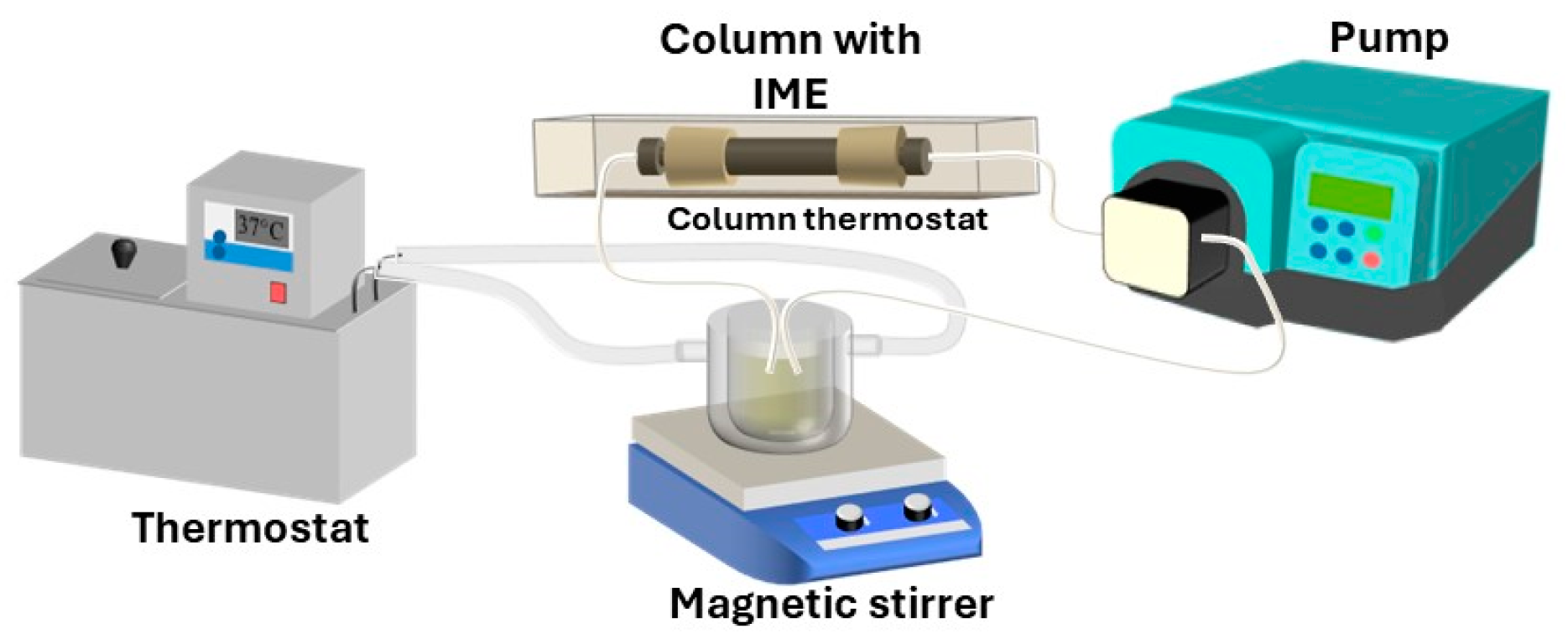
A column packed with the immobilized enzyme was inserted into the high-performance liquid chromatography system (HPLC, Hitachi LaChrom, interface model D-7000; UV-Vis detector L-7400 set to 420 nm, pump L-7100, all parts from Hitachi High-Technologies Corporation, Tokio, Japan) and placed in the column thermostat set to 37 °C. The whole system is shown in Figure 8. The system was equipped with a 3 mL injection loop for injecting the required quantity of substrate solution into the IME column. The loop volume was sufficient to achieve steady-state conditions manifested by a stable detector signal for at least 1 min. After the system’s stabilization at 37 °C and a mobile-phase flow rate of 1 mL/min (50 mM citrate-phosphate buffer at pH 4.5), a series of substrate solutions with known concentrations of 4-nitrophenyl-2-acetamido-2-deoxy-ß-D-glucopyranoside was injected while the spectrophotometric signal was registered, and a steady-state value was determined.
From the obtained values, the concentration of the steady-state product was calculated based on system calibration on standard solutions of 4-nitrophenol in the citrate–phosphate buffer (50 mM, pH 4.5) at the above-mentioned temperature and flow rate.
Before kinetic measurements, the amount of immobilized enzyme in the packed column was optimized to ensure that the substrate conversion was below 5% to operate the IME column as a differential reactor, finally providing 0.035 mL of the IME particles. In such a case, the rate of enzyme reaction along the active bed can be assumed to be constant, and its value can be calculated as the steady-state output concentration of the product (cp) divided by the residence time (tr):
v = d c p d t c p t r
The value of residence time was calculated as follows:
t r = V R ε F
where VR is the reactor volume, which is equal to the total volume of the active bed, and F indicates the flow rate. The value of bed void fraction (ε) was estimated to be 0.4, which is an approximated value for beds packed with particles whose diameter is at least 10 times smaller than the bed diameter [34].
The inhibition study was performed by injecting different solutions (with substrate concentrations ranging from 0.4 to 2.0 mM, and inhibitor concentrations ranging from 0.1 to 1.0 µM for PUGNAc and from 10 to 90 µM for NAG-thiazoline) into the column. Between injections, the column was washed with citrate–phosphate buffer (50 mM, pH 4.5).

3.5.3. Crushed Particles of Immobilized Hex

First, a sample of immobilized enzyme particles was crushed in a mortar to a powder and mixed with citrate–phosphate buffer (50 mM, pH 4.5) to form a homogeneous suspension. To evaluate the kinetic properties, a suitable quantity of the suspension was mixed with the reaction mixtures containing different concentrations of substrate (4NPGlcNAc) in an Eppendorf tube and shaken at 750 rpm and 37 °C using the Eppendorf Thermomixer Comfort (Eppendorf, Cambridge, UK). Aliquots of 50 µL were collected at specific time intervals, added to 3.0 mL of 0.1 M Na2CO3 solution, and filtered using 0.45 μm syringe filters. The released 4-nitrophenol was measured using a spectrophotometer at 420 nm (Eppendorf BioSpectrometer® Basic, Eppendorf, Germany).

3.6. Mathematical Modeling

3.6.1. Kinetics of Hex

To describe the kinetic experimental data, the following kinetic models were used:
Michaelis Menten   equation   v = V m a x c s K m + c s
Uncompetitive   substrate   inhibition   v = V m a x c s K m + c s + c s 2 K i s
Uncompetitive   substrate   inhibition   and   non-competitive   product   inhibition   v = V m a x c s K m + c s + c s 2 K i s 1 + c p K i p
Uncompetitive   substrate   inhibition   and   competitive   product   inhibition   v = V m a x c s K m 1 + c p K i p + c s + c s 2 K i s
In the above-mentioned equations, Vmax is the maximum reaction rate, Km is the Michaelis constant, Kis is the substrate inhibition constant, Kip is the product inhibition constant, cs and cp are the concentrations of substrate and product, respectively, and v is the reaction rate.
For the mathematical modeling of the enzymatic reaction in a batch system, the above equations were used, along with the following differential substrate and product balance equations:
d c s d t = v
d c p d t = v
Differential Equations (7) and (8) were solved using the initial condition corresponding to experimental initial concentrations of substrate and product.
t = 0 : c s = c s 0 ; c p = 0
The differential equations were solved, and kinetic parameters were estimated using MATLAB R2022a (MathWorks, Inc., Natick, MA, USA). The ode45 procedure based on the explicit Runge–Kutta formula was used for solving differential equations, and parameters in kinetic equations were fitted to the experimental data by Levenberg–Marquardt non-linear regression using the lsqnonlin procedure. Standard deviations of parameters were estimated from the square roots of diagonal elements of a variance–covariance matrix.

3.6.2. Kinetic Modeling of Reaction with Immobilized Hex

The mathematical model described in this section was used to evaluate the kinetic measurements performed in a system with immobilized cells with infinite recirculation, as shown in Figure 7. The system was operated in a way that facilitated its mathematical description in terms of the batch-stirred reactor. The progress of the reaction S → P in a batch reactor catalyzed by an enzyme immobilized in porous spherical particles can be calculated by solving the following non-steady, non-linear reaction–diffusion equation:
D e i 2 c i r 2 + 2 r c i r + ν i v = ε p c i t
where v is the reaction rate in the particle, Dei is the effective diffusion coefficient, r is the particle radial coordinate, εp is the particle porosity, and ci is the compound concentration in the particle. The index i is S or P for the substrate or product. The stoichiometric coefficient νi is −1 for the substrate and 1 for the product. Equation (10) for substrate and product was solved with the following initial condition (Equation (11)) and boundary condition (Equation (12)).
t = 0 :   0 r R :     c i = 0 ;   c S b = c S 0 ;   c P b = 0
t > 0 : r = 0 :   c i r = 0
r = R : D e i S p c i r R = V l c i b t
Index b in Equations (11) and (12) denotes bulk concentration. The initial condition reflects the moment at the beginning of the reaction when the substrate bulk concentration cSb is cSo, while its concentration in the particles is zero. The product concentration is initially zero everywhere. The boundary condition in the particle center (r = 0) is based on the symmetry of the concentration profile in the particle with its minimum at the particle center. The boundary condition corresponding to the particle surface (r = R) reflects the material balance in a batch reactor, according to which the number of moles of a compound passing through the outer surface of the particle per unit of time is equal to the rate of molar change in the reaction medium.
The value of the effective diffusion coefficient of the substrate was estimated from the equation
D e S = ε p D S
where the value of particle porosity εp = 0.53 was obtained from another study [35]. The substrate-free diffusivity (DS) in water at 37 °C was calculated using the Wilke–Chang equation [36] and was found to be 4.89 × 10⁻⁸ dm2/s. Regarding similar properties of substrate and product, the same value of diffusion coefficient was used for both compounds.
To calculate the reaction course, the spatial derivatives were approximated by finite differences using first-order and second-order central differences. The derivatives in Equation (12) were transformed into algebraic equations by three-point forward and backward difference equations. Using this method, the partial differential equations for substrate and product (Equation (10)) were transformed into ordinary differential equations, which were solved in MATLAB using the ode15s procedure. Parameters in the kinetic term were fitted to experimental data by Levenberg–Marquardt non-linear regression using the lsqnonlin procedure.

4. Conclusions

In this study, the characterization of Hex in free and immobilized forms was reported. The study on kinetics revealed a slightly higher Km for crushed immobilized particles (0.5249 ± 0.0031 mM) compared with the Km of the free enzyme (0.4150 ± 0.0004 mM), indicating the presence of mass transfer limitations in the biocatalyst particles. The mathematical model that included mass transfer limitations provided a Km value of 0.3493 ± 2.5 × 10⁻⁷ mM. The calculation of the substrate concentration profiles in the particle showed that the activity of the immobilized enzyme was so high that the reaction occurred at a maximum distance of 100 µm below the surface of the particle due to the substrate concentration of zero in the deeper part of the particle.
The study on kinetics was complemented by the assessment of inhibitory effects, for which a continuous flow system with an immobilized enzyme in a packed-bed mini-reactor was used. Two well-known inhibitors of Hex, i.e., PUGNAc and NAG-thiazoline, were tested. PUGNAc exhibited a strong inhibition effect with a Ki value of 0.908 µM.
These findings provide a framework for pharmaceutical and industrial applications of immobilized Hex, where rapid and efficient screening methods are necessary for developing new drugs and products.

Author Contributions

Conceptualization, H.H. and V.Š.; methodology, H.H., V.Š. and M.B.; formal analysis, V.Š.; investigation, H.H. and M.B.; data curation and mathematical modeling, V.Š. and M.B.; writing—original draft preparation, V.Š. and M.B.; writing—review and editing, H.H. and V.Š.; supervision, H.H. and V.Š.; funding acquisition, M.R. and V.Š. All authors have read and agreed to the published version of the manuscript.

Funding

This work was funded by the Slovak Research and Development Agency, according to the agreements Nr. APVV-20–0208 and Nr. APVV-22-0161.

Data Availability Statement

The data presented in this study are available on request from the corresponding author.

Conflicts of Interest

The authors declare no conflicts of interest.

References

  1. Khan, M.R. Immobilized Enzymes: A Comprehensive Review. Bull. Natl. Res. Cent. 2021, 45, 207. [Google Scholar] [CrossRef]
  2. Moo-Young, M. Comprehensive Biotechnology, 2nd ed.; Elsevier: Amsterdam, The Netherlands, 2011; ISBN 9780080885049. [Google Scholar]
  3. Konopka, J.B. N-Acetylglucosamine Functions in Cell Signaling. Scientifica 2012, 2012, 489208. [Google Scholar] [CrossRef] [PubMed]
  4. Schmölzer, K.; Weingarten, M.; Baldenius, K.; Nidetzky, B. Correction: Lacto-N-Tetraose Synthesis by Wild-Type and Glycosynthase Variants of the β-N-Hexosaminidase from Bifidobacterium Bifidum. Org. Biomol. Chem. 2019, 17, 5849. [Google Scholar] [CrossRef] [PubMed]
  5. Liu, Y.; Yan, Q.; Ma, J.; Yang, S.; Li, T.; Jiang, Z. Production of Lacto-N-Triose II and Lacto-N-Neotetraose from Chitin by a Novel β-N-Acetylhexosaminidase Expressed in Pichia Pastoris. ACS Sustain. Chem. Eng. 2020, 8, 15466–15474. [Google Scholar] [CrossRef]
  6. Liu, Y.; Yan, Q.; Ma, J.; Wang, J.; Jiang, Z.; Yang, S. Directed Evolution of a β-N-Acetylhexosaminidase from Haloferula sp. for Lacto-N-Triose II and Lacto-N-Neotetraose Synthesis from Chitin. Enzyme Microb. Technol. 2023, 164, 110177. [Google Scholar] [CrossRef]
  7. Bojarová, P.; Kulik, N.; Slámová, K.; Hubálek, M.; Kotik, M.; Cvačka, J.; Pelantová, H.; Křen, V. Selective β-N-Acetylhexosaminidase from Aspergillus Versicolor—A Tool for Producing Bioactive Carbohydrates. Appl. Microbiol. Biotechnol. 2019, 103, 1737–1753. [Google Scholar] [CrossRef]
  8. Wang, S.Y.; Laborda, P.; Lu, A.M.; Wang, M.; Duan, X.C.; Liu, L.; Voglmeir, J. Chemo-Enzymatic Approach to Access Diastereopure α-Substituted GlcNAc Derivatives. J. Carbohydr. Chem. 2016, 35, 423–434. [Google Scholar] [CrossRef]
  9. James, A.N., II; William, H.; Baricos, Y.-T.L.; Chambers, R.P.; Cohen4, W. Characterization of Immobilized β-N-Acetylhexosaminidase. Anal. Lett. 1977, 10, 501–514. [Google Scholar] [CrossRef]
  10. Yeung, K.-K.; Owen, A.J.; Dain, J.A. Effect of Immobilization on Stability and Properties of N-Acetyl-β-d-Hexosaminidase from Turbo Cornutus. Carbohydr. Res. 1979, 75, 295–304. [Google Scholar] [CrossRef]
  11. Sakai, K.; Uchiyama, T.; Matahira, Y.; Nanjo, F. Immobilization of Chitinolytic Enzymes and Continuous Production of N-Acetylglucosamine with the Immobilized Enzymes. J. Ferment. Bioeng. 1991, 72, 168–172. [Google Scholar] [CrossRef]
  12. Ruzic, L.; Bolivar, J.M.; Nidetzky, B. Glycosynthase Reaction Meets the Flow: Continuous Synthesis of Lacto-N-Triose II by Engineered β-Hexosaminidase Immobilized on Solid Support. Biotechnol. Bioeng. 2020, 117, 1597–1602. [Google Scholar] [CrossRef]
  13. Hronská, H.; Štefuca, V.; Ondrejková, E.; Bláhová, M.; Višňovský, J.; Rosenberg, M. Chemo-Enzymatic Production of 4-Nitrophenyl-2-Acetamido-2-Deoxy-α-D-Galactopyranoside Using Immobilized β-N-Acetylhexosaminidase. Catalysts 2022, 12, 474. [Google Scholar] [CrossRef]
  14. Ryšlavá, H.; Kalendová, A.; Doubnerová, V.; Skočdopol, P.; Kumar, V.; Kukačka, Z.; Pompach, P.; Vaněk, O.; Slámová, K.; Bojarová, P.; et al. Enzymatic Characterization and Molecular Modeling of an Evolutionarily Interesting Fungal β-N-Acetylhexosaminidase. FEBS J. 2011, 278, 2469–2484. [Google Scholar] [CrossRef] [PubMed]
  15. Edwards, R.G.; Thomas, P.; Westwood, J.H. The Purification and Properties of a Beta-N-Acetylhexosaminidase from Trichomonas Foetus. Biochem. J. 1975, 151, 145–148. [Google Scholar] [CrossRef] [PubMed]
  16. Yamamoto, K.; Lee, K.M.; Kumagai, H.; Tochdcura, T. Purification and Characterization of β-N-Acetylhexosaminidase from Penicillium Oxalicum. Agric. Biol. Chem. 1985, 49, 611–619. [Google Scholar] [CrossRef]
  17. Li, S.-C.; Li, Y.-T. Studies on the Glycosidases of Jack Bean Meal. J. Biol. Chem. 1970, 245, 5153–5160. [Google Scholar] [CrossRef]
  18. Rodriguez, J.; Copa-Patino, J.L.; Reyes, F.; Pérez-Leblic, M.I. A Β-N-acetylhexosaminidase from Penicillium Oxalicum Implicated in Its Cell-wall Degradation. Lett. Appl. Microbiol. 1994, 19, 217–220. [Google Scholar] [CrossRef]
  19. Škerlová, J.; Bláha, J.; Pachl, P.; Hofbauerová, K.; Kukačka, Z.; Man, P.; Pompach, P.; Novák, P.; Otwinowski, Z.; Brynda, J.; et al. Crystal Structure of Native β-N-Acetylhexosaminidase Isolated from Aspergillus Oryzae Sheds Light onto Its Substrate Specificity, High Stability, and Regulation by Propeptide. FEBS J. 2018, 285, 580–598. [Google Scholar] [CrossRef]
  20. Mei, Y.; Miller, L.; Gao, W.; Gross, R.A. Imaging the Distribution and Secondary Structure of Immobilized Enzymes Using Infrared Microspectroscopy. Biomacromolecules 2003, 4, 70–74. [Google Scholar] [CrossRef]
  21. Štefuca, V.; Gemeiner, P. Investigation of Catalytic Properties of Immobilized Enzymes and Cells by Flow Microcalorimetry. In Thermal Biosensors, Bioactivity, Bioaffinitty; Bhatia, P.K., Danielsson, B., Gemeiner, P., Grabley, S., Lammers, F., Mukhopadhyay, A., Ramanathan, K., Saleemuddin, M., Scheper, T., Stefuca, V., et al., Eds.; Springer: Berlin/Heidelberg, Germany, 1999; pp. 69–99. ISBN 978-3-540-49811-7. [Google Scholar]
  22. Siebert, D.A.; Caon, N.B.; Alberton, M.D.; Vitali, L.; Parize, A.L.; Micke, G.A. Immobilized Acetylcholinesterase in Magnetic Nanoparticles for In-Line Inhibition Studies Using a Capillary Electrophoresis System. Anal. Chim. Acta 2023, 1275, 341566. [Google Scholar] [CrossRef]
  23. Cerdeira Ferreira, L.M.; da Costa, E.T.; do Lago, C.L.; Angnes, L. Miniaturized Flow System Based on Enzyme Modified PMMA Microreactor for Amperometric Determination of Glucose. Biosens. Bioelectron. 2013, 47, 539–544. [Google Scholar] [CrossRef] [PubMed]
  24. Bajić, M.; Plazl, I.; Stloukal, R.; Žnidaršič-Plazl, P. Development of a Miniaturized Packed Bed Reactor with ω-Transaminase Immobilized in LentiKats®. Process Biochem. 2017, 52, 63–72. [Google Scholar] [CrossRef]
  25. De Oliveira, P.C.O.; Lessa, R.C.; Ceroullo, M.S.; Wegermann, C.A.; De Moraes, M.C. On-Flow Enzymatic Inhibitor Screening: The Emerging Success of Liquid Chromatography-Based Assays. Front. Anal. Sci. 2022, 2, 1004113. [Google Scholar] [CrossRef]
  26. Meekrathok, P.; Stubbs, K.A.; Aunkham, A.; Kaewmaneewat, A.; Kardkuntod, A.; Bulmer, D.M.; van den Berg, B.; Suginta, W. NAG-Thiazoline Is a Potent Inhibitor of the Vibrio Campbellii GH20 β-N-Acetylglucosaminidase. FEBS J. 2020, 287, 4982–4995. [Google Scholar] [CrossRef]
  27. Stubbs, K.A.; Zhang, N.; Vocadlo, D.J. A Divergent Synthesis of 2-Acyl Derivatives of PUGNAc Yields Selective Inhibitors of O-GlcNAcase. Org. Biomol. Chem. 2006, 4, 839–845. [Google Scholar] [CrossRef]
  28. Whitworth, G.E.; Macauley, M.S.; Stubbs, K.A.; Dennis, R.J.; Taylor, E.J.; Davies, G.J.; Greig, I.R.; Vocadlo, D.J. Analysis of PUGNAc and NAG-Thiazoline as Transition State Analogues for Human O-GlcNAcase:  Mechanistic and Structural Insights into Inhibitor Selectivity and Transition State Poise. J. Am. Chem. Soc. 2007, 129, 635–644. [Google Scholar] [CrossRef]
  29. Knapp, S.; Vocadlo, D.; Gao, Z.; Kirk, B.; Lou, J.; Withers, S.G. NAG-Thiazoline, An N-Acetyl-β-Hexosaminidase Inhibitor That Implicates Acetamido Participation. J. Am. Chem. Soc. 1996, 118, 6804–6805. [Google Scholar] [CrossRef]
  30. Plíhal, O.; Sklenář, J.; Hofbauerová, K.; Novák, P.; Man, P.; Pompach, P.; Kavan, D.; Ryšlavá, H.; Weignerová, L.; Charvátová-Pišvejcová, A.; et al. Large Propeptides of Fungal β-N-Acetylhexosaminidases Are Novel Enzyme Regulators That Must Be Intracellularly Processed to Control Activity, Dimerization, and Secretion into the Extracellular Environment. Biochemistry 2007, 46, 2719–2734. [Google Scholar] [CrossRef]
  31. Huňková, Z.; Křen, V.; Ščigelová, M.; Weignerová, L.; Scheel, O.; Thiem, J. Induction of β-N-Acetylhexosaminidase in Aspergillus Oryzae. Biotechnol. Lett. 1996, 18, 725–730. [Google Scholar] [CrossRef]
  32. Slámová, K.; Bojarová, P.; Gerstorferová, D.; Fliedrová, B.; Hofmeisterová, J.; Fiala, M.; Pompach, P.; Křen, V. Sequencing, Cloning and High-Yield Expression of a Fungal β-N-Acetylhexosaminidase in Pichia Pastoris. Protein Expr. Purif. 2012, 82, 212–217. [Google Scholar] [CrossRef]
  33. Bradford, M.M. A Rapid and Sensitive Method for the Quantitation of Microgram Quantities of Protein Utilizing the Principle of Protein-Dye Binding. Anal. Biochem. 1976, 72, 248–254. [Google Scholar] [CrossRef] [PubMed]
  34. Benyahia, F.; O’Neill, K.E. Enhanced Voidage Correlations for Packed Beds of Various Particle Shapes and Sizes. Part. Sci. Technol. 2005, 23, 169–177. [Google Scholar] [CrossRef]
  35. Anand, A.; Gnanasekaran, P.; Allgeier, A.M.; Weatherley, L.R. Study and Deployment of Methacrylate-Based Polymer Resins for Immobilized Lipase Catalyzed Triglyceride Hydrolysis. Food Bioprod. Process. 2020, 123, 164–176. [Google Scholar] [CrossRef]
  36. Wilke, C.R.; Chang, P. Correlation of Diffusion Coefficients in Dilute Solutions. AIChE J. 1955, 1, 264–270. [Google Scholar] [CrossRef]
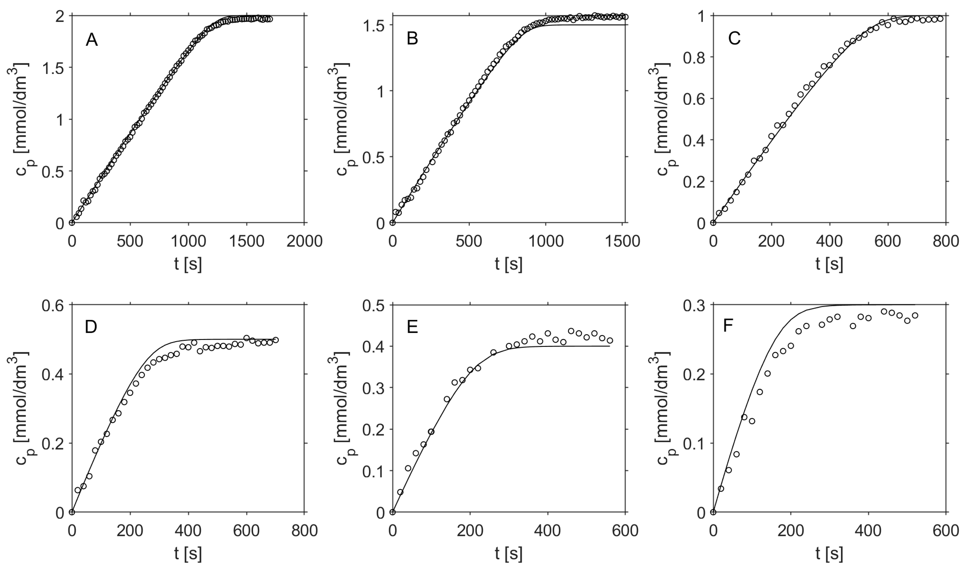
Figure 1. Progress curves of enzymatic hydrolysis of 4NPGlcNAc catalyzed by free hexosaminidase. Points correspond to experimental data, and lines represent data calculated from the Michaelis–Menten equation with uncompetitive substrate and non-competitive product inhibition. Initial substrate concentrations (in mmol/dm3): (A): 2.0; (B): 1.5; (C): 1.0; (D): 0.5; (E): 0.4; and (F): 0.3.
Catalysts 14 00725 g001
Figure 2. The evaluation of reaction kinetics with crushed particles was compared using different models. Left panel: Michaelis–Menten model with uncompetitive substrate inhibition and non-competitive product inhibition; Right panel: Simple Michaelis–Menten kinetics. Initial substrate concentrations (mmol/dm3): 0.2 (•); 0.7 (✱); 1.0 (■); 1.5 (▲); and 2.0 (▼).
Catalysts 14 00725 g002
Figure 3. Reaction kinetics with intact immobilized enzyme particles. Comparison of experimental data (points) and data calculated from Michaelis–Menten kinetics, including mass transfer effects (solid lines). Initial substrate concentrations (mmol/dm3): 0.25 (▲); 0.4 (■); 0.7 (✱); 1.0 (•).
Catalysts 14 00725 g003
Figure 4. Influence of the position of the boundary of immobilized Hex on the best RSS obtained by non-linear regression.
Catalysts 14 00725 g004
Figure 5. Evolution of substrate concentration profiles in the immobilized Hex particle during the reaction.
Catalysts 14 00725 g005
Figure 6. Evolution of substrate concentration profiles in the immobilized Hex particles, assuming particle radii of 50 μm (left panel) and 20 μm (right panel).
Catalysts 14 00725 g006
Figure 7. Experimental setup for measuring the kinetics of immobilized Hex with infinite recirculation of the reaction mixture.
Catalysts 14 00725 g007
Figure 8. The continuous flow system used for measuring the activity of immobilized Hex.
Catalysts 14 00725 g008
Table 1. Kinetic parameters of four different models tested with free Hex.
ModelVmax (×10−3 mM/s)Km (mM)Kis (mM)Kip (mM)RSS
M–M1.7452 ± 0.00020.0560 ± 0.0001--1.395
M–M with substrate inhibition4.1187 ± 0.00240.4876 ± 0.00081.2157 ± 0.0021-0.454
M–M with uncompetitive substrate and non-competitive product inhibition4.1441 ± 0.00240.4105 ± 0.00246.8619 ± 0.00601.3611 ± 0.00140.175
M–M with uncompetitive substrate and competitive product inhibition3.7795 ± 0.00260.3263 ± 0.00052.5736 ± 0.00421.4210 ± 0.00180.248
M–M = Michaelis–Menten kinetic model.
Table 2. The kinetic parameters of four different models tested using crushed particles with immobilized Hex.
ModelVmax (mM/s)Km (mM)Kis (mM)Kip (mM)RSS
M–M(7.431 ± 0.016) × 10−30.5249 ± 0.0031--0.0158
M–M with substrate inhibition(1.452 ± 0.023) × 10−31.362 ± 0.0272.018 ± 0.063-0.0087
M–M with uncompetitive substrate and non-competitive product inhibition(1.448 ± 0.022) × 10−31.326 ± 0.02531.4 ± 1.72.074 ± 0.0620.0081
M–M with uncompetitive substrate and competitive product inhibition(1.425± 0.022) × 10−31.300 ± 0.02623.9 ± 1.72.090 ± 0.0640.0083
M–M = Michaelis–Menten kinetic model.
Table 3. A summary of the Km values for free Hex, immobilized crushed particles, and immobilized intact particles.
Enzyme PreparationMathematical Model UsedKm (mM)
Free enzymeM–M0.4105 ± 0.0004
Immobilized Hex–crushed particlesM–M0.5249 ± 0.0031
Immobilized Hex–intact particlesM–M1.168 ± 0.014
Immobilized Hex–intact particlesMass transfer + M–M0.3493 ± 2.5 × 10⁻⁷
Table 4. Kinetic and inhibition parameters of immobilized Hex.
ModelVmax (mM/s)Km (mM)Kis (µM)RSS
PUGNAc competitive0.686.150.917.032
PUGNAc non-competitive0.787.311.137.129
NAG-thiazoline competitive0.322.471892.628
NAG-thiazoline non-competitive0.352.802982.534
Disclaimer/Publisher’s Note: The statements, opinions and data contained in all publications are solely those of the individual author(s) and contributor(s) and not of MDPI and/or the editor(s). MDPI and/or the editor(s) disclaim responsibility for any injury to people or property resulting from any ideas, methods, instructions or products referred to in the content.