Article

Feedback Inhibition of DszC, a Crucial Enzyme for Crude Oil Biodessulfurization

LAQV, REQUIMTE, Departamento de Química e Bioquímica, Faculdade de Ciências, Universidade do Porto, Rua do Campo Alegre, s/n, 4169-007 Porto, Portugal
*
Author to whom correspondence should be addressed.
These authors contributed equally to this work.
Catalysts 2023, 13(4), 736; https://doi.org/10.3390/catal13040736
Submission received: 3 March 2023 / Revised: 5 April 2023 / Accepted: 10 April 2023 / Published: 13 April 2023
(This article belongs to the Special Issue Designing Catalytic Desulfurization Processes to Prepare Clean Fuels)

Abstract

:
The Rhodococcus erythropolis (strain IGTS8) bacterium has a tremendous industrial interest as it can remove sulfur from crude oil through its four-enzyme (DszA-D) 4S metabolic pathway. DszC is one of the rate-limiting enzymes of the pathway and the one that most suffers from feedback inhibition. We have combined molecular docking and molecular dynamics simulations to identify binding sites through which two products of the 4S pathway, 2-hydroxybiphenyl and 2′-hydroxybiphenyl-2-sulfinate, induce DszC feedback inhibition. We have identified four potential binding sites: two adjacent binding sites close to the 280–295 lid loop proposed to contribute to DszC oligomerization and proper binding of the flavin mononucleotide cofactor, and two other close to the active site of DszC and the substrate binding site. By considering (i) the occupancy of the binding sites and (ii) the similar inhibitor poses, we propose that the mechanism of feedback inhibition of DszC occurs through disturbance of the DszC oligomerization and consequent binding of the flavin mononucleotide due to the weakening of the interactions between the 280–295 lid loop, and both the 131–142 loop and the C-terminal tail. Nevertheless, inhibitor binding close to the active site or the substrate binding sites also compromises critical interactions within the active site of DszC. The disclosed molecular details provide valuable insight for future rational enzyme engineering protocols to develop DszC mutants more resistant against the observed feedback inhibition mechanism.

1. Introduction

The high demand for crude oil continues [1] as greener energy production alternatives grow slower than current energy demands [2]. As a result, large amounts of harmful gaseous emissions, including CO2, sulfur oxides (SOx) or nitric oxides (NOx), threaten the environment and push governments to rule on strategies to reduce emissions of such pollutants in the near future.
Since the 1990s, legislation from many countries has been pursuing ultra-low concentrations of sulfur in fossil fuels [3,4,5,6,7]. Since 2012, U.S.A. regulations state that the sulfur content in diesel and gasoline should not exceed 15 ppm [8,9], while in the European Union, the limit has been ten ppm since 2011 for most vehicles [10,11]. As the most significant oil reservoirs across the world (Middle East, Gulf of Mexico, South America) are increasingly composed of sulfur-rich compounds [12], new ways to efficiently reduce sulfur concentrations in crude oil are being sought.
Heteroaromatic sulfur compounds, namely thiophenes and benzothiophenes [5,6,7], are responsible for almost 70% of the sulfur concentration in crude oil [13] and are pretty poorly desulphurized by the conventional hydrodesulfurization (HDS) method. Other less-used methods include extractive or oxidative desulfurization, but these still do not represent efficient alternatives for industry [3,14,15]. Biodesulfurization is a promising alternative to complement or replace HDS in crude oil desulfurization [13,16]. In particular, the process uses living organisms or their enzymes [16] to remove sulfur from organosulfur compounds and is highly selective for heteroaromatic sulfur compounds, while preserving their calorific content with negligible environmental impacts compared to current desulfurization alternatives [16,17].

1.1. Engineering the 4S Pathway

For a few decades now, the 4S pathway of the bacterium Rhodococcus erythropolis (strain IGTS8) has been studied as a potential biodesulfurization tool [18,19,20]. The 4S pathway encompasses four enzymes and uses dibenzothiophene and its derivatives in sulfur-rich crude oil as a substrate to store energy in aromatic carbon skeletons. The metabolism of DBT consumption by the bacterium, depicted in Figure 1, requires the action of two FMNH2-dependent monooxygenases, DszA and DszC, the desulfinase DszB and the NADH-dependent FMN oxidoreductase, DszD; and produces 2-hydroxybiphenyl (2-HBP) and sulfite (SO32−).
Despite its appealing features, the efficiency of the 4S pathway is not up to the standards required for an industrial-scale application, particularly when compared with the currently employed chemical processes [19,21,22]. There are two leading causes for this: the rate-limiting kinetics of the enzymes DszC and DszB, which catalyze the initial double-oxidation of sulfur and the final cleavage of the C-S bond, releasing the aromatic hydrocarbon product too slowly and the mechanism of feedback inhibition, in which the products of enzymes of the 4S pathway bind the DszA-C enzymes and decrease their activity [23].
Genetic engineering has been the preferred approach to improve the efficiency of the 4S pathway. Nevertheless, while previous attempts improved the efficiency of the pathway up to 20-fold [22,24,25,26], the 500-fold improvement required for industrial implementation remains unachieved [27]. Most of these studies were made with directed evolution, which relies on successive rounds of random mutagenesis from which the most successful candidate mutants are selected to proceed for further rounds. While successful in improving more promiscuous enzymes, it involves laborious work, often leading to efficiency dead-ends [28].
More recently, we have focused on determining the reaction mechanism of the rate-limiting enzymes DszC and DszB with atomistic detail using hybrid quantum mechanics/molecular mechanics calculations [29,30]. In such a way, we could identify each reaction cycle’s rate-limiting step and the critical residues for their catalytic power and exact function. We also identified candidate residues for site-directed mutagenesis that should increase the reaction rate through rational enzyme engineering [30,31]. Our rational enzyme-design methodology can be used alone or combined with directed evolution, representing a promising strategy for state-of-the-art enzyme engineering [32,33].

1.2. The Feedback Inhibition Mechanism of the 4S Pathway

While higher desulfurization activities should be achievable by enzyme immobilization or genetic engineering, the reduced activity of the 4S pathway is also caused by an increase in 2-HBP in the medium during biodesulfurization [23]. It is known that the enzymes DszA-C are inhibited by the 2-HBP product, but the molecular details of this inhibition mechanism remain unknown [21].
A comprehensive kinetic study by Abin-Fuentes evaluated the dependence in activity of the 4S pathway in resting cell cultures and through in vitro experiments with each enzyme in the pathway [23]. The authors confirmed that the feedback inhibition mechanism of the 4S pathway results from the accumulation of the final product within the biocatalyst at concentrations that can surpass the hundreds of μM. In agreement with other work [34,35,36], the DszB and DszC enzymes were identified as the kinetic bottlenecks of the 4S pathway, with a catalytic rate, kcat, of 1.7 and 1.6 min−1; and a catalytic efficiency of 1.3 and 1.1 μM−1·min−1. In addition, dose-response experiments on the DszA-C enzymes with 2-HBP and the 2′-hydroxybiphenyl-2-sulfinate (HBPS) product of DszA showed that DszC is the most severely affected by the feedback inhibition caused by the 2-HBP product of DszB and also, by the 2′-hydroxybiphenyl-2-sulfinate (HBPS) product of DszA, with half maximal inhibitory concentration (IC50) of 15 and 50 μM. DszA and DszB are also affected, although to a lesser extent [23]. The sensibility to feedback inhibition follows the order DszC > DszA > DszB. In particular, the inhibition of DszC by 2-HBP and HBPS also fitted that of a noncompetitive kinetic model, with inibition constants, Ki, of 40 and 13.5 μM. As such, the binding of 2-HBP and HBPS should not compete with that of the DBT substrate, although it can still compete with FMN binding. As DszC is also one of the least efficient enzymes in the 4S pathway, understanding its feedback inhibition mechanism is particularly important. Nevertheless, no structural studies can be found in the literature on this subject.
DszC is functional as a homodimer [37], although it has also been determined as a tetramer (dimer of homodimers) from gel filtration and X-ray experiments [38,39]. The enzyme binds a flavin mononucletide (FMN) cofactor at the interface of the homodimer and the DBT substrate in a large pocket composed of an inner hydrophobic chamber and an outer hydrophilic chamber [39]. From the available structural data on the enzyme, the oligomerization of DszC occurs prevalently by a C-terminal domain encompassing residues Ala236-Ser417 [38,39], with more specific contributions from an Arg338 located on an α-helix close to the DBT binding pocket and the Tyr410-Ser417 terminal loop [39]. A conformational shift after FMN binding by the Gln280-Asp295 loop, contributing for FMN binding, and the Ser131-Lys142 loop of another monomer would then lead to a close conformation ready for catalysis [38,40]. Mutation of any of the key catalytic residues identified from QM/MM calculations by Barbosa et al. (Ser163, His92, Tyr96, His391, and Asn129) or residues contributing either for FMN binding or DszC dimerization, namely Arg338 or the Tyr410-Ser417 terminal loop, led to a loss of enzyme activity [38].
One of the most relevant studies on DszC engineering dated from 2019, when the mechanism of feedback inhibition in DszC by HBPS and 2-HBP was analyzed with a combination of desensitization engineering and DszC overexpression [25]. The authors produced double mutants that were 13.8-fold faster than the wild-type DszC, indicating that the mutation of a Trp327 was important for increased DszC efficiency. However, they could not identify the enzyme patches or sites where the feedback inhibitors bind.
Recent in silico protocols have supported that the combination of molecular docking and molecular dynamics (MD) simulations can be used to characterize the structure of protein: small molecule models [41,42]. As such, a combination of molecular docking and MD simulations will be used here to identify the regions of DszC that bind the HBPS and 2-HPB molecules, underpinning the inhibition mechanisms by the latter, thus paving ways to develop rational mutants that circumvent the feedback mechanism without affecting the catalytic machinery.

2. Results and Discussion

We ran two 200 ns MD simulations with different starting conditions: in the first, each system was simulated after a standard minimization and equilibration protocol; in the second one, which will be referred to as induced-fit simulation, the ligand molecules (2-HBP and HBPS) were kept fixed with positional harmonic restraints, while DszC was kept free for 6 ns in order to induce a fitting of the DszC complex to the starting pose in each binding site. The strategy aimed to investigate whether the final binding sites identified in the MD simulations depended on fitting the DszC complex to the starting poses of the ligands in each binding site. We stress that our MD simulations are limited by the short simulation time when comparing to binding/unbinding events, which take microsseconds to seconds to occur and do not take into account complex oligomerization events in solution, depending on the available concentration of DszC or interactions with other molecules/structures in the bacterial cytosol. Such events would require either enhanced sampling simulation techniques [43], only affordable in exascale computing platforms; or multiscale resolution methods [44], which also compromise the molecular detail of the interactions taking place between the DszC and 2-HBP and HBPS. Hence, the results of our work should establish a groundwork for subsequent study with experimental methods, either in vitro or in vivo, that may provide further structural, kinetic, or thermodynamic data to complement our findings.
Throughout the final 200 ns MD simulations considered for analysis, there was no substantial change in the overall conformation of DszC, as evidenced by the root-mean-square deviations below 3 Å for the backbone atoms of the DszC homodimer (Figures S1 and S2). However, there was variability in 2-HBP and HBPS that required further analysis. Hence, we proceeded with a contact analysis between the 2-HBP and HBPS and the DszC residues within an interacting distance from 3.5 to 5 Å (summarized in Figures S3–S6). We chose these cutoffs to account for short- and medium-range interactions. In such a way, we could inspect whether the ligands interacted with the binding sites initially proposed from the docking calculations or if new binding sites were identified.

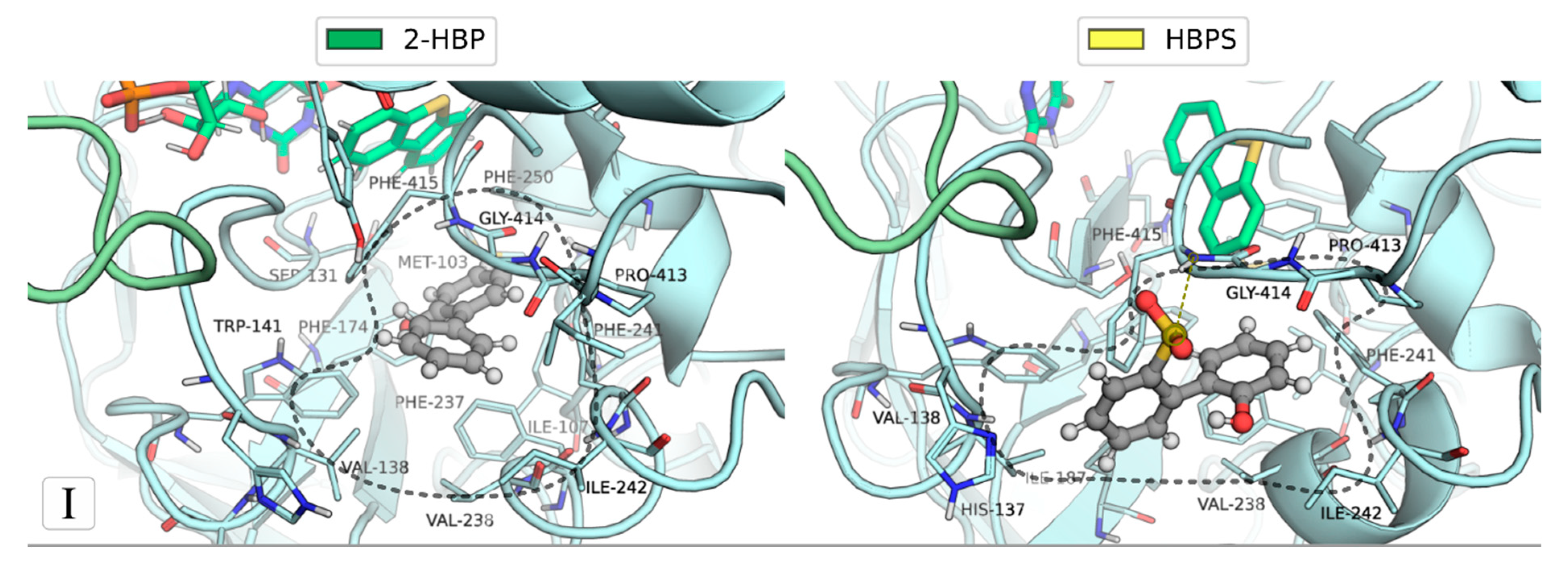
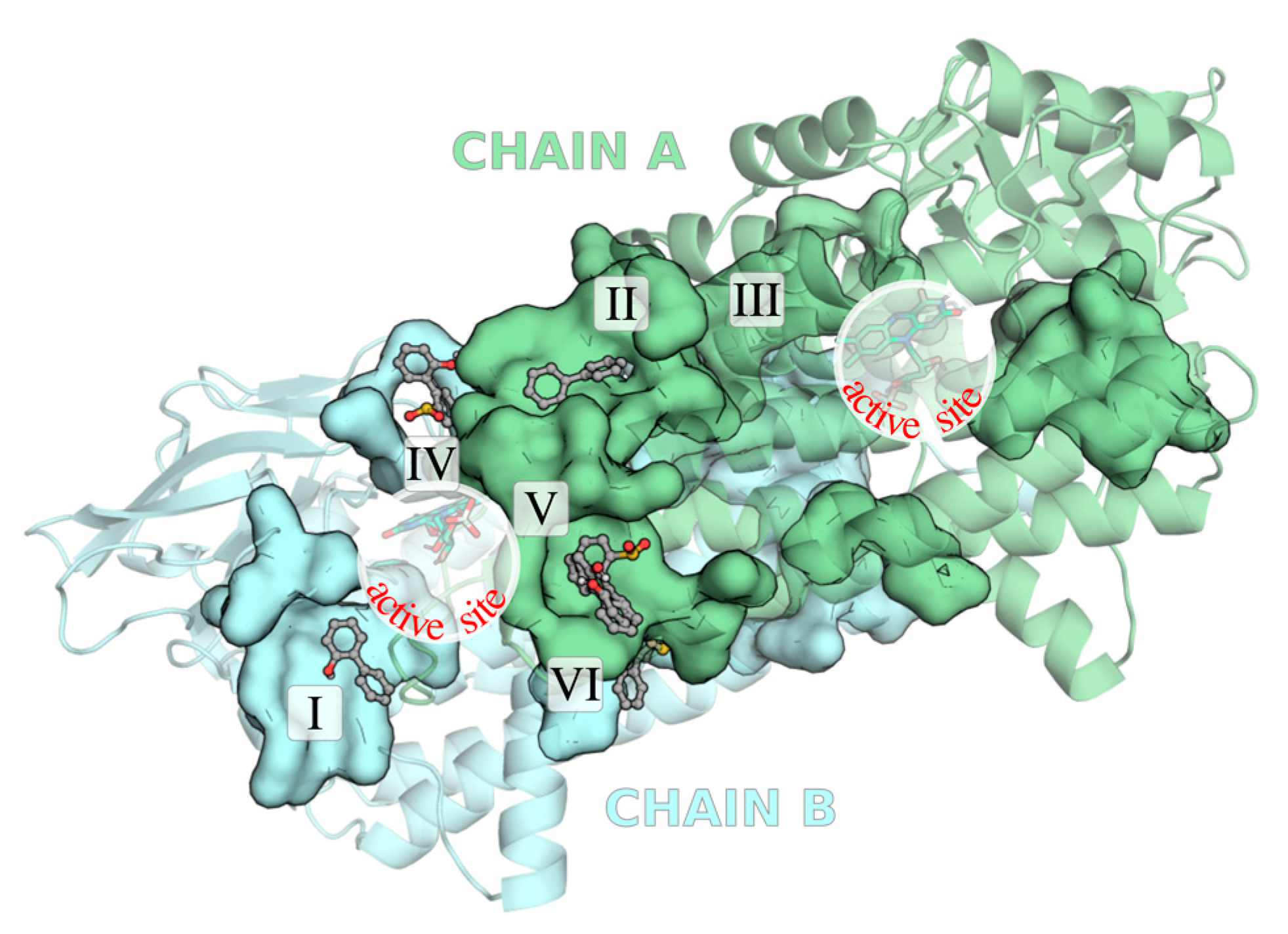
2.1. Identification of the Binding Sites of 2-HBP and HBPS

From the overall contact analysis between 2-HBP and HBSP and the residues at a minimum distance of 3.5 Å, summarized in Figure 2, it is clear that 2-HBP and HBPS moved from their initial docking binding sites and explored further binding sites. As a result, three additional binding sites were identified (VII, VIII, and IX), with sites VII and IX being identified in simulations involving 2-HBP and HBPS. Other than site III, all binding sites are mostly found at the C-terminal domain of the enzyme (Ala236-Ser417), which is the main one responsible for DszC and cofactor binding [38,39], with sites V-VII and IX being composed exclusively of residues from this domain.
A closer look at Figure 2 indicates that four out of the six binding sites identified in the docking calculations (sites I, II, IV, and V) interacted with either 2-HBP or HBPS throughout the MD simulations, and three of them (sites I, II, and V) were occupied by both. In particular, HBPS exhibited frequent interaction (more than 60% of the simulation time) with binding sites identified by docking for 2-HBP (sites I and II), which would be expected given the similarity between both molecules, although the same was not observed for 2-HBP.
When comparing whether a pre-equilibration of the enzyme to favor the initial poses of 2-HBP and HBPS influences the resulting binding sites for 2-HBP and HBPS (Figures S7–S10), we observe that site V is the one occupied in most simulations, in particular when interacting with 2-HBP, although its occupation can range from 30 to 80%. Furthermore, an analysis of the individual contacts between each ligand and the nearby DszC residues (Figures S3–S6) also indicated that the occupation of the initial binding sites was not favored when an induced-fit equilibration was attempted, although a higher frequency of close contacts between the ligands and DszC residues was generally observed. The simulations following an induced-fit equilibration suggested most of the new binding sites identified (as can be confirmed in Figures S4 and S6).
To identify representative binding sites for 2-HBP and HBPS binding, we performed a cluster analysis over the root-mean-square deviation of residues interacting within 3.5 Å of the 2-HBP and HBPS for more than 40% of the simulation (the detailed contact analysis per ligand follows in Figures S3–S6). In Figure 3, we depict the resulting seven representative binding sites.
The binding sites I, IV, and VII are located nearby the active site of DszC. The binding site I encompasses the entrance to the binding site of the DBT substrate and includes Phe415, known to interact directly with DBT [39], whereas the binding site VII, which lies close to the site I, encompasses the α10-helix (Thr334-Gly368) adjacent to the α8-helix (Leu249-Gln280), also found at the binding site of DBT. Both binding sites also include several residues (Gly347-Leu351 or Pro413-Phe415, sites I and VII in Figure 3) close to Arg338, whose mutation by Ala affected DszC activity and oligomerization [39]. The binding site IV comprises several residues involved in the binding of the FMN cofactor, namely the conserved Phe161 and Trp205; and Arg370, whose mutation by Ala have led to inactive forms of DszC [38], although Trp205 establishes close contacts less often.
Aside from the binding site VIII (located at the surface of each DszC monomer), binding sites II, V, and IX are close to regions that might contribute to the dimerization of DszC. In particular, site V includes residues right next to the Gln280-Asp295 residues composing a flexible lid loop, which is proposed to be involved in the open/closed-conformation interchange required for DszC activity [38,40] alongside site II, which includes residues at the N-terminus of the lid loop (Ala272-Gln280) and the C-terminus of α11-helix (Tyr376-Arg380). Finally, the binding site IX lies well at the dimerization interface of the homodimer required for a catalytically-competent form of the enzyme.

2.2. Ranking of the Binding Sites

In order to identify the most favorable binding sites among all identified, we used four criteria: (i) to be common to both simulations (with or without induced fit equilibration), as that indicates that ligands exhibited high affinity regardless of the initial conditions of the simulation; (ii) the contacts between residues composing the site and ligands should be stable in time (>60% of the simulation time); (iii) the binding should not rely strongly on ligand concentration and, thus, similar contacts should be found regardless of the number of ligands in the simulation; and (iv) as both 2-HBP and HBPS induce feedback inhibition and share a similar molecular shape, they should bind in a similar binding site.
Most consensual binding site: binding site V. No binding site filling the four requisites was found, but one filled most criteria. The binding site V, composed of residues Arg274, Thr277, Arg278, Thr293, Thr298, Ile299, and Tyr302, was observed to interact with both 2-HBP and HBPS in all but one simulation through short contacts often lasting > 60% of the time (Figure 2 and Figure S7–S10). Furthermore, it bound more often HBPS than 2-HBP, which is consistent with the higher inhibitory effect of HBPS suggested by experimental IC50 (15 μM vs. 50 μM of 2-HBP) [23]. Finally, the binding site is located right by the Gln280-Asp295 lid loop, which, together with the adjacent Ser131-Lys142 loop, promotes the proper binding of the FMN cofactor required for DszC activity [38,40].
Upon clustering the trajectories using the RMSd of the residues composing the binding site, we obtained a representative binding mode of 2-HBP and HBPS, depicted in Figure 4, which shows a similar binding pose for both ligands.
The binding of 2-HBP and HBPS results from the hydrophobic packing of the sulfinated phenyl ring in the hydrophobic cavity formed by the sidechain of residues Tyr302, Thr277, Thr298-Ile299, and Thr293. The charged guanidinium head of the nearby Arg278 is kept facing out of the pocket by a salt interaction with the nearby Glu275, and a hydrogen bond is observed between the 2-hydroxyl of both 2-HBP and HBPS with the guanidinium head of Arg274; as such, no significant polar or ionic interactions can be formed within the hydrophobic cavity accommodating the ligands.
Upon comparison of the conformation of the binding site upon ligand binding and in the control simulation, we observed that the hydrophobic cavity outlined by Tyr302, Thr277, Thr293, and Thr298-Ile299 was previously occupied by the sidechain of Arg274. Hence, the aromatic rings of 2-HBP and HBPS replaced Arg274, and the 2-hydroxyl substitution and solvent exposure kept the Arg274 pointing outwards the binding pocket. We also observe that the sulfinate group in HBPS faces the solvent, although ionic interactions with Arg274 could occur. Since the binding of the ligands does not seem to rely on the sulfinate group distinguishing 2-HBP and HBPS, this binding mode aligns with our hypothesis that 2-HBP and HBPS can inhibit DszC activity through a similar mode of action.
Other significantly consensual binding sites: II and VII. The binding sites II and VII were identified in four of the eight simulations (Figures S7 and S9 for binding site II, and Figures S7–S9 for binding site VII). They were observed for both 2-HBP and HBPS molecules and with a number of contacts for more than 40% of the simulation in at least half of the simulations where they were identified. Unlike for binding site V, where docking calculations produced starting poses for 2-HBP and HBPS at this binding site, the binding site II was observed to bind HBPS even though there were no starting poses of HBPS occupying it, and binding site VII was not identified by docking calculations to start with. As for binding site V, binding site II is also close to the Gln280-Asp295 loop required for DszC activity [38,40]. The binding site VII is nearby the DBT binding site and it is located in the α10-helix, including the conserved Arg338, which is essential for DszC activity and oligomerization [39].
Figure 5 depicts a representative mode of interaction for 2-HBP and HBPS at binding sites II and VII, obtained from the clustering of the simulation in which the binding sites registered the highest occupation, following the RMSd of the residues composing each binding site.
The binding site II is rich in hydrophobic and bulky residues, such as Tyr276, Tyr376, and Phe378, that form a compact hydrophobic patch where the biphenyl body of the ligands lies; and it is then outlined by several polar and charged residues, Glu275, Thr279, Gln280, Tyr376, Arg375, and Arg380. The phosphate tail of the FMN cofactor also establishes a conserved hydrogen bond with Tyr276. The binding of 2-HBP and HBPS should be promoted mainly through hydrophobic contacts with residues Tyr276, Tyr376, and Phe378, as few polar/charged interactions between the ligands and the binding pocket are observed, although the 2-hydroxyl substituent also forms lasting hydrogen bonds with either the backbone-CO of Arg375 or Tyr376, as can also be confirmed in Figure 2 by the high frequency of contacts with Arg375 and Tyr376, respectively, for HBPS and 2-HBP. Again, the sulfinate group of HBPS does not establish any specific lasting interactions and is mostly facing the bulk.
The binding site VII finds its foundation on the α10-helix of DszC, and it is delimited by residues in both the α9- (adjacent to the DBT binding site) and the α12-helices. However, contrary to binding site II, it comprises several polar/charged residues (Glu318, Lys344, Ser346, His404, and Thr405) and lacks bulky hydrophobic residues (it is rich in Ala and Val). Consequently, the resulting binding modes of 2-HBP and HBPS are different. While HBPS interacts with a hydrophobic patch composed of the α10-helix residues Gly347, Val348, and Leu351 through the 2-sulfinatephenyl moiety and might establish hydrogen bonds with the Lys344 and the Glu318 of the α9-helix through the 2′-hydroxyl of the 2′-hydroxyphenyl moiety, 2-HBP binds a hydrophic patch at the opposite side of the α10-helix (composed of residues Val343, Gly347, and Ala350; and residues Ile398 and Val401 of the α12-helix) through the 2-hydroxybiphenyl moiety and establishes a hydrogen bond with Ser346. Nevertheless, a closer look at the summary of the frequency of contacts in Figure 2 also suggests that HBPS may also bind similarly to 2-HBP, as a high frequency of contacts with the Val401 in the α12-helix was also observed.
A binding site with frequent close contacts at the DBT binding site: binding site I. Finally, the binding sites I and IX were also observed to be occupied by both 2-HBP and HBPS; however, they were only occupied for over 60% of the simulation in two out of the eight simulations (and under higher concentrations of 2-HBP or HBPS). Upon clustering the RMSd of the residues composing the binding sites, we could only obtain a representative binding mode for the binding site I for both 2-HBP and HBPS (Figure 6).
Interestingly, the binding site I lies at the binding site of the DBT substrate and includes residues from the inner (Phe237, Phe415, and Thr416) and outer (Asn134 and Ile187) chambers composing the DBT binding site [39]. Other residues include Val138, Trp141, Val238, Phe241, Ile242, Pro413, and Gly414. The binding site I seems to be able to accommodate both DBT and 2-HBP or HBPS simultaneously, which is in line with the evidence pointing out that inhibition of DszC activity by 2-HBP and HBPS noncompetitive towards DBT.
There is an evident contribution from bulky hydrophobic residues for ligand binding, in particular for 2-HBP, which is entrapped in a hydrophobic cage formed by Val138, Trp141, Phe174, Phe237, Val238, Phe241, Ile242, Phe250, and Phe415, with the few polar groups available facing outwards the binding site. On the other hand, HBPS is not as buried in the binding site, sitting on a hydrophobic patch shaped by the same the Val138, Trp141, Phe237, Val238, Phe241, Ile242, and Phe415, but also including Ile 187 and the Pro413; and forms a hydrogen bond the backbone-NH of the conserved Phe415.

2.3. Mechanism of Feedback Inhibition by 2-HBT and HBPS

Since the inhibition of DszC by 2-HBP and HBPS is noncompetitive, we would expect that the most likely inhibition mechanisms should rely on the disruption of the chemical environment of the active site, which lies at the interface of the DszC dimer, namely the weakening of the binding of the FMN cofactor or its interactions with key catalytic residues of DszC (Ser163, His92, Tyr96, His391, and Asn129), or the weakening of the dimerization of DszC, and consequent change of the quaternary structure, which is critical for the activity of DszC as it ensures proper FMN binding [38,39,40]. Our consensus protocol indicated that two of the most frequently occupied binding sites (sites V and II) involve a Gln280–Asp295 loop, which was suggested from X-ray crystallography to contribute to proper FMN binding and formation of the catalytic closed-conformation of DszC by interacting with the Ser131–Lys142 loop of the neighboring monomer composing the catalytically active homodimer [38,40]. Hence, the binding of 2-HBP or HBPS to these binding sites could constitute an inhibition mechanism of DszC activity that would not compromise the efficiency of DBT binding, but rather its oxidation by the FMN cofactor.
Binding sites I and VII may also be structurally related as the binding site VII includes several residues along the α10-helix, which includes an Arg338 proposed to contribute to DszC oligomerization, and binding site I includes several residues at the Tyr410–Ser417 terminal loop of DszC; both the mutation of Arg338 by Ala or the deletion of the terminal loop have been observed to significantly impair DszC activity and oligomerization [39]. In particular, the authors discussed that these changes affected substrate binding by compromising the hydrophobic inner chamber to which DBT binds. These binding sites are also quite close to the inner chamber of the DBT binding site (refer to binding site VII in Figure 5 and the binding site I in Figure 6) and would thus be unlikely to inhibit DszC activity in light of the noncompetitive inhibition model proposed for DszC. However, the authors stated that DszC kinetics showed DBT inhibition, and that DszC kinetics was best described by a combination of noncompetitive inhibition and substrate inhibition models [23]. In such a model, the binding of 2-HBP and HBPS could happen regardless of DBT binding, and the binding of DBT by the active site or even at the active site would not be prohibitive. Although it may be an artifact of low sampling, binding sites I and VII are located at the active site where DBT was modeled. Hence, we believe DszC inhibition by 2-HBP and HBPS might also involve changes in the DBT binding site promoted by these ligands.
To assess whether the binding sites were changing the dynamics of relevant regions of DszC, we started by performing a comparison of the root-mean-square fluctuation (RMSf) of the simulations for both 2-HBP and HBPS ligands where binding sites I, II, V, and VII were prevalently found (both simulations where five 2-HBP ligands were included; and the simulations where four and two HBPS ligands were included, and an “induced-fit” equilibration was performed, as summarized in Table S1), against a control simulation where neither 2-HBP nor HBPS ligands were present. In Figure 7a, we summarize the results by plotting the highest RMSf fluctuations obtained per residue for the simulations involving either 2-HBP or HBPS ligands, considering the abovementioned selected simulations, using the RMSf obtained by the control simulation as the baseline. The results for the individual simulations can be found in Figures S11 and S12 in SI.
The core of the β-domain (residues 125 to 233) comprises most residues with a high RMSf in the control simulation for which large fluctuations were registered upon binding of 2-HBP and HBPS. On a closer analysis, significantly higher fluctuations are observed for the 280–295 lid loop of one of the monomers, the 131–142 loop, and the C-terminal domain of the other, in particular, when 2-HBP is present, and binding pockets II and V are the most occupied (Figure S11 and Table S1). These regions are structurally close: the 280–295 lid loop interacts with the 131–142 loop of the adjacent monomer to promote a tight binding of FMN and the active close conformation of DszC, and the 131–142 loop interacts with the 410–417 tail of the C-terminal domain to lock the DBT binding site from the bulk (Figure 7b). The increase in RMSf was more modest, mostly below 0.5 Å, regardless of the occupation of any binding site by HBPS (Figure S12 in SI), although most increases in RMSf above 0.5 Å are observed when binding site V was most occupied. Given the proximity of binding sites II and V to the lid loop, and its role in the proper binding of the FMN cofactor, we hypothesize that one possible inhibition mechanism by 2-HBP and HBPS would involve the weakening of interactions between the 280–295 loop and the 131–142 loop of the adjacent monomer, which would compromise FMN binding, and the 131–142 loop and the C-terminal tail, which would repercute on DBT binding. In particular, since the highest increase in RMSf in these regions occurred only when both binding sites II and V were dominantly occupied in the simulation (refer to Figure S7, no induced fit and Figure S11, 2-HBP in SI), we speculate that they might work cooperatively.
Regarding the C-terminal domain, larger RMSf fluctuations were observed in simulations including 2-HBP ligands, particularly at the α12-helix; and when I, VII, and IX were the most occupied (refer to Figure S11 in SI). The same was not observed in simulations with HBPS, although there was also occupation of binding sites I and VII. However, as shown in Figure 5 and Figure 6, the binding mode of HBPS and 2-HBP differ significantly; and, in particular, 2-HBP interacts with more residues at the α12-helix, which would suggest that the binding mode of 2-HBP could be more effective to induce higher fluctuations in the α12-helix of DszC. Both binding sites are also close to the active site of DszC, where the reactive complex for DBT oxidation can be found (Figure 7b); and could thus introduce rearrangements in the active site, resulting in a lower rate of DBT oxidation by DszC. To test this hypothesis, we analyzed the contacts with heavy atoms of DszC residues within 3 Å of the FMN cofactor and the DBT substrate, particularly by residues His92, Tyr96, Asn129, Ser163, and His391, as pointed out by Barbosa et al. to be catalytically relevant [29]. Although the results were not entirely conclusive, there were fewer contacts (below 40%) between FMN or DBT, and the catalytic residues His92, Ser163, and His391 only when 2-HBP bound sites I and VII. As such, the binding mode observed for 2-HBP should likely lead to DszC inhibition.
Although more extended simulations and further analysis could produce more definite results, the location of these binding sites close to critical regions of the DszC catalytic machinery and the fact that the binding sites were observed under different simulation conditions indicate that these binding sites might contribute to the feedback inhibition. Site-directed mutations targeting these binding sites could therefore have very positive effects on increasing the enzyme efficiency in the presence of the products of the reaction.

3. Materials and Methods

3.1. Structure Selection and Preparation

The active form of DszC consists of a homodimer, with one catalytic site per each 412-residue monomer. The catalysis occurs close to the interface of the two monomers and requires molecular oxygen; the reduced flavin mononucleotide (FMN); the DBT substrate; and the residues His92, Tyr96, Asn129, Ser163, His388, and His391 from each monomer.
The DszC Michaelis complex was previously modeled by Barbosa et al. [29] based on two PDB structures available in the Protein Data Bank: 3X0Y, containing a DszC tetramer in complex with oxidized FMN [40]; and 5XDE, containing the homolog TdsC enzyme in complex with oxidized FMN and the DBT substrate [45]. The protonation of each residue was based on the pKa values provided by the PROPKA software for a pH of 7 [46] and visual analysis of the residue’s environment with the VMD software. The active site residues were protonated accordingly to the previous results of Barbosa et al. [29].
Each active site was modeled at a different stage of the catalytic cycle of DszC: one included the anionic form of the reduced FMN cofactor (FMNH) prior to molecular oxygen binding; the other included the C4a-hydroperoxyflavin (C4aOOH) intermediate resultant from the molecular oxygen activation mechanism and the DBT substrate. In such a way, we explored distinct hypotheses for the mechanism of DszC inhibition: either by preventing proper FMN or DBT binding, or by preventing the rate-limiting step of DBT oxidation.

3.2. Docking of 2-HBP and HBPS

There are neither structures of DszC or other similar enzymes with bound 2-HBP or HBPS, nor information about where the interaction between the inhibitors and the enzyme occurs. As such, we resorted to molecular docking, using the Autodock Vina software [47] to predict where 2-HBP and HBPS bind. The inhibition of DszC by the 2-HBP and HBPS is known to be noncompetitive [23]. As such, we used a docking strategy where 2-HBP and HBPS were docked independently to the DszC complex, considering a search space of 126 × 126 × 126 Å3 centered in the DszC homodimer. Each docking calculation comprehended independent docking runs with 16 steps of sequential random perturbation and local optimization with the Broyden-Fletcher-Goldfarb-Shanno algorithm, from which a maximum output of 40 ligand poses within an energy range of 3 kcal·mol−1 was considered for subsequent analysis.
The ligand poses showed very similar binding affinities (between −6.5 and −5.4 kcal mol−1) and were scattered over the surface of the DszC complex. As a result, the final selection of binding sites considered subsequent molecular dynamics simulations; and also considered the number of poses found in the same pocket, that is, the number of poses interacting with the same residues. Table 1 summarizes the six DszC binding sites that were selected for HBPS or 2-HBP.
The poses considered for MD simulations were those that ranked higher in affinity for each binding site. In Figure 8, the final six binding sites are represented, as well as the initial poses of 2-HBP and HBPS that are considered for assembling systems for further molecular dynamics simulations. Most binding sites occur near the homodimer interface, and 2-HBP and HBPS share only three.
From these results, we proceeded with MD simulations with four systems composed of the DszC reactive complex, including 2-HBP or HBPS. In addition, we modeled a fifth system (control) consisting of without DszC only, without 2-HBP or HBPS.
In two DszC:ligand systems, 2-HBP and HBPS were modeled at every binding site occupied by multiple poses or simultaneously by both 2-HBP and HBPS poses. This means that one included five 2-HBP molecules (sites I, II in both monomers, IV, V, and VI), and the other included four HBPS molecules (sites IV, V in both monomers, and VI). The other two DszC:ligand systems considered only the occupation of the binding sites common to 2-HBP and HBPS (V and VI in Table 1)—one included two 2-HBP molecules, and the other included two HBPS molecules. All systems are included in the Supporting Information (SI).

3.3. System Parameterization and Simulation Setup

The DszC homodimer was parameterized with the FF14SB force field. Parameters for the FMNH, C4aOOH, and DBT were retrieved from the previous work of Barbosa et al. [29]. Intramolecular and Lennard-Jones parameters for 2-HBP and HBPS molecules were generated from the General AMBER Force Field (GAFF) after a geometry optimization in vacuum at the HF/6-31* level. Atomic charges were calculated at the HF/6-31* level with the restrained electrostatic potential (RESP) fitting. The TIP3P water model was used to solvate the assembled complexes. The Xleap module of AMBER18 was used to assemble, solvate, and neutralize all the simulated systems. In total, 33 Na+ ions (35 for the systems containing HBPS) were added to neutralize the system’s global charge. The resulting complex was surrounded by a rectangular box of TIP3P water molecules distancing a minimum distance of 16 Å from the solute.
The energy minimization protocol was performed in four steps where different components of the system were sequentially released: (i) water and ions, (ii) small ligands (FMNH, C4aOOH, DBT, and 2-HBP or HBPS), (iii) DszC side chains, and (iv) all atoms in the system. Positional harmonic restraints of 1000 kJ·mol−1·nm−2 were used during the protocol. Afterwards, a 100 ps annealing simulation was conducted to linearly increase the system’s temperature to 300 K, using the V-rescale thermostat, followed by an additional 100 ps NVT simulation. The V-rescale thermostat was applied to two groups (the solute and the non-solute), considering a time constant of 0.1 ps. Next, to equilibrate the density of the solvent, a 4 ns MD simulation in the NPT ensemble was performed, using 1000 kJ·mol−1·nm−2 positional harmonic restraints in the solute. Again, the V-rescale thermostat was used for a constant temperature of 300 K, with a time constant of 0.4 ps. Whenever positional restraints were used, the Berendsen barostat was used to simulate a constant pressure of 1 bar, considering a time constant of 2 ps; otherwise, the Parrinello-Rahman barostat was considered with a time constant of 5 ps. All MD simulations were run with periodic boundary conditions, constraining hydrogen–covalent bonds with the LINCS algorithm and using an integration step of 2 fs. Particle–particle electrostatic and Lennard-Jones interactions were considered for a cutoff of 10 Å, beyond which electrostatic interactions were treated with the particle-mesh Ewald method, and Lennard-Jones interactions were damped.
Two different production runs were performed for 210 ns to study the interaction between DszC and 2-HBP or HBPS: in the first, no restraints were applied to the system, whereas the second was preceded by a 6 ns simulation where positional harmonic restraints (force constant of 1000 kJ·mol−1·nm−2) were applied to 2-HBP or HBPS. The last 200 ns were considered for the analysis. All topologies and input files are included in SI.

4. Conclusions

The 4S pathway is inhibited by some of the pathway’s products, namely 2-HBP and HBPS, which bind DszA-C noncompetitively and lower their efficiency. So far, DszC is known to be the most affected by this mechanism, but no molecular details on the interaction between these molecules and the enzyme were known. As such, we combined molecular docking to generate hypothetical binding sites for 2-HBP and HBPS, and molecular dynamics simulations to explore the binding sites. By varying the number of molecules simultaneously binding DszC, the initial conditions of the simulations, and by using replicate simulations, we identified four binding sites that are strong candidates for 2-HBP or HBPS binding, and for being at the root of the feedback inhibition of DszC.
Two of them are located close to the interface of the DszC homodimer, whose proper structure is required for enzyme function. The binding of 2-HBP and HBPS at this site further induced changes in adjacent loops in different monomers (280–295 and 131–142), interacting with the phosphate tip of the FMN cofactor required for catalysis and contributing to DszC oligomerization. The remaining two binding sites are close to the active site of DszC and might consequently lead to changes in active site dynamics that may affect its reaction rate.
These results may guide rational enzyme engineering efforts to derive more efficient forms of DszC, also improving the efficiency of the 4S pathway toward its industrial application.

Supplementary Materials

The following supporting information can be downloaded at: https://www.mdpi.com/article/10.3390/catal13040736/s1, Figure S1. Graphical representation of the RMSd of the protein backbone over time; Figure S2. Graphical representation of the backbone RMSd fluctuation of the concentrated and control systems upon 200 ns simulation; Figure S3. Bar plot of the frequency of contacts between different 2-HBP ligands and the DszC residues in the 200 ns MD simulations including five 2-HBP molecules; Figure S4. Bar plot of the frequency of contacts between different 2-HBP ligands and the DszC residues in the 200 ns MD simulations including two 2-HBP molecules; Figure S5. Bar plot of the frequency of contacts between different 2-HBP ligands and the DszC residues in the 200 ns MD simulations including four HBPS molecules; Figure S6. Bar plot of the frequency of contacts between different 2-HBP ligands and the DszC residues in the 200 ns MD simulations including two HBPS molecules; Figure S7. Summary bar plot with the frequency of contacts of 2-HBP molecules with DszC residues at a minimum distance of 3.5 Å for the 200 ns molecular dynamics including five 2-HBP ligands, following the “no induced fit” and the “induced fit” protocols; Figure S8. Summary bar plot with the frequency of contacts of 2-HBP molecules with DszC residues at a minimum distance of 3.5 Å for the 200 ns molecular dynamics including two 2-HBP ligands, following the “no induced fit” and the “induced fit” protocols; Figure S9. Summary bar plot with the frequency of contacts of 2-HBP molecules with DszC residues at a minimum distance of 3.5 Å for the 200 ns molecular dynamics including four HBPS ligands, following the “no induced fit” and the “induced fit” protocols; Figure S10. Summary bar plot with the frequency of contacts of 2-HBP molecules with DszC residues at a minimum distance of 3.5 Å for the 200 ns molecular dynamics including two HBPS ligands, following the “no induced fit” and the “induced fit” protocols; Figure S11. Bar plot of the RMSf registered for each of the two simulations where the best ranked binding sites (V, II, VII, and I) were occupied by 2-HBP; Figure S12. Bar plot of the RMSf registered for each of the two simulations where the best ranked binding sites (V, II, VII, and I) were occupied by HBPS; Table S1. Summary of the ranking of the binding sites identified for the eight 200 ns MD simulations ran. All files required to reproduce the MD simulations ran in Gromacs are provided in a ZIP file (gromacs_files). Raw data files from the contact, clustering, and root-mean-square fluctuation analysis are provided in a ZIP file (gromacs_analysis).

Author Contributions

R.P.P.N.: conceptualization, formal analysis, investigation, methodology, project administration, resources, validation, visualization, writing—original draft, writing—review and editing. B.A.: formal analysis, investigation, methodology, resources, validation, visualization, writing—original draft. M.J.R.: methodology, resources, visualization, writing—review and editing. P.A.F.: conceptualization, funding acquisition, methodology, project administration, resources, visualization, writing—review and editing. All authors have read and agreed to the published version of the manuscript.

Funding

This research was funded by FCT (Fundação para a Ciência e Tecnologia), grant number PTDC/QUI-QFI/28714/2017; and FCT/MCTES, grant number UIBD/50006/2020 and UIDP/50006/2020.

Data Availability Statement

Data that support the findings of this study are available from the corresponding author upon request.

Acknowledgments

The work was supported by UIBD/50006/2020 and UIDP/50006/2020 with funding from FCT/MCTES through national funds. The authors thank FCT (Fundação para a Ciência e Tecnologia) for financing through project PTDC/QUI-QFI/28714/2017. RPPN thanks FCT for funding through the Individual Call to Scientific Employment Stimulus (Ref. 2021.00391.CEECIND/CP1662/CT0003).

Conflicts of Interest

The authors declare no conflict of interest.

References

  1. US Energy Information Administration. International Energy Outlook 2019 with Projections to 2050; U.S. Department of Energy: Washington, DC, USA, 2019. [Google Scholar]
  2. IRENA. Renewable Energy Statistics 2019; The International Renewable Energy Agency: Abu Dhabi, United Arab Emirates, 2019. [Google Scholar]
  3. Javadli, R.; de Klerk, A. Desulfurization of heavy oil. Appl. Petrochem. Res. 2012, 1, 3–19. [Google Scholar] [CrossRef] [Green Version]
  4. Boniek, D.; Figueiredo, D.; dos Santos, A.F.B.; Stoianoff, M.A.D. Biodesulfurization: A mini review about the immediate search for the future technology. Clean Technol. Environ. Policy 2015, 17, 29–37. [Google Scholar] [CrossRef]
  5. Ali, S.H.; Hamad, D.M.; Albusairi, B.H.; Fahim, M.A. Removal of Dibenzothiophenes from Fuels by Oxy-desulfurization. Energy Fuels 2009, 23, 5986–5994. [Google Scholar] [CrossRef]
  6. Hernandez-Maldonado, A.J.; Yang, R.T. Desulfurization of transportation fuels by adsorption. Catal. Rev. 2004, 46, 111–150. [Google Scholar] [CrossRef]
  7. Shafi, R.; Hutchings, G.J. Hydrodesulfurization of hindered dibenzothiophenes: An overview. Catal. Today 2000, 59, 423–442. [Google Scholar] [CrossRef]
  8. Environmental Protection Agency. Technical Amendments to the Highway and Nonroad Diesel Regulations. Fed. Regist. 2006, 71, 25706–25726. [Google Scholar]
  9. Environmental Protection Agency. Diesel Fuel Standards and Rulemakings. Available online: https://www.epa.gov/diesel-fuel-standards/diesel-fuel-standards-and-rulemakings (accessed on 9 April 2023).
  10. Agency, E.E. Directive 2003/17/EC of the European Parliament and of the Council of 3 March 2003. Off. J. Eur. Union 2003, L76, 10–19. [Google Scholar]
  11. Agency, E.E. Directive 2009/30/EC of the European Parliament and of the Council of 23 April 2009. Off. J. Eur. Union 2009, L140, 88–113. [Google Scholar]
  12. Kilbane, J.J.; Le Borgne, S. Petroleum biorefining: The selective removal of sulfur, nitrogen, and metals. In Studies in Surface Science and Catalysis; Vazquez-Duhalt, R., Quintero-Ramirez, R., Eds.; Elsevier: Amsterdam, The Netherlands, 2004; Volume 151, pp. 29–65. [Google Scholar]
  13. Mohebali, G.; Ball, A.S. Biodesulfurization of diesel fuels—Past, present and future perspectives. Int. Biodeterior. Biodegrad. 2016, 110, 163–180. [Google Scholar] [CrossRef]
  14. Ismagilov, Z.; Yashnik, S.; Kerzhentsev, M.; Parmon, V.; Bourane, A.; Al-Shahrani, F.M.; Hajji, A.A.; Koseoglu, O.R. Oxidative Desulfurization of Hydrocarbon Fuels. Catal. Rev. 2011, 53, 199–255. [Google Scholar] [CrossRef]
  15. Hossain, M.N.; Park, H.C.; Choi, H.S. A Comprehensive Review on Catalytic Oxidative Desulfurization of Liquid Fuel Oil. Catalysts 2019, 9, 229. [Google Scholar] [CrossRef] [Green Version]
  16. Le Borgne, S.; Quintero, R. Biotechnological processes for the refining of petroleum. Fuel Process. Technol. 2003, 81, 155–169. [Google Scholar] [CrossRef]
  17. Montiel, C.; Rodolfo, Q.; Aburto, J. Petroleum biotechnology: Technology trends for the future. Afr. J. Biotechnol. 2009, 8, 2653–2666. [Google Scholar]
  18. Gallagher, J.R.; Olson, E.S.; Stanley, D.C. Microbial desulfurization of dibenzothiophene: A sulfur-specific pathway. FEMS Microbiol. Lett. 1993, 107, 31–35. [Google Scholar] [CrossRef]
  19. Gray, K.A.; Mrachko, G.T.; Squires, C.H. Biodesulfurization of fossil fuels. Curr. Opin. Microbiol. 2003, 6, 229–235. [Google Scholar] [CrossRef] [PubMed]
  20. Gupta, N.; Roychoudhury, P.K.; Deb, J.K. Biotechnology of desulfurization of diesel: Prospects and challenges. Appl. Microbiol. Biotechnol. 2005, 66, 356–366. [Google Scholar] [CrossRef]
  21. Kilbane, J.J. Biodesulfurization: How to Make it Work? Arab. J. Sci. Eng. 2017, 42, 1–9. [Google Scholar] [CrossRef]
  22. Kamali, N.; Tavallaie, M.; Bambai, B.; Karkhane, A.A.; Miri, M. Site-directed mutagenesis enhances the activity of NADH-FMN oxidoreductase (DszD) activity of Rhodococcus erythropolis. Biotechnol. Lett. 2010, 32, 921–927. [Google Scholar] [CrossRef]
  23. Abin-Fuentes, A.; Mohamed, M.E.; Wang, D.I.C.; Prathera, K.L.J. Exploring the Mechanism of Biocatalyst Inhibition in Microbial Desulfurization. Appl. Environ. Microbiol. 2013, 79, 7807–7817. [Google Scholar] [CrossRef] [Green Version]
  24. Pan, J.; Wu, F.; Wang, J.; Yu, L.Q.; Khayyat, N.H.; Stark, B.C.; Kilbane, J.J. Enhancement of desulfurization activity by enzymes of the Rhodococcus dsz operon through coexpression of a high sulfur peptide and directed evolution. Fuel 2013, 112, 385–390, Corrigendum in Fuel 2013, 113, 766. [Google Scholar] [CrossRef]
  25. Li, L.; Liao, Y.; Luo, Y.; Zhang, G.; Liao, X.; Zhang, W.; Zheng, S.; Han, S.; Lin, Y.; Liang, S. Improved Efficiency of the Desulfurization of Oil Sulfur Compounds in Escherichia coli Using a Combination of Desensitization Engineering and DszC Overexpression. ACS Synth. Biol. 2019, 8, 1441–1451. [Google Scholar] [CrossRef]
  26. Ohshiro, T.; Ohkita, R.; Takikawa, T.; Manabe, M.; Lee, W.C.; Tanokura, M.; Izumi, Y. Improvement of 2′-Hydroxybiphenyl-2-sulfinate Desulfinase, an Enzyme Involved in the Dibenzothiophene Desulfurization Pathway, from Rhodococcus erythropolis KA2-5-1 by Site-Directed Mutagenesis. Biosci. Biotechnol. Biochem. 2007, 71, 2815–2821. [Google Scholar] [CrossRef]
  27. Kilbane, J.J. Microbial biocatalyst developments to upgrade fossil fuels. Curr. Opin. Biotechnol. 2006, 17, 305–314. [Google Scholar] [CrossRef] [PubMed]
  28. Tokuriki, N.; Stricher, F.; Serrano, L.; Tawfik, D.S. How Protein Stability and New Functions Trade Off. PLoS Comput. Biol. 2008, 4, e1000002. [Google Scholar] [CrossRef] [PubMed] [Green Version]
  29. Barbosa, A.C.C.; Neves, R.P.P.; Sousa, S.F.; Ramos, M.J.; Fernandes, P.A. Mechanistic Studies of a Flavin Monooxygenase: Sulfur Oxidation of Dibenzothiophenes by DszC. ACS Catal. 2018, 8, 9298–9311. [Google Scholar] [CrossRef]
  30. Sousa, J.P.M.; Neves, R.P.P.; Sousa, S.F.; Ramos, M.J.; Fernandes, P.A. Reaction Mechanism and Determinants for Efficient Catalysis by DszB, a Key Enzyme for Crude Oil Bio-desulfurization. ACS Catal. 2020, 10, 9545–9554. [Google Scholar] [CrossRef]
  31. Neves, R.P.P.; Ramos, M.J.; Fernandes, P.A. Engineering DszC Mutants from Transition State Macrodipole Considerations and Evolutionary Sequence Analysis. J. Chem. Inf. Model. 2022, 63, 20. [Google Scholar] [CrossRef]
  32. Chen, M.M.Y.; Snow, C.D.; Vizcarra, C.L.; Mayo, S.L.; Arnold, F.H. Comparison of random mutagenesis and semi-rational designed libraries for improved cytochrome P450 BM3-catalyzed hydroxylation of small alkanes. Protein Eng. Des. Sel. 2012, 25, 171–178. [Google Scholar] [CrossRef] [PubMed] [Green Version]
  33. Welborn, V.V.; Head-Gordon, T. Computational Design of Synthetic Enzymes. Chem. Rev. 2019, 119, 6613–6630. [Google Scholar] [CrossRef] [Green Version]
  34. Gray, K.A.; Pogrebinsky, O.S.; Mrachko, G.T.; Xi, L.; Monticello, D.J.; Squires, C.H. Molecular mechanisms of biocatalytic desulfurization of fossil fuels. Nat. Biotechnol. 1996, 14, 1705–1709. [Google Scholar] [CrossRef]
  35. Watkins, L.M.; Rodriguez, R.; Schneider, D.; Broderick, R.; Cruz, M.; Chambers, R.; Ruckman, E.; Cody, M.; Mrachko, G.T. Purification and characterization of the aromatic desulfinase, 2-(2′-hydroxyphenyl)benzenesulfinate desulfinase. Arch. Biochem. Biophys. 2003, 415, 14–23. [Google Scholar] [CrossRef] [PubMed]
  36. Matsubara, T.; Ohshiro, T.; Nishina, Y.; Izumi, Y. Purification, Characterization, and Overexpression of Flavin Reductase Involved in Dibenzothiophene Desulfurization by<em>Rhodococcus erythropolis</em> D-1. Appl. Environ. Microbiol. 2001, 67, 1179. [Google Scholar] [CrossRef] [PubMed] [Green Version]
  37. Lei, B.F.; Tu, S.C. Gene overexpression, purification, and identification of a desulfurization enzyme from Rhodococcus sp strain IGTS8 as a sulfide/sulfoxide monooxygenase. J. Bacteriol. 1996, 178, 5699–5705. [Google Scholar] [CrossRef] [Green Version]
  38. Liu, S.H.; Zhang, C.G.; Su, T.T.; Wei, T.D.; Zhu, D.Y.; Wang, K.; Huang, Y.; Dong, Y.H.; Yin, K.; Xu, S.J.; et al. Crystal structure of DszC from Rhodococcus sp. XP at 1.79 angstrom. Proteins 2014, 82, 1708–1720. [Google Scholar] [CrossRef]
  39. Zhang, L.; Duan, X.L.; Zhou, D.M.; Dong, Z.; Ji, K.H.; Meng, W.Y.; Li, G.Q.; Li, X.; Yang, H.T.; Ma, T.; et al. Structural insights into the stabilization of active, tetrameric DszC by its C-terminus. Proteins 2014, 82, 2733–2743. [Google Scholar] [CrossRef]
  40. Guan, L.J.; Lee, W.C.; Wang, S.; Ohshiro, T.; Izumi, Y.; Ohtsuka, J.; Tanokura, M. Crystal structures of apo-DszC and FMN-bound DszC from Rhodococcus erythropolis D-1. FEBS J. 2015, 282, 3126–3135. [Google Scholar] [CrossRef] [PubMed]
  41. Doerr, S.; Harvey, M.J.; Noe, F.; De Fabritiis, G. HTMD: High-Throughput Molecular Dynamics for Molecular Discovery. J. Chem. Theory Comput. 2016, 12, 1845–1852. [Google Scholar] [CrossRef]
  42. Pan, A.C.; Jacobson, D.; Yatsenko, K.; Sritharan, D.; Weinreich, T.M.; Shaw, D.E. Atomic-level characterization of protein-protein association. Proc. Natl. Acad. Sci. USA 2019, 116, 4244–4249. [Google Scholar] [CrossRef] [Green Version]
  43. Ahmad, K.; Rizzi, A.; Capelli, R.; Mandelli, D.; Lyu, W.P.; Carloni, P. Enhanced-Sampling Simulations for the Estimation of Ligand Binding Kinetics: Current Status and Perspective. Front. Mol. Biosci. 2022, 9, 899805. [Google Scholar] [CrossRef]
  44. Stevens, J.A.; Grunewald, F.; van Tilburg, P.A.M.; Konig, M.; Gilbert, B.R.; Brier, T.A.; Thornburg, Z.R.; Luthey-Schulten, Z.; Marrink, S.J. Molecular dynamics simulation of an entire cell. Front. Chem. 2023, 11, 1106495. [Google Scholar] [CrossRef]
  45. Hino, T.; Hamamoto, H.; Suzuki, H.; Yagi, H.; Ohshiro, T.; Nagano, S. Crystal structures of TdsC, a dibenzothiophene monooxygenase from the thermophile Paenibacillus sp A11-2, reveal potential for expanding its substrate selectivity. J. Biol. Chem. 2017, 292, 15804–15813. [Google Scholar] [CrossRef] [PubMed] [Green Version]
  46. Olsson, M.H.M.; Sondergaard, C.R.; Rostkowski, M.; Jensen, J.H. PROPKA3: Consistent Treatment of Internal and Surface Residues in Empirical pKa Predictions. J. Chem. Theory Comput. 2011, 7, 525–537. [Google Scholar] [CrossRef] [PubMed]
  47. Trott, O.; Olson, A.J. Software News and Update AutoDock Vina: Improving the Speed and Accuracy of Docking with a New Scoring Function, Efficient Optimization, and Multithreading. J. Comput. Chem. 2010, 31, 455–461. [Google Scholar] [CrossRef] [PubMed] [Green Version]
Figure 1. The 4S pathway starts with DszC performing the double oxidation of the dibenzothiophene (DBT) to DBT-sulfoxide (DBTO) and DBT-sulfone (DBTO2), respectively, followed by the first carbon-sulfur bond cleavage performed by DszA; and finally, the formation of 2-hydroxybiphenyl (2-HBP) and sulfite SO32− by the action of DszB.
Catalysts 13 00736 g001
Figure 2. Summary bar plot with the frequency of contacts between 2-HBP (in green) and HBPS (in yellow) with DszC residues at a minimum distance of 3.5 Å, considering all the eight 200 ns MD simulations. The detailed individual plots are shows in Figures S7–S10. The frequency shown corresponds to the maximum frequency of contacts for 2-HBP or HBPS—only residues that contact for more than 20% of the simulation time are shown.
Catalysts 13 00736 g002
Figure 3. The sites where 2-HBP and HBPS bound most of the time. The residues with contacts within 3.5 Å of 2-HBP and HBPS are labeled and shown in stick representation. Binding sites that differ from the predicted in the docking calculations are labeled red (VII, VIII, and IX).
Catalysts 13 00736 g003
Figure 4. Representative pose of 2-HBP and HBPS at the binding site V. 2-HBP and HBPS are represented in ball-and-stick representation with carbons colored in gray. All residues composing the binding pocket and those within 4 Å of 2-HBP and HBPS are represented in stick representation. Residues within 4 Å of either 2-HBP and HBPS are labeled. Each monomer of DszC is represented in cartoon and colored green (chain A) or cyan (chain B). Hydrophobic patches are outlined with dashed gray lines, and hydrogen bonds are represented with dashed green lines.
Catalysts 13 00736 g004
Figure 5. Representative binding mode of 2-HBP and HBPS at the binding sites II (on top) and VII (on bottom). 2-HBP and HBPS are represented in ball-and-stick representation with carbons colored in gray. All residues composing the binding pocket and those within 4 Å of 2-HBP and HBPS are represented in stick representation. Residues within 4 Å of either 2-HBP and HBPS are labeled. Each monomer of DszC is represented in cartoon and colored green (chain A) or cyan (chain B). Hydrophobic patches are outlined with dashed gray lines, and hydrogen bonds are represented with dashed green lines.
Catalysts 13 00736 g005
Figure 6. Representative pose of 2-HBP and HBPS at the binding sites I. 2-HBP and HBPS are represented in ball-and-stick representation with carbons colored in gray. All residues composing the binding pocket and those within 4 Å of 2-HBP and HBPS are represented in stick representation. Residues within 4 Å of either 2-HBP and HBPS are labeled. Each monomer of DszC is represented in cartoon and colored green (chain A) or cyan (chain B). Hydrophobic patches are outlined with dashed gray lines, and hydrogen bonds are represented with dashed green lines.
Catalysts 13 00736 g006
Figure 7. (a) Bar plot of the maximum RMSf registered for the simulations where the best-ranked binding sites (V, II, VII, and I) were occupied by either 2-HBP or HBPS; the RMSf values from the simulation with no 2-HBP and HBPS were used as a reference height for each bar, and only residues with an RMSf variation above 0.5 Å in at least one simulation were represented. (b) Representation of the elements with the highest increase in RMSf upon occupation of the binding sites V, II, VII, or I: the lid loop, the 131–142 loop, the 410–417 C-tail, and the α12-helix; the binding sites V, II, VII, and I are represented in surface representation; and the FMN cofactor and DBT substrate are represented in stick representation. (c) Bar plot representing the frequency of contacts between the FMN cofactor and the DBT substrate, and DszC residues within 3 Å for the simulations where binding sites I and VII were the most occupied by 2-HBP or HBPS; only residues with a frequency of contacts higher than 40% are represented and the vital active sites residues identified by Barbosa et al. [29] are highlighted in bold.
Catalysts 13 00736 g007
Figure 8. The six binding sites identified from the docking calculations are shown in surface representation. 2-HBP and HBPS ligands selected from the docking protocol are also shown in ball-and-stick representation. The DszC homodimer is represented in cartoon with the FMN cofactor and DBT substrate represented in sticks.
Catalysts 13 00736 g008
Table 1. Binding sites that were identified after an analysis of the docking assays with 2-HBP and HBPS. The number of poses found for each ligand at each binding site is indicated in superscript. Whenever the binding site is composed of residues from the two monomers, the corresponding chains are identified with A/B superscripts.
Binding SiteLigand (no. poses)Residues
I2-HBP (6)Asn134; Ser136; Val138; Trp141; Ile187; Phe237; Val238; Phe241; Ile242; Pro413; Gly414; Phe415; Thr416
II *2-HBP (6)Arg32; Val36; Ala272; Glu275; Tyr276; Thr279; Gln280; Tyr376; Phe378; Arg380; Phe381
III *2-HBP (3)/HBPS (1)Arg40 AB; Ala206 A; Ala207 A; Ile208 A; Arg211 AB; Pro374 B; Asp379 B
IVHBPS (5)Arg370 A; His373 A; Arg375 A; His160 B; Phe161 B; Asp204 B; Trp205 B; Ser217 B
V *2-HBP (2)/HBPS (10)Arg274; Thr277; Arg278; Thr293; Thr298; Ile299; Tyr302; Thr306
VI2-HBP (1)/HBPS (2)Ser301 A; Glu304 A; Glu365 A; Thr353 B; Asn354 B; Val394 B; Ser395 B; Ile398 B
* binding sites occupied in both monomers.
Disclaimer/Publisher’s Note: The statements, opinions and data contained in all publications are solely those of the individual author(s) and contributor(s) and not of MDPI and/or the editor(s). MDPI and/or the editor(s) disclaim responsibility for any injury to people or property resulting from any ideas, methods, instructions or products referred to in the content.

