Article

Synthesis and Characterization of Fe Doped Aurivillius-Phase PbBi2Nb2O9 Perovskite and Their Photocatalytic Activity on the Degradation of Methylene Blue

Green Materials and Process R&D Group, Korea Institute of Industrial Technology, 55 Jongga-ro, Jung-gu, Ulsan 44413, Republic of Korea
*
Author to whom correspondence should be addressed.
Catalysts 2023, 13(2), 399; https://doi.org/10.3390/catal13020399
Submission received: 10 November 2022 / Revised: 23 January 2023 / Accepted: 26 January 2023 / Published: 13 February 2023

Abstract

:
A simple solid-state reaction was applied to synthesize Fe-doped perovskite-type PBFNO catalysts, and methylene blue decomposition studies were performed in the form of visible light according to the changes in the Fe doping content (0.4 to 1.9 mol ratio compared with Bi mol) and the amount of catalyst used (0.05 to 0.2 g used). As the Fe doping content increases, the absorbance and bang gap energy of the PBFNOs sample rapidly increase and decrease, respectively, because the Fe dopant in the PBNO lattice acts as an intermediate band between the valence and conduction bands of the PBNO and reduces the band gap energy. As a result, it showed a performance degradation of approximately 42% compared to the maximum performance. In addition, the presence of Fe dopants in the PBNO lattice greatly reduces the intensity of the photoluminescent lines. This is because the Fe dopant can play an important role in light-induced electron transfer and as a hole trap, reducing the recombination rate. Additionally, when too much photocatalyst was used (>0.1 g used), the Fe dopant played an important role as a light-induced electron transfer and hole trap, reducing the recombination rate and lowering the overall photocatalytic activity by 51%. In particular, 0.1 g of PBNO-0.2-F showed continuous catalytic activity, even when the photocatalytic reaction proceeded for 180 min. Therefore, this study demonstrates that the Fe-doped aurivillius-phase PBFNO photocatalyst is very promising for the dye manufacturing industry.

1. Introduction

Dyes are notorious water pollutants, produced from many different industrial processes making, such as, paper, textile, food, leather, and cosmetic [1,2,3]. Some dyes and their reduction products in the water environment are reported as highly carcinogenic or mutagenic compounds [1,2,4,5]. Therefore, it is important to treat effluents from the dye industry before discharging them into the natural water ecosystem [4,6]. Dyes are colored by organic substances consisting of two main groups, namely chromophores and auxochromes, depending on the color and its intensity, respectively [4,7]. Based on chromophores, there are roughly 20–30 different groups of dyes, including azo, anthraquinone, phthalocyanine, and triarylmethane, which are known as the most dominant groups [4]. Among the chromophores, azo and anthraquinone comprise a large percentage of dyes (around 85%) [4].
Many techniques have been developed to degrade dye compounds in colored wastewaters. Generally, the degradation techniques for dye removal can be classified into three main categories: (i) chemical techniques, such as chemical oxidation [8], electrochemical degradation [9], ionic exchange [10], and ozonation [11]; (ii) physical techniques, such as adsorption [12,13] and membrane filtration [14]; and (iii) biological techniques, such as aerobic and anaerobic degradations [1]. Among the dye removal methods, physical methods only transfer dye compounds from one phase to another phase, without any degradation; thus, additional treatments of the effluents are required [4,6,15]. In addition, biological methods take a long time and require a large space to completely remove the dyes in wastewaters. On the other hand, chemical methods are extensively adopted for the degradation of dyes in wastewaters to remove harmless compounds, such as carbon dioxide, water, and mineral salts [4,6,16,17,18]. In particular, the photocatalysis has recently been spotlighted as an emerging technology for the degradation of dyes in the water environment [17,18]. Belousov et al. found that there is a synergetic effect between various physicochemical properties, such as adsorption ability, morphology, bond strength, and surface molar ratio, such that the adsorption capacity dictates the photocatalytic activity [19,20].
Over the last decade, photocatalysis has usually been employed to applications using the ultraviolet (UV) light spectrum, rather than using the range of visible light. However, many recent studies have focused on the development of high active photocatalysts under visible light irradiation. Despite such efforts, photocatalysts under conventional visible light are still unstable and less active. Thus, operating photocatalysts with a high activity under visible light is still a key issue in the photocatalysis research. Recent studies show that a novel oxide photocatalyst, PbBi2Nb2O9 (PBNO), an aurivillius-phase perovskite, is an effective material as a photocatalyst, splitting water into O2 or H2, isopropyl alcohol degradation to CO2, and photocurrent production under visible light [21]. The previous study of our research group demonstrates that the aurivillius-phase PBNO perovskite as a visible light photocatalyst can be applied to remove methylene blue (MB), which is one of the representative components in dye industry effluent [22]. The PBNO photocatalyst in the study showed approximately 89% of MB removal, which verified its photocatalytic activity under the visible light irradiation [22].
Recently, comprehensive and systematic reviews regarding the use of photocatalysts have been conducted. In particular, Belousov et al. [23] reemphasizes the importance of the specific surface area in the development process of pyrochlore oxides as photocatalysts. Wei et al. [24] suggested various strategies to improve the perovskite light utilization, charge separation and to create more active sites. Several studies have reported the outstanding catalytic activity of Bi-containing perovskites [19,25]. It is noted that the physico-chemical properties of the photocatalyst strongly influence its reaction capability under visible light [19]. Recent studies show that some active oxide material, under UV light irradiation, could be transformed into visible light photocatalysts when using a possible alternative dopant such as transition metal. The aurivillius-phase perovskites are generally formulated as (Bi2O2)2+(An-1BnO3n+1)2−, where A and B are the two types of cations that enter the perovskite units [26,27,28,29,30]. Singh et al. [31] reported that the redox active sites that facilitate the catalytic reaction could be formed by replacing the B cations in the perovskite structure with a reducible early transition metal, such as cobalt (Co), manganese (Mn), and iron (Fe). Furthermore, the combination of two different ions at the B site could provoke synergistic effects, resulting in enhanced catalytic activity [24,25,31]. Moura et al. [32] also announced that doped Fe3+ in a photocatalyst lattice could act as an electron scavenger that inhibits the recombination of the photoinduced electron and hole. In particular, Fe3+, as a dopant of a photocatalyst, was able to form multi band levels between a valence band and a conduction band, so that they improve the photocatalytic activity [32]. Therefore, one of the promising approaches to developing a new photocatalyst with high activity under visible light irradiation is the modification of the optical properties of the existing photocatalysts that are active under UV light, which can be conducted by substitutional doping.
In the present study, we successfully synthesized the Fe-doped PBNO perovskites (PbBi2−xFexNb2O9, denoted as PBFNOs) using a solid-state method with different Bi and Fe molar ratios. In order to find out the effects of the doped Fe ion at the B site of a perovskite, we investigated the photocatalytic activities of PBFNOs through the MB degradation experiments under visible light irradiation. In particular, we focused on modifying the PBNO through Fe doping, which results in a modification of the optical properties. It is expected that PBFNO perovskites may increase the photocatalytic activity under visible light irradiation. The effects of various factors, including reaction time, Fe doping ratio, and photocatalyst dose, were also investigated. In addition, the synthesized PBNO and PBFNO samples were analyzed with several analytical instruments, including scanning electron microscope-energy dispersive (SEM-EDX), X-ray diffraction (XRD), (ultraviolet visible near infrared) UV-Vis-NIR, UV-Vis spectrophotometer, and photoluminescence spectroscopy. The interaction among the photocatalytic activity, as well as the physical and chemical properties of the PBFNO photocatalysts, were discussed. Furthermore, it is noted that PBNO showed good structural stability during the water decomposition under the visible light [22]. Although no defective Fe-doped PBNOs were found in the experiments in the present study, the stability of Fe-Doped PBNO needs further investigation.

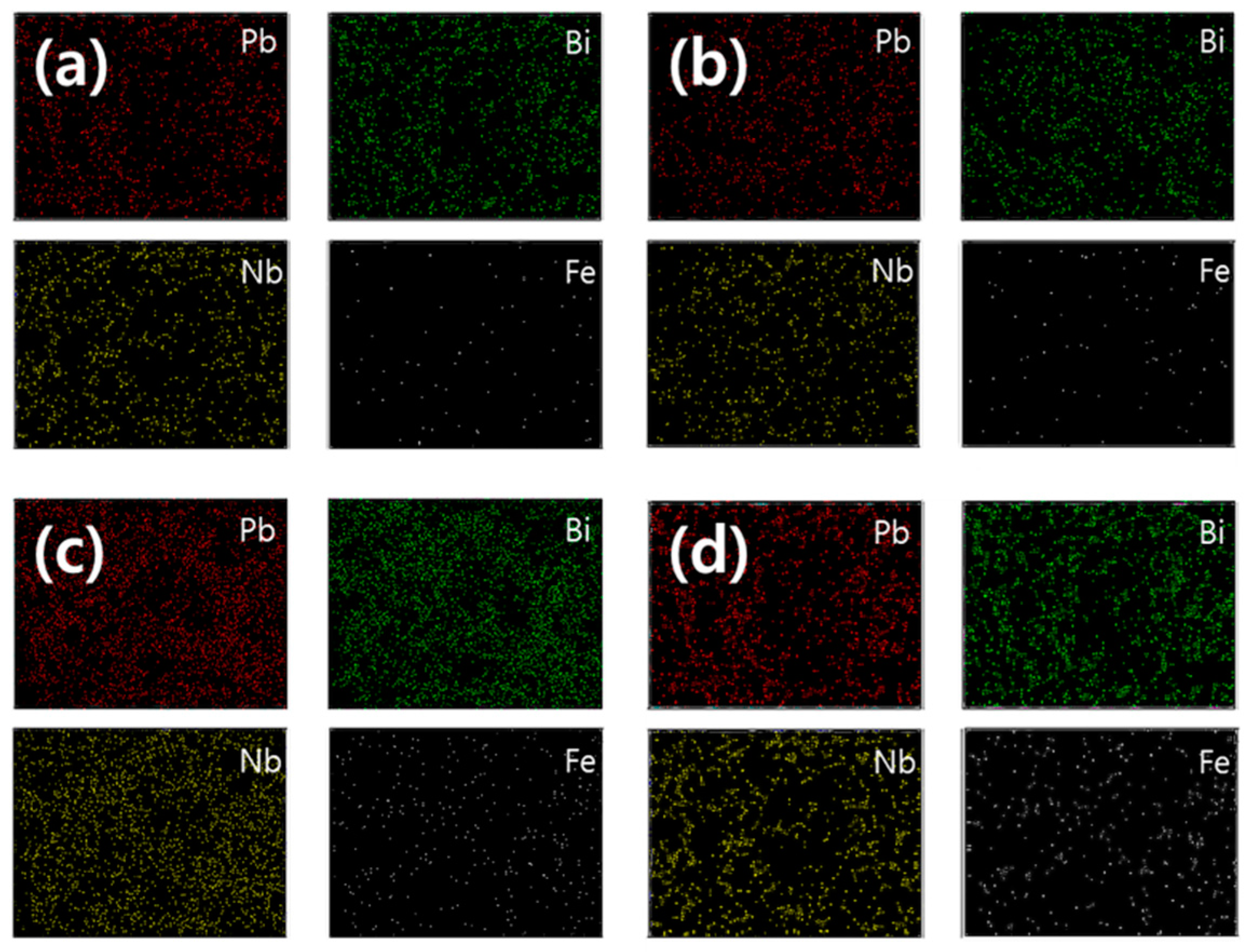
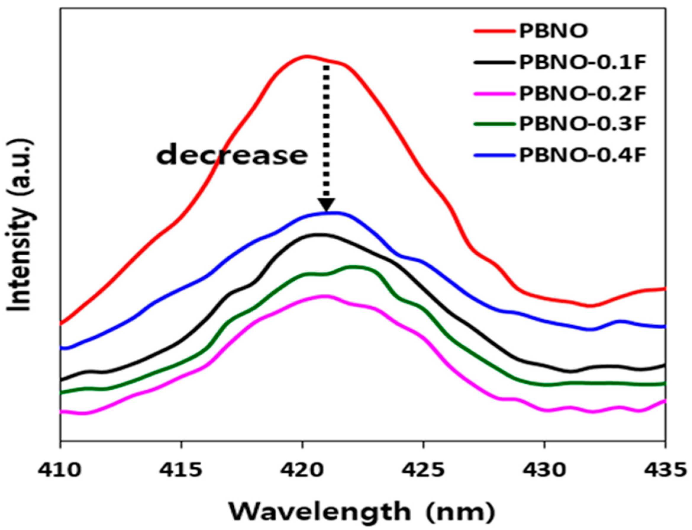
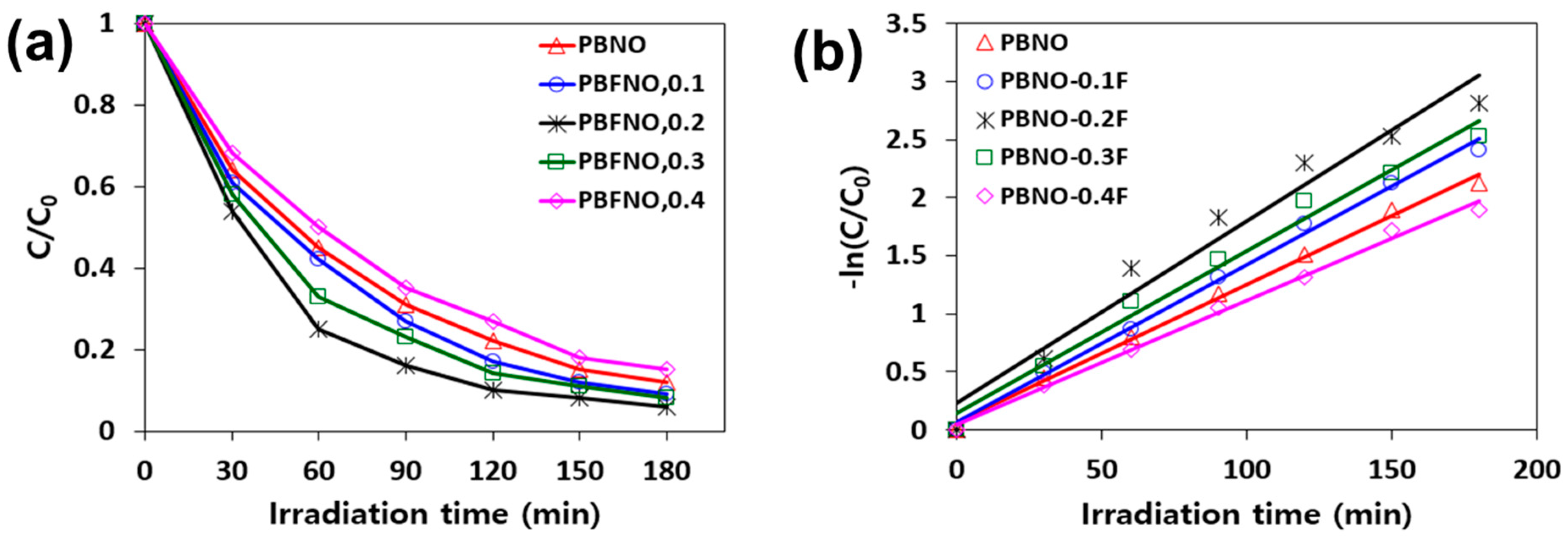
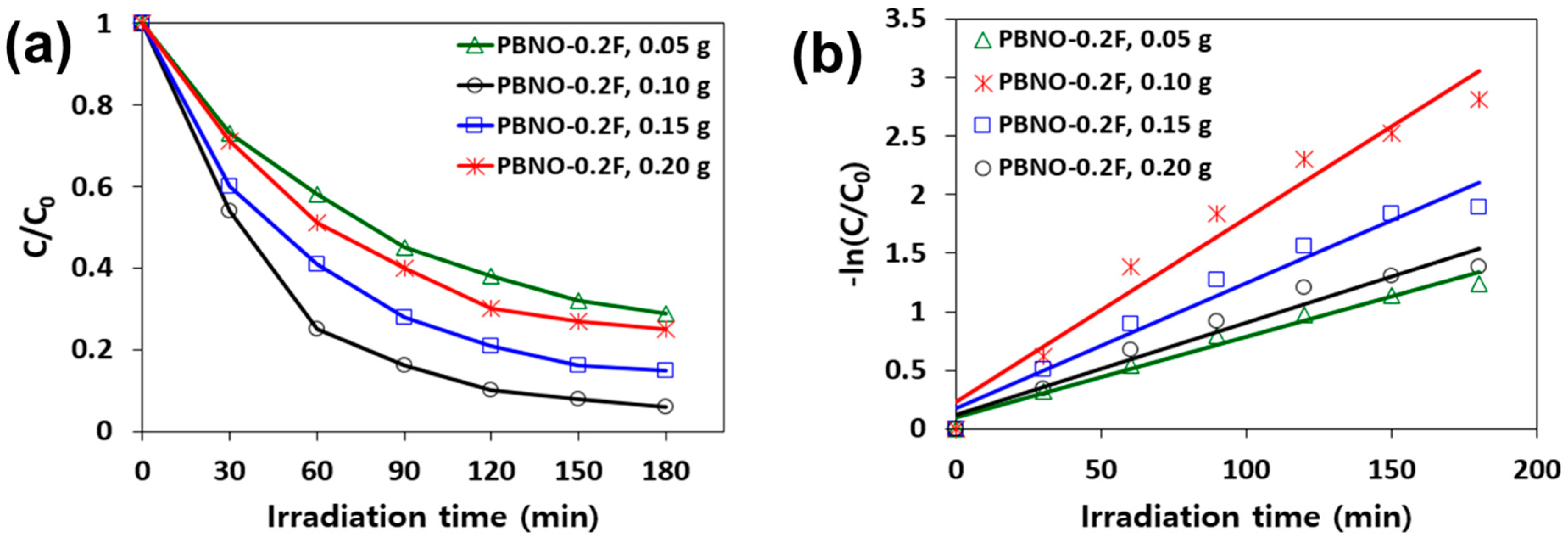
2. Results and Discussion

SEM-EDX analyses were conducted to find out the morphology, local composition, and elemental distribution on the bulk and surface of PBFNOs and PBNO. As shown in Figure 1 and Figure S1, the SEM images of the as-prepared PBFNO samples indicate good combination and connection among the particles. The particle sizes of the as-synthesized PBFNO samples decrease with the increased Fe doping ratio in the PBNO lattice because the ionic radius of doped Fe3+ (0.64 Å) is relatively small compared to the radius of Bi3+ (0.96 Å), resulting in little collapse of the crystal structure of the PBNO. The EDX elemental mapping images and atomic percentage are shown in Figure 2 and Table S1; they demonstrate a good distribution of the Pb, Bi, Fe, and Nb elements onto the surface of the as-prepared PBFNO photocatalysts. It can be clearly seen from Figure 2 that the amount of distributed Fe element on the surface of the as-synthesized PBFNOs increases as a function of the Fe doping ratio. Thus, the SEM-EDX analyses support the evidence that the PBFNO photocatalysts were successfully synthesized by a simple solid-state method.
An XRD measurement was carried out to verify the crystal structure of the as-synthesized PBNO and PBNO samples. As shown in Figure 3, the XRD pattern of the as-prepared PBNO sample is consistent with the standard XRD pattern (JCPDS No. 86–1333), which means that the PBNO was successfully synthesized by the simple solid-state reaction method used in the present study. The peak positions of the as-synthesized PBNO and PBFNO samples represent the single phase layered perovskite with a cubic crystal structure [24,31,33,34]. No impurity peaks were observed in the XRD patterns of the as-synthesized PBNO and PBFNO samples, indicating that the secondary phases, such as α-Fe, α-Fe2O3, γ-Fe2O3, Fe3O4, Pb-ferrite, Bi-ferrite, and Nb-ferrite, or other impurity phases did not form after calcination at 950 °C for 5 h [32,33,34]. In addition, as shown in the inset of Figure 3, the shift of the main diffraction peaks (115) of the perovskite toward higher 2θ values occurred. This corresponds to the peak broadening after Fe doping due to the small decrease in the lattice distance according to the substitution of the bigger ionic radius of Bi3+ (0.96 Å) by the smaller ionic radius of Fe3+ (0.64 Å) [32]. In other words, the doping Fe ions were incorporated into the structures of the PBNO and replaced the Bi ions, which caused the peak shifts and peak broadening. Furthermore, the data obtained from the XRD patterns of the samples have been used to determine its lattice parameters, unit cell volume, and crystallite size using the Debye-Scherrer method [35]. According to the Scherrer Equation, the wavelength of the X-rays are in the range between 0.01 nm to 10 nm. Hence, X-rays can easily penetrate the crystal structure of most materials in order to analyze its physical properties. The particle size is estimated by the Scherrer formula:
Dp = (0.94∙λ)/(β∙Cosθ)
where Dp = Average crystallite size, β = Line broadening in radians, θ = Bragg angle, λ = X-ray wavelength.
As shown in Table 1, the lattice parameters and unit-cell volume of the as-prepared PBNO sample decreases with the increasing Fe doping ratio because the lattice parameters and unit cell volume highly depend on the ionic radius of the substituting cation on the Bi-site. The substituting Fe ion, which has a smaller ionic radius compared to the Bi ion, caused the decrease in the lattice parameters and unit cell volume [32]. The crystalline size of the PBNO also gradually decreased when the Fe doping ratio increased, which is in agreement with the result of the SEM. This result means that the Fe dopants were successfully incorporated in the PBNO lattice during the synthesis process.
Figure 4 shows the UV-Vis absorbance spectra of the as-synthesized PBFNO samples with different Fe doping ratios. As a comparison, the spectrum of the pure PBNO sample was also examined, as shown in Figure 4. It can be clearly seen that the absorption edge for the pure PBNO is approximately 500 nm, while the PBFNO composites exhibited a strong absorption of the visible light and a significant red shift of the absorbance band edge with respect to the pure PBNO. It is notable that, as the Fe doping ratios increases, the as-prepared PBFNO composites remarkably increases its visible light absorbance, with a large red shift of the absorbance edge to the visible light region. It could be concluded that Fe doping into the PBNO lattice tunes the electronic structure and shifts the light absorbance edge to the more visible light region [36]. In addition, the results represent that the red shift of the absorption edge was brought about by the charge transfer (or electron transfer) between the d electrons of Fe and the valence or conduction bands of the PBNO [36]. Thus, Fe could produce a new electron state inside the electronic structure of the PBNO, which can catch the excited electrons from the valence band of the PBNO and suppress the charge carrier recombination. Based on these findings, we anticipated that the as-prepared PBFNO composites may present much better photocatalytic activity under visible light.
Furthermore, the band gap energies of the as-prepared PBNO and PBFNO samples were calculated by extrapolating the linear region of the Tauc plot of [F(R)E]2 versus photon energy (eV), combined with the Kubelka-Munk method [37,38,39,40]. It is well-known that, in a Tauc plot, an intersection point of a tangent line from a transformed curve to the photon energy axis is the band gap energy of a material [37,38,41]. Figure 5 shows that the estimated band gap energies of the as-prepared PBNO and PBFNOs with different Fe doping ratios at 0.1, 0.2, 0.3, and 0.4 were 2.64, 2.40, 2.32, 2.30, and 2.29 eV, respectively. This result reveals that the band gap energies of the as-synthesized PBFNO samples slightly decreases as the Fe doping ratios increase. These results verify that the Fe dopants could form an intermediate band between the valence and the conduction bands of the as-prepared PBNO sample, resulting in the production of a visible light sensitive photocatalyst [42]. Thus, it is easily possible to excite an electron, even under the visible light irradiation, leading to the improved visible light activity for the removal of MB in the water environment [32]. Meanwhile, the band gap energies of the as-synthesized PBFNO samples can be stabilized when the Fe doping ratio increases to above 0.2. This might be attributed to the maximum Fe doping ratio in the PBNO lattice [43]. The obtained result means that the proper amounts of doped Fe3+ ions in the PBNO lattice could leverage the photocatalytic activity of the PBNO photocatalyst.
The photoluminescence spectra of the as-synthesized PBNO and PBFNO samples were analyzed to deduce the recombination rate of the photoinduced electrons and holes, which significantly influence the photocatalytic activity. The excitation of all of the samples was carried out over the wavelength ranges of 260–480 nm, with a 250 nm excitation wavelength. It is well-known that a low intensity of photoluminescence presents a low recombination of photoinduced electrons and holes. On the contrary, a high intensity of photoluminescence means a high recombination rate and a decrease in the photocatalytic activity. It is clearly seen from Figure 6 that the intensity of the emission spectrum of the as-synthesized PBNO sample is relatively higher than the as-prepared PBFNO samples. This means that the photoinduced electrons and holes of the as-synthesized PBNO sample can be easily combined. In contrast, the relative intensity of the as-prepared PBFNO samples significantly decreased after the Fe doping in the PBNO lattice. This indicates that the recombination of the photoinduced electrons and holes was impeded by the Fe dopants, which acted as both electron and hole traps [32,42,44,45,46,47,48]. The surface defects of the as-synthesized PBNO sample caused by Fe doping could serve as friendly trap sites of the exited electrons or holes, resulting in restraining their recombination. In particular, among the as-prepared PBFNO samples, the PBNO-0.2F sample shows the lowest intensity of its photoluminescence line, which indicates that the 0.2 molar ratio of Fe can effectively suppress the electron-hole pair recombination. It is found that a proper Fe doping ratio can provide an electron acceptor, preventing a direct recombination of the photoinduced electrons and holes. When the Fe doping is relatively small, the recombination rate of the electron and hole pairs is higher due to the lack of sufficient traps. On the other hand, when the Fe doping is relatively high, both the light absorption and the generation of electron-hole pairs decrease. From these results, we can infer that the optimum ratio of the Fe dopant into the PBNO lattice could efficiently act as trap for the photoinduced electron and hole pairs to enhance the separation of charge carriers [49]. Therefore, the as-synthesized PBFNO-0.2F sample is suggested to be a more suitable photocatalyst in comparison to the other samples.
The photocatalytic activities of the as-synthesized PBNO and PBFNOs with different Fe doping ratios were evaluated based on the MB degradation in an aqueous solution under the visible light irradiation. Figure 7 shows that, for all of the samples, the degradation efficiency of the MB increases as the reaction progresses. It can be seen that most of the as-prepared PBFNO samples show higher photocatalytic activity than the pure PBNO sample, with the exception of the PBNO-0.4F sample. In particular, it was found that the 0.2 molar ratio of the Fe doped PBNO sample achieved the highest MB degradation performance, with 94.1% of efficiency, compared to other samples. However, the MB degradation efficiency steadily decreases when the addition of Fe dopants is above the 0.2 molar ratio. In particular, the PBNO-0.4F sample shows lower MB degradation efficiency than the pristine PBNO sample. The added Fe dopants, above the optimum molar ratio, may be attributed to acting as recombination centers through the quantum tunneling effect, rather than an intermediate band [41,50,51]. Furthermore, it has also been commonly reported that a photocatalyst with narrow band gap energy has less redox ability [52] because the theoretical background of the photocatalytic system is similar to the electrochemical cell. The relationship between the band gap energy and the redox potential can be explained according to the following equation (Equation (1)) [52]:
G = nFE
where ΔG is the free energy change of the redox process occurring in the system; n is the number of moles of electrons transferred involved in the redox process; F is the Faraday constant, E represents the band gap energy of a photocatalyst. This equation also implies that the narrowing of the band gap energy by Fe doping is not always favorable for the photocatalytic activity, indicating that there is an optimum doping ratio. In this study, the PBNO-0.4F sample, which has the narrowest band gap energy among the as-prepared samples, shows the lowest MB degradation efficiency. However, the PBNO-0.2F sample was found to be the optimum one for the MB degradation. The apparent rate constants derived from the pseudo first-order model evidently support the MB degradation efficiency of the as-prepared photocatalyst samples. As shown in Table 2, the highest value of the apparent rate constant was obtained by the as-prepared PBNO-0.2F sample (0.0156 min−1), which was 1.32 times higher than the pristine PBNO sample (0.0118 min−1). The coefficients of the determination (R2) were reasonably high (R2 = 0.9628–0.9954) to fit the pseudo first-order model. The possible pathway of MB degradation by the as-synthesized PBNO and PBFNO samples under the visible light is given by the equations below (Equations (2)–(8)) [3,18].
PB(F)NOs + hv (visible light) → e(cb) + h+(vb)
H2O + h+(vb) → OH + H+
O2 + e(cb) → O2−•
O2−• + H+ → HO2
2HO2 → H2O2 + O2
H2O2 → 2OH
OH + MB → 16CO2 + HCl + H2SO4 + 3HNO3 + 6H2O
Furthermore, the amount of photocatalyst was one of the important factors in the MB degradation under visible light irradiation. The PBNO-0.2F indicates the highest MB degradation efficiency among the samples, where the amount of photocatalyst used varied between 0.05 and 0.20 g in the 100 mL of the artificial MB solution. As shown in Figure 8, it was found that the MB degradation efficiency sharply increases when the amount of the PBNO-0.2F increases up to 0.1 g. It is noted that the number of active sites on the surface of the PBNO-0.2F could increase by increasing its amount. Many studies have reported the importance of O2 and H2O molecules absorbed on the PBNO surfaces to form hydroxyl radicals (OH) and/or superoxide anions (O2−•) [2,22]. However, the MB degradation efficiency gradually decreases with the increasing amount of the PBNO-0.2F, above 0.1 g. This might indicate that there are two possible reasons for the decrease in the MB degradation efficiency as to why the amount of photocatalysts increases above the optimum value. Firstly, the excessive amount of photocatalysts can cause light scattering through suspensions and, thus, the amount of the visible light could not sufficiently reach the obscured photocatalysts through the suspensions [2,5,8,53,54,55,56]. Secondly, the particle agglomeration by the excessive increase in the photocatalyst dose, which leads to the reduction in the specific surface area of the photocatalyst. This has a negative influence on the total amount of light absorption to the photocatalysts [2,57]. In addition, the agglomeration of particles causes the isolation of dopants far from the surface of the photocatalysts, resulting in a lowering of the charge transfer efficiency [52]. The highest value of the apparent rate constant was observed when the 0.10 g of PBNO-0.2F (0.0156 min−1) was used for MB degradation; this was 2.26 times higher than when the 0.05 g of PBNO-0.2F was used, as indicated in Table 3. The value of the apparent rate constant was completely in accordance with the MB degradation efficiency. Table 4 shows the catalytic activity of PBNO-0.2F with some other reported photocatalysts. It also confirms the superior performance of the present works.

3. Experimental Details

3.1. Synthesis

Lead(II) Oxide (PbO, >99%, ACS Reagent), Bismuth(III) Oxide (Bi2O3, >98.0%, purum), Iron(III) Oxide (Fe2O3, >99.995% Trace metals basis), and Niobium(V) Oxide (Nb2O5, 99.99%, Trace metal basis) were purchased from Sigma-aldrich, Korea. A simple solid-state reaction was applied to synthesize the aurivillius-phase PBFNOs perovskites. Certain amounts of stoichiometric mixtures of PbO, Bi2O3, Fe2O3, and Nb2O5, with the addition of a small amount of distilled water, were ground in a mortar for 30 min, then calcined by a muffle furnace at 950 °C for 5 h with 10 °C min−1 of heating rate to obtain the PBFNOs products. The various molar ratios of Bi and Fe (1.9:0.1, 1.8:0.2, 1.7:0.3, and 1.6:0.4) were applied to find out the effect of the Fe doping ratio. The prepared photocatalysts were named PBNO-0.1F, PBNO-0.2F, PBNO-0.3F, and PBNO-0.4F, respectively, where 0.1, 0.2, 0.3 and 0.4 represent the molar ratios of the Fe dopant in the PBNO. The as-prepared PBFNO perovskites were kept in the red bottles to prevent a photochemical reaction by external light sources before being used.

3.2. Materials Characterization

The scanning electron microscope (SEM, Hitachi, SU8020, Tokyo, Japan) was used to observe the surface morphologies of the as-prepared PBNO and PBFNO samples. The energy dispersive X-ray spectroscopy (EDX, Horiba, EMAX, Kyoto, Japan) connected to the SEM was used to obtain the elemental compositions and the elemental mapping images of the as-synthesized samples. The X-ray diffraction (XRD) patterns of the as-prepared PBNO and PBFNO samples were analyzed using a Philips X’Pert-MPD system equipped with a Cu Kα radiation (λ = 1.5405 Å) source and operated at a scan rate of 0.02° s−1 over the 2θ range of 20–80°. The light absorption abilities of the as-synthesized PBNO and PBFNO samples were measured by the UV-Vis-NIR (Agilent, Cary 5000, Santa Clara, CA, USA) over the wavelength range of 300–800 nm. The light reflectance of the as-prepared PBNO and PBFNO samples were obtained by using a UV-Vis spectrophotometer (Perkin Elmer, LAMBDA 650, Waltham, MA, USA), equipped with an integrating sphere over the wavelength range of 200–800 nm. The photoluminescence (PL) spectra of the as-synthesized PBNO and PBFNO samples were collected using a Spectrofluorometer (Jasco International, FP-8500ST, Tokyo, Japan), equipped with a 150 W Xenon lamp and a photomultiplier tube detector and operated at a scan rate of 600 nm min−1 over the wavelength ranges of 260–480 nm with 250 nm excitation wavelength. To calculate the MB concentration, the light absorbance spectra of the MB solutions before and after photocatalytic reaction were obtained using a UV-Vis spectrophotometer (Perkin Elmer, LAMBDA 650).

3.3. Photocatalytic Characterization

The MB degradation experiment was carried out to investigate the photocatalytic degradation efficiency of MB in the solution by the as-prepared PBNO and PBFNO samples under visible light irradiation. First, 100 mL of the artificial MB solution (10 ppm) and 0.1 g of the as-synthesized photocatalysts were magnetically stirred in a glass beaker at 720 rpm for 3 h. The 150 W Xenon lamp was applied as a light source to simulate the sunlight. The pH of the solution was fixed at pH 12, which showed the best MB degradation efficiency in the previous study by our group. Thus, the surface structure of the as-synthesized PBNO did not significantly affect the MB adsorption efficiency, while the pH of the alkaline solution plays a key role for enhancing the adsorption capacity [22] The MB solution samples were collected every 30 min to find out the MB degradation efficiency as a function of the reaction time. The MB degradation experiment was also conducted with different amounts of photocatalyst (0.05–0.20 g). The light absorbance spectra of the MB solution at 662 nm wavelength were utilized to calculate the MB degradation efficiency, which is generally assumed to be proportional to the MB concentration. According to the following equation, the MB degradation efficiency can be calculated (Equation (9)).
E   % =   C 0 C i C i × 100 =   A 0 A i A i × 100
where E is the MB degradation efficiency; C0 is the initial MB concentration; Ci is the final MB concentration; A0 is the initial light absorbance; Ai is the final light absorbance. The pseudo first-order kinetic model was applied to compare the photocatalytic degradation rate of MB in solution by the as-prepared PBNO and PBFNO samples. The general equation for the pseudo first-order model is also given below (Equation (10))
ln C t / C 0 = k t
where Ct is the MB concentration at the reaction time t (mg L−1); C0 is the MB concentration at the initial reaction time 0 (mg L−1); k is apparent rate constant (min−1).

4. Conclusions

The as-prepared PBNO and PBFNOs with various Fe doping ratios were successfully synthesized by a simple solid-state reaction for the degradation of MB in solution under visible light irradiation. The light absorption ability of the PBFNOs was enhanced with the increasing Fe doping ratio because the Fe dopants that exist in the PBNO lattice act as an intermediate band between the valence and conduction bands of the PBNO. The band gap energy of the PBFNOs remarkably decreased compared to the pristine PBNO, leading to easy charge transfer. This has a positive effect on the photocatalytic activity by generating highly reactive species, such as hydroxyl radicals (OH) and superoxide anions (O2−•). The intensity of the photoluminescence spectra was also dramatically reduced after Fe doping in the PBNO lattice because the Fe dopants act as photoinduced electron and hole traps, leading to a suppressed recombination rate. In this study, the optimum Fe doping ratio in the PBNO lattice was found to be a 0.2 molar ratio and showed the highest MB degradation efficiency. In addition, the 1 g L−1 of PBNO-0.2F was found to be the optimum amount for the degradation of 10 mg L−1 MB. The excessive amount of photocatalysts above the optimum value caused the scattering of irradiated light by suspensions and the agglomeration of photocatalyst particles incurring a reduction in the available surface area for the light harvest. This study verifies that the Fe dopants could enhance the photocatalytic activity of the PBNO by acting as intermediate band and electron-hole traps; thus, the PBFNOs showed a superior MB degradation performance, even under visible light irradiation.

Supplementary Materials

The following supporting information can be downloaded at: https://www.mdpi.com/article/10.3390/catal13020399/s1. Figure S1: SEM-EX images of as-synthesized non-doped PBNO. Table S1. SEM-EDX atomic percent results of as-synthesized PBNO and PBNO-0.2F.

Author Contributions

Y.G.: formal analysis, investigation, writing-original draft; M.K.: data curation, formal analysis, investigation; H.S.K.: conceptualization, writing-review and editing; D.-H.L.: conceptualization, project administration, writing-original draft, writing-review and editing. All authors have read and agreed to the published version of the manuscript.

Funding

This work was supported by the Technology Innovation Program (20005690, Development of three-dimensional metal structured De-NOx catalyst) funded by the Ministry of Trade, Industry and Energy (MOTIE, Korea).

Data Availability Statement

Data is contained within the article or Supplementary Materials.

Acknowledgments

We thank Mino Woo for his contribution to revise the manuscript during the review process.

Conflicts of Interest

The authors declare no conflict of interest.

References

  1. Nguyen-Le, M.T.; Lee, B.K. High temperature synthesis of interfacial functionalized carboxylate mesoporous TiO2 for effective adsorption of cationic dyes. Chem. Eng. J. 2015, 281, 20–33. [Google Scholar] [CrossRef]
  2. Hassena, H. Photocatalytic Degradation of Methylene Blue by Using Al2O3/Fe2O3 Nano Composite under Visible Light. Mod. Chem. Appl. 2016, 4, 3–7. [Google Scholar] [CrossRef]
  3. Reddy, J.R.; Kurra, S.; Guje, R.; Palla, S.; Veldurthi, N.K.; Ravi, G.; Vithal, M. Photocatalytic degradation of methylene blue on nitrogen doped layered perovskites, CsM2Nb3O10 (M=Ba and Sr). Ceram. Int. 2015, 41, 2869–2875. [Google Scholar] [CrossRef]
  4. Tabari, T.; Tavakkoli, H.; Zargaran, P.; Beiknejad, D. Fabrication of perovskite-type oxide BaPbO3 nanoparticles and their efficiency in photodegradation of methylene blue. S. Afr. J. Chem. 2012, 65, 239–244. [Google Scholar]
  5. Ansari, R.; Mosayebzadeh, Z. Removal of basic dye methylene blue from aqueous solutions using sawdust and sawdust coated with polypyrrole. J. Iran. Chem. Soc. 2010, 7, 339–350. [Google Scholar] [CrossRef]
  6. Dariani, R.S.; Esmaeili, A.; Mortezaali, A.; Dehghanpour, S. Photocatalytic reaction and degradation of methylene blue on TiO2 nano-sized particles. Optik 2016, 127, 7143–7154. [Google Scholar] [CrossRef]
  7. Christie, R. Colour Chemistry; RSC Paperbacks; The Royal Society of Chemistry: Cambridge, UK, 2001; ISBN 978-0-85404-573-0. [Google Scholar]
  8. Kasiri, M.B.; Khataee, A.R. Photooxidative decolorization of two organic dyes with different chemical structures by UV/H2O2 process: Experimental design. Desalination 2011, 270, 151–159. [Google Scholar] [CrossRef]
  9. Yi, F.; Chen, S.; Yuan, C. Effect of activated carbon fiber anode structure and electrolysis conditions on electrochemical degradation of dye wastewater. J. Hazard. Mater. 2008, 157, 79–87. [Google Scholar] [CrossRef]
  10. Labanda, J.; Sabaté, J.; Llorens, J. Modeling of the dynamic adsorption of an anionic dye through ion-exchange membrane adsorber. J. Memb. Sci. 2009, 340, 234–240. [Google Scholar] [CrossRef]
  11. Tehrani-Bagha, A.R.; Mahmoodi, N.M.; Menger, F.M. Degradation of a persistent organic dye from colored textile wastewater by ozonation. Desalination 2010, 260, 34–38. [Google Scholar] [CrossRef]
  12. Chatterjee, S.; Lee, D.S.; Lee, M.W.; Woo, S.H. Congo red adsorption from aqueous solutions by using chitosan hydrogel beads impregnated with nonionic or anionic surfactant. Bioresour. Technol. 2009, 100, 3862–3868. [Google Scholar] [CrossRef]
  13. Xia, C.; Jing, Y.; Jia, Y.; Yue, D.; Ma, J.; Yin, X. Adsorption properties of congo red from aqueous solution on modified hectorite: Kinetic and thermodynamic studies. Desalination 2011, 265, 81–87. [Google Scholar] [CrossRef]
  14. Ahmad, A.L.; Puasa, S.W. Reactive dyes decolourization from an aqueous solution by combined coagulation/micellar-enhanced ultrafiltration process. Chem. Eng. J. 2007, 132, 257–265. [Google Scholar] [CrossRef]
  15. Slokar, Y.M.; Majcen Le Marechal, A. Methods of decoloration of textile wastewaters. Dye. Pigment. 1998, 37, 335–356. [Google Scholar] [CrossRef]
  16. Wang, L.; Zhang, J.; Wang, A. Removal of methylene blue from aqueous solution using chitosan-g-poly(acrylic acid)/montmorillonite superadsorbent nanocomposite. Colloids Surf. A Physicochem. Eng. Asp. 2008, 322, 47–53. [Google Scholar] [CrossRef]
  17. Torres-Martínez, L.M.; Cruz-López, A.; Juárez-Ramírez, I.; Meza-de la Rosa, M.E. Methylene blue degradation by NaTaO3 sol-gel doped with Sm and La. J. Hazard. Mater. 2009, 165, 774–779. [Google Scholar] [CrossRef] [PubMed]
  18. Houas, A.; Lachheb, H.; Ksibi, M.; Elaloui, E.; Guillard, C.; Herrmann, J.M. Photocatalytic degradation pathway of methylene blue in water. Appl. Catal. B Environ. 2001, 31, 145–157. [Google Scholar] [CrossRef]
  19. Belsousov, A.S.; Suleimanov, E.V.; Parkhacheva, A.A.; Fukina, D.G.; Koryagin, A.V.; Titaev, D.N.; Lazarev, M.A. Synthesis and Characterization of Bi2WxMo1−xO6 Solid Solutions and Their Application in Photocatalytic Desulfurization under Visible Light. Processes 2022, 10, 789. [Google Scholar] [CrossRef]
  20. Serverino, D.; Junqueira, H.C.; Gugliotti, M.; Gabrielli, D.S.; Baptista, M.S. Influence of negatively charged interfaces on the ground and excited state properties of methylene blue. J. Photochem. Photobiol. A Chem. 2003, 77, 459–468. [Google Scholar] [CrossRef]
  21. Gyu Kim, H.; Won Hwang, D.; Sung Lee, J. An Undoped, Single-Phase Oxide Photocatalyst Working under Visible Light. J. Am. Chem. Soc. 2004, 126, 8912–8913. [Google Scholar] [CrossRef]
  22. Lee, C.-H.; Kim, H.G.; Gu, Y.; Lim, D.-H. A Study of Photocatalytic Degradation of Methylene Blue in Aqueous Solution Using Perovskite Structured PbBi2Nb2O9. Nanosci. Nanotechnol. Lett. 2018, 10, 1179–1186. [Google Scholar] [CrossRef]
  23. Belousov, A.S.; Suleimanov, E.V.; Fukina, D.G. Pyrochlore oxides as visible light-responsive photocatalysts. New J. Chem. 2021, 45, 22531–22558. [Google Scholar] [CrossRef]
  24. Wei, K.; Faraj, Y.; Yao, G.; Xie, R.; Lai, B. Strategies for improving perovskite photocatalysts reactivity for organic pollutants degradation: A review on recent progress. Chem. Eng. J. 2021, 414, 128783. [Google Scholar] [CrossRef]
  25. Maniyazagan, M.; Hussain, M.; Kang, W.S.; Kim, S.J. Hierarchical Sr-Bi2WO6 photocatalyst for the degradation of 4-nitrophenol and methylene blue. J. Ind. Eng. Chem. 2022, 110, 168–177. [Google Scholar] [CrossRef]
  26. Kendall, K.R.; Navas, C.; Thomas, J.K.; Zur Loye, H.C. Recent developments in oxide ion conductors: Aurivillius phases. Chem. Mater. 1996, 8, 642–649. [Google Scholar] [CrossRef]
  27. Aurivillius, B. Mixed Bismuth Oxides with Layer Lattices 1. The Structure Type of CaNb2Bi2O9. Ark. Kemi 1949, 1, 463–480. [Google Scholar]
  28. Aurivillius, B. Mixed bismuth oxides with layer lattices II. Structure of Bi4Ti3O12. Ark. Kemi 1949, 1, 499–512. [Google Scholar]
  29. Aurivillius, B. Mixed oxides with layer lattices III. Structure of BaBi4Ti4O15. Ark. Kemi 1950, 39, 5711–5715. [Google Scholar]
  30. Rae, A.D.; Thompson, J.G.; Withers, R.L. Structure refinement of commensurately modulated bismuth strontium tantalate, Bi2SrTa2O9. Acta Crystallogr. Sect. B 1992, 48, 418–428. [Google Scholar] [CrossRef]
  31. Singh, C.; Wagle, A.; Rakesh, M. Doped LaCoO3 perovskite with Fe: A catalyst with potential antibacterial activity. Vacuum 2017, 146, 468–473. [Google Scholar] [CrossRef]
  32. Moura, K.F.; Chantelle, L.; Rosendo, D.; Longo, E.; Dos Santos, I.M.G. Effect of Fe3+ doping in the photocatalytic properties of BaSnO3 perovskite. Mater. Res. 2017, 20, 317–324. [Google Scholar] [CrossRef]
  33. Kim, J.; Jun, A.; Shin, J.; Kim, G. Effect of Fe doping on layered GdBa0.5Sr0.5Co2O5+δ perovskite cathodes for intermediate temperature solid oxide fuel cells. J. Am. Ceram. Soc. 2014, 97, 651–656. [Google Scholar] [CrossRef]
  34. Bousslama, W.; Elhouichet, H.; Férid, M. Enhanced photocatalytic activity of Fe doped ZnO nanocrystals under sunlight irradiation. Optik 2017, 134, 88–98. [Google Scholar] [CrossRef]
  35. Burton, A.W.; Ong, K.; Rea, T.; Chan, I.Y. On the estimation of average crystallite size of zeolites from the Scherrer equation: A critical evaluation of its application to zeolites with one-dimensional pore systems. Microporous Mesoporous Mater. 2009, 117, 75–90. [Google Scholar] [CrossRef]
  36. Teh, C.M.; Mohamed, A.R. Roles of titanium dioxide and ion-doped titanium dioxide on photocatalytic degradation of organic pollutants (phenolic compounds and dyes) in aqueous solutions: A review. J. Alloys Compd. 2011, 509, 1648–1660. [Google Scholar] [CrossRef]
  37. Pham, T.D.; Lee, B.K. Novel capture and photocatalytic conversion of CO2 into solar fuels by metals co-doped TiO2 deposited on PU under visible light. Appl. Catal. A Gen. 2017, 529, 40–48. [Google Scholar] [CrossRef]
  38. Pham, T.D.; Lee, B.K. Novel photocatalytic activity of Cu@V co-doped TiO2/PU for CO2 reduction with H2O vapor to produce solar fuels under visible light. J. Catal. 2017, 345, 87–95. [Google Scholar] [CrossRef]
  39. Laokiat, L.; Khemthong, P.; Grisdanurak, N.; Sreearunothai, P.; Pattanasiriwisawa, W.; Klysubun, W. Photocatalytic degradation of benzene, toluene, ethylbenzene, and xylene (BTEX) using transition metal-doped titanium dioxide immobilized on fiberglass cloth. Korean J. Chem. Eng. 2012, 29, 377–383. [Google Scholar] [CrossRef]
  40. Tauc, J.; Grigorovici, R.; Vancu, A. Optical Properties and Electronic Structure of Amorphous Germanium. Phys. Status Solidi 1966, 15, 627–637. [Google Scholar] [CrossRef]
  41. Zhou, M.; Yu, J.; Cheng, B. Effects of Fe-doping on the photocatalytic activity of mesoporous TiO2 powders prepared by an ultrasonic method. J. Hazard. Mater. 2006, 137, 1838–1847. [Google Scholar] [CrossRef]
  42. Garza-Arévalo, J.I.; García-Montes, I.; Reyes, M.H.; Guzmán-Mar, J.L.; Rodríguez-González, V.; Reyes, L.H. Fe doped TiO2 photocatalyst for the removal of As(III) under visible radiation and its potential application on the treatment of As-contaminated groundwater. Mater. Res. Bull. 2016, 73, 145–152. [Google Scholar] [CrossRef]
  43. Nguyen, T.D.C.; Nguyen, T.P.L.C.; Mai, H.T.T.; Dao, V.D.; Nguyen, M.P.; Nguyen, V.N. Novel photocatalytic conversion of CO2 by vanadium-doped tantalum nitride for valuable solar fuel production. J. Catal. 2017, 352, 67–74. [Google Scholar] [CrossRef]
  44. Pham, T.D.; Lee, B.K.; Lee, C.H. The advanced removal of benzene from aerosols by photocatalytic oxidation and adsorption of Cu-TiO2/PU under visible light irradiation. Appl. Catal. B Environ. 2016, 182, 172–183. [Google Scholar] [CrossRef]
  45. Sood, S.; Umar, A.; Mehta, S.K.; Kansal, S.K. Highly effective Fe-doped TiO2 nanoparticles photocatalysts for visible-light driven photocatalytic degradation of toxic organic compounds. J. Colloid Interface Sci. 2015, 450, 213–223. [Google Scholar] [CrossRef]
  46. Ma, J.; He, H.; Liu, F. Effect of Fe on the photocatalytic removal of NOx over visible light responsive Fe/TiO2 catalysts. Appl. Catal. B Environ. 2015, 179, 21–28. [Google Scholar] [CrossRef]
  47. Tian, F.; Wu, Z.; Tong, Y.; Wu, Z.; Cravotto, G. Microwave-Assisted Synthesis of Carbon-Based (N, Fe)-Codoped TiO2 for the Photocatalytic Degradation of Formaldehyde. Nanoscale Res. Lett. 2015, 10, 1–2. [Google Scholar] [CrossRef]
  48. Li, Z.; Shen, W.; He, W.; Zu, X. Effect of Fe-doped TiO2 nanoparticle derived from modified hydrothermal process on the photocatalytic degradation performance on methylene blue. J. Hazard. Mater. 2008, 155, 590–594. [Google Scholar] [CrossRef]
  49. Fuerte, A.; Hernández-Alonso, M.D.; Maira, A.J.; Martínez-Arias, A.; Fernández-García, M.; Conesa, J.C.; Soria, J. Visible light-activated nanosized doped-TiO2 photocatalysts. Catal. Commun. 2001, 2718–2719. [Google Scholar] [CrossRef]
  50. Choi, W.; Termin, A.; Hoffmann, M.R. The role of metal ion dopants in quantum-sized TiO2: Correlation between photoreactivity and charge carrier recombination dynamics. J. Phys. Chem. 1994, 98, 13669–13679. [Google Scholar] [CrossRef]
  51. Zhang, Z.; Wang, C.C.; Zakaria, R.; Ying, J.Y. Role of particle size in nanocrystalline TiO2-based photocatalysts. J. Phys. Chem. B 1998, 102, 10871–10878. [Google Scholar] [CrossRef]
  52. Yu, J.C.; Zhang, L.; Yu, J. Direct Sonochemical Preparation and Characterization of Highly Active Mesoporous TiO2 with a Bicrystalline Framework. Chem. Mater. 2002, 14, 4647–4653. [Google Scholar] [CrossRef]
  53. Byrappa, K.; Subramani, A.K.; Ananda, S.; Lokanatha Rai, K.M.; Dinesh, R.; Yoshimura, M. Photocatalytic degradation of rhodamine B dye using hydrothermally synthesized ZnO. Bull. Mater. Sci. 2006, 29, 433–438. [Google Scholar] [CrossRef]
  54. Chakrabarti, S.; Dutta, B.K. Photocatalytic degradation of model textile dyes in wastewater using ZnO as semiconductor catalyst. J. Hazard. Mater. 2004, 112, 269–278. [Google Scholar] [CrossRef] [PubMed]
  55. Huang, M.; Xu, C.; Wu, Z.; Huang, Y.; Lin, J.; Wu, J. Photocatalytic discolorization of methyl orange solution by Pt modified TiO2 loaded on natural zeolite. Dye. Pigment. 2008, 77, 327–334. [Google Scholar] [CrossRef]
  56. Shen, H.; Xue, T.; Wang, Y.; Cao, G.; Lu, Y.; Fang, G. Photocatalytic property of perovskite LaFeO3 synthesized by sol-gel process and vacuum microwave calcination. Mater. Res. Bull. 2016, 84, 15–24. [Google Scholar] [CrossRef]
  57. Bresolin, B.M.; Hammouda, S.B.; Sillanpaa, M. Methylammonium iodo bismuthate perovskite (CH3NH3)3Bi2I9 as new effective visible light-responsive photocatalyst for degradation of environment pollutants. J. Photochem. Photobio. A Chem. 2019, 379, 116–126. [Google Scholar] [CrossRef]
Figure 1. SEM images of as-synthesized (a) PBNO-0.1F, (b) PBNO-0.2F, (c) PBNO-0.3F, and (d) PBNO-0.4F samples.
Catalysts 13 00399 g001
Figure 2. EDX elemental mapping images of as-synthesized (a) PBNO-0.1F, (b) PBNO-0.2F, (c) PBNO-0.3F, and (d) PBNO-0.4F samples.
Catalysts 13 00399 g002
Figure 3. XRD patterns of as-prepared PBNO and PBFNO samples by solid-state reaction.
Catalysts 13 00399 g003
Figure 4. UV-Vis absorbance spectra of as-prepared PBNO and PBFNO samples with different Fe doping ratios.
Catalysts 13 00399 g004
Figure 5. Tauc plot of as-prepared PBNO and PBFNO samples with different Fe doping ratios.
Catalysts 13 00399 g005
Figure 6. Photoluminescence spectra of as-prepared PBNO and PBFNO samples with different Fe doping ratios.
Catalysts 13 00399 g006
Figure 7. The results of MB degradation by as-prepared PBNO and PBFNO samples with different Fe doping ratio. (a) MB degradation trend as a function of reaction time (b) Pseudo first-order kinetic plot for MB degradation.
Catalysts 13 00399 g007
Figure 8. The effect of the PBNO-0.2F amounts on the MB degradation. (a) MB degradation trend as a function of reaction time (b) Pseudo first-order kinetic plot for MB degradation.
Catalysts 13 00399 g008
Table 1. The calculated lattice parameters, unit cell volume, and crystalline sizes of as-prepared PBNO and PBFNO samples.
CatalystLattice ParametersUnit Cell
Volume
Crystalline Size
a (Å)b (Å)c (Å)(Å3)(nm)
PBNO5492550325,550772.1828.57
PBNO-0.1F5473548525,452764.0524.49
PBNO-0.2F5465547925,405760.7021.44
PBNO-0.3F5460547625,380758.8419.06
PBNO-0.4F5458547525,368758.6217.16
Table 2. The values of k and R2 for the pseudo first-order kinetic model at each given Fe doping ratio (amount of photocatalyst = 0.1 g, volume of MB solution = 100 mL, concentration of MB solution = 0.1 mg L−1, pH = 12, reaction time = 180 min).
Catalystsk (min−1)R2
PBNO0.01180.9954
PBNO-0.1F0.01340.9953
PBNO-0.2F0.01560.9628
PBNO-0.3F0.01400.9837
PBNO-0.4F0.01050.9951
Table 3. The values of k and R2 for the pseudo first-order kinetic model at each given amount of photocatalyst (type of photocatalyst = PBNO-0.2F, volume of MB solution = 100 mL, concentration of MB solution = 0.1 mg L−1, pH = 12, reaction time = 180 min).
Amount of PBNO-0.2Fk (min−1)R2
0.05 g0.00690.9762
0.10 g0.01560.9628
0.15 g0.01050.9636
0.20 g0.00770.9567
Table 4. Comparison of the photo catalytic potential of the PBNO-0.2F with previous reported photo catalysts.
CatalystCatalyst Amount (g)Volume of MB Solution (mL)Concentration
of MB Solution (mg/L)
pHApparent Rate Constant
(K, min−1)
R2Ref.
TiO20.11000.1120.00730.9965[22]
PBNO0.11000.1120.01230.9984[22]
PBNO-0.1F0.11000.1120.01340.9954This work
PBNO-0.2F0.11000.1120.01560.9628This work
PBNO-0.3F0.11000.1120.01400.9837This work
MW-700–30500.050.00910.9978[56]
CC-700500.050.00800.9990[56]
BMI0.051000.020.01290.9698[57]
Disclaimer/Publisher’s Note: The statements, opinions and data contained in all publications are solely those of the individual author(s) and contributor(s) and not of MDPI and/or the editor(s). MDPI and/or the editor(s) disclaim responsibility for any injury to people or property resulting from any ideas, methods, instructions or products referred to in the content.

