Article

Ultrasonic Preparation of PN for the Photodegradation of 17β-Estradiol in Water and Biotoxicity Assessment of 17β-Estradiol after Degradation

1
Laboratory of Ministry of Education for Coastal and Wetland Ecosystems, College of the Environment and Ecology, Xiamen University, Xiamen 361102, China
2
Department of Environmental Engineering, National I-Lan University, Yilan 260007, Taiwan
*
Authors to whom correspondence should be addressed.
Catalysts 2023, 13(2), 332; https://doi.org/10.3390/catal13020332
Submission received: 28 December 2022 / Revised: 27 January 2023 / Accepted: 28 January 2023 / Published: 2 February 2023

Abstract

:
This study prepares a novel phosphorene (PN) and loads it onto TiO2 to fabricate PN-TiO2 and effectively photodegrade the hydrophobic environmental hormone 17β-estradiol in aqueous solutions. The effect of the PN on degradation efficiency is systematically investigated. It is observed that the doping of TiO2 with PN significantly enhances its photocatalytic and adsorption properties compared with that in the absence of PN; that is, the addition improves the adsorption capability of the composite. The optimal PN weight content is found to be 0.5%. The performance of the PN-TiO2 photocatalyst in degrading E2 is around 67.5%. However, its photodegradation efficiency gradually decreases when the PN content is further increased. This optimal PN content directly suggests synergistic interactions affecting the photodegrading efficiency. Compared with other PN-based photocatalysts mentioned in the literature, this PN-based material possesses striking advantages, such as higher energy efficiency, greater removal capacity, and superior cost-effectiveness. Further, the decrease in the biotoxicity of the water after treatment is evident in observing the development of zebrafish embryos. The studies of the catalyst performed on the zebrafish show that it results in a higher mortality rate at 96 h with a superior hatching rate and healthy fish development. In summary, the prepared PN-based materials exhibited promising photocatalytic capabilities for the removal and biotoxicity reduction of 17β-estradiol in aqueous solutions.

Graphical Abstract

1. Introduction

For decades, environmental hormones (EHs) have been used extensively to augment agricultural production for exponentially growing population needs. However, although these hormones provide some benefits, they inevitably pose a serious threat to human and wildlife health. In fact, some studies have suggested that EHs have a negative impact on the reproductive systems, nervous systems, and immune systems of organisms, leading to slow and malformed development, decreased sperm counts, poor sperm motility, and increased cancer risks [1,2,3].
Regarding environmental hormones, 17β-estradiol (E2) is a typical steroid estrogen used significantly for the reproduction of aquatic organisms even at extremely low concentrations. Biegel et al. [4] reported that this estrogen caused the stripes of male crocodiles to be transformed to that of female crocodiles as well as causing the crocodiles to exhibit female-like reproductive behavior. Diamante [5] also observed that zebrafish embryo underwent some changes, including curved body axis, yolk-sac edema, and pericardial edema, after treatment with E2. Angus [6] reported that high dosages of E2 led to the death of juvenile fishes in water bodies. Consequently, considerable attention has been paid to the development of techniques for the removal of E2, e.g., biodegradation [7,8,9], ion exchange [10,11], adsorption [12,13], and photocatalysis [14,15].
Although biodegradation could facilitate the decomposition of E2 into inorganic matter to some extent, the applications of biodegradation are still limited due to the long period of degradation and the strict conditions for bacterial growth. Bimetal-organic framework-derived nanotubes and cellulose aerogels have also been studied as photocatalysts for peroxymonosulfate activation [16]. By contrast, ion exchange and adsorption technologies are incapable of degrading E2.
Compared with these methods, photocatalysis is popularly regarded as the most appropriate method for E2 removal due to its high removal efficiency, shorter degradation period, and decomposable intermediates. In the photoreaction process, light irradiates the semiconductor to cause the excitation of electrons from the valence band (VB) to the conduction band (CB), creating positively charged holes (h+) and negatively charged electrons (e) available for photochemistry. This process also accelerates the photocatalysis process, where a catalyst promotes secondary photoreactions due to the formation of the strong oxidizing capabilities of holes and the reduction potentials of electrons [16].
Among typical catalysts (e.g., TiO2, CeO2, ZnO, SnO2), TiO2 is widely used in the photocatalysis process as it is a chemically stable, low-cost, and nontoxic material with promising reusability [17]. Recently, significant attention has been paid to the heterogeneous doping of TiO2 to enhance adsorption under visible light irradiation by decreasing its band gap and preventing the recombination of electrons and holes. However, several investigations have utilized expensive heavy metals (e.g., Au, Ag, and Pt) as a dopant [18,19], increasing the overall cost and pollution risk. Consequently, an environmentally friendly and biocompatible substitute [20], PN-TiO2, has been proposed for the photo-decomposition of E2.
Regarding novel materials, phosphorene (PN) is a novel two-dimensional (2D) material with a high current on/off ratio (~104–105) and high electron mobility (~200–1000 cm2V−1s−1) at room temperature. [20,21]. Thus, it can be used to dope TiO2, to enhance the excitation of electrons from VB to CB, and inhibit electron-hole recombination, leading to the increased generation of hydroxyl radicals (·OH). Furthermore, Wang et al. [22] reported that ·OH and 1O2 can be generated when PN is irradiated with ultraviolet (UV) and visible light, respectively. This strongly supports the notion of PN as an electrochemically feasible photocatalyst. Thus, the photocatalytic performance of TiO2 may be improved if it is doped with PN. In addition, PN possesses a large specific surface area [23] that aids the adsorption of contaminants onto reactive sites at the surface of TiO2, significantly promoting the removal of E2.
Previous studies have attempted to employ TiO2 for the photodegradation of E2. It was found that degradation efficiency increased with increasing TiO2 concentration. The highest degradation efficiency could be achieved as the TiO2 concentration rose to 0.5 mg ml−1. However, the degradation efficiency would decrease as the TiO2 concentration ranged from 0.5 to 1 mg ml−1 [24]. Another study achieved 20% removal of E2 at just 60 min of contact time using a TiO2 photocatalyst compared to a ZnO photocatalyst [24]. Very few studies have been conducted to explore the capabilities of a TiO2 photocatalyst for E2 degradation, hence the need for further studies.
In this study, PN is fabricated to mix with various amounts (0.5% weight, 1.0% weight, and 3.0% weight) of PN and TiO2 using microwave heating to synthesize PN-TiO2 composites (e.g., 0.5% PN-TiO2, 1.0% PN-TiO2 and 3.0% PN-TiO2). The degradation efficiencies of these composites are investigated to quantify the performance of E2 elimination. Although several studies [14,15,24,25,26] have investigated the degradation efficiency of TiO2 or TiO2-based composites, the removal capacities reported in these studies were still low and operation inevitably required strong illumination. In this study, only an 8 W UV light source is used to demonstrate the treatability of effective photocatalysis with cost and energy-effectiveness. This study exhibits the most representative PN-based hybrid for the elimination of hydrophobic and refractory pollutants in aqueous solutions [27,28,29,30,31].

2. Results

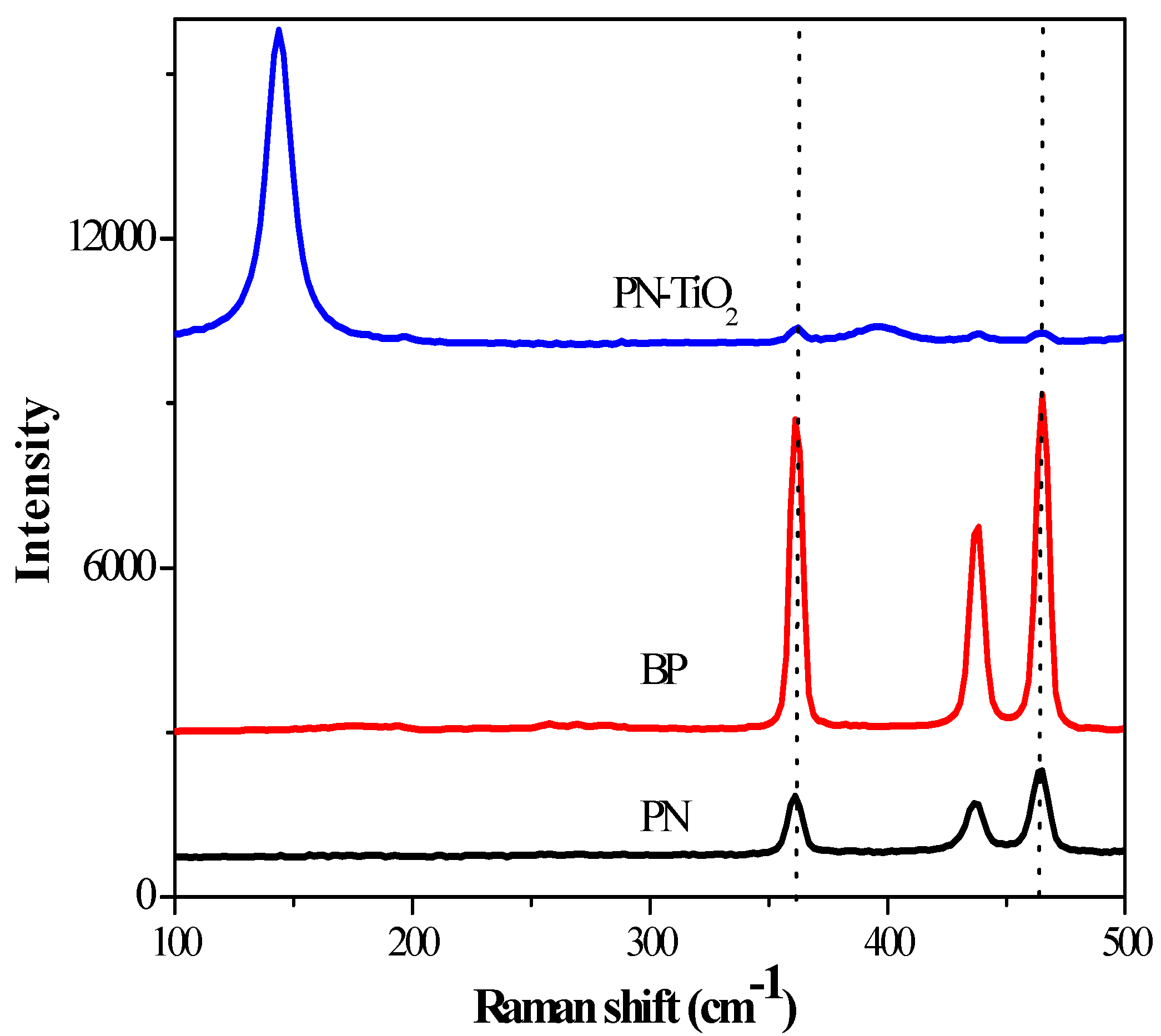
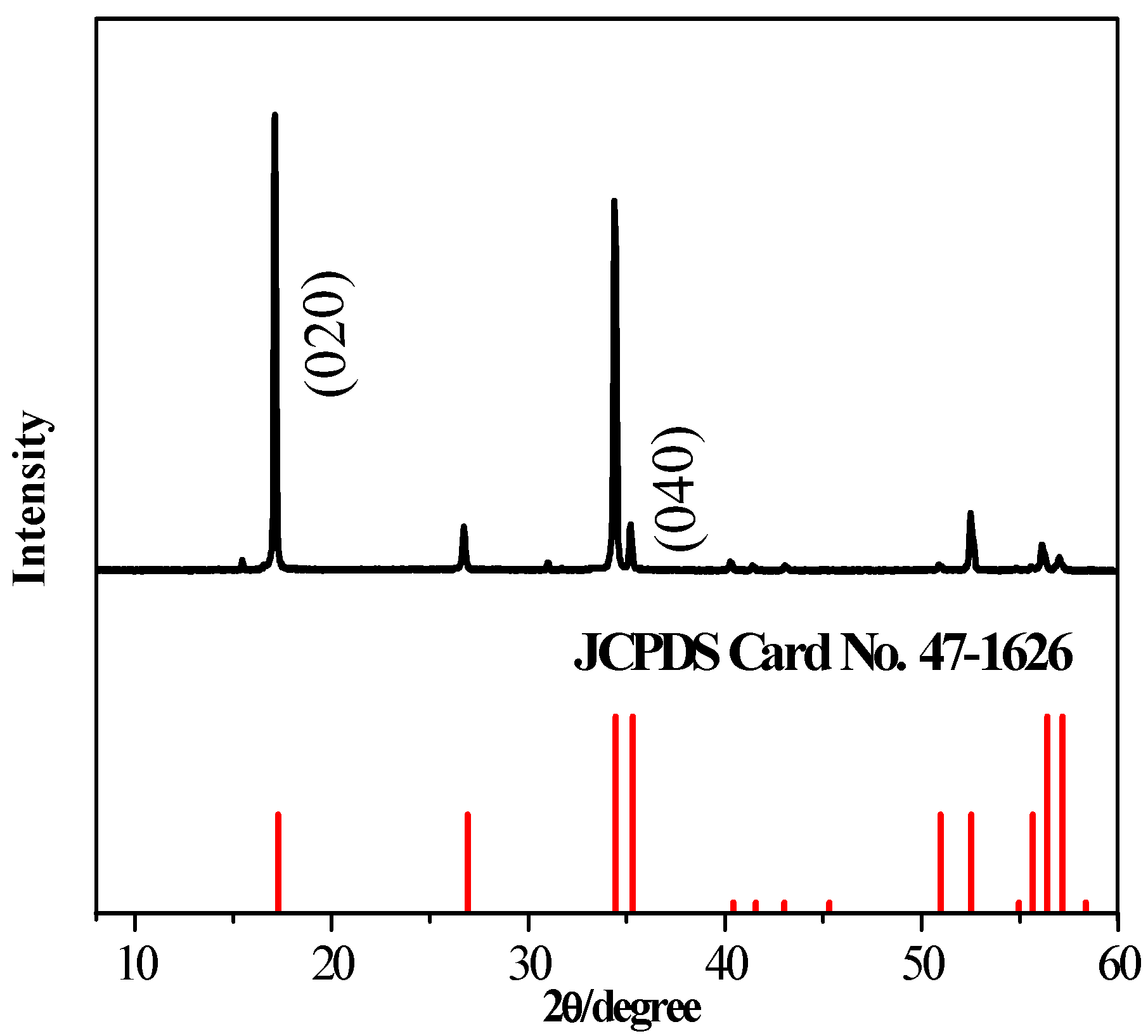
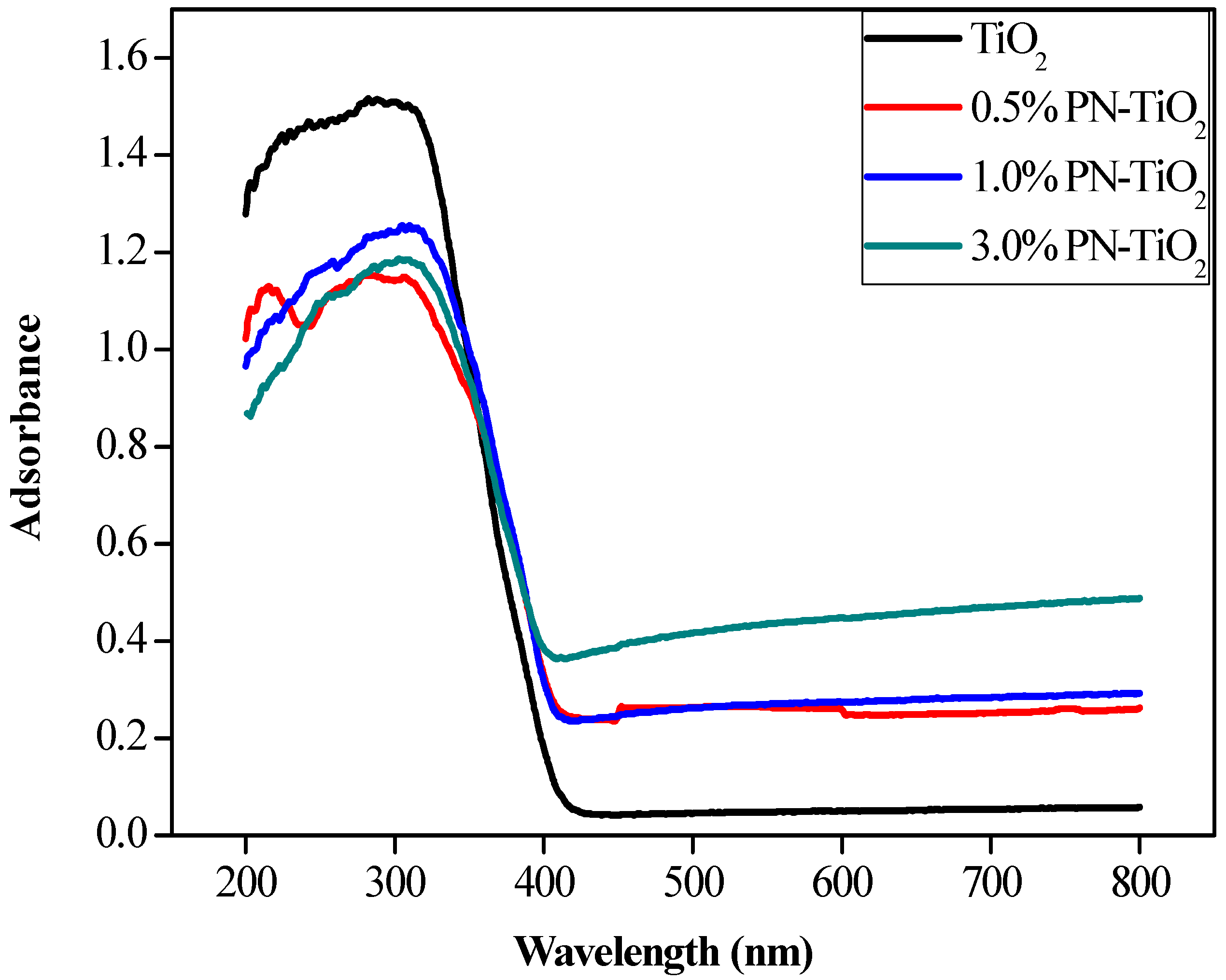
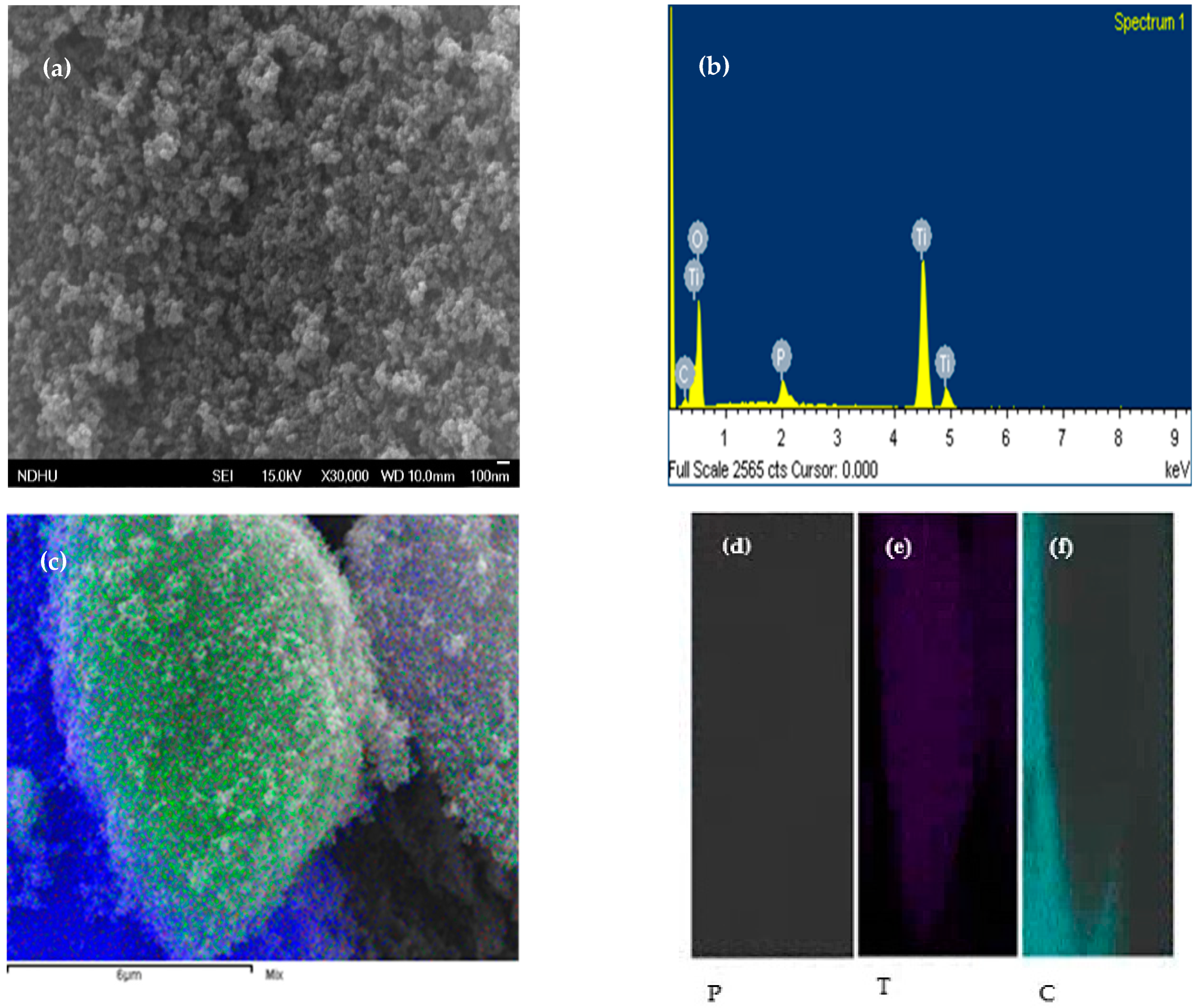
2.1. Material Characterization

To confirm the formation of composites through sample characterization, TEM, XRD, Raman, FT-IR, XPS, and EDX analyses were performed. The surface morphology of the crystalline composites could be easily observed from TEM images as indicated in Figure 1. Figure 1a clearly exhibits the ultrathin film structure of the samples confirmed to be the characterization of 2D material as it was revealed. Moreover, the electron diffraction patterns of the selected area as shown in Figure 1b also confirmed the crystalline nature of the sample. To further determine the thickness of the PN, the AFM characterization technique was applied through an optical contrast analysis (Figure 2a). The results indicated that the clear edges of the PN were still sharpened, suggesting that oxidation was unlikely to have occurred during material preparation. Moreover, the height profile from Figure 2b showed that the thickness of the PN was approximately 3.0–5.0 nm. According to Liu et al. [2], the thickness of a single layer of PN is approximately 0.9 nm, suggesting that the number of sample layers could have been approximately 3–5. This is consistent with the findings of the TEM.
To identify the substructure and electronic features of PN and PN-TiO2 composite, Raman spectra were implemented as shown in Figure 3. The results indicated that three Raman characteristic peaks of black phosphorous (BP) and PN were displayed at approximately 361 cm−1, 436 cm−1 and 463 cm−1. These peak wavelengths corresponded to phosphorous. It was confirmed that the second sample prepared was indeed a phosphorous homologous substance. An apparent shift was observed for the composite samples at 361 cm−1 and 463 cm−1, indicating a comparatively smaller thickness than that of BP. This was due to a positive relationship between the extent of the shift and the PN thickness as reported in the literature [1]. There were more and less obvious additional peaks at 144 cm−1 and 391 cm−1, respectively, in the Raman spectroscopy of PN-TiO2. The two characteristic peaks corresponded to the typical figure of Raman spectra for anatase, directly indicating that the TiO2 sample was P25 and the ratio of anatase to rutile was 80/20 [2]. Moreover, the formation of three signals identified the approximate locations of 361 cm−1, 436 cm−1 and 463 cm−1 in the curve of PN-TiO2 composites, suggesting that PN was successfully combined with TiO2 [3].
In addition, XRD patterns of the samples were obtained to reveal the crystalline structure and phase purity of TiO2 (Figure 4). Figure 4 illustrates the apparent peaks for (hkl) 020 and 040 planes according to JCPDS card no. 47-1626. The results of both the TEM and AFM confirmed that the PN was prepared successfully. All the relatively high characteristic peaks in the XRD image of the TiO2 reflected its anatase structure. The TiO2 used in this study was P25 with anatase at approximately 80%. The absence of the characteristic peaks of other impurities simply suggests the high purity of the composites studied.
To observe the changes in the absorption spectra of these four composites with different PN content levels at 0.5% PN-TiO2, 1.0% PN-TiO2, and 3.0% PN-TiO2, a UV–vis spectrophotometric analysis was carried out at a range between 200 nm and 800 nm (Figure 5). As indicated in Figure 5, its results directly pointed out that gradually increasing capacities of adsorption within visible light ranges occurred with the increase in PN content. Although there was a slight decrease in the adsorption value within UV light, the Eg value of these composites determined by Equation (1) was still lower, and the order of this decrease in adsorption was as follows: Eg3.0% PN-TiO2 (2.6 eV) < Eg1.0% PN-TiO2 (2.8 eV) ≈ Eg0.5% PN-TiO2 (2.8 eV) < EgTiO2 (3.0 eV)
Eg = 1240/λ
where parameter λ is the node value of the tangent of the spectra and X axis.
To explore the characterization of material preparation, the surface morphology of PN-TiO2 was also studied via SEM (Figure 6a). As the SEM image indicated, the particle of TiO2 was evenly distributed with nearly no particle aggregation. Figure 6b shows a typical EDS image of the PN-TiO2 composite. The EDS image showed that the composite consisted of P, Ti, and O elements. Combining the results obtained from both Figure 6c and the elemental maps of PN-TiO2, the three elements were completely mixed and P was successfully loaded onto TiO2.

2.2. Photodegradation Performance of PN-TiO2

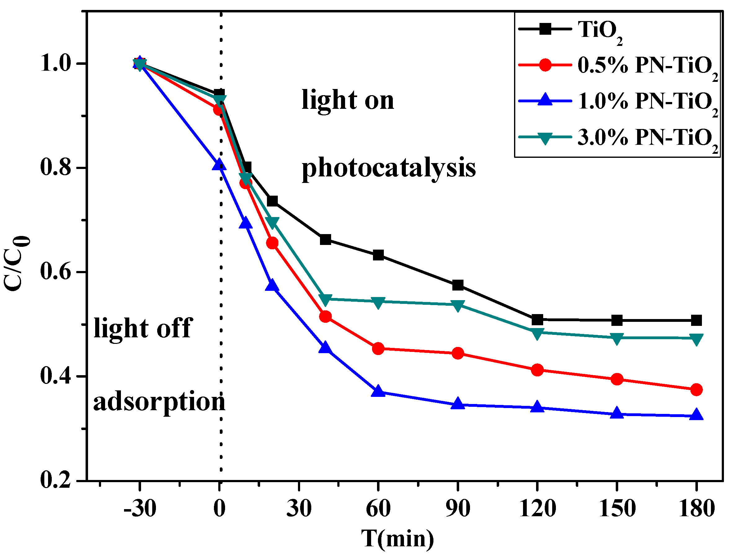
2.2.1. Effects of Different Levels of PN Content

To investigate the photocatalytic efficiency of composites with different levels of PN content, 0.05 g of TiO2, 0.50% PN-TiO2, 1% PN-TiO2, and 3.00% PN-TiO2 were individually mixed with 500 mL of 10 ppm E2 solution for comparative study (Figure 7). As Figure 7 indicates, the removal of E2 was significantly improved with an increase in PN content. In addition, bare TiO2 was observed to be more hydrophilic, leading to the lower adsorption of E2 on the surface of the catalyst. However, it is observed that the photodegradation of E2 was enhanced for 0.5% PN, but a further increase in the PN content decreased degradation performance. This may be attributed to the fact that PN as an electron acceptor may prevent electron-hole recombination caused by TiO2 [31,32,33,34,35]. Moreover, h+ may react with water and oxygen to generate ·OH to eliminate E2, as shown in Equations (2)–(6). ·OH can also be generated due to the excitation of PN by UV light [22]. Thus, increased photodegradation efficiency is shown where PN is doped into TiO2. However, the excessive PN content still affected the absorption of the catalyst, leading to decreased catalytic activity. This was confirmed by the UV–vis absorption spectra of PN-TiO2 (Figure 5). Furthermore, it was also observed that PN could be significantly accumulated when PN content was very high; thus, it could not prevent electron-hole recombination. Consequently, the catalytic activity of the PN-TiO2 composite was attenuated [35].
hv + TiO2 → e + h+
h+ + H2O + O2 → ·OH + O2−·
O2−· + HO2· + H+ → H2O2 + O2
e + H2O2 → OH + ·OH
h+/·OH + E2 → byproducts

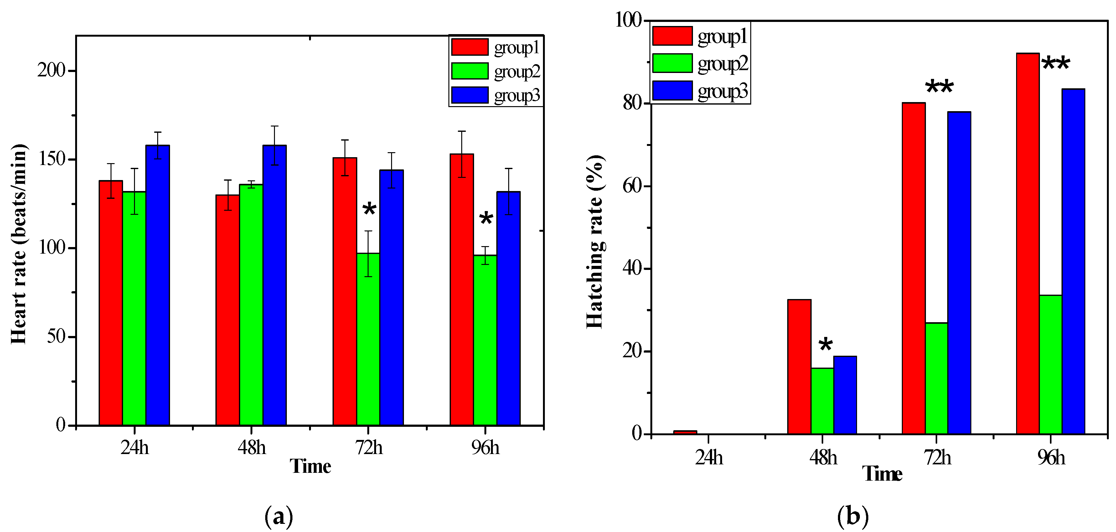
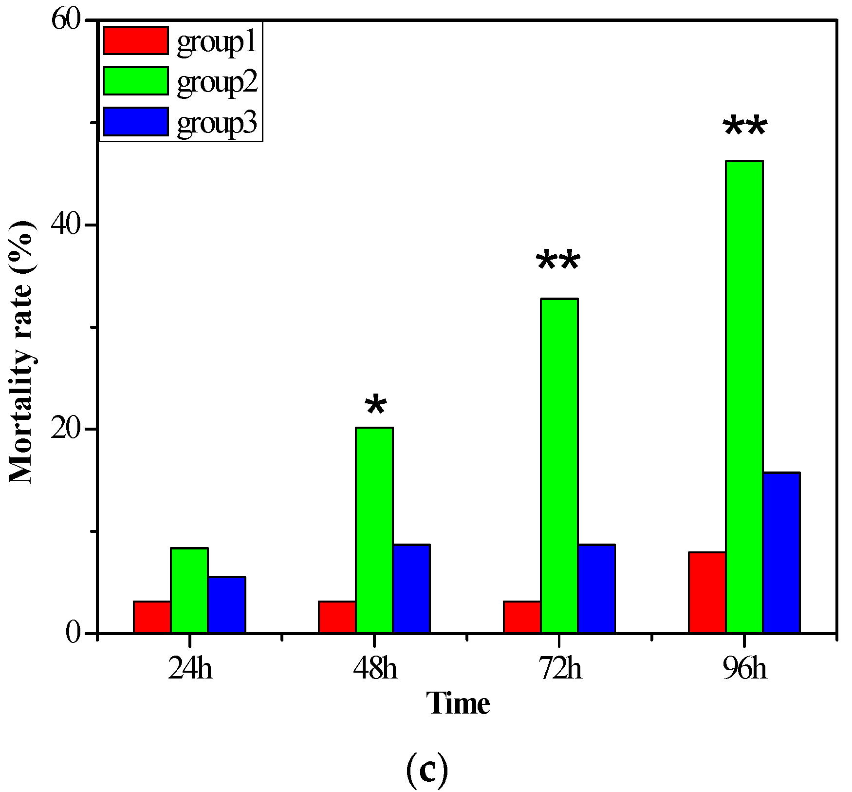
2.2.2. The Biotoxicity of the Water after Degradation

To evaluate the biotoxicity of the treated water, zebrafish were used as the model organism. Ten fish in each group were exposed to the clean water (group 1), the wastewater containing E2 (group 2), and the treated water (group 3), respectively [36]. The hatching rates, mortality rates, and heart rates were observed to investigate the effects of the water before and after degradation on the development of zebrafish embryos. Thus, it could be determined whether the photocatalysis degradation system was effective.
Figure 8 shows that heart rates increased with the development of the embryos in a natural environment (group 1), which was in contrast to those in the water containing E2 (group 2 and group 3). Heart rates in group 3 were slightly higher in the beginning and then decreased with time accumulation. This indicates that a certain concentration of E2 may lead to some response within a short timeframe; however, a biotoxicity effect emerged afterward, causing heart rates in group 3 to decrease at 48–96 h. Furthermore, a significant decrease was observed in group 2, demonstrating that higher concentrations of E2 have a negative effect on the metabolic activity of zebrafish embryos and inhibit their growth. These results correspond with those of Ulhaq [37]. In addition, the difference between group 2 and group 3 shows that photocatalysis degradation decreased the activity of E2 and its biotoxicity.
According to Figure 2, zebrafish eggs started to hatch out from the second day, and the highest hatching rate appeared on the third day. However, there was a significant difference between the hatching rate of group 2 and those of the other two groups. Furthermore, the hatching rate in group 2 was significantly lower throughout the whole hatching process, which demonstrates that a higher concentration of E2 inhibits the development of zebrafish embryos leading to lower hatching rates, which is consistent with the findings of a report by Lee [38]. There was also no significant difference between group 1 and group 3, indicating that the water environment in the two groups was similar.
Figure 3 shows that the mortality rate of embryos became higher in the 3 groups with the accumulation of time, and the mortality rate was highest in group 2. It can be inferred that higher E2 concentrations may lead to the higher mortality of zebrafish embryos. A study by Ren [39] also mentioned that higher levels of E2 (>10 µM) would lead to zebrafish malformation or mortality. Moreover, it can be observed that the mortality rate of zebrafish embryos in group 3 was significantly lower than that in group 2 and was similar to that in group 1, suggesting that the water degradation led to the lower mortality rate of zebrafish embryos.
In conclusion, the photocatalysis system was effective for the degradation of E2 and could decrease the negative effects of E2 on the development of zebrafish embryos.

3. Discussion

3.1. Comparative Assessment of Degradation Performance

To compare the degradation efficiencies of other materials, the degradation performances of similar materials obtained from the literature were adopted (Table 1). Evidently, the results of this study indicated that the removal rate and photocatalytic capacity of the model used in our study are promising. Its efficiency with the 10 ppm E2 solution led to an achievement of 79.5% removal within 3 h, which was a far superior result to those in reported data [14,15,24,25]. For example, its removal capacity was 39.8 mg/g. Alvarez-Corena [36,37] reported that degradation efficiency was significantly enhanced when O2 was added to pure TiO2 for E2-containing wastewater treatment. In addition, the degradation rate was 0.53 min−1, which is 5–10 times higher than that reported in other studies. However, O2 was required to be supplied continuously during this process, inevitably leading to a higher cost for practical applications. Li et al. [38] and Fernandez [39] reported degradation efficiencies of 99.5% and 90% when Fe-Bi2SiO5 and P25 were utilized to decompose E2 at 3 ppm and 1 ppm in an aqueous solution, respectively. According to the Langmuir–Hinshelwood mechanism, these higher efficiencies were likely attributed to the lower E2 concentration. Consequently, their removal capacities were approximately 5.97 mg/g and 1.80 mg/g, which were around 1/8 and 1/20 times lower than that in our study, respectively. Moreover, a high-intensity UV lamp was still required to be used in these two studies and cost-effectiveness problems were still of great concern. Yang et al. [26] used a 20 W UV lamp in their study. FTG (surface fluorinated TiO2/reduced graphene oxide) and TiO2-RGO (reduced graphene oxide) were prepared to remove 3 ppm of E2 solution, and their degradation efficiencies reached up to 99.8% and 83.3% in 180 min, respectively. These efficiencies were higher than that of P25 (74.4%) and of the model in our study (79.5%). FTG in particular was capable of completely removing 3 ppm of E2 in wastewater in 3 h. This is possibly due to the vital role of F- in photocatalysis. The adsorption of F on the surface of TiO2 improved the crystallization of the TiO2 anatase phase. However, the ·OH radicals generated on the surface of F-TiO2 were more mobile than those generated on pure TiO2 under UV irradiation [40]. Further, F- is harmful to the environment and ecosystem, meaning that a secondary pollutant could be generated without recycling. Furthermore, a UV lamp of a higher intensity was used in the study, and the removal capacity was still far lower than our study.
This study used a PN and PN-TiO2 composite as a photocatalyst. Although the mass% of the PN was only 0.5% in the composite, its degradation efficiency was more significantly improved than that of bare TiO2. Furthermore, our use of a UV lamp with very low intensity (8 W) suggests that this technique is not only cost-effective but also energy-efficient. This is consistent with the aim of green sustainability and environmental friendliness. The removal capacity of 39.8 mg/g was still far higher than that reported in the literature (Table 2). However, the degradation rate still requires further improvement from a kinetic perspective for overall performance consideration.

3.2. Significance of this Study

Recently, a novel 2D material, PN, has attracted considerable attention in studies owing to its physical, electrical, and chemical characteristics. Most of these studies have focused on the electrical applications of PN (e.g., solar cells and barrier diodes) [27,28,29,30], and a few studies have investigated its photocatalytic properties (Table 2). In addition, degradation efficiency [33] and catalyst instability were mentioned when PN was used as a photocatalyst, as it could readily react with water and oxygen to generate PO43− [31,41,42]. In this study, PN was mixed with the commonly used oxide TiO2 to prepare a PN-TiO2 hybrid. The advantages of this hybrid can be summarized as follows: (1) PN exhibited synergistic interactions with photodegradation due to its inherent photocatalytic properties; (2) The proposed doping of PN (instead of expensive metals) into TiO2 was biocompatible and cost-effective; and (3) PN-TiO2 composites were stable, preventing the reaction of PN with water and oxygen. Lee et al. [31] reported black phosphorus (BP) @TiO2 as a promising photocatalyst for dye photodegradation. However, the mass content of PN was very high in this composite, thus increasing the overall cost, since PN is still not economically feasible compared with TiO2 [42,43]. Regarding synergistic photocatalysis, PN inhibits electron-hole recombination due to its high electron mobility, improving the degradation efficiency of the hybrid for pollutant elimination. Moreover, the addition of PN increased degradation efficiency by 30% in the aqueous phase compared to that exhibited by bare TiO2. The maximum degradation efficiency was achieved at an optimum PN content at 1.0% wt. Thus, the overall cost was still far lower than that of the model reported by Lee [38]. This study indicates the considerable prospects of the fabrication of other PN–oxide hybrids (e.g., PN-WO3 and PN-CeO2) that may exhibit comparable photodegradation performance.

4. Material and Methods

4.1. Chemical Reagents

Bulk black phosphorus (BP) was obtained from XFNANO. Model pollutant 17β-estradiol (E2, purity ≥ 98%) was purchased from Sigma. Nano-TiO2 was provided by ACROS. Ultrapure hydroelectricity was prepared using the OTUN ultrapure water system (0R007XXM1, ELGA). Stock solutions of E2 were prepared by dissolving approximately 0.1 mg of E2 in 100 mL of methanol and stirring well for 5 min. The solution was then maintained at 4 °C for further use.

4.2. Preparation of Phosphorene Nanosheets (PN) and Phosphorene—TiO2 Composites (PN-TiO2)

PN was prepared by exfoliating the bulk black phosphorous in liquid via ultrasonic treatment. Sequentially, 25 mg of bulk sample was dispersed into 50 mL ethanol (purity ≥ 99.5%) followed by purging N2 to remove residual dissolved oxygen (i.e., crucial step preventing PN oxidation). Then, the mixture was placed in ice water for 12 h sonication. The brown suspension was centrifuged at 3000 rpm for 10 min to harvest the supernatant. Then, the solution was bubbled with N2 for 5 min and then maintained in 4 °C for preparation of PN-TiO2. The supernatant was then dried at 60 °C for approximately 8 h to obtain PN powder. Hereafter, PN content at 0.5%, 1.0%, and 3.0% was used to mix with TiO2, which was then added to 15 mL DI water. Next, the solutions were well stirred under ultrasonication for 1 h and then microwaved (200 °C, 400 W) for 30 min. Finally, the mixtures were dried at 60 °C to harvest PN-TiO2 composites (denoted as 0.5% PN-TiO2, 1.0% PN-TiO2, and 3.0% PN-TiO2, respectively). The characterizations of PN and PN-TiO2 were then implemented via transmission electron microscope (TEM, JEM-F200 "F2", Taipei, Taiwan), atomic force microscope (AFM, NX7 STinstruments, Taipei, Taiwan), Raman spectroscopy (DXR3 SmartRaman Spectrometer, Taipei, Taiwan), X-ray diffraction (XRD, Bruker D2 phaser, Yilan, Taiwan), UV–vis spectrophotometer (Hitachi U-3900, Yilan, Taiwan), field emission scanning electron microscope (FE-SEM, Jeol IT-100, Yilan, Taiwan), energy dispersive spectrometer (EDS, Jeol IT-100, Yilan, Taiwan), and elemental maps.

4.3. Photodegradation Reaction

The cylindrical reactor (7 cm diameter and 40 cm height) was used for the treatability study of photocatalytic degradation. In addition, an 8 W UV lamp (0.55 mW/cm2) and a tungsten halogen lamp (500 W output power) as sources of minimum energy supply were equipped to provide sufficient energy to trigger photodegradation process to take place. Subsequently, 0.05 g of PN-TiO2 was supplemented into 500 mL of 10 ppm E2 diluted in 2% methanol diluted with DI water. The reaction was first conducted in the dark for 30 min to achieve equilibrium for absorption, and then UV light was switched on to trigger photocatalysis. All the experiments were conducted at inherent solution pH and 30 ± 0.2 °C at 300 rpm. The samples were then conducted (without removing photocatalysts) using a Millipore filter with pore size of 0.22 µm. E2 concentration of samples was analyzed through high-performance liquid chromatography (HPLC, ThermoFisher, Yilan, Taiwan). The flow phase was a solution mixture of ultrapure water and acetonitrile in 48:52 volumetric ratio eluting the pollutant with 1.2 mL/mins of flow rate. In addition to 20 μL of injection volume and 25 °C of column, the retention time was set to 5.5 min for E2.

5. Conclusions

The enhanced photodegradation of 17β-estradiol by phosphorene-basing composites was reported with the successful preparation of PN and PN-TiO2 photocatalysts through detailed characterization. The extent of the degradation of E2 reached up to 67.5% with PN-TiO2 under UV light within 180 min, which was an almost two-fold greater result than that of bare TiO2. To the best of our knowledge, this study represents the first attempt to combine PN with other catalysts for the degradation of refractory and hydrophobic pollutants (e.g., E2). During the reaction process, PN could adsorb E2 onto the surface of TiO2. The studies of the catalyst performed on zebrafish showed a higher mortality rate at 96 h as well as a superior hatching rate and healthier fish development. Thus, it could facilitate photodegradation due to the formation of the ·OH radical. In addition, the combination of e with h+ could be restrained by PN and could also generate ·OH in combination with TiO2 to attack E2, which is the reason why the E2 degradation efficiency and removal capacity of PN-TiO2 were higher than that of bare TiO2. However, some intermediates with high estrogen activity could possibly be produced during this photodegradation process, causing a potential threat to aquatic organisms and humans [5].

Author Contributions

Conceptualization, K.M. and C.-T.C.; methodology, K.Z., K.M.; investigation, K.M. and C.-T.C.; data curation, C.-T.C.; writing—original draft preparation, C.-T.C.; writing—review and editing, C.-T.C.; supervision, K.Z.; project administration, K.Z.; funding acquisition, K.Z. All authors have read and agreed to the published version of the manuscript.

Funding

This work was supported by the National Key Research and Development Program of China (2016YFC0502901) and the National Key Research and Development Program of China (2017YFC0506103). The authors would like to thank the Ministry of Science and Technology of Taiwan for providing financial support for project MOST 106-2622-E-197-003-CC3.

Data Availability Statement

Not available.

Conflicts of Interest

The authors declare no conflict of interest.

References

  1. Finn, R.S.; Aleshin, A.; Slamon, D.J. Targeting the cyclin-dependent kinases (CDK) 4/6 in estrogen receptor-positive breast cancers. Breast Cancer Res. 2016, 18, 17. [Google Scholar] [CrossRef] [PubMed]
  2. Pendse, S.; Maertens, A.; Rosenberg, M.; Roy, D.; Fasani, R.; Vantangoli, M.; Madnick, S.; Boekelheide, K.; Fornace, A.; Odwin, S.-A. Information-dependent enrichment analysis reveals time-dependent transcriptional regula-tion of the estrogen pathway of toxicity. Arch. Toxicol. 2017, 91, 1749–1762. [Google Scholar] [CrossRef] [PubMed]
  3. Sanders, J.; Coulter, S.; Knudsen, G.; Dunnick, J.; Kissling, G.; Birnbaum, L. Disruption of estrogen ho-meostasis as a mechanism for uterine toxicity in Wistar Han rats treated with tetrabromobisphenol A. Toxicol. Appl. Pharmacol. 2016, 298, 31–39. [Google Scholar] [CrossRef] [PubMed]
  4. Biegel, L.B.; Hirshfield, A.N.; O’Connor, J.C.; Ladics, G.S.; Cook, J.C.; Frame, S.R.; Flaws, J.A.; Elliott, G.S.; Silbergeld, E.K.; Van Pelt, C.S.; et al. 90-Day Feeding and One-Generation Reproduction Study in Crl:CD BR Rats with 17β-Estradiol. Toxicol. Sci. 1998, 44, 116–142. [Google Scholar] [CrossRef] [PubMed]
  5. Diamante, G.; Menjivar-Cervantes, N.; Leung, M.S.; Volz, D.C.; Schlenk, D. Contribution of G protein-coupled estrogen receptor 1 (GPER) to 17β-estradiol-induced develop-mental toxicity in zebrafish. Aquat. Toxicol. 2017, 186, 180–187. [Google Scholar] [CrossRef] [PubMed]
  6. Angus, R.A.; Stanko, J.; Jenkins, R.L.; Watson, R.D. Effects of 17α-ethynylestradiol on sexual development of male western mosquitofish (Gambusia affinis). Comp. Biochem. Physiol. Part C Toxicol. Pharmacol. 2005, 140, 330–339. [Google Scholar] [CrossRef] [PubMed]
  7. Li, M.; Zhao, X.; Zhang, X.; Wu, D.; Leng, S. Biodegradation of 17β-estradiol by Bacterial Co-culture Isolated from Manure. Sci. Rep. 2018, 8, 3787. [Google Scholar] [CrossRef]
  8. Xiong, W.; Peng, W.; Liang, R. Identification and genome analysis of Deinococcus actinosclerus SJTR1, a novel 17β-estradiol degradation bacterium. 3 Biotech 2018, 8, 433. [Google Scholar] [CrossRef]
  9. Yu, Q.; Wang, P.; Liu, D.; Gao, R.; Shao, H.; Zhao, H.; Ma, Z.; Wang, D.; Huo, H. Degradation characteristics and metabolic pathway of 17β-estradiol (E2) by Rhodococcus sp. DS201. Biotechnol. Bioprocess Eng. 2016, 21, 804–813. [Google Scholar] [CrossRef]
  10. Mazlan, W.S.; Mokhtar, H.; Saâ, N.; Tajuddin, R.M. Fabrication of ultrafiltration membrane using addtive to extract hormone from poultry wastewater. J. Teknol. 2016, 78, 121–126. [Google Scholar] [CrossRef] [Green Version]
  11. Tong, Y.; McNamara, P.J.; Mayer, B.K. Fate and impacts of triclosan, sulfamethoxazole, and 17β-estradiol during nutrient recovery via ion ex-change and struvite precipitation. Environ. Sci. Water Res. Technol. 2017, 3, 1109–1119. [Google Scholar] [CrossRef]
  12. Jiang, L.-H.; Liu, Y.-G.; Zeng, G.-M.; Xiao, F.-Y.; Hu, X.-J.; Hu, X.; Wang, H.; Li, T.-T.; Zhou, L.; Tan, X.-F. Removal of 17β-estradiol by few-layered graphene oxide nanosheets from aqueous solutions: External influence and adsorption mechanism. Chem. Eng. J. 2016, 284, 93–102. [Google Scholar] [CrossRef]
  13. Jiang, L.; Liu, Y.; Liu, S.; Hu, X.; Zeng, G.; Hu, X.; Liu, S.; Liu, S.; Huang, B.; Li, M. Fabrication of β-cyclodextrin/poly (l-glutamic acid) supported magnetic graphene oxide and its adsorption behavior for 17β-estradiol. Chem. Eng. J. 2017, 308, 597–605. [Google Scholar] [CrossRef]
  14. Kim, S.; Cho, H.; Joo, H.; Her, N.; Han, J.; Yi, K.; Kim, J.-O.; Yoon, J. Evaluation of performance with small and scale-up rotating and flat reactors; photocatalytic degradation of bisphenol A, 17β–estradiol, and 17α–ethynyl estradiol under solar irradiation. J. Hazard. Mater. 2017, 336, 21–32. [Google Scholar] [CrossRef]
  15. Kovacic, M.; Kopcic, N.; Kusic, H.; Bozic, A.L. Solar driven degradation of 17β-estradiol using composite photocatalytic materials and artificial irradiation source: Influence of process and water matrix parameters. J. Photochem. Photobiol. A Chem. 2018, 361, 48–61. [Google Scholar] [CrossRef]
  16. Saravanan, R.; Gracia, F.; Stephen, A. Basic principles, mechanism, and challenges of photocatalysis. In Nanocomposites for Visible Light-induced Photocatalysis; Springer: Berlin/Heidelberg, Germany, 2017; pp. 19–40. [Google Scholar]
  17. Xing, Z.; Zhang, J.; Cui, J.; Yin, J.; Zhao, T.; Kuang, J.; Xiu, Z.; Wan, N.; Zhou, W. Recent advances in floating TiO2-based photocatalysts for environmental application. Appl. Catal. B Environ. 2018, 225, 452–467. [Google Scholar] [CrossRef]
  18. Tahir, K.; Ahmad, A.; Li, B.; Nazir, S.; Khan, A.; Nasir, T.; Khan, Z.; Naz, R.; Raza, M. Visible light photo cata-lytic inactivation of bacteria and photo degradation of methylene blue with Ag/TiO2 nanocomposite prepared by a novel method. J. Photochem. Photobiol. B Biol. 2016, 162, 189–198. [Google Scholar] [CrossRef]
  19. Vaiano, V.; Iervolino, G.; Sannino, D.; Murcia, J.; Hidalgo, M.; Ciambelli, P.; Navío, J. Photocatalytic removal of patent blue V dye on Au-TiO2 and Pt-TiO2 catalysts. Appl. Catal. B Environ. 2016, 188, 134–146. [Google Scholar] [CrossRef]
  20. Bagheri, S.; Mansouri, N.; Aghaie, E. Phosphorene: A new competitor for graphene. Int. J. Hydrog. Energy 2016, 41, 4085–4095. [Google Scholar] [CrossRef]
  21. Khandelwal, A.; Mani, K.; Karigerasi, M.H.; Lahiri, I. Phosphorene–the two-dimensional black phosphorous: Properties, synthesis and applications. Mater. Sci. Eng. B 2017, 221, 17–34. [Google Scholar] [CrossRef]
  22. Wang, H.; Jiang, S.; Shao, W.; Zhang, X.; Chen, S.; Sun, X.; Zhang, Q.; Luo, Y.; Xie, Y. Optically Switchable Photocatalysis in Ultrathin Black Phosphorus Nanosheets. J. Am. Chem. Soc. 2018, 140, 3474–3480. [Google Scholar] [CrossRef] [PubMed]
  23. Hu, Z.T.; Liu, J.; Yan, X.; Oh, W.D.; Lim, T.T. Low-temperature synthesis of graphene/Bi2Fe4O9 composite for synergistic adsorp-tion-photocatalytic degradation of hydrophobic pollutant under solar irradiation. Chem. Eng. J. 2015, 262, 1022–1032. [Google Scholar] [CrossRef]
  24. Zatloukalová, K.; Obalová, L.; Koči, K.; Čapek, L.; Matěj, Z.; Šnajdhaufová, H.; Ryczkowski, J.; Słowik, G. Photo-catalytic degradation of endocrine disruptor compounds in water over immobilized TiO2 photocatalysts. Iran. J. Chem. Chem. Eng. (IJCCE) 2017, 36, 29–38. [Google Scholar]
  25. Mboula, V.M.; Héquet, V.; Andrès, Y.; Gru, Y.; Colin, R.; Doña-Rodríguez, J.; Pastrana-Martínez, L.; Silva, A.; Leleu, M.; Tindall, A.; et al. Photocatalytic degradation of estradiol under simulated solar light and assessment of estrogenic activity. Appl. Catal. B Environ. 2015, 162, 437–444. [Google Scholar] [CrossRef]
  26. Yang, Y.; Luo, L.; Xiao, M.; Li, H.; Pan, X.; Jiang, F. One-step hydrothermal synthesis of surface fluorinated TiO2/reduced graphene oxide nanocomposites for photocatalytic degradation of estrogens. Mater. Sci. Semicond. Process. 2015, 40, 183–193. [Google Scholar] [CrossRef]
  27. Padilha, J.E.; Fazzio, A.; Da Silva, A.J.R. van der Waals Heterostructure of Phosphorene and Graphene: Tuning the Schottky Barrier and Doping by Electrostatic Gating. Phys. Rev. Lett. 2015, 114, 066803. [Google Scholar] [CrossRef]
  28. Hu, W.; Wang, T.; Yang, J. Tunable Schottky contacts in hybrid graphene–phosphorene nanocomposites. J. Mater. Chem. C 2015, 3, 4756–4761. [Google Scholar] [CrossRef]
  29. Guo, H.; Lu, N.; Dai, J.; Wu, X.; Zeng, X. Phosphorene nanoribbons, phosphorus nanotubes, and van der Waals multilayers. Journal of Physical Chemistry C 2014, 118, 14051–14059. [Google Scholar] [CrossRef]
  30. Luo, Y.; Zhang, S.; Chen, W.; Jia, Y. Interlayer coupling effects on electronic properties of the phosphorene/h-BN van der Walls heterostructure: A first principles investigation. Phys. B Condens. Matter 2018, 534, 51–55. [Google Scholar] [CrossRef]
  31. Lee, H.U.; Lee, S.C.; Won, J.; Son, B.-C.; Choi, S.; Kim, Y.; Park, S.Y.; Kim, H.-S.; Lee, Y.-C.; Lee, J. Stable semiconductor black phosphorus (BP)@titanium dioxide (TiO2) hybrid photocatalysts. Sci. Rep. 2015, 5, srep08691. [Google Scholar] [CrossRef]
  32. Liu, H.; Neal, A.T.; Zhu, Z.; Luo, Z.; Xu, X.; Tomanek, D.; Ye, P.D. Phosphorene: An Unexplored 2D Semiconductor with a High Hole Mobility. ACS Nano 2014, 8, 4033–4041. [Google Scholar] [CrossRef] [PubMed] [Green Version]
  33. Wang, H.; Yang, X.; Shao, W.; Chen, S.; Xie, J.; Zhang, X.; Wang, J.; Xie, Y. Ultrathin Black Phosphorus Nanosheets for Efficient Singlet Oxygen Generation. J. Am. Chem. Soc. 2015, 137, 11376–11382. [Google Scholar] [CrossRef] [PubMed]
  34. León, A.; Reuquen, P.; Garín, C.; Segura, R.; Vargas, P.; Zapata, P.; Orihuela, P.A. FTIR and Raman Characterization of TiO2 Nanoparticles Coated with Polyethylene Glycol as Carrier for 2-Methoxyestradiol. Appl. Sci. 2017, 7, 49. [Google Scholar] [CrossRef]
  35. Nawaz, M.; Miran, W.; Jang, J.; Lee, D.S. One-step hydrothermal synthesis of porous 3D reduced graphene ox-ide/TiO2 aerogel for carbamazepine photodegradation in aqueous solution. Appl. Catal. B Environ. 2017, 203, 85–95. [Google Scholar] [CrossRef]
  36. Woomer, A.H.; Farnsworth, T.W.; Hu, J.; Wells, R.A.; Donley, C.L.; Warren, S.C. Phosphorene: Synthesis, Scale-Up, and Quantitative Optical Spectroscopy. ACS Nano 2015, 9, 8869–8884. [Google Scholar] [CrossRef]
  37. Ulhaq, Z.S.; Kishida, M. Brain Aromatase Modulates Serotonergic Neuron by Regulating Serotonin Levels in Zebrafish Embryos and Larvae. Front. Endocrinol. 2018, 9, 230. [Google Scholar] [CrossRef]
  38. Lee, D.H.; Jo, Y.J.; Eom, H.J.; Yum, S.; Rhee, J.S. Nonylphenol induces mortality and reduces hatching rate through increase of oxidative stress and dysfunction of antioxidant defense system in marine medaka embryo. Mol. Cell. Toxicol. 2018, 14, 437–444. [Google Scholar] [CrossRef]
  39. Ren, X.; Lu, F.; Cui, Y.; Wang, X.; Bai, C.; Chen, J.; Huang, C.; Yang, D. Protective effects of genistein and estradiol on PAHs-induced developmental toxicity in zebrafish embryos. Hum. Exp. Toxicol. 2012, 31, 1161–1169. [Google Scholar] [CrossRef]
  40. Alvarez-Corena, J.R.; Bergendahl, J.A.; Hart, F.L. Advanced oxidation of five contaminants in water by UV/TiO2: Reaction kinetics and by-products identification. J. Environ. Manag. 2016, 181, 544–551. [Google Scholar] [CrossRef]
  41. Li, L.; Long, Y.; Chen, Y.; Wang, S.; Wang, L.; Zhang, S.; Jiang, F. Facile synthesis of Fe/Bi2SiO5 nanocomposite with enhanced photocatalytic activity for degradation of 17β-Estradiol(E2). Solid State Sci. 2018, 83, 143–151. [Google Scholar] [CrossRef]
  42. Fernández, L. Impact of operating conditions on the removal of endocrine disrupting chemicals by membrane photocatalytic reactor. Environ. Technol. 2014, 35, 2068–2074. [Google Scholar] [CrossRef] [PubMed]
  43. Vijayabalan, A.; Selvam, K.; Velmurugan, R.; Swaminathan, M. Photocatalytic activity of surface fluorinated TiO2-P25 in the degradation of Reactive Orange 4. J. Hazard. Mater. 2009, 172, 914–921. [Google Scholar] [CrossRef] [PubMed]
Figure 1. (a) TEM image and (b) electron diffraction pattern of PN.
Catalysts 13 00332 g001
Figure 2. AFM spectra of composite (a) external surface morphology (b) height profile.
Catalysts 13 00332 g002
Figure 3. Raman spectra of BP, PN, and PN-TiO2.
Catalysts 13 00332 g003
Figure 4. XRD pattern of TiO2.
Catalysts 13 00332 g004
Figure 5. UV–vis absorption spectra of PN-TiO2 with various PN content levels.
Catalysts 13 00332 g005
Figure 6. SEM images: (a) SEM spectrum, (b) EDS of PN-TiO2 composite, (c) overlapping of P, Ti, and C elements, (df) elemental maps of 3% PN-TiO2.
Catalysts 13 00332 g006
Figure 7. Photodegradation efficiency of different PN-TiO2 composites. (t < 0 and t > 0 denoted dark adsorption and light photodegradation, respectively).
Catalysts 13 00332 g007
Figure 8. The heart rates (a), hatching rates (b), and mortality rates (c) of zebrafish embryos in three groups (* in each graph indicated significant differences, p < 0.05; ** in each graph indicated very significant differences, p < 0.005).
Catalysts 13 00332 g008aCatalysts 13 00332 g008b
Table 1. Comparison of degradation efficiency and removal capacity of this study with the literature.
ReferenceMaterialsC0 (L−1) *Degradation EfficiencyLight SourceK1 * (min−1)Removal Capacity *
(mg/g)
[24]Immobilized
TiO2
44 µg45% (180 min)36 W UV 0.0030.0132
[37]TiO2 + O22 mg93% (25 min)100 W UV0.5318.6
[15]TiO2-FeZ1.36 mg78.1% (90 min)450 W Xe lamp0.01525.3
[26]FTG43 mg99.8% (180 min)20 W UV0.035Unsaturation
[26]TiO2-RGO3 mg83.3% (180 min)20 W UV0.0108.33
[26]P253 mg74.4% (180 min)20 W UV0.0087.44
[14]NTT0.54 mg65% (150 min)3300 µW/cm2 UV0.0073.51
[38]Fe-Bi2SiO53 mg99.5% (60 min)20 W UV0.0695.97
[39]P251 mg90% (3 h)64 W UV0.0131.8
[25]4%GO-TiO21 mg48% (60 min)450 W Xe lamp0.01124
This studyPN-TiO210 mg67.5%Visible/UV0.00667.5
* C0 is the initial concentration; K1 is the first order constant; removal capacity = mass of pollutant (mg)/mass of material (g).
Table 2. PN and PN-based hybrids and their fields of application.
ReferenceMaterialField of Application
[27]PN–GN hybridPerpendicular electric field
[28]PN–GN hybridSchottky barrier diode
[29]PN-MoS2Electric field/solar cell
[30]PN-h-BNElectric field
[22]PNPhotocatalysis for DPBF
[31](more)PN-TiO2Photocatalysis for dye
This study(less)PN-TiO2Photocatalysis for 17β-estradiol
PN, GN, BN, and DPBF indicate phosphorene, graphene, hexagonal boron nitride, and 1,3-diphenylisobenzofuran, respectively.
Disclaimer/Publisher’s Note: The statements, opinions and data contained in all publications are solely those of the individual author(s) and contributor(s) and not of MDPI and/or the editor(s). MDPI and/or the editor(s) disclaim responsibility for any injury to people or property resulting from any ideas, methods, instructions or products referred to in the content.

