Article

The Stability of Dimeric D-amino Acid Oxidase from Porcine Kidney Strongly Depends on the Buffer Nature and Concentration

by
Diego Carballares
1,
Javier Rocha-Martin
2,* and
Roberto Fernandez-Lafuente
1,3,*
1
Departamento de Biocatálisis, ICP-CSIC, C/Marie Curie 2, Campus UAM-CSIC, Cantoblanco, 28049 Madrid, Spain
2
Department of Biochemistry and Molecular Biology, Faculty of Biology, Complutense University of Madrid, José Antonio Novais 12, 28040 Madrid, Spain
3
Center of Excellence in Bionanoscience Research, External Scientific Advisory Academic, King Abdulaziz University, Jeddah 21589, Saudi Arabia
*
Authors to whom correspondence should be addressed.
Catalysts 2022, 12(9), 1009; https://doi.org/10.3390/catal12091009
Submission received: 11 August 2022 / Revised: 1 September 2022 / Accepted: 4 September 2022 / Published: 6 September 2022
(This article belongs to the Special Issue Promising Industrial Enzymes)

Abstract

:
The first step of the inactivation of the enzyme D-amino acid oxidase (DAAO) from porcine kidney at pH 5 and 7 is the enzyme subunit dissociation, while FAD dissociation has not a relevant role. At pH 9, both dissociation phenomena affect the enzyme stability. A strong effect of the buffer nature and concentration on enzyme stability was found, mainly at pH 7 and 9 (it was possible at the same temperature to have the enzyme fully inactivated in 5 mM of Hepes while maintaining 100% in 5 mM of glycine). The effect of the concentration of buffer on enzyme stability depended on the buffer: at pH 5, the acetate buffer had no clear effect, while Tris, Hepes and glycine (at pH 7) and carbonate (at pH 9) decreased enzyme stability when increasing their concentrations; phosphate concentration had the opposite effect. The presence of 250 mM of NaCl usually increased enzyme stability, but this did not occur in all cases. The effects were usually more significant when using low concentrations of DAAO and were not reverted upon adding exogenous FAD. However, when using an immobilized DAAO biocatalyst which presented enzyme subunits attached to the support, where dissociation was not possible, this effect of the buffer nature on enzyme stability almost disappeared. This suggested that the buffers were somehow altering the association/dissociation equilibrium of the enzyme.

1. Introduction

D-amino acid oxidases (DAAOs) are dimeric flavin adenine dinucleotide (FAD)-containing flavoenzymes that catalyze the oxidative deamination of D-amino acids yielding the corresponding imino acid, that is spontaneously hydrolyzed to yield α-keto acid and ammonia, producing hydrogen peroxide in the re-oxidation of FAD [1,2,3,4,5,6]. These enzymes have found applications in diverse sectors such as analytical chemistry (biosensors) [7,8,9,10] and in the production of keto-acids [11,12,13,14]. A specific application of DAAOs is in the production of semisynthetic cephalosporins, producing glutaryl 7- aminocephalosporanic acid from the oxidation of cephalosporin C [15,16,17,18,19]. The side-production of hydrogen peroxide is the key to its use as a D-amino acid biosensor, as usually this reagent generates the detection signal, while it can become a problem in biocatalysis, as it is an enzyme inactivating reagent and can also produce the oxidative decarboxylation of keto-acids [20]. In the 7-amino cephalosporanic acid production, the decarboxylation of the keto-adipic intermediate is persuaded in the reaction design, as this keto-acid is not a good substrate for existing deacylating enzymes (glutaryl acylases), although new synthetic routes have been proposed to shortcut this matter [21]. In this way, coimmobilization with catalase is the standard solution [20]. There are some publications preparing fusion enzymes (DAAO-glutaryl acylase) for 7-amino cephalosporanic production [22], even with the problems that these fusion proteins can raise [23].
There are DAAOs from many different origins [1,2,3,4,5,6]; however, in this paper we focus on the DAAO from porcine kidney. The enzyme’s structure was resolved a long time ago [24,25,26,27]. This enzyme has found diverse applications [28,29,30,31], therefore, it has been cloned and over-expressed in diverse microorganisms since a long time ago [32,33]. This enzyme has been stabilized by site-directed mutagenesis [34]. The dimeric character of this enzyme makes it that the subunit dissociation plays a relevant role on the enzyme’s inactivation [35,36]. This makes it that any factor of difficulty of the enzyme subunits dissociation may increase the enzyme stability (and the opposite) [37]. For example, multimeric enzyme stability may be greatly increased if we prevent the enzyme dissociation by intramolecular crosslinking [37] using a polymer [38,39] or support (i.e., via multi-subunit immobilization) [40]. Although the industrial application of this enzyme is mainly in immobilized form [41,42,43,44,45], to know how to handle and store this enzyme may prevent many problems in obtaining reproducible biocatalysts [46].
In this context, to analyze the stability of this enzyme under different conditions may be of great interest, as this can determine the conditions range where the enzyme may be handled (e.g., for storing or immobilizing the enzyme). Special attention has been paid to the effect of enzyme concentration on enzyme stability for multimeric and FAD (non-covalently attached to the enzyme)-depended enzymes [37]. In some instances, such as in the case of the DAAO from Rhodotorula gracilis, it has been shown that the release of FAD from the enzyme constituted an important method of enzyme inactivation [47]. Moreover, special attention has been paid in this research to the effect of the concentration and nature of different buffers. While the medium composition is usually systematically studied on its effect on enzyme activity [48,49,50,51,52,53], in only some few examples was it analyzed their effect on the enzyme stability. However, in some instances, they can play a critical role on enzyme stability [54,55,56]. This may be especially relevant for a multimeric enzyme and a FAD-dependent enzyme, as the buffers can play some role in reinforcing or decreasing the multimeric and active form of the enzyme. In this way, this paper is focused on the analysis of the effects of the medium nature on DAAO stability.

2. Results and Discussion

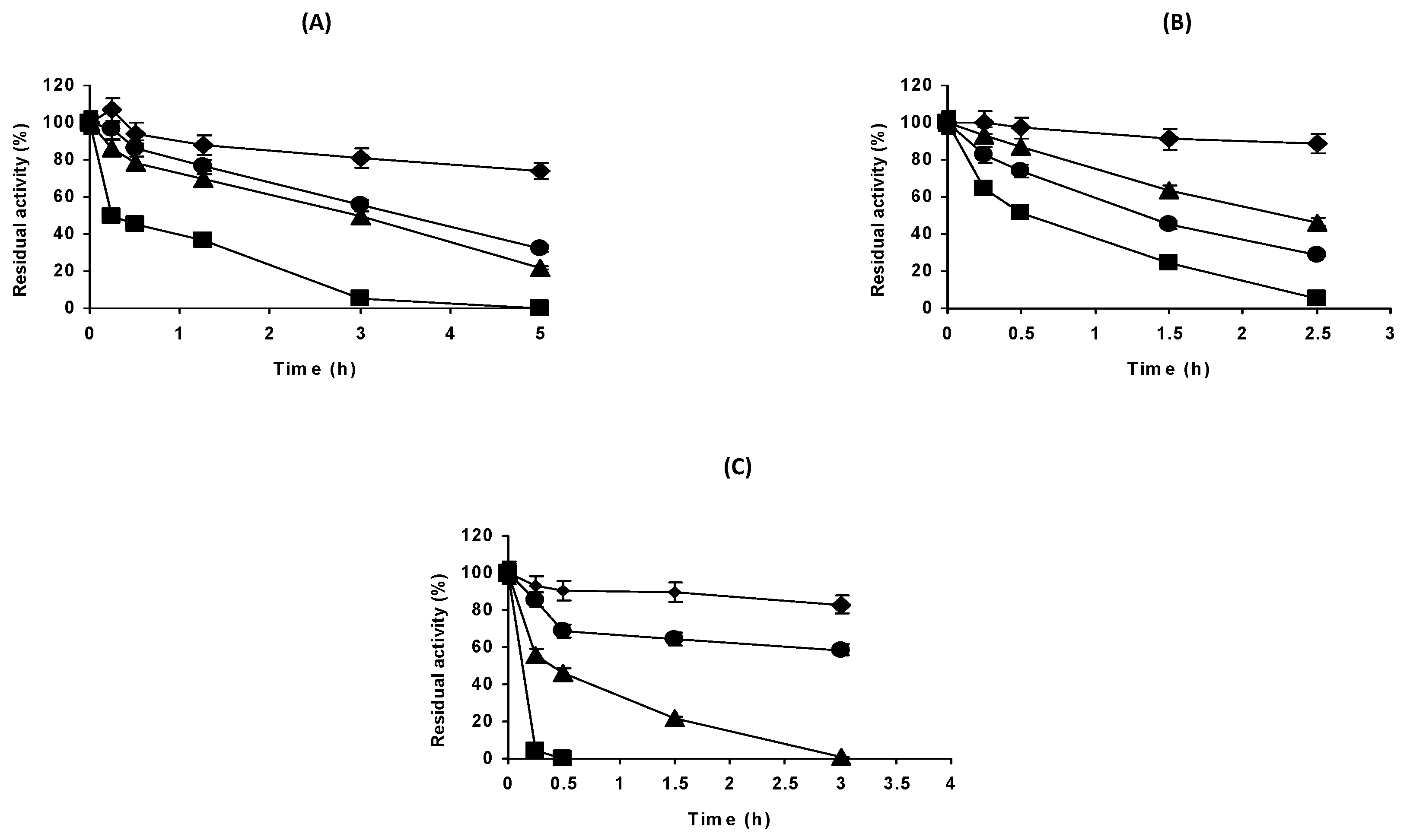
2.1. Effect of the Enzyme Concentration on the Stability of DAAO at Different pH Values

Figure 1 shows the inactivation courses of DAAO at pH 5, 7 and 9. In all cases, the enzyme stability was lower when the enzyme concentration decreased, and this was not reversed upon adding 1 mg/mL of bovine serum albumin (not shown results).
The intensity of the enzyme concentration effect on its stability was slightly clearer at pH 9 than at pH 5. We investigated if the dissociation of FAD was the reason for the enzyme concentration dependence (Figure 2), but we did not find a relevant effect of the addition of this compound during the enzyme inactivation at pH 5 and 7 using the lowest enzyme concentration of Figure 1, while at pH 9 the addition of the external cofactor presented some stabilizing effect. This stabilizing effect of the addition of exogenous FAD at pH 9 was not concentration dependent; it seemed that the minimal concentration was enough to prevent FAD dissociation. In any case, the enzyme stability in the presence of FAD remained much lower than when a higher enzyme concentration was inactivated, suggesting that the enzyme subunit dissociation was also very important at pH 9.
In this way, the results suggested that the first DAAO inactivation step, at all the studied pH values, was the dissociation of the enzyme subunits.

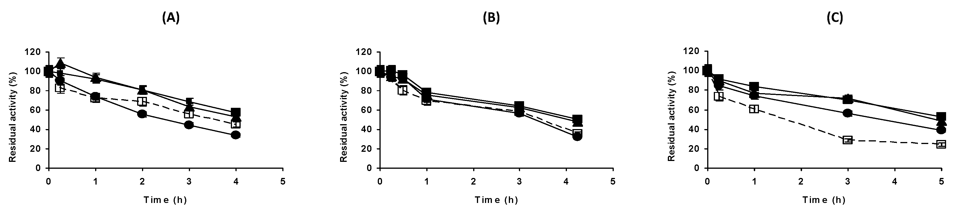
2.2. Effect of the Buffer Nature on the Stability of DAAO at pH 7

Figure 3 shows the inactivation courses of DAAO in the presence of different buffers at pH 7, using the enzyme concentrations of 0.2 and 1 mg/mL. Due to the much higher stability of the enzyme at 1 mg/mL, we used 45 °C in these experiments versus the 40 °C used for the diluted enzyme.
The figure shows an impressive effect of the buffer nature on the inactivation courses. While the diluted enzyme maintained 100% of the initial activity in 5 mM of glycine, it was fully inactivated in 5 mM of Hepes, the inactivation courses in between were Tris-HCL and phosphate (this last one was the one that provided the second lowest enzyme stability). The situation was completely different using the concentrated enzyme. In this case, the enzyme stability presented a similar stability in phosphate and glycine buffers, higher than in Tris-HCL in both cases. Hepes remained as the buffer that permitted a lower stability of the enzyme.
To analyze if the buffer nature effects could somehow be related to the favoring of the release of FAD from the enzyme, we analyzed the effect of adding 2 mM of exogenous FAD on the diluted (0.2 mg/mL) enzyme stability. Temperature was optimized to have reliable inactivation courses. Figure 4 shows the lack of effect of the external addition of FAD in all cases at pH 7. In this way, it could be discarded that the dissociation of the FAD was responsible for the effect of the buffer nature on enzyme stability.

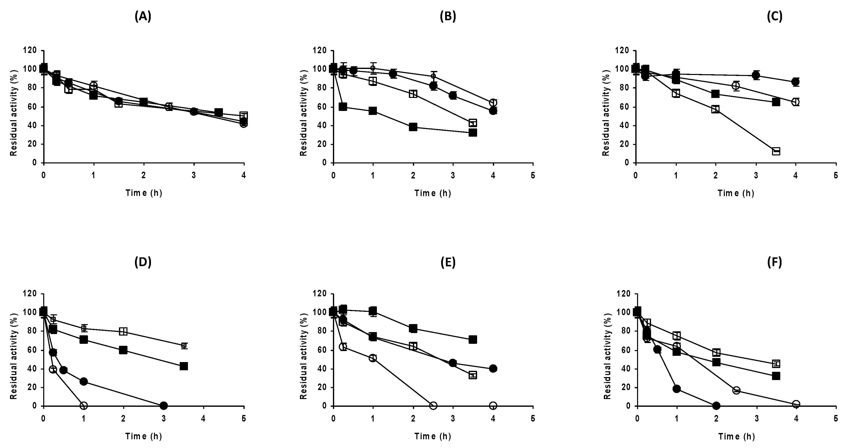
2.3. Effect of the Buffer Concentration at Different Enzyme Concentrations

Next, we analyzed the effect of the buffer concentration at pH 5, 7 and 9 using 0.2 mg/mL of the enzyme (Figure 5). At pH 5, the sodium acetate concentration effect was almost negligible, and it was not possible to find any trend. At pH 7, the increase in Tris-HCL concentration from 5 mM to 50 mM decreased the enzyme stability, but further increases in the Tris-HCL concentration presented a negligible effect on enzyme stability. The situation was completely different using sodium phosphate, which permitted a lower stability than Tris-HCL using 5 mM of buffer. The increase in sodium phosphate concentration permitted to significantly increase the enzyme stability in a proportional way: the higher the phosphate concentration, the higher the enzyme stability. The increase in Hepes concentration produced a dramatic decrease in enzyme stability (we only assayed 5, 50 and 125 mM of this buffer). Glycine, which permitted the highest stability when presented at 5 mM, decreased the enzyme stability when its concentration was increased. That is, at pH 7, increases in Tris-HCL, glycine and Hepes concentrations presented a negative effect on the stability of the diluted enzyme, while phosphate had the reversed effect. At pH 9, the effect of the carbonate salts also presented a very negative effect: the enzyme stability decreased when the buffer concentration increased.
Using 1 mg/mL of the enzyme (Figure 6), the situation was similar: at pH 5, the increase in acetate did not present any effect on enzyme stability. However, the situation was completely different using Tris-HCL at pH 7 when comparing the highly concentrated enzyme to the diluted one (Figure 5). In this instance, a higher concentration of Tris-HCL favored the enzyme stability. The increase in phosphate concentration, clearly positive using 0.2 mg/mL of DAAO, had a scarce effect when using 1 mg/mL of the enzyme. The increase in Hepes concentration maintained the negative effect on enzyme stability observed using the diluted enzyme, but with a smaller intensity. At pH 9, the increase in carbonate concentration from 5 to 50 mM had a negative effect, but further increase in the buffer concentration had not relevant effect.
In order to analyze if the ionic strength effect could explain at least partially the buffers effect on enzyme stability, the inactivations in the presence of different buffers was performed via adding 250 mM of NaCl, at both 0.2 and 1 mg/mL of the enzyme, and 5 and 250 mM of the buffers (see Figure 7 and Figure 8, respectively). The use of high ionic strength and free enzymes might have promoted some difficulties on the understanding of the results, as the enzyme aggregation might have been favored under these conditions, and this effect might have been more significant for the most concentrated enzyme. These aggregated forms might have been inactive (producing the enzyme inactivation) or might have maintained the activity and been a more stable form of the enzyme (producing an apparent enzyme stabilization).
At pH 5, using acetate, NaCl produced a similar increase in DAAO stability at both buffer concentrations (Figure 7). Using Tris-HCL at pH 7, the addition of 250 mM of NaCl was able to neutralize the negative effect of the increase in Tris-HCL from 5 to 250 mM on the enzyme stability. The explanation was not simple. Upon increasing the Tris-HCL concentration, the stability decreased and this could be reverted using NaCl. As when using 250 mM of Tris-HCL, the ionic strength was higher than using 5 mM, it was hard to believe that a general increment in ionic strength might have been responsible for this negative effect on enzyme stability of increasing the Tris-HCL concentration, even more so considering that the effect could be prevented upon adding 250 mM of NaCl. Perhaps there was some specific place in the enzyme with low affinity for Tris-HCL, that produced a negative effect for the enzyme stability, and this interaction was weakened using 250 mM of NaCl. However, using phosphate, the effect of adding 250 mM of NaCl was marginal in both concentrations (5 and 250 mM). These results were also hard to explain; if the ionic strength was positive, why did the stability not improve when using 5 mM of phosphate and adding 250 mM NaCl? If the interaction of phosphate was in a specific way, it was curious that the increase in the ionic strength was unable to reduce this effect. It could not be discarded that some low affinity place for phosphate might have been responsible for the effects on enzyme stability.
The case of Hepes was also a surprise. While, when using 5 mM of Hepes, the addition of 250 mM of NaCl was clearly negative, using 250 mM of Hepes, the effect of 250 mM of NaCl was clearly positive, in fact, the enzyme became more stable than using 5 mM of Hepes in the absence of salt.
Using glycine, the picture of the effect of NaCl on DAAO stability was again completely different: it drastically decreased the enzyme stability, perhaps by preventing some positive effect of this molecule on the enzyme stability. At pH 9, the use of NaCl slightly increased the enzyme stability at both carbonate concentrations. We did not observe any enzyme precipitations during the inactivation experiments, but this did not mean that some large enzyme aggregates and soluble forms of the enzyme could not have been formed.
Using 1 mg/mL of DAAO, the effects of NaCl at pH 5 on enzyme stability were not so clear (Figure 8). In the presence of Tris-HCL at pH 7, the enzyme stability increased after adding NaCl, and even more when using 5 mM of Tris-HCL. The enzyme stability decreased, in a more significant way when using 5 mM of phosphate. The effects of the NaCl addition on DAAO stability using Hepes were mixed and the opposite to the use of the 0.2 mg/mL enzyme: it was positive at 5 mM of Hepes and negative using 250 mM, while when using glycine, the effects were always negative, and even more intense using 250 mM of glycine. In carbonate at pH 9, the enzyme stability increased when adding NaCl.
We did not observe any enzyme precipitations during the inactivation experiments, but this did not mean that some large enzyme aggregates and soluble forms of the enzyme could not have been formed.
In this way, the effects of the buffer nature on the enzyme stability depended on the concentration of the enzyme. Focusing on pH 7, Tris gave the highest enzyme stability using the low enzyme concentration, and its increase decreased the enzyme stability; Hepes was always very negative for enzyme stability and phosphate improved enzyme stability when increasing its concentration. Using the more concentrated enzyme, the increase in Tris concentration was not negative for enzyme stability, an even increased it, while phosphate concentration lost effect on enzyme stability and Hepes remained as very negative. The lack of effect of FAD suggested that this was not responsible for the effect: the addition of NaCl was positive or genitive, depending on the buffer. In this way, the situation was quite complex, and an empiric analysis seemed to be necessary before deciding the best conditions to handle and store the enzyme.

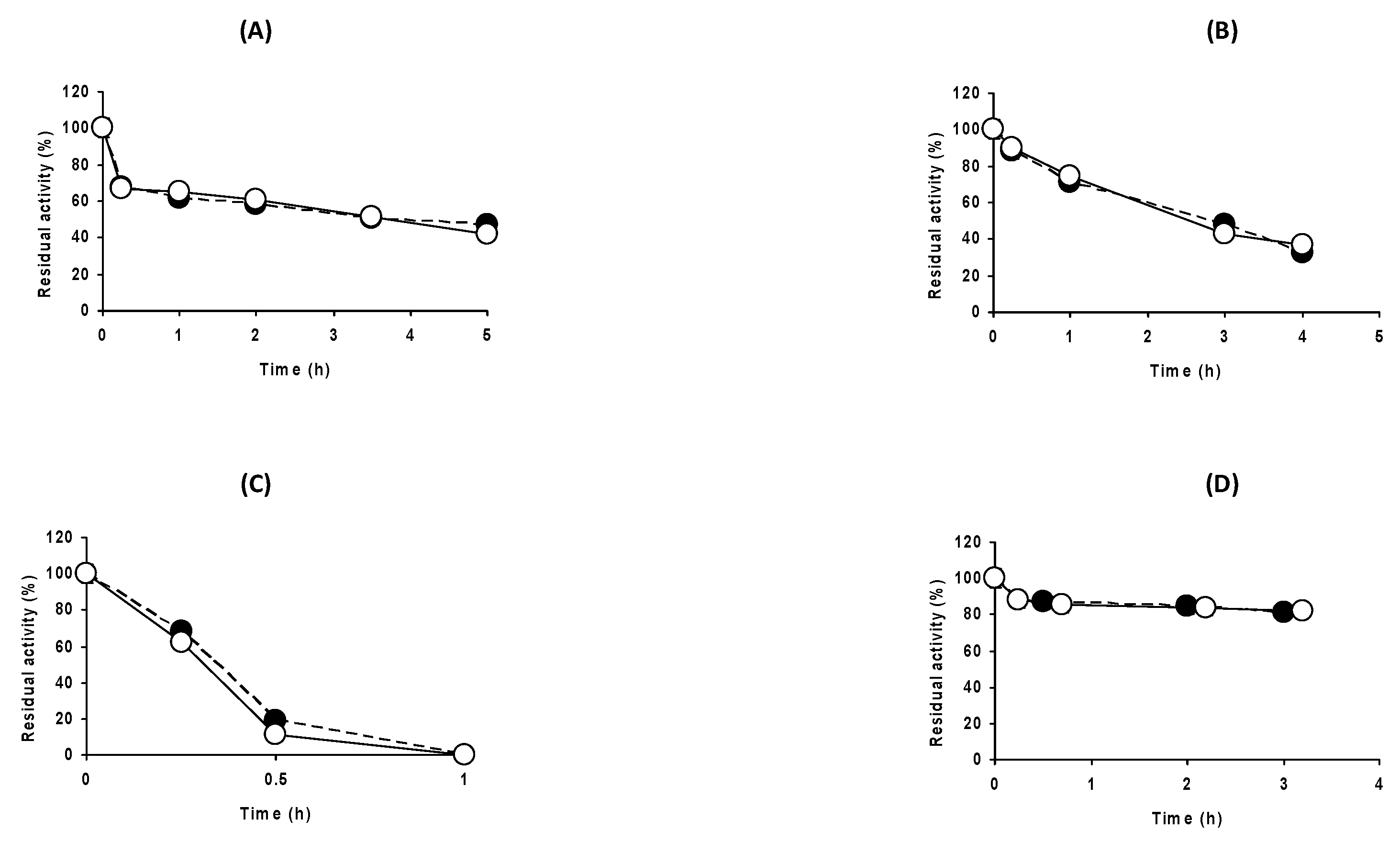
2.4. Effect of the Buffers’ Nature on the Stability of Immobilized DAAO When Enzyme Dissociation Was Not Possible

The results showed above show strong and complex effects of the buffer nature and concentration on enzyme stability, effects that might not be decreased upon adding FAD. As the enzyme inactivation started mainly with the dissociation of the enzyme subunit (Figure 1), the current results could have been based on the effects of the buffers on this enzyme subunit dissociation, but with the current information, we could not discard that the effects of the buffers were directly on the tri-dimensional structure of the enzyme [54,55,56]. One possibility to refuse the hypothesis of the role of the buffers on the enzyme dissociation, was if the effect was no longer visible using an enzyme formulation where the enzyme could not dissociate. To this goal, we used an immobilized biocatalyst of DAAO, immobilized in glutaraldehyde-agarose. Figure S1 shows the SDS-PAGE of this biocatalyst. It shows that this biocatalyst could not release any enzyme subunit to the medium when submitted to boiling in breaking buffer, making not possible the enzyme inactivation by subunit dissociation.
Figure 9 shows the inactivation of this enzyme biocatalyst using the different buffers utilized in this paper, where it was not possible to find clear differences in the stabilities of this biocatalyst using the different buffers. This result supports that the main reason for the effect of the different buffers on enzyme stability may be founded on the prevention/favoring of enzyme dissociation.

3. Materials and Methods

3.1. Materials

Glutaraldehyde solution grade I, 25% (v/v) in water and D-amino acid oxidase from porcine kidney (DAAO) (7.2 U/mg of powder) were acquired from Sigma Aldrich (Madrid, Spain). The 4BCL agarose beads standard was purchased from ABT (Burgos, Spain). 2,20-azino-bis (3-ethylbenzothiazoline-6-sulfonic acid) diammonium salt (ABTS®) was purchased from Roche (Mannheim, Germany). Alfa Aesar (Heysham, UK) provided D-alanine. Glycine, flavin adenine dinucleotide (FAD) disodium salt and horseradish peroxidase (268 U/mg of protein) were acquired from Fisher scientific Spain (Madrid, Spain). LMW-SDS markers (14.4–97.0 KDa) for electrophoresis reference were purchased from GE Healthcare Life Sciences (Madrid, Spain). Protein concentration was determined using Bradford’s method using bovine serum albumin as standard [57]. All other reagents were of analytical grade.

3.2. Methods

All experiments were performed in triplicate (as minimum) and the values were presented as mean values and standard deviations.

3.2.1. Determination of DAAO Activity

The activity of DAAO was determined via ABTS® coupled assay using a spectrophotometer thermoregulated at 25 °C, equipped with a magnetic stirring device. The ABTS® oxidization was caused by the peroxidase catalyzed reaction, using the hydrogen peroxide released in the DAAO oxidation reaction. It was checked that using double and half concentrations of peroxidase, the determined DAAO activity was identical. It was performed using 2.2 mL of 100 mM sodium carbonate at pH 8.3, containing 200 µL of D-alanine at 200 mM, 100 µL of ABTS® at 10 mg/mL prepared in 100 mM sodium phosphate at pH 7.0 and 50 µL of horseradish peroxidase at 0.1 mg/mL prepared in 100 mM sodium phosphate at pH 7.0. The reaction started when 10–100 µL of the enzyme sample was added to the cuvette. The oxidation of ABTS® was monitored using the change in absorbance of 414 nm (ε414 = 36,000 M−1 cm−1 under these conditions) [55]. One unit (U) of activity was defined as the amount of enzyme that oxidized 1 µmol of substrate per minute under the specified conditions.

3.2.2. Immobilization of DAAO on Glutaraldehyde-Amino-Agarose Beads

This immobilization protocol provided a biocatalyst where both enzyme subunits were immobilized on the support, preventing any risk of enzyme subunit dissociation [58].

Preparation of Glutaraldehyde-Amino-Agarose Beads

The amino-glutaraldehyde-agarose-activated support was prepared from monoaminoethyl-N-aminoethyl (MANAE) agarose, produced as previously described [59]. The support (10 g) was added to 90 mL of sodium phosphate containing 10% glutaraldehyde at pH 7 and 25 °C for 16 h. After, the support was extensively washed with distilled water to eliminate any remaining glutaraldehyde molecules.

Preparation of DAAO Glutaraldehyde-Amino-Agarose Beads

Immobilization on glutaraldehyde-amino-agarose was performed using 5 g of support per 50 mL of enzyme solution (0.2 mg/mL), prepared in 5 mM sodium phosphate at pH 7.0 during 1 h. The enzymatic activity of the suspension and supernatant was followed during the whole process using the ABTS® assay described above. Finally, the biocatalyst was washed with distilled water, vacuum dried and stored at 4–6 °C.

3.2.3. Thermal Inactivations of DAAO under Different Conditions

The enzyme concentration during the inactivation experiments was varied (0.2 mg/mL to 1 mg/mL). The enzyme was diluted in 5, 50, 125 or 250 mM of sodium phosphate, Tris-HCL, Hepes or glycine, adjusting the pH at 7.0, sodium acetate at pH 5.0 and sodium carbonate buffer at pH 9.0 (the pH was adjusted using HCl or NaOH). In some instances, 250 mM of NaCl or 2 mM of FAD were added. Samples were periodically withdrawn, and the residual activity was quantified using the ABTS® assay described above. In some instances, the immobilized enzyme was utilized (at a concentration of 0.2 mg protein/mL of inactivation media). The inactivation temperatures were selected to ensure reliable inactivation courses, and the error in the established temperatures was ±1 °C for different experiments.

3.2.4. SDS-PAGE of Enzyme Preparations

SDS-PAGE experiments were carried out following Laemmli’s protocol [60]. The samples (the free enzyme and the glutaraldehyde biocatalyst) were diluted in 4% SDS (w/v) and 10% mercaptoethanol (v/v). These suspensions were boiled for 8 min. In the case of the immobilized biocatalyst, the support was discarded after centrifuging at 10,000 rpm for 5 min using a Midi centrifuge (FisherBrand, Madrid, Spain). After injecting 15 µL aliquots of each sample, and 5 µL of low molecular weight marker proteins (LMW-SDS Marker 14.4–97 kDa), to carry out the SDS-PAGE analysis, the experiment was run at 100 V. Finally, gels were stained utilizing Coomassie brilliant blue stain.

4. Conclusions

The results presented in this paper offered a very clear example of the role of the medium nature on the stability of an enzyme, in this case, a multimeric enzyme where the first step of the inactivation was the enzyme subunit dissociation, as was shown by the effect of the enzyme concentration on the inactivation courses. However, they also show a very complex situation, suggesting that several phenomena (some positive, some negative) were simultaneously occurring to determine the enzyme stability. Some buffers permitted higher enzyme stability when they were used at higher concentrations, while other ones offered the opposite situation. This could not be directly correlated with the ionic strength of the buffers, as the addition of NaCl could increase or decrease the enzyme stability, depending on the buffer and enzyme concentration used in the inactivation experiments. This wide diversity of results made it not possible, with the available information, to formulate any hypothesis to explain the results; it seems that the effect of the media on this DAAO stability must be empirically analyzed before deciding the conditions of handling or storing of the enzyme.
The results (mainly the use of the immobilized biocatalyst where both enzyme subunits were immobilized) suggest that the main effect of the buffer was related to the favoring/disfavoring of the enzyme dissociation, as all buffers’ effects were almost gone using this stabilized biocatalyst. However, the buffer selection is critical during the handling and storage. Moreover, these results show, in a very clear way, that the stabilization achieved with the immobilization of the enzyme may be easily enlarged, for example performing the inactivation of the enzyme at low concentration and using Hepes as a buffer.

Supplementary Materials

The following are available online at https://www.mdpi.com/article/10.3390/catal12091009/s1, Figure S1: SDS-PAGE gels of the analysis of free enzyme and immobilized at 5 mM sodium phosphate buffer at pH 7.0 and 25 °C over glutaraldehyde agarose support. Lane 1: molecular weight markers; Lane 2: free enzyme (1 mg/mL); Lane 3: free enzyme (0.2 mg/mL); Lane 4: DAAO (0.2 mg/mL) immobilized on glutaraldehyde agarose support. Other specifications can be found in the Materials and Methods section.

Author Contributions

D.C. performed the experiments; J.R.-M. and R.F.-L. designed and supervised the experiments; all authors participated in the writing of the paper. All authors have read and agreed to the published version of the manuscript.

Funding

This research received no external funding.

Data Availability Statement

Data can be obtained from the authors.

Acknowledgments

DC thanks to Ministerio de Ciencia e Innovación-Spanish Government by a FPI (PRE2018-084809).

Conflicts of Interest

The authors declare no conflict of interest.

References

  1. Tishkov, V.I.; Khoronenkova, S.V. D-Amino acid oxidase: Structure, catalytic mechanism, and practical application. Biochemistry 2005, 70, 40–54. [Google Scholar] [PubMed]
  2. Takamatsu, S.; Lee, I.; Lee, J.; Asano, R.; Tsugawa, W.; Ikebukuro, K.; Dick, J.E.; Sode, K. Transient potentiometry based D-serine sensor using engineered D-amino acid oxidase showing quasi-direct electron transfer property. Biosens. Bioelectron. 2022, 200, 113927. [Google Scholar] [CrossRef] [PubMed]
  3. Khoronenkova, S.V.; Tishkov, V.I. D-Amino acid oxidase: Physiological role and applications. Biochemistry 2008, 73, 1511–1518. [Google Scholar] [CrossRef] [PubMed]
  4. Pollegioni, L.; Molla, G.; Sacchi, S.; Rosini, E.; Verga, R.; Pilone, M.S. Properties and applications of microbial D-Amino acid oxidases: Current state and perspectives. Appl. Microbiol. Biotechnol. 2008, 78, 1–16. [Google Scholar] [CrossRef] [PubMed]
  5. Pollegioni, L.; Molla, G. New biotech applications from evolved D-Amino acid oxidases. Trends Biotechnol. 2011, 29, 276–283. [Google Scholar] [CrossRef] [PubMed]
  6. Barber, M.S.; Giesecke, U.; Reichert, A.; Minas, W. Industrial enzymatic production of cephalosporin-based β-Lactams. In Molecular Biotechnolgy of Fungal Beta-Lactam Antibiotics and Related Peptide Synthetases; Springer: Berlin/Heidelberg, Germany, 2004; pp. 179–215. [Google Scholar]
  7. Moussa, S.; Murtas, G.; Pollegioni, L.; Mauzeroll, J. Enhancing electrochemical biosensor selectivity with engineered D-Amino acid oxidase enzymes for D-Serine and D-Alanine quantification. ACS Appl. Bio. Mater. 2021, 4, 5598–5604. [Google Scholar] [CrossRef] [PubMed]
  8. Tian, T.; Liu, M.; Chen, L.; Zhang, F.; Yao, X.; Zhao, H.; Li, X. D-Amino acid electrochemical biosensor based on D-Amino acid oxidase: Mechanism and high performance against enantiomer interference. Biosens. Bioelectron. 2020, 151, 111971. [Google Scholar] [CrossRef] [PubMed]
  9. Nieh, C.H.; Kitazumi, Y.; Shirai, O.; Kano, K. Sensitive D-Amino acid biosensor based on oxidase/peroxidase system mediated by pentacyanoferrate-bound polymer. Biosens. Bioelectron. 2013, 47, 350–355. [Google Scholar] [CrossRef]
  10. Lata, S.; Batra, B.; Kumar, P.; Pundir, C.S. Construction of an amperometric D-Amino acid biosensor based on D-Amino acid oxidase/carboxylated mutliwalled carbon nanotube/copper nanoparticles/polyalinine modified gold electrode. Anal. Biochem. 2013, 437, 1–9. [Google Scholar] [CrossRef]
  11. Upadhya, R.; Nagajyothi; Bhat, S.G. Stabilization of D-Amino acid oxidase and catalase in permeabilized Rhodotorula gracilis cells and its application for the preparation of α-Ketoacids. Biotechnol. Bioeng. 2000, 68, 430–436. [Google Scholar] [CrossRef]
  12. Upadhya, R.; Nagajyothi, H.; Bhat, S.G. D-Amino acid oxidase and catalase of detergent permeabilized Rhodotorula gracilis cells and its potential use for the synthesis of α-keto acids. Process Biochem. 1999, 35, 7–13. [Google Scholar] [CrossRef]
  13. Fernández-Lafuente, R.; Rodriguez, V.; Guisán, J.M. The coimmobilization of D-Amino acid oxidase and catalase enables the quantitative transformation of D-Amino acids (D-Phenylalanine) into α-Keto acids (phenylpyruvic acid). Enzym. Microb. Technol. 1998, 23, 28–33. [Google Scholar] [CrossRef]
  14. Butò, S.; Pollegioni, L.; D’Angiuro, L.; Pilone, M.S. Evaluation of D-Amino acid oxidase from Rhodotorula gracilis for the production of α-keto acids: A reactor system. Biotechnol. Bioeng. 1994, 44, 1288–1294. [Google Scholar] [CrossRef] [PubMed]
  15. Wong, K.S.; Fong, W.P.; Tsang, P.W.K. Entrapment of a Trigonopsis variabilis D-Amino acid oxidase variant F54Y for oxidative deamination of cephalosporin C. Eng. Life Sci. 2011, 11, 491–495. [Google Scholar] [CrossRef]
  16. Yu, H.L.; Li-Gong, C.; Yang, L.; Xi-Long, Y.; Guo-Yi, B.; Xue-Ming, D. Effect of hydrogen peroxide on the conversion of cephalosporin C to glutaryl-7-ACA by D-Amino acid oxidase. J. Chem. Eng. Chin. Univ. 2006, 20, 79–84. [Google Scholar]
  17. Shih-Yun, L.; Wang, J.D.; Lin, J.H. Expression of Trigonopsis variabilis D-Amino acid oxidase in transgenic rice for cephalosporin production. Bot. Stud. 2009, 50, 181–192. [Google Scholar]
  18. Pollegioni, L.; Caldinelli, L.; Molla, G.; Sacchi, S.; Pilone, M.S. Catalytic properties of D-Amino acid oxidase in cephalosporin C bioconversion: A comparison between proteins from different sources. Biotechnol. Prog. 2004, 20, 467–473. [Google Scholar] [CrossRef]
  19. Volpato, G.; Rodrigues, R.C.; Fernandez-Lafuente, R. Use of enzymes in the production of semi-synthetic penicillins and cephalosporins: Drawbacks and perspectives. Curr. Med. Chem. 2010, 17, 3855–3873. [Google Scholar] [CrossRef] [PubMed]
  20. Hernandez, K.; Berenguer-Murcia, A.; Rodrigues, R.C.; Fernandez-Lafuente, R. Hydrogen peroxide in biocatalysis. A dangerous liaison. Curr. Org. Chem. 2012, 16, 2652–2672. [Google Scholar] [CrossRef]
  21. Lopez-Gallego, F.; Batencor, L.; Hidalgo, A.; Mateo, C.; Fernandez-Lafuente, R.; Guisan, J.M. One-pot conversion of cephalosporin C to 7-Aminocephalosporanic acid in the absence of hydrogen peroxide. Adv. Synth. Catal. 2005, 347, 1804–1810. [Google Scholar] [CrossRef]
  22. Luo, H.; Li, Q.; Yu, H.; Shen, Z. Construction and application of fusion proteins of D-Amino acid oxidase and glutaryl-7-Aminocephalosporanic acid acylase for direct bioconversion of cephalosporin C to 7-Aminocephalosporanic acid. Biotechnol. Lett. 2004, 26, 939–945. [Google Scholar] [CrossRef]
  23. Carballares, D.; Morellon-Sterling, R.; Fernandez-Lafuente, R. Design of artificial enzymes bearing several active centers: New trends, opportunities and problems. Int. J. Mol. Sci. 2022, 23, 5304. [Google Scholar] [CrossRef]
  24. Mizutani, H.; Miyahara, I.; Hirotsu, K.; Nishina, Y.; Shiga, K.; Setoyama, C.; Miura, R. Three-Dimensional structure of porcine kidney D-Amino acid oxidase at 3.0 a resolution. J. Biochem. 1996, 120, 14–17. [Google Scholar] [CrossRef] [PubMed]
  25. Fukui, K.; Watanabe, F.; Shibata, T.; Miyake, Y. Molecular cloning and sequence analysis of cDNAs encoding porcine kidney D-Amino acid oxidase. Biochemistry 1987, 26, 3612–3618. [Google Scholar] [CrossRef]
  26. Mizutani, H.; Miyahara, I.; Hirotsu, K.; Nishina, Y.; Shiga, K.; Setoyama, C.; Miura, R. Three-dimensional structure of the purple intermediate of porcine kidney D-Amino acid oxidase. Optimization of the oxidative half-reaction through alignment of the product with reduced flavin. J. Biochem. 2000, 128, 73–81. [Google Scholar] [CrossRef] [PubMed]
  27. Setoyama, C.; Miura, R.; Nishina, Y.; Shiga, K.; Mizutani, H.; Miyahara, I.; Hirotsu, K. Crystallization of expressed porcine kidney D-Amino acid oxidase and preliminary X-ray crystallographic characterization. J. Biochem. 1996, 119, 1114–1117. [Google Scholar] [CrossRef] [PubMed]
  28. Yasukawa, K.; Motojima, F.; Ono, A.; Asano, Y. Expansion of the substrate specificity of porcine kidney D-Amino acid oxidase for S-stereoselective oxidation of 4-Cl-benzhydrylamine. ChemCatChem 2018, 10, 3500–3505. [Google Scholar] [CrossRef]
  29. Kawahara, N.; Yasukawa, K.; Asano, Y. New enzymatic methods for the synthesis of primary α-Aminonitriles and unnatural α-Amino acids by oxidative cyanation of primary amines with D-Amino acid oxidase from porcine kidney. Green Chem. 2017, 19, 418–424. [Google Scholar] [CrossRef]
  30. Yasukawa, K.; Kawahara, N.; Motojima, F.; Nakano, S.; Asano, Y. Porcine kidney D-Amino acid oxidase-derived R-Amine oxidases with new substrate specificities. Enzymes 2020, 47, 117–136. [Google Scholar]
  31. Kawahara, N.; Palasin, K.; Asano, Y. Novel enzymatic method for imine synthesis via the oxidation of primary amines using D-Amino acid oxidase from porcine kidney. Catalysts 2022, 12, 511. [Google Scholar] [CrossRef]
  32. Watanabe, F.; Fukui, K.; Momoi, K.; Miyake, Y. Expression of normal and abnormal porcine kidney D-Amino acid oxidases in Escherichia coli: Purification and characterization of the enzymes. Biochem. Biophys. Res. Commun. 1989, 165, 62–73. [Google Scholar] [CrossRef]
  33. Ciccarelli, E.; Massaer, M.; Guillaume, J.P.; Herzog, A.; Loriau, R.; Cravador, A.; Jacobs, P.; Bollen, A. Porcine D-Amino acid oxidase: Production of the biologically active enzyme in Escherichia coli. Biochem. Biophys. Res. Commun. 1989, 161, 865–872. [Google Scholar] [CrossRef]
  34. Bakke, M.; Setoyama, C.; Miura, R.; Kajiyama, N. Thermostabilization of porcine kidney D-Amino acid oxidase by a single Amino acid substitution. Biotechnol. Bioeng. 2006, 93, 1023–1027. [Google Scholar] [CrossRef] [PubMed]
  35. Atroshenko, D.L.; Pometun, A.A.; Savin, S.S.; Tishkov, V.I. Determination of the kinetic parameters of a wild-type D-Amino acid oxidase from yeast and its mutant forms in a reaction of cephalosporin C oxidation. Mosc. Univ. Chem. Bull. 2019, 74, 169–172. [Google Scholar] [CrossRef]
  36. Poltorak, O.M.; Chukhray, E.S.; Torshin, I. Dissociative thermal inactivation, stability, and activity of oligomeric enzymes. Biochemistry 1998, 63, 303–311. [Google Scholar]
  37. Fernandez-Lafuente, R. Stabilization of multimeric enzymes: Strategies to prevent subunit dissociation. Enzym. Microb. Technol. 2009, 45, 405–418. [Google Scholar]
  38. Bolivar, J.M.; Rocha-Martin, J.; Mateo, C.; Cava, F.; Berenguer, J.; Fernandez-Lafuente, R.; Guisan, J.M. Coating of soluble and immobilized enzymes with ionic polymers: Full stabilization of the quaternary structure of multimeric enzymes. Biomacromolecules 2009, 10, 742–747. [Google Scholar] [CrossRef]
  39. Garcia-Galan, C.; Barbosa, O.; Fernandez-Lafuente, R. Stabilization of the hexameric glutamate dehydrogenase from Escherichia coli by cations and polyethyleneimine. Enzym. Microb. Technol. 2013, 52, 211–217. [Google Scholar] [CrossRef]
  40. Rodrigues, R.C.; Berenguer-Murcia, Á.; Carballares, D.; Morellon-Sterling, R.; Fernandez-Lafuente, R. Stabilization of enzymes via immobilization: Multipoint covalent attachment and other stabilization strategies. Biotechnol. Adv. 2021, 52, 107821. [Google Scholar]
  41. Findrik, Z.; Tusić, M.; Vasić-Rački, D. Stabilization of D-Amino acid oxidase via covalent immobilization and mathematical model of D-Methionine oxidative deamination catalyzed by immobilized enzyme. Chem. Biochem. Eng. Q. 2016, 30, 93–102. [Google Scholar] [CrossRef]
  42. Mateo, C.; Palomo, J.M.; Fernandez-Lorente, G.; Guisan, J.M.; Fernandez-Lafuente, R. Improvement of enzyme activity, stability and selectivity via immobilization techniques. Enzym. Microb. Technol. 2007, 40, 1451–1463. [Google Scholar]
  43. Garcia-Galan, C.; Berenguer-Murcia, Á.; Fernandez-Lafuente, R.; Rodrigues, R.C. Potential of different enzyme immobilization strategies to improve enzyme performance. Adv. Synth. Catal. 2011, 353, 2885–2904. [Google Scholar]
  44. Di Cosimo, R.; Mc Auliffe, J.; Poulose, A.J.; Bohlmann, G. Industrial use of immobilized enzymes. Chem. Soc. Rev. 2013, 42, 6437–6474. [Google Scholar]
  45. Liese, A.; Hilterhaus, L. Evaluation of immobilized enzymes for industrial applications. Chem. Soc. Rev. 2013, 42, 6236–6249. [Google Scholar] [CrossRef] [PubMed]
  46. Bolivar, J.M.; Woodley, J.M.; Fernandez-Lafuente, R. Is Enzyme Immobilization a Mature Discipline? Some Critical Considerations to Capitalize on the Benefits of Immobilization. Chem. Soc. Rev. 2022. submitted. [Google Scholar]
  47. Betancor, L.; Hidalgo, A.; Fernández-Lorente, G.; Mateo, C.; Rodríguez, V.; Fuentes, M.; López-Gallego, F.; Fernández-Lafuente, R.; Guisan, J.M. Use of physicochemical tools to determine the choice of optimal enzyme: Stabilization of D-Amino acid oxidase. Biotechnol. Prog. 2003, 19, 784–788. [Google Scholar] [CrossRef] [PubMed]
  48. Bauduin, P.; Nohmie, F.; Touraud, D.; Neueder, R.; Kunz, W.; Ninham, B.W. Hofmeister specific-ion effects on enzyme activity and buffer pH: Horseradish peroxidase in citrate buffer. J. Mol. Liq. 2006, 123, 14–19. [Google Scholar]
  49. Rej, R.; Vanderlinde, R.E. Effects of buffers on aspartate Aminotransferase activity and association of the enzyme with pyridoxal phosphate. Clin. Chem. 1975, 21, 1585–1591. [Google Scholar]
  50. Haije, W.G. Influence of buffer conditions on the activities of some iso-enzymes of alkaline phosphatase in the serum. Clin. Chim. Acta 1973, 48, 23–26. [Google Scholar]
  51. Garry, P.J. Serum cholinesterase variants: Examination of several differential inhibitors, salts, and buffers used to measure enzyme activity. Clin. Chem. 1971, 17, 183–191. [Google Scholar] [PubMed]
  52. Singh, S.; Sharma, S.; Agarwal, S.K. A simple purification procedure of buffalo lung cathepsin H, its properties and influence of buffer constituents on the enzyme activity. Biochem. Biophys. Rep. 2020, 22, 100739. [Google Scholar]
  53. Nicholas, P.C. Fructose-1,6-bisphosphate aldolases from guinea-pig cerebral cortex: Variation of enzyme activity with substrate concentration observed in Tris/HCl buffer. Biochem. Soc. Trans. 1989, 17, 227–228. [Google Scholar] [CrossRef]
  54. Zaak, H.; Fernandez-Lopez, L.; Velasco-Lozano, S.; Alcaraz-Fructuoso, M.T.; Sassi, M.; Lopez-Gallego, F.; Fernandez-Lafuente, R. Effect of high salt concentrations on the stability of immobilized lipases: Dramatic deleterious effects of phosphate anions. Process Biochem. 2017, 62, 128–134. [Google Scholar]
  55. Kornecki, J.F.; Carballares, D.; Morellon-Sterling, R.; Siar, E.H.; Kashefi, S.; Chafiaa, M.; Arana-Peña, S.; Rios, N.S.; Gonçalves, L.R.B.; Fernandez-Lafuente, R. Influence of phosphate anions on the stability of immobilized enzymes. Effect of enzyme nature, immobilization protocol and inactivation conditions. Process Biochem. 2020, 95, 288–296. [Google Scholar]
  56. Braham, S.A.; Siar, E.-H.; Arana-Peña, S.; Carballares, D.; Morellon-Sterling, R.; Bavandi, H.; de Andrades, D.; Kornecki, J.F.; Fernandez-Lafuente, R. Effect of concentrated salts solutions on the stability of immobilized enzymes: Influence of inactivation conditions and immobilization protocol. Molecules 2021, 26, 968. [Google Scholar] [CrossRef] [PubMed]
  57. Bradford, M.M. A rapid and sensitive method for the quantitation of microgram quantities of protein utilizing the principle of protein-dye binding. Anal. Biochem. 1976, 72, 248–254. [Google Scholar] [CrossRef]
  58. Carballares, D.; Rocha-Martín, J.; Fernandez–Lafuente, R. Immobilization-Stabilization of the Dimeric D-Amino Acid Oxidase from Porcine Kidney. Process Biochem. submitted.
  59. Fernandez-Lafuente, R.; Rosell, C.M.; Rodriguez, V.; Santana, C.; Soler, G.; Bastida, A.; Guisán, J.M. Preparation of activated supports containing low pK Amino groups. A new tool for protein immobilization via the carboxyl coupling method. Enzym. Microb. Technol. 1993, 15, 546–550. [Google Scholar] [CrossRef]
  60. Laemmli, U.K. Cleavage of structural proteins during the assembly of the head of bacteriophage T4. Nature 1970, 227, 680–685. [Google Scholar] [PubMed]
Figure 1. Thermal inactivation courses of free DAAO at different concentrations. The enzyme was inactivated in 5 mM of different buffers at 40 °C. (A): 5 mM of sodium acetate at pH 5.0; (B): 5 mM of Tris-HCL at pH 7.0 and (C): 5 mM of sodium carbonate at pH 9.0. Solid squares: 0.01 mg/mL; solid triangles: 0.1 mg/mL; solid circles: 0.5 mg/mL and solid rhombi: 2.5 mg/mL. Other specifications are described in the Methods section.
Catalysts 12 01009 g001
Figure 2. Effect of FAD addition and inactivation pH on the stability of free DAAO (0.2 mg/mL). (A): 5 mM of sodium acetate buffer at pH 5.0; (B): 5 mM of Tris-HCL buffer at pH 7.0 and (C): 5 mM of sodium carbonate buffer at pH 9.0. Solid squares: free enzyme (0.2 mg/mL); solid circles: free enzyme with 0.11 mM of FAD; solid tringles: free enzyme with 0.33 mM of FAD and solid rhombi: free enzyme with 2.0 mM of FAD. Other specifications are described in the Methods section.
Catalysts 12 01009 g002
Figure 3. Thermal inactivation courses of free DAAO (A): (0.2 mg/mL, at 40 °C) and (B): (1 mg/mL, at 45 °C). The enzyme was inactivated with 5 mM of different buffers at pH 7.0. Solid squares: sodium phosphate buffer; solid triangles: Tris-HCl buffer; solid circles: Hepes buffer and solid rhombi: glycine buffer. Other specifications are described in the Methods section.
Catalysts 12 01009 g003
Figure 4. Effect of 2 mM of FAD addition on the effect of the buffer nature on the stability of free DAAO (0.2 mg/mL). (A): Tris-HCl buffer at pH 7.0 and 40 °C; (B): sodium phosphate buffer at pH 7.0 and 40 °C; (C): Hepes buffer at pH 7.0 and 35 °C and (D): glycine buffer at pH 7.0 and 40 °C. Solid circles: 5 mM of buffer and empty circles: 5 mM of buffer with 2 mM of FAD. Other specifications are described in the Methods section.
Catalysts 12 01009 g004
Figure 5. Effect of buffer concentration on the stability of free DAAO (0.2 mg/mL). (A): sodium acetate buffer at pH 5.0 and 40 °C; (B): Tris-HCl buffer at pH 7.0 and 40 °C; (C): sodium phosphate buffer at pH 7.0 and 40 °C; (D): Hepes buffer at pH 7.0 and 35 °C; (E): glycine buffer at pH 7.0 and 40 °C and (F): sodium carbonate buffer at pH 9.0 and 35 °C. Solid squares: 5 mM; solid triangles: 50 mM; solid rhombi: 125 mM and solid circles: 250 mM. Other specifications are described in the Methods section.
Catalysts 12 01009 g005
Figure 6. Effect of ionic strength and inactivation pH on the stability of free DAAO (1 mg/mL). (A): sodium acetate buffer at pH 5.0 and 45 °C; (B): Tris-HCl buffer at pH 7.0 and 45 °C; (C): sodium phosphate buffer at pH 7.0 and 45 °C; (D): Hepes buffer at pH 7.0 and 35 °C; (E): glycine buffer at pH 7.0 and 45 °C and (F): sodium carbonate buffer at pH 9.0 and 45 °C. Solid squares: 5 mM; solid triangles: 50 mM; solid rhombi: 125 mM and solid circles: 250 mM. Other specifications are described in the Methods section.
Catalysts 12 01009 g006
Figure 7. Effect of 250 mM of NaCl addition on the stability of free DAAO (0.2 mg/mL) in different media. (A): sodium acetate buffer at pH 5.0 and 40 °C; (B): Tris-HCl buffer at pH 7.0 and 40 °C; (C): sodium phosphate buffer at pH 7.0 and 40 °C; (D): Hepes buffer at pH 7.0 and 35 °C; (E): glycine buffer at pH 7.0 and 40 °C and (F): sodium carbonate buffer at pH 9.0 and 40 °C. Solid symbols: without NaCl; empty symbols: with NaCl. Squares: 5 mM and circles: 250 mM of buffer concentration. Other specifications are described in the Methods section.
Catalysts 12 01009 g007
Figure 8. Effect of 250 mM of NaCl addition on the stability of free enzyme DAAO (1 mg/mL. (A): sodium acetate buffer at pH 5.0 and 40 °C; (B): Tris-HCl buffer at pH 7.0 and 40 °C; (C): sodium phosphate buffer at pH 7.0 and 40 °C; (D): Hepes buffer at pH 7.0 and 35 °C; (E): glycine buffer at pH 7.0 and 40 °C and (F): sodium carbonate buffer at pH 9.0 and 40 °C. Solid symbols: without NaCl; empty symbols: with NaCl. Squares: 5 mM and circles: 250 mM of buffer concentration. Other specifications are described in the Methods section.
Catalysts 12 01009 g008
Figure 9. Inactivation courses of DAAO (2 mg/g of biocatalyst, diluted 1/10 with the inactivation media) immobilized on glutaraldehyde agarose support. The inactivation was performed in 5 mM of different buffers at pH 7.0 and 40 °C. Solid squares: Tris-HCL; solid triangles: sodium phosphate buffer; solid circles: Hepes buffer and solid rhombi: glycine buffer. Other specifications are described in the Methods section.
Catalysts 12 01009 g009
Publisher’s Note: MDPI stays neutral with regard to jurisdictional claims in published maps and institutional affiliations.

