Article

Screening of Cellulolytic Bacteria from Various Ecosystems and Their Cellulases Production under Multi-Stress Conditions

1
Laboratory of Applied Microbiology, Department of Microbiology, Faculty of Natural and Life Sciences, University Ferhat Abbas of Setif, Setif 19000, Algeria
2
East Azarbaijan Agricultural and Natural Resources Research and Education Centre, Plant Protection Research Department, Agricultural Research, Education and Extension Organization (AREEO), Tabriz 5355179854, Iran
3
Department of Biology and Genetics, Institute of Biology, Zoology and Radiobiology, University of Veterinary Medicine and Pharmacy in Košice, 04181 Kosice, Slovakia
4
Marine Biodiscovery Centre, Department of Chemistry, University of Aberdeen, Old Aberdeen AB24 3UE, UK
5
Laboratory of Soil Biology, University of Neuchatel, 11 Rue Emile Argand, CH-2000 Neuchatel, Switzerland
*
Author to whom correspondence should be addressed.
Catalysts 2022, 12(7), 769; https://doi.org/10.3390/catal12070769
Submission received: 17 May 2022 / Revised: 30 June 2022 / Accepted: 9 July 2022 / Published: 11 July 2022
(This article belongs to the Special Issue Catalysis in Biomass Valorization)

Abstract

:
Cellulose represents the most abundant component of plant biomass on earth; it is degraded by cellulases, specific enzymes produced by microorganisms. However, cellulases of bacterial origin attract more interest due to their natural diversity and ability to inhabit a variety of niches, allowing the selection of cellulolytic strains resistant to environmental stresses. The screening of the cellulolytic activity of 398 bacteria isolated from various ecosystems in Algeria (cave, ruins, chott, thermal station, and rhizosphere of arid and semi-arid regions) was performed by the appearance of a hydrolysis zone on carboxymethylcellulose (CMC) medium. The cellulase activity on CMC (1%) broth allowed to select 26 strains among which 12 had the best activity (0.3 U/mL to 2.2 U/mL). Optimization of physicochemical parameters (salinity: 0–1 M NaCl; pH: 3, 4, 7, 9, and 11; temperature: 30, 45, and 50 °C; PEG8000: 0 and 30%) involved in growth and cellulose production showed that the majority of strains were mesophilic, neutrophilic, or alkali- tolerant and tolerant to 30% of PEG8000. The cellulase activity and stability under different stress allowed to retain five strains, which the most efficient. Based on the 16S-rRNA sequencing results, they belonged to the genus Bacillus. The physicochemical properties of cellulases (crude extract) showed a CMCase active over a wide range of pH (4 to 11), optimal at 50 °C and 60 °C. The inhibiting salinity effect on the activity was not detected and was negligible on the enzymatic stability. The residual CMCase activity remained between 40 and 70% in a temperature range between 40 and 70 °C, was stable over a wide range of saline concentrations (0–2000 mM), and was weakly affected at 30% of PEG8000. The crude enzyme extract was able to hydrolyze both soluble and insoluble cellulosic substrates. The evaluation of the hydrolysis capacity of lignocellulosic waste revealed the ability of tested strains to degrade wheat bran, barley bran, and corncob. In addition, the enzyme showed significant multi-stress resistance on solid and liquid media. By these characteristics, these cellulolytic bacteria could be attractive to be used in various industrial and biotechnology applications.

1. Introduction

Each year, approximately 998 million tons of lignocellulosic waste are generated by agricultural activities [1,2]. In terrestrial environments, Lignocellulose is the main product of photosynthesis and represents on average the most abundant renewable plant biomass bio-resource in the world [3]. Lignocellulosic material is mainly composed of three structural polymers, namely cellulose, hemicellulose, and lignin, and is one of the best options as an ecological raw material of industrial and biotechnological interest, as it is both inexpensive and easily available [4,5]. Their valorization reduces the use of fossil fuels and preserves the natural environment [6]. Several methods are available for the pretreatment of lignocellulose, namely physical, chemical, and biological methods. The application of microorganisms or microbial enzymes for the pretreatment of lignocellulosic materials is very attractive and has attracted huge attention from the industry. This interest would be attributed to the results of the application of lignocellulosic biomass for the production of bioethanol by enzymatic hydrolysis as a sustainable alternative to fossil fuels [7]. Cellulose hydrolysis includes a group of enzymes (β-glucosidases, endoglucanases, and exoglucanases) known as cellulase, which breaks β-1,4-glycosidic links synergistically while releasing glucose molecules [7]. According to global enzyme market reports, cellulase is the most demanded enzyme, thus covering around 20% of the global market [8]. In the United States, the cellulase market has grown exponentially and reached USD 400 million per year [9]. Cellulase is increasingly applied in various fields, namely health, where it is used as a treatment for biofilms produced by Pseudomonas as an alternative to antibiotics [10]; the textile industry (bio-washing, biopolishing, bio-stonewashing, and bleaching of denim) [11]; biotechnology (production of bioethanol and valorization of lignocellulosic material); the paper industry (bio-bleaching and elimination of ink); and the foods and beverages industry (production of fruit juices and enhancement of aromas and flavors). Cellulase is also used to produce detergents and cleaning and washing agents [10,11].
Several microorganisms can produce cellulases: including fungi, bacteria, and actinomycetes. Currently, the majority of laboratory and commercial cellulases are obtained from fungi: mainly Trichoderma, Aspergillus, and Penicillium due to their high enzymatic activity and hydrolysis capacity [12,13]. However, cellulases of bacterial origin are more attractive due to the natural diversity and high growth rate of the bacteria that secrete them. More importantly, bacteria inhabit a wide variety of environmental niches capable of selecting cellulolytic strains that are extremely resistant to environmental stresses [3]. Moreover, these strains often produce enzymes that are stable under extreme conditions. Many bacterial genera are characterized in the literature as extracellular cellulase producers, including Acetivibrio, Alteromonas, Bacillus, Clostridium, Cellulomonas, and Ruminococcus [13]. The genus Bacillus remains the most studied and the most cited [14]. The species of this genus are isolated from a wide variety of environmental niches allowing them to resist physico-chemical stresses and are thus promising producers of alkalophilic, thermophilic, psychrophilic, acidophilic, and halophilic cellulolytic enzymes [15]. Moreover, Bacillus spp. are among the most attractive bacteria in industrial biotechnology; in particular, for the production of cellulases, they are generally easily and quickly culturable, have very few nutritional requirements, and produce large quantities of enzymes. In addition, great efforts have been devoted recently to the identification of strains of extremophilic Bacillus: halophiles, thermophiles, psychrophiles, acidophiles and alkalophiles, xerophiles, and their genes involved in the production of cellulases [16]. The objectives of this study were to evaluate the cellulase activity of bacteria isolated from various environmental biotopes, to optimize certain physico-chemical factors involved in the production of cellulase, then to determine the stability of the enzyme against multiple physico-chemical factors, and to evaluate the capacity of the enzyme to degrade certain cellulosic substrates, which proves useful for its subsequent industrial application.

2. Results

2.1. Screening for Cellulase-Producing Strains

The characterization of cellulase activity was carried out on 398 isolates from various environments. A rate of 6.5% (n = 26/398) (Table 1) of the tested isolates expressed CMCase activity, with enzymatic indexes varying between 0.34 and 5.2. The analysis of variance of the effect of strain on cellulase production was significant at p ≤ 0.05 (Figure 1). The isolates B6, Ba8, B6, HOT4, and P1 showed significant cellulase production with a maximum in P1 revealed by Congo red solution and B6 and HOT4 revealed by Lugol’s solution. However, the other isolates showed appreciable or negligible cellulase activity. The effect of the revelation solution was also significant. Indeed, the Ei (Enzymatic index) obtained by Lugol’s solution were better than those obtained by Congo red solution (Figure 2).

2.2. Evaluation of the Enzymatic Activity

The quantification of the enzymatic activity (U/mL), in liquid medium was carried out on the 26 cellulase-producing isolates. Almost half of the isolates (n = 12) expressed significant enzymatic activity (from 0.3 to 2.2 U/mL). These were Ba8, Ba13, D4, HOT1, HOT2, HOT3, HOT4, EP8, ET11, ET12, RK2, and RT10, while the others had very low (B1, B6, B10, B25, D6, HOT5, HOT6, E17, and P1) or no (Ba1, C16, T10, L4, and N4) enzymatic activity (Figure 3).

2.3. Effect of Physicochemical Parameters on Bacterial Growth and Cellulase Production

The study of the effect of physico-chemical parameters on bacterial growth and cellulase production was carried out on 12 isolates selected according to their enzymatic performance in liquid medium.

2.3.1. Effect of pH on Bacterial Growth

The effect of culture medium pH on bacterial growth was significant at p ≤ 0.05. All the isolates were neutrophilic and moderately alkalo-tolerant (pH 9). It should be noted that the strongly acidic and basic pH inhibited the growth of all the bacteria tested (Figure S1).

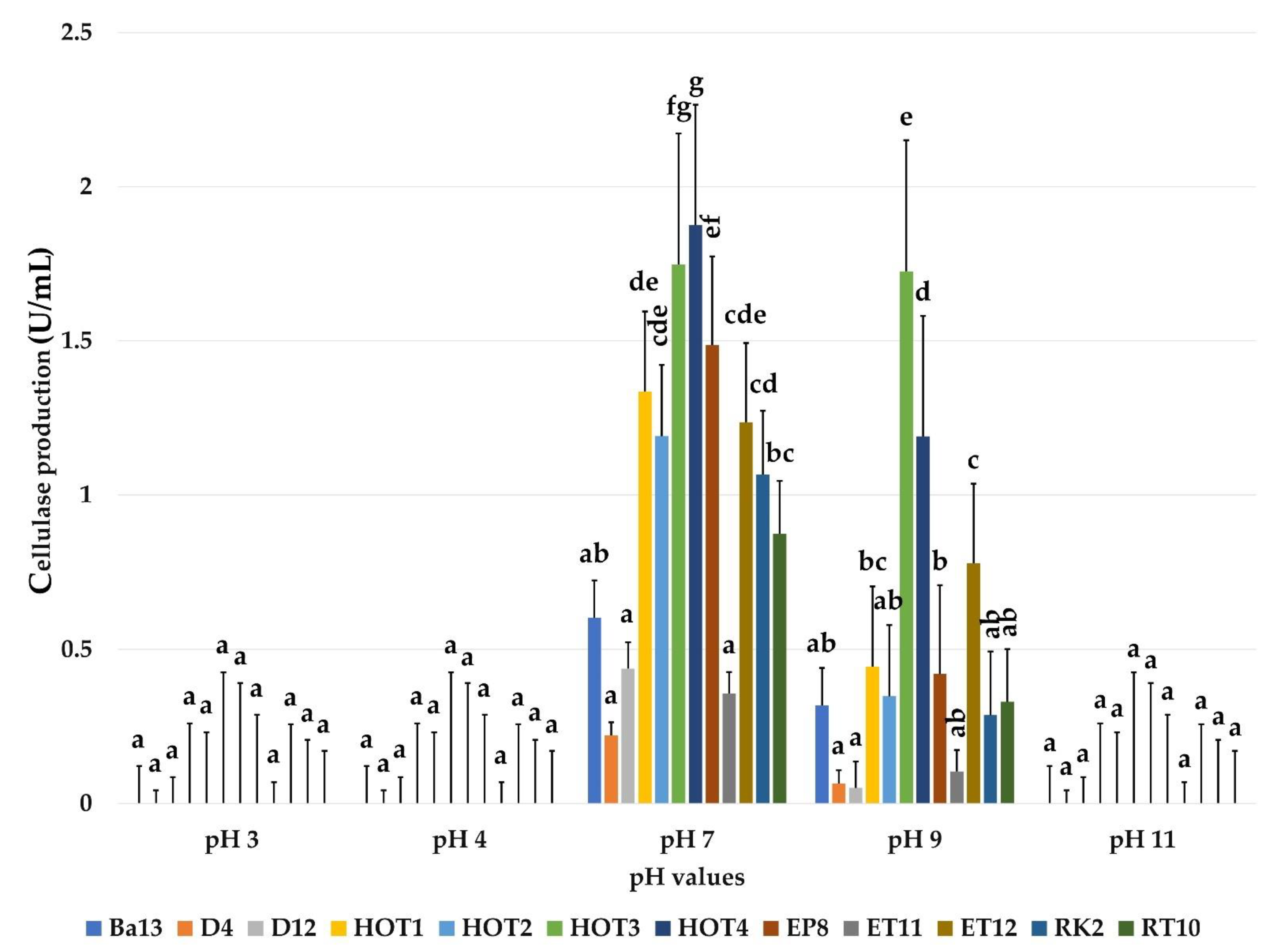
2.3.2. Effect of pH on Cellulase Production

The effect of culture medium pH on cellulase production was significant at p ≤ 0.05. Cellulase activity was optimal at neutral pH for all isolates. Some isolates (HOT3, HOT4, and ET12) produced significant CMCase at pH 9. The others, although showing visible growth on CMC medium, synthesized very small quantities of this enzyme (Figure 4).

2.3.3. Effect of Temperature on Bacterial Growth

The effect of heat treatment was selective (Figure S2). Indeed, bacterial growth at 45 °C and 50 °C decreased significantly compared to the optimal temperature of 30 °C for the majority of isolates. However, four isolates, one isolated from the thermal waters of Hammam Ouled Tebben (HOT1) and the other three from the rhizosphere of Terfez (EP8, ET11, and ET12), had optimal growth temperatures of 45 °C and 50 °C, respectively.

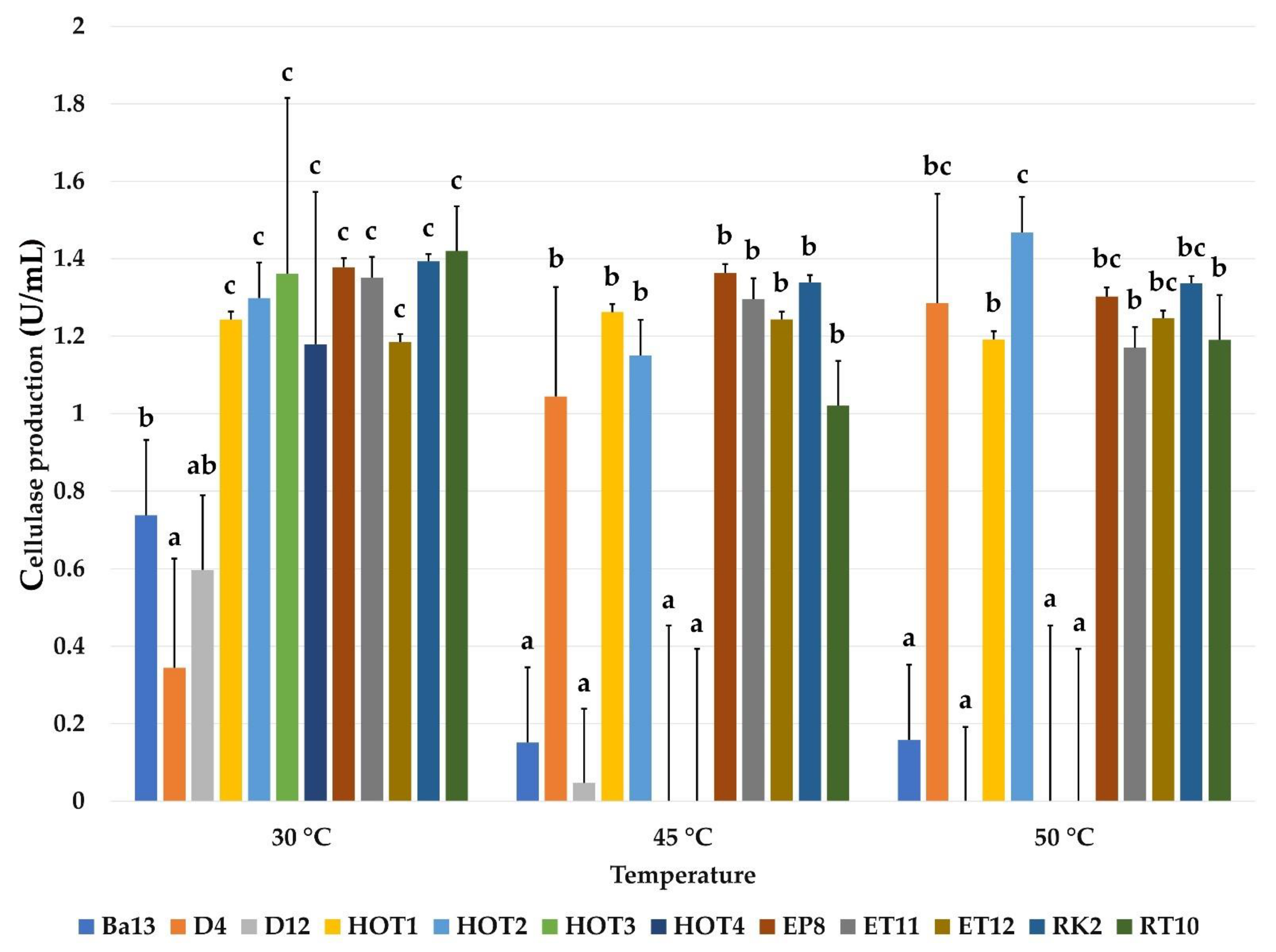
2.3.4. Effect of Temperature on Cellulase Production

The effect of incubation temperature on cellulase production revealed that the majority of the tested isolates were able to produce cellulase with stable cellulase activity at high temperatures of 45 °C and 50 °C. However, the synthesis of this enzyme for Ba13, D12, HOT3, and HOT4 bacteria was inhibited by heat stress (Figure 5).

2.3.5. Effect of Salinity on Bacterial Growth

The inhibiting effect of culture medium salinity on bacterial growth was visible (p ≤ 0.05) from 400 mM NaCl. However, Ba13, D4, D12, HOT1, HOT2, ET11, ET12, RK2, and RT10 were able to grow at this level of salinity. The isolates HOT1 and RT10 were tolerant to NaCl concentrations ranging from 800 to 1000 mM (Figure S3).

2.3.6. Effect of Salinity on Cellulase Production

The effect of salt stress on cellulase production was significant at p ≤ 0.05. Half of the isolates were capable of cellulase synthesis at high salinity levels of 400 to 800 mM NaCl. However, at 1000 mM, isolates HOT1, EP8, ET11, and RT10 showed a low but non-negligible enzymatic activity (Figure 6).

2.3.7. Effect of Water Activity (aw) on Bacterial Growth

The effect of water activity on the bacterial growth of the 12 isolates was determined by adding increasing levels of PEG8000 to the liquid CMC medium incubated at 30 °C/72 h. All isolates were able to grow at PEG8000 concentrations up to 30%. It should be noted that EP8, RK2, and RT10 developed better at 30% PEG8000 than at 0% (Figure S4).

2.3.8. Effect of Water Activity on Cellulase Production

The effect of water stress on the production of cellulase was determined by measuring the enzymatic activity. The production of cellulase clearly decreased at 30% of PEG8000 for the majority of the tested isolates except for HOT1, which produced enzyme normally (Figure 7). However, D4 showed no cellulase activity at 30% of PEG8000.
In order to select the most efficient bacteria with the best stress-tolerance abilities of the combined effect of pH, temperature, salinity, and PEG8000, strains HOT1, HOT2, EP8, ET11, and ET12 were chosen for molecular identification and for the study of enzymatic activity and stability (Figure 8).

2.4. Molecular Identification of Bacterial Strains and Their Phylogenetic Positions

The phylogenetic tree based on the comparison of the 16S-rDNA sequences of the strains with the similar sequences strains available in the databases (GenBank) allowed to confirm that all the strains belonged to Bacillus genus (Figure S5). The HOT1 isolate was identified as B. velezensis; HOT2, EP8, and ET11 belonged to B. subtilis; while ET12 and RK2 belonged to B. mojavensis and B. cereus, respectively. Their sequences were submitted in GenBank, and accession numbers were implied in the tree. LC521988 Lactobacillus hokkaidonensis strain JCM was used as outgroup.

2.5. Effect of Physico-Chemical Parameters on Cellulase Activity and Stability

2.5.1. Effect of pH on Cellulase Activity and Stability

The impact of varying pH values on enzyme activity and stability was remarkable. This enzyme was active in a wide range of pH ranging from 4 to 11, with an optimum at pH 8, but it was inactive at pH 3. The residual activity was also strongly maintained under the influence of this same parameter or according to the strains tested. Further, 50% to 83% of the cellulase activity was preserved at pH 4 and approximately 70% at pH 7 (Figure S6).

2.5.2. Effect of Temperature on Cellulase Activity and Stability

The influence of temperature on enzymatic activity and stability was studied at increasing temperatures ranging from 30 to 70 °C. The CMCase activity values were remarkable at the different temperatures studied and reached their maximum levels at 50 °C and 60 °C for all the strains. Concerning the stability expressed in residual enzymatic activity, it was optimal at 40 °C and 50 °C with a relative activity of 70%, and it was remarkably preserved and notable at 70 °C, where all the strains kept more than 40% of residual activity (Figure S7).

2.5.3. Effect of Salinity on Cellulase Activity and Stability

The effect of salinity on cellulase activity and stability was significant at p ≤ 0.05. The cellulases synthesized by the selected strains showed potential for CMC degradation under considerably high salinities up to 2000 mM (Figure S8). The residual cellulase activity was stable at 500 mM and remained high (70% of the initial activity) at 2000 mM with a stability for the HOT1 strain of more than 80% under the effect of this concentration of NaCl.

2.5.4. Effect of Water Activity (aw) on Cellulase Activity and Stability

The evaluation of the influence of the lack of water availability in the medium by the presence of PEG8000 at 30% was not effective on the initial and residual activity of the enzyme. However, the enzymatic stability was maintained and varied from 40 to 75% depending on the isolates in the presence or absence of PEG8000 (Figure S9).

2.6. Hydrolysis of Different Substrates

The substrate specificity was determined by incubating the crude enzyme extract with different substrates. The analysis of the variance of the hydrolysis of the different substrates was significant at p ≤ 0.05. The enzyme extract had a significantly high activity with CMC (from 1.13 U/mL to 1.6 U/mL) and wheat bran. This enzyme extract was capable of hydrolyzing insoluble (crystalline) cellulosic substrates such as fiber cellulose and filter paper. In addition, the degradation power of cellulose and filter paper was similar for all strains (Figure 9).

2.7. Estimation of Total Sugars

The analysis of variance of the amount of total sugars was not significant; all the isolates produce approximate amounts of total sugars (from 335 μg/mL to 450 μg/mL) (Figure 10).

2.8. Estimation of Total Proteins

The analysis of variance of the quantity of total proteins was significant at p ≤ 0.05. HOT1, EP8, and ET12 produce similar amounts of protein (from 1505 μg/mL to 1676 μg/mL) (Figure 11).

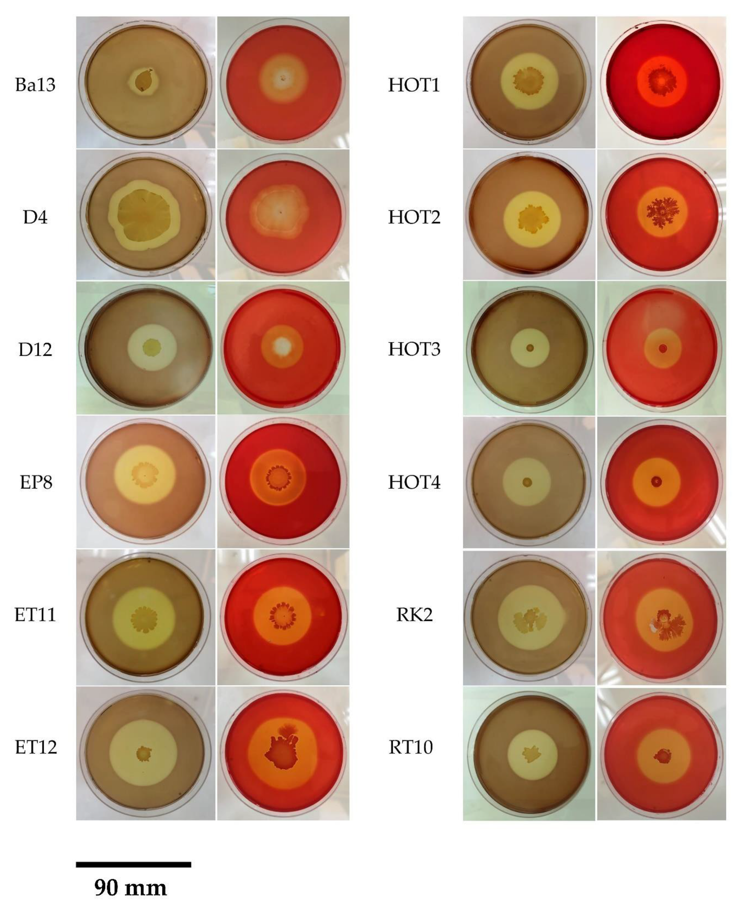
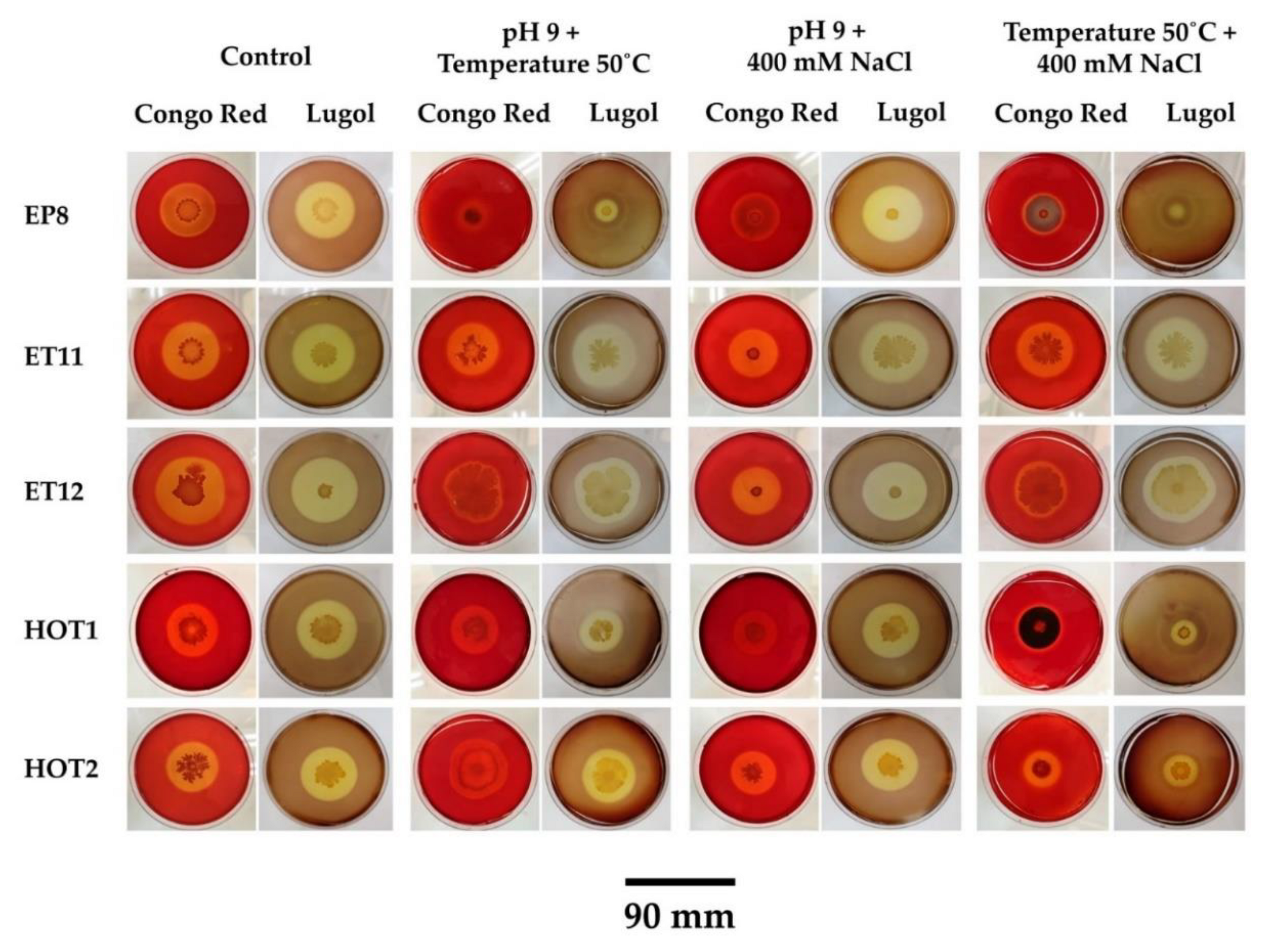
2.9. Effect of Multiple Stress on the Growth and Production of Cellulase on Solid Medium

The influence of a single stress, double stress, and multiple stress on the production of cellulase in solid medium was significant at p ≤ 0.05. In addition, the analysis of variance showed a significant difference in the revelator effect, thus confirming that the Enzymatic index obtained by Lugol’s solution were better than those obtained by Congo red solution (Figure 12, Figure 13 and Figure 14). The production of the enzyme was maintained under pH 9, while the temperature and the salinity strongly influenced this production except for the strain EP8, which showed a better production compared to the control. Thus, pH 9 combined with 400 mM NaCl did not impact the Ei, whereas a decrease was observed with the other two double stresses. The triple stress also influenced the production of cellulases on solid medium except for ET11, which has been shown to be very resistant (Figure 15).

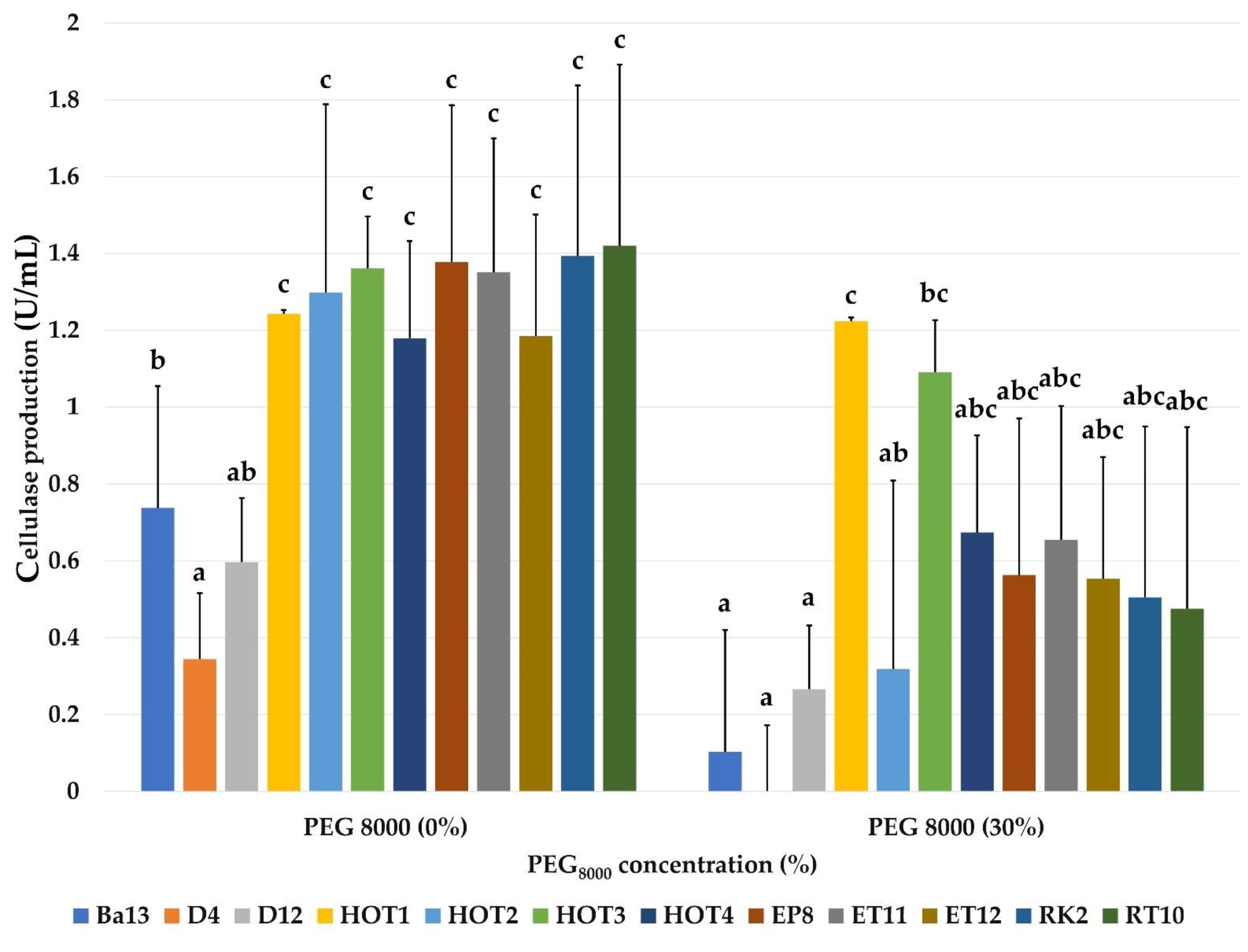
2.10. Effect of Multiple Stress on the Growth and Production of Cellulase on Liquid Medium

The effect of multiple stress on bacterial growth and cellulase production was significant at p ≤ 0.05 (Figure 16). HOT1 has shown itself to be insensitive to pH 9, to 400 mM NaCl, and to their combination, where it showed an optimum of growth at 400 mM NaCl, while HOT2 was very resistant to all stresses and showed an OD of 0.71 at pH 9 and in the presence of 400 mM NaCl. However, the effect of stress was not significant on EP8, ET11, and ET12. It should be noted that ET11 was completely inhibited under the effect of triple stress. The production of the enzyme was also strongly maintained for all the strains (Figure 17).

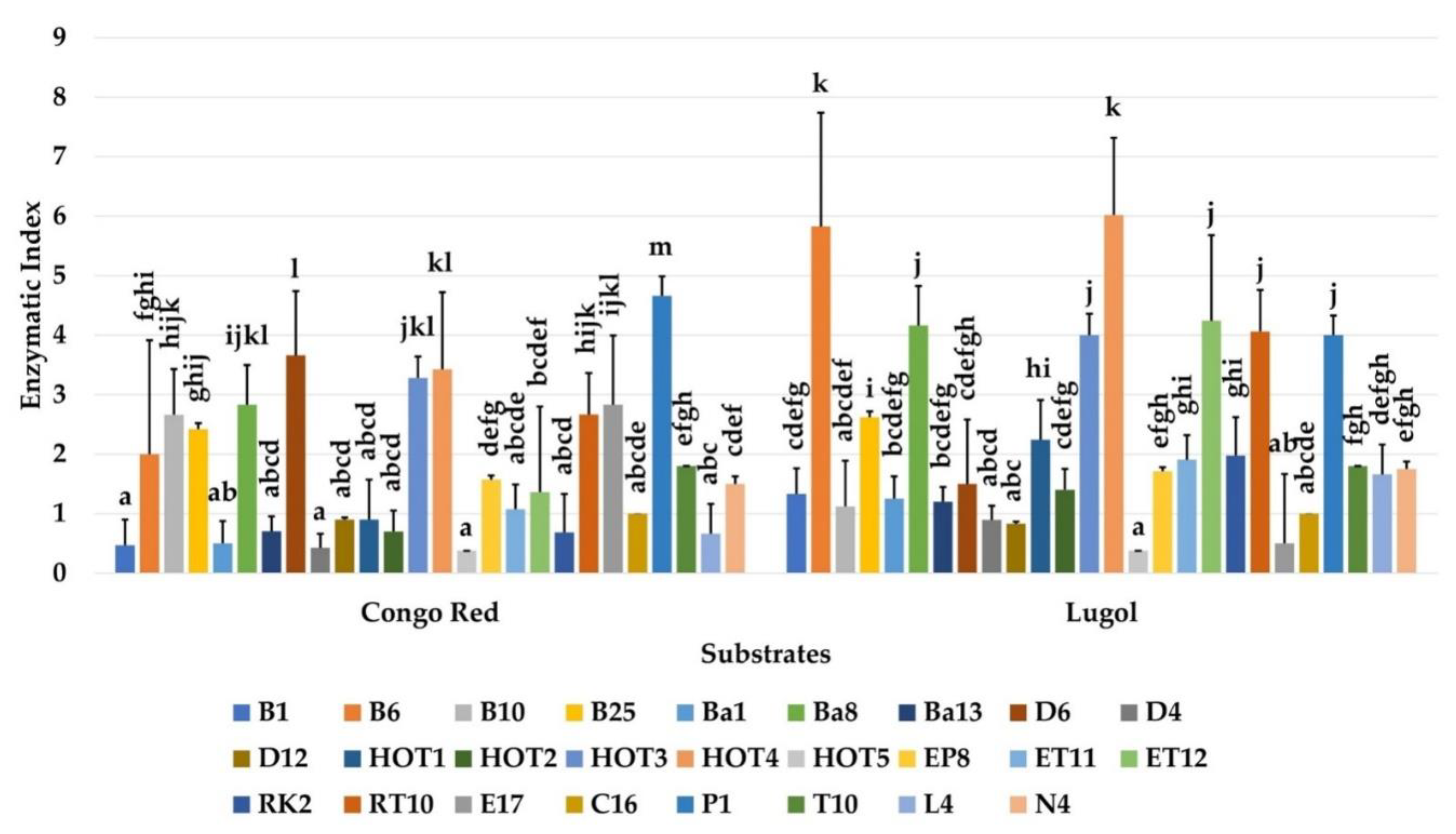
2.11. Effect of Various Cellulosic Substrates on Cellulase Production

The analysis of variance of the ability of the strains to degrade certain lignocellulosic substrates demonstrated that wheat bran was the most digestible substrate by all the strains where degradation of more than 53% was observed, while barley bran was degraded from 37% to 47% (Figure 18A).
Low degradation was observed with corncob. Estimation of the total proteins produced from each of the substrates revealed that significant amounts were produced from wheat bran up to 888 µg/mL produced by the ET11 strain, while non-significant variable amounts were produced from barley bran and corncob, which varied between 131 µg/mL and 353 µg/mL (Figure 18B). Thus, very appreciable cellulase activities were observed by HOT2 and EP8 for wheat bran and by EP8 and ET11 for barley bran but decreased considerably with corncob (Figure 18C). The highest specific activity was recorded by HOT2 with wheat bran, but very low values were observed with corncob for all the strains (Figure 18D). Appreciable amounts of biomass were obtained with wheat bran and barley bran but more or less low with corncob (Figure 18E).

3. Discussion

Due to widespread applications, cellulase is produced by many industries around the world. Despite the enormous use of natural cellulosic sources, there are abundant amounts of cellulose that are not being exploited and that could be used more effectively. In this context, more research is needed for cost-effective and economically feasible production of cellulases allowing wider applicability in different processes. The use of enzymes obtained from microorganisms could open new ways to convert complex polysaccharides into fermentable sugars and cause an increase in the efficiency of the hydrolysis of lignocellulosic biomass. Analysis of our results on the production of cellulase by bacterial isolates showed that out of the 398 strains tested, only 26 showed areas of hydrolysis around the revealed colonies. These bacteria therefore possess cellulases capable of degrading CMC. These cellulases are endoglucanases and β-glucosidases since CMC is a soluble cellulose [17]. The production capacity of isolates is explained by the adaptation of this bacteria to their biotope. According to our results, only 1% of the strains (n = 200) coming from the marvelous cave of Jijel, consisting mainly of mineral material, have cellulase abilities. Among the other strains, consisting of actinobacteria isolated from the archeological site of Djemila (data not shown) (n = 70), bacteria isolated from the rhizosphere of Terfez (n= 80), or barley and wheat soils (n = 40) and bacteria isolated from the thermal spring of Ouled Tebben (n = 8), only 5.7%, 6.25%, 25%, and 62.5%, respectively, have cellulase activities. Since cellulases are inducible enzymes, they are synthesized by microorganisms during their growth on lignocellulosic materials [18]. The growth or the presence of cellulolytic bacteria in the rhizosphere can be explained by the richness of these environments in organic matter brought by lignocellulosic residues of plants and animals [19]. Moreover, the rhizosphere or the rhizospheric soil can be abundant in bacteria having an arsenal of hydrolytic enzymes [19].
The sampling carried out at the level of the thermal spring of Ouled Tebben allowed the isolation of only eight bacterial strains, five of which were cellulase positive, and the presence of a reduced bacterial population is explained by the high temperature of the thermal waters. These natural high-temperature environments will certainly influence the resident microbial flora; the microorganisms able to grow and survive in these environments where the temperature exceeds 50 °C are thermotolerant or thermophilic [20]. A similar study demonstrated that only 16% of the bacteria isolated from the hot springs of western Algeria have cellulases; this type of unexploited biotope could constitute a promising source of thermophilic enzymes of great industrial importance [21].
Although actinobacteria can dominate the microbial communities of archaeological sites and historical monuments, and their beneficial presence is implicated in the preservation and bio-conservation of these sites [22], their cellulolytic potential is rarely established. Antonelli et al. [23] revealed, through new sequencing technologies, the presence of bacteria with cellulolytic capacities from archaeological sites in Italy, the majority of which are non-culturable.
A recent study by Houfani et al. [24] showed the richness of cellulolytic bacteria in different Algerian terrestrial and aquatic ecosystems, such as soils and thermal waters. Bacterial communities were characterized by high-throughput 16S amplicon sequencing followed by in silico prediction of their functional potential. The highest lignocellulolytic activity was recorded in forest and garden soils, while activities in agricultural, arid, and desert soils were generally low. Proteobacteria, Firmicutes, and Actinobacteria phyla dominated all soils.
However, the results of the primary screening have shown that the Lugol’s solution gives better revelation compared to the Congo red solution. The intensity of the black-bluish complex and the clarity of the hydrolysis zone give better contrast color and yield easily measurable diameters [25]. In addition to the high toxicity of Congo red, the handling time is also remarkable: the revelation by Lugol’s solution requires only 5 min of contact and does not require washing, while the Congo red solution remains at least 20 min between coloring and discoloration with NaCl [25]. However, it should be noted that revelation by Lugol’s solution may lead to false-positive results. It has been reported that amylase activity of certain bacteria and fungi due to the presence of starch in bacteriological agar may give rise to areas hydrolysis around the colonies even in the absence of cellulase activity [26].
Enzyme activity varies by species. Our results indicated that isolates from the Terfez rhizosphere and those from thermal waters performed best with a CMCase activity of 0.7 to 2.2 U/mL. These results should be compared with those of Maryam et al. [27], who indicated that the CMCasic activity is 1.99 U/mL for B. cellulosillyticus and lower by 0.07 U/mL for B. subtilis AS3 [28]. However, other strains of Bacillus are capable of having a higher endoglucanase activity, from 7.3 U/mL to 9.1 U/mL [29]. It should be noted that no strain of actinobacteria showed cellulase activity on CMC-based liquid medium, and this can be explained by the slow growth and nutritional requirements of these microorganisms.
The optimization of physico-chemical factors is a key step in the success of enzymatic processes, as defining the optimum of each variable allows increased performance and better production [30].
Temperature, medium pH, salinity, and water activity are critical parameters that control the growth and production of enzymes by microorganisms, and their effects generally differ from one organism to another [31]. The pH of the medium is a very important factor that controls the production of cellulase [31]. All the strains studied were neutrophils or moderately alkalo-tolerant (pH 7–9). The cellulase activity was optimal at neutral pH for all the strains and varied between 0.3U/mL and 1.8 U/mL. Among them, HOT3, HOT4, and ET12 produced a CMCase at pH 9 in an appreciable way by manifesting a CMCase activity of 1.8 U/mL, 1.3 U/mL, and 0.8 U/mL, respectively. Although the others showed visible growth on CMC medium, they synthesized very small quantities of this enzyme. Moreover, it is well-established that most cellulases produced by microorganisms have a better production between pH values of 6 to 9 [32]. Other studies report that the optimum pH for the production of this enzyme by the B. pumilus EWBCM1 strain is 6, with an activity reaching 0.25 U/mL [33]. It should also be noted that the strongly acidic and basic pH inhibits the growth of all the bacteria tested. These results are similar to those reported by Shankar and Isaiarasu [33].
Temperature influences the growth and production of extracellular enzymes by modifying the physical properties of the cell membrane [32]. This physiological parameter is essential to determine because beyond its optimal value, bacterial cells are killed, and their metabolites are damaged, and below that, their metabolism is inactivated [34]. The optimal growth temperature and cellulase activity depend on the strain and the isolation site. All the strains studied were mesophilic and had a CMCase activity varying between 0.38 U/mL and 1.4 U/mL at 30 °C. However, the majority of strains from the Terfez rhizosphere (EP8, ET11, ET12, RK2, and RT10) and thermal waters (HOT1 and HOT2) were thermotolerant, and their optimal cellulase activities (1.2 U/mL and 1.44 U/mL) were at 45 °C and 50 °C. Many studies place the optimum temperature for growth and production of the enzyme of cellulolytic bacteria at 40 °C. Thus, Shankar and Isaiarasu [33] showed that B. pumilus EWBCM1 exhibits optimal but weaker activity (0.24 U/mL) at this temperature, which decreases sharply at 60 °C (0.02 U/mL). These results were confirmed by Immanuel et al. [35], revealing that cellulolytic bacteria belonging to genera Cellulomonas, Bacillus, and Micrococcus produce endoglucanase at maximum levels at 40 °C. However, these observations are not to be generalized, as certain strains of Pseudomonas fluorescens have optimal cellulase capacities at 35 °C [32]. The same is true for B. pumilus EWBCM1, which has a CMCase activity of 0.27 U/mL at 37 °C [33], whereas in other strains of Bacillus, such as B. halodurans CAS 1, the optimum temperatures for cellulase growth and production are at 55 °C and 60 °C, respectively [36].
Growth on culture media containing 400 mM and 800 mM NaCl concentrations was observed in strains Ba13, D4, D12, HOT1, HOT2, EP8, ET11, ET12, RK2, and RT10. These can be qualified according to Kushner [37] as halotolerant. However, HOT1 and RT10 were tolerant to 1000 mM with an enzymatic activity of 0.7 U/mL and 0.55 U/mL, respectively. Half of the strains were able to synthesize the cellulase enzyme at high salinity levels of 400–800 mM NaCl. This is the case for most aerobic or facultative aero-anaerobic and salt-tolerant bacterial isolates from different habitats, such as saline lakes and estuarine waters capable of producing cellulase at these saline concentrations [38]. Indeed, it has been established that B. pumilus has maximum cellulotic power at 2.5% NaCl (about 400 mM) [33], while a strain of Gracilibacillus sp. reaches maximum production at 10% NaCl [39].
The response of water activity effect on bacterial growth and on the production of cellulase in the presence of 30% PEG8000 indicated osmo-tolerance and residual production of the enzyme for all the strains tested with an optimum at 1.22 U/mL of HOT1. Drought tolerance is a faculty possessed by Gram-positive and Gram-negative bacteria. PGPR strains Klebsiella sp., Enterobacter ludwigii, and Flavobacterium sp. isolated from wheat rhizosphere have the ability to grow at 12% up to 20% PEG8000 [40]. Others from the waters and belonging to the species B. cereus, B. subtilis, and Serratia sp. grow well in media containing 50% PEG8000 [41]. As for the production of cellulase in the presence of this hyperosmotic agent, it should be noted that no previous study in the scientific literature on the effect of physico-chemical factors mentions this agent. However, it is crucial to determine the quantity of water available and to control the “aw” for a good enzyme production [33].
The effect of pH on the activity of the cellulases produced was examined at various pH values ranging from 3 to 11. The optimum pH for cellulase activity was 1.8 U/mL at pH 8 for strains HOT1, EP8, and ET11 and at pH4 for HOT2 and ET12 with activity of 1.75 U/mL. Residual activity was also strongly maintained under the influence of this same parameter where, depending on the strains tested, 50% to 83% (1.1 U/mL to 1.65 U/mL) of cellulase activity was preserved at pH 4 and approximately 70% (1.4 U/mL) at pH 7. This result is consistent with those found with different strains of Bacillus [42]. Indeed, according to Dos Santos et al. [43], more than 60% residual activity is maintained at pH 4. Cellulases are generally stable over a wide range of pH ranging from 5 to 10. Recently, many researchers have tried to exploit microbes for the isolation of thermostable alkaline enzymes due to their considerable industrial potential. Given this, higher temperature and pH stability found with purified cellulases could be useful for challenging industrial applications [44].
The effect of temperature on enzymatic activity was determined over a wide temperature range (30–70 °C) with optimal activity at 40 °C and 50 °C for all strains. However, EP8 was the most efficient at all the temperatures tested, with an activity around 1.7 U/mL. Concerning the stability expressed in residual enzymatic activity, it was optimal at 40 °C and 50 °C, with a relative activity of 0.8–1.4 U/mL with all strains; it was also appreciable at 70 °C, where a residual activity of 0.7 U/mL was noted. These results on cellulase activity and stability are mixed compared to those reported by many authors. Indeed, according to Dos Santos et al. [43], the total activity is optimal at 60 °C for a strain of Bacillus sp. isolated from marine environment, and the enzymatic stability is also preserved up to 40% at 70 °C. These data are similar to those of our study. On the other hand, in other works, the enzymatic activity and stability under the thermal effect are greater at higher temperatures at 75 °C in Anobacillus flavithermus isolated from thermal waters [45].
The limiting effect of the increasing contents of NaCl (0–2000 mM) on the activity and the stability of the cellulases of the tested strains was negligible. Indeed, the strains retained a high enzymatic power (1.6–1.82 U/mL) at all salinity levels. The halo-stability showed that the crude cellulase extracts were 70% stable at 500 mM and that they retained a level of 80% of their initial activity even at 2000 mM, exceptionally with HOT1, HOT2, and ET11. It should be noted that these results on the activity of our extracts are much higher than those described by many authors. The latter, working on CMCase purified from a strain of Bacillus, showed that this enzyme retains only 34% of its activity at 30% NaCl [36], while most cellulases reported to date were only stable between 5% and 20% NaCl [46,47]. The characteristics of this enzyme with high salt tolerance are important for future applications in various biotechnological processes that depend on high salinity or osmotic pressures [36].
The evaluation of the influence of the lack of water availability by the presence of PEG8000 at 30% was not effective on the initial and residual activity of the enzyme, which was 1.7 U/mL. However, the enzymatic stability was maintained and fluctuated from 40 to 75% (0.68 U/mL to 1.2 U/mL) depending on the strains in the presence or absence of PEG8000.
The substrate specificity was determined by incubating the crude enzyme extract with different substrates. The enzymes studied had a significant activity with the CMC for all the strains and varied between 1.1 U/mL and 1.6 U/mL. An activity was also measured on wheat bran (lignocellulosic waste) that varied from 1.1 U/mL to 1.5 U/mL. The substrate specificity of cellulases is an issue that has altered the view of synergy between distinct enzyme molecules to achieve cellulose hydrolysis [48]. Enzymes with overlapping specificities have been reported. The ability to degrade crystalline cellulose was commonly considered synonymous with exoglucanases. It has been demonstrated that the endoglucanases of Bacillus sp. and fungi exhibiting avicelase and filter paper hydrolysis (FPase) activities in addition to CMCase activity can do so with the help of an exo-activity residing in the same molecule [49].
The FPase activity was evaluated between 1U/mL and 1.3 U/mL. An appreciable activity of 0.55 U/mL to 0.8 U/mL was measured on cellulose (powder + cotton fiber). This is probably due to a certain affinity of the enzymatic extract towards powdered cellulose. From these results, it seems more appropriate to refer to this enzyme as an endo- and exo- type of cellulose [17]. It should also be noted that adsorption of the enzyme onto cellulose is essential for solubilization [50]. A previous study showed that a fragment of the C-terminus of the full-length endoglucanase gene product plays an important role in the interaction of the enzyme with the insoluble substrate [51]. Soluble substrates including low-DP (degree of polymerization) celluloses can be dissolved in water due to their chemical substitutions. Ion-substituted carboxymethylcellulose (CMC) is often used to determine the activity of endoglucanase, called CMCase, because endoglucanases cleave intramolecular β-1,4-glucosidic bonds in a random fashion, resulting in a dramatic reduction in DP of the CMC [9]. Insoluble cellulose-containing substrates for cellulase activity assays including nearly pure celluloses (cotton fiber, Whatman filter paper, bacterial cellulose, microcrystalline, and amorphous cellulose) are water-insoluble celluloses. Cotton fiber is made from natural cotton after removal of impurities such as wax, pectin, and colored matter [52]. Whatman filter paper no. 1 is made from long-staple cotton pulp [53].
The concentration of total sugars varied relatively in the same way for all the strains, and it varied from 335 μg/mL to 450 μg/mL; these monomeric sugars are produced from the hydrolysis of the substrate (CMC), which reflects that this substrate is highly digestible by the strains tested. The estimate of the total sugar content in a culture supernatant on medium containing lignocellulosic materials from a strain of B. cellulosilyticus is 179.84 mg/mL [27].
Total protein concentration varied relatively in the same way for the five strains studied. The contents of this product varied from 1505–1676 μg/mL, and they were remarkably higher than those obtained by Roussos and Raimbault [54] from a liquid culture Trichoderma harzianum, by Palma et al. [55] on a culture of Bjerkandera sp. in liquid medium based on glucose and peptone, and by Ladeira et al. [56] on the supernatant of cultures of Bacillus sp. SMIA2 in sugar cane bagasse. The proteins can come partly from the complex lignocellulosic biomass (e.g., wheat bran) or from cells after lysis and can also constitute different enzymes secreted to digest the substrate. Although expensive purification procedures may be required to remove them, most industrial enzymes are consumed in the presence of impurities from culture supernatants [56].
The effect of multiple stresses on growth and cellulase production was studied at pH 9, 400 mM NaCl, and 50 °C. The results of this study revealed that the growth and the production of cellulases of the five bacterial isolates were not affected by the presence of 400 mM NaCl in the culture medium since the cellulase activities in the presence of this stress were better than those obtained from the NaCl-free medium ranging from 1.887 U/mL to 2.179 U/mL. The incubation temperature also had no effect on growth or cellulase excretion, especially for the HOT2 strain, which had a growth optimum of 50 °C. A similar result was obtained with pH 9, where no strain was sensitive to this effect. The analysis of the effect of double and triple stress revealed that none of the combinations influenced the growth of the isolates except for the ET11 strain, which did not show any growth under the effect of the three stresses; the combination of the two factors pH 9 and 400 mM NaCl was optimal for the growth of HOT1 and HOT2. Thus, the results of the multi-stress showed that the double and the triple stress did not influence the production of cellulase and that all the strains were moderate thermo-alkalo-halophiles. This physiological characteristic makes these strains effective candidates for the recovery of agricultural waste.
The capacity of the five strains studied to degrade certain lignocellulosic substrates was evaluated by determining the quantity of degraded substrate, quantity of proteins and biomass produced, and the enzymatic and specific activities. Our results indicated a moderate hydrolysis capacity of barley bran with rates ranging from 37.33% to 47.33% after three days of incubation; this hydrolysis made it possible to produce relatively high rates of protein with a better production of 888.5 µg/mL by strain ET11. This degradation indicated that the tested strains possess the barley bran hydrolysis enzymes, namely cellulases, amylases, xylanases, and hemicellulases, given that barley bran is rich in cellulose, starch, hemicellulose, and arabinoxylan [57,58]. However, its high insoluble fiber content and the presence of lignin in its composition prevent it from being further degraded. This finding correlates with the cellulase activity of the strains, which varied between 1.021 U/mL and 2.098 U/mL except for the ET12 strain, which gives a low activity of 0.521 U/mL and with the specific activity levels that were relatively low. This result is comparable to that of Mihajlovski et al. [59], who obtained a CMCase activity of 0.405 U/mL from a culture with barley bran as a carbon source. Estimation of the amount of biomass produced indicates that barley bran could be a good carbon source for bacterial cultures as well as for the production of hydrolytic enzymes.
For the hydrolysis of wheat bran, our strains were found to be very effective in the degradation of this substrate, with degradation ranging from 53.33% to 61.33%, with better degradation recorded by ET11; a similar result was reported by Vu et al. [60], who observed that Bacillus strains caused a loss of wheat bran mass ranging from 54% to 60% after 7 days of incubation. This confirms the observation of the presence of a cellulolytic and hemicellulolytic enzymatic complex in the culture supernatant, considering that wheat is composed of 50% carbohydrates, of which cellulose and hemicellulose constitute more than 70% [61]. However, the quantity of proteins produced was appreciable for all the strains; these proteins released in the medium normally contain the enzymes of hydrolysis including the cellulases, the latter showing relatively high enzymatic activities with a better activity of 3.055 U/mL by the EP8 strain. These results correlate with the specific activity, thus indicating the effectiveness of the cellulases produced in large quantities in just 72 h. In addition, since wheat bran is digestible, it made it possible to produce significant amounts of biomass by all the strains.
The analysis of the corncob degradation capacity showed that all the strains had a low hydrolysis capacity on this substrate, with a maximum rate of 16.66% observed by the EP8 strain. This result affirms that no enzymatic treatment is able to effectively hydrolyze corncob given its characteristic composition of a complex of cellulose and hemicellulose, which are coated by a recalcitrant matrix of lignin composed of phenolic macromolecules; the latter decreases access of hydrolytic enzymes to celluloses and hemicelluloses and requires physicochemical pre-treatment to degrade it [62]. Thus, the reduction in the quantity of proteins and cellulases produced was remarkable and led to a significant decrease in specific activity. Concerning the microbial biomass, our results indicated significant quantities ranging from 0.025–0.05 g.

4. Materials and Methods

4.1. Biological Material

The study of cellulase activity was performed on seven collections of bacterial isolates (n = 398) from various ecological niches. The bacterial isolates were a collection of the Laboratory of Applied Microbiology (University Ferhat Abbas, Setif, Algeria) (Table 2).

4.2. Screening for Cellulase-Producing Isolates

All the bacterial strains were screened for their cellulolytic activity on solid medium. The procedure was performed by spotting 2 μL of the bacterial culture on 1% (w/v) carboxymethylcellulose (CMC) agar incubated at 30 °C/72 h [63]. Lugol’s solution (iodine 1%) and Congo red solution (1%) were used to reveal the CMC hydrolysis zone, which appeared around the colony spontaneously after the addition of iodine solution, but the revelation of the cellulase activity with Congo red solution required 15 min followed by discoloration with a NaCl solution (1M) for 10 min. To indicate the cellulose activity, the diameter of clear zones around colonies on CMC agar was measured, and the enzymatic index (Ei) was determined according to Ferbiyanto et al. [64] by the following formula:
Ei = (Halo diameter-colony diameter)/colony diameter

4.3. Cellulase Production on Liquid Medium

Cellulase production in a liquid medium was performed to evaluate the ability of efficient bacterial isolates selected (n = 26) to produce the enzyme. First, 100 μL of the bacterial cultures on LB (Luria Bertani) broth, incubated at 30 °C/24 h, were inoculated in tubes containing 10 mL of (1%) CMC broth and then incubated at 30 °C/72 h under stirring. The cultures were centrifuged (5000 rpm/10 min). The supernatant containing the crude enzyme was stored at 4 °C for subsequent analysis [33].

4.4. Evaluation of Cellulase Activity

CMCase activity was determined by the 3,5-dinitrosalicylic acid (DNS) method by measuring the amount of reducing sugars released [65]. The reaction mixture was prepared by mixing 2 mL of supernatant with 1 mL of acetate buffer (50 mM, pH 4) containing CMC (1%). After incubation at 50 °C/30 min, the reaction was stopped by adding 2 mL of DNS reagent, followed by boiling for 5 min and cooling to stabilize the color. The optical density (OD) of the samples was read at 540 nm. One unit (U) of enzyme activity was defined as the amount of enzyme releasing 1 µmol of reducing sugar per minute using glucose as a standard [66].

4.5. Effect of Physico-Chemical Parameters on Bacterial Growth and Cellulase Production Capacity

Based on the results obtained, selected strains (n = 12) were assessed for their optimum bacterial growth and cellulose activity under various abiotic stresses. Bacterial growth was determined by measuring the OD at 630 nm, and the production of cellulase was estimated by determining the enzymatic activity using the DNS method as described previously.

4.5.1. pH

The influence of the pH variation of the culture medium on bacterial growth and cellulase production capacity was tested in CMC liquid medium adjusted to values of 3, 4, 7, 9, and 11. The cultures were incubated at 30 °C for 72 h.

4.5.2. Temperature

To determine the effect of temperature stress on the growth of the isolates studied and the production of enzyme, the incubation of the cultures in CMC medium was carried out at 30 °C, 45 °C, and 50 °C for 72 h.

4.5.3. Salinity

The effect of salt stress on bacterial growth and cellulase production was determined in CMC medium at increasing NaCl concentrations of 0, 400, 800, and 1000 mM and incubated at 30° C/72 h.

4.5.4. Water activity (aw)

The effect of aw on cellulase growth and production was evaluated by adding polyethylene glycol (PEG8000) at concentrations of 0% and 30%. The cultures were incubated, as before, at 30 °C/72 h.

5. Effect of Physico-Chemical Parameters on Cellulase Activity and Stability

According to the results of the ability to grow and produce cellulase under different physicochemical conditions, five isolates were chosen to study their enzymatic activity and stability under multiple stress parameters.

5.1. pH

TThe influence of pH variation on the activity and stability of CMCase was evaluated at pH values ranging from 3 to 11 using the following buffers: acetate buffers (pH 3), citrate buffer (pH 4–6), phosphate buffer (pH 7–11), and glycine buffer (pH 9–10). The effect of pH on enzyme stability was determined by incubating the enzymes with the appropriate pH buffer (3 to 11) for one hour at room temperature. The residual activity was assayed by the standard method of DNS.

5.1.1. Temperature

The effect of temperature on enzyme activity and stability was determined at different temperatures (30–70 °C). The enzymatic activity was tested in acetate buffer (pH 4) in the presence of 1% of CMC, while the stability of the enzyme was evaluated for 60 min in the same buffer free of this substrate. Then, the residual activity of the CMCase was measured, under the same conditions described above, by the standard DNS method.

5.1.2. Salinity

The optimal NaCl concentration for CMCase activity was determined by incubating the enzyme with a buffer solution containing 1% CMC for 30 min at different NaCl concentrations (0–2000 mM). Enzyme stability was assessed by incubating the enzyme in CMC-free buffer at the same NaCl concentrations for one hour at room temperature. After adding 1% CMC, the DNS method was used for residual CMCase activity assay.

5.1.3. Water Activity (aw)

The effect of water activity on CMCase activity was evaluated by adding polyethylene glycol (PEG8000) at two concentrations (0% and 30%). CMCase stability was estimated by incubating the enzyme in the absence of CMC in buffer containing different concentrations of polyethylene glycol (PEG8000) for 30 min at 50 °C. The standard DNS method was used to measure glucose release.

5.2. Molecular Identification of the Selected Isolates

The five bacterial isolates were phylogenetically identified. DNA extraction, amplification and sequencing were performed according to Kerbab et al. [67]. Phylogenetic analyses were realized according to Mefteh et al. [68].

5.3. Hydrolysis of Different Substrates

The objective of this study was to evaluate the ability of this crude enzyme to hydrolyze different cellulosic substrates. The substrates tested were addition to CMC, powdered cellulose, wheat bran, and filter paper (Whatman No 40). A volume of 2 mL of supernatant was mixed with 1 mL of pH 4 acetate buffer containing 1% of each following substrates (cellulose powder, CMC, or wheat bran) and incubated at 50 °C/30 min. Concerning the filter paper, pieces of size (1×6 cm) corresponding to 50 mg were immersed in the same volume of buffer supplemented with 2 mL of supernatant. Incubation was carried out at 50 °C/60 min. The dosage of reducing sugars was carried out by the DNS method with glucose as a reference.

5.4. Estimation of Total Sugars

The total sugars were determined according to the method of Dubois et al. [69], the principle of which is based on the following reaction: concentrated sulfuric acid added causes, when hot, the departure of several molecules of water from the sugars. This dehydration is accompanied by the formation of a hydroxy-methylfurfural (HMF) in the case of hexose and of a furfural in the case of a pentose. These compounds condense with phenol to give colored complexes (yellow-orange). The intensity of the coloration is proportional to the concentration of the oases. The reaction mixture was composed of 1 mL of the sample, 1 mL of a 5% phenol solution, and 5 mL of sulfuric acid. The optical density was measured at 490 nm. The values obtained were expressed into glucose concentrations by reference to a previously established calibration curve.

5.5. Estimated of Total Proteins

The assay of total proteins in the enzymatic extracts was carried out according to the method of Lowry et al. [70]. The principle of this method was based on the blue coloration developed by proteins following a reaction between Folin’s reagent and amino acids. First, 5 mL of the reaction mixture (Lowry reagent) were added to 1 mL of the sample and 0.5 mL of Folin’s reagent diluted to ½. The intensity of the coloration was proportional to the protein concentration contained in the extracts after incubation for 30 min at room temperature and protected from light. Absorbance was read at 750 nm. The concentration was determined by reference to a calibration curve with BSA (bovine serum albumin).

5.6. Effect of Multiple Stress on Growth and Cellulase Production on Solid Medium

The influence of multiple stresses on cellulolytic activity in a solid medium was determined on a CMC medium adjusted to pH 9, 400 mM NaCl, and incubated at 50 °C. Demonstration of cellulolytic activity was carried out by spotting 2 μL of the bacterial culture on CMC agar. Lugol’s solution and Congo red solution (1%) were used to reveal the CMC hydrolysis zone that appeared around the colony, and the enzymatic index was calculated as previously described. The enzymatic indices of single stress and the combination of two and three stresses were compared to a control.

5.7. Effect of Multiple Stress on Growth and Cellulase Production on Liquid Medium

The multiple stress tolerance of the strains in a liquid medium and their ability to produce cellulases was evaluated by inoculating 100 µL of the bacterial suspension in 10 mL of CMC broth (1%) adjusted to pH 9 and 400 mM of NaCl incubated at 50 °C/72 h under constant stirring. The bacterial growth was determined by measuring the OD at 630 nm. The cultures were centrifuged (5000 rpm/10 min), and the supernatant containing the crude enzyme was used to determine the enzyme activity using the DNS method.

5.8. Effect of Various Cellulosic Substrates on the Production of Cellulases

The evaluation of the capacity of the strains to degrade various cellulosic substrates and to produce cellulases was carried out by replacing the CMC by 1% of wheat bran, 1% of barley bran, and 1% of corncob; the incubation was carried out at 30 °C/72 h under constant agitation. After incubation, the cultures were first filtered to recover the non-degraded substrate, which was then washed and dried at 30 °C and weighed daily until obtaining a constant weight to determine the percentage of degradation. The cultures were then centrifuged at 5000 rpm/15 min, the pellet was dried at 30 °C and weighed daily until obtaining a constant weight to determine the amount of biomass, and the supernatant containing the enzyme and the proteins was used to determine the enzyme activity and protein concentration, which were estimated as previously described. The specific activity was calculated according to Islam and Roy [71] as follows: enzyme unit (U/mL)/protein (mg).

5.9. Statistical Analysis

All experiments were performed in duplicate, with results expressed as mean ± standard deviation (SD); statistical analysis of data was performed using IBM-SPSS v. 24, and two-way ANOVA was used to identify variance between different treatments. Results were considered significant when p-values were less than 0.05. Post hoc comparison tests of Tukey’s HSD test were performed when a significant difference was found.

6. Conclusions

Microorganisms are very attractive for their immense potential production of cellulases. Screening of the cellulase activity of 398 various isolates allowed to select five high-performance strains belonging to the genus Bacillus. The physicochemical properties of cellulases (crude extract) showed a CMCase active over a wide range of pH (4 to 11), which was optimal at 50 °C and 60 °C. The residual CMCase activity remained between 40 and 70% in a temperature range between 40 and 70 °C and was stable over a wide range of saline concentrations (0–2000 mM) and weakly affected by PEG8000. The enzymatic extract of our isolates was able to hydrolyze soluble and insoluble substrates and appeared to be a mixture of endo and exo-cellulase type. Our isolates showed interesting multiple-stress-tolerance properties and were moderately thermo-alkalo-halotolerant. The significant potential of selected Bacillus strains to use lignocellulosic residues as a carbon source has led to increased production of CMCases; therefore, these isolates can be used as an eco-friendly source for biofuel and bioethanol production.
Further studies on purification, characterization of cellulase, identification of genes involved in its production and stability, and its large-scale production and application in different commercial fields constitute an interesting strategy. The purified cellulase can be used for various purposes in detergent industries, food industries, and pharmaceutical industries. Continued research on enzymatic engineering of cellulase after purification and its investigation on genetic level seems fully interesting to boost production and enzyme stability. Various studies have been made for the improvement of the stability of the enzyme or the search for a stable enzyme. High activity and stability of cellulase enzymes between neutral and alkaline pH and at high temperatures will be useful in various industrial and biotechnology applications.

Supplementary Materials

The following supporting information can be downloaded at: https://www.mdpi.com/article/10.3390/catal12070769/s1, Figure S1: Effect of pH on bacterial growth (ODʎ630 nm) in CMC broth (1%) of the 12 isolates tested. Results are expressed as mean-standard error of duplicate OD values. The effects of acid and alkaline concentrations corresponding to each treatment and not sharing the same letters are significantly different according to Tukey’s post hoc HSD test; Figure S2: Effect of temperature on bacterial growth (ODʎ630 nm) of the 12 isolates tested. Results are expressed as mean-standard error of duplicate OD values. The growth temperature effects corresponding to each treatment and not sharing the same letters are significantly different according to Tukey’s post hoc HSD test; Figure S3: Effect of salinity on bacterial growth (ODʎ630 nm) of the 12 isolates tested. Results are expressed as mean-standard error of duplicate OD values. The effects of the saline concentrations corresponding to each treatment and not sharing the same letters are significantly different, according to the post hoc Tukey HSD test; Figure S4: Effect of water activity on bacterial growth (ODʎ630 nm) of the 12 isolates tested. Results are expressed as mean-standard error of duplicate OD values. The effects of the PEG8000 concentrations corresponding to each treatment and not sharing the same letters are significantly different according to Tukey’s post hoc HSD test; Figure S5: Maximum likelihood phylogenetic tree of Bacillus strains based on comparison of the 16S-rRNA gene sequence of bacterial strains with some phylogenetically related strains. Supports for branches were assessed by bootstrap resampling of the dataset with 1000 replicates. Lactobacillus hokkaidonensis JCM was used as outgroup; Figure S6: Effect of pH on (A) enzymatic activity and (B) stability of the strains tested. The results are expressed as the mean-standard error of the values of the enzymatic unit in duplicate. The pH effects corresponding to each treatment and not sharing the same letters are significantly different according to Tukey’s post hoc HSD test; Figure S7: Effect of temperature on (A) enzymatic activity and (B) stability of the strains tested. The results are expressed as the mean-standard error of the values of the enzymatic unit in duplicate. The temperature effects corresponding to each treatment and not sharing the same letters are significantly different according to Tukey’s post hoc HSD test; Figure S8: Effect of salinity on (A) enzymatic activity and (B) stability of the strains tested. The results are expressed as the mean-standard error of the values of the enzymatic unit in duplicate. The effects of saline concentrations corresponding to each treatment and not sharing the same letters are significantly different according to Tukey’s post hoc HSD test; Figure S9: Effect of water activity on (A) cellulase activity and (B) stability of the strains tested. The results are expressed as the mean-standard error of the values of the enzymatic unit in duplicate. The effects of the PEG8000 concentrations corresponding to each treatment and not sharing the same letters are significantly different according to Tukey’s post hoc HSD test.

Author Contributions

Conceptualization, A.S., H.C.-S., A.B. (Amel Balla), A.B. (Amel Boudechicha), and L.B.; methodology, A.S., H.C.-S., A.C.B., and L.B.; software, A.C.B., F.N.A., A.S., H.C.-S., and L.B.; validation, A.B. (Amel Balla), A.S., H.C.-S., and L.B.; formal analysis, A.S., H.C.-S., A.C.B., L.L., A.S., and L.B.; investigation, A.S., H.C.-S., and L.B.; resources, A.S., H.C.-S., and L.B.; data curation, A.B. (Amel Balla), A.B. (Amel Boudechicha), A.C.B., L.L., F.N.A. and L.B.; writing—original draft preparation, A.S., H.C.-S., and L.B.; writing—review and editing, A.S., H.C.-S., A.B. (Amel Balla), A.B. (Amel Boudechicha), A.C.B., and L.B.; visualization, A.S., H.C.-S., A.B. (Amel Balla), A.B. (Amel Boudechicha), A.C.B., L.L., F.N.A., and L.B.; supervision, A.S., H.C.-S., and L.B.; project administration, A.S., H.C.-S., and L.B.; funding acquisition, A.S., H.C.-S., and L.B. All authors have read and agreed to the published version of the manuscript.

Funding

This research received no external funding.

Conflicts of Interest

The authors declare no conflict of interest.

Sample Availability

Samples of the bacteria are available from the corresponding author.

References

  1. Periyasamy, S.; Karthik, V.; Senthil Kumar, P.S.; Isabel, J.B.; Temesgen, T.; Hunegnaw, B.M.; Melese, B.B.; Mohamed, B.A.; Vo, D.-V.N. Chemical, physical and biological methods to convert lignocellulosic waste into value-added products. A Review. En-Viron. Chem. Letters 2022, 20, 1129–1152. [Google Scholar] [CrossRef]
  2. Ja’afar, J.A.N.; Shitu, A. Utilization of Lignocellulosic Agro-Waste as an Alternative Carbon Source for Industrial Enzyme Production. In Waste Management, Processing and Valorisation; Embrandiri, A., Kassaw, G.M., Geto, A.K., Wogayehu, B.T., Embrandiri, M., Eds.; Springer: Singapore, 2022; pp. 221–233. [Google Scholar]
  3. Himanshu; Saini, J.K. Diversity and Biotechnological Importance of Cellulolytic Microorganisms from Biodiversity Hotspots; Academic Press: Pune, India, 2021; pp. 207–230. [Google Scholar] [CrossRef]
  4. Sahay, S. Deconstruction of lignocelluloses: Potential biological approaches. In Handbook of Biofuels; Academic Press: Cambridge, MA, USA, 2021; pp. 207–232. [Google Scholar] [CrossRef]
  5. Fatima, A.; Yasir, S.; Khan, M.S.; Manan, S.; Ullah, M.W.; Ul-Islam, M. Plant extract-loaded bacterial cellulose composite membrane for potential biomedical applications. J. Bioresour. Bioprod. 2021, 6, 26–32. [Google Scholar] [CrossRef]
  6. Xia, Z.; Li, J.; Zhang, J.; Zhang, X.; Zheng, X.; Zhang, J. Processing and valorization of cellulose, lignin and lignocellulose using ionic liquids. J. Bioresour. Bioprod. 2020, 5, 79–95. [Google Scholar] [CrossRef]
  7. Jayasekara, S.; Ratnayake, R. Microbial cellulases: An overview and applications. In Cellulose; Pascual, A.R., Martín, M.E.E., Eds.; IntechOpen: London, UK, 2019; pp. 1–21. [Google Scholar] [CrossRef] [Green Version]
  8. Singh, A.; Bajar, S.; Devi, A.; Pant, D. An overview on the recent developments in fungal cellulase production and their industrial applications. Bioresour. Technol. Rep. 2021, 14, 100652. [Google Scholar] [CrossRef]
  9. Zhang, Y.H.P.; Himmel, M.E.; Mielenz, J.R. Outlook for cellulase improvement: Screening and selection strategies. Biotechnol. Adv. 2006, 24, 452–481. [Google Scholar] [CrossRef] [PubMed]
  10. Lavanya, D.K.P.K.; Kulkarni, P.K.; Dixit, M.; Raavi, P.K.; Krishna, L.N.V. Sources of cellulose and their applications—A review. Int. J. Drug Dev. Res. 2011, 2, 19–38. [Google Scholar]
  11. Singh, S.; Jaiswal, D.K.; Sivakumar, N.; Verma, J.P. Developing efficient thermophilic cellulose degrading consortium for glucose production from different agro-residues. Front. Energy Res. 2019, 7, 61. [Google Scholar] [CrossRef]
  12. Nagendran, S.; Hallen-Adams, H.E.; Paper, J.M.; Aslam, N.; Walton, J.D. Reduced genomic potential for secreted plant cell-wall-degrading enzymes in the ectomycorrhizal fungus Amanita bisporigera, based on the secretome of Trichoderma reesei. Fungal Geneti. Biol. 2009, 46, 427–435. [Google Scholar] [CrossRef] [Green Version]
  13. Saratale, G.D.; Saratale, R.G.; Oh, S.E. Production and characterization of multiple cellulolytic enzymes by isolated Streptomyces sp. MDS. Biomass Bioenergy 2012, 47, 302–315. [Google Scholar] [CrossRef]
  14. Sadhu, S.; Maiti, T.K. Cellulase production by bacteria: A review. Br. Microbiol. Res. J. 2013, 3, 235–258. [Google Scholar] [CrossRef] [Green Version]
  15. Malik, W.A.; Javed, S. Biochemical Characterization of Cellulase from Bacillus subtilis Strain and its Effect on Digestibility and Structural Modifications of Lignocellulose Rich Biomass. Front. Bioeng. Biotechnol. 2021, 9, 800265. [Google Scholar] [CrossRef]
  16. Chen, G.Q.; Jiang, X.R. Next generation industrial biotechnology based on extremophilic bacteria. Curr. Opin. Biotechnol. 2018, 50, 94–100. [Google Scholar] [CrossRef]
  17. Sharma, A.; Tewari, R.; Rana, S.S.; Soni, R.; Soni, S.K. Cellulases: Classification, methods of determination and industrial applications. Appl. Biochem. Biotechnol. 2016, 179, 1346–1380. [Google Scholar] [CrossRef]
  18. Gaur, R.; Tiwari, S. Isolation, production, purification and characterization of an organic-solvent-thermostable alkalophilic cellulase from Bacillus vallismortis RG-07. BMC Biotechnol. 2015, 15, 1–12. [Google Scholar] [CrossRef] [Green Version]
  19. Nwachukwu, B.C.; Ayangbenro, A.S.; Babalola, O.O. Elucidating the rhizosphere associated bacteria for environmental sustainability. Agriculture 2021, 11, 75. [Google Scholar] [CrossRef]
  20. Wilson, C.K.; King, G.M. Short-Term Exposure to Thermophilic Temperatures Facilitates CO Uptake by Thermophiles Maintained under Predominantly Mesophilic Conditions. Microorganisms 2022, 10, 656. [Google Scholar] [CrossRef]
  21. Khelil, O.; Cheba, B. Thermophilic cellulolytic microorganisms from western Algerian sources: Promising isolates for cellulosic biomass recycling. Proc. Technol. 2014, 12, 519–528. [Google Scholar] [CrossRef] [Green Version]
  22. McNamara, C.J.; Perry, T.D.; Bearce, K.A.; Hernandez-Duque, G.; Mitchell, R. Epilithic and endolithic bacterial communities in limestone from a Maya archaeological site. Microb. Ecol. 2006, 51, 51–64. [Google Scholar] [CrossRef]
  23. Antonelli, F.; Esposito, A.; Galotta, G.; Davidde Petriaggi, B.; Piazza, S.; Romagnoli, M.; Guerrieri, F. Microbiota in waterlogged archaeological wood: Use of Next-Generation Sequencing to evaluate the risk of biodegradation. Appl. Sci. 2020, 10, 4636. [Google Scholar] [CrossRef]
  24. Houfani, A.A.; Větrovský, T.; Navarrete, O.U.; Štursová, M.; Tláskal, V.; Beiko, R.G.; Boucherba, N.; Baldrian, P.; Benallaoua, S.; Jorquera, M.A. Cellulase−hemicellulase activities and bacterial community composition of different soils from Algerian ecosystems. Microb. Ecol. 2019, 77, 713–725. [Google Scholar] [CrossRef]
  25. Kasana, R.C.; Salwan, R.; Dhar, H.; Dutt, S.; Gulati, A. A rapid and easy method for the detection of microbial cellulases on agar plates using Gram’s iodine. Curr. Microbiol. 2008, 57, 503–507. [Google Scholar] [CrossRef] [PubMed]
  26. Meddeb-Mouelhi, F.; Moisan, J.K.; Beauregard, M. A comparison of plate assay methods for detecting extracellular cellulase and xylanase activity. Enzym. Microb. Technol. 2014, 66, 16–19. [Google Scholar] [CrossRef] [PubMed]
  27. Maryam, B.; Qadir, A.; Zameer, M.; Ahmad, S.R.; Nelofer, R.; Jamil, N.; Arzoo, S.; Afzaal, R. Production of Cellulases by Bacillus cellulosilyticus Using Lignocellulosic Material. Pol. J. Environ. Stud. 2018, 27, 2659–2667. [Google Scholar] [CrossRef]
  28. Deka, D.; Bhargavi, P.; Sharma, A.; Goyal, D.; Jawed, M.; Goyal, A. Enhancement of cellulase activity from a new strain of Bacillus subtilis by medium optimization and analysis with various cellulosic substrates. Enzym. Res. 2011, 2011, 151656. [Google Scholar] [CrossRef] [Green Version]
  29. Mawadza, C.; Hatti-Kaul, R.; Zvauya, R.; Mattiasson, B. Purification and characterization of cellulases produced by two Bacillus strains. J. Biotechnol. 2000, 83, 177–187. [Google Scholar] [CrossRef]
  30. Nnaemeka, I.C.; Maxwell, O.; Christain, A.O. Optimization and kinetic studies for enzymatic hydrolysis and fermentation of colocynthis vulgaris Shrad seeds shell for bioethanol production. J. Bioresour. Bioprod. 2021, 6, 45–64. [Google Scholar] [CrossRef]
  31. Sarsan, S.; Merugu, R. Role of bioprocess parameters to improve cellulase production: Part II. In New and Future Developments in Microbial Biotechnology and Bioengineering. From Cellulose to Cellulase: Strategies to Improve Biofuel Production; Srivastava, N., Srivastava, M., Mishra, P.K., Ramteke, P.W., Singh, R.L., Eds.; Elsevier: Pune, India, 2019; pp. 77–97. [Google Scholar]
  32. Bakare, M.K.; Adewale, I.O.; Ajayi, A.; Shonukan, O.O. Purification and characterization of cellulase from the wild-type and two improved mutants of Pseudomonas fluorescens. Afr. J. Biotechnol. 2005, 4, 898–904. [Google Scholar]
  33. Shankar, T.; Isaiarasu, L. Cellulase production by Bacillus pumilus EWBCM1 under varying cultural conditions. Middle East J. Sci. Res. 2011, 8, 40–45. [Google Scholar]
  34. Raju, E.V.N.; Divakar, G. Production of pectinase by using Bacillus circulans isolated from dump yards of vegetable wastes. Int. J. Pharmaceut. Sci. Res. 2013, 4, 2615–2622. [Google Scholar]
  35. Immanuel, G.; Dhanusha, R.; Prema, P.; Palavesam, A.J.I.J. Effect of different growth parameters on endoglucanase enzyme activity by bacteria isolated from coir retting effluents of estuarine environment. Int. J. Environ. Sci. Technol. 2006, 3, 25–34. [Google Scholar] [CrossRef] [Green Version]
  36. Annamalai, N.; Thavasi, R.; Vijayalakshmi, S.; Balasubramanian, T. A novel thermostable and halostable carboxymethylcellulase from marine bacterium Bacillus licheniformis AU01. World J. Microbiol. Biotechnol. 2011, 27, 2111–2115. [Google Scholar] [CrossRef]
  37. Kushner, D.J. Life in high salt and solute concentrations. In Microbial Life in Extreme Environments; Kushner, D.J., Ed.; Academic Press: London, UK, 1978; pp. 317–368. [Google Scholar]
  38. Vargas, V.A.; Delgado, O.D.; Hatti-Kaul, R.; Mattiasson, B. Bacillus bogoriensis sp. nov., a novel alkaliphilic, halotolerant bacterium isolated from a Kenyan soda lake. Int. J. Sys. Evol. Microbiol. 2005, 55, 899–902. [Google Scholar] [CrossRef] [Green Version]
  39. Yu, H.Y.; Li, X. Alkali-stable cellulase from a halophilic isolate, Gracilibacillus sp. SK1 and its application in lignocellulosic saccharification for ethanol production. Biomass. Bioenergy. 2015, 81, 19–25. [Google Scholar] [CrossRef]
  40. Gontia-Mishra, I.; Sapre, S.; Sharma, A.; Tiwari, S. Amelioration of drought tolerance in wheat by the interaction of plant growth-promoting rhizobacteria. Plant Biol. 2016, 18, 992–1000. [Google Scholar] [CrossRef]
  41. Vardharajula, S.; Zulfikar Ali, S.; Grover, M.; Reddy, G.; Bandi, V. Drought-tolerant plant growth promoting Bacillus spp.: Effect on growth, osmolytes, and antioxidant status of maize under drought stress. J. Plant Interact. 2011, 6, 1–14. [Google Scholar] [CrossRef]
  42. Lee, Y.J.; Kim, B.K.; Lee, B.H.; Jo, K.I.; Lee, N.K.; Chung, C.H.; Lee, Y.C.; Lee, J.W. Purification and characterization of cellulase produced by Bacillus amyoliquefaciens DL-3 utilizing rice hull. Bioresour. Technol. 2008, 99, 378–386. [Google Scholar] [CrossRef]
  43. Dos Santos, Y.Q.; De Veras, B.O.; De Franca, A.F.J.; Gorlach-Lira, K.; Velasques, J.; Migliolo, L.; Dos Santos, E.A. A new salt-tolerant thermostable cellulase from a marine Bacillus sp. strain. J. Microbiol. Biotechnol. 2018, 28, 1078–1085. [Google Scholar] [CrossRef]
  44. Samiullah, T.R.; Bakhsh, A.; Rao, A.Q.; Naz, M.; Saleem, M. Isolation, purification and characterization of extracellular β-glucosidase from Bacillus sp. Adv. Environ. Biol. 2009, 3, 269–277. [Google Scholar]
  45. Sheble Ibrahim, A.S.; El-diwany, A. Isolation and identification of new cellulases thermophilic bacteria from an Egyptian hot spring and some properties of the crude enzyme. Aust. J. Basic Appl. Sci. 2007, 1, 473–478. [Google Scholar]
  46. Wang, C.Y.; Hsieh, Y.R.; Ng, C.C.; Chan, H.; Lin, H.T.; Tzeng, W.S.; Shyu, Y.T. Purification and characterization of a novel halostable cellulase from Salinivibrio sp. strain NTU-05. Enzyme Microb. Technol. 2009, 44, 373–379. [Google Scholar] [CrossRef]
  47. Trivedi, N.; Gupta, V.; Kumar, M.; Kumari, P.; Reddy, C.R.K.; Jha, B. An alkali-halotolerant cellulase from Bacillus flexus isolated from green seaweed Ulva lactuca. Carbohydr. Polym. 2011, 83, 891–897. [Google Scholar] [CrossRef]
  48. Gilbert, H.J.; Hazlewood, G.P. Bacterial cellulases and xylanases. Microbiology 1993, 139, 187–194. [Google Scholar] [CrossRef] [Green Version]
  49. Li, X.; Lin, W.; Gao, P.; Chen, F. Endoglucanase S, a novel endocellulase exhibiting exoglucanase activity from a newly isolated Streptomyces sp. LX. J. Appl. Microbiol. 1998, 85, 347–356. [Google Scholar] [CrossRef]
  50. Coughlan, M.P.; Mayer, F. The cellulase decomposing bacteria and their enzyme systems. In The Prokaryotes, 2nd ed.; Balowes, A., Trurer, H., Dworkin, M., Harder, W., Schleifer, K.H., Eds.; Springer: New York, NY, USA, 1992; Volume 1, pp. 460–516. [Google Scholar]
  51. Hefford, M.A.; Laderoute, K.; Willick, G.E.; Yaguchi, M.; Seligy, V.L. Bipartite organization of the Bacillus subtilis endo-β-1, 4-glucanase revealed by C-terminal mutations. Protein Eng. Des. Sel. 1992, 5, 433–439. [Google Scholar] [CrossRef] [PubMed]
  52. Wood, T.M.; McCRAE, S.I. The cellulase of Trichoderma koningii, Purification and properties of some endoglucanase components with special reference to their action on cellulose when acting alone and in synergism with the cellobiohydrolase. Biochem. J. 1978, 171, 61–72. [Google Scholar] [CrossRef] [PubMed] [Green Version]
  53. Dong, X.M.; Revol, J.F.; Gray, D.G. Effect of microcrystallite preparation conditions on the formation of colloid crystals of cellulose. Cellulose 1998, 5, 19–32. [Google Scholar] [CrossRef]
  54. Roussos, S.; Raimbault, M. Hydrolysis of cellulose by fungi. II. Production of cellulases by Trichoderma harzianium by fermentation in liquid media. Ann. Microbiol. 1982, 133, 465–474. [Google Scholar]
  55. Palma, C.; Martınez, A.T.; Lema, J.M.; Martınez, M.J. Different fungal manganese-oxidizing peroxidases: A comparison between Bjerkandera sp. and Phanerochaete chrysosporium. J. Biotechnol. 2000, 77, 235–245. [Google Scholar] [CrossRef]
  56. Ladeira, S.A.; Cruz, E.; Delatorre, A.B.; Barbosa, J.B.; Leal Martins, M.L. Cellulase production by thermophilic Bacillus sp: SMIA-2 and its detergent compatibility. Electron. J. Biotechnol. 2015, 18, 110–115. [Google Scholar] [CrossRef] [Green Version]
  57. Fastnaught, C.E. Barley fiber. In Handbook of Dietary Fiber; Cho, S.S., Dreher, M.L., Eds.; CRC Press: Boca Raton, FL, USA, 2001; pp. 519–542. [Google Scholar]
  58. Sullivan, P.; O’Flaherty, J.; Brunton, N.; Gee, V.L.; Arendt, E.; Gallagher, E. Chemical composition and microstructure of milled barley fractions. Eur. Food Res. Technol. 2010, 230, 579–595. [Google Scholar] [CrossRef]
  59. Mihajlovski, K.; Buntić, A.; Milić, M.; Rajilić-Stojanović, M.; Dimitrijević-Branković, S. From agricultural waste to biofuel: Enzymatic potential of a bacterial isolate Streptomyces fulvissimus CKS7 for bioethanol production. Waste Biomass Valorization 2021, 12, 165–174. [Google Scholar] [CrossRef]
  60. Vu, V.; Farkas, C.; Riyad, O.; Bujna, E.; Kilin, A.; Sipiczki, G.; Sharma, M.; Usmani, Z.; Gupta, V.K.; Nguyen, Q.D. Enhancement of the enzymatic hydrolysis efficiency of wheat bran using the Bacillus strains and their consortium. Bioresour. Technol. 2022, 343, 126092. [Google Scholar] [CrossRef] [PubMed]
  61. Kabel, M.A.; Bos, G.; Zeevalking, J.; Voragen, A.G.; Schols, H.A. Effect of pretreatment severity on xylan solubility and enzymatic breakdown of the remaining cellulose from wheat straw. Bioresour. Technol. 2007, 98, 2034–2042. [Google Scholar] [CrossRef] [PubMed]
  62. Pointner, M.; Kuttner, P.; Obrlik, T.; Jager, A.; Kahr, H. Composition of corncobs as a substrate for fermentation of biofuels. Agron. Res. 2014, 12, 391–396. [Google Scholar]
  63. Rawat, R.; Tewari, L. Purification and characterization of an acidothermophilic cellulase enzyme produced by Bacillus subtilis strain LFS3. Extremophiles 2012, 16, 637–644. [Google Scholar] [CrossRef]
  64. Ferbiyanto, A.; Rusmana, I.; Raffiudin, R. Characterization and identification of cellulolytic bacteria from gut of worker Macrotermes gilvus. Hayati J. Biosci. 2015, 22, 197–200. [Google Scholar] [CrossRef] [Green Version]
  65. Miller, G.L. Use of dinitrosalicylic acid reagent for determination of reducing sugar. Anal. Chem. 1959, 31, 426–428. [Google Scholar] [CrossRef]
  66. Hussain, A.A.; Abdel-Salam, M.S.; Abo-Ghalia, H.H.; Hegazy, W.K.; Hafez, S.S. Optimization and molecular identification of novel cellulose degrading bacteria isolated from Egyptian environment. J. Genet. Eng. Biotechnol. 2017, 15, 77–85. [Google Scholar] [CrossRef]
  67. Kerbab, S.; Silini, A.; Chenari Bouket, A.; Cherif-Silini, H.; Eshelli, M.; El Houda Rabhi, N.; Belbahri, L. Mitigation of NaCl stress in wheat by rhizosphere engineering using salt habitat adapted PGPR halotolerant bacteria. Appl. Sci. 2021, 11, 1034. [Google Scholar] [CrossRef]
  68. Mefteh, F.B.; Chenari Bouket, A.; Daoud, A.; Luptakova, L.; Alenezi, F.N.; Gharsallah, N.; Belbahri, L. Metagenomic insights and genomic analysis of phosphogypsum and its associated plant endophytic microbiomes reveals valuable actors for waste bioremediation. Microorganisms 2019, 7, 382. [Google Scholar] [CrossRef] [Green Version]
  69. Dubois, M.; Gilles, K.A.; Hamilton, J.K.; Rebers, P.T.; Smith, F. Colorimetric method for determination of sugars and related substances. Anal. Chem. 1956, 28, 350–356. [Google Scholar] [CrossRef]
  70. Lowry, O.C.; Rosebrough, N.; Farr, A.; Randall, R. Protein measurement with the Folin phenol reagent. J. Biol. Chem. 1951, 193, 265–275. [Google Scholar] [CrossRef]
  71. Islam, F.; Roy, N. Screening, purification and characterization of cellulase from cellulase producing bacteria in molasses. BMC Res. Notes 2018, 11, 1–6. [Google Scholar] [CrossRef] [Green Version]
Figure 1. Enzymatic index of CMCase activity in CMC agar (1%) of the 26 isolates revealed by Congo red and Lugol solutions. The results are expressed as the mean ± standard error of the values of the enzymatic indexes in duplicate. The effects of the revelators corresponding to each treatment and not sharing the same letters are significantly different according to Tukey’s post hoc HSD test.
Catalysts 12 00769 g001
Figure 2. Illustration of CMC hydrolysis zones by the tested isolates (Ba13, D4, D12, EP8, ET11, ET12, HOT1, HOT2, HOT3, HOT4, RK2, and RT10) revealed by Congo red and Lugol solutions.
Catalysts 12 00769 g002
Figure 3. CMCase activity in CMC broth (1%) of the 26 isolates tested. Results are expressed as mean-standard error of duplicate OD values. The cellulase activities corresponding to each treatment and not sharing the same letters are significantly different according to Tukey’s post hoc HSD test.
Catalysts 12 00769 g003
Figure 4. Effect of pH on the production of CMCase in CMC broth (1%) of the 12 isolates tested. The results are expressed as the mean-standard error of the values of the enzymatic units in duplicate. The effects of acid and alkaline concentrations corresponding to each treatment and not sharing the same letters are significantly different according to Tukey’s post hoc HSD test.
Catalysts 12 00769 g004
Figure 5. Effect of temperature on the production of CMCase in CMC broth (1%) of the 12 isolates tested. The results are expressed as the mean-standard error of the values of the enzymatic units in duplicate. The production temperature effects corresponding to each treatment and not sharing the same letters are significantly different according to Tukey’s post hoc HSD test.
Catalysts 12 00769 g005
Figure 6. Effect of NaCl concentration on the production of CMCase in CMC broth (1%) of the 12 isolates tested. The results are expressed as the mean-standard error of the values of the enzymatic units in duplicate. The effects of saline concentration of production corresponding to each treatment and not sharing the same letters are significantly different according to Tukey’s post hoc HSD test.
Catalysts 12 00769 g006
Figure 7. Effect of water stress on the production of CMCase in CMC broth (1%) of the 12 isolates tested. The results are expressed as the mean-standard error of the values of the enzymatic unit in duplicate. The effects of the PEG8000 concentrations corresponding to each treatment and not sharing the same letters are significantly different according to Tukey’s post hoc HSD test.
Catalysts 12 00769 g007
Figure 8. Heat map showing the response of the isolates to different stress tolerance (pH, temperature, NaCl, and PEG). The bacterial isolates (HOT1, HOT2, EP8, ET11, and ET12) having the best capacities are highlighted with the same colors.
Catalysts 12 00769 g008
Figure 9. Cellulase activity on different substrates of the strains tested. The results are expressed as the mean-standard error of the values of the enzymatic unit in duplicate. The cellulase activities on different substrates corresponding to each treatment and not sharing the same letters are significantly different according to Tukey’s post hoc HSD test.
Catalysts 12 00769 g009
Figure 10. Estimation of the total sugars produced by the strains tested.
Catalysts 12 00769 g010
Figure 11. Estimation of the total proteins produced by the strains tested.
Catalysts 12 00769 g011
Figure 12. Illustration of the CMC hydrolysis zone by the strains tested in the presence of a single stress revealed by Congo red and Lugol solutions.
Catalysts 12 00769 g012
Figure 13. Illustration of the CMC hydrolysis zone by the strains tested in the presence of double stress revealed by Congo red and Lugol solutions.
Catalysts 12 00769 g013
Figure 14. Illustration of the CMC hydrolysis zone by the strains tested in the presence of triple stress revealed by Congo red and Lugol solutions.
Catalysts 12 00769 g014
Figure 15. Effect of multiple stress on cellulase production on solid medium. The results are expressed as the mean-standard error of the values of the enzymatic indexes in duplicate. The effects of the revelators corresponding to each treatment and not sharing the same letters are significantly different according to Tukey’s post hoc HSD test.
Catalysts 12 00769 g015
Figure 16. Effect of multiple stresses on bacterial growth in liquid medium. Results are expressed as mean-standard error of duplicate OD values. The stresses effect corresponding to each treatment and not sharing the same letters are significantly different according to Tukey’s post hoc HSD test.
Catalysts 12 00769 g016
Figure 17. Effect of multiple stresses (A: temperature ‘50 °C’, pH 9 and 400 mM NaCl separately; B: pH 9 + temperature ‘50 °C’, pH 9 + 400 mM NaCl and temperature ‘50 °C’ + 400 mM NaCl; C: temperature ‘50 °C’+ pH 9 + 400 mM NaCl) on cellulase production in liquid medium. The results are expressed as the mean-standard error of the values of the enzymatic unit in duplicate. The effects of the stresses corresponding to each treatment and not sharing the same letters are significantly different according to Tukey’s post hoc HSD test.
Catalysts 12 00769 g017
Figure 18. Effects of the cellulase producing Bacillus on: (A) the degradation percentage, (B) estimation of total proteins produced, (C) cellulase activities, (D) specific activities, and (E) quantity of biomass produced of different lignocellulosic substrates barley bran, wheat bran, and corncob. Data present mean ± standard error. Bars labelled with different letters are significantly different among the treatments at p < 0.05 using the Tukey’s HSD test. In each bar groups, bars labelled with the same letter are not significantly different from each other according to Tukey’s HSD at p < 0.05.
Catalysts 12 00769 g018
Table 1. Selected bacterial isolates (n = 26) with cellulase activity from various environments.
Bacterial IsolatesSampling SitesNumber of Active Strains (%)
P1, T10, L4, N4Archaeological site (rocks and Roman ruins) of Djemila-Setif4/70 (5.71)
EP8, ET11, ET12, RK2, RT10Rhizosphere of Terfez (Boussaada region)5/80 (6.25)
B1,B6, B10, B25Wheat rhizosphere soil (Setif region)4/20 (20)
Ba1, Ba8, Ba13Wheat rhizosphere soil (Boussaada region)3/11 (27)
D4, D6, D12Barley rhizosphere soil (Djelfa region)3/9 (33.3)
E17, C16The marvelous cave (Jijel region)2/200 (1)
HOT1, HOT2, HOT3, HOT4, HOT5Thermal spring of Hammam Ouled Tebben-Setif5/8 (62.5)
Table 2. Number of bacterial isolates recovered from different sampling sites.
Sampling SitesSampling Site CoordinatesNumber of Isolates
Archaeological site (rocks and Roman ruins) of Djemila-Setif36.319096, 5.73613570
Rhizosphere of Terfez (Boussaada region)35.217234, 4.27326380
Wheat rhizosphere soil (Setif region)36.316769, 5.44836720
Wheat rhizosphere soil (Boussaada region)35.348036, 4.40856811
Barley rhizosphere soil (Djelfa region)35.062900, 3.5351549
The marvelous cave (Jijel region)36.667344, 5.475292200
Thermal spring of Hammam Ouled Tebben-Setif35.787571, 5.1236048
Publisher’s Note: MDPI stays neutral with regard to jurisdictional claims in published maps and institutional affiliations.

