Article

Enzymatic Formation of Protectin Dx and Its Production by Whole-Cell Reaction Using Recombinant Lipoxygenases

1
Department of Integrative Bioscience and Biotechnology, Konkuk University, Seoul 05029, Korea
2
Department of Bioscience and Biotechnology, Konkuk University, Seoul 05029, Korea
3
National Center for Inter-University Research Facilities (NCIRF), Seoul National University, Seoul 08826, Korea
4
School of Biological Sciences and Technology, Chonnam National University, Gwangju 61186, Korea
*
Authors to whom correspondence should be addressed.
Catalysts 2022, 12(10), 1145; https://doi.org/10.3390/catal12101145
Submission received: 17 August 2022 / Revised: 21 September 2022 / Accepted: 28 September 2022 / Published: 30 September 2022
(This article belongs to the Special Issue Enzymatic Catalysis in Bio-Based Chemicals Production)

Abstract

:
In the human body, docosahexaenoic acid (DHA) contained in fish oil is converted to trace amounts of specialized pro-resolving mediators (SPMs) as the principal bioactive metabolites for their pharmacological effects. Protectin Dx (PDX), an SPM, is an important medicinal compound with biological activities such as modulation of endogenous antioxidant systems, inflammation pro-resolving action, and inhibition of influenza virus replication. Although it can be biotechnologically synthesized from DHA, it has not yet been produced quantitatively. Here, we found that 15S-lipoxygenase from Burkholderia thailandensis (BT 15SLOX) converted 10S-hydroxydocosahexaenoic acid (10S-HDHA) to PDX using enzymatic reactions, which was confirmed by LC-MS/MS and NMR analyses. Thus, whole-cell reactions of Escherichia coli cells expressing BT 15SLOX were performed in flasks to produce PDX from lipase-treated DHA-enriched fish oil along with E. coli cells expressing Mus musculus (mouse) 8S-lipoxygenase (MO 8SLOX) that converted DHA to 10S-HDHA. First, 1 mM DHA (DHA-enriched fish oil hydrolysate, DFOH) was obtained from 455 mg/L DHA-enriched fish oil by lipase for 1 h. Second, E. coli cells expressing MO 8SLOX converted 1 mM DHA in DFOH to 0.43 mM 10S-HDHA for 6 h. Finally, E. coli cells expressing BT 15SLOX converted 0.43 mM 10S-HDHA in MO 8SLOX-treated DFOH to 0.30 mM (108 mg/L) PDX for 5 h. Consequently, DHA-enriched fish oil at 455 mg/L was converted to 108 mg/L PDX after a total of 12 h (conversion yield: 24% (w/w); productivity: 4.5 mg/L/h). This study is the first report on the quantitative production of PDX via biotechnological approaches.

1. Introduction

Fish oil, one of the main dietary sources of docosahexaenoic acid (DHA), is used in the supplement, pharmaceutical, and functional food industries, owing to its health benefits [1,2,3,4,5]. Adequate intake of fish oil as an unhydrolyzed raw material helps in the regulation of various immune responses, including inflammation in the human body, by converting DHA to trace amounts of specialized pro-resolving mediators (SPMs), such as the resolvin, protectin, and maresin families [6,7,8]. SPMs, a type of lipid mediator, are signaling molecules essential for intracellular communication and homeostasis in humans [9]. They are generated in humans by transcellular reactions of M2 macrophages in response to tissue damage and infection and promote the resolution of inflammation at the trace level [10,11,12]. These mediators have attracted great interest in recent years because of their diverse important roles, such as modulation of endogenous antioxidant systems [13], reduction of pain [14], relief of fever [12], recovery of homeostasis [15], resolution of insulin resistance [16], regeneration of injured tissues [17], restoration of vascular integrity and perfusion [18], production of human antibodies [19], and host defense to infection [20,21]. Nevertheless, SPMs are still sold only for reagent use due to the lack of production technology. Although SPM Active® (Metagenic, Aliso Viejo, CA, USA) as a dietary supplement is on the market, it only consists of precursors of SPMs, such as 18-hydroxyeicosapentaenoic acid, 17-hydroxydocosahexaenoic acid, and 14-hydroxydocosahexaenoic acid. Therefore, for the commercialization of SPMs as functional health foods or pharmaceuticals, it is necessary to develop technologies to produce them.
The chemical structures and abbreviations of SPMs and their analogs are summarized in Table S1. Among the protectin family, protectin D1 (PD1), 10R,17S-dihydroxydocosahexaenoic acid (10R,17S-DiHDHA), is well known to possess strong anti-inflammatory [22], anti-apoptotic [23], and neuroprotective activities [24,25], and protectin Dx (PDX), 10S,17S-DiHDHA, is structurally different from PD1, with a different stereochemistry of the hydroxyl group at the C10 position. PD1 exhibits Z and E configurations for the double bonds at the C13 and C15 positions, whereas PDX shows E and Z configurations [26,27]. PDX has been reported to be an important medicinal compound, with anti-inflammatory [28], antioxidant [29,30], antiviral [28], and anti-fibroproliferative [31] effects. Chemical synthesis of PDX is an expensive and pollution-causing process because of its synthesis with 29-step reactions and the use of heavy metals, such as Pd (PPh3)2Cl2 and CrCl2, carcinogens, such as benzene and tetrahydrofuran, and hazardous substances [32]. However, enzymatic synthesis of natural or synthetic compounds is a viable alternative route to chemical synthesis, which has become an important remarkable system in industries [33,34]. PDX can also be biologically synthesized by a double-dioxygenating lipoxygenase (LOX)-mediated reaction in peritonitis exudate, human leukocyte suspension, and in soybean 15SLOX incubated with 2−30 μM DHA [26,35]. However, PDX produced by biological methods only has been qualitatively identified; thus, quantitative production must still be performed. Moreover, the production of SPM from inexpensive natural oil has not yet been attempted. To economically provide the PDX sample for efficacy experiments, it is essential to biotechnologically and quantitatively produce PDX using a higher concentration of substrate than previous biological synthesis.
Double-dioxygenating LOXs have converted DHA to SPM and SPM analogs, such as resolvin D5 (RvD5, 7S,17S-DiHDHA) and 10-cis-12-trans-7S-epimer of maresin 1 (7S,14S-DiHDHA), with their enzymatic activities [36,37]. Although PDX is biosynthesized from DHA by double-dioxygenating of MO 8SLOX, the activity at the second dioxygenation is too low (680-fold lower than that of Archangium violaceum 15SLOX) [36]. Thus, only MO 8SLOX cannot be used for the quantitative production of PDX. As an alternative route, PDX can be produced from 10S-hydroxydocosahexaenoic acid (10S-HDHA), which is converted from DHA by MO 8SLOX, by 15SLOX, which can hydroxylate the C17 position in 10S-HDHA as a substrate. We have already produced 10S-HDHA from DHA using whole-cell reaction of Escherichia coli cells expressing MO 8SLOX in flasks [38]. Therefore, the production of PDX from DHA can be completed by applying 15SLOX, which can convert to PDX from 10S-HDHA, to the whole-cell reaction.
In this study, we found that 15SLOX from Burkholderia thailandensis (BT 15SLOX) converted 10S-HDHA, 8S-hydroxyeicosatetraenoic acid (8S-HETE), and 8S-hydroxyeicosapentaenoic acid (8S-HEPE) to PDX (10S,17S-DiHDHA), 8S,15S-dihydroxyeicosatetraenoic acid (8S,15S-DiHETE), and 8S,15S-dihydroxyeicosapentaenoic acid (8S,15S-DiHEPE), respectively. To quantitatively produce expensive PDX from inexpensive DHA-enriched fish oil, lipase-treated DHA-enriched fish oil hydrolysate (DFOH) was used as the substrate, and a two-step whole-cell biotransformation process via a 10S-HDHA intermediate was established using E. coli cells expressing MO 8SLOX and BT 15SLOX (Figure 1).

2. Results and Discussion

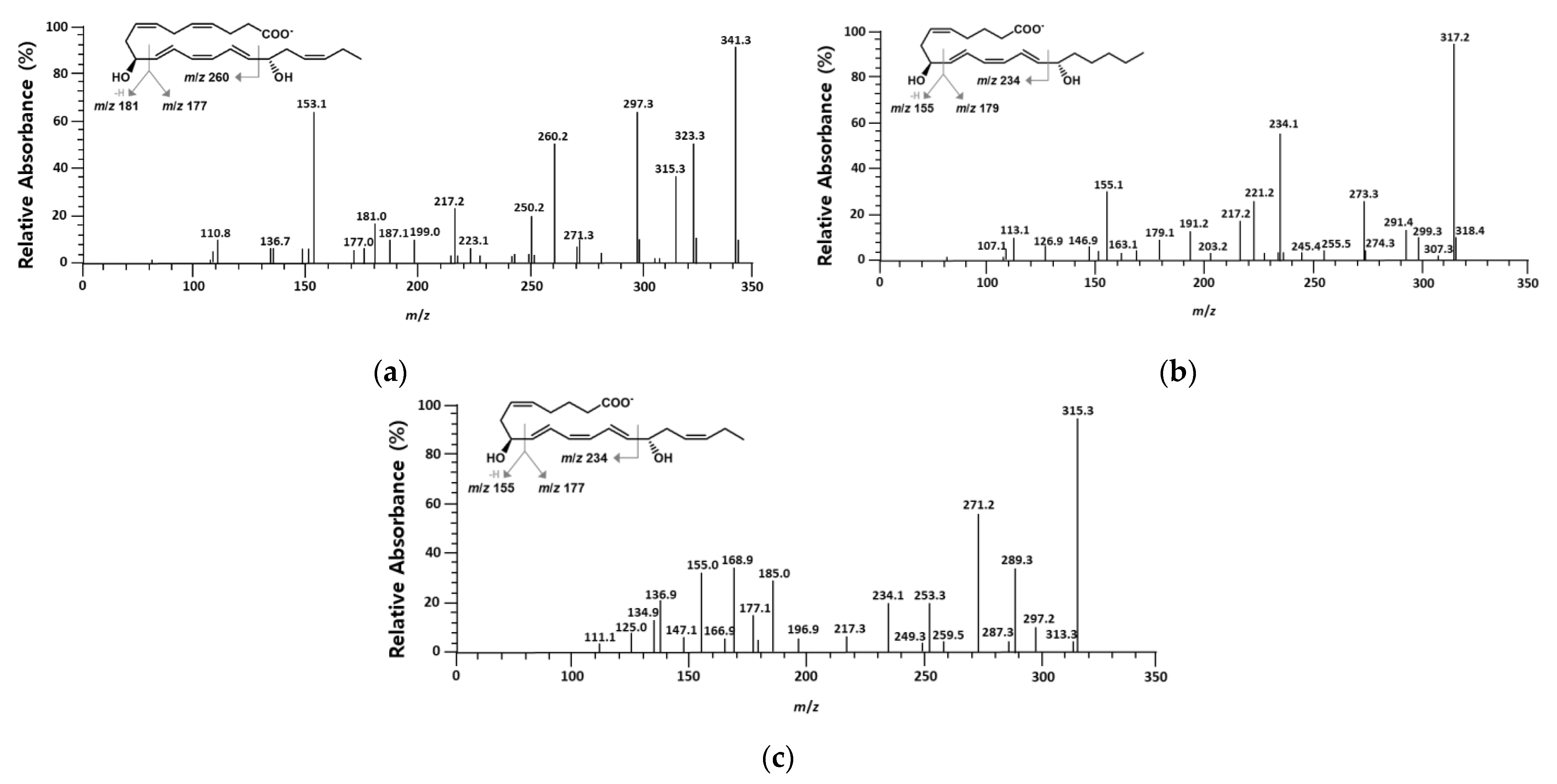
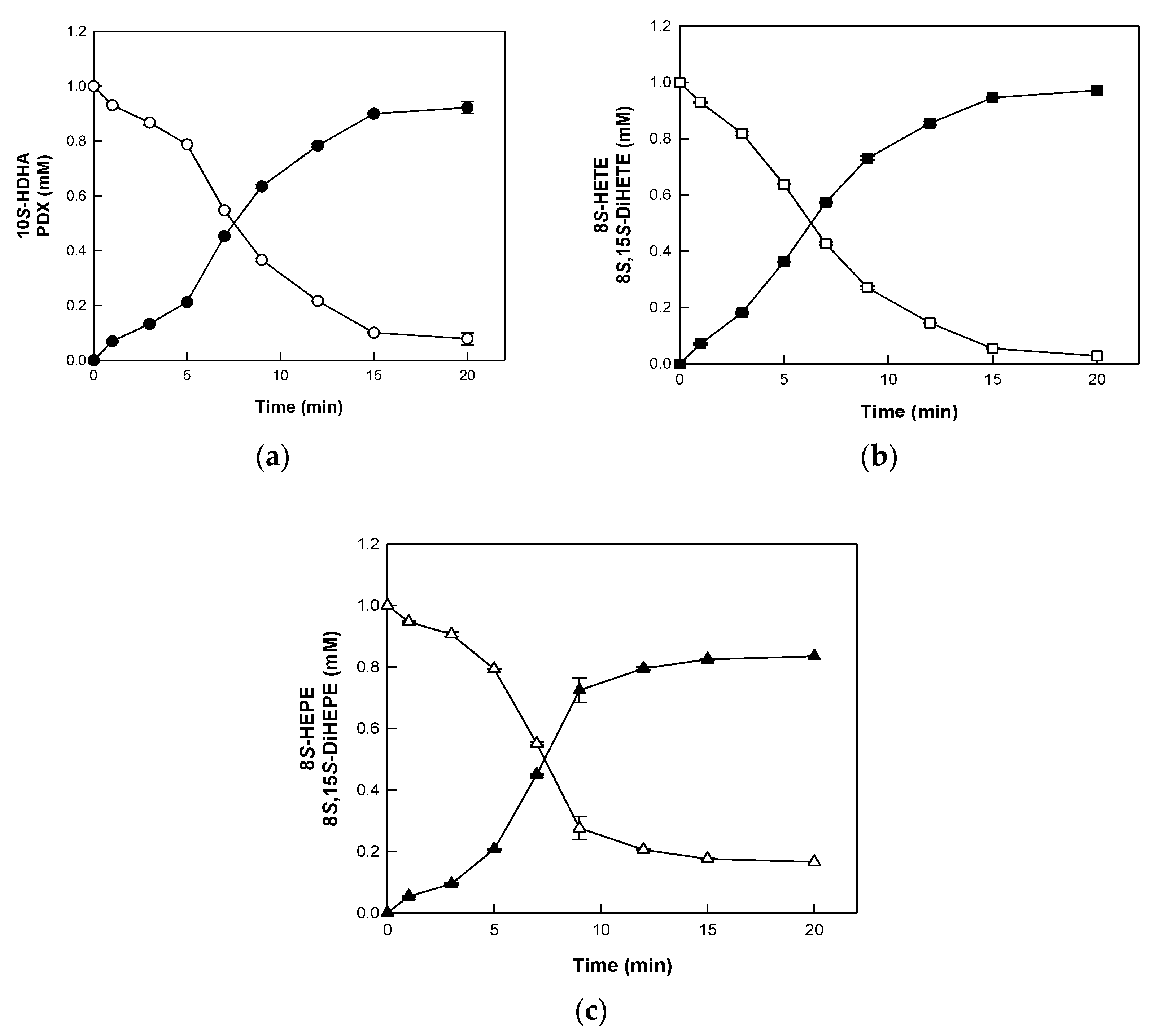
2.1. Identification of Products Obtained from the Conversion of 10S-HDHA, 8S-HETE, and 8S-HEPE by BT 15SLOX

Hydroxylation at the C17 position of 10S-HDHA is a key step in the production of the DiHFA (dihydroxy fatty acid) PDX. To accurately identify the conversion of hydroxy fatty acids (HFAs) into DiHFAs, enzymatic reaction using BT 15SLOX was performed. The DiHFA products P1, P2, and P3 converted from HFA substrates 10S-HDHA, 8S-HETE, and 8S-HEPE, which were converted from DHA, arachidonic acid (AA), and eicosapentaenoic acid (EPA) by E. coli cells expressing MO 8SLOX, respectively [38], were detected with retention times of 3.46, 3.72, and 4.25 min in high-performance liquid chromatography (HPLC), respectively (Figure S1). The total molecular masses of the products derived from 10S-HDHA, 8S-HETE, and 8S-HEPE were indicated by the peaks at mass per charge (m/z) values of 359.6, 335.6, and 333.7 in their LC-MS spectra (Figure S2), corresponding to the molecular masses of DiHDHA, DiHETE, and DiHEPE, respectively. The fragment peaks at m/z 341, 317, and 315 in Figure 2 were formed by the loss of H2O from the total molecular masses. The fragment peaks at m/z 181 and 177, and m/z 260 in the LC-MS/MS spectra of the product from 10S-HDHA resulted from cleavages between the C10 and C11 positions and between the C16 and C17 positions, respectively (Figure 2a). The fragment peaks at m/z 155 and 179, and m/z 234 of the product from 8S-HETE, indicate the cleavages between the C8 and C9 positions and between the C14 and C15 positions, respectively (Figure 2b). In the Liquid chromatography–tandem mass spectrometry (LC-MS/MS) spectra of the product from 8S-HEPE, the fragment peaks at m/z 155 and 177, and m/z 234, indicate the cleavages between the C8 and C9 positions and between the C14 and C15 positions, respectively (Figure 2c).
The stereochemistry of 10,17-DiHDHA was confirmed by 1D and 2D Nuclear magnetic resonance (NMR) spectroscopy (Table 1 and Figure S3) by comparing with a previous report [26]. The coupling constants of J11-12 and J15-16 were 15 Hz, indicating that the double bond is in E geometry at 5.70 and 6.68 ppm, respectively. Four Z geometric double bonds were contained at 5.45–5.31 ppm of H-4,7,19 and 5.93 ppm of H-13. The 10S-H and 17S-H peaks had 4.14 and 4.12 ppm, respectively, indicating that the product obtained from the conversion of 10S-HDHA by BT 15SLOX is 10S,17S-DiHDHA and that BT 15SLOX is a PDX-producing enzyme.

2.2. Substrate Specificity of BT 15SLOX for PUFAs and HFAs

Since the maximal activity of BT 15SLOX was observed at pH 7.5 and 25 °C [27], the substrate specificity of BT 15SLOX was determined at this pH and temperature by measuring the specific activities for PUFAs, such as DHA, AA, and EPA, and HFAs, such as 10S-HDHA, 8S-HETE, and 8S-HEPE (Table 2). The specific activity of BT 15SLOX towards PUFAs followed the order AA > EPA > DHA, whereas that towards HFAs followed the reverse order, 10S-HDHA > 8S-HEPE > 8S-HETE. Among the activities towards HFAs, the activity towards 10S-HDHA was approximately 1.9- and 3.0-fold higher than that towards 8S-HETE and 8S-HEPE, respectively. These results indicate that BT 15SLOX is an efficient enzyme for producing PDX from 10S-HDHA derived from DHA.

2.3. Biotransformation of HFAs to DiHFAs by BT 15SLOX

The conversion of HFAs, 10S-HDHA, 8S-HETE, and 8S-HEPE to the DiHFAs 10S,17S-DiHDHA (PDX), 8S,15S-DiHETE, and 8S,15S-DiHEPE, respectively, was investigated at 25 °C and pH 7.5 with 1 mM HFA, and 1.5 mg/mL enzyme in the presence of 10 mM cysteine for 20 min (Figure 3). BT 15SLOX converted 1 mM of 10S-HDHA, 8S-HETE, and 8S-HEPE to 0.92 mM PDX, 0.97 mM 8S,15S-DiHETE, and 0.83 mM 8S,15S-DiHEPE for 20 min, with molar conversion yields of 92, 97, and 83%, and productivities of 2.76, 2.91, and 2.50 mM/h, respectively. The conversion rate and productivity towards the substrates followed the order: 8S-HETE > 10S-HDHA > 8S-HEPE, which was different from the order of the specific activity. This may be due to the differences in the initial and overall rates and/or used substrate concentration (0.2 mM and 1 mM). To date, DiHFAs have been quantitatively produced by double-dioxygenating microbial LOXs, including 15SLOX from A. violaceum [36] and 12SLOX from Endozoicomonas numazuensis [37], which convert DHA to 7S,17S-DiHDHA (RvD5) and 7S,14S-DiHDHA, with molar conversion yields of 40 and 52%, respectively. In contrast, in the present study, DiHFA was produced by the combined reactions of two single-dioxygenating LOXs. Therefore, the biotransformation of HFAs to DiHFAs in this study could not be quantitatively compared with other studies. Nevertheless, the high yield of 92% for the conversion of 10S-HDHA to PDX indicated that BT 15SLOX is a promising biocatalyst for PDX production.

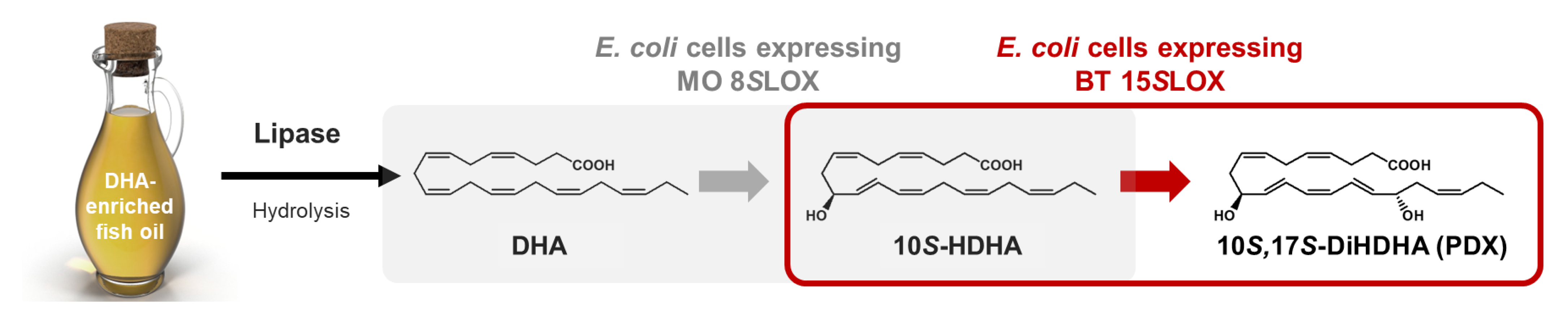
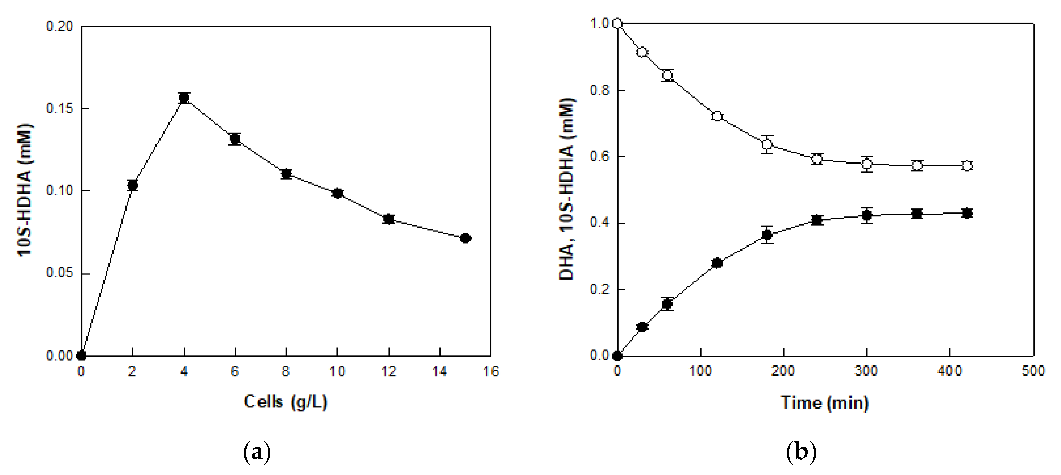
2.4. Production of 10S-HDHA from DHA-Enriched Fish Oil via DHA in DFOH by E. coli Cells Expressing MO 8SLOX

DHA-enriched fish oil, which was industrially produced and directly ingestible as a dietary supplement [3,5,39], was used as a cheap raw material for PDX production. DFOH containing 1 mM DHA was obtained from 455 mg/L DHA-enriched fish oil by lipase from T. lanuginose after 1 h of treatment for use as a substrate for 10S-HDHA production. Whole-cell reaction using E. coli cells has been used for HFA and DiHFA productions because it is more economical and stable than the enzymatic reaction [38,40]. Therefore, two E. coli cells expressing MO 8SLOX and BT 15SLOX were used for the production of 10S-HDHA and PDX from DHA and 10S-HDHA, respectively.
In the first-step of the whole-cell biotransformation, DHA in lipase-treated DFOH was converted to 10S-HDHA by E. coli cells expressing MO 8SLOX. The maximal activity of the cells was observed at pH 7.5 and at 35 °C (Figure S4). These results were the same as the optimal pH and temperature for producing 8S-HETE from AA using E. coli cells expressing MO 8SLOX [38].
The optimal concentration of E. coli cells expressing MO 8SLOX to produce 10S-HDHA from DHA in DFOH was assessed at pH 7.5 and 35 °C for 1 h by varying the cell concentration from 2 to 15 g/L (Figure 4a). 10S-HDHA production was highest at a cell concentration of 4 g/L, indicating that the optimal cell concentration was 4 g/L. Under the conditions of pH 7.5, 35 °C, 4 g/L cells, and 1 mM DHA in DFOH, 10S-HDHA production was performed for 7 h (Figure 4b). E. coli cells expressing MO 8SLOX produced 0.43 mM 10S-HDHA from 1 mM DHA in DFOH for 5 h, with a molar conversion yield of 43%, which was approximately 1.5-fold lower than that from reagent-grade DHA [38]. This reduction may be due to the influence of other fatty acids, such as AA and EPA, or glycerol in DFOH.

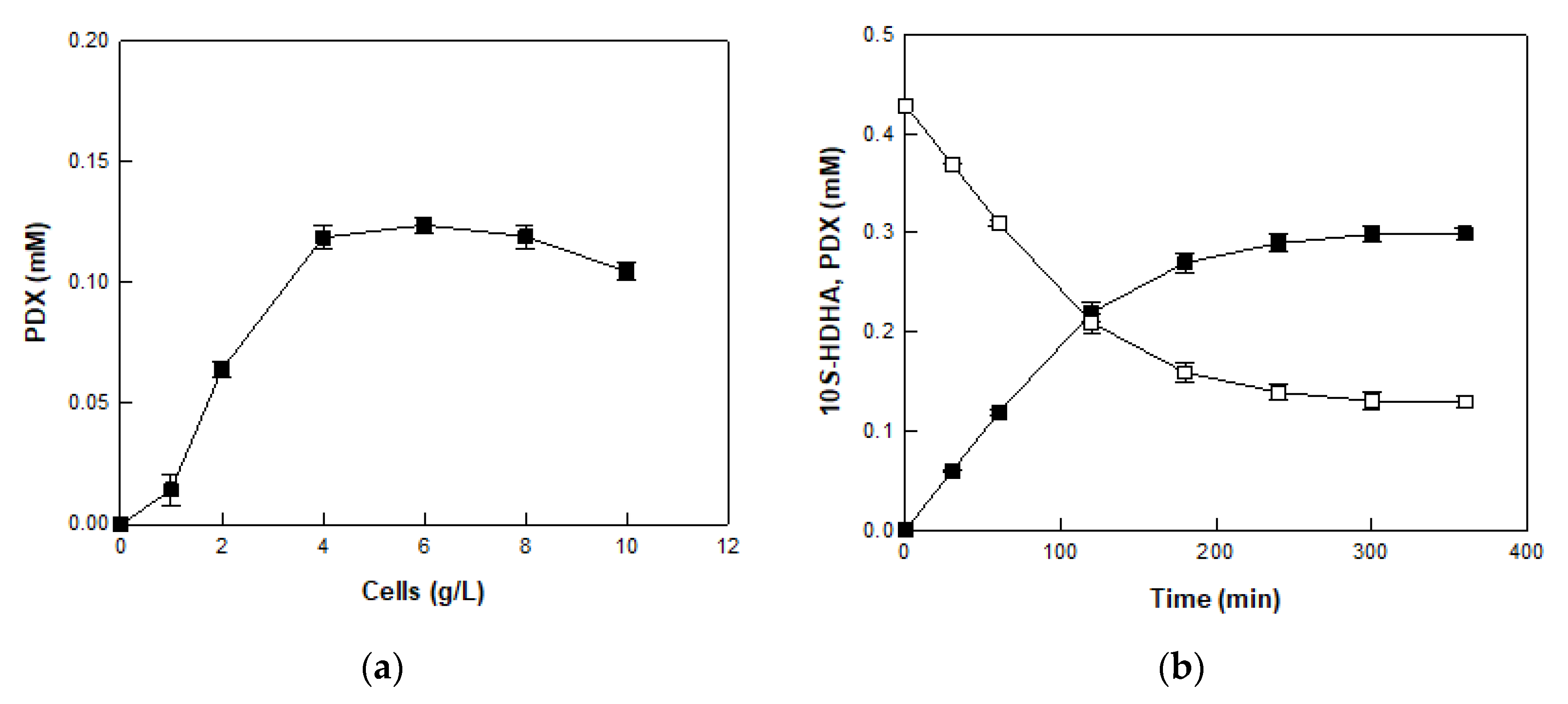
2.5. Production of PDX from 10S-HDHA in MO 8SLOX-Treated DFOH by E. coli Cells Expressing BT 15SLOX

In the second-step of the whole-cell biotransformation, 10S-HDHA in 8SLOX-treated DFOH was converted to PDX by E. coli cells expressing BT 15SLOX. The maximal activity of the cells was observed at pH 7.5 and 30 °C (Figure S5). The optimal pH for producing PDX from 10S-HDHA in DFOH was the same as that of 13-hydroxyoctadecadienoic acid produced from linoleic acid using the same enzyme, whereas the optimal temperature was 5 °C higher [27].
The effect of the concentration of recombinant E. coli cells on PDX production was studied with 0.43 mM 10S-HDHA in MO 8SLOX-treated DFOH by varying the cell concentration from 1 to 10 g/L (Figure 5a). PDX production increased with increasing cell concentration, but the production rate significantly decreased at concentrations above 4 g/L. Therefore, the optimum cell concentration was determined to be 4 g/L, and the optimal reaction conditions to produce PDX were pH 7.5, 30 °C, and 4 g/L cells. Under these conditions, PDX production was performed with 10S-HDHA in MO 8SLOX-treated DFOH for 6 h (Figure 5b). E. coli cells expressing BT 15SLOX produced 0.30 mM PDX from 0.43 mM 10S-HDHA in MO 8SLOX-treated DFOH for 5 h, with a productivity of 60 μM/h and molar conversion yield of 70%.
Table 3 shows the conversion yield and productivity of PDX from DHA-enriched fish oil at each step, which was summarized as follows: DHA-enriched fish oil at 455 mg/L was hydrolyzed to DFOH containing 1 mM (328 mg/L) DHA by lipase after 1 h. DFOH containing 1 mM DHA was converted to 0.43 mM (148 mg/L) 10S-HDHA by E. coli cells expressing MO 8SLOX after 6 h. PDX at 0.30 mM (108 mg/L) was produced from 0.43 mM 10S-HDHA in MO 8SLOX-treated DFOH by E. coli cells expressing BT 15SLOX after 5 h. Consequently, 455 mg/L DHA-enriched fish oil was converted to 108 mg/L PDX after 12 h, with a conversion yield of 24% (w/w) and a productivity of 4.5 mg/L/h. Only a few studies have reported the qualitative biosynthesis of PDX [26,41]. For instance, human 15SLOX converted 2 μM 17S-hydroperoxydocosahexaenoic acid to PDX as a minor product, accounting for only 28% of all product peaks in the LC-MS trace [41]. Thus, we performed the first quantitative production of PDX. However, this study still has a limitation in that it involves several reaction steps to produce PDX from the oil. In the future, it is necessary to develop the production of PDX using a one-pot or one-cell reaction.

3. Materials and Methods

3.1. Materials

Lipase from Thermomyces lanuginosa and PUFA standards, including DHA, AA, and EPA, were purchased from Genofocus (Daejeon, Republic of Korea) and Sigma-Aldrich (St. Louis, MO, USA), respectively. HFA standards, including 8S-HETE and 8S-HEPE, and DiHFA standards, including PDX and 8S,15S-DiHETE, were purchased from Cayman Chemical (Ann Arbor, MI, USA). 10S-HDHA and 8S,15S-DiHEPE, which were obtained from the conversion of DHA and 8S-HEPE by MO 8SLOX and BT 15SLOX, respectively, were purified using a semi-preparative HPLC system, as described previously [36,42]. The purified 10S-HDHA was used as a standard and substrate to produce PDX and the purified 8S,15S-DiHEPE was used as a standard. DHA-enriched fish oil (73% (w/v) DHA) was purchased from AK & MN BioFarm (Ulsan, Republic of Korea). The hydrolysis of DHA-enriched fish oil to DFOH by lipase from T. lanuginosa was performed with 455 mg/L oil and 0.5 mg/mL lipase at 37 °C and pH 7.5 with shaking at 250 rpm for 1 h. Complementary DNA (cDNA) encoding MO 8SLOX was provided by the Korea Human Gene Bank, Medical Genomics Research Center (Daejeon, Republic of Korea).

3.2. Gene Cloning and Culture Conditions

cDNA encoding MO 8SLOX and genomic DNA of B. thailandensis KCTC 23190 were used for cloning of 8SLOX and 15SLOX, respectively. E. coli ER2566 (host cells) and pET-28a (+) plasmids (expression vectors) were used for cloning of both genes. Gene cloning of MO 8SLOX and BT 15SLOX was performed as previously described [27,38]. E. coli cells expressing MO 8SLOX and BT 15SLOX were cultured in Luria-Bertani (LB) medium supplemented with kanamycin (50 μg/mL) at 37 °C, with shaking at 200 rpm. Protein expression was induced by adding isopropyl-β-d-thiogalactopyranoside (IPTG, 0.1 mM) once an OD600 = 0.6 was reached. The cells were further grown for 16 h at 150 rpm at 16 °C.

3.3. Enzyme Purification

E. coli cells expressing BT 15SLOX were harvested from the culture broth by centrifugation (6000× g, 30 min, 4 °C), washed twice using 0.85% NaCl solution, and resuspended using lysis buffer (50 mM phosphate buffer pH 7.5, 300 mM NaCl, 1 mg/mL lysozyme). Cells were lysed by sonication on ice for 25 min (power 25%, pulse on 40 s, and pulse off 20 s with a sonic dismembrator (Model 100; Fisher Scientific, Pittsburgh, PA, USA)). The lysed cells were centrifuged (13,000× g, 20 min, 4 °C), and the supernatant was used to purify the expressed enzyme following standard techniques of His-trap HP (GE Healthcare, Piscataway, NJ, USA) and a fast protein liquid chromatography (Bio-Rad, Hercules, CA, USA) systems. BT 15SLOX purification from cell lysate followed the described in our previous publication and the purified enzyme showed a single band in SDS-PAGE [27].

3.4. Enzyme Reaction

The substrate specificity of BT 15SLOX for PUFAs (DHA, AA, EPA) and HFAs (10S-HDHA, 8S-HETE, 8S-HEPE) was investigated by incubating at 30 °C for 10 min in a 50 mM 4-(2-hydroxyethyl)-1-piperazine ethanesulfonic acid (HEPES) buffer (pH 7.5) containing 0.2 mM substrate and 0.1 mg/mL BT 15SLOX with a reducing agent (10 mM cysteine). The activity of BT 15SLOX for each substrate was evaluated by quantitatively measuring the product via an HPLC system (Agilent 1100, Santa Clara, CA, USA). The conversion of 10S-HDHA, 8S-HETE, and 8S-HEPE to PDX, 8S,15S-DiHETE, and 8S,15S-DiHEPE, respectively, were performed in a 50 mM HEPES buffer (pH 7.5) containing 1 mM substrate and 1.5 mg/mL BT 15SLOX with 10 mM cysteine at 30 °C for 20 min.

3.5. Whole-Cell Reaction

The optimal pH and temperature to produce 10S-HDHA from DHA in DFOH or to produce PDX from 10S-HDHA in DFOH obtained from treating E. coli cells expressing MO 8SLOX to DHA (MO 8SLOX-treated DFOH) were determined; pH was varied from 6.0 to 9.0 using 50 mM 2-(N-morpholino) ethane sulfonic acid (MES, pH 6.0−6.5), HEPES (pH 6.5−8.0), 3-[4-(2-hydroxyethyl)-1-piperazinyl] propane sulfonic acid (EPPS, pH 8.0−8.5), and 2-(cyclohexylamino)ethane-1-sulfonic acid (CHES, pH 8.5−9.0) buffers at a constant temperature of 30 or 35 °C and temperature was varied from 15 or 10 °C to 45 or 40 °C in 50 mM HEPES buffer at a constant pH of 7.5, respectively. The reactions were conducted in the presence of 10 mM cysteine with 1 mM DHA in DFOH and 1 g/L E. coli cells expressing MO 8SLOX for 10S-HDHA production. The reactions were performed in the presence of 10 mM cysteine with 0.43 mM 10S-HDHA in MO 8SLOX-treated DFOH and 4 g/L E. coli cells expressing BT 15SLOX for PDX production. The optimal cell concentration for producing 10S-HDHA was determined by varying the concentration of E. coli cells expressing MO 8SLOX from 2 to 15 g/L with 1 mM DHA in DFOH with 10 mM cysteine (pH 7.5, 35 °C, and 1 h). The optimal cell concentration to produce PDX, the reactions were performed with different concentrations of E. coli cells expressing BT 15SLOX from 1 to 10 g/L and MO 8SLOX-treated DFOH containing 0.43 mM 10S-HDHA in the presence of 10 mM cysteine at pH 7.5 and 30 °C for 1 h.

3.6. HPLC Analysis

An equal volume of ethyl acetate was used to extract the reaction mixtures, and the extracted solution was evaporated until completely dry, and then methanol was added for HPLC analysis. Substrates and their products were analyzed using a Nucleosil C18 column (3.2 × 150 mm, 5-μm particle size; Phenomenex, Torrance, CA, USA) and monitored at 202 nm, and their concentrations were calculated as described in our previous publication [36]. The column was eluted at 35 °C with mobile phase A (MPA, acetonitrile/water/acetic acid, 50/50/0.1, v/v/v) and mobile phase B (MPB, acetonitrile/acetic acid, 100/0.1, v/v) gradient with a flow rate of 0.25 mL/min as follows: 100% MPA for 0−5 min, from 100% MPA to 100% MPB 5−21 min, from 100% MPB to 100% MPA for 21−27 min, and 100% MPA for 27−30 min.

3.7. LC-MS/MS and NMR Analyses

LC-MS/MS analysis of the DiHFAs was performed using a Thermo-Finnigan LCQ Deca XP plus ion trap mass spectrometer (Thermo Fisher Scientific, Waltham, MA, USA) at the National Instrumentation Center for Environmental Management of Seoul National University (Seoul, Republic of Korea). Sample ionization was carried out by electrospray ionization with the following settings: capillary temperature (275 °C), ion source voltage (5 kV), nebulizer gas (207 kPa), capillary voltage (15 V, negative mode), average scan time (0.6 s), average time to change polarity (1.2 s), and abundance of precursor ions at collision energy (35%). NMR spectroscopy was used to confirm the chemical structure of PDX by recording 1D (1H, 13C, and 1H homo decoupling) and 2D (COSY, ROESY, HSQC, and HMBC) NMR spectra using a Bruker Avance III HD (850 MHz) equipped with a triple-resonance inverse cryoprobe at the National Center for Inter-University Research Facilities of Seoul National University. Deuterated methanol (MeOD) was used as the solvent and internal standard (1H:δ = 3.31, 13C:δ = 49.15). All chemical shifts are shown in δ (ppm).

4. Conclusions

The production of the SPM PDX from DHA-enriched fish oil was demonstrated via an enzymatic hydrolysis reaction and two-step whole-cell biotransformation process. DHA in DFOH obtained from 455 mg/L oil by 0.5 mg/mL lipase was converted to 148 mg/L 10S-HDHA by 4 g/L whole E. coli cells expressing MO 8SLOX and was subsequently converted to 108 mg/L PDX by 4 g/L whole E. coli cells expressing BT 15SLOX. To the best of our knowledge, this is the first quantitative biotechnological production of PDX. PDX has been chemically synthesized, which is an expensive and pollution-causing method, whereas the amounts produced by biological methods are trace to date. Thus, the establishment of the biotransformation process from inexpensive natural oil to expensive SPM is meaningful. Our results would contribute to the economical production of PDX, thereby also providing sufficient supply of it for studying its efficacy.

Supplementary Materials

The following supporting information can be downloaded at: https://www.mdpi.com/article/10.3390/catal12101145/s1, Figure S1: HPLC chromatograms for the products obtained from the conversion of (a) 10S-HDHA, (b) 8S-HETE, and (c) 8S-HEPE by BT 15SLOX. P1, P2, and P3 represent unknown products derived from 10S-HDHA, 8S-HETE, and 8S-HEPE, respectively; Figure S2: LC-MS spectra for the products obtained from the conversion of (a) 10S-HDHA, (b) 8S-HETE, and (c) 8S-HEPE by BT 15SLOX; Figure S3: 1D (1H and 13C) and 2D (COSY, ROSEY, HSQC, and HMBC) NMR spectra of 10S,17S-DiHDHA (PDX) using NMR spectroscopy (850 MHz NMR, MeDO). (a) 1H NMR spectrum of PDX. (b) 13C NMR spectrum of PDX. (c) COSY spectrum of PDX. (d) ROSEY spectrum of PDX. (e) HSQC spectrum of PDX. (f) HMBC spectrum of PDX; Figure S4: Effects of pH and temperature on the production of 10S-HDHA from DHA in DFOH using E. coli cells expressing MO 8SLOX. (a) Effect of pH. The reactions were performed in 50 mM MES (pH 6.0−6.5, ○), HEPES (pH 6.5−8.0, ■), EPPS (pH 8.0−8.5, △), and CHES (pH 8.5−9.0, ♦) buffers with 1 g/L of E. coli cells expressing MO 8SLOX and DFOH containing 1 mM DHA at 35 °C for 10 min. (b) Effect of temperature. The reactions were performed in 50 mM HEPES buffer (pH 7.5) with 1 g/L of E. coli cells expressing MO 8SLOX and DFOH containing 1 mM DHA by varying the temperature from 15 to 45 °C for 10 min. Data represent the means of three separate experiments, and error bars represent the standard deviation; Figure S5: Effects of pH and temperature on the production of PDX from 10S-HDHA in 8SLOX-treated DFOH using E. coli cells expressing BT 15SLOX. (a) Effect of pH. The reactions were performed in 50 mM MES (pH 6.0−6.5, ○), HEPES (pH 6.5−8.0, ■), EPPS (pH 8.0−8.5, △), and CHES (pH 8.5−9.0, ♦) buffers with 1 g/L cells expressing BT 15SLOX and 8SLOX-treated DFOH containing 0.43 mM 10S-HDHA at 35 °C for 10 min. (b) Effect of temperature. The reactions were performed in 50 mM HEPES buffer (pH 7.5) with 1 g/L of E. coli cells expressing BT 15SLOX and 8SLOX-treated DFOH containing 0.43 mM 10S-HDHA by varying the temperature from 10 to 40 °C for 10 min. Data represent the means of three separate experiments, and error bars represent the standard deviation; Table S1: The chemical structures and abbreviations of SPMs and their analogs.

Author Contributions

Conceptualization, K.-C.S., M.-J.S. and D.-K.O.; methodology, K.-C.S., T.-E.L., S.-E.K. and M.-J.S.; formal analysis, K.-C.S., T.-E.L., Y.-J.K. and M.-J.S.; investigation, K.-C.S. and D.-K.O.; data curation, K.-C.S., T.-E.L. and M.-J.S.; writing—original draft, K.-C.S., M.-J.S. and D.-K.O.; writing—reviewing and editing, K.-C.S., M.-J.S. and D.-K.O.; supervision, K.-C.S. and D.-K.O.; funding acquisition, K.-C.S., M.-J.S. and D.-K.O. All authors have read and agreed to the published version of the manuscript.

Funding

This work was supported by the Basic Research Lab Program (NRF-2020R1A4A1018648), the Basic Science Research Program (NRF-2020R1I1A1A01063145), and National Research Foundation (NRF) grant (NRF-2022R1C1C2003774) funded by the Korean Ministry of Science and ICT.

Data Availability Statement

Not applicable.

Acknowledgments

This paper was supported by the KU Research Professor Program of Konkuk University.

Conflicts of Interest

The authors declare no conflict of interest.

References

  1. Venugopalan, V.K.; Gopakumar, L.R.; Kumaran, A.K.; Chatterjee, N.S.; Soman, V.; Peeralil, S.; Mathew, S.; McClements, D.J.; Nagarajarao, R.C. Encapsulation and protection of omega-3-rich fish oils using food-grade delivery systems. Foods 2021, 10, 1566. [Google Scholar] [CrossRef] [PubMed]
  2. Ghelichi, S.; Shabanpour, B.; Pourashouri, P.; Hajfathalian, M.; Jacobsen, C. Extraction of unsaturated fatty acid-rich oil from common carp (Cyprinus carpio) roe and production of defatted roe hydrolysates with functional, antioxidant, and antibacterial properties. J. Sci. Food Agric. 2018, 98, 1407–1415. [Google Scholar] [CrossRef] [PubMed]
  3. Chang, P.K.; Tsai, M.F.; Huang, C.Y.; Lee, C.L.; Lin, C.; Shieh, C.J.; Kuo, C.H. Chitosan-based anti-oxidation delivery nano-platform: Applications in the encapsulation of DHA-enriched fish oil. Mar. Drugs 2021, 19, 470. [Google Scholar] [CrossRef] [PubMed]
  4. Li, Z.; Chen, H.; Su, J.; Wang, W.; Chen, H.; Yang, B.; Wang, Y. Highly efficient and enzyme-recoverable method for enzymatic concentrating omega-3 fatty acids generated by hydrolysis of fish oil in a substrate-constituted three-liquid-phase system. J. Agric. Food Chem. 2019, 67, 2570–2580. [Google Scholar] [CrossRef] [PubMed]
  5. Zhang, Z.; Liu, F.; Ma, X.; Huang, H.; Wang, Y. Two-stage enzymatic preparation of eicosapentaenoic acid (EPA) and docosahexaenoic acid (DHA) enriched fish oil triacylglycerols. J. Agric. Food Chem. 2018, 66, 218–227. [Google Scholar] [CrossRef]
  6. Nordgren, T.M.; Anderson Berry, A.; Van Ormer, M.; Zoucha, S.; Elliott, E.; Johnson, R.; McGinn, E.; Cave, C.; Rilett, K.; Weishaar, K.; et al. Omega-3 fatty acid supplementation, pro-resolving mediators, and clinical outcomes in maternal-infant pairs. Nutrients 2019, 11, 98. [Google Scholar] [CrossRef]
  7. Grenon, S.M.; Owens, C.D.; Nosova, E.V.; Hughes-Fulford, M.; Alley, H.F.; Chong, K.; Perez, S.; Yen, P.K.; Boscardin, J.; Hellmann, J.; et al. Short-term, high-dose fish oil supplementation increases the production of omega-3 fatty acid-derived mediators in patients with peripheral artery disease (the OMEGA-PAD I Trial). J. Am. Heart Assoc. 2015, 4, e002034. [Google Scholar] [CrossRef]
  8. Ramirez, J.L.; Gasper, W.J.; Khetani, S.A.; Zahner, G.J.; Hills, N.K.; Mitchell, P.T.; Sansbury, B.E.; Conte, M.S.; Spite, M.; Grenon, S.M. Fish oil increases specialized pro-resolving lipid mediators in PAD (The OMEGA-PAD II Trial). J. Surg. Res. 2019, 238, 164–174. [Google Scholar] [CrossRef]
  9. Basil, M.C.; Levy, B.D. Specialized pro-resolving mediators: Endogenous regulators of infection and inflammation. Nat. Rev. Immunol. 2016, 16, 51–67. [Google Scholar] [CrossRef]
  10. Sugimoto, M.A.; Vago, J.P.; Perretti, M.; Teixeira, M.M. Mediators of the resolution of the inflammatory response. Trends Immunol. 2019, 40, 212–227. [Google Scholar] [CrossRef]
  11. Serhan, C.N. Pro-resolving lipid mediators are leads for resolution physiology. Nature 2014, 510, 92–101. [Google Scholar] [CrossRef] [PubMed]
  12. Chiang, N.; Fredman, G.; Bäckhed, F.; Oh, S.F.; Vickery, T.; Schmidt, B.A.; Serhan, C.N. Infection regulates pro-resolving mediators that lower antibiotic requirements. Nature 2012, 484, 524–528. [Google Scholar] [CrossRef] [PubMed]
  13. Leuti, A.; Maccarrone, M.; Chiurchiù, V. Proresolving lipid mediators: Endogenous modulators of oxidative stress. Oxidative Med. Cell Longev. 2019, 2019, 8107265. [Google Scholar] [CrossRef] [PubMed]
  14. Chávez-Castillo, M.; Ortega, Á.; Cudris-Torres, L.; Duran, P.; Rojas, M.; Manzano, A.; Garrido, B.; Salazar, J.; Silva, A.; Rojas-Gomez, D.M.; et al. Specialized pro-resolving lipid mediators: The future of chronic pain therapy? Int. J. Mol. Sci. 2021, 22, 10370. [Google Scholar] [CrossRef] [PubMed]
  15. Ishihara, T.; Yoshida, M.; Arita, M. Omega-3 fatty acid-derived mediators that control inflammation and tissue homeostasis. Int. Immunol. 2019, 31, 559–567. [Google Scholar] [CrossRef]
  16. White, P.J.; St-Pierre, P.; Charbonneau, A.; Mitchell, P.L.; St-Amand, E.; Marcotte, B.; Marette, A. Protectin DX alleviates insulin resistance by activating a myokine-liver glucoregulatory axis. Nat. Med. 2014, 20, 664–669. [Google Scholar] [CrossRef]
  17. Gobbetti, T.; Dalli, J.; Colas, R.A.; Federici Canova, D.; Aursnes, M.; Bonnet, D.; Alric, L.; Vergnolle, N.; Deraison, C.; Hansen, T.V.; et al. Protectin D1(n-3 DPA) and resolvin D5(n-3 DPA) are effectors of intestinal protection. Proc. Natl. Acad. Sci. USA 2017, 114, 3963–3968. [Google Scholar] [CrossRef]
  18. Bäck, M.; Yurdagul, A., Jr.; Tabas, I.; Öörni, K.; Kovanen, P.T. Inflammation and its resolution in atherosclerosis: Mediators and therapeutic opportunities. Nat. Rev. Cardiol. 2019, 16, 389–406. [Google Scholar] [CrossRef]
  19. Ramon, S.; Gao, F.; Serhan, C.N.; Phipps, R.P. Specialized proresolving mediators enhance human B cell differentiation to antibody-secreting cells. J. Immunol. 2012, 189, 1036–1042. [Google Scholar] [CrossRef]
  20. Spite, M.; Norling, L.V.; Summers, L.; Yang, R.; Cooper, D.; Petasis, N.A.; Flower, R.J.; Perretti, M.; Serhan, C.N. Resolvin D2 is a potent regulator of leukocytes and controls microbial sepsis. Nature 2009, 461, 1287–1291. [Google Scholar] [CrossRef] [Green Version]
  21. Morita, M.; Kuba, K.; Ichikawa, A.; Nakayama, M.; Katahira, J.; Iwamoto, R.; Watanebe, T.; Sakabe, S.; Daidoji, T.; Nakamura, S.; et al. The lipid mediator protectin D1 inhibits influenza virus replication and improves severe influenza. Cell 2013, 153, 112–125. [Google Scholar] [CrossRef] [PubMed]
  22. Iwuchukwu, I.; Nguyen, D.; Shirazian, A.; Asatryan, A.; Jun, B.; Bazan, N.G. Neuroprotectin D1, a lipid anti-inflammatory mediator, in patients with intracerebral hemorrhage. Biochimie 2022, 195, 16–18. [Google Scholar] [CrossRef] [PubMed]
  23. Antony, R.; Lukiw, W.J.; Bazan, N.G. Neuroprotectin D1 induces dephosphorylation of Bcl-xL in a PP2A-dependent manner during oxidative stress and promotes retinal pigment epithelial cell survival. J. Biol. Chem. 2010, 285, 18301–18308. [Google Scholar] [CrossRef]
  24. Belayev, L.; Hong, S.H.; Menghani, H.; Marcell, S.J.; Obenaus, A.; Freitas, R.S.; Khoutorova, L.; Balaszczuk, V.; Jun, B.; Oriá, R.B.; et al. Docosanoids promote neurogenesis and angiogenesis, blood-brain barrier integrity, penumbra protection, and neurobehavioral recovery after experimental ischemic stroke. Mol. Neurobiol. 2018, 55, 7090–7106. [Google Scholar] [CrossRef] [PubMed]
  25. Asatryan, A.; Bazan, N.G. Molecular mechanisms of signaling via the docosanoid neuroprotectin D1 for cellular homeostasis and neuroprotection. J. Biol. Chem. 2017, 292, 12390–12397. [Google Scholar] [CrossRef]
  26. Chen, P.; Fenet, B.; Michaud, S.; Tomczyk, N.; Véricel, E.; Lagarde, M.; Guichardant, M. Full characterization of PDX, a neuroprotectin/protectin D1 isomer, which inhibits blood platelet aggregation. FEBS Lett. 2009, 583, 3478–3484. [Google Scholar] [CrossRef]
  27. An, J.U.; Kim, B.J.; Hong, S.H.; Oh, D.K. Characterization of an omega-6 linoleate lipoxygenase from Burkholderia thailandensis and its application in the production of 13-hydroxyoctadecadienoic acid. Appl. Microbiol. Biotechnol. 2015, 99, 5487–5497. [Google Scholar] [CrossRef]
  28. Lagarde, M.; Guichardant, M.; Bernoud-Hubac, N. Anti-inflammatory and anti-virus potential of poxytrins, especially protectin DX. Biochimie 2020, 179, 281–284. [Google Scholar] [CrossRef]
  29. Hwang, H.-J.; Jung, T.W.; Kim, J.W.; Kim, J.A.; Lee, Y.B.; Hong, S.H.; Roh, E.; Choi, K.M.; Baik, S.H.; Yoo, H.J. Protectin DX prevents H2O2-mediated oxidative stress in vascular endothelial cells via an AMPK-dependent mechanism. Cell. Signal. 2019, 53, 14–21. [Google Scholar] [CrossRef]
  30. Liu, M.; Boussetta, T.; Makni-Maalej, K.; Fay, M.; Driss, F.; El-Benna, J.; Lagarde, M.; Guichardant, M. Protectin DX, a double lipoxygenase product of DHA, inhibits both ROS production in human neutrophils and cyclooxygenase activities. Lipids 2014, 49, 49–57. [Google Scholar] [CrossRef] [Green Version]
  31. Yang, J.X.; Li, M.; Hu, X.; Lu, J.C.; Wang, Q.; Lu, S.Y.; Gao, F.; Jin, S.W.; Zheng, S.X. Protectin DX promotes epithelial injury repair and inhibits fibroproliferation partly via ALX/PI3K signalling pathway. J. Cell. Mol. Med. 2020, 24, 14001–14012. [Google Scholar] [CrossRef] [PubMed]
  32. Sancéau, J.Y.; Maltais, R.; Poirier, D.; Marette, A. Total synthesis of the antidiabetic (type 2) lipid mediator protectin DX/PDX. J. Org. Chem. 2019, 84, 495–505. [Google Scholar] [CrossRef] [PubMed]
  33. Wu, S.; Snajdrova, R.; Moore, J.C.; Baldenius, K.; Bornscheuer, U.T. Biocatalysis: Enzymatic Synthesis for Industrial Applications. Angew. Chem. Int. Ed. Engl. 2021, 60, 88–119. [Google Scholar] [CrossRef] [PubMed]
  34. Bell, E.L.; Finnigan, W.; France, S.P.; Green, A.P.; Hayes, M.A.; Hepworth, L.J.; Lovelock, S.L.; Niikura, H.; Osuna, S.; Romero, E. Biocatalysis. Nat. Rev. Methods Primers 2021, 1, 1–21. [Google Scholar] [CrossRef]
  35. Serhan, C.N.; Gotlinger, K.; Hong, S.; Lu, Y.; Siegelman, J.; Baer, T.; Yang, R.; Colgan, S.P.; Petasis, N.A. Anti-inflammatory actions of neuroprotectin D1/protectin D1 and its natural stereoisomers: Assignments of dihydroxy-containing docosatrienes. J. Immunol. 2006, 176, 1848–1859. [Google Scholar] [CrossRef]
  36. Lee, J.; An, J.U.; Kim, T.H.; Ko, Y.J.; Park, J.B.; Oh, D.K. Discovery and engineering of a microbial double-oxygenating lipoxygenase for synthesis of dihydroxy fatty acids as specialized proresolving mediators. ACS Sustain. Chem. Eng. 2020, 8, 16172–16183. [Google Scholar] [CrossRef]
  37. Kim, T.H.; Lee, J.; Kim, S.E.; Oh, D.K. Biocatalytic synthesis of dihydroxy fatty acids as lipid mediators from polyunsaturated fatty acids by double dioxygenation of the microbial 12S-lipoxygenase. Biotechnol. Bioeng. 2021, 118, 3094–3104. [Google Scholar] [CrossRef]
  38. Shin, K.C.; Kang, W.R.; Seo, M.J.; Kim, D.W.; Oh, D.K. Production of 8S- and 10S-hydroxy polyunsaturated fatty acids by recombinant Escherichia coli cells expressing mouse arachidonate 8S-lipoxygenase. Biotechnol. Lett. 2019, 41, 575–582. [Google Scholar] [CrossRef]
  39. Abbott, K.; Burrows, T.L.; Acharya, S.; Thota, R.N.; Garg, M.L. Dietary supplementation with docosahexaenoic acid rich fish oil increases circulating levels of testosterone in overweight and obese men. Prostaglandins Leukot. Essent. Fatty Acids 2020, 163, 102204. [Google Scholar] [CrossRef]
  40. Shin, K.C.; Seo, M.J.; Kang, S.H.; Oh, D.K. Production of 8,11-dihydroxy fatty acids from oleic and palmitoleic acids by Escherichia coli cells expressing variant 6,8-linoleate diol synthases from Penicillium oxalicum. Biotechnol. Prog. 2022, 7, e3267. [Google Scholar] [CrossRef]
  41. Tsai, W.C.; Kalyanaraman, C.; Yamaguchi, A.; Holinstat, M.; Jacobson, M.P.; Holman, T.R. In Vitro biosynthetic pathway investigations of neuroprotectin D1 (NPD1) and protectin DX (PDX) by human 12-lipoxygenase, 15-lipoxygenase-1, and 15-lipoxygenase-2. Biochemistry 2021, 60, 1741–1754. [Google Scholar] [CrossRef] [PubMed]
  42. Seo, M.J.; Shin, K.C.; Oh, D.K. Production of 5,8-dihydroxy-9,12(Z,Z)-octadecadienoic acid from linoleic acid by whole recombinant Escherichia coli cells expressing diol synthase from Aspergillus nidulans. Appl. Microbiol. Biotechnol. 2014, 98, 7447–7456. [Google Scholar] [CrossRef] [PubMed]
Figure 1. Biosynthetic pathway of 10S,17S-DiHDHA (PDX) from DHA-enriched fish oil. DHA, which is hydrolyzed from DHA-enriched fish oil by lipase, is converted to PDX via 10S-HDHA by mouse 8S-LOX and BT 15S-LOX.
Catalysts 12 01145 g001
Figure 2. LC-MS/MS spectra of the products obtained from the conversion of (a) 10S-HDHA, (b) 8S-HETE, and (c) 8S-HEPE by BT 15SLOX. The inset shows the major fragments formed.
Catalysts 12 01145 g002
Figure 3. Biotransformation of HFAs to DiFHAs by BT 15SLOX. (a) Biotransformation of 10S-HDHA (empty circle) into PDX (filled circle). (b) Biotransformation of 8S-HETE (empty square) to 8S,15S-DiHETE (filled square). (c) Biotransformation of 8S-HEPE (empty up-triangle) to 8S,15S-DiHEPE (filled up-triangle). The reactions were performed with BT 15SLOX (1.5 mg/mL), each substrate (7 mM), and cysteine (10 mM) in 50 mM HEPES buffer (pH 7.5) at 30 °C for 20 min. Data represent the means of three experiments, and error bars represent the standard deviations.
Catalysts 12 01145 g003
Figure 4. (a) Effects of cell concentration and (b) time-course reactions to produce 10S-HDHA (filled circle) from DHA (empty circle) in DFOH. The reactions were performed with E. coli cells expressing MO 8SLOX, substrate (DHA), and cysteine in 50 mM HEPES buffer (pH 7.5) at 35 °C for 1 h. Data represent the means of three experiments and error bars represent standard deviations.
Catalysts 12 01145 g004
Figure 5. (a) Effects of cell concentration and (b) time-course reactions to produce PDX (filled square) from 10S-HDHA (empty square) in MO 8SLOX-treated DFOH. The reactions were performed with E. coli cells expressing BT 15SLOX, substrate (10S-HDHA), and cysteine in 50 mM HEPES buffer (pH 7.5) at 30 °C for 1 h. Data represent the means of three experiments and error bars represent standard deviations.
Catalysts 12 01145 g005
Table 1. 1H and 13C NMR data of PDX.
Proton Number1H (δ)MultipletJ (Hz)Protons13C (δ)
1 177.4
22.29m 2H35.3
32.23m 2H24.1
45.35m 1H129.4
55.35m 1H130.2
62.80m 2H26.8
75.40m 1H131.1
85.40m 1H126.6
92.31, 2.29m 2H36.5
104.14td6.53, 6.121H73.2
11 dd15.05, 6.121H138.2
12 dd15.051H126.6
13 d9.831H130.1
14 d9.831H130.1
15 dd15.071H126.6
16 ddd15.07, 6.54, 2.651H138.2
174.12td6.58, 6.541H73.3
182.28, 2.24m 2H36.3
195.33m 1H125.6
205.43m 1H134.8
212.02td7.50, 7.152H21.8
220.92t7.503H14.7
Table 2. Substrate specificity of BT 15SLOX for PUFAs and HFAs.
SubstrateProductSpecific Activity (μmol/min/mg)
DHA17S-HDHA2.74 ± 0.06
AA15S-HETE4.89 ± 0.19
EPA15S-HEPE3.09 ± 0.06
10S-HDHAPDX1.17 ± 0.01
8S-HETE8S,15S-DiHETE0.38 ± 0.01
8S-HEPE8S,15S-DiHEPE0.61 ± 0.02
Table 3. Conversion and productivity of PDX from DHA-enriched oil at each step.
Step
(Biocatalyst)
Substrate
(mg/L) [mM]
Product
(mg/L) [mM]
Productivity
(mg/L/h) [μM/h]
Step Yield
(g/g) [mol/mol]
Total Yield
(g/g)
Hydrolysis
(lipase from Thermomyces lanuginose)
DHA-enriched fish oil
(455)
DHA
(328 ± 11)
[1.00 ± 0.03]

(328 ± 11)
[1000 ± 34]
(0.72)0.72
1st hydroxylation
(E. coli cells expressing MO 8S-LOX)
DHA in DFOH
(328)
[1.00]
10S-HDHA
(148 ± 4.1)
[0.43 ± 0.01]

(24.7 ± 0.68)
[72 ± 2.0]

(0.45)
[0.43]
0.33
2nd hydroxylation
(E. coli cells expressing BT 15S-LOX)
10S-HDHA in MO 8S-LOX treated DFOH
(148)
[0.43]
PDX
(108 ± 1.8)
[0.30 ± 0.01]

(21.6 ± 0.36)
[60 ± 1.0]

(0.73)
[0.70]
0.24
Publisher’s Note: MDPI stays neutral with regard to jurisdictional claims in published maps and institutional affiliations.

