Article

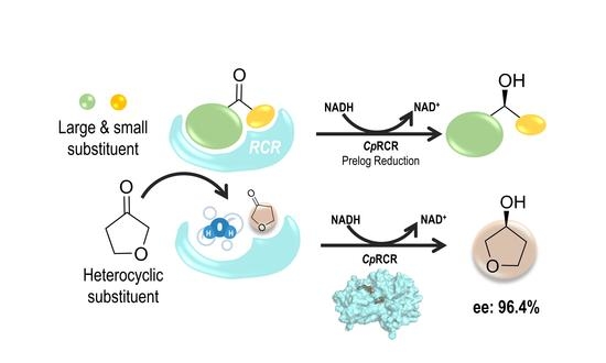
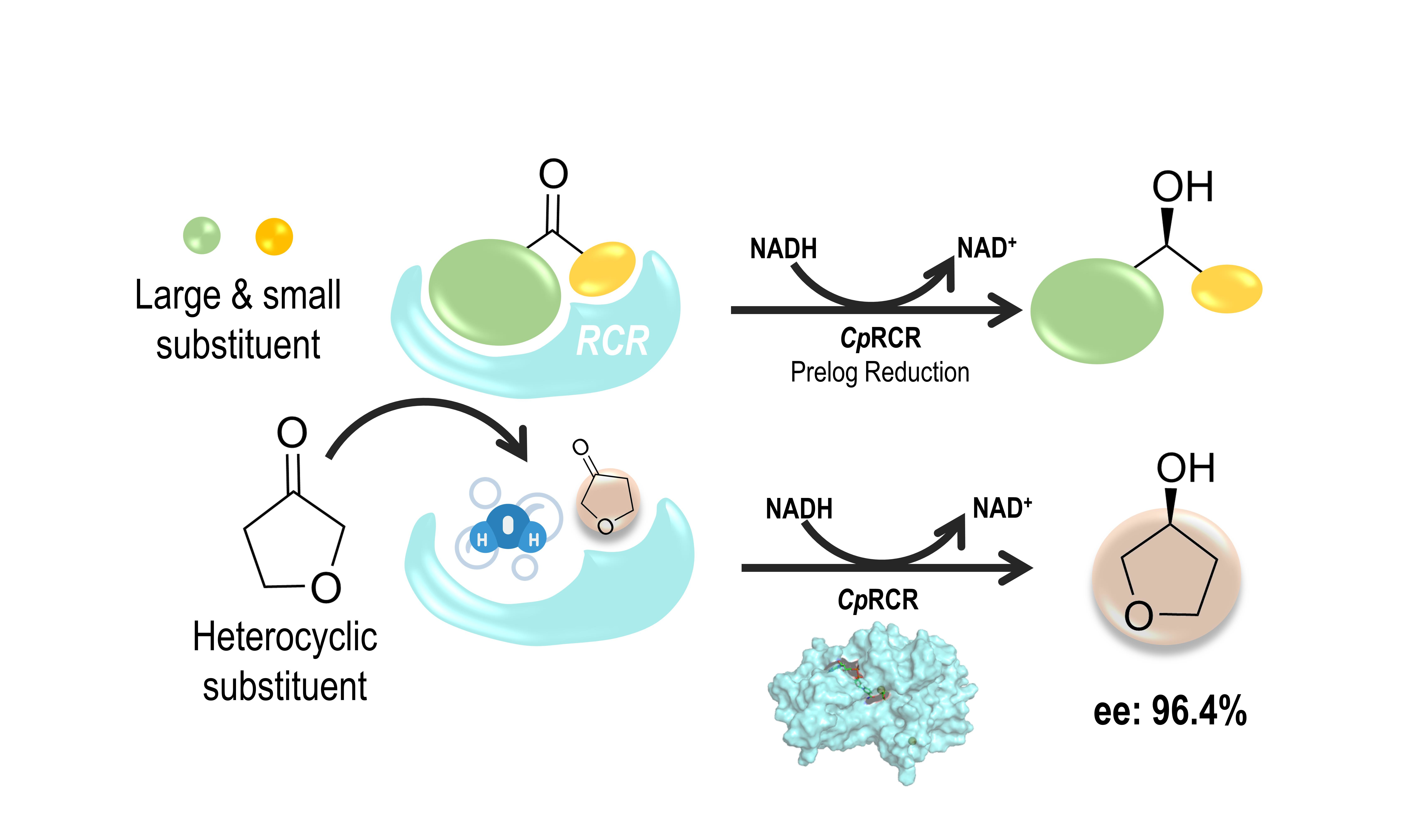
Origin of the Unexpected Enantioselectivity in the Enzymatic Reductions of 5-Membered-Ring Heterocyclic Ketones Catalyzed by Candida parapsilosis Carbonyl Reductases

1
State Key Laboratory of Microbial Metabolism, Joint International Research Laboratory of Metabolic and Developmental Sciences, School of Life Sciences and Biotechnology, Shanghai Jiao Tong University, Shanghai 200240, China
2
School of Biotechnology and Key Laboratory of Industrial Biotechnology, Ministry of Education, Jiangnan University, Wuxi 214122, China
3
Ribose Medical Technology Co., Ltd., Fuzhou 350200, China
*
Authors to whom correspondence should be addressed.
These authors contributed equally to this work.
Catalysts 2022, 12(10), 1086; https://doi.org/10.3390/catal12101086
Submission received: 6 July 2022 / Revised: 23 July 2022 / Accepted: 25 July 2022 / Published: 21 September 2022
(This article belongs to the Special Issue Advances in Biocatalysis and Enzyme Engineering)

Abstract

:
Candida parapsilosis carbonyl reductases (CpRCR) have been widely used for the reductive conversion of ketone precursors and chiral alcohol products in pharmaceutical industries. The enzymatic enantioselectivity is believed to be related to the shape complementation between the cavities in the enzymes and the substitutions of the ketone substrates. In this work, we reported an unexpected enantioselectivity in the enzyme reductions of dihydrofuran-3(2H)-one (DHF) to (S)-tetrahydrofuran-3-ol (DHF-ol, enantiomeric excess: 96.4%), while dihydrothiophen-3(2H)-one substrate (DHT) was unproductive under the same experimental conditions. To rationalize the exclusive S-configuration and the specific reactivity of DHF, we carried out molecular dynamics simulations for the reacting complexations of DHF with CpRCR, and DHT with CpRCR. Our calculations indicate that DHF preferentially binds to the small cavity near L119, F285, and W286, while the large cavity near the α1 helix was mainly occupied by solvent water molecules. Moreover, the pre-reaction state analysis suggests that the pro-S conformations were more abundant than the pro-R, in particular for DHF. This suggests that the non-polar interaction of substrate C4-C5 methylene contacting the hydrophobic side-chains of L119-F285-W286, and the polar interaction of funanyl oxygen exposing the solvent environment play important roles in the enantioselectivity and reactivity. The phylogenetic tree of CpRCR homologues implies that a variety of amino acid combinations at positions 285 and 286 were available and thereby potentially useful for redesigning enantioselective reductions of 5-membered-ring heterocyclic ketones.

Graphical Abstract

1. Introduction

Chiral alcohols are highly demanded in pharmaceutics; therefore, the reductive conversions of ketone precursors are one of the practical and green approaches for preparing enantiomeric alcohols [1]. Besides the typical secondary alcohols with two separated substituents, heterocyclic alcohols are also demanded for their specific biological activity [2,3,4]. For example, (S)-tetrahydrofuran-3-ol is an important intermediate for Amprenavir, a human immunodeficiency virus (HIV) inhibitor drug for the treatment of acquired immune deficiency syndrome (AIDS) [1]. However, the organic synthesis of unbranched heterocyclic alcohols is highly challenging due to its pseudo-symmetry of the ketone skeleton [5]. For example, the chemical synthesis method, via radical cyclization of preassembled alkenes [6] or the employment of co-catalyzed hydrolytic kinetic resolution [7] can ultimately produce (S)-tetrahydrofuran-3-ol. However, these methods suffer from the limitations of long sequential reactions which include low % ee, and low yield, all of which conflict with the ideals of green and sustainable chemistry. Thus, whether alcohol dehydrogenases could provide an efficient approach for the enantioselective reduction of heterocyclic ketones like dihydrofuran-3(2H)-one (DHF) becomes an intriguing topic [3].
Candida parapsilosis carbonyl reductase (CpRCR) is a zinc-mediated medium-chain ketone reductase, which has been extensively investigated because of its specific enantioselectivity and thermostability for noncyclic ketone reductions. CpRCR generally catalyzes α-ketoglutarate, aryl, and aliphatic ketone variances to their corresponding chiral alcohols [8]. Similar to other alcohol dehydrogenases (ADH), these ketone substrates bind the catalytic zinc ion and their two individual substituents on the carbonyl group and sit on the catalytic pocket with a specific orientation [6]. It was believed that CpRCR provided two distinct cavities in the active site for the two substituents of ketone; a large cavity formed by the α-helice 1 with residues 46–52 for the large substituent group, and a small cavity near F285 and W286 for the small substituent group [8,9]. Accordingly, continuing efforts have been made in CpRCR protein engineering to enhance catalytic efficiency and substrate promiscuity, via modifying the binding pocket to meet as many substrates as possible [10]. However, the suitability of heterocyclic ketone variants is neglected owing to the two-substituents-to-two-cavities presupposition in prior protein engineering.
In this study, we measured the product chirality of CpRCR-catalyzed DHF reductions. Our experiments revealed that CpRCR was capable of efficiently promoting the asymmetric reduction of DHF and generates (S)-tetrahydrofuran-3-ol (S-DHF-3-ol) with an enantiomeric excess of 96.4%, while the analogue dihydrothiophen-3(2H)-one (DHT) failed to produce any alcohol products. To rationalize the unexpected enantioselectivity in the experiment, we carried out molecular modelling for the CpRCR-DHF and CpRCR-DHT complexes. The molecular dynamic simulations suggest that pro-S structures to the S-chiral alcohol are dramatically promoted by the molecular interactions in the small cavity. Moreover, with comparison of the two similar heterocyclic ketones DHF and DHT, the pre-reaction state analysis indicates that numerous factors are involved in the enzymatic reactivity, including the hydrophobic interactions of L119 and W286 on the heterocyclic ketones. Given the importance of the small cavity, we also conducted a homology-based sequence alignment to scan the residue pairs corresponding to positions 285 and 286, in an attempt to predict the possible applications of the variable shapes of small cavities for the small heterocyclic ketones.

2. Results

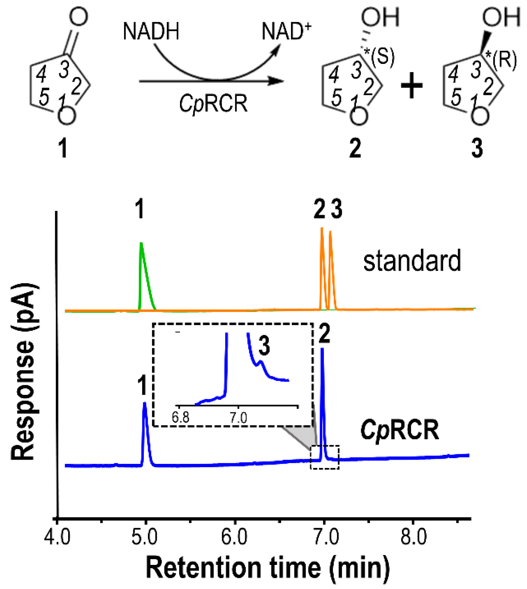
2.1. Kinetics and Stereoselectivity of DHF and DHT with CpRCR

We measured the CpRCR activity of the heterocyclic ketone reduction with substrates DHF and DHT, using the Michaelis-Menten kinetic parameterization method. The kcat, Km, and kcat/Km values were determined to be 2.75 ± 0.11 s−1, 1.15 ± 0.07 mM, and 2.39 mM−1s−1, respectively. Compared with previous studies on other substrates, such as α-ketoglutarate and aryl ketones, the catalytic efficiency of DHF is at a moderate level [6]. In contrast, we did not observe any reaction when DHT was used as a substrate under the same experimental conditions.
The enantioselectivity was determined with gas chromatography (GC), using standard DHF-3-one (1), (S)-isomeric (2), and racemic (2 & 3) DHF-3-ol. As shown in Figure 1, the retention time of (S)-isomer is slightly shorter than that of (R)-isomer by about 6 s in the GC enantiomer separation. As shown in Figure 1, the CpRCR-catalyzed reduction of DHF-3-one exhibits a very high enantioselectivity, with a dramatic preference for (S)-isomer at 96.4% enantiomeric excess (% ee). This result is parallel to a modified biocatalyst Thermoethanolicus brokii (TbSADH) in previous studies [9]. However, this is the first time that wild type CpRCR was observed to efficiently catalyze the enantioselective reduction without any further protein engineering.

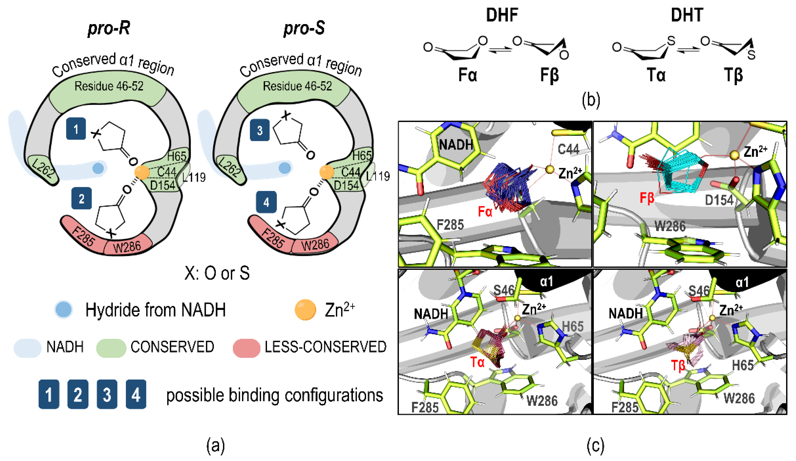
2.2. Plausible Binding Orientations

It is reported that disubstituted ketones, such as acetophenone and α-ketoglutarate ketone variants, follow Prelog’s orientation, in which the large substituent group sits in the large cavity and the small substituent group in the small cavity. In contrast, the 5-m-r heterocyclic ketones may place the cyclic ring in either the large or small cavity (Figure 2a). Basically, the large cavity of CpRCR majorly consists of residues in a sequential range of 46–52 (α1 region), while the small cavity consists of only two residues, i.e., F285 and W286. Comparatively, the two residues are less conserved than those of the α1 region.
As shown in Figure 2b, both the half-chair conformers (α and β) were used as small ligands in the molecular docking. Figure 2c shows the docking results of low-energy conformers , , , and . Intriguingly, all the low-energy docking poses led to the small-cavity orientations. This was very unusual, as the previous studies about the medium-sized ketones suggested the large cavity was preferentially occupied in molecular dockings [10].
One of the possible rationales is that the small cavity provides stronger hydrophobic interactions with the side-chains of F285 and W286, because the methylene groups of the heterocyclic substrates were consistently found to be <3 Å toward F285 and W286. Intriguingly, conformer (α conformer of DHF) presents a considerable number of pro-R poses toward the R-isomer, while only one of the low-energy poses with conformer adopted the reactive pro-R configuration (others are unproductive in reduction due to the lack of the proper O-Zn coordination). In the case of DHT, the docking poses of conformers and strongly preferred pro-S configuration. The molecular docking suggests that the O/S swap in the 5-m-r heterocyclic ketone substrates significantly influences the binding orientation.

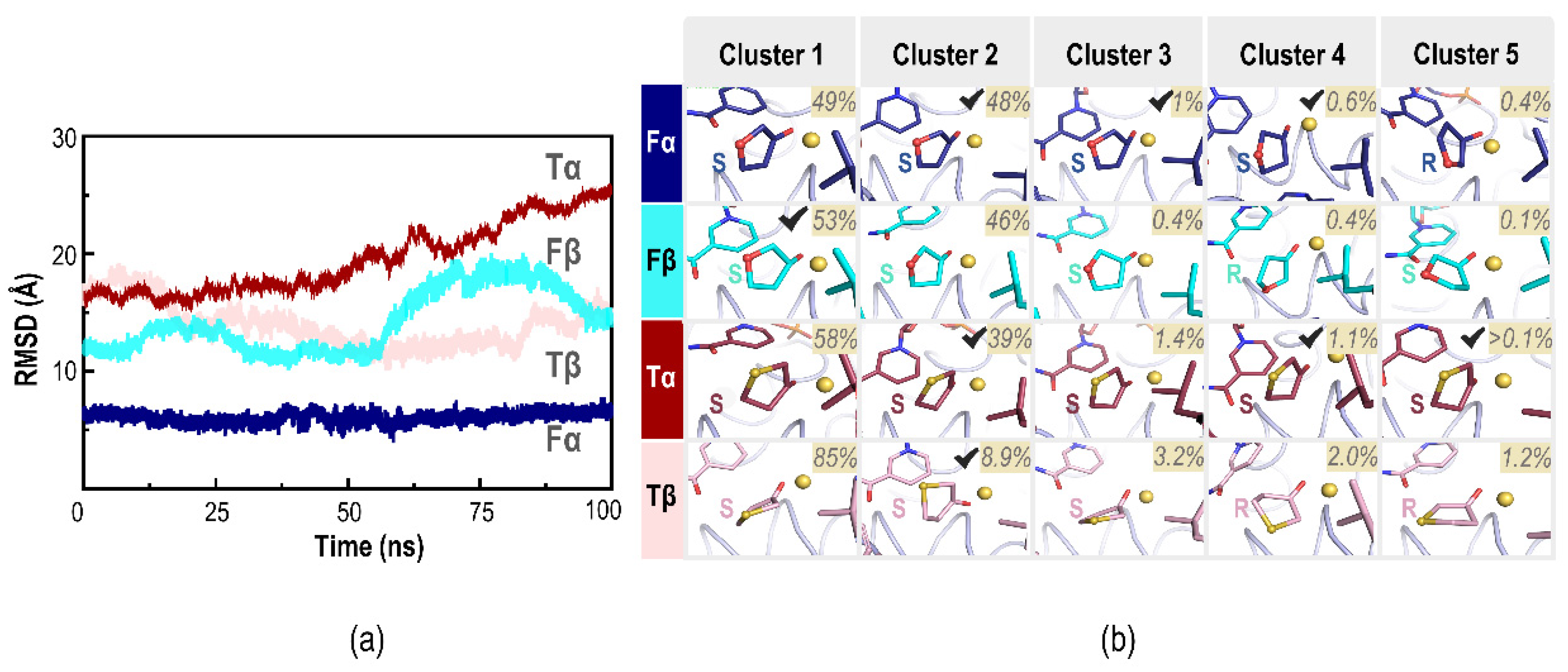
2.3. Molecular Dynamics Simulations

It is harmless to use the lowest-energy pose of each molecular docking as the starting geometry in the following-up molecular dynamics simulations, because the spacious cavities of CpRCR allow the small 5-m-r heterocyclic ketones to freely move back-and-forth in the enzyme. Figure 3a exhibits the root mean squared deviation (RSMD) of the substrates with the reference of CpRCR backbone Cα atoms. The relative stabilities of substrates in the enzyme are in the order of >> ~ >> . This indicates that conformer likely fits the cavity shape well.
Figure 3b presents the clustering analysis on the MD trajectories. In the cases of DHF, over 98% of conformations remain in the pro-S configuration, such as clusters -1/2/3/4 and -1/2/3/5; only ~0.4% in the pro-R configuration (i.e., cluster -5 and -4). A similar tendency was observed in the DHT cases. Thus, even though the 5-m-r heterocyclic ketones only occupied the small cavity near F285-W286, the pro-S configuration is dominant in the enzyme-substrate complexation. This is in good agreement with the experimental observation of S-preferential enantioselectivity.
It should be noted that the coenzyme NADH shifted away from the substrates in the MD simulations. For example, NADH moved further away from the substrate in the pro-S cluster and Tβ cluster 1 (3.6 Å, 3.5 Å). With the consideration of coenzyme distances, DHF formed a higher population of the reacting pro-S configuration than DHT. A previous study by Nie and Zhao et al. [10] suggests an open-close motion between the co-factor binding domain and the catalytic domain, accompanied with clockwise and counterclockwise twists from the view point of the active site. The residue RMSD (Figure S4) shows the β-conformer substrates’ (i.e., and ) tight binding to the small cavity eventually leads to these dynamic movements.

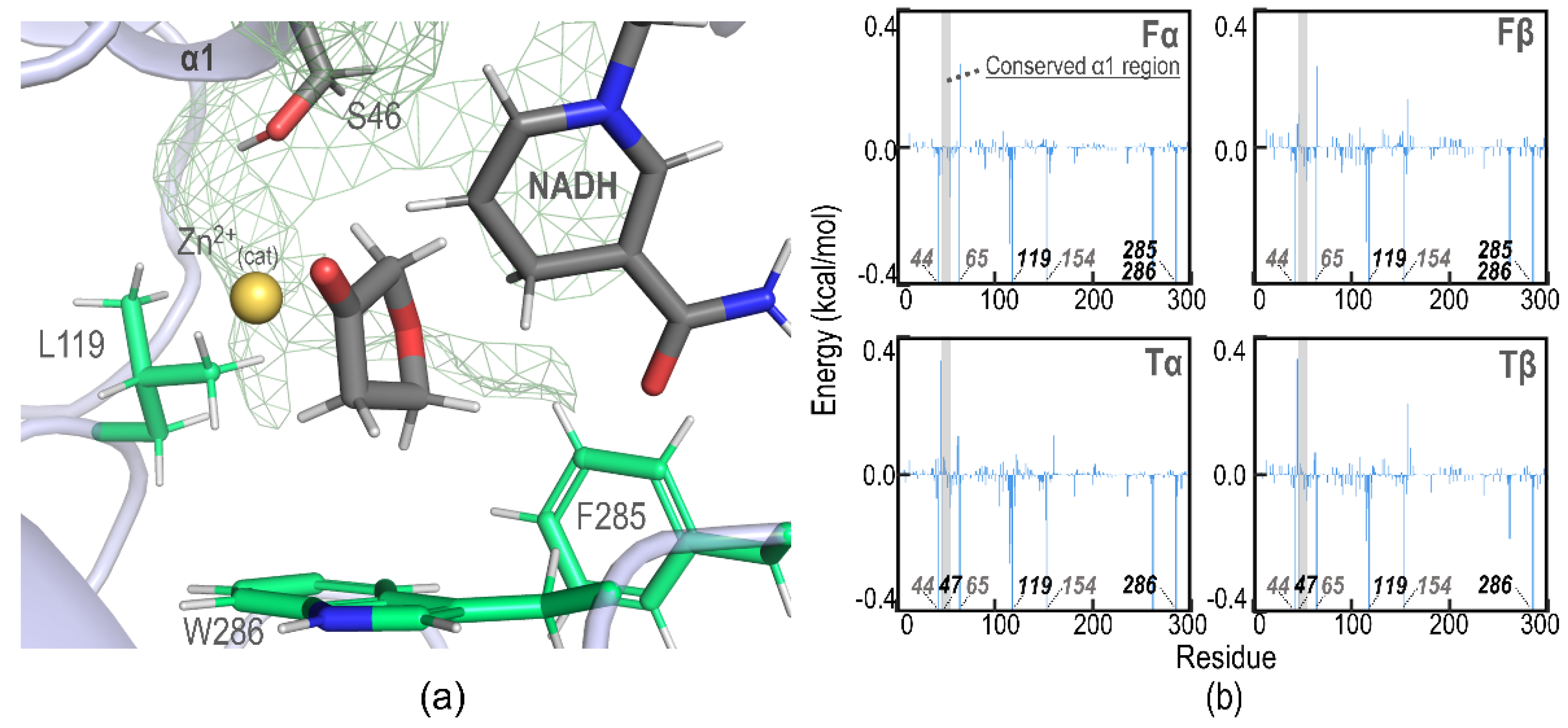
2.4. Binding Affinity Analysis

As shown in Figure 4a,b, the C4-C5 methylene groups were in a geometrical range of contacts with the aromatic rings of F285 and W286, probably benefiting from the hydrophobic and C-H⋯π interactions. Figure 4b illustrated the MM/PBSA binding energy decompositions for the substrate and enzyme. The zinc-coordinating residues, C44-H65-D154, significantly contribute to the binding energy due to their extremely short distances to the substrates (all of them bind the catalytic zinc center together). Furthermore, L119 and W296 were found to be second in rank, with a binding energy larger than 0.5 kcal/mol, while the conserved α1 region (the large cavity) only contributes a tiny amount of binding affinity. The L119 contribution is reasonable because it is located on the side borders between the large and small cavities of CpRCR (Figure 2a). More importantly, the binding affinity analysis suggests that the non-polar contacts between the substrate C4-C5 methylene group and L119-F285-W286 side-chains could be the dominant driving force for the formation of pro-S conformations.

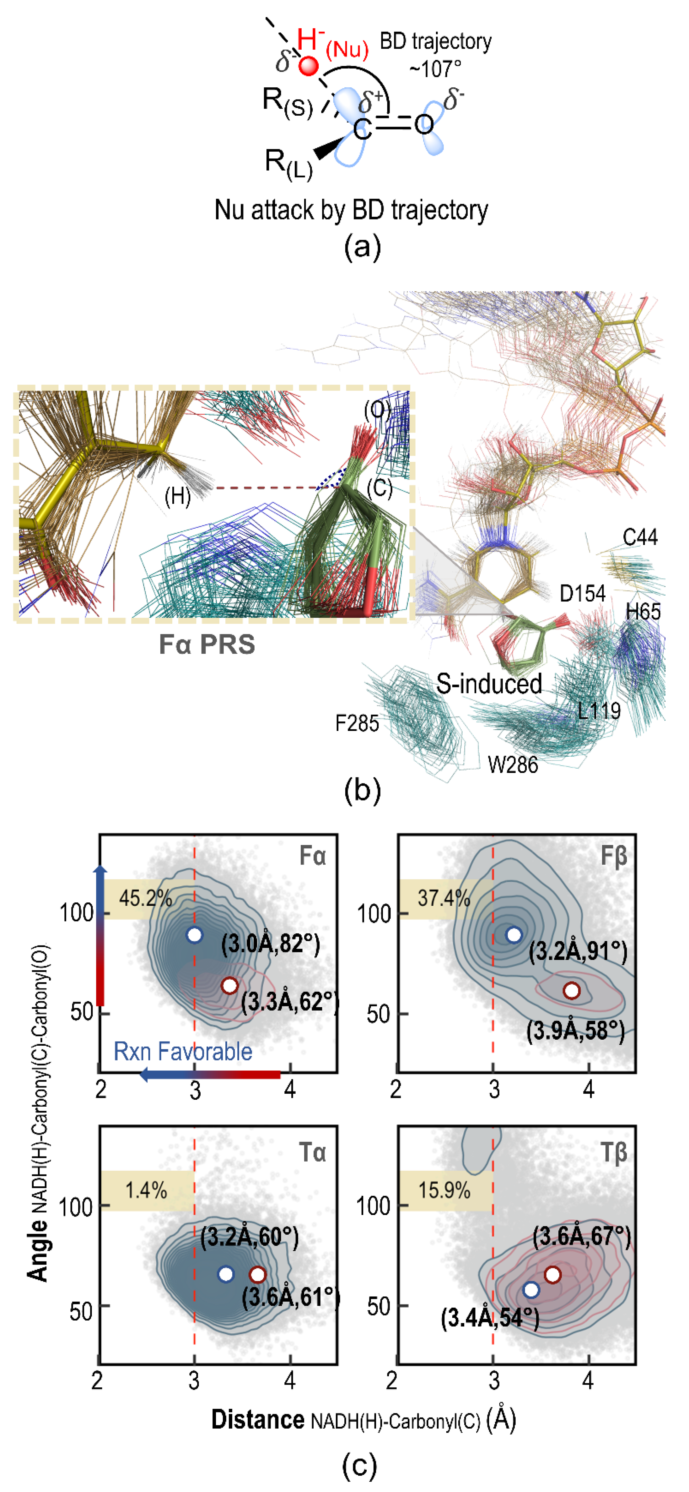
2.5. Pre-Reaction State Analysis

Figure 5 shows the pre-reaction state (PRS) analysis on the geometrical information in the MD simulations. It has been reported that the hydride transfer is the rate-determining step for the CpRCR-catalyzed reduction [11]. PRS analysis has been thus applied for the linear substrates, similar to the aryl-substituted ketones in the previous CpRCR studies [10,12]. In this work, we employed similar PRS protocols to measure the reactivity of 5-m-r heterocyclic ketones bound in the small cavity, as in the molecular dockings and MD simulations, by observing the pre-reaction state (Figure 5a) among all the possible conformations of enzyme-substrate complexes.
As shown in Figure 5b,c, the PRS populations were estimated with the cut-off attacking distances within 3 Å and the Bürgi-Dunitz angles in a reactive range of 107 ± 10°. The overall reacting populations were then estimated to be 45.2, 37.4, 1.4, and 15.9% for , , , and , respectively. Therefore, DHF appears more reactive than DHT in terms of total PRS population by about 6-fold, which signifies a large depreciation of the O/S swap in the substrates if the reaction barrier difference were neglectable. Interestingly, the PRS area was smaller in the and trajectories but the corresponding substrate RMSD values were much larger, indicating that DHT appears more dynamic in the enzyme but hardly orients the proper attacking angle in the small cavity. One the other hand, no matter with or conformer, the complexation of DHF and CpRCR appears well-fitting, with the larger PRS conformations in preference of pro-S configurations. The pro-S conformations were prevalently by 0.87:0.13 and 0.85:0.15 (pro-S:pro-R) in the MD simulations starting with and conformers, respectively.
Because both the 5-m-r heterocyclic substrates (DHF and DHT) preferentially sit in the L119-F285-W286 small cavity, the decreasing PRS population of DHT indicates that the furanyl oxygen atom plays a crucial role to maintain the reacting conformations. In the pro-S configuration, the furanyl oxygen and thiophenyl sulfur atoms are exposed to the large cavity that is occupied by water molecules (see the green mesh surface in Figure 4a), while they have to face down relative to the L119-F285-W286 hydrophobic hemisphere in the pro-R configurations. In contrast, the sulfur element is less electronegative and more hydrophobic than the oxygen element. Thus, the pro-S configuration would be more favored in the cases of DHF, due to the stronger electrostatic interactions (or even hydrogen bonding) in pro-S conformations and less hydrophobic interactions in pro-R conformations. The vice versa scenario holds for DHT–no significant benefits were observed in both pro-S and pro-R configurations. The steric repulsion may cause DHT to remain less frequently in the pro-R configuration as well, and flip up to the pro-S configuration that is unfavored too. As a result, DHT becomes more unsettled in the CpRCR active site and cannot form a pre-reaction state well. In contrast, DHF presents a good induced-fit in the pro-S conformations, because the exposed furanyl oxygen atom interacts with the solvent molecules in the large cavity. The trajectory analysis showed that a solvent-ether (furanyl oxygen) H-bond presents by a chance of 42.5% and 32.7% in the MD simulations starting with conformers and , respectively.

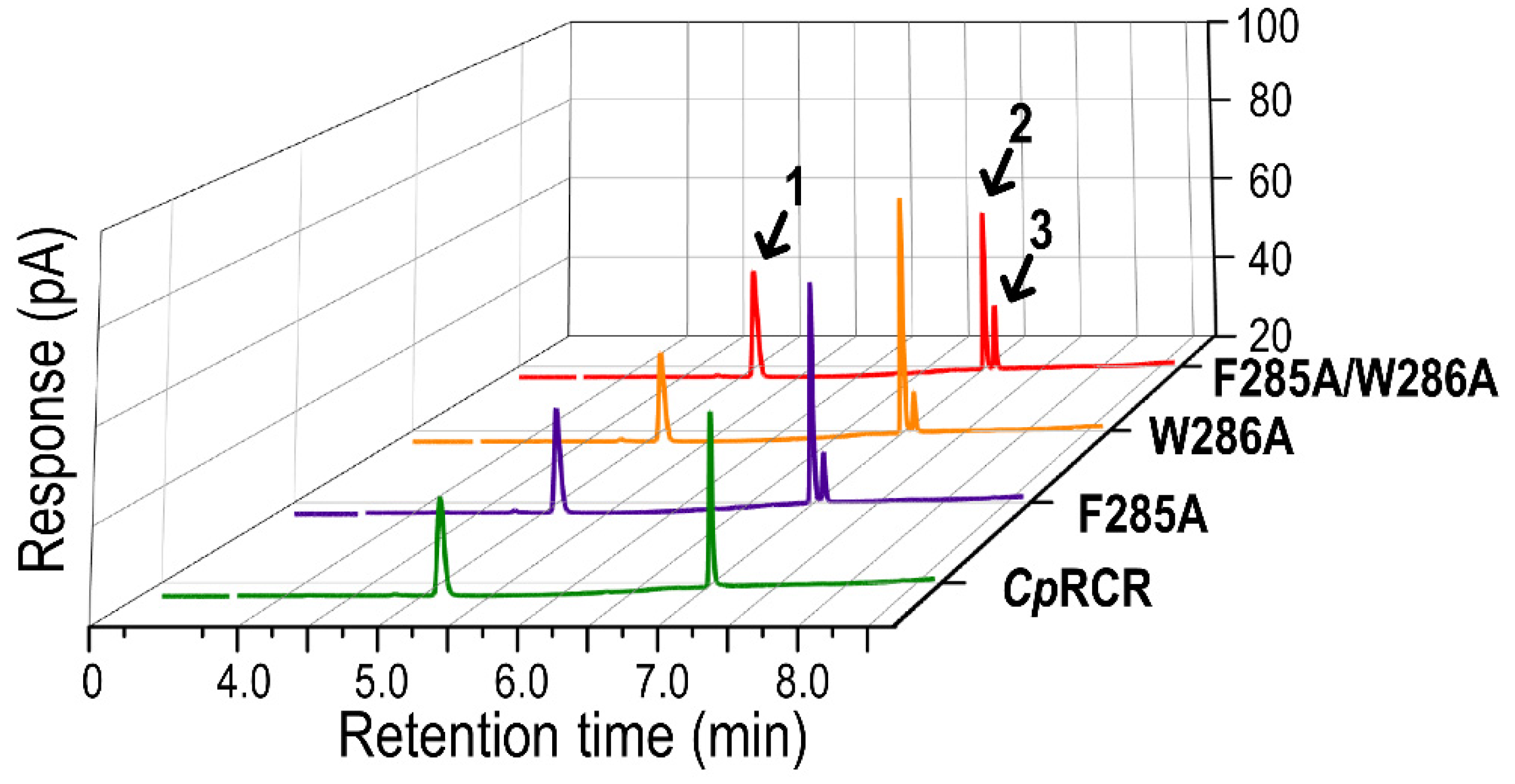
2.6. Experimental Validation

To verify whether the small-cavity induced-fit plays an important role for the enantioselectivity of CpRCR-catalyzed DHF reductions, we carried out alanine mutations on F285 and W286. L119 was not considered in the experimental validation because it is located at the border between the small- and large- cavities in the protein. The enantioselectivities were measured with the same biochemical assay in Figure 1. As shown in Figure 6, enantiomeric excess (% ee) of CpRCR variants F285A, W286A, and F285A/W286A are 67.7, 75.0, and 48.3% in the same S-preference, respectively, far away from the wild type value of 96.4%. Therefore, the origin of enantioselectivity is due to the specific interactions between DHF and bulky aromatic side chains in the small cavity.
The enzyme kinetics were also experimentally studied with the enzyme variants. The kcat/Km values were determined to be about 2.19, 0.41, and 0.69 mM−1s−1 for F285A, W286A, and F285A/W286A, respectively. This indicates that W286 is the key residue for the induced-fit, in agreement with the van der Waals interactions of DHF C4-C5 methylene groups with the tryptophan aromatic indole ring. However, the Km values were determined to be 0.34 ± 0.02, 1.14 ± 0.07, and 0.29 ± 0.03 mM, respectively. This indicates that F285A may significantly enhance the substrate binding affinity, probably via promoting van der Waals contacts to W286. The contradictory effect on substrate binding and catalytic efficiency is common in enzyme studies—the stronger binding is not an equivalent of a higher catalytic efficiency, as the highest-probable E·S structures are not the highest reactive pre-reaction state (Figure 5c)

2.7. Prespective on Small Cavities in Alcohol Dehydrogenases

Encouraged by the experimental and computational accomplishments, we searched all alcohol dehydrogenase orthologs to understand the amino acid patterns of the small cavity. Indeed, F285-W286 have been extensively investigated as mutational hotspots in the CpRCR reductions of linearly disubstituted ketones, for the purpose of fitting the small substituents. Here we take a closer look to analyze the potential relationship between F285-W286 mutagenesis and the small cavity, via sequence alignments of these homologous enzymes.
The annotation in the phylogenetic analysis indicates the amino acids of residues 285–286 CpRCR equivalent positions in the homologous sequence and the classification of organisms (archaea, bacteria, and eukaryote), respectively. As shown in Figure 7, CpRCR was clustered together with those from yeast species, forming a well-supported monophyletic clade. Alcohol dehydrogenases evolve independently in prokaryotes, eukaryotes, and archaea, presented as three distinct clades. A small proportion of bacterial ADH also contains small cavities like those in eukaryotes.
Among the 140 orthologs, 12 combinations of residue-pairs were collected with the reference of CpRCR positions 285 and 286. The majority of the bacterial sequences tend to contain Y285-W286, whereas eukaryotes such as fungi and yeast contain both F285-W286 and F285-G286. The coexistence of F285 and W286 was highly conserved with 37% population, and the isolated distributions of individual F285 and W286 appeared in 46% and 74% of population, respectively. The F285-W286 co-evolutionary inclination indicates that the 5-m-r heterocyclic enantioselectivity might be widely spread in alcohol dehydrogenases.
It also raises the question of whether more spacious cavities with certain combinations, such as the amino acid combination of Phe-Cys, Phe-Gly, or Leu-Ala, may lose the enantioselectivity with the DHT substrate. Nevertheless, the diverse combinations could alter the enantioselective outcome, as the polar residues such as Tyr and Glu are able to interact with the funanyl oxygen and thiophenyl sulfur atoms of heterocyclic substrates in pro-R configuration via additional hydrogen bonding network. Hence, the involvement of the polar property residues in the catalytic activity in bacteria warrants further investigations.

3. Discussion

In this study, the enzymatic enantioselectivity and reactivity were measured with experimental and computational methods. Although the two substrates were highly similar in structure, the experimental results indicated the S-isomer was generated with 96.4% enantiomeric excess in the DHF enzymatic reaction, while no detectable ketone-alcohol conversion was observed for DHT. Thus, the O/S swap definitively brings significant changes and allows comparative studies of pre-organization and induced-fit between the substrates and proteins.
Different from the linear-disubstituted ketone substrates, the small-ring heterocyclic ketone substrates only occupied the small cavity formed by F285 and W286, leaving the large cavity for water molecules. By using four conformers of , , , and in the molecular dockings and MD simulations, we have successfully rationalized the relative reactivity and enantio-preference. The molecular dockings concluded that the heterocyclic ketones are bound in the active site between helix α1 and F285-W265. The low-energy poses illustrate that both pro-R and pro-S configurations are available for CpRCR-catalyzed reductions.
The following-up molecular dynamic simulations suggest that conformer prevails and the MM/PBSA binding energy decomposition confirms that residues L119, F285, and W286 substantially contribute to the hydrophobic interactions, besides the zinc-coordinating groups (Zn-C44/H65/D154). However, very weak interactions are observed between the substrates and the α1 region, supporting that the interactions from the small cavity dominate in the substrate-enzyme complexation.
The pre-organization of the 5-m-r heterocyclic ketone and CpRCR is novel and different from linear-disubstituted ketones in that the large substituent is in the large cavity and the small substituent in the small cavity. The traditional induced-fit model was generally supported for two reasons: (1) a high conservation presents for the key residues of the large cavity (helix α1) in comparison to the small cavity; and (2) the small cavities of CpRCR do not have enough spaces for the large-sized ketones. Hypothetically, the small-ring heterocyclic ketones would occupy the large cavity in priority, if both the small and large cavities had enough spaces. However, our molecular modeling indicated that DHF preferentially takes the small cavity because of the favored hydrophobic interactions with W286, L119, and F285. The disappeared reactivity of DHT may stem from adverse effects of fast motion in the active site.
Although F285 and W286 were previously reported to be “mutational hotspots”, the mechanistic understandings vary from the linear substrates as the shapes of 5-m-r ketone substrates change dramatically. The sequence alignment of alcohol dehydrogenases’ analogous proteins presented the 12 possible combinations of amino acid pairs at CpRCR positions 285 and 286. The diversity could bring different compartment shapes of “small cavity” and, thereby, be predicted to change reactivity and enantioselectivity for the small-ring heterocyclic ketones via different induced-fit effects.

4. Materials and Methods

4.1. Materials

DNA polymerase and kits for DNA cloning and amplification were purchased from Takara-Bio Co., Takara Kusatsu, Japan. Oligonucleotides were synthesized by Sangon Biotech, Shanghai, China. Ketone substrates, coenzyme NADH, and standard chiral alcohols were ordered from Sigma-Aldrich, Saint Louis, Illinois, USA. Chromatography-grade hexane and isopropanol used in high performance liquid chromatography (HPLC) were purchased from Aladdin Co., Shanghai, China.

4.2. Gene Expression and Enzyme Purification

Recombinant plasmid pET-32Xa/LIC-CpRCR (WT) was previously constructed [9]. A single transformant was cultured in the Luria-Bertani (LB) medium including 100 μg·mL−1 ampicillin and 0.2 mM zinc acetate at 37 °C and 200 rpm, until optical density of the culture at wavelength 600 nm reached 0.6–0.8. Then, the target proteins were further expressed at 30 °C for 8 h. After harvesting, the cells were resuspended in 25 mM pH 8.0 Tris-HCl buffer, with 150 mM NaCl and 20 mM imidazole. At 4 °C, the cells were lysed by sonication and centrifuged to collect the supernatant. The His-tag protein was purified by affinity chromatography, using an eluent of ~300 mM imidazole solution. The purified protein was verified to be a single band in SDS-PAGE and thereafter considered for subsequent experiments.

4.3. Enzyme Kinetic Assay

The standard assay of enzyme activity was measured by the change of coenzyme NADH absorbance at 340 nm. The standard assay was performed at 30 °C in a reaction mixture of 100 μL, composed of 100 mM sodium phosphate buffer, pH 6.5, 0.1–15 mM substrate, and 0.5 mM NADH, together with appropriate amounts of the purified enzymes. One unit of enzyme activity was defined as the amount of enzyme required to oxidize 1 μmol NADH per min under the standard assay conditions [8]. The kinetic parameters were obtained through GraphPad Prism 7 to curve-fit the Michaelis-Menten equation, and the specific activity at different substrate concentrations (0.1–15 mM) was measured. All data were averaged over three replicates.

4.4. Stereoselectivity Assay

Enzymatic reduction was performed by adding 10 mM substrate, 10 mM NADH, 10% isopropanol, and a proper amount of enzyme to 100 mM phosphate buffer (pH 6.5), and then allowing them to react at 30 °C and 200 rpm for 8 h. After the reaction mixture was extracted with ethyl acetate, the organic layer was filtered through a 0.22-μm PVDF syringe filter (Sangon Biotech, Shanghai, China). The reaction products were analyzed by chiral GC (7890A, Agilent (Palo Alto, CA, USA)) equipped with an FID detector 5150. The specific peak of (R)-/(S)-alcohol was detected by gas chromatography, with comparison of standard chiral alcohol samples. As typical, a hydrodex-β-TBDAc column was used in the GC enantiomeric separation; the column temperature was set at 125 °C and changed after 1 min, by 3 °C/min to 135 °C, and then held for 1 min, followed by 50 °C/min to 200 °C, and held for 2 min.

4.5. Computational System Preparation

X-ray crystal structure of CpRCR (PDB: 3wle) was obtained from the public PDB database [9]. The preliminary protonation state was assigned with the H++ online server at pH = 7 [13]. C44-C95-C98-C101-C104 were manually deprotonated and H65 was set as HID to fit the zinc-amino acid coordination. The 3-dimensional structures of the substrate were constructed with the Gaussview program [14] and then optimized with the B3LYP/6-31G density functional theory [15]. Mirror structures of substrates were used in the molecular docking with the Autodock version 4.2 [16]. Atomic charges in the MD simulation were pre-calculated using the restrained electrostatic potential (RESP) method in the antechamber module of the Amber14 package [17]. The forcefield parameters of the catalytic zinc site were generated with the python-based metal center parameter builder (MCPB) method [18], and those for the structural zinc site were directly added with the ZAFF force fields in the literature [18]. The force field parameters of NADH were also set as in the literature [19]. The ff14SB force field was applied for remaining amino acids in the protein, and the TIP3P force field for the solvent water molecules. The enzyme-substrate complex was loaded in a cubic box with the tleap module of the AMBER package, to generate the topology and coordinate files, with a thickness of > 10 Å water layer from every protein surface.

4.6. Molecular Dynamics Simulations

All simulations were assigned with the PMEMD.cuda program in the AMBER package [20]. Two-step minimizations was followed to relax the water molecules and then the solvated complex. Langevin dynamic at collision frequency of 2ps−1 was applied to raise the system temperature from 0 to 300 K. Thereafter, 50-ps of equilibration was carried out under constant pressure and temperature ensemble (NPT) before recording trajectories. Multiple replicates of 100 ns simulations were conducted, with the non-bonded cutoff of 10.0 Å. The snapshots were recorded every 1000 steps, in the integration time of 2.0 fs. The resulting trajectories were analyzed with the cpptraj commands of Amber tools 18 [21]. The water-substrate hydrogen bonding analysis was conducted with the criteria distance of 3.0 Å in the cpptraj list of commands.

4.7. Molcular Mechanics-Poisson-Boltzmann Surface Area (MM/PBSA)

The MM/PBSA approach was employed to measure the binding free energy of the substrates , , , using the following calculations method—
Δ G b i n d = G c o m p l e x   ( G p r o t e i n s u b s t r a t e   + G p r o t e i n )
and
Δ G b i n d = Δ H T Δ S     E M M   + Δ G s o l v ( T Δ S )
where
Δ E M M = Δ E i n t Δ E v d W + Δ E e l e
and
Δ G s o l v = Δ G P B Δ G S A
Each term was individually measured on 100 equally distributed snapshots taken from 100 ns MD trajectories and the binding energies were analyzed statistically. The solvation free energy ( Δ G S A ) was measured by the solvent-accessible surface area (SASA), via the LCPO algorithm [22] with the Poisson Boltzmann ( Δ G P B ) model [23].

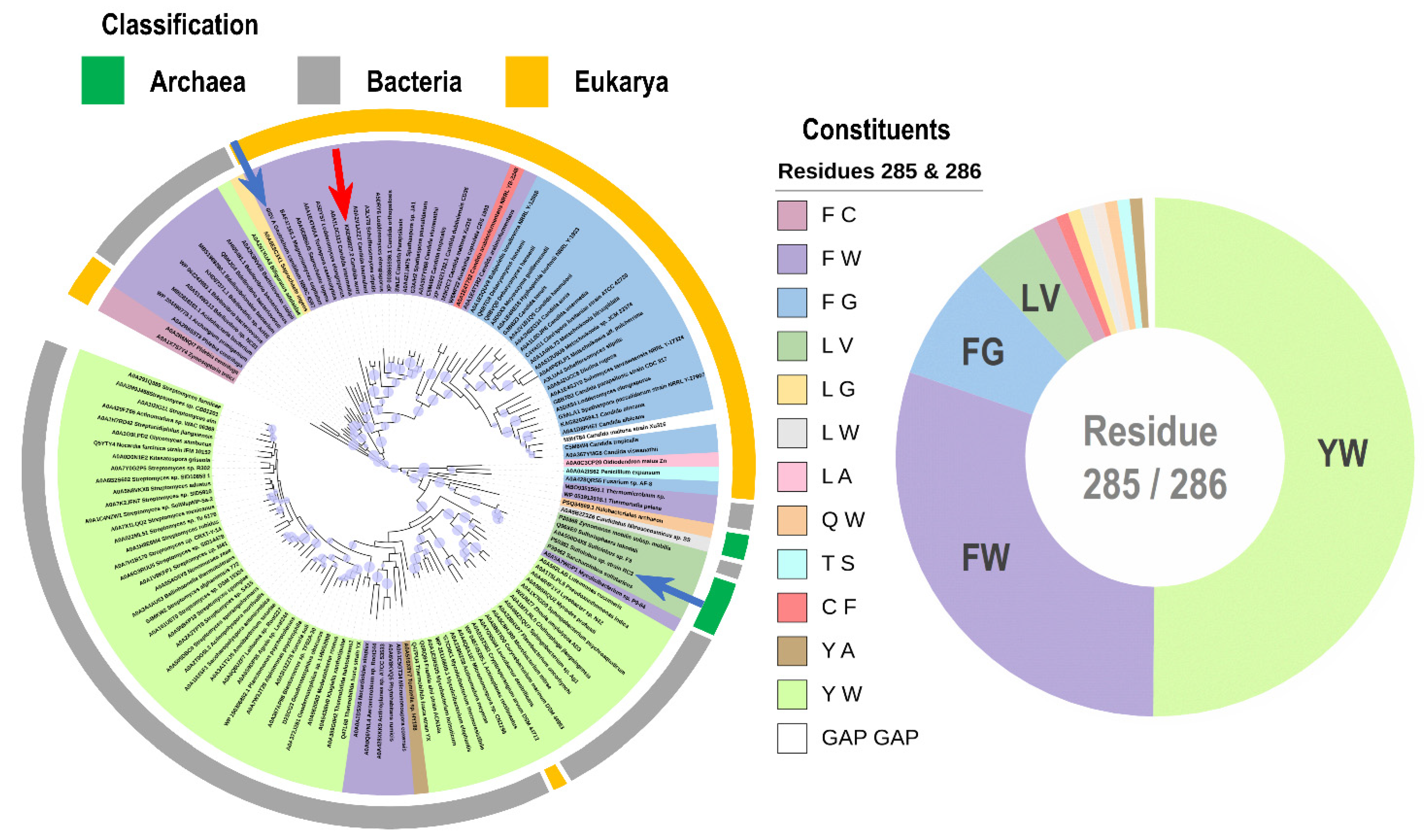
4.8. Homology Sequence Analysis and Phyolgenetic Tree

The medium chain alcohol dehydrogenase from Candida parapsilosis (GenBank: DQ295067.1) was used as a query to search CpRCR analogues in the NCBI nonredundant protein sequences (nr) and Uniprot databases [24] via protein BLAST [25]. The data set was refined by excluding sequences that had less than 35% identity with the query sequence, and sequences with expected values (E) of more than 10−30 and coverage of less than 60%. Redundant sequences were then filtered using CD-HIT Suite software [26] with an identity percentage cut-off value set at 0.90. Multiple sequence alignments were then performed using the Clustal Omega program [27]. Phylogenetic reconstructions were performed with the maximum likelihood model inference as implemented in the Mega software [28], with 1000 bootstrap replicates. The phylogenetic tree was visualized with the iTOL version-5 software (Heidelberg, Germany) [29].

Supplementary Materials

The following supporting information can be downloaded at: https://www.mdpi.com/article/10.3390/catal12101086/s1, Figure S1: GC-MS Chromatogram of (S)-tetrahydrofuran-3-ol and (R)-tetrahydrofuran-3-ol as a standard; Figure S2: GC-MS chromatogram of dihydrofuran-3(2H)-one as a standard; Figure S3: GC-MS chromatogram of CpRCR reaction mixture; Figure S4: CpRCR Residue wide RMSD measured from 100ns MD simulation for each sample; Figure S5: GC-MS chromatogram of CpRCR-F285A, CpRCR-W286A, CpRCR-F285A/W286A reaction mixture; Figure S6: Amino acid sequence alignment of CpRCR with other proteins in the medium-chain alcohol dehydrogenase phylogenetic tree with >50% identity; Figure S7: Structural alignment of CpRCR, Geotrichum candidum and Sulfolobus tokodaii labeled in red and blue arrow in Figure 7.

Author Contributions

Conceptualization, proofreading, and supervision by Y.-L.Z.; experimental design supervision Y.N.; MD simulations and manuscript draft by B.R.S.; Experiment by J.G. and Q.C.; Bioinformatical analysis by Y.L. (Yvette Ley), Y.L. (Yihan Liu) and S.L. All authors have read and agreed to the published version of the manuscript.

Funding

This research was funded by grants from the National Natural Science Foundation of China (grant number 31970041) and the National Key R&D Program of China (grant number 2020YFA0907700 and 2018YFA0901200). The APC was partially funded by company Ribose.

Data Availability Statement

The authors confirm that the data supporting the findings of this study are available within the article and its Supplementary Materials. Derived data supporting the findings of this study are available upon request.

Acknowledgments

B.R.S. deeply appreciate Ting Shi and Ho-Jin Lee for enlightening discussions.

Conflicts of Interest

The authors declare no conflict of interest.

References

  1. Gladiali, S.; Alberico, E. Asymmetric transfer hydrogenation: Chiral ligands and applications. Chem. Soc. Rev. 2006, 35, 226–236. [Google Scholar] [CrossRef] [PubMed]
  2. Qin, L.; Wu, L.; Nie, Y.; Xu, Y. Biosynthesis of chiral cyclic and heterocyclic alcohols via CO/C–H/C–O asymmetric reactions. Catal. Sci. Technol. 2021, 11, 2637–2651. [Google Scholar] [CrossRef]
  3. Grunberg, E.; Titsworth, E.H. Chemotherapeutic Properties of Heterocyclic Compounds: Monocyclic Compounds with Five-Membered Rings. Annu. Rev. Microbiol. 1973, 27, 317–346. [Google Scholar] [CrossRef]
  4. Albarrán-Velo, J.; González-Martínez, D.; Gotor-Fernández, V. Stereoselective biocatalysis: A mature technology for the asymmetric synthesis of pharmaceutical building blocks. Biocatal. Biotransform. 2018, 36, 102–130. [Google Scholar] [CrossRef]
  5. Ager, D.J.; Prakash, I.; Schaad, D.R. 1,2-Amino Alcohols and Their Heterocyclic Derivatives as Chiral Auxiliaries in Asymmetric Synthesis. Chem. Rev. 1996, 96, 835–876. [Google Scholar] [CrossRef]
  6. Tiecco, M.; Testaferri, L.; Marini, F.; Sternativo, S.; Santi, C.; Bagnoli, L.; Temperini, A. Intramolecular addition of carbon radicals to aldehydes: Synthesis of enantiopure tetrahydrofuran-3-ols. Tetrahedron 2007, 63, 5482–5489. [Google Scholar] [CrossRef]
  7. Gadakh, S.K.; Santhosh Reddy, R.; Sudalai, A. Enantioselective synthesis of HIV protease inhibitor amprenavir via Co-catalyzed HKR of 2-(1-azido-2-phenylethyl)oxirane. Tetrahedron Asymmetry 2012, 23, 898–903. [Google Scholar] [CrossRef]
  8. Nie, Y.; Wang, S.; Xu, Y.; Luo, S.; Zhao, Y.-L.; Xiao, R.; Montelione, G.T.; Hunt, J.F.; Szyperski, T. Enzyme Engineering Based on X-ray Structures and Kinetic Profiling of Substrate Libraries: Alcohol Dehydrogenases for Stereospecific Synthesis of a Broad Range of Chiral Alcohols. ACS Catal. 2018, 8, 5145–5152. [Google Scholar] [CrossRef]
  9. Wang, S.; Nie, Y.; Xu, Y.; Zhang, R.; Ko, T.-P.; Huang, C.-H.; Chan, H.-C.; Guo, R.-T.; Xiao, R. Unconserved substrate-binding sites direct the stereoselectivity of medium-chain alcohol dehydrogenase. Chem. Commun. 2014, 50, 7770–7772. [Google Scholar] [CrossRef]
  10. Gu, J.; Sim, B.R.; Li, J.; Yu, Y.; Qin, L.; Wu, L.; Shen, Y.; Nie, Y.; Zhao, Y.-L.; Xu, Y. Evolutionary coupling-inspired engineering of alcohol dehydrogenase reveals the influence of distant sites on its catalytic efficiency for stereospecific synthesis of chiral alcohols. Comput. Struct. Biotechnol. J. 2021, 19, 5864–5873. [Google Scholar] [CrossRef]
  11. Dhoke, G.V.; Davari, M.D.; Schwaneberg, U.; Bocola, M. QM/MM calculations revealing the resting and catalytic states in zinc-dependent medium-chain dehydrogenases/reductases. ACS Catal. 2015, 5, 3207–3215. [Google Scholar] [CrossRef]
  12. Byuri, S.; Yilei, Z. Assessment on the pre-reaction state of enzyme: Could we understand catalytic activity with near transition-state molecular dynamic simulation?—A review. Synth. Biol. J. 2022, 3, 567. [Google Scholar]
  13. Jurrus, E.; Engel, D.; Star, K.; Monson, K.; Brandi, J.; Felberg, L.E.; Brookes, D.H.; Wilson, L.; Chen, J.; Liles, K.; et al. Improvements to the APBS biomolecular solvation software suite. Protein. Sci. 2018, 27, 112–128. [Google Scholar] [CrossRef] [PubMed]
  14. Frisch, M.; Trucks, G.; Schlegel, H.; Scuseria, G.; Robb, M.; Cheeseman, J.; Scalmani, G.; Barone, V.; Petersson, G.; Nakatsuji, H. Gaussian 16; Gaussian, Inc.: Wallingford, CT, USA, 2016. [Google Scholar]
  15. Kruse, H.; Goerigk, L.; Grimme, S. Why the Standard B3LYP/6-31G* Model Chemistry Should Not Be Used in DFT Calculations of Molecular Thermochemistry: Understanding and Correcting the Problem. J. Org. Chem. 2012, 77, 10824–10834. [Google Scholar] [CrossRef] [PubMed]
  16. Trott, O.; Olson, A.J. AutoDock Vina: Improving the speed and accuracy of docking with a new scoring function, efficient optimization, and multithreading. J. Comput. Chem. 2010, 31, 455–461. [Google Scholar] [CrossRef] [PubMed]
  17. Case, D.A.; Babin, V.; Berryman, J.T.; Betz, R.M.; Cai, Q.; Cerutti, D.S.; Cheatham, T.E., III; Darden, T.A.; Duke, R.E.; Gohlke, H.; et al. AMBER 14; University of California: San Francisco, CA, USA, 2014. [Google Scholar]
  18. Li, P.; Merz, K.M. MCPB.py: A Python Based Metal Center Parameter Builder. J. Chem. Inf. Modeling 2016, 56, 599–604. [Google Scholar] [CrossRef]
  19. Walker, R.C.; De Souza, M.M.; Mercer, I.P.; Gould, I.R.; Klug, D.R. Large and fast relaxations inside a protein: Calculation and measurement of reorganization energies in alcohol dehydrogenase. J. Phys. Chem. B 2002, 106, 11658–11665. [Google Scholar] [CrossRef]
  20. Salomon-Ferrer, R.; Götz, A.W.; Poole, D.; Le Grand, S.; Walker, R.C. Routine Microsecond Molecular Dynamics Simulations with AMBER on GPUs. 2. Explicit Solvent Particle Mesh Ewald. J. Chem. Theory Comput. 2013, 9, 3878–3888. [Google Scholar] [CrossRef]
  21. Roe, D.R.; Cheatham, T.E. PTRAJ and CPPTRAJ: Software for Processing and Analysis of Molecular Dynamics Trajectory Data. J. Chem. Theory Comput. 2013, 9, 3084–3095. [Google Scholar] [CrossRef]
  22. Weiser, J.; Shenkin, P.S.; Still, W.C. Approximate solvent-accessible surface areas from tetrahedrally directed neighbor densities. Biopolym. Orig. Res. Biomol. 1999, 50, 373–380. [Google Scholar] [CrossRef]
  23. Miller, B.R., 3rd; McGee, T.D., Jr.; Swails, J.M.; Homeyer, N.; Gohlke, H.; Roitberg, A.E. MMPBSA.py: An Efficient Program for End-State Free Energy Calculations. J. Chem. Theory Comput. 2012, 8, 3314–3321. [Google Scholar] [CrossRef] [PubMed]
  24. Consortium, T.U. UniProt: The universal protein knowledgebase in 2021. Nucleic Acids Res. 2020, 49, D480–D489. [Google Scholar] [CrossRef] [PubMed]
  25. Altschul, S.F.; Gish, W.; Miller, W.; Myers, E.W.; Lipman, D.J. Basic local alignment search tool. J. Mol. Biol. 1990, 215, 403–410. [Google Scholar] [CrossRef]
  26. Huang, Y.; Niu, B.; Gao, Y.; Fu, L.; Li, W. CD-HIT Suite: A web server for clustering and comparing biological sequences. Bioinformatics 2010, 26, 680–682. [Google Scholar] [CrossRef] [PubMed]
  27. Madeira, F.; Pearce, M.; Tivey, A.R.N.; Basutkar, P.; Lee, J.; Edbali, O.; Madhusoodanan, N.; Kolesnikov, A.; Lopez, R. Search and sequence analysis tools services from EMBL-EBI in 2022. Nucleic Acids Res. 2022, 50, W276–W279. [Google Scholar] [CrossRef]
  28. Kumar, S.; Stecher, G.; Li, M.; Knyaz, C.; Tamura, K. MEGA X: Molecular Evolutionary Genetics Analysis across Computing Platforms. Mol. Biol. Evol. 2018, 35, 1547–1549. [Google Scholar] [CrossRef]
  29. Letunic, I.; Bork, P. Interactive Tree Of Life (iTOL) v5: An online tool for phylogenetic tree display and annotation. Nucleic Acids Res. 2021, 49, W293–W296. [Google Scholar] [CrossRef]
Figure 1. GC traces for chiral characterization, using the standards DHF-3-one (1), (S)-DHF-3-ol (2), and racemic DHF-3-ol (2 & 3). The enzymatic reduction was performed for 8 h at 30 °C with the reaction mixtures of CpRCR (refer to methods section for details). For GC, Hydrodex-β-TBDAc column was used; after the first minute at 125 °C, the temperature increased by 3 °C/min to 135 °C and was held for 1 min, and then increased by 50 °C/min to 200 °C and held for 2 min.
Catalysts 12 01086 g001
Figure 2. Molecular dockings of the heterocyclic ketones in CpRCR (a)Four plausible orientations of the 5-m-r heterocyclic ketones in the large and small cavities of CpRCR. The highly conserved region of the large cavity and catalytic triad were colored in green, and the less-conserved small cavities comprising of F285-W286 were colored in brown. (b) half-chair conformers (the two mirror structures labeled as α and β) for two substrate ligands (DHF and DHT) in molecular docking, i.e., , , , and . (c) Diagram of the molecular docking results of the DHF and DHT substrates inside CpRCR. The superpositions of docking poses, using 50 poses only as representative which constitute both pro-R and pro-S conformations.
Catalysts 12 01086 g002
Figure 3. Dynamic structures of substrates in CpRCR. (a) the time-resolved RMSD plot of the 5-m-r heterocylic ketones with the reference of protein backbones, upon the four trajectories starting from the lowest-energy docking poses, (b) the clustering analysis of the MD simulations. NADH is shown behind the substrate. The catalytic zinc is shown in yellow orange. The oxygen and the sulfur atoms are indicated with a sphere (colored red and yellow) to determine the substrate orientation (R, up; S, down). The clusters with the attacking distance of coenzyme NADH were marked with black ticks.
Catalysts 12 01086 g003
Figure 4. Induced fit of substrate into the L119−F285−W286 small cavity. (a) While the substrate ( in this figure) is bound to the small cavity, the remaining surface area is exposed (green mesh). L119 borders between the large and the small cavity. (b) MMPB-SA binding affinity analysis of residue wide decomposition. The residues with binding energy stronger than 0.4 kcal/mol were marked. The grey zone denotes the conserved α1 region constructing the large cavity. The grey numbers are the zinc coordinating residues.
Catalysts 12 01086 g004
Figure 5. Pre-reaction state (PRS) analysis of the 5-m-r heterocyclic ketones bound in CpRCR. (a) The frontier molecular orbital difination of PRS for the hydride transfer, where the nucleophilic hydride attacks the carbonyl carbon atom along the maximal orbital-overlapping direction known as the Bürgi-Dunitz (BD) angle. (b) The pro-S snapshots of in the MD simulations from the most reactive region of panel c (yellow box). The PRS parameters for the primary orbital interactions in the rate-determining step were defined as the distance between the coenzyme NADH C4N-bearing hydride atom and the carbonyl carbon atom of ketones, plus the attacking angle of O=C---C4N. (c) the PRS population of pro-S configuration in the , , , and . The values in the yellow areas are percentage of frames found with the optimal distance and angle for the hydride transfer. The background contour maps present the distributions of pro-S and pro-R conformations in the MD simulation, colored by blue and red, respectively.
Catalysts 12 01086 g005
Figure 6. The GC traces of the wild type CpRCR and variants F285A, W286A, and F285A/W286A-catalzyed DHF reduction. Compounds 13 correspond to DHF, (S)-DHF-ol, and (R)-DHF-ol, respectively.
Catalysts 12 01086 g006
Figure 7. Phylogenetic tree of analogous proteins of alcohol dehydrogenases, with the maximum likelihood (ML) of 1000 bootstraps. The red indicator is CpRCR and the blue indicators are ADH from Geotrichum candidum and Sulfolobus tokodaii (Figure S7). The proportion of the different combinations of amino acid residues at 285 and 286 sites was shown in the pie chart at the bottom-right corner.
Catalysts 12 01086 g007
Publisher’s Note: MDPI stays neutral with regard to jurisdictional claims in published maps and institutional affiliations.

