Review

Contemporary Enzyme-Based Methods for Recombinant Proteins In Vitro Phosphorylation

Department of Biological and Biochemical Sciences, Faculty of Chemical Technology, University of Pardubice, 532 10 Pardubice, Czech Republic
*
Author to whom correspondence should be addressed.
Catalysts 2021, 11(8), 1007; https://doi.org/10.3390/catal11081007
Submission received: 31 July 2021 / Revised: 19 August 2021 / Accepted: 19 August 2021 / Published: 20 August 2021
(This article belongs to the Special Issue Recent Advances in Biocatalysis and Metabolic Engineering)

Abstract

:
Phosphorylation is a reversible, enzyme-controlled posttranslational process affecting approximately one-third of all proteins in eukaryotic cells at any given time. Any deviation in the degree and/or site of phosphorylation leads to an abnormal conformation of proteins, resulting in a decline or loss of their function. Knowledge of phosphorylation-related pathways is essential for understanding the understanding of the disease pathogenesis and for the design of new therapeutic strategies. Recent availability of various kinases at an affordable price differs in activity, specificity, and stability and provides the opportunity of studying and modulating this reaction in vitro. We can exploit this knowledge for other applications. There is an enormous potential to produce fully decorated and active recombinant proteins, either for biomedical or cosmetic applications. Closely related is the possibility to exploit current achievements and develop new safe and efficacious vaccines, drugs, and immunomodulators. In this review, we outlined the current enzyme-based possibilities for in vitro phosphorylation of peptides and recombinant proteins and the added value that immobilized kinases provide.

Graphical Abstract

1. Introduction

In the past, it has been repeatedly proven that the most common posttranslational modifications (PTM)—such as glycosylation, phosphorylation, and acylation—significantly affect the folding process, the final native structure, and consequently the biological activity of protein molecules [1]. Polypeptide chains as a product of translation must undergo various posttranslational modifications to be assembled properly to reach a final three-dimensional conformation. Phosphorylation is also considered a key protein regulatory modification, often leading to substantial structural changes, which directly turn protein activity on or off, causing changes in its interacting molecules or subcellular localization [2,3,4,5].
Phosphorylation is a reversible and rapid mechanism under the control of kinases, whereas dephosphorylation is controlled by phosphatases. Most phosphorylation proceeds in a highly site-specific manner, with the addition of the phosphate group (PO4), preferably occurring at the side chains (R groups) of the three amino acids: serine, threonine, and tyrosine. We note that phosphorylation in different R groups can cause different outcomes. Nonphysiological and out-of-control changes in phosphorylation often lead to fundamental alterations in biological regulation. Imbalanced expression of kinases means an imbalance in signaling pathways. Protein kinase dysfunction leads to abnormal signaling network activity, resulting in altered cellular functions in tumor cells [6].
It follows from the above that phosphorylation of the polypeptide chain is vital to attain a three-dimensional conformation associated with proper biochemical and biological nature and reactivity. A strategy based on the chemical synthesis of short polypeptide chains that contain amino acids bearing posttranslational modifications was shown to be suitable, especially for peptides and small proteins up to a maximum of 50 amino acids in length [7]. Because the importance of protein phosphorylation for the proper functioning of cells and tissues is estimated, there is growing interest in in vitro phosphorylation. One of the challenges facing the biotechnology process of recombinant proteins today is to prepare a product identical to a native protein of eukaryotic origin using a cheaper and more extensive bacterial protein expression system.
Proper folding of polypeptide chains is an important prerequisite to obtain a protein in its native structure [3,8]. The key to the successful production of pharmaceutical-grade recombinant proteins is to identify host cells with maximum efficiency in the expression of candidate proteins that are safe and effective at a reasonable cost [9]. However, in many cases, the lower cost and less demanding production of eukaryotic-derived recombinant proteins in bacterial cells is not suitable [10]. Although the baculovirus expression vector system offers PTMs similar to proteins of mammalian origin, low protein yield finally results in a higher cost of production. Fortunately, plant, viral, or eukaryotic expression systems [11,12,13] are possible alternatives even if they are more demanding financially, and labor-intensive. For large proteins and proteins that require glycosylation or phosphorylation, mammalian cell lines (e.g., Chinese hamster ovary), yeasts such as Saccharomyces cerevisiae and Pichia pastoris, or transgenic plants such as Arabidopsis thaliana are suitable production organisms routinely used in practice. Compared to bacterial production cells (Escherichia coli) and without going into detail, this eukaryotic cell-based approach is more expensive and laborious or can lead to qualitative changes in protein structure [11,14,15,16].
However, results published in the last 10 to 15 years showed that large-scale phosphorylation of recombinant proteins in vitro is not a routine matter [10,17,18,19]. There are major drawbacks complicating extensive production: (i) kinases are enzymes that are very sensitive to gentle changes in the environment, and their catalytic activity and stability are difficult to regulate; (ii) the activity of kinases is also controlled by site-specific phosphorylation/dephosphorylation; (iii) the soluble form of kinases added to the recombinant protein solution contaminates the final product, and another purification step is needed. The strategy that is logically offered is based on the immobilization of the kinase molecule in the solid phase, which can easily and mostly quantitatively be removed from the final product. In terms of industrial production, the possibility of repeated use is also an indispensable advantage. In vitro phosphorylation using enzyme reactors emerges as a promising strategy.
A better understanding of the protein phosphorylation process is required to fully understand the mechanisms of cell signaling and to evaluate the relationship between changes in key protein phosphorylation and some diseases of civilization [2,20,21,22,23,24,25]. That is the reason why we need standard proteins and peptides with proper phosphorylation, e.g., hyperphosphorylated tau protein in its pathological form, to reveal the causes of neurodegeneration [17,26,27,28]. In particular, mutations causing dysregulation of kinase or phosphatase activity play an important role in many other diseases (see Figure 1) [21,29,30,31,32].
Recombinant (phospho)proteins are a suitable tool not only for the cell regulation study, as we have already mentioned. They are also widely applied as the main component of new generation vaccines [33,34] or for the production of specific monoclonal antibodies that recognize specific antigens, phosphoproteins, or site-specific amino acid phosphorylation [35], clearly reviewed by [6]. A typical example is the work of Freivalds et al. (2011), in which the authors described the production of phosphorylated yeast-derived hepatitis B core protein to carry foreign peptides [16]. Zakhartchouk et al. (2005), reported on the SARS-CoV N protein expressed and phosphorylated in a replication-defective human adenovirus 5 vector. Vaccination with this phosphorylated form of the N protein generated protective humoral and T cell-mediated immune responses mediated by SARS-CoV compared to a vaccine prepared with proteins lacking adequate phosphorylation [5].

2. In Vitro Phosphorylation of Recombinant Proteins: Various Methodological Approaches

There are currently several options for the manufacturing of (multi)phosphorylated peptides and proteins of synthetic respective recombinant origin:
  • Fmoc SPSS using modified peptides
  • SPSS combined with microwave radiation
  • Protein semisynthesis
  • Protein synthesis and biotechnology combination
  • Enzyme-based phosphorylation approach
However, approaches for preparing phosphorylated peptides or large proteins differ fundamentally. Production of phosphopeptides with a length of up to 70 amino acids is based mainly on chemical solid-phase peptide synthesis (SPPS). Fmoc SPSS with modified peptides is the method of choice for peptide synthesis. Phosphopeptides are produced by the specific incorporation of protected phospho-amino acids, the so-called building block approach [7,36]. However, this method is limited to peptides with a maximum of three phosphorylation sites [37,38]. SPSS with microwave radiation increases the efficiency of synthesis and allows the synthesis of phosphopeptides and multiphosphopeptides [39,40].
The semisynthetic approach offers phosphorylated polypeptides and proteins over the commonly synthesized length of 50 amino acids on a large scale by combining synthetic and recombinant prepared fragments. In 2020, Thompson and Muir published an extensive review of protein semisynthesis methods, including chemical and biological methods, in which they also reported the semisynthesis of phosphopeptides/proteins α-synuclein, tau protein, and huntingtin [41].
Another possibility combines protein synthesis and a biotechnological approach, as described in [42,43]. This approach used a cell-free protein synthesis (CFPS) platform employing crude Escherichia coli extracts (see Figure 2). The platform was enriched with the components of the orthogonal translational system (phosphoseryl-tRNA synthetase, tRNASep, and the elongation factor EF-Sep), which used the so-called co-translational incorporation of phosphoserine into the protein.
The enzyme-based phosphorylation approach has several possible configurations. If the target peptides/proteins and kinase(s) are in soluble form, then in the majority cases a preparative liquid chromatography must follow (Figure 3a) [4]. From the point of view of the purity of the final product, another option seems to be more advantageous: a process in which one of the components is bound to a solid phase, the enzyme or the target molecules to be phosphorylated (see Figure 3b) [44]. If the recombinant (poly)peptide product has an affinity tag (e.g., protein A and lacZ, polyHis, glutathione S-transferase (GST), or maltose binding protein) this tag fixes the (poly)peptide on a solid support, and specific in vitro phosphorylation by soluble kinases follows on. After washing, the release of the phosphorylated product occurs by specific anchor cleavage [45,46,47,48]. A similar mechanism serves to covalently immobilize recombinant proteins that have affinity and phosphorylatable anchors after their in vitro phosphorylation (see Figure 4) [45,49].
One should not forget one other in vitro phosphorylation technique which exploits the full spectrum of kinase substrates and phosphorylation activity in eukaryotic cell extracts or the next alternative technique, where site-specific mutations that incorporate aspartate/glutamate amino acids mimic phosphorylated amino acids such as phosphoserine/phosphothreonine [50,51].
The selection of suitable kinase(s) for in vitro phosphorylation is strictly determined by the presence or absence of consensus sequences in the polypeptide chains to be phosphorylated [52,53,54,55]. Acquisition of active purified kinases or phosphatases may seem to be an obstacle, but several companies provide recombinant enzymes of the required quality at a reasonable price. The use of kinases extracted and purified from stimulated mammalian cells for in vitro phosphorylation poses additional risks. Contaminating co-purified kinases can compete with artificially added kinases, leading to variations in phosphorylation, affecting the properties of the resulting protein [56].
One of the challenges for in vitro phosphorylation is the fact that many proteins and peptides are naturally multiphosphorylated. The spectrum of phosphorylated amino acids within the polypeptide chain is the result of the synergistic catalysis of several kinases that complement each other. It is also common that final protein phosphorylation is processed by the prephosphorylation step. The general protocol for multiple in vitro phosphorylation involves a sequential cascade of different soluble [57,58] or immobilized kinases (see Figure 5) [59,60]. There are several possibilities: kinases can be gradually added to the polypeptide(s) solution. However, there is a risk that some kinases will interact with each other and alter the resulting activity. A similar effect is visible when working with kinases from the crude extracts, as mentioned above. In the case of sequential phosphorylation, we must not forget that sophisticated control of each phosphorylation step is required [59]. In cases where kinases do not interact with each other, multiphosphorylation at once is recommended [46,61].

3. Overview of Protein Kinases and Phosphatases Suitable for In Vitro Phosphorylation

Protein kinases transfer a phosphoric acid residue from ATP to a substrate. The phosphorylation process is located at the catalytic site of the enzyme, allowing binding of ATP or GTP and specific protein substrate. It should be noted that kinase activity itself is often controlled by phosphorylation and dephosphorylation of the kinase domain activation loop [62]. Based on the sequence similarity of the catalytic domains, more than 500 kinases are divided into nine groups; each group is then divided into families and often subfamilies with respect to their substrate specificity [56,63]. Protein kinases belong to a broad group of serine/threonine kinases (STK) or tyrosine kinases (TK).
The state of protein phosphorylation varies according to the balance between the activity of protein kinases and protein phosphatases in its vicinity. Thus, phosphatases are actively involved in the regulation of protein phosphorylation [64,65]. Phosphatases generally have substrate specificities broader than those of kinases. This phenomenon is clearly described in the work of Martin et al. in 2013, where abnormal phosphorylation of the tau protein is a result of increased kinase activity typical and compromised phosphatase activity [66].
In the following part, we provide a brief description of the protein kinases and phosphatases recently used for in vitro phosphorylation.
Mitogen-activated protein kinases (MAPKs) include p38, c-Jun amino N-terminal kinases (JNK1/2/3), and extracellular regulated kinases (ERK1/2, ERK5). These kinases convert extracellular stimuli into a wide range of cellular responses. The first cloned and characterized was mammalian ERK1 in the 1990s, with ERK2 sharing 83% amino acid identity. Both isoforms are expressed in all tissues, with an emphasis on the brain and skeletal muscle [3]. Historically, in 1992, ERK1 and ERK2 kinases purified from PC12 cells and 3T3 cells, respectively, were used for in vitro phosphorylation of tyrosine hydroxylase, an enzyme that plays a crucial role in catecholamine biosynthesis [23]. Both ERK1 and ERK2 phosphorylation appeared to be selective for serine 31 tyrosine hydroxylase, and this phosphorylation appeared to be regulated by multiple signaling pathways at the time. In 1998, Veeranna et al. published a paper on the use of mitogen-activated protein kinase-1 (MEK1) and ERK1/2 in vitro to map phosphorylation sites. Recombinant (mutant) MEK1 produced in E. coli contained substitutions at the phosphorylation site S218 and S222. This work elucidated the role of neuronal kinases, MEK1 and ERK1/2, in the phosphorylation of purified recombinant polypeptide motifs and neurofilament proteins, with MEK phosphorylating ERK2, which in turn was activated to phosphorylate the peptide sequence lysine-serine-proline repeats [24]. More recent work by Lei et al., in 2018 described a controllable in vitro phosphorylation of immobilized recombinant heavy subunit of neurofilaments (NFH-SA) by recombinant MAPK1 (ERK2) inactivated recombinant MEK. As a result, a biophysical view of the structure–phosphorylation relationship of this type of protein was revealed [44].
In the 1990s, in vitro studies of the pathologic mechanisms of Alzheimer’s disease (AD) confirmed phosphorylation of the recombinant tau protein by purified porcine brain ERK on serine-proline and threonine-proline motifs [26]. Later, in 2013, Mendoza et al. confirmed the successful in vitro phosphorylation of tau protein using Western blotting with phospho-tau-specific antibodies using STK checkpoint kinases 1 and 2 (Chk1, Chk2), and cell cycle kinases activated by DNA damage. [67].
Thakur et al. in 2007 studied c-Jun N-terminal kinase (JNK, MAPK family) phosphorylation and reported that JNK is activated in AD patients by oxidative stress and can lead to defense-protective adaptations or cell apoptosis [27]. The JNK1 and JNK2 isoforms are expressed in all mammalian tissues, while the JNK3 isoform is present mainly in the brain, cardiac tissue, or testes. Yoshida et al. reported in 2004 for the first time the in vitro phosphorylation of the recombinant tau protein by the JNK isoforms JNK1, JNK2, and JNK3 [17]. Furthermore, phospho-c-Jun (serine 73) has been found to be strongly associated with neurofibrillary tangles and granulovacuolar degeneration in neuron nuclei in the hippocampal regions of the AD brain [27]. The last in vitro study here demonstrated multisite phosphorylation of the recombinant N-terminal transcription factor c-Jun (T91/T93/T95) by a single JNK kinase [61,68]. In other studies, JNKs were considered attractive therapeutic targets through the development of JNK-inhibiting molecules. Ngoei et al. demonstrated in 2013 that the novel cell-permeable 18 amino acid peptide PYC98 inhibits JNK1 activity against c-Jun. In vitro assays revealed that in addition to inhibiting phosphorylation, c-Jun JNK1 inhibited other substrates: a transmembrane receptor protein in a peptide derived from the epidermal growth factor receptor (EGFR) of humans, transcription factor ATF2, and DCX microtubular regulatory protein. JNK2 and JNK3 activities against c-Jun were also inhibited [28].
As mentioned above, ERK1/2 and JNK kinases may imbalance physiological tau phosphorylation with all the consequences leading to neurodegeneration. In this context, Veeranna et al. in 2011 reported that an in vitro decrease in protein phosphatase (PP) activity is the basis for hyperphosphorylation of age-related neurofilaments in mice. To this end, purified PP2A or PP2B phosphatases, which successfully dephosphorylated the heavy neurofilament subunit or its highly phosphorylated carboxyl-terminal domain in vitro, after prior in vitro phosphorylation with recombinant ERK2 and MEK1 [69].
Another enzyme studied and used for substrate in vitro phosphorylation is age-related and pathological AD glycogen synthase kinase-3 (GSK3). GSK3 is an STK that phosphorylates and inhibits glycogen synthase and, as a result, inhibits glycogen synthesis from glucose units. GSK3 includes two mammalian isoforms termed GSK-3α and GSK-3β. GSK3 is present in all tissues of mammals. Dysregulation of GSK-3β modulates the production and accumulation of Aβ peptides in AD. Hyperactive GSK-3β promotes phosphorylation and formation of toxic tau species, therefore, GSK-3β represents a good therapeutic target against AD [70]. Another issue addressed by in vitro phosphorylation was whether Aβ42 directly stimulates GSK-3α. Purified GSK-3α and tau in the ATP buffer system were used in an in vitro kinase assay, and Aβ42 was found to increase GSK-3 activity of GSK-3α threefold in terms of tau phosphorylation under the assay conditions [71].
Another kinase clearly involved in the pathological processes of tauopathies is protein kinase R (PKR), also involved in inflammation. PKR is an interferon-induced kinase that plays a key role in an innate immune response to defend against viral infection. PKR is autophosphorylated after dsRNA binding and subsequently phosphorylates cellular substrates, such as the eukaryotic translation initiation factor eIF2α [72]. Reimer et al. reported that PKR-mediated phosphorylation actively displaces tau from microtubules in cells. Through in vitro phosphorylation and regulation of recombinant kinase activity in cells and acute brain tissue, they found that inflammatory-associated kinase, PKR, directly phosphorylates numerous abnormal and disease-modifying residues in the tau protein, independent of GSK3-β, including threonine 181, serine 199/202, threonine 231, serine 262, 396, 404, and 409 [73].
Protein kinase A (PKA) is a STK dependent on cAMP from the AGC family that is significantly involved in the regulation of cell differentiation and proliferation, memory, and metabolism. It occurs in all mammalian cells [74]. To confirm the role of PKA in lipid cellular metabolism, Dong et al. in 2014 performed MS analysis of recombinant n-terminal sterol regulatory element (SREBP-1) binding proteins by purified PKA in vitro phosphorylation. As a result of these experiments, they identified SREBP serine 331/332 of lipid cellular metabolism in the kidneys and other tissues, being a direct target of PKA phosphorylation [75]. In recent years, protein phosphorylation has been shown to be associated with softness of meat and prevent proteolytic susceptibility of myofibrillar proteins to degradation by μ-calpain during meat maturation. Such findings were confirmed by in vitro phosphorylation and dephosphorylation of myofibrillar proteins using PKA and ALP [76]. Regulation of reverse protein phosphorylation was studied by Sugiyama et al. in 2012 by in vitro enzyme catalysis by recombinant protein phosphatases PP1 and PP2A and PKA. Using in vitro phosphorylation/dephosphorylation of target molecules, the authors could confirm that phospholipase C-related (PRIP) directly interacts with the catalytic subunits of two different phosphatases in cell signaling in a mutually exclusive manner and that these interactions are regulated by phosphorylation [48].
Casein kinase 2 (CK-II) belongs to the CMGC group and the CK2 family and is one of the participants studied in the in vitro process of high phosphorylation and multiphosphorylation of calmodulin [77]. Pan et al. described an in vitro phosphorylation method for the accurate characterization of six specific phosphospecies of recombinant calmodulin [57]. The use of recombinant α-casein kinase II (CKIIα) in vitro was involved in the site-specific binding of affinity proteins to the surface of a zirconium phosphonate on microarrays. The principle was the efficient phosphorylation of a newly developed peptide tag, genetically fused at the C-terminal of proteins. By these phosphopeptide-based probes, proteins were quantitatively captured on the zirconated surface of the microarray platform [45,49,78].
Qin et al. in 1998 studied the association of phosphorylation discrepancies, with a deviated level of Toxoplasma gondii protein kinase-3 (TPK3), a homolog of the shaggy/GSK3 family. Through in vitro experiments, the authors confirmed that TPK3 belongs to highly conserved protein kinases that play an important role in cell death regulation, nuclear signaling, and hormonal regulation. The in vitro GST-TPK3 kinase assay showed that TPK3 autophosphorylates itself and then phosphorylates a protein phosphatase-2 inhibitor, a specific substrate for GSK3 kinase [79]. Another study of Toxoplasma gondii-secreted serine/threonine kinase called ROP18 using in vitro kinase assays explained the inhibitory mechanism of degradation of natural parasites by macrophages. Phosphorylation of immunity-related GTPases by ROP18 kinase implicates resistance to a variety of intracellular pathogens such as Chlamydia, Mycobacteria, Listeria, and Salmonella [80].
c-Src is a well-characterized nonreceptor tyrosine kinase, which belongs to the TK family, allowing the phosphorylation of proteins and peptides at tyrosine residues. c- c-Src kinase is involved in the process of differentiating macrophages and SLC11A1 activity, an integral membrane protein of the myeloid lineage, as described by Xu et al. in 2018. In this work, the authors studied the mechanism of activation of myeloid lineage cells in the defense immune response by administering a strong phorbol myristate acetate (PMA) promoter. In vitro phosphorylation of the SLC11A1 protein on tyrosine 15 by recombinant c-Src kinase occurred after PMA administration and led to differentiation of human promyelocytic leukemia cells into macrophages [81]. Focal adhesion kinase (FAK), the non-receptor TK, was used in in vitro experiments as a substrate for c-Src kinase. FAK is a key regulator of cell adhesion and migration and has been found to be overexpressed in many types of cancer diseases. Cable et al. demonstrated in 2012, by phosphorylation in vitro with c-Src, two pH-dependent sites of tyrosine 926 and tyrosine 1008 in the focal adhesion C-terminal targeting the FAK domain. Thus, they demonstrated the role of FAK in the promotion of metastasis and invasion in vivo by linking the FAT domain to the MAPK pathway through its interaction with the growth factor receptor 2 binding protein [82].
Many other authors performed peptide/protein in vitro phosphorylation with recombinant or native and purified STKs to monitor protein phosphorylation of target proteins, to study signaling pathways, and to reveal the regulation mechanisms of many pathological processes occurring in mammalian cells or tissues.
It is not easy to ensure that peptides carry several phosphate groups in close proximity, even at a precise location. Multiphosphorylation in vitro is a challenge that is slowly being met. There are several strategies for preparing multiphosphorylated peptides. The review by Samarasihareaddy et al. in 2020 [83] drew attention to the critical steps in their preparation.
Various protein kinases and phosphatases were preferentially applied for the (multi)phosphorylation of the clinically interesting protein tristetraprolin (TTP) [84]. Historically, the first in vitro phosphorylation of recombinant TTP was provided by recombinant active MAPK by Taylor et al. in 1995 [74]. Since then, various authors have performed recombinant TTP kinase-based phosphorylation. Phosphorylation by three enzymes of the MAP kinase family—p42, p38, and JNK—did not appear to affect TTP activity [85,86]. Additional protein kinases were added for in vitro phosphorylation—namely, GSK3b, PKA, PKB, PKC [18].
Erdem et al. in 2017 also demonstrated multiphosphorylation of proteins in vitro. Kinase assays revealed that recombinant CDK5, PKC-α, PKA, p38 MAPK, CAMKIIa, and GSK-3β mediated phosphorylation. The list of protein kinases responsible for the phosphorylation of Kv7.2 GST fusion proteins has been revised [87].

4. In Vitro Protein Phosphorylation by Immobilized Kinases

Generally, a reaction catalyzed by enzymes in soluble form is usually associated with drawbacks, such as enzyme instability due to reaction conditions [88]. This is based on feedback inhibition by the enzyme’s end product and the related reproducibility of a catalyzed reaction [89,90]. To control the process of recombinant protein phosphorylation, stable reaction conditions must be ensured throughout the whole catalysis, e.g., pH and molarity of buffer, temperature, organic solvents, presence of cofactors, etc. An already proven strategy and promising future view of enzyme-based phosphorylation is (non)covalent immobilization of enzyme molecules in the solid phase. Enzyme molecules can be attached by interactions ranging from reversible physical adsorption, ionic linkages, and affinity binding, to irreversible and more stable covalent bonds [91]. No immobilization method should limit or affect the resulting enzyme activity and specificity. A correctly chosen immobilization strategy enables controlling the orientation of the covalently bound enzyme molecules. The common criterion is to maintain the enzyme activity with the steric accessibility of all active sites and stability, where contact with the inert matrix must not affect its native conformation. Increased steric accessibility of active sites can be achieved by incorporating the spacer arm. Flexible, inert, hydrophilic molecules distancing the enzyme from the support reduce any possible steric hindrance [92,93].
The ability to easily and efficiently separate enzyme molecules from the product is an indisputable advantage, especially in the case of large-scale production of phosphorylated recombinant proteins. Reusability is another significant benefit, reflected mainly in the cost per mg of the final product.
Methods with immobilized enzymes are currently commonly used and widespread wherever enzymes are used for catalysis, not only in biotechnology. The selection of a proper carrier is closely related to the reaction conditions and mode of operation—e.g., batch, column, plug-flow, or fluidized bed reactor systems. Certain physicochemical characteristics of the support material should also be considered (available functional groups available for covalent bonding, mechanical and chemical stability, regeneration feasibility and nontoxicity) [94].
Currently, micro/nanoparticles, nanofibers, nanotubes, or nanocomposites [95,96,97,98,99] are preferred because of their inherently large surface area, mechanical properties, and reduced diffusion limitation [100,101]. The combination of such micro/nanostructures with superparamagnetic activity offers other benefits that are already effectively exploited in many large-scale biotechnological processes [102,103]. Future aspects of substrate phosphorylation by magnetically active carriers can be found in microanalytical platforms such as microfluidic devices shown in [103].
However, in the case of immobilized enzymes, it is necessary to draw attention to parameters that are not associated with free enzyme catalysis: diffusion-related restrictions and decrease in enzyme mobility. These factors could affect the mobility of substrates and cofactors, the mass transfer of substrates and products [104]. However, the mass transfer resistance has been shown to decrease with increased flow rates and increased stirring. The immobilized enzyme operates under diffusion-limiting conditions. A thin diffusion layer in the case of a solid nanostructured phase results in limited mass transfer effects [105].
Enzyme reactors with immobilized kinases and/or phosphatases were commonly used for in vitro phosphorylation of low-molecular weight substances, such as organic molecules, energy storage molecules, and dyes. Work on the use of immobilized kinases for the modification of these predominantly low-molecular-weight substances appeared in the 1980s and 1990s and has found applications in many fields [106,107,108,109]. A wide range of immobilized enzymes with kinase activity have already been successfully used in practice, for example, nucleoside/nucleotide kinases [110,111,112], flavokinase [106], polyphosphate kinase 2 [99,113,114,115], polyphosphate glucokinase [116], acetate kinase [107] and/or butyrate kinase [117], hexokinase [108,118,119,120], glycerol kinase [95,121,122,123,124,125], or adenylate kinase [98]. More recently, recombinant protein kinases have started to be used: those that are naturally less readily available [126]. Their activity is often related to their own phosphorylation, which limits research and its application in this area. Only with the development of large-scale production of recombinant proteins and new knowledge gained related to phosphorylation at the molecular level (e.g., in the study of pathological processes such as tau protein), researchers turned to immobilized protein kinases and their applications. However, immobilized kinases applied to the phosphorylation of proteins or polypeptides have not yet found a wider application in practice, as shown in Table 1. In the following paragraphs, we characterize these immobilized protein kinases. Even in the case of immobilized kinases, several different methodological approaches can be traced, as the examples show, these are covalent, affinity, or affinity irreversible bonds. These examples clearly illustrate the benefits such as simple, rapid, and quantitative separation of the enzyme from the reaction mixture, advanced and wider stability of the enzyme to temperature, solvents, pH, contaminants, and impurities [127].
The first immobilized protein kinases were biotinylated and captured on the surface of the sensor chip by immobilization with avidin [126]. Active and stable surfaces of captured protein kinases were applied for the measurement of molecular interactions using surface plasmon resonance (SPR) technology. The authors evaluated the kinase activity by measuring the affinities of ATP and ADP for each kinase and confirmed the surface activity of all kinases except kinase C. Then, drug discovery of potential inhibitors revealed correct uptake of kinase C, but inactive form or lack of Mn2+ instead of Mg2+ for binding of ADP/ATP. Immobilized protein kinases are available on the market. An example of such systems comprises biotinylated protein kinases immobilized by direct amine binding using the SPR biosensor platform technique with the aim of studying the effect of pH on the active state of the kinase [128]. Kinase (40 kDa) was preincubated with ATP and then immobilized at pH 4.5, 5.0, and 5.5 acetate buffer. To prevent inactivation of protein kinase during binding in a low ionic strength buffer and at a pH below its pI, a higher pH was achieved by increasing the concentration of kinase in solution. The bound kinase activity verified by the small molecule binding reaction revealed a very narrow pH value, which ensured the active state of the kinase.
Other cases of immobilization of recombinant protein kinase on solid support were reported via affinity tags. First, GST-tagged recombinant interleukin-2-inducible T cell kinase (ITK) and GST-tagged recombinant epidermal growth factor receptor (EGFR) were immobilized by irreversible and specific covalent modification of the tyrosine 111 residue of Schistosoma japonicum GST (sjGST) tag on modified sepharose 4B beads (Figure 6). Kinase activities were measured by a homogeneous time-resolved fluorescence (HTRF)-based enzyme assay. Compared to the free form of sjGST-tagged kinases, the activity of the immobilized enzyme retained 70% of the original kinase activity. The authors demonstrated the ability to preserve kinase activity after immobilization and the applicability to site-specific phosphorylation [129].
Wang et al. [130] described the principle of click chemistry with a different type of probe, namely, a fluorophosphonate-reactive probe (Figure 7), to maintain kinase activity. sjGST-tagged kinases, B lymphoid tyrosine kinase (BLK) and lymphocyte-specific protein tyrosine kinase (LCK), were immobilized site-selectively and irreversibly with fluorophosphonate compounds on the pyrimidine-based scaffold. Immobilized kinase activities were monitored by kinase assay and Western blot analysis, using the activity of untreated kinases as a reference. Based on these results, the immobilized kinases exhibited apparent 84% and 75% of the original enzymatic activity for BLKs and 48% and 66% of the original enzymatic activity for LCK loading concentrations 2 ng/µL and 4 ng/µL. Western blot analysis with both kinases reported even higher percentage yields and confirmed successful immobilizations [130].
In 2015, Li et al. described the preparation of a magnetic bead-based carrier with immobilized recombinant glycogen synthase kinase-3β (GSK-3β). The reusability of the kinase carriers examined under various reaction temperatures reported a rapid decrease in kinase activity to 20% of the original activity at 37 ° C in 4 cycles and to 47% of the original activity at 25 °C in 10 cycles. However, below 4 °C, the carrier maintained the enzyme activity in 10 cycles at approximately 90% [60].
Hromadkova et al. (2018) described how to prepare a carrier with covalently bound GSK-3β and ERK2 and under what conditions to use this carrier for specific in vitro phosphorylation of tau protein. The authors used magnetically active microparticles of various functionalities and recombinant GSK-3β and ERK2 kinases that were covalently immobilized. They studied the extent to which the immobilization strategy affected the parameters associated with enzyme activity and operational and storage stability by standard low molecular substrate phosphorylation [59]. Covalent GSK-3β loaded SeraMag carboxyl beads were fully active after 6 weeks of storage at 4 °C and more than 95% activity was maintained after 10 cycles of reuse compared to the soluble form of the enzyme. For SeraMag beads loaded with ERK2, only 36% of free enzyme activity was detected after the 10th repetition. The purpose of the work was sequential in vitro phosphorylation to prepare a pathological hyperphosphorylated form of tau protein, which is not yet available on the market [59].

5. Monitoring Kinase Activity and Quality of Peptide/Protein Phosphorylation

For monitoring the efficiency and quality of in vitro phosphorylation/dephosphorylation, highly sensitive biochemical, and analytical methods are needed. In addition, there is a need for a proper tool to control the course of phosphorylation and identify the sites of phosphorylated amino acids along the polypeptide chain.
Protein phosphorylations are monitored by methods based on different principles and their advantages and disadvantages in detection have been clearly presented in, e.g., Kim et al. (2010), Wu et al. (2008), and Lopéz et al. (2011) [25,131,132]. Radiolabeling with a phosphorus isotope is primarily based on the kinase-mediated transfer of phosphoryl groups containing these isotopes to proteins and subsequent electrophoretic protein separation [23]. Western blotting using phosphospecific antibodies is very sensitive, with antibodies that recognize phosphorylated forms of serine, threonine, or tyrosine. The modified product can be identified by the unique specificity of the antibodies, in which the phosphorylated amino acid is clearly distinguished [61]. Other methods use metal-chelating compounds that selectively bind to a phosphoryl group. Metal ion complexes Zn2+, Mn2+, and Ca2+ are common materials for phosphate recognition [131]. The methods are then used for phospho-specific coloring or visualization (some commercial products are Stains-All TM, Pro-Q Diamond TM, Phos-tagTM, and pIMAGO TM). The gradual improvement of mass spectrometry (MS) techniques has led to the establishment of the field of phosphoproteomics, which, in combination with bioinformatics, has made it possible to obtain connections with existing molecular information [25,132]. MS techniques are routinely combined with a preanalytical chromatographic step such as HPLC-ESI-MS [4]. Radioactive labeling of phosphoproteins using 32P, also in combination with mass spectrometry and Edman sequencing methods, allowed analysis and accurate determination of phosphoprotein phosphorylation sites [133]. More recently, a method was developed that combines the labeling of the phosphate group of phosphoproteins using the stable isotope [γ-18O4] ATP with MS and is suitable for the specific and quantitative analysis of phosphorylation sites without the need for radioisotopes [134]. A method used for the specific enrichment of phosphopeptides before MS analysis can be, for example, the following: immobilized metal affinity chromatography or electrophoresis (IMAC, resp. IMAEP), hydrophilic interactive chromatography (HILIC), TiO2 and ZiO2 based affinity chromatography, katex, resp. annex exchange chromatography (SCX, SAX). Despite the variety of methods available today, the method of choice is always MS in combination with HPLC or ultra-HPLC. This is a method which, in a single analysis, provides all data on the site of phosphorylation, the proportion of phosphorylated amino acids, and the method can be sequenced in the MS/MS combination. Quantification is also possible within the framework of other modifications.
This review set itself the task of informing about the possibilities that science and advanced technology offer today, and it is no longer a problem to prepare in vitro bioactive molecules together with all posttranslational modifications efficiently and with high accuracy.

6. Conclusions

Controlled phosphorylation seems to be widely used, and standard procedures, conditions, and approaches need to be implemented. Postsynthetic enzymatic phosphorylation of peptides and proteins, which is not limited by the length of the polypeptide chain, is suitable for native, synthetic, and recombinant proteins. Kinases and protein kinases immobilized on solid supports have demonstrated a coordinated and reliable result in the form of phosphorylated proteins, especially in the postsynthetic phosphorylation of recombinant oligopeptides and proteins. Large-scale production of phosphorylated proteins, as well as protein multiphosphorylation, remains a challenge. However, the properties of immobilized enzymes, such as preserved enzyme activity and reuse, indicate a good choice.
For recombinant proteins, there will be a trend in methods that are fast, robust, and more accurate; phosphorylation methods and conditions that will be controllable so that the product has desirable properties and it is not necessary to purify the product after phosphorylation. Price will play an important role, because phosphoproteins will be produced in bulk (for biological treatment, biotechnological applications, diagnostic methods, etc.), so the possibility of reusing the system will be desirable, which the system with immobilized kinases offers and will dominate in the future. In this way, therapy and diagnostics will reach a higher quality level, be more accurate, more targeted, and more subtle. The possibility of therapies (immunomodulation, substitution of missing proteins, enzymes, etc.) will be more targeted. The application of a milligram amount of phosphoprotein in biologic therapy will not be as significant during therapy, the intervention can be expected to have minor side effects and reduce immunoreactivity, which is a big problem today.

Author Contributions

M.S. and Z.B. prepared and reviewed the manuscript. Both authors have read and agreed to the published version of the manuscript.

Funding

This research was funded by OP RDE project OP VVV “IT4 Neuro(degeneration)” number CZ.02.1.01/0.0/0.0/18_069/0010054.

Acknowledgments

The authors thank the valuable help of Zaan Bester for her suggestions and advices with English-writing style.

Conflicts of Interest

The authors declare no conflict of interest.

Abbreviations

ADAlzheimer’s disease
AGCPKA, PKG and PKC family protein group
ALPalkaline phosphatase
BLK B lymphoid tyrosine kinase
CAMKthe Ca2+/calmodulin-dependent protein kinase group
CDKcyclin dependent kinase
CFPScell-free protein synthesis
Chk1, Chk2checkpoint kinases 1 and 2
CK1casein kinase 1 group
CKIIαα-casein kinase II
CLKCdc2-like kinase family
CMGCgroup of MAPK, CDK, GSK3, CLK
EGFR epidermal growth factor receptor
ERKextracellular-regulated kinase
ESI MS electrospray ionization mass spectrometry
FAKfocal adhesion kinase
GSK-3glycogen synthase kinase-3
GSTglutathione S-Transferase
HPLChigh performance liquid chromatography
ITKinterleukin 2-inducible T cell kinase
JNKc-Jun amino N-terminal kinase
LCK lymphocyte specific protein tyrosine kinase
MAPKmitogen-activated protein kinase
MEK, MKKmitogen-activated protein kinases kinase
MSmass spectrometry
NFH-SAneurofilament heavy subunit
PMA phorbol myristate acetate
PPprotein phosphatase
PKA, PKC, PKG, PKRprotein kinase A, C, G, and R
PTMposttranslational modification
SPR surface plasmon resonance
SPSSsolid-phase peptide synthesis
STEyeast kinase homologues Sterile 7, Sterile 11, Sterile 20
STKserine/threonine kinases
TKtyrosine kinases
TKLtyrosine kinases like
TPK3toxoplasma protein kinase-3
TTPtristetraprolin

References

  1. Walsh, G. Posttranslational Modifications to Improve Biopharmaceuticals. Mod. Biopharm. 2013, 445–467. [Google Scholar] [CrossRef]
  2. Qiu, Y.; Poppleton, E.; Mekkat, A.; Yu, H.; Banerjee, S.; Wiley, S.E.; Dixon, J.E.; Kaplan, D.L.; Lin, Y.-S.; Brodsky, B. Enzymatic Phosphorylation of Ser in a Type I Collagen Peptide. Biophys. J. 2018, 115, 2327–2335. [Google Scholar] [CrossRef] [Green Version]
  3. Cargnello, M.; Roux, P.P. Activation and Function of the MAPKs and Their Substrates, the MAPK-Activated Protein Kinases. Microbiol. Mol. Biol. Rev. 2011, 75, 50–83. [Google Scholar] [CrossRef] [PubMed] [Green Version]
  4. Schulze, W.X. Proteomics approaches to understand protein phosphorylation in pathway modulation. Curr. Opin. Plant. Biol. 2010, 13, 279–286. [Google Scholar] [CrossRef]
  5. Zakhartchouk, A.N.; Viswanathan, S.; Mahony, J.B.; Gauldie, J.; Babiuk, L.A. Severe acute respiratory syndrome coronavirus nucleocapsid protein expressed by an adenovirus vector is phosphorylated and immunogenic in mice. J. Gen. Virol. 2005, 86, 211–215. [Google Scholar] [CrossRef] [PubMed]
  6. Pierobon, M.; Petricoin, E.F., III; Wulfkuhle, J.D. Phosphoprotein-based drug target activation mapping for precision oncology: A view to the future. Expert Rev. Proteom. 2018, 15, 851–853. [Google Scholar] [CrossRef] [Green Version]
  7. Fields, G.B. Introduction to Peptide Synthesis. Curr. Protoc. Protein Sci. 2001, 26, 58–59. [Google Scholar] [CrossRef] [PubMed] [Green Version]
  8. Walsh, G. Post-translational modifications of protein biopharmaceuticals. Drug Discov. Today 2010, 15, 773–780. [Google Scholar] [CrossRef] [PubMed]
  9. Rodney, J.Y.H.; Milo, G. Transforming proteins and genes into drugs. In Biotechnology and Biopharmaceuticals; John Wiley & Sons, Inc.: Hoboken, NJ, USA, 2013; p. 1. ISBN 9781118660485. [Google Scholar]
  10. Rosano, G.L.; Ceccarelli, E.A. Recombinant protein expression in Escherichia coli: Advances and challenges. Front. Microbiol. 2014, 5, 172. [Google Scholar] [CrossRef] [PubMed] [Green Version]
  11. Overton, T.W. Recombinant protein production in bacterial hosts. Drug Discov. Today 2014, 19, 590–601. [Google Scholar] [CrossRef]
  12. Clark, D.P.; Pazdernik, N.J. Chapter 10—Recombinant Proteins. In Biotechnology, 2nd ed.; Academic Cell: Boston, MA, USA, 2016; pp. 335–363. ISBN 978-0-12-385015-7. [Google Scholar]
  13. Abd-Aziz, N.; Tan, B.C.; Rejab, N.A.; Othman, R.Y.; Khalid, N. A New Plant Expression System for Producing Pharmaceutical Proteins. Mol. Biotechnol. 2020, 62, 240–251. [Google Scholar] [CrossRef] [PubMed]
  14. Kaufmann, H.; Mazur, X.; Fussenegger, M.; Bailey, J.E. Influence of low temperature on productivity, proteome and protein phosphorylation of CHO cells. Biotechnol. Bioeng. 1999, 63, 573–582. [Google Scholar] [CrossRef]
  15. Go, E.P.; Liao, H.-X.; Alam, S.M.; Hua, D.; Haynes, B.F.; Desaire, H. Characterization of Host-Cell Line Specific Glycosylation Profiles of Early Transmitted/Founder HIV-1 gp120 Envelope Proteins. J. Proteome Res. 2013, 12, 1223–1234. [Google Scholar] [CrossRef] [PubMed] [Green Version]
  16. Freivalds, J.; Dislers, A.; Ose, V.; Pumpens, P.; Tars, K.; Kazaks, A. Highly efficient production of phosphorylated hepatitis B core particles in yeast Pichia pastoris. Protein Expr. Purif. 2011, 75, 218–224. [Google Scholar] [CrossRef]
  17. Yoshida, H.; Hastie, J.; Mclauchlan, H.; Cohen, P.; Goedert, M. Phosphorylation of microtubule-associated protein tau by isoforms of c-Jun N-terminal kinase (JNK). J. Neurochem. 2004, 90, 352–358. [Google Scholar] [CrossRef]
  18. Cao, H.; Lin, R. Phosphorylation of Recombinant Tristetraprolin In vitro. Protein J. 2008, 27, 163–169. [Google Scholar] [CrossRef] [Green Version]
  19. Palomares, L.A.; Estrada-Moncada, S.; Ramírez, O.T. Production of Recombinant Proteins BT. In Recombinant Gene Expression: Reviews and Protocols; Balbás, P., Lorence, A., Eds.; Humana Press: Totowa, NJ, USA, 2004; pp. 15–51. ISBN 978-1-59259-774-1. [Google Scholar]
  20. Raghow, R.; Dong, Q.; Elam, M.B. Phosphorylation dependent proteostasis of sterol regulatory element binding proteins. Biochim. Biophys. Acta BBA Mol. Cell Biol. Lipids 2019, 1864, 1145–1156. [Google Scholar] [CrossRef]
  21. Cohen, P. The role of protein phosphorylation in human health and disease: Delivered on June 30th 2001 at the FEBS Meeting in Lisbon. Eur. J. Biochem. 2001, 268, 5001–5010. [Google Scholar] [CrossRef] [PubMed]
  22. Ardito, F.; Giuliani, M.; Perrone, D.; Troiano, G.; Muzio, L.L. The crucial role of protein phosphorylation in cell signaling and its use as targeted therapy (Review). Int. J. Mol. Med. 2017, 40, 271–280. [Google Scholar] [CrossRef] [Green Version]
  23. Haycock, J.W.; Ahn, N.G.; Cobb, M.H.; Krebs, E.G. ERK1 and ERK2, two microtubule-associated protein 2 kinases, mediate the phosphorylation of tyrosine hydroxylase at serine-31 in situ. Proc. Natl. Acad. Sci. USA 1992, 89, 2365–2369. [Google Scholar] [CrossRef] [PubMed] [Green Version]
  24. Veeranna; Amin, N.D.; Ahn, N.G.; Jaffe, H.; Winters, C.A.; Grant, P.; Pant, H.C. Mitogen-Activated Protein Kinases (Erk1,2) Phosphorylate Lys-Ser-Pro (KSP) Repeats in Neurofilament Proteins NF-H and NF-M. J. Neurosci. 1998, 18, 4008–4021. [Google Scholar] [CrossRef]
  25. Ferreira, A.; López, E.; Matthiesen, R.; López, I.; Ashman, K.; Mendieta, J.; Wesselink, J.-J.; Gómez-Puertas, P. Functional phosphoproteomics for current immunology research. J. Integr. OMICS 2011, 1, 1–16. [Google Scholar] [CrossRef]
  26. Drewes, G.; Lichtenberg-Kraag, B.; Döring, F.; Mandelkow, E.M.; Biernat, J.; Goris, J.; Dorée, M.; Mandelkow, E. Mitogen activated protein (MAP) kinase transforms tau protein into an Alzheimer-like state. EMBO J. 1992, 11, 2131–2138. [Google Scholar] [CrossRef] [PubMed]
  27. Thakur, A.; Wang, X.; Siedlak, S.L.; Perry, G.; Smith, M.A.; Zhu, X. c-Jun phosphorylation in Alzheimer disease. J. Neurosci. Res. 2007, 85, 1668–1673. [Google Scholar] [CrossRef] [PubMed]
  28. Ngoei, K.R.W.; Catimel, B.; Milech, N.; Watt, P.M.; Bogoyevitch, M.A. A novel retro-inverso peptide is a preferential JNK substrate-competitive inhibitor. Int. J. Biochem. Cell Biol. 2013, 45, 1939–1950. [Google Scholar] [CrossRef] [PubMed]
  29. Shchemelinin, I.; Šefc, L.; Nečas, E. Protein kinases, their function and implication in cancer and other diseases (Review). Folia Biol. 2006, 52, 81–101. [Google Scholar]
  30. Cohen, P. Immune diseases caused by mutations in kinases and components of the ubiquitin system. Nat. Immunol. 2014, 15, 521–529. [Google Scholar] [CrossRef] [PubMed] [Green Version]
  31. Atkins, R.J.; Stylli, S.S.; Luwor, R.B.; Kaye, A.H.; Hovens, C.M. Glycogen synthase kinase-3β (GSK-3β) and its dysregulation in glioblastoma multiforme. J. Clin. Neurosci. 2013, 20, 1185–1192. [Google Scholar] [CrossRef]
  32. Stoothoff, W.H.; Johnson, G.V.W. Tau phosphorylation: Physiological and pathological consequences. Biochim. Biophys. Acta BBA Mol. Basis Dis. 2005, 1739, 280–297. [Google Scholar] [CrossRef] [Green Version]
  33. Stachyra, A.; Grzelak, S.; Basałaj, K.; Zawistowska-Deniziak, A.; Bień-Kalinowska, J. Immunization with a Recombinant Protein of Trichinella britovi 14-3-3 Triggers an Immune Response but No Protection in Mice. Vaccines 2020, 8, 515. [Google Scholar] [CrossRef] [PubMed]
  34. Norais, N.; Hall, J.A.; Gross, L.; Tang, D.; Kaur, S.; Chamberlain, S.H.; Burke, R.L.; Marcus, F. Evidence for a phosphorylation site in cytomegalovirus glycoprotein gB. J. Virol. 1996, 70, 5716–5719. [Google Scholar] [CrossRef] [Green Version]
  35. Fauvel, B.; Yasri, A. Antibodies directed against receptor tyrosine kinases: Current and future strategies to fight cancer. mAbs 2014, 6, 838–851. [Google Scholar] [CrossRef] [PubMed] [Green Version]
  36. Smet-Nocca, C.; Wieruszeski, J.-M.; Buée, L.; Landrieu, I.; Lippens, G. Regulation of Pin1 peptidyl-prolylcis/transisomerase activity by its WW binding module on a multi-phosphorylated peptide of Tau protein. FEBS Lett. 2005, 579, 4159–4164. [Google Scholar] [CrossRef] [Green Version]
  37. Højlys-Larsen, K.B.; Jensen, K.J. Solid-Phase Synthesis of Phosphopeptides. Methods Mol. Biol. 2013, 1047, 191–199. [Google Scholar] [CrossRef] [PubMed]
  38. Mora, N.; Lacombe, J.M.; Pavia, A.A. A new approach to phosphoserine and phosphothreonine synthons suitable for the stepwise synthesis of phosphopeptides. Tetrahedron Lett. 1993, 34, 2461–2464. [Google Scholar] [CrossRef]
  39. Samarasimhareddy, M.; Mayer, D.; Metanis, N.; Veprintsev, D.; Hurevich, M.; Friedler, A. A targeted approach for the synthesis of multi-phosphorylated peptides: A tool for studying the role of phosphorylation patterns in proteins. Org. Biomol. Chem. 2019, 17, 9284–9290. [Google Scholar] [CrossRef] [PubMed]
  40. Harris, P.W.R.; Williams, G.M.; Shepherd, P.; Brimble, M.A. The Synthesis of Phosphopeptides Using Microwave-assisted Solid Phase Peptide Synthesis. Int. J. Pept. Res. Ther. 2008, 14, 387–392. [Google Scholar] [CrossRef]
  41. Thompson, R.E.; Muir, T.W. Chemoenzymatic Semisynthesis of Proteins. Chem. Rev. 2020, 120, 3051–3126. [Google Scholar] [CrossRef] [PubMed]
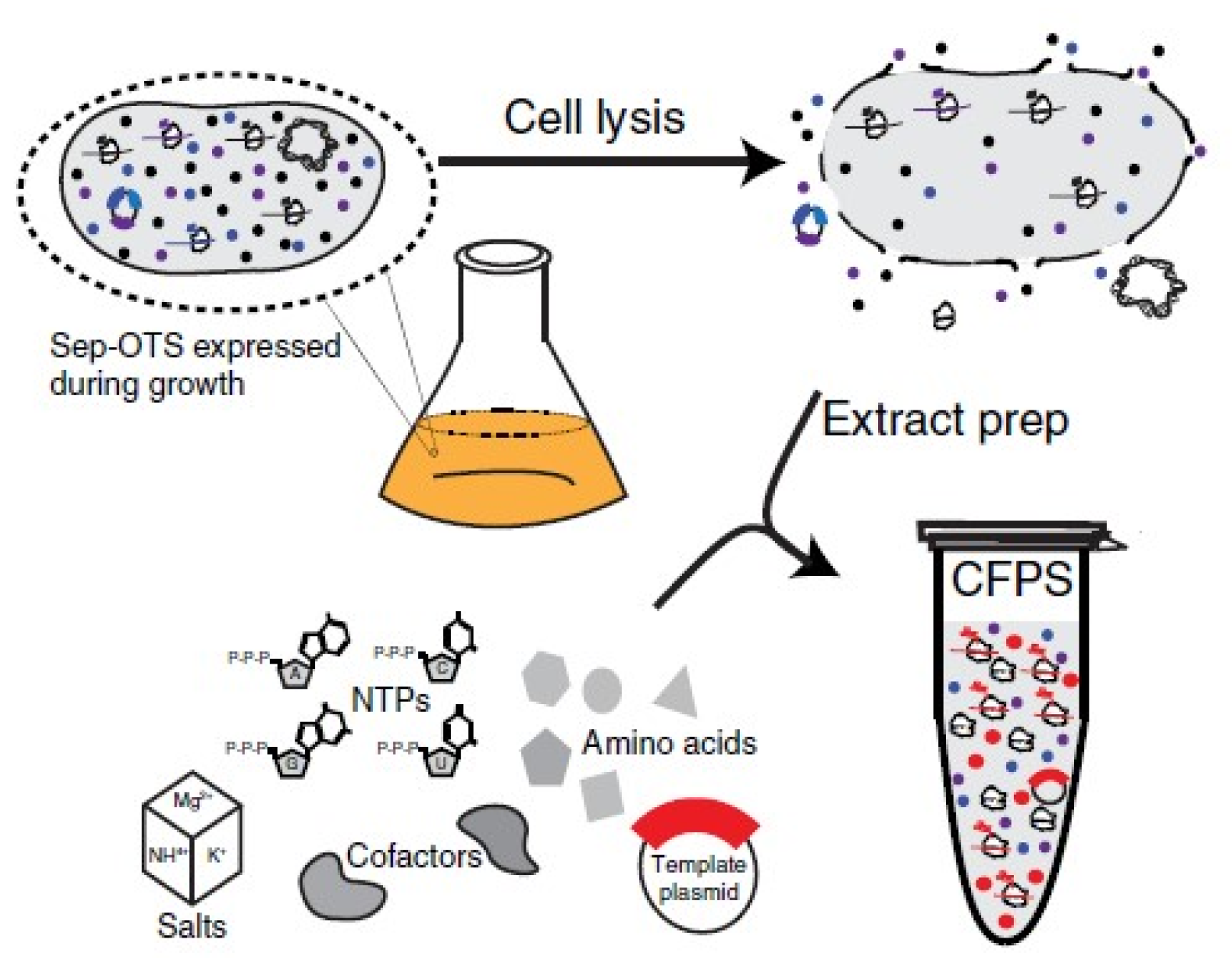
  42. Carlson, E.D.; Gan, R.; Hodgman, C.E.; Jewett, M.C. Cell-free protein synthesis: Applications come of age. Biotechnol. Adv. 2012, 30, 1185–1194. [Google Scholar] [CrossRef] [PubMed] [Green Version]
  43. Oza, J.P.; Aerni, H.R.; Pirman, N.L.; Barber, K.W.; Ter Haar, C.M.; Rogulina, S.; Amrofell, M.B.; Isaacs, F.J.; Rinehart, J.; Jewett, M.C. Robust production of recombinant phosphoproteins using cell-free protein synthesis. Nat. Commun. 2015, 6, 8168. [Google Scholar] [CrossRef] [Green Version]
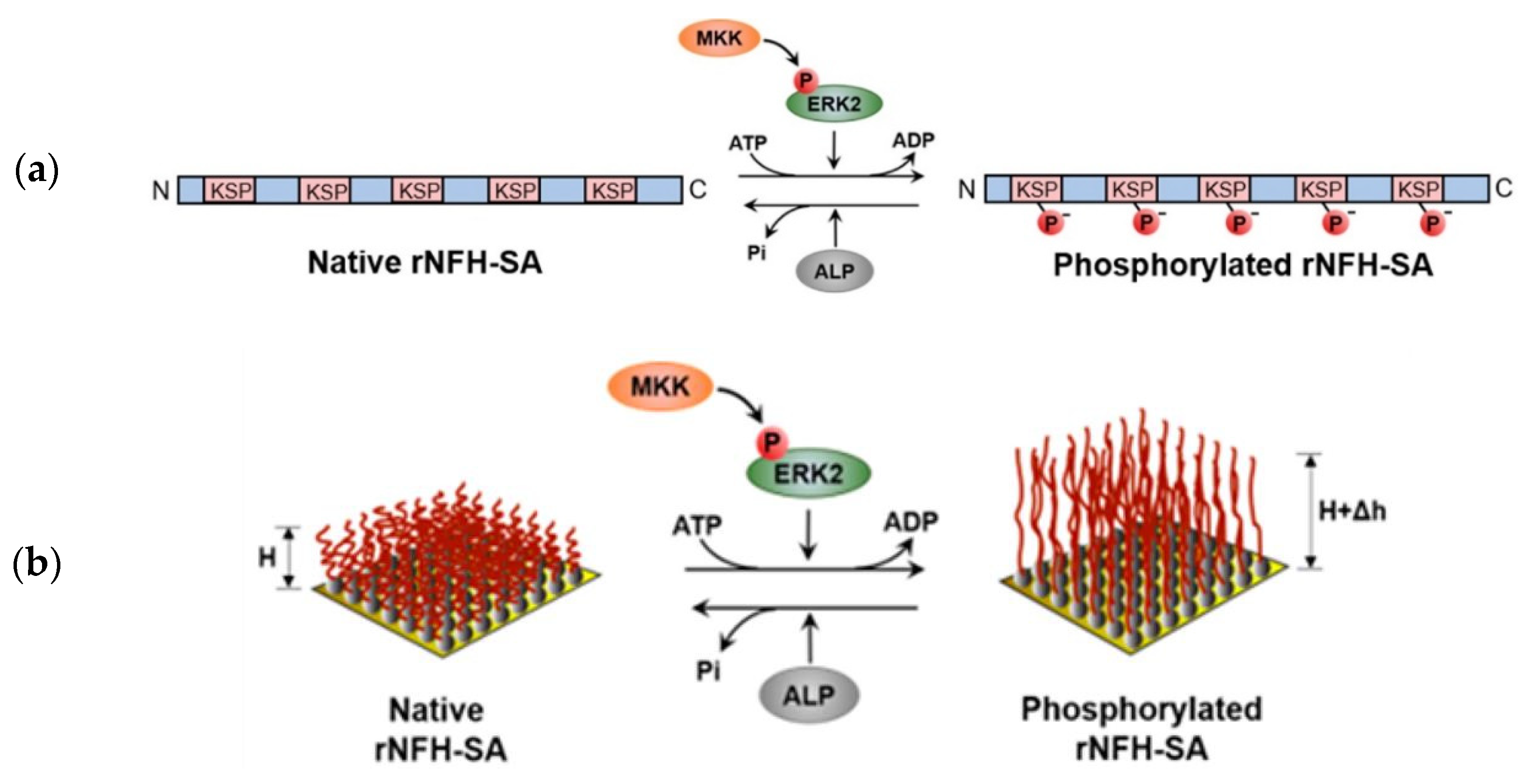
  44. Lei, R.; Lee, J.P.; Francis, M.B.; Kumar, S. Structural Regulation of a Neurofilament-Inspired Intrinsically Disordered Protein Brush by Multisite Phosphorylation. Biochemistry 2018, 57, 4019–4028. [Google Scholar] [CrossRef]
  45. Goux, M.; Fateh, A.; Defontaine, A.; Cinier, M.; Tellier, C. In vivo phosphorylation of a peptide tag for protein purification. Biotechnol. Lett. 2016, 38, 767–772. [Google Scholar] [CrossRef] [PubMed]
  46. Lei, R. Multi-site Phosphorylation Modulates the Conformation and Electrostatic Response of Intrinsically Disordered Protein Brushes. Biophys. J. 2018, 114, 590a. [Google Scholar] [CrossRef]
  47. Srinivasan, N.; Bhagawati, M.; Ananthanarayanan, B.; Kumar, S. Stimuli-sensitive intrinsically disordered protein brushes. Nat. Commun. 2014, 5, 1–8. [Google Scholar] [CrossRef] [PubMed] [Green Version]
  48. Sugiyama, G.; Takeuchi, H.; Nagano, K.; Gao, J.; Ohyama, Y.; Mori, Y.; Hirata, M. Regulated Interaction of Protein Phosphatase 1 and Protein Phosphatase 2A with Phospholipase C-Related but Catalytically Inactive Protein. Biochemistry 2012, 51, 3394–3403. [Google Scholar] [CrossRef]
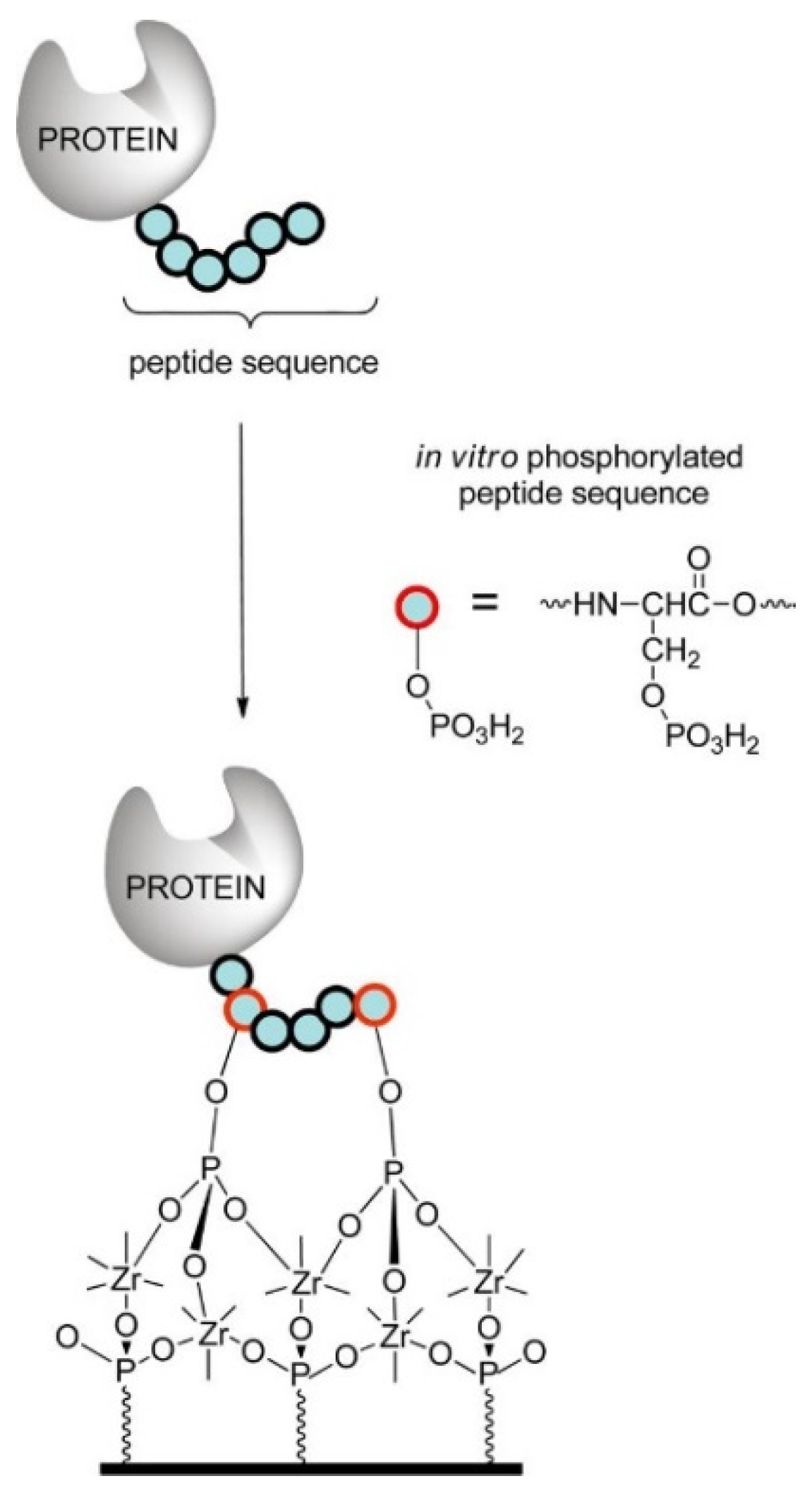
  49. Liu, H.; Queffélec, C.; Charlier, C.; Defontaine, A.; Fateh, A.; Tellier, C.; Talham, D.R.; Bujoli, B. Design and Optimization of a Phosphopeptide Anchor for Specific Immobilization of a Capture Protein on Zirconium Phosphonate Modified Supports. Langmuir 2014, 30, 13949–13955. [Google Scholar] [CrossRef]
  50. Lan, Y.T.; Li, J.; Liao, W.-Y.; Ou, J.-H. Roles of the Three Major Phosphorylation Sites of Hepatitis B Virus Core Protein in Viral Replication. Virology 1999, 259, 342–348. [Google Scholar] [CrossRef] [Green Version]
  51. Roux, P.P.; Thibault, P. The Coming of Age of Phosphoproteomics—from Large Data Sets to Inference of Protein Functions. Mol. Cell. Proteom. 2013, 12, 3453–3464. [Google Scholar] [CrossRef] [Green Version]
  52. Deng, Y.; Alicea-Velázquez, N.L.; Bannwarth, L.; Lehtonen, S.I.; Boggon, T.J.; Cheng, H.-C.; Hytönen, V.P.; Turk, B.E. Global Analysis of Human Nonreceptor Tyrosine Kinase Specificity Using High-Density Peptide Microarrays. J. Proteome Res. 2014, 13, 4339–4346. [Google Scholar] [CrossRef] [Green Version]
  53. Kobe, B.; Kampmann, T.; Forwood, J.K.; Listwan, P.; Brinkworth, R.I. Substrate specificity of protein kinases and computational prediction of substrates. Biochim. Biophys. Acta BBA Proteins Proteom. 2005, 1754, 200–209. [Google Scholar] [CrossRef]
  54. Ubersax, J.A.; Ferrell, J.E., Jr. Mechanisms of specificity in protein phosphorylation. Nat. Rev. Mol. Cell Biol. 2007, 8, 530–541. [Google Scholar] [CrossRef] [PubMed]
  55. Hornbeck, P.V.; Kornhauser, J.M.; Latham, V.; Murray, B.; Nandhikonda, V.; Nord, A.; Skrzypek, E.; Wheeler, T.; Zhang, B.; Gnad, F. 15 years of PhosphoSitePlus®: Integrating post-translationally modified sites, disease variants and isoforms. Nucleic Acids Res. 2019, 47, D433–D441. [Google Scholar] [CrossRef] [Green Version]
  56. Johnson, S.A.; Hunter, T. Kinomics: Methods for deciphering the kinome. Nat. Methods 2005, 2, 17–25. [Google Scholar] [CrossRef] [PubMed]
  57. Pan, J.; Zhang, S.; Borchers, C.H. Protein species-specific characterization of conformational change induced by multisite phosphorylation. J. Proteom. 2016, 134, 138–143. [Google Scholar] [CrossRef]
  58. Lee, J.; Samson, A.A.S.; Song, J.M. Direct On-Paper Inkjet Printing of Kinase-to-Kinase Phosphorylation Cascade Reactions. ACS Omega 2019, 4, 7866–7873. [Google Scholar] [CrossRef]
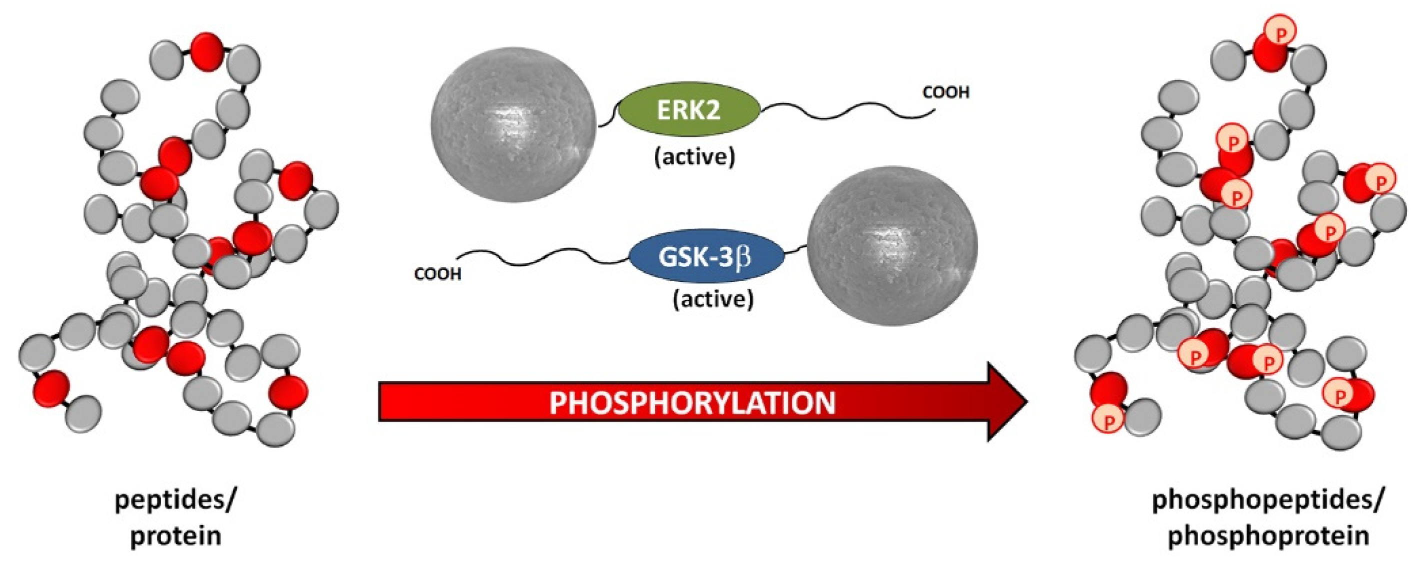
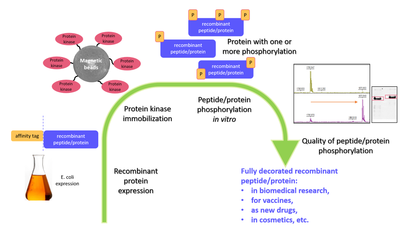
  59. Hromadkova, L.; Kupcik, R.; Vajrychova, M.; Prikryl, P.; Charvatova, A.; Jankovicova, B.; Ripova, D.; Bilkova, Z.; Slovakova, M. Kinase-loaded magnetic beads for sequential in vitro phosphorylation of peptides and proteins. Analyst 2018, 143, 466–474. [Google Scholar] [CrossRef] [PubMed]
  60. Li, Y.; Xu, J.; Chen, Y.; Mei, Z.; Xiao, Y. Screening of inhibitors of glycogen synthase kinase-3β from traditional Chinese medicines using enzyme-immobilized magnetic beads combined with high-performance liquid chromatography. J. Chromatogr. A 2015, 1425, 8–16. [Google Scholar] [CrossRef]
  61. Reddy, C.E.; Albanito, L.; De Marco, P.; Aiello, D.; Maggiolini, M.; Napoli, A.; Musti, A.M. Multisite phosphorylation of c-Jun at threonine 91/93/95 triggers the onset of c-Jun pro-apoptotic activity in cerebellar granule neurons. Cell Death Dis. 2013, 4, e852. [Google Scholar] [CrossRef]
  62. Quintero, O.A.; Unrath, W.C.; Stevens, S.M.J.; Manor, U.; Kachar, B.; Yengo, C.M. Myosin 3A Kinase Activity Is Regulated by Phosphorylation of the Kinase Domain Activation Loop. J. Biol. Chem. 2013, 288, 37126–37137. [Google Scholar] [CrossRef] [Green Version]
  63. Manning, G.; Whyte, D.B.; Martinez, R.; Hunter, T.; Sudarsanam, S. The Protein Kinase Complement of the Human Genome. Science 2002, 298, 1912–1934. [Google Scholar] [CrossRef] [Green Version]
  64. Zolnierowicz, S.; Bollen, M. Protein phosphorylation and protein phosphatases De Panne, Belgium, 19–24 September 1999. EMBO J. 2000, 19, 483–488. [Google Scholar] [CrossRef] [Green Version]
  65. Moorhead, G.B.G.; De Wever, V.; Templeton, G.; Kerk, D. Evolution of protein phosphatases in plants and animals. Biochem. J. 2009, 417, 401–409. [Google Scholar] [CrossRef] [Green Version]
  66. Martin, L.; Latypova, X.; Wilson, C.M.; Magnaudeix, A.; Perrin, M.-L.; Terro, F. Tau protein phosphatases in Alzheimer’s disease: The leading role of PP2A. Ageing Res. Rev. 2013, 12, 39–49. [Google Scholar] [CrossRef]
  67. Mendoza, J.; Sekiya, M.; Taniguchi, T.; Iijima, K.M.; Wang, R.; Ando, K. Global Analysis of Phosphorylation of Tau by the Checkpoint Kinases Chk1 and Chk2in vitro. J. Proteome Res. 2013, 12, 2654–2665. [Google Scholar] [CrossRef] [PubMed] [Green Version]
  68. Morton, S.; Davis, R.J.; McLaren, A.; Cohen, P. A reinvestigation of the multisite phosphorylation of the transcription factor c-Jun. EMBO J. 2003, 22, 3876–3886. [Google Scholar] [CrossRef] [Green Version]
  69. Veeranna; Yang, D.-S.; Lee, J.-H.; Vinod, K.Y.; Stavrides, P.; Amin, N.D.; Pant, H.C.; Nixon, R.A. Declining phosphatases underlie aging-related hyperphosphorylation of neurofilaments. Neurobiol. Aging 2011, 32, 2016–2029. [Google Scholar] [CrossRef] [Green Version]
  70. Lauretti, E.; Dincer, O.; Praticò, D. Glycogen synthase kinase-3 signaling in Alzheimer’s disease. Biochim. Biophys. Acta BBA Mol. Cell Res. 2020, 1867, 118664. [Google Scholar] [CrossRef]
  71. Dunning, C.J.; McGauran, G.; Willén, K.; Gouras, G.K.; O’Connell, D.J.; Linse, S. Direct High Affinity Interaction between Aβ42 and GSK3α Stimulates Hyperphosphorylation of Tau. A New Molecular Link in Alzheimer’s Disease? ACS Chem. Neurosci. 2016, 7, 161–170. [Google Scholar] [CrossRef] [Green Version]
  72. Wong, C.J.; Launer-Felty, K.; Cole, J.L. Analysis of PKR–RNA Interactions by Sedimentation Velocity. Methods Enzymol. 2011, 488, 59–79. [Google Scholar] [CrossRef] [PubMed] [Green Version]
  73. Reimer, L.; Betzer, C.; Kofoed, R.H.; Volbracht, C.; Fog, K.; Kurhade, C.; Nilsson, E.; Överby, A.K.; Jensen, P.H. PKR kinase directly regulates tau expression and Alzheimer’s disease-related tau phosphorylation. Brain Pathol. 2021, 31, 103–119. [Google Scholar] [CrossRef] [PubMed]
  74. Taylor, G.A.; Thompson, M.J.; Lai, W.S.; Blackshear, P.J. Phosphorylation of Tristetraprolin, a Potential Zinc Finger Transcription Factor, by Mitogen Stimulation in Intact Cells and by Mitogen-activated Protein Kinase in Vitro. J. Biol. Chem. 1995, 270, 13341–13347. [Google Scholar] [CrossRef] [Green Version]
  75. Dong, Q.; Giorgianni, F.; Deng, X.; Beranova-Giorgianni, S.; Bridges, D.; Park, E.A.; Raghow, R.; Elam, M.B. Phosphorylation of sterol regulatory element binding protein-1a by protein kinase A (PKA) regulates transcriptional activity. Biochem. Biophys. Res. Commun. 2014, 449, 449–454. [Google Scholar] [CrossRef] [Green Version]
  76. Li, Z.; Li, X.; Gao, X.; Shen, Q.W.; Du, M.; Zhang, D. Phosphorylation prevents in vitro myofibrillar proteins degradation by μ-calpain. Food Chem. 2017, 218, 455–462. [Google Scholar] [CrossRef] [PubMed]
  77. Benaim, G.; Villalobo, A. Phosphorylation of calmodulin. Functional implications. Eur. J. Biochem. 2002, 269, 3619–3631. [Google Scholar] [CrossRef] [PubMed]
  78. Cinier, M.; Petit, M.; Pecorari, F.; Talham, D.R.; Bujoli, B.; Tellier, C. Engineering of a phosphorylatable tag for specific protein binding on zirconium phosphonate based microarrays. JBIC J. Biol. Inorg. Chem. 2012, 17, 399–407. [Google Scholar] [CrossRef]
  79. Qin, C.-L.; Tang, J.; Kim, K. Cloning and in vitro expression of TPK3, a Toxoplasma gondii homologue of shaggy/glycogen synthase kinase-3 kinases1. Mol. Biochem. Parasitol. 1998, 93, 273–283. [Google Scholar] [CrossRef]
  80. Fentress, S.J.; Behnke, M.S.; Dunay, I.R.; Mashayekhi, M.; Rommereim, L.M.; Fox, B.A.; Bzik, D.J.; Taylor, G.A.; Turk, B.E.; Lichti, C.F.; et al. Phosphorylation of Immunity-Related GTPases by a Toxoplasma gondii-Secreted Kinase Promotes Macrophage Survival and Virulence. Cell Host Microbe 2010, 8, 484–495. [Google Scholar] [CrossRef] [PubMed] [Green Version]
  81. Xu, Y.Z.; Thuraisingam, T.; Kanagaratham, C.; Tao, S.; Radzioch, D. c-Src kinase is involved in the tyrosine phosphorylation and activity of SLC11A1 in differentiating macrophages. PLoS ONE 2018, 13, e0196230. [Google Scholar] [CrossRef] [PubMed] [Green Version]
  82. Cable, J.; Prutzman, K.; Gunawardena, H.P.; Schaller, M.D.; Chen, X.; Campbell, S.L. In Vitro Phosphorylation of the Focal Adhesion Targeting Domain of Focal Adhesion Kinase by Src Kinase. Biochemistry 2012, 51, 2213–2223. [Google Scholar] [CrossRef] [Green Version]
  83. Samarasimhareddy, M.; Mayer, G.; Hurevich, M.; Friedler, A. Multiphosphorylated peptides: Importance, synthetic strategies, and applications for studying biological mechanisms. Org. Biomol. Chem. 2020, 18, 3405–3422. [Google Scholar] [CrossRef]
  84. Clark, A.R.; Dean, J.L.E.E. The control of inflammation via the phosphorylation and dephosphorylation of tristetraprolin: A tale of two phosphatases. Biochem. Soc. Trans. 2016, 44, 1321–1337. [Google Scholar] [CrossRef] [Green Version]
  85. Cao, H.; Dzineku, F.; Blackshear, P.J. Expression and purification of recombinant tristetraprolin that can bind to tumor necrosis factor-α mRNA and serve as a substrate for mitogen-activated protein kinases. Arch. Biochem. Biophys. 2003, 412, 106–120. [Google Scholar] [CrossRef]
  86. Cao, H. Expression, Purification, and Biochemical Characterization of the Antiinflammatory Tristetraprolin: A Zinc-Dependent mRNA Binding Protein Affected by Posttranslational Modifications†,‡. Biochemistry 2004, 43, 13724–13738. [Google Scholar] [CrossRef] [Green Version]
  87. Erdem, F.A.; Salzer, I.; Heo, S.; Chen, W.; Jung, G.; Lubec, G.; Boehm, S.; Yang, J.-W. Updating In Vivo and In Vitro Phosphorylation and Methylation Sites of Voltage-Gated Kv7.2 Potassium Channels. Proteomics 2017, 17, 1700015. [Google Scholar] [CrossRef]
  88. Homaei, A.A.; Sariri, R.; Vianello, F.; Stevanato, R. Enzyme immobilization: An update. J. Chem. Biol. 2013, 6, 185–205. [Google Scholar] [CrossRef] [PubMed] [Green Version]
  89. Bisswanger, H. Enzyme assays. Perspect. Sci. 2014, 1, 41–55. [Google Scholar] [CrossRef] [Green Version]
  90. Hernandez, K.; Fernandez-Lafuente, R. Control of protein immobilization: Coupling immobilization and site-directed mutagenesis to improve biocatalyst or biosensor performance. Enzym. Microb. Technol. 2011, 48, 107–122. [Google Scholar] [CrossRef]
  91. Brena, B.; Batista-Viera, F. Methods in BiotechnologyTM. In Immobilization of Enzymes and Cells, 2nd ed.; Guisan, J.M., Ed.; Humana Press: Totowa, NJ, USA, 2006; Volume 22, ISBN 978-1-58829-290-2. [Google Scholar]
  92. Jesionowski, T.; Zdarta, J.; Krajewska, B. Enzyme immobilization by adsorption: A review. Adsorption 2014, 20, 801–821. [Google Scholar] [CrossRef] [Green Version]
  93. De Moraes, M.C.; Temporini, C.; Calleri, E.; Bruni, G.; Ducati, R.G.; Santos, D.S.; Cardoso, C.L.; Cass, Q.B.; Massolini, G. Evaluation of capillary chromatographic supports for immobilized human purine nucleoside phosphorylase in frontal affinity chromatography studies. J. Chromatogr. A 2014, 1338, 77–84. [Google Scholar] [CrossRef] [PubMed]
  94. Foresti, M.L.; Ferreira, M.L. Chitosan-immobilized lipases for the catalysis of fatty acid esterifications. Enzym. Microb. Technol. 2007, 40, 769–777. [Google Scholar] [CrossRef]
  95. Narwal, V.; Pundir, C.S. Development of glycerol biosensor based on co-immobilization of enzyme nanoparticles onto graphene oxide nanoparticles decorated pencil graphite electrode. Int. J. Biol. Macromol. 2019, 127, 57–65. [Google Scholar] [CrossRef]
  96. Machín, B.; Chaves, S.; Ávila, C.; Pera, L.M.; Chehín, R.N.; Pingitore, E.V. Highly reusable invertase biocatalyst: Biological fibrils functionalized by photocrosslinking. Food Chem. 2020, 331, 127322. [Google Scholar] [CrossRef]
  97. Wang, L.; Wei, L.; Chen, Y.; Jiang, R. Specific and reversible immobilization of NADH oxidase on functionalized carbon nanotubes. J. Biotechnol. 2010, 150, 57–63. [Google Scholar] [CrossRef]
  98. Hetmann, A.; Wujak, M.; Bolibok, P.; Zięba, W.; Wiśniewski, M.; Roszek, K. Novel biocatalytic systems for maintaining the nucleotide balance based on adenylate kinase immobilized on carbon nanostructures. Mater. Sci. Eng. C 2018, 88, 130–139. [Google Scholar] [CrossRef] [PubMed]
  99. Niu, H.; Ding, M.; Sun, X.; Zhuang, W.; Liu, D.; Ying, H.; Zhu, C.; Chen, Y. Immobilization of a polyphosphate kinase 2 by coordinative self-assembly of his-tagged units with metal-organic frameworks and its application in ATP regeneration from AMP. Colloids Surf. B Biointerfaces 2019, 181, 261–269. [Google Scholar] [CrossRef]
  100. Saifuddin, N.; Raziah, A.Z.; Junizah, A.R. Carbon Nanotubes: A Review on Structure and Their Interaction with Proteins. J. Chem. 2013, 2013, 1–18. [Google Scholar] [CrossRef]
  101. Zaitsev, S.Y.; Savina, A.A.; Zaitsev, I.S. Biochemical aspects of lipase immobilization at polysaccharides for biotechnology. Adv. Colloid Interface Sci. 2019, 272, 102016. [Google Scholar] [CrossRef]
  102. Rotková, J.; Šuláková, R.; Korecká, L.; Zdražilová, P.; Jandová, M.; Lenfeld, J.; Horák, D.; Bilkova, Z. Laccase immobilized on magnetic carriers for biotechnology applications. J. Magn. Magn. Mater. 2009, 321, 1335–1340. [Google Scholar] [CrossRef]
  103. Slovakova, M.; Minc, N.; Bilkova, Z.; Smadja, C.; Faigle, W.; Fütterer, C.; Taverna, M.; Viovy, J.-L. Use of self assembled magnetic beads for on-chip protein digestion. Lab. Chip 2005, 5, 935–942. [Google Scholar] [CrossRef]
  104. Ferey, J.; Da Silva, D.; Colas, C.; Nehmé, R.; Lafite, P.; Roy, V.; Morin, P.; Daniellou, R.; Agrofoglio, L.; Maunit, B. Monitoring of successive phosphorylations of thymidine using free and immobilized human nucleoside/nucleotide kinases by Flow Injection Analysis with High-Resolution Mass Spectrometry. Anal. Chim. Acta 2019, 1049, 115–122. [Google Scholar] [CrossRef] [PubMed]
  105. Svitel, J.; Boukari, H.; Van Ryk, D.; Willson, R.C.; Schuck, P. Probing the Functional Heterogeneity of Surface Binding Sites by Analysis of Experimental Binding Traces and the Effect of Mass Transport Limitation. Biophys. J. 2007, 92, 1742–1758. [Google Scholar] [CrossRef] [Green Version]
  106. Merrill, A.H.; McCormick, D.B. Preparation and properties of immobilized flavokinase. Biotechnol. Bioeng. 1979, 21, 1629–1638. [Google Scholar] [CrossRef]
  107. Berke, W.; Schüz, H.-J.; Wandrey, C.; Morr, M.; Denda, G.; Kula, M.-R. Continuous regeneration of ATP in enzyme membrane reactor for enzymatic syntheses. Biotechnol. Bioeng. 1988, 32, 130–139. [Google Scholar] [CrossRef] [PubMed]
  108. Ropp, P.A.; Cheng, P.-W. Enzymatic synthesis of UDP-GlcN by a two step hollow fiber enzyme reactor system. Anal. Biochem. 1990, 187, 104–108. [Google Scholar] [CrossRef]
  109. Fonong, T. Immobilized enzyme assay of creatine kinase with amperometric detection. Anal. Biochem. 1989, 176, 234–238. [Google Scholar] [CrossRef]
  110. Ferey, J.; Da Silva, D.; Colas, C.; Lafite, P.; Topalis, D.; Roy, V.; Agrofoglio, L.A.; Daniellou, R.; Maunit, B. Monitoring of phosphorylation using immobilized kinases by on-line enzyme bioreactors hyphenated with High-Resolution Mass Spectrometry. Talanta 2019, 205, 120120. [Google Scholar] [CrossRef] [PubMed]
  111. Serra, I.; Conti, S.; Piškur, J.; Clausen, A.R.; Terreni, M.; Ubiali, D.; Munch-Petersen, B. Immobilized Drosophila melanogaster Deoxyribonucleoside Kinase (DmdNK) as a High Performing Biocatalyst for the Synthesis of Purine Arabinonucleotides. Adv. Synth. Catal. 2014, 356, 563–570. [Google Scholar] [CrossRef]
  112. Wu, S.; Li, J.; Hu, C.; Tian, J.; Zhang, T.; Chen, N.; Fan, X. Concomitant use of immobilized uridine-cytidine kinase and polyphosphate kinase for 5′-cytidine monophosphate production. Chin. J. Biotechnol 2020, 36, 1002–1011. [Google Scholar] [CrossRef]
  113. Hoffman, R.C., Jr.; Wyman, P.L.; Smith, L.E.; Nolt, C.L.; Conley, J.L.; Hevel, J.M.; Warren, J.P.; Reiner, G.A.; Moe, O.A., Jr. Immobilized polyphosphate kinase: Preparation, properties, and potential for use in adenosine 5′-triphosphate regeneration. Biotechnol. Appl. Biochem. 1988, 10, 107–117. [Google Scholar] [CrossRef]
  114. Suzuki, S.; Hara, R.; Kino, K. Production of aminoacyl prolines using the adenylation domain of nonribosomal peptide synthetase with class III polyphosphate kinase 2-mediated ATP regeneration. J. Biosci. Bioeng. 2018, 125, 644–648. [Google Scholar] [CrossRef]
  115. Sun, X.; Niu, H.; Song, J.; Jiang, D.; Leng, J.; Zhuang, W.; Chen, Y.; Liu, D.; Ying, H. Preparation of a Copper Polyphosphate Kinase Hybrid Nanoflower and Its Application in ADP Regeneration from AMP. ACS Omega 2020, 5, 9991–9998. [Google Scholar] [CrossRef]
  116. Kowalczyk, T.H.; Szymona, O. Glucose determination using immobilized polyphosphate glucokinase. Anal. Biochem. 1991, 197, 326–332. [Google Scholar] [CrossRef]
  117. Gabris, C.; Bengelsdorf, F.R.; Dürre, P. Analysis of the key enzymes of butyric and acetic acid fermentation in biogas reactors. Microb. Biotechnol. 2015, 8, 865–873. [Google Scholar] [CrossRef]
  118. Castro, L.B.R.; Silva, F.F.; Carmona-Ribeiro, A.M.; Kappl, M.; Petri, D.F.S. Immobilization of Hexokinase onto Chitosan Decorated Particles†. J. Phys. Chem. B 2007, 111, 8520–8526. [Google Scholar] [CrossRef] [PubMed]
  119. Cui, Y.; Barford, J.P.; Renneberg, R. Amperometric trienzyme ATP biosensors based on the coimmobilization of salicylate hydroxylase, glucose-6-phosphate dehydrogenase, and hexokinase. Sensors Actuators B Chem. 2008, 132, 1–4. [Google Scholar] [CrossRef]
  120. Kucherenko, I.S.; Kucherenko, D.Y.; Soldatkin, O.O.; Lagarde, F.; Dzyadevych, S.V.; Soldatkin, A.P. A novel conductometric biosensor based on hexokinase for determination of adenosine triphosphate. Talanta 2016, 150, 469–475. [Google Scholar] [CrossRef]
  121. Narwal, V.; Pundir, C.S. Fabrication of glycerol biosensor based on co-immobilization of enzyme nanoparticles onto pencil graphite electrode. Anal. Biochem. 2018, 555, 94–103. [Google Scholar] [CrossRef]
  122. Chauhan, N.; Narang, J.; Pundir, C.S. Covalent immobilization of lipase, glycerol kinase, glycerol-3-phosphate oxidase & horseradish peroxidase onto plasticized polyvinyl chloride (PVC) strip & its application in serum triglyceride determination. Indian J. Med. Res. 2014, 139, 603–609. [Google Scholar]
  123. Minakshi, C.S.; Pundir, C.S. Co-immobilization of lipase, glycerol kinase, glycerol-3-phosphate oxidase and peroxidase on to aryl amine glass beads affixed on plastic strip for determination of triglycerides in serum. Indian J. Biochem. Biophys. 2008, 45, 111–115. [Google Scholar]
  124. Kalia, V.; Pundir, C.S. Co-immobilization of lipase, glycerol kinase, glycerol-3-phosphate oxidase and peroxidase onto alkylamine glass beads through glutaraldehyde coupling. Indian J. Biochem. Biophys. 2002, 39, 342–346. [Google Scholar] [PubMed]
  125. Pundir, C.S.; Aggarwal, V. Amperometric triglyceride bionanosensor based on nanoparticles of lipase, glycerol kinase, glycerol-3-phosphate oxidase. Anal. Biochem. 2017, 517, 56–63. [Google Scholar] [CrossRef]
  126. Taka’aki, M.; Kenischi, M.; Takuya, M.; Tetsu, M.; Masateru, O.; Yasuhiko, S. High-Throughput Profiling of Kinase Inhibitors Selectivity Using the ProteOnTM XPR36 Protein Interaction Array System. Available online: https://www.bio-rad.com/webroot/web/pdf/lsr/literature/Bulletin_5960A.pdf (accessed on 11 March 2021).
  127. Liese, A.; Hilterhaus, L. Evaluation of immobilized enzymes for industrial applications. Chem. Soc. Rev. 2013, 42, 6236–6249. [Google Scholar] [CrossRef]
  128. Popplewell, J.; Luo, R. Kinase Immobilization for Small Molecule Inhibition Studies. Bioradiations Articles. Available online: https://www.bioradiations.com/immobilization-of-active-kinases-for-small-molecule-inhibition-studies/ (accessed on 11 March 2021).
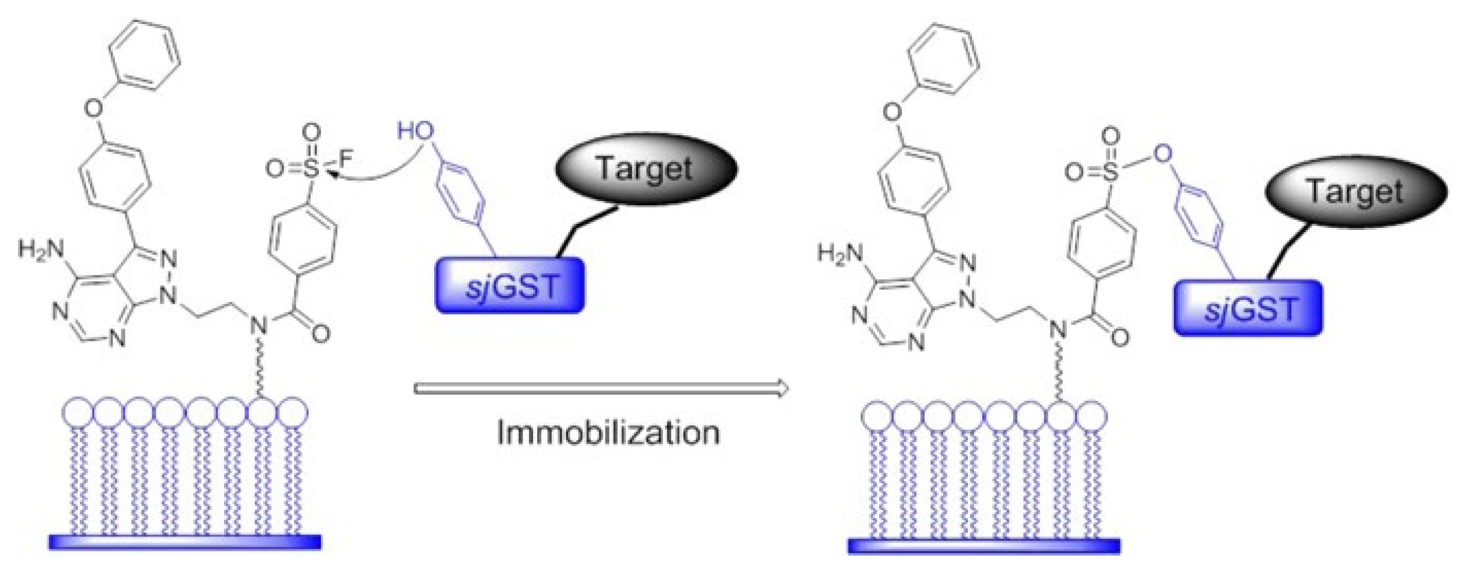
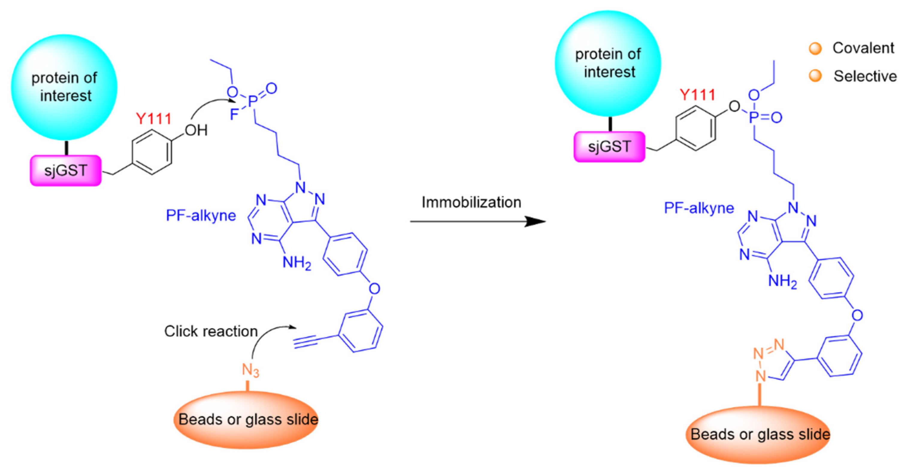
  129. Zhou, Y.; Guo, T.; Tang, G.; Wu, H.; Wong, N.-K.; Pan, Z. Site-Selective Protein Immobilization by Covalent Modification of GST Fusion Proteins. Bioconjug. Chem. 2014, 25, 1911–1915. [Google Scholar] [CrossRef]
  130. Wang, X.; Guo, T.; Chen, J.; Li, X.; Zhou, Y.; Pan, Z. Covalent and selective immobilization of GST fusion proteins with fluorophosphonate-based probes. Chem. Commun. 2018, 54, 4661–4664. [Google Scholar] [CrossRef]
  131. Kim, M.; Shin, D.-S.; Kim, J.; Lee, Y.-S. Substrate screening of protein kinases: Detection methods and combinatorial peptide libraries. Biopolymers 2010, 94, 753–762. [Google Scholar] [CrossRef] [PubMed]
  132. Wu, H.-Y.; Liao, P.-C. Analysis of protein phosphorylation using mass spectrometry. Chang. Gung Med. J. 2008, 31, 217–227. [Google Scholar]
  133. Campbell, D.G.; Morrice, N.A. Identification of protein phosphorylation sites by a combination of mass spectrometry and solid phase Edman sequencing. J. Biomol. Tech. JBT 2002, 13, 119–130. [Google Scholar] [PubMed]
  134. Molden, R.C.; Goya, J.; Khan, Z.; Garcia, B.A. Stable Isotope Labeling of Phosphoproteins for Large-scale Phosphorylation Rate Determination. Mol. Cell. Proteom. 2014, 13, 1106–1118. [Google Scholar] [CrossRef] [PubMed] [Green Version]
Figure 1. Diagram illustrating events that can contribute to neuronal dysfunction in AD and/or frontotemporal dementia and parkinsonism related to tauopathies of tauopathies of tauopathies of chromosome 17 (FTDP-17), and the crucial role played by alterations in tau protein phosphorylation. Reproduced with permission from [32]; published by Elsevier, 2005.
Catalysts 11 01007 g001
Figure 2. Phosphoprotein production by the combination of protein synthesis and a biotechnological approach: cell-free protein synthesis adapted for phosphoprotein biosynthesis in the presence of L-phosphoserine (Sep). The orthogonal translational system for phosphoprotein synthesis consists of phosphoseryl-tRNA synthetase, tRNASep, and the elongation factor. tRNASep is aminoacylated with Sep-Sep tRNA synthetase (SepRS). EF-Sep then delivers Sep-tRNASep to the ribosome. Reprinted with permission from [43]. Copyright 2015 Springer Nature.
Catalysts 11 01007 g002
Figure 3. Enzymatic phosphorylation of: (a) recombinant heavy subunit of neurofilaments (NFH-SA) and; (b) immobilized recombinant heavy subunit of neurofilaments (NFH-SA) brushes using a two-kinase process involving treatment with ERK2 preactivated with MEK. Reprinted with permission from [44]. Copyright (2018) American Chemical Society.
Catalysts 11 01007 g003
Figure 4. Covalent attachment of proteins to zirconium phosphonate surfaces via phosphopeptide anchors. Reprinted with permission from [49]. Copyright (2014) American Chemical Society.
Catalysts 11 01007 g004
Figure 5. Sequential in vitro phosphorylation of recombinant tau protein by a system of two immobilized active kinases. The prephosphorylation of the tau protein was provided by immobilized ERK2, followed by phosphorylation by immobilized GSK-3β [59].
Catalysts 11 01007 g005
Figure 6. Scheme of site-selective covalent immobilization of the recombinant protein kinases ITK and EGFR with fused sjGST tags through irreversible and specific covalent modification of the tyrosine 111 residue of the sjGST tag. Immobilization involved the use of small-molecule pyrimidine-based probes with a reactive group of sulfonyl fluoride. Reprinted with permission from [129]. Copyright (2012) American Chemical Society.
Catalysts 11 01007 g006
Figure 7. Site-selective immobilization of sjGST-tagged proteins containing the tyrosine residue 111 through a small pyrimidine-based molecule with a fluorophosphonate-reactive group in the bead. Reprinted with permission from [130]. Copyright (2018) Royal Society of Chemistry.
Catalysts 11 01007 g007
Table 1. List of immobilized protein kinases applied for in vitro phosphorylation.
Protein KinaseSolid SupportMethod of Immobilization and PurposeRef.
Biotinylated kinasesNeutrAvidin surface sensor chipBiotin-avidin affinity bond for high-throughput profiling of kinase inhibitor selectivity[126,128]
ITK, EGFR, BLK, and LCK recombinant, GST-taggedCH-sepharose 4B beads; Site selective covalent kinase immobilization[129,130]
GSK-3β, GST-tagged recombinantGlutathione beads, magnetic; Affinity kinase immobilization for kinase inhibitors screening[60]
GSK-3β, recombinant; ERK2/MAPK1 recombinant, activeSeraMag SpeedBeads, carboxyl; 0.816 μm; magnetic
BcMag, aldehyde; magnetic
Covalent kinase immobilization for in vitro phosphorylation[59]
GSK-3β His-tagged, recombinant, MAPK2 His-tagged, recombinantSIMAG-IDA/Co3+, SIMAG-IDA/Ni2+ magnetic; Affinity irreversible kinase immobilization for in vitro phosphorylation[59]
Publisher’s Note: MDPI stays neutral with regard to jurisdictional claims in published maps and institutional affiliations.

