Article

Efficient 1-Hydroxy-2-Butanone Production from 1,2-Butanediol by Whole Cells of Engineered E. coli

1
Key Laboratory of Biopesticide and Chemical Biology, Ministry of Education, College of Life Sciences, Fujian Agriculture and Forestry University, Fuzhou 350002, China
2
Department of Pharmacy, The First Affiliated Hospital of Xiamen University, Xiamen 361022, China
3
College of Plant Protection, Fujian Agriculture and Forestry University, Fuzhou 350002, China
*
Authors to whom correspondence should be addressed.
These authors contributed equally to this work.
Catalysts 2021, 11(10), 1184; https://doi.org/10.3390/catal11101184
Submission received: 2 September 2021 / Revised: 24 September 2021 / Accepted: 27 September 2021 / Published: 28 September 2021
(This article belongs to the Special Issue Enzyme-Mediated Stereoselective Synthesis II)

Abstract

:
1-Hydroxy-2-butanone (HB) is a key intermediate for anti-tuberculosis pharmaceutical ethambutol. Commercially available HB is primarily obtained by the oxidation of 1,2-butanediol (1,2-BD) using chemical catalysts. In present study, seven enzymes including diol dehydrogenases, secondary alcohol dehydrogenases and glycerol dehydrogenase were chosen to evaluate their abilities in the conversion of 1,2-BD to HB. The results showed that (2R, 3R)- and (2S, 3S)-butanediol dehydrogenase (BDH) from Serratia sp. T241 could efficiently transform (R)- and (S)-1,2-BD into HB respectively. Furthermore, two biocatalysts co-expressing (2R, 3R)-/(2S, 3S)-BDH, NADH oxidase and hemoglobin protein in Escherichia coli were developed to convert 1,2-BD mixture into HB, and the transformation conditions were optimized. Maximum HB yield of 341.35 and 188.80 mM could be achieved from 440 mM (R)-1,2-BD and 360 mM (S)-1,2-BD by E. coli (pET-rrbdh-nox-vgb) and E. coli (pET-ssbdh-nox-vgb) under the optimized conditions. In addition, two biocatalysts showed the ability in chiral resolution of 1,2-BD isomers, and 135.68 mM (S)-1,2-BD and 112.43 mM (R)-1,2-BD with the purity of 100% could be obtained from 300 and 200 mM 1,2-BD mixture by E. coli (pET-rrbdh-nox-vgb) and E. coli (pET-ssbdh-nox-vgb), respectively. These results provided potential application for HB production from 1,2-BD mixture and chiral resolution of (R)-1,2-BD and (S)-1,2-BD.

Graphical Abstract

1. Introduction

1-hydroxy-2-butanone (HB) containing carbonyl and hydroxyl groups, is an important intermediate for the synthesis of anti-tuberculosis pharmaceutical ethambutol [1,2]. However, few studies about HB synthesis were reported in past literature. Currently, commercially available HB is primarily produced industrially by the oxidation of 1,2-butanediol (1,2-BD) using various chemical catalysts. Thus, 1,2-BD could be transformed into α-hydroxyketone with the space-time yield of 19.71 g/d by the catalyst of CuO : ZnO : ZrO2 : Al2O3 with the ratios of 12 : 1 : 2 : 2, and the product HB occupied a molar fraction of 98.1% with a small amount of by-products including butanone, 1-butanol, and butanoic acid [3]. In addition, HB and another product, hydroxyacetone (HA), were also produced directly from cellulose by Sn–Ni intermetallic compound catalysts, and 66.4 mg HA and 71.2 mg HB could be achieved from 200 mg cellulose under optimum conditions, resulting in space-time HB yield of 1.71 g/d [2]. However, harsh reaction conditions and low product yields result in a higher cost and potential environmental pollution. Compared with chemical synthesis methods, biosynthesis strategy via whole cell or enzymatic biocatalysis was paid increasing attention due to mild reaction conditions and high catalytic efficiency [4,5]. Therefore, developing a biosynthesis method for HB production is desired and urgent.
A literature search showed that HB biosynthesis and related enzymes were rarely reported in previous studies. Fortunately, various diol dehydrogenases have shown the abilities in the oxidation of diol to the corresponding ketone alcohol [6,7]. Further, 2,3-Butanediol dehydrogenase (BDH), one of diol dehydrogenases, can catalyze the dehydrogenation of 2,3-butanediol (2,3-BD) into acetoin (AC, a stereoisomer of HB) [8,9,10,11,12]. Furthermore, the BDH enzymes from Serratia marcescens H30 and Paenibacillus polymyxa ATCC 12321 also showed the activities for 1,2-pentadiol and 1,2-propanediol as substrates [13,14]. These results exhibited a broad substrate spectrum for the diol oxidation by BDH enzymes, which provided application potential for HB production from 1,2-BD. According to the configuration of the substrate 2,3-BD including (2S, 3S)-BD, meso-2,3-BD, and (2R, 3R)- BD, BDHs can be classified into three types: (2S, 3S)-BDH, meso-BDH, and (2R, 3R)-BDH, which were involved in stereoisomeric formation of 2,3-BD and AC from various carbon sources by many microorganisms [14,15,16,17]. Among three types of BDH, (2S, 3S)-BDH showed substrate specificity and only catalyzed (2S, 3S)-BD into (3S)-AC using NAD+ as coenzyme. Meso-BDH could convert meso-2,3-BD and (2S, 3S)-BD as substrates into (3R)-AC and (3S)-AC respectively in the presence of NAD+, and exhibit (S)-type configuration specificity. In contrast, (2R, 3R)-BDH with (R)-type configuration specificity showed the activity for the conversion of meso-2,3-BD and (2R, 3R)-BD into (3S)-AC and (3R)-AC, respectively [8,9,14,15,16]. Moreover, a secondary alcohol dehydrogenase (SADH) from Burkholderia sp. was found to convert secondary alcohol into secondary ketone, and exhibited a maximum specific activity when 2-propanol was used as substrate. Substrate specificity assays showed that 1,2-BD could be oxidated into HB by the SADH enzyme, and a relative activity of 20% for 1,2-BD as substrate compared with that of 2-propanol could be achieved [18]. In addition, glycerol dehydrogenases (GDHs), catalyzing the dehydrogenation of glycerol to dihydroxyacetone, also showed the ability in the oxidation of 1,2-BD to HB in previous studies [4,19,20,21]. The relative activity of 90%, 60%, and 45% for 1,2-BD as substrate by three GDHs from Schizosaccharomyces pombe, Cellulomonas sp., and Enterobacter aerogenes respectively could be achieved when compared with that of glycerol as substrate [4,21]. Thus, both of the two types of enzymes may be used as an alternative enzyme for HB production from 1,2-BD as substrate.
In this study, three diol dehydrogenases, three secondary alcohol dehydrogenases, and one glycerol dehydrogenase were chosen to evaluate their abilities in converting 1,2-BD into HB. These enzymes consisted of (2S, 3S)-BDH, meso-BDH, (2R, 3R)-BDH, and GDH from Serratia sp. T241 [16], CbADH from Clotridium beijerinckii [22], CaADH and its variant CaADH:S199A from Clostridium autoethanogenum [10,23,24]. Enzyme activity assays showed that all the tested enzymes could use a mixture of 1,2-BD (45% (S)-1,2-BD, and 55% (R)-1,2-BD) as a substrate with coenzyme NAD+ or NADP+. Furthermore, substrate stereospecificity analysis revealed that (2R, 3R)-BDH and GDH exhibited R-type configuration specificity and only converted (R)-1,2-BD into HB, whereas (S)-1,2-BD was the substrate of other five enzymes. Finally, two new biocatalysts co-expressing (2R, 3R)-/(2S, 3S)-BDH, NADH oxidase (NOX), and hemoglobin protein (VHB) in E. coli were developed for the conversion of 1,2-BD mixture to HB. This study provided potential application for HB production from 1,2-BD mixture and chiral resolution of (R)-1,2-BD and (S)-1,2-BD.

2. Results and Discussion

2.1. Screening of Enzymes for HB Production from 1,2-BD

Previous studies showed that diol dehydrogenases such as 2,3-butanediol dehydrogenase (BDH) could efficiently catalyze the interconversion of 2,3-BD and AC [8,9,10,11,12]. Substrate specificity assays indicated that the BDH enzyme could oxidate 1,2-pentadiol and 1,2-propanediol into the corresponding secondary ketone in the presence of NAD+, suggesting the BDH enzyme might have potential for the conversion of 1,2-BD to HB [13,14]. Thus, BDHs as candidate enzymes were firstly investigated for the feasibility of HB production from 1,2-BD. Considering the existence of different substrate stereospecificities for various BDHs, we developed three recombinant E. coli strains harboring the plasmids pET-ssbdh, pET-mbdh, and pET-rrbdh for heterologous expression of (2S, 3S)-BDH, meso-BDH, and (2R, 3R)-BDH from Serratia sp. T241. Expression and purification of the enzymes were given in Figure S1. Three enzymes after purification showed different specific activities when 1,2-BD mixture was used as substrate with the coenzyme NAD+ (Table 1), suggesting 1,2-BD might be a substrate for HB production by BDH enzymes. Among three enzymes, maximum specific activity with 5.02 U/mg was observed in the presence of 50 mM 1,2-BD mixture with 0.3 mM NAD+ as coenzyme by (2R, 3R)-BDH. Subsequently, two secondary alcohol dehydrogenases (CbADH and CaADH) from C. beijerinckii and C. autoethanogenum were chosen to evaluate the activities for 1,2-BD mixture as substrate. In addition, the variant of CaADH:S199A showed improved activity for the oxidation of secondary alcohol to ketone [24]. Therefore, we expressed the three enzymes of CbADH, CaADH, and CaADH:S199A in E. coli (Figure S1). Assays of enzyme activity showed that 1.43, 0.23, and 0.90 U/mg of the specific activities for purified CbADH, CaADH, and CaADH:S199A, respectively, could be obtained by using 50 mM 1,2-BD mixture with 0.3 mM NADP+ as coenzyme (Table 1). Finally, glycerol dehydrogenase (GDH) was also used a candidate for the oxidation of 1,2-BD mixture since GDH enzyme in the previous studies showed considerable activity for 1,2-BD as substrate [4,21]. Our previous study showed that GDH enzyme played dual function in glycerol utilization and 2,3-BD stereoisomers formation in Serratia sp. T241 [16]. Herein, the GDH enzyme from Serratia sp. T241 was expressed in E. coli and purified. The purified GDH enzyme exhibited relatively high activity with 2.28 U/mg for 1,2-BD mixture oxidation with NAD+ as coenzyme (Table 1). These results showed that all the seven candidate enzymes had application potential for HB production from 1,2-BD mixture.

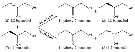
2.2. Analysis of Substrate Stereospecificity

1,2-BD contains one stereocenter and has two stereoisomers with (S)-1,2- and (R)-1,2-forms. Thus, these candidate enzymes might exhibit different substrate stereoselectivities when 1,2-BD mixture was used as substrate. To reveal the substrate stereospecificities of seven candidate enzymes, the whole cell biocatalysis method was established to replace the enzyme reaction due to its low 1,2-BD conversion rate and HB yield. The substrate of 100 mM 1,2-BD mixture supplemented in LB medium could be transformed into HB during the cultivation process when the recombinant strains were cultivated and induced by IPTG, and simultaneously NAD(P)H generated by the oxidation of 1,2-BD mixture could be utilized for cell growth and metabolism to regenerate NAD(P)+. As a result, (2R, 3R)-BDH and GDH from Serratia sp. T241 mainly converted (R)-1,2-BD into HB with the final concentration of 51.09 and 23.75 mM respectively (Table 2 and Figure S2). The (2R, 3R)-BDH enzyme exhibited higher catalytic efficiency superior to that of GDH, which was consistent to the results of enzyme activity assays (Table 1). The other five enzymes showed the ability of primarily converting (S)-1,2-BD into HB with the final concentration range from 8.77 to 40.54 mM (Table 2). However, maximum HB concentration with 40.54 mM could be achieved by the (2S, 3S)-BDH enzyme, which was inconsistent with the results of enzyme activity assay. Among the five enzymes, CbADH and CaADH:S199A exhibited higher specific activities for 1,2-BD mixture as substrate compared with that of (2S, 3S)-BDH (Table 1). A major reason was that the enzymes of CbADH and CaADH:S199A were mainly expressed in inclusion bodies in the recombinant E. coli strains, and only a small amount of soluble enzymes were obtained as shown in Figure S3. The results above implied that (2R, 3R)-BDH and (2S, 3S)-BDH from Serratia sp. T241 were suitable candidates for HB production from (R)-1,2-BD and (S)-1,2-BD respectively among seven enzymes.

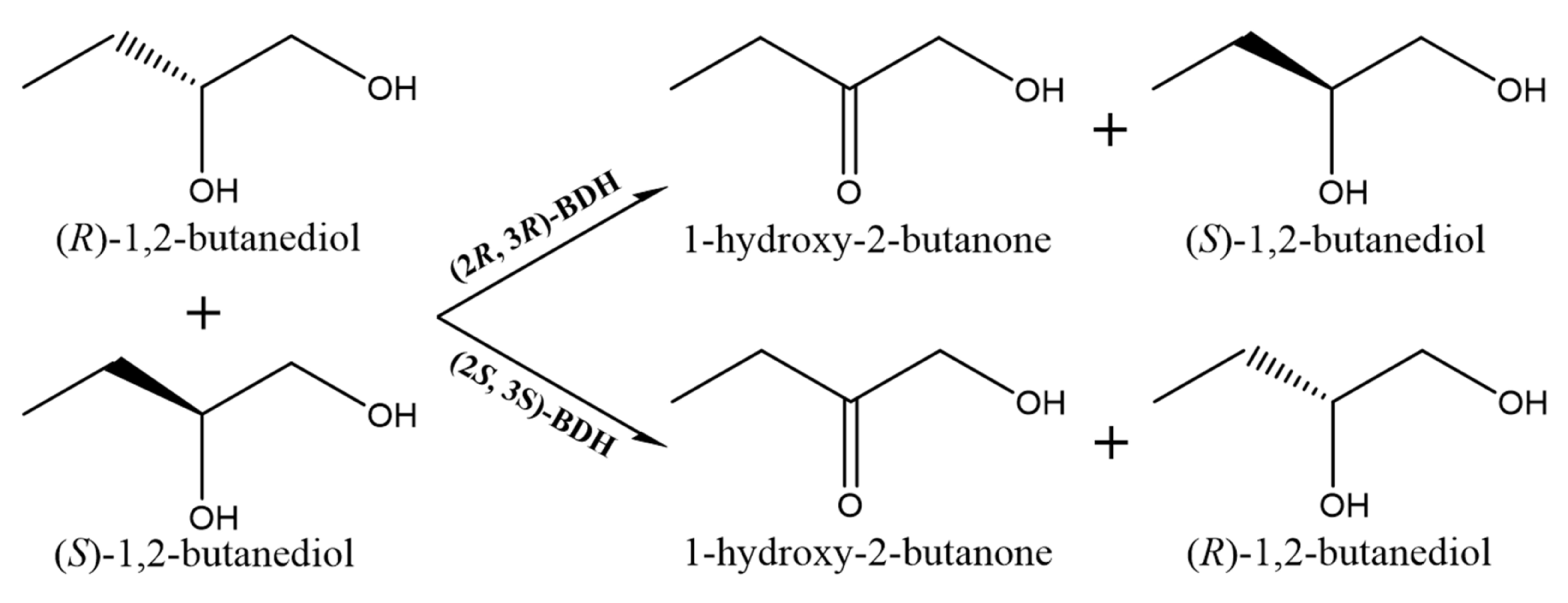
2.3. Enzymatic Properties of (2R, 3R)-BDH and (2S, 3S)-BDH for 1,2-BD Mixture

The effects of different temperature and pH value on (2R, 3R)-BDH and (2S, 3S)-BDH activity were investigated by using 50 mM 1,2-BD mixture and 0.3 mM NAD+ as substrate and coenzyme, respectively. The influence of the buffer (pH 6–12) were investigated at room temperature using 50 mM potassium phosphate buffers (pH 6–8), glycine-NaOH buffers (pH 9–10), and Na2HPO4–NaOH buffers (pH 11–12). The results indicated that the activities of two enzymes were obviously increased with the increase of pH values (Figure 1A,B). Maximum enzyme activities for the oxidation of 1,2-BD mixture by (2R, 3R)-BDH and (2S, 3S)-BDH could be observed at pH 11 and 10, respectively. The influence of temperature (20–60 °C) were assayed at pH 11 for (2R, 3R)-BDH and pH 10 for (2S, 3S)-BDH. The results in Figure 1C,D indicated that the optimum temperature for both enzymes was found to be 50 °C.
The kinetic parameters for the (2R, 3R)-BDH and (2S, 3S)-BDH enzymes were determined in a reaction system containing 10 μg purified enzyme, 0.3 mM NAD+ and 3-100 mM 1,2-BD mixture at room temperature under the optimum pH conditions. The Km and kcat values of kinetic parameters for both enzymes were calculated using non-linear regression fitting of Michaelis–Menten equation. As a result, the values of Km and kcat for the oxidation of 1,2-BD mixture as substrate were 0.97 mM and 16.61 S−1 for (2R, 3R)-BDH, 2.60 mM and 0.70 S−1 for (2S, 3S)-BDH (Table S3 and Figure S4). The kcat/Km value of 17.07 S−1 mM−1 for (2R, 3R)-BDH showed significantly higher catalytic efficiency compared with that of (2S, 3S)-BDH (0.27 S−1 mM−1) when 1,2-BD mixture was used as substrate.

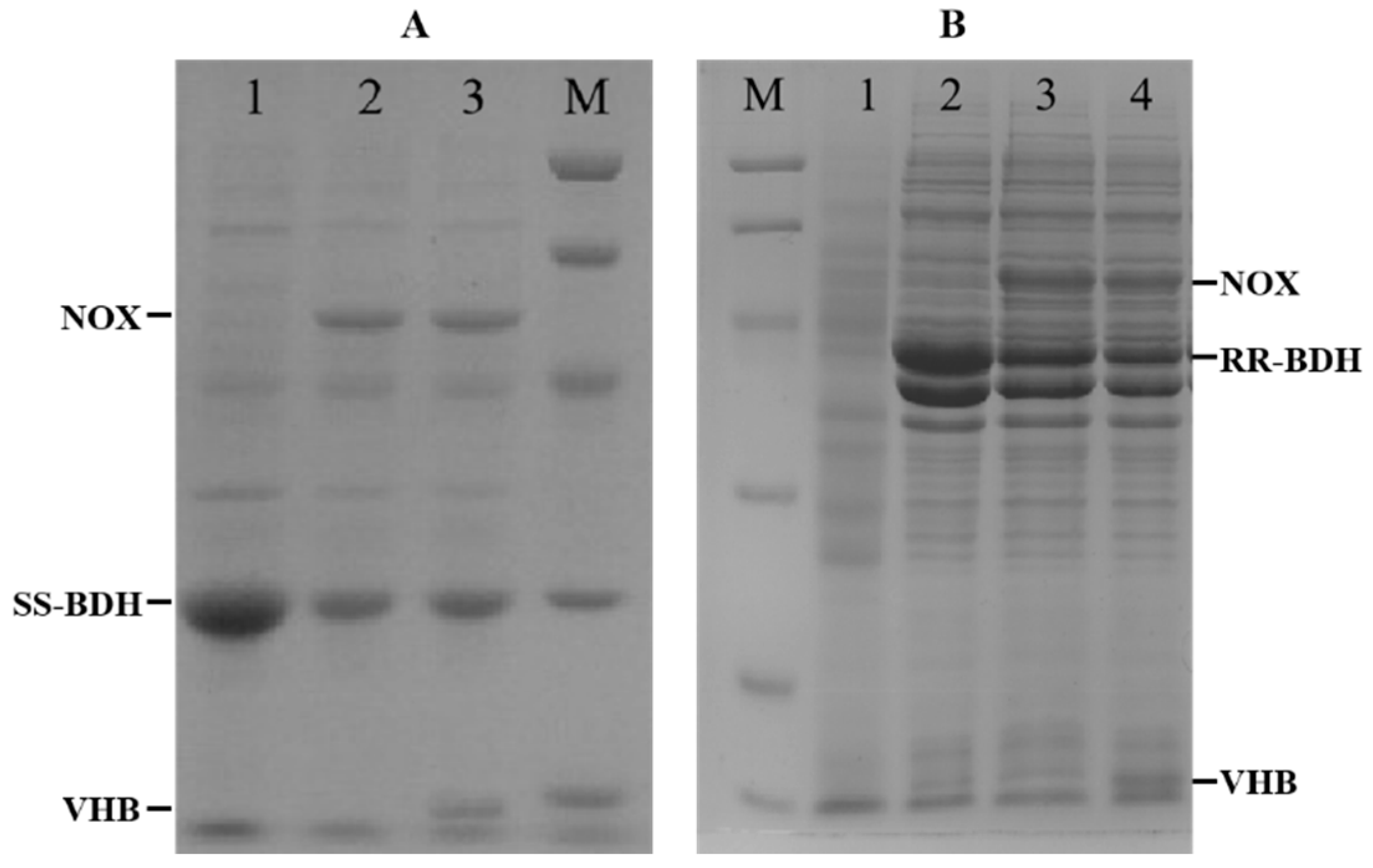
2.4. Construction of Whole Cell Biocatalysts

As described in our previous study, (2R, 3R)-BDH and (2S, 3S)-BDH from Serratia sp. T241 belonged to NAD(H)-dependent dehydrogenase [16]. For continuous HB production, the biocatalytic reaction required NAD+ regeneration via the consumption of NADH generated by the oxidation of 1,2-BD mixture. Thus, two biocatalysts including E. coli (pET-rrbdh-nox) and E. coli (pET-ssbdh-nox) carrying the genes of rrbdh/nox and ssbdh/nox respectively were developed and a ribosomal binding site (RBS) was employed for their co-expression between rrbdh/ssbdh and nox (Table S1). In the biocatalytic system, (2R, 3R)-BDH and (2S, 3S)-BDH converted 1,2-BD into HB together with generated NADH which was simultaneously oxidized into NAD+ by NOX enzyme (Figure 2). The enzyme expression levels in two biocatalysts were analyzed by using SDS-PAGE, and the E. coli strains harboring pET28a, pET-ssbdh and pET-rrbdh were used as controls. As shown in Figure 3, two bands for (2S, 3S)-BDH/NOX and (2R, 3R)-BDH/NOX were clearly observed, suggesting the genes of ssbdh/nox and rrbdh/nox could be induced by IPTG as inducer and co-expressed in E. coli. However, the co-expression of (2S, 3S)-BDH/NOX and (2R, 3R)-BDH/NOX in E. coli resulted in the decrease of expression levels for (2S, 3S)-BDH and (2R, 3R)-BDH partially due to metabolic burden of the host strain. Furthermore, the crude enzyme activities of (2S, 3S)-BDH and (2R, 3R)-BDH from two biocatalysts by ultrasonication were determined using 1,2-BD mixture as substrate at room temperature under the optimum pH, and NADH as substrate was used to assay the activity of NOX enzyme in two biocatalysts [25]. The crude enzyme extract from induced cells of E. coli (pET-ssbdh-nox) exhibited the activities of 0.25 U/mg for 1,2-BD mixture and 2.02 U/mg for NADH, whereas a higher activity of 9.02 U/mg for 1,2-BD mixture with 2.49 U/mg for NADH could be achieved by the crude enzyme from induced cells of E. coli (pET-rrbdh-nox) (Table S4). These obtained results demonstrated that two biocatalysts of E. coli (pET-ssbdh-nox) and E. coli (pET-rrbdh-nox) for co-expression of (2S, 3S)-BDH/NOX and (2R, 3R)-BDH/NOX was constructed successfully.

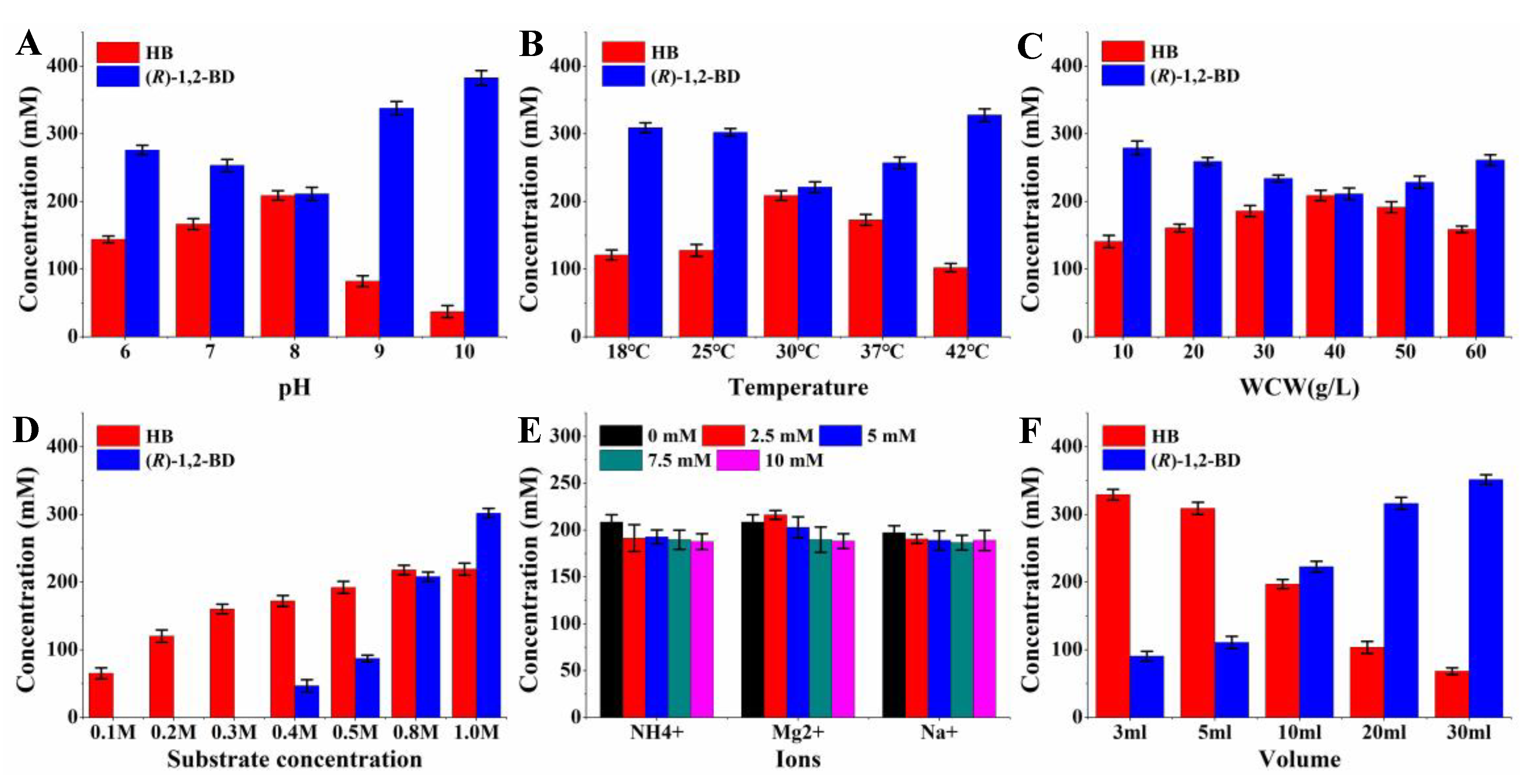
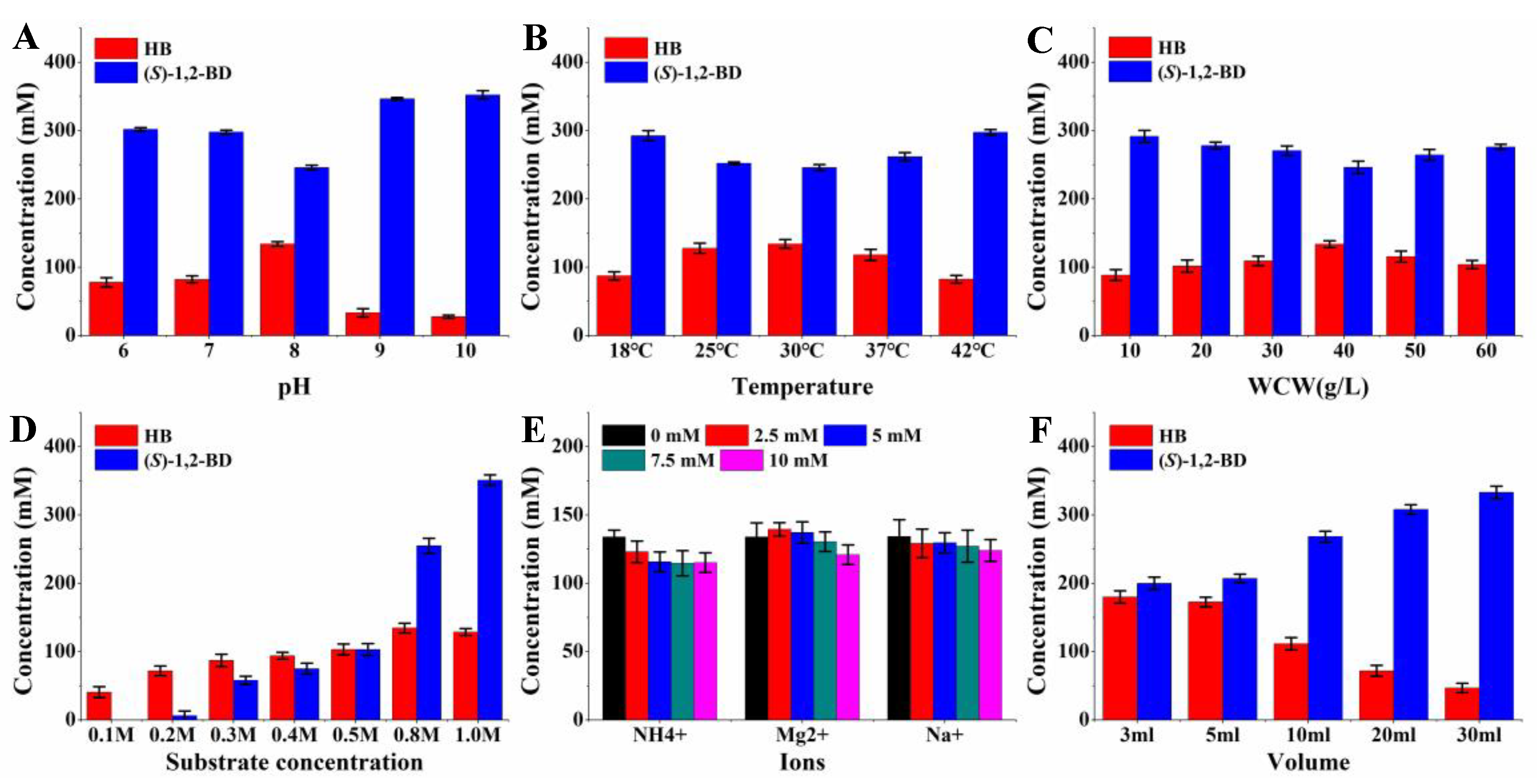
2.5. HB Production from 1,2-BD Mixture by Whole-Cell Catalysis

The initial biotransformation tests for the production of HB from 1,2-BD mixture by E. coli (pET-rrbdh) and E. coli (pET-rrbdh-nox), E. coli (pET-ssbdh) and E. coli (pET-ssbdh-nox) were conducted for 12 h in 50 mM potassium phosphate buffer (pH 8.0) with 40 g/L of wet induced cells and 800 mM 1,2-BD mixture, and the E. coli (pET28a) strain was used as control. The results showed that 202.98 and 124.59 mM of HB could be obtained from 800 mM 1,2-BD mixture (about 440 mM (R)-1,2-BD and 360 mM (S)-1,2-BD) by E. coli (pET-rrbdh-nox) cells and E. coli (pET-ssbdh-nox) cells, respectively, while the biocatalysts of E. coli (pET-rrbdh) and E. coli (pET-ssbdh) only produced 158.91- and 76.78-mM HB, implying that coenzyme regeneration could efficiently improve HB production in the reaction system (Table 3). In addition, a small amount of HB with 2.08 mM could be produced by the control of E. coli (pET28a) cells, demonstrating some non-specific dehydrogenases in E. coli might contribute to HB production via the oxidation of 1,2-BD mixture. A large amount of residual 1,2-BD in the reaction mixture required the optimization of reaction conditions to improve HB yield.
To further improve HB yield, firstly, the bioconversion conditions including pH, temperature, and WCW were optimized via single factor test by E. coli (pET-rrbdh-nox) cells and E. coli (pET-ssbdh-nox) cells. The effects of different pH values on HB production were investigated in a reaction system containing 800 mM 1,2-BD mixture and 40 g/L wet cells at 30 °C for 12 h. As shown in Figure 4A and Figure 5A, a maximum HB yield of 208.81 and 134.17 mM by E. coli (pET-rrbdh-nox) cells and E. coli (pET-ssbdh-nox) cells was achieved at pH 8.0, which was obviously lower than the optimum pH values of (2R, 3R)-BDH and (2S, 3S)-BDH (Figure 1). A probable reason is that the NOX enzyme for coenzyme regeneration showed less activity under alkaline environment [26]. Thus, the optimum pH of 8.0 was chosen to carry out the following experiments. The influence of temperature on the production of HB was determined from 18 to 42 °C. As a result, the highest HB yield by both biocatalysts could be observed at 30 °C (Figure 5B and Figure 6B), which was chosen as the optimum reaction temperature. Subsequently, different concentrations of two biocatalysts were used to evaluate its effects on the production of HB from 1,2-BD mixture under the optimum pH and temperature. The results were presented in Figure 4C and Figure 5C, which showed that the optimum WCW was 40 g/L, resulting in the HB production of 208.78 and 133.94 mM by E. coli (pET-rrbdh-nox) cells and E. coli (pET-ssbdh-nox) cells, respectively.
During the biocatalytic process, a suitable substrate concentration favored to improve product yield and avoid substrate inhibition. As shown in Figure 4D and Figure 5D, a rapid increase of HB concentration was observed with the increase of substrate concentration from 100 mM to 800 mM by both biocatalysts. The E. coli (pET-rrbdh-nox) cells could transform (R)-1,2-BD from 1,2-BD mixture completely into HB when the substrate concentration was less than 300 mM. Additionally, 100 mM (R)-1,2-BD in 1,2-BD mixture could be transformed into HB completely by the E. coli (pET-ssbdh-nox) cells (Figure 4D and Figure 5D), indicating that the catalytic efficiency of (2R, 3R)-BDH was superior to that of (2S, 3S)-BDH during the 1,2-BD oxidation process. Maximum HB yield of 219.05 and 134.59 mM could be achieved from 800 mM 1,2-BD mixture by E. coli (pET-rrbdh-nox) and E. coli (pET-ssbdh-nox) respectively. Of all the listed metal ions, the supplement of Mn2+, Zn2+, Ca2+, Cu2+, Fe2+, and Fe3+ in the reaction system caused partial precipitate. Therefore, the effects of ions including NH4+, Mg2+, and Na+ on HB production by both biocatalysts were studied by using 800 mM 1,2-BD mixture and 40 g/L wet cells under optimum pH and temperature. The results indicated that Mg2+ could slightly improve HB production at the concentration of 2.5 mM, resulting in the increase of HB yield with 3.73% and 4.14% by E. coli (pET-rrbdh-nox) and E. coli (pET-ssbdh-nox), respectively (Figure 4E and Figure 5E). In previous studies, oxygen supply was regarded as an important limiting factor in coenzyme regeneration reaction system since oxygen was a substrate of NOX enzyme [8,9]. Herein, different reaction volumes (3–30 mL) in 50 mL flasks were employed to explore the effects of oxygen supply on the production of HB from 1,2-BD mixture. As shown in Figure 4F and Figure 5F, the product yield in the biotransformation reaction remarkably decreased with the increase of the reaction volume, which showed that oxygen supply played a key limiting role on HB production from 1,2-BD mixture by both biocatalysts. Ultimately, the HB concentration of 329.22 and 179.99 mM could be achieved in 12 h by E. coli (pET-rrbdh-nox) cells and E. coli (pET-ssbdh-nox) cells when the reaction volume was 3 mL. Compared with those before optimization, the HB concentration was improved by 62.19% and 44.47% under the optimal bioconversion conditions by E. coli (pET-rrbdh-nox) and E. coli (pET-ssbdh-nox), respectively. However, two biocatalysts could not convert 1,2-BD mixture into HB completely. Two potential reasons for explanation might be involved in the reaction system. On one hand, two enzymes of (2S, 3S)-BDH and (2R, 3R)-BDH in both biocatalysts were reversible enzymes with the ability of interconversion between 1,2-BD and HB [13,27,28]. Alkaline environment favored the oxidation of alcohol into ketone, which was readily reduced to produce alcohol under acidic reaction mixture [11,14,16]. In the current reaction system, the relatively low pH compared with the optimum pH vales of (2S, 3S)-BDH and (2R, 3R)-BDH was used to maintain the activity of NOX enzyme in two biocatalysts. Currently, available NOX enzymes reported in previous studies exhibited excellently catalytic efficiency under acidic environment and low activities under alkaline condition. Therefore, shifting the optimum pH value for NOX enzymes might be a feasible strategy to improve HB production from 1,2-BD mixture. On the other hand, two stereo isomers existed in 1,2-BD mixture may interfere with the bioconversion reaction each other via substrate mass transfer and/or substrate inhibition since (S)-1,2-BD and (R)-1,2-BD were specific substrates for (2S, 3S)-BDH and (2R, 3R)-BDH respectively. Further experiments were necessary for finding the reasons in future.

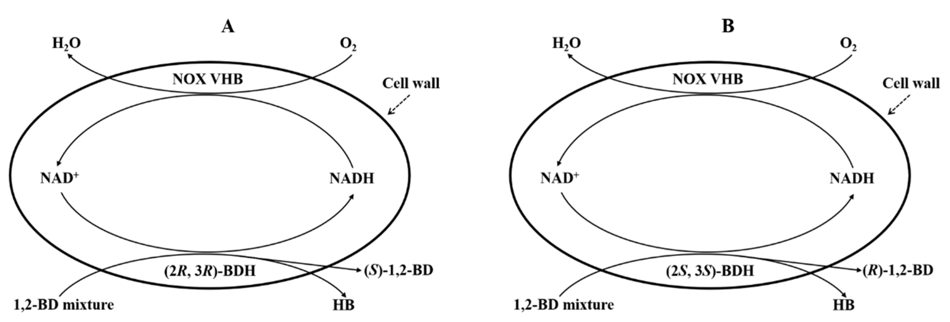
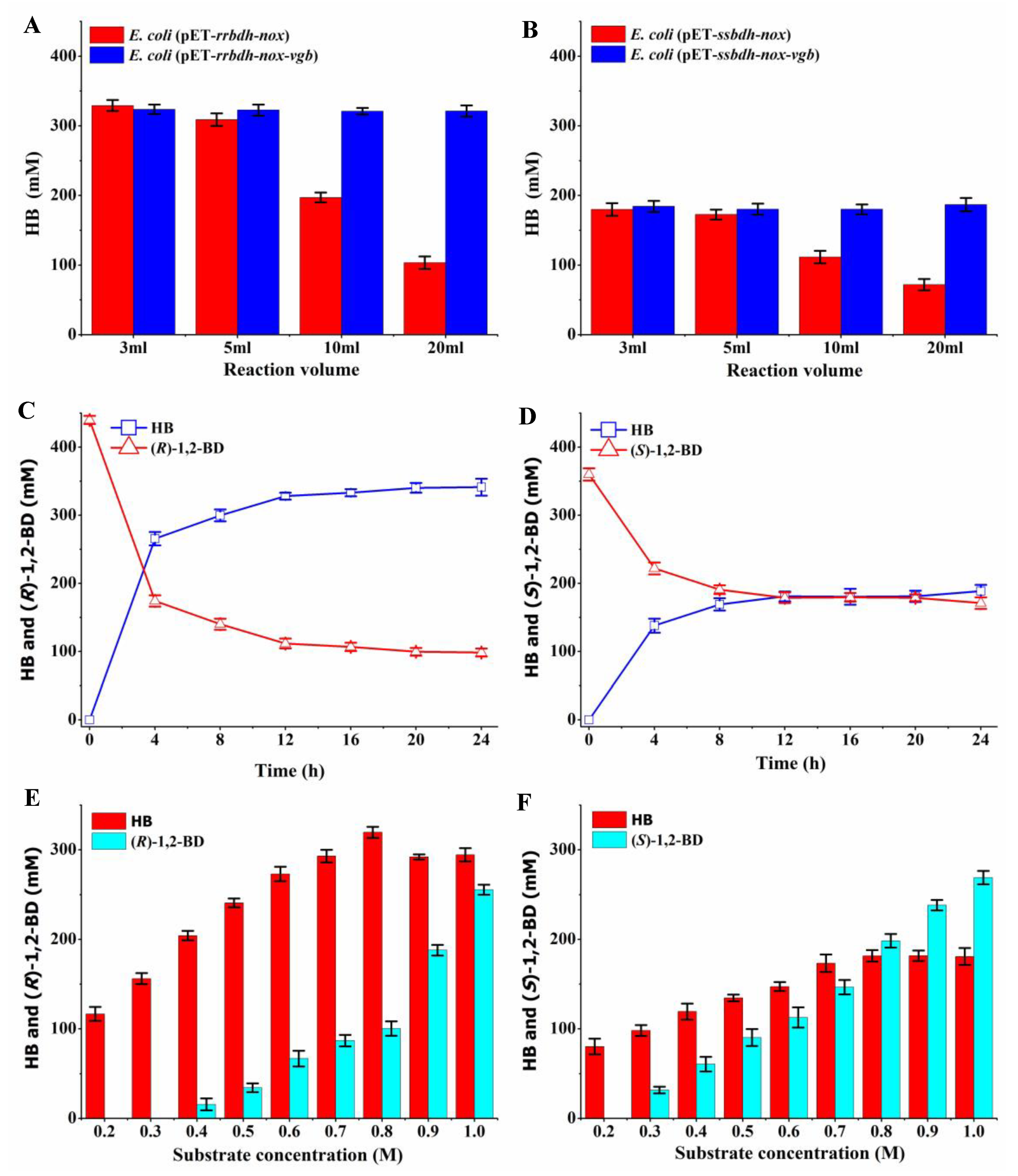
2.6. Effects of VHB Expression on the Production of HB from 1,2-BD Mixture

As observed in Figure 4F and Figure 5F, HB production from 1,2-BD mixture by two biocatalysts during the biocatalytic process was significantly dependent on oxygen supply, which might be an important limiting factor for scale-up production in industry. Our previous studies indicated that VHB protein co-expressed with BDH and NOX in E. coli could efficiently improve the yield of AC from 2,3-BD under oxygen supply-limited condition [8,9]. Therefore, another two new biocatalysts of E. coli (pET-rrbdh-nox-vgb) and E. coli (pET-ssbdh-nox-vgb) expressing VHB protein encoded by the vgb gene from Vitreoscilla were developed. As observed in Figure 3, three encoded proteins in E. coli (pET-rrbdh-nox-vgb) cells and E. coli (pET-ssbdh-nox-vgb) cells were successfully co-expressed, and the corresponding enzyme activities could be detected as shown in Table S4. Under the optimum bioconversion conditions, the effects of VHB co-expression on the production of HB from 1,2-BD mixture by E. coli (pET-rrbdh-nox-vgb) cells and E. coli (pET-ssbdh-nox-vgb) cells were determined under different reaction volumes, and E. coli (pET-rrbdh-nox) and E. coli (pET-ssbdh-nox) were used as the controls. The results (Figure 6A,B) showed that HB production from 1,2-BD mixture by E. coli (pET-rrbdh-nox-vgb) and E. coli (pET-ssbdh-nox-vgb) was kept at constant concentration of about 320 and 180 mM, respectively, under different reaction volumes (3–20 mL), demonstrating that the co-expression of VHB protein in E. coli (pET-rrbdh-nox) and E. coli (pET-ssbdh-nox) improved oxygen supply and thus favored scale-up HB production. The time-course profiles of HB production from 1,2-BD mixture by E. coli (pET-rrbdh-nox-vgb) and E. coli (pET-ssbdh-nox-vgb) were given in Figure 6C,D. A rapid increase of HB concentration with the decrease of the corresponding substrate ((R)-1,2-BD and (S)-1,2-BD) could be observed before 8 h. Thereafter, HB production by the two biocatalysts grew slowly until 24 h. Ultimately, maximum HB concentration of 341.35 and 188.80 mM could be achieved from 440 mM (R)-1,2-BD and 360 mM (S)-1,2-BD by E. coli (pET-rrbdh-nox-vgb) and E. coli (pET-ssbdh-nox-vgb), respectively. The residual substrate with 98.65 mM (R)-1,2-BD and 171.20 mM (S)-1,2-BD in two reaction systems could not be transformed into HB during the whole biocatalytic process. Thus, different substrate concentrations in the range of 0.2–1.0 M were used to evaluate the yield by the two biocatalysts. The results showed that (R)-1,2-BD in 300 mM 1,2-BD mixture could be completely converted into HB by the E. coli (pET-rrbdh-nox-vgb) biocatalyst, which resulted in 164.21 mM HB and 135.68 mM enantiopure (S)-1,2-BD with 100% purity (Figure 6E and Figure S5). Similar results were also observed in Figure 6F and Figure S6. In total, 200 mM of 1,2-BD mixture could be converted into HB (86.34 mM) by E. coli (pET-ssbdh-nox-vgb), and the enantiopure (R)-1,2-BD of 112.43 mM with 100% purity was retained in the reaction system. These results showed that the two biocatalysts of E. coli (pET-rrbdh-nox-vgb) and E. coli (pET-ssbdh-nox-vgb) provided a feasible strategy for chiral resolution of (R)-1,2-BD and (S)-1,2-BD except HB production from 1,2-BD mixture.

3. Materials and Methods

3.1. Strains, Plasmids, Primers, and Chemicals

The strains, plasmids, and primers used in this work were listed in Table 1 and Table S1. All the strains in this work were cultivated in Luria-Bertani (LB) medium. Antibiotics were used to maintain the plasmid in E. coli with 50 μg/mL kanamycin or 100 μg/mL ampicillin. Primers synthesis for PCR amplification and commercial sequencing were performed by Sangon Biotech (Shanghai, China). Enzymes and other reagents molecular manipulation were obtained from TaKaRa Biotech (Dalian, China) and YEASEN (Shanghai, China). The plasmid preparation and genomic DNA extraction were carried out by using Mini-prep Kits (OMEGA, Shanghai, China). The chemical standards of (S)-1,2-BD (99%), (R)-1,2-BD (99%), and HB (99%) were from Sinopharm (Beijing, China). The reaction substrate of 1,2-BD mixture (about 45% (S)-1,2-BD, and 55% (R)-1,2-BD) was obtained from TCI (Tokyo Chemical Industry Co., Ltd., Tokyo, Japan). All other chemicals were purchased from Sigma-Aldrich (Shanghai, China).

3.2. Recombinant Enzyme Expression and Purification

The genes encoding (2S, 3S)-BDH, meso-BDH, (2R, 3R)-BDH, and GDH were PCR-amplified using the genomic DNA template of Serratia sp. T241 with the primers in Table S1. The PCR-amplified products were purified and then inserted into the plasmid pET-28a (+) at the restriction sites of EcoRI and HindIII, generating the recombinant expression plasmids designated as pET-ssbdh, pET-mbdh, pET-rrbdh, and pET-gdh, respectively. The genes for CbADH from Clotridium beijerinckii and CbADH from Clostridium autoethanogenum were synthesized and cloned into pET28a vector by General Biosystems, Inc. (Anhui, China), resulting in the recombinant plasmid pET-Caadh and pET-Cbadh. The CbADH:S199A variant was constructed by using site-directed mutagenesis via PCR-amplification of whole plasmid pET28a-Caadh. All the obtained recombinant plasmids were confirmed through restriction analysis and DNA commercial sequencing, and then introduced into competent E. coli BL21(DE3) cells for enzyme expression using heat shock transformation.
For recombinant enzyme expression, the E. coli strains harboring pET-ssbdh, pET-mbdh, pET-rrbdh, pET-gdh, pET-Caadh, pET-Cbadh, and pET-Caadh:S199A were cultured at 37 °C in a 500-mL conical flask containing 100 mL fresh LB medium for 2.5 h, and subsequently supplemented isopropyl-beta-D-thiogalactopyranoside (IPTG) to 0.5 mM for protein expression. The cells were harvested by centrifugation after induction at 18 °C for 24 h. The cell precipitates were resuspended and treated by ultrasonication in an ice bath. The crude enzyme was obtained by centrifugation at 13,000 × g for 10 min to remove the debris for further purification. The purification of seven recombinant enzymes was performed by using a Ni-NTA superflow column according to the purification protocol (Qiagen, Hilden, Germany). The eluates were desalted and concentrated by Amicon Ultra-15 (Merck Millipore Ltd., German). These purified enzymes were then analyzed using SDS-PAGE, and assayed for the activity of 1,2-BD oxidation.

3.3. Enzyme Assays

For the activity assays of seven candidate enzymes, the reaction mixtures consisting of 50 mM 1,2-BD mixture, and 0.3 mM NAD+ for (2S, 3S)-BDH, meso-BDH, (2R, 3R)-BDH, and GDH or 0.3 mM NADP+ for CbADH, CbADH, and CbADH:S199A were prepared in 1 mL potassium phosphate buffer (50 mM, pH 8.0). The purified enzyme (20 μL) was added into the reaction mixture to initiate the reaction at room temperature, and the reduction of NAD(P)+ to NAD(P)H was monitored spectrophotometrically as the absorbance increase at 340 nm (ε340 = 6.22 mM−1 cm−1). Assays of the enzyme activities were performed in triplicate. One unit of the enzyme activity was defined as the amount of enzyme to catalyze 1μmol of NAD(P)+ in one minute. The concentrations of all the tested enzymes were determined by using BCA assay kit from Tiangen (Shanghai, China) with bovine serum albumin served as a standard sample. The activity of NOX enzyme was determined as described previously [25].
The enzyme kinetic parameters of (2R, 3R)-BDH and (2S, 3S)-BDH were determined by using a reaction system containing 0.3 mM NAD+, 10 μg purified enzyme, and 3–100 mM 1,2-BD mixture at room temperature under the corresponding optimum pH. All assays were determined in triplicate. The Km and kcat values of kinetic parameters for both enzymes were calculated using the Michealis–Menten equation and the non-linear regression fitting.

3.4. Analysis of Substrate Stereospecificity

The whole cell biocatalysis method was used to investigate the substrate stereospecificity of seven candidate enzymes. In brief, the recombinant strains were cultivated at 37 °C in LB medium with the substrate of 100 mM 1,2-BD (45% (S)-1,2-BD, and 55% (R)-1,2-BD). After 2.5 h cultivation, 0.5 mM ITPG was supplemented into the culture medium for inducing enzyme expression at 25 °C for 10h. Meanwhile, the expressed enzymes could convert the substrate 1,2-BD mixture into HB during the cultivation process. The residual substrate ((S)-1,2-BD, and (R)-1,2-BD) in the broth were analyzed and quantified by a GC system equipped with a flame ionization detector (Agilent 7820A, anta Clara, California, USA) and a chiral column of Supelco β-DEX™ 120 (30-m length, 0.25-mm inner diameter) using nitrogen as carrier gas. Prior to analysis, the supernatant after centrifuge at 13,000 × g for removing the cells was extracted by using ethyl acetate with equal volume as well as n-butanol as internal standard added. The GC operation conditions were as follows: injector temperature of 250 °C and detector temperature of 300 °C were set, respectively; column temperature firstly was maintained at 80 °C for 5 min, and then programed with an increment of 2 °C min−1 to 150 °C. The product of HB in the broth was determined and quantified by a HPLC system (Ultimate 3000, Thermo Fisher Scientific, Waltham, MA, USA) equipped with a Carbomix H-NP column (7.8 × 300 mm, 10 μm) at the flow rate of 0.5 mL/min and the wavelength of 191 nm in a mobile phase of 1% phosphoric acid. The loading volume of the sample after filtration was 5 μL, and the column temperature was controlled at 70 °C. The concentrations of (S)-1,2-BD, (R)-1,2-BD and HB in the broth were quantified by using calibration curves.

3.5. Construction of Whole Cell Biocatalysts

The resources including strains, and plasmids used in this work were shown in Table 4 and the primers were given in Table S1. The genomic DNA of Serratia sp. T241 extracted by using the Bacterial Genomic DNA Kit of OMEGA were used as template for amplifying the rrbdh and ssbdh genes. The nox and vgb genes encoding NADH oxidase and VHB protein was derived from the pET-nox (GenBank ID: AAN04047) and pBR322-vgb (GenBank ID: KM108313.1) vectors developed in our previous study [8,9]. The detailed information of gene sequences was presented in Table S2. Hieff Clone Plus Multi One Step Cloning Kit (YEASEN, Shanghai, China) was used to develop co-expressing plasmids of pET-rrbdh-nox, pET-rrbdh-nox-vgb, pET-ssbdh-nox, and pET-ssbdh-nox-vgb according to the provided protocol. Briefly, the genes of rrbdh and nox were PCR-amplified using the designed primers of P1/P2 and P3/P4 with adjacent oligos overlapped by 15–25 bp at the end of each fragment. The amplified products were purified and assembled into the vector pET28a linearized by double digestion of BamHI and HindIII, creating the recombinant plasmid pET-rrbdh-nox. Similarly, the plasmids of pET-rrbdh-nox-vgb, pET-ssbdh-nox, and pET-ssbdh-nox-vgb were constructed by using the corresponding primers of P5-P20 listed in Table S1. The obtained recombinant plasmids were transformed into E. coli BL21(DE3) cells, and subsequently induced by the addition of IPTG. The enzyme expression and activity in the induced cells was analyzed by SDS-PAGE and assayed by using 1,2-BD mixture as substrate.

3.6. HB Production from 1,2-BD Mixture by Whole-Cell Catalysis

The initial biotransformation tests for the production of HB from 1,2-BD mixture by E. coli (pET-rrbdh), E. coli (pET-rrbdh-nox), E. coli (pET-ssbdh), and E. coli (pET-ssbdh-nox) were carried out in 10 mL reaction system (50 mM potassium phosphate buffer (pH 8.0), 40 g/L of wet induced cells, 800 mM 1,2-BD mixture) in 100 mL conical flask at 30 °C for 12 h, and the E. coli (pET28a) strain was used as control. To improve the bioconversion rate of 1,2-BD mixture for HB production, six factors including reaction pH (6–10), reaction temperature (18–42 °C), wet induced cell weight (WCW, 10–60 g/L), 1,2-BD mixture concentration (0.1–1 M), ions (Mn2+, Zn2+, Ca2+, Cu2+, Fe2+, Fe3+, NH4+, Mg2+, Na+ at the final concentration of 0–10 mM), and reaction volume (3–30 mL) were optimized by single factor tests by E. coli (pET-rrbdh-nox) cells and E. coli (pET-ssbdh-nox) cells. The optimization of reaction volume was conducted in 50 mL flask, and the experiments of the other five factors were carried out in 10 mL centrifuge tube containing 1 mL reaction mixture. All the biotransformation tests were carried out in triplicate. The substrate and product in the reaction solution were analyzed and quantified by using GC and HPLC system respectively.

3.7. Effect of VHB Co-Expression on HB Production from 1,2-BD Mixture

The effects of VHB co-expression on the production of HB from 1,2-BD mixture were investigated using different reaction volume (3–20 mL) in a 50 mL flask under the optimized bioconversion conditions by E. coli (pET-rrbdh-nox-vgb) cells and E. coli (pET-ssbdh-nox-vgb) cells, and the recombinant strains of E. coli (pET-rrbdh-nox) and E. coli (pET-ssbdh-nox) were used as the control, respectively. The substrate and product in the reaction solutions were quantified and compared by using GC and HPLC system. All the experiments were conducted in triplicate.

4. Conclusions

In this study, two enzymes of (2R, 3R)-BDH and (2S, 3S)-BDH from Serratia sp. T241 were identified and exhibited the abilities in the oxidation of 1,2-BD to HB. Substrate stereospecificity assays indicated that (R)-1,2-BD and (S)-1,2-BD were the substrates of (2R, 3R)-BDH and (2S, 3S)-BDH, respectively. Furthermore, two biocatalysts of E. coli (pET-rrbdh-nox-vgb) and E. coli (pET-ssbdh-nox-vgb) were developed for HB biosynthesis from 1,2-BD mixture. Under the optimized bioconversion conditions, 341.35 and 188.80 mM of HB could be achieved from 440 mM (R)-1,2-BD and 360 mM (S)-1,2-BD in 800 mM 1,2-BD mixture by E. coli (pET-rrbdh-nox-vgb) and E. coli (pET-ssbdh-nox-vgb), respectively. In addition, the two biocatalysts exhibited the ability in chiral resolution of (R)-1,2-BD and (S)-1,2-BD in 1,2-BD mixture. 135.68 mM (S)-1,2-BD and 112.43 mM (R)-1,2-BD with the purity of 100% could be obtained from 300 and 200 mM 1,2-BD mixture by E. coli (pET-rrbdh-nox-vgb) and E. coli (pET-ssbdh-nox-vgb), respectively. These results provided potential application for HB production from 1,2-BD mixture and chiral resolution of (R)-1,2-BD and (S)-1,2-BD.

Supplementary Materials

The following are available online at https://www.mdpi.com/article/10.3390/catal11101184/s1, Figure S1. Analysis of expression and purification for SSBDH, GDH, RRBDH, mBDH (A), and CaADH, CaADH:S199A, CbADH (B) by SDS-PAGE. (A) Lane M, marker (97.2, 66.4, 44.3, 29, 20.01, 14.3 kDa); Lane 1-4, SSBDH (control, whole cell, soluble protein, and purified enzyme); Lane 5–8, GDH (whole cell, soluble protein, and purified enzyme); Lane 9–12, RRBDH (control, whole cell, soluble protein, and purified enzyme); Lane 13–15, mBDH (whole cell, soluble protein, and purified enzyme); (B) Lane M, marker (97.2, 66.4, 44.3, 29, 20.01, 14.3 kDa); Lane 1–4, CaADH, (control, whole cell, soluble protein, and purified enzyme); Lane 5–7, CaADH:S199A, (whole cell, soluble protein, and purified enzyme); Lane 8–11, CbADH (control, whole cell, soluble protein, and purified enzyme), Figure S2: Chiral analysis of the product in the bioconversion reactions by whole cell biocatalysis. (A) Standard chemical; (B) E. coli (pET28a); (C) E. coli (pET-rrbdh); (D) E. coli (pET-gdh); (E) E. coli (pET-ssbdh); (F) E. coli (pET-mbdh); (G) E. coli (pET-Caadh); (H) E. coli (pET-Caadh:S199A); (I) E. coli (pET-Cbadh), Figure S3: Analysis of expression level for RRBDH, SSBDH, GDH, mBDH, CaADH, CaADH:S199A, and CbADH in E. coli by SDS-PAGE. A: Lane M, marker (97.2, 66.4, 44.3, 29, 20.01, 14.3 kDa); Lane 1–3, RRBDH (whole cell, soluble protein, insoluble protein); Lane 4–6, SSBDH (whole cell, soluble protein, insoluble protein); Lane 7–9, GDH (whole cell, soluble protein, insoluble protein); Lane 10–12, mBDH, (whole cell, soluble protein, insoluble protein); B: Lane M, marker (97.2, 66.4, 44.3, 29, 20.01, 14.3 kDa); Lane 1–3, CaADH (whole cell, soluble protein, insoluble protein); Lane 4–6, CaADH:S199A (whole cell, soluble protein, insoluble protein); Lane 7–9, CbADH (whole cell, soluble protein, insoluble protein), Figure S4: Analysis of kinetic parameters for (2R, 3R)-BDH and (2S, 3S)-BDH using 1,2-BD mixture as substrate, Figure S5: Chiral resolution of (S)-1,2-BD from 300 mM (A) and 400 mM (B) 1,2-BD mixture by E. coli (pET-rrbdh-nox-vgb), Figure S6: Chiral resolution of (R)-1,2-BD from 200 mM (A) and 300 mM (B) 1,2-BD mixture by E. coli (pET-ssbdh-nox-vgb), Table S1: The primers used in this study, Table S2: Sequence information of the genes used in this study, Table S3: Kinetic parameters of (2R, 3R)-BDH and (2S, 3S)-BDH for 1,2-BD mixture, Table S4. Enzyme activities of the recombinant E. coli strains.

Author Contributions

L.Z. and B.H. conceived this study and designed the experiments; H.L. and J.X. developed all the recombinant strains and performed biocatalytic experiments; W.S. and H.G. determined enzyme activity; K.H. provided technology support; W.H. and J.Q. performed GC and HPLC analysis; B.H. and L.Z. wrote and edited the manuscript. All authors have read and agreed to the published version of the manuscript.

Funding

This work was supported by the National Natural Science Foundation of China (no. 81673542 and no. U1803232), the National Key R&D Program of China (no. 2017YFE0122000), and Key Project from Fujian Provincial Department of Science and Technology (no. 2020N5005).

Conflicts of Interest

The authors declare no competing financial interest.

References

  1. Chen, F.F.; Cosgrove, S.C.; Birmingham, W.R.; Mangas-Sanchez, J.; Citoler, J.; Thompson, M.P.; Zheng, G.W.; Xu, J.H.; Turner, N.J. Enantioselective synthesis of chiral vicinal amino alcohols using amine dehydrogenases. ACS Catal. 2019, 9, 11813–11818. [Google Scholar] [CrossRef]
  2. Wang, H.Y.; Zhu, C.H.; Liu, Q.Y.; Tan, J.; Wang, C.G.; Liang, Z.; Ma, L.L. Selective Conversion of cellulose to hydroxyacetone and 1-hydroxy-2-butanone with Sn-Ni bimetallic catalysts. ChemSusChem 2019, 12, 2154–2160. [Google Scholar] [CrossRef]
  3. Sato, S.; Takahashi, R.; Sodesawa, T.; Fukuda, H.; Sekine, T.; Tsukuda, E. Synthesis of a-hydroxyketones from 1,2-diols over Cu-based catalyst. Catal. Commun. 2005, 6, 607–610. [Google Scholar] [CrossRef]
  4. Lee, L.G.; Whitesides, G.M. Preparation of optically active 1,2-diols and α-hydroxy ketones using glycerol dehydrogenase as catalyst: Limits to enzyme-catalyzed synthesis due to noncompetitive and mixed inhibition by product. J. Org. Chem. 1986, 51, 25–36. [Google Scholar] [CrossRef]
  5. Hu, N.J.; Li, S.Y.; Liu, Y.C. Recent advances in biocatalysis and metabolic engineering. Catalysts 2021, 11, 1052. [Google Scholar] [CrossRef]
  6. Hohn-Bentz, H.; Radler, F. Bacterial 2,3-butanediol dehydrogenases. Arch. Microbiol. 1983, 116, 197–203. [Google Scholar] [CrossRef]
  7. Xu, G.C.; Bian, Y.Q.; Han, R.Z.; Dong, J.J.; Ni, Y. Cloning, expression, and chararacterization of budC gene encoding meso-2,3-butanediol dehydrogenase from Bacillus licheniformis. Appl. Biochem. Biotechnol. 2016, 178, 604–717. [Google Scholar] [CrossRef] [PubMed]
  8. Guo, Z.W.; Zhao, X.H.; He, Y.Z.; Yang, T.X.; Gao, H.F.; Li, G.X.; Chen, F.X.; Sun, M.J.; Lee, J.K.; Zhang, L.Y. Efficient (3R)-acetoin production from meso-2,3-butanediol using a new whole-cell biocatalyst with co-expression of meso-2,3-butanediol dehydrogenase, NADH oxidase and Vitreoscilla hemoglobin. J. Microbiol. Biotechnol. 2017, 27, 92–100. [Google Scholar] [CrossRef] [PubMed] [Green Version]
  9. He, Y.Z.; Chen, F.X.; Sun, M.J.; Gao, H.F.; Guo, Z.W.; Lin, H.; Chen, J.B.; Jin, W.S.; Yang, Y.L.; Zhang, L.Y.; et al. Efficient (3S)-acetoin and (2S, 3S)-2,3-butanediol production from meso-2,3-butanediol using whole-cell biocatalysis. Molecules 2018, 23, 691. [Google Scholar] [CrossRef] [Green Version]
  10. Zhang, L.Y.; Singh, R.; Sivakumar, D.; Guo, Z.W.; Li, J.H.; Chen, F.B.; He, Y.Z.; Guan, X.; Kang, Y.C.; Lee, J.K. An artificial synthetic pathway for acetoin, 2,3-butanediol and 2-butanol production from ethanol using cell free multi-enzymes catalysis. Green Chem. 2018, 20, 230–242. [Google Scholar] [CrossRef]
  11. Cui, Z.Z.; Zhao, Y.J.; Mao, Y.F.; Shi, T.; Lu, L.X.; Ma, H.W.; Wang, Z.W.; Chen, T. In vitro biosynthesis of optically pure D-(-)-acetoin from meso-2,3-butanediol using 2,3-butanediol dehydrogenase and NADH oxidase. J. Chem. Technol. Biotechnol. 2019, 94, 2547–2554. [Google Scholar] [CrossRef]
  12. Subramanian, V.; Lunin, V.V.; Farmer, S.J.; Alahuhta, M.; Moore, K.T.; Ho, A.; Chaudhari, V.B.; Zhang, M.; Himmel, M.E.; Decker, S.R. Phylogenetics-based identification and characterization of a superior 2,3-butanediol dehydrogenase for Zymomonas mobilis expression. Biotechnol. Biofuels 2020, 13, 186. [Google Scholar] [CrossRef] [PubMed]
  13. Yu, B.; Sun, J.B.; Bommareddy, R.R.; Song, L.F.; Zeng, A.P. Novel (2R, 3R)-2,3-butanediol dehydrogenase from potential industrial strain Paenibacillus polymyxa ATCC 12321. Appl. Environ. Microb. 2011, 77, 4230–4233. [Google Scholar] [CrossRef] [Green Version]
  14. Zhang, L.Y.; Xu, Q.M.; Zhan, S.R.; Li, Y.Y.; Lin, H.; Sun, S.J.; Sha, L.; Hu, K.H.; Guan, X.; Shen, Y.L. A new NAD(H)-dependent meso-2,3-butanediol dehydrogenase from an industrially potential strain Serratia marcescens H30. Appl. Microbiol. Biotechnol. 2014, 98, 1175–1184. [Google Scholar] [CrossRef] [PubMed]
  15. Chen, C.; Wei, D.; Shi, J.P.; Wang, M.; Hao, J. Mechanism of 2,3-butanediol stereoisomer formation in Klebsiella pneumoniae. Appl. Microbiol. Biotechnol. 2014, 98, 4603–4613. [Google Scholar] [CrossRef]
  16. Zhang, L.Y.; Guo, Z.W.; Chen, J.B.; Xu, Q.M.; Lin, H.; Hu, K.H.; Guan, X.; Shen, Y.L. Mechanism of 2,3-butanediol stereoisomers formation in a newly isolated Serratia sp. T241. Sci. Rep. 2016, 6, 19257. [Google Scholar] [CrossRef] [PubMed] [Green Version]
  17. Pasaye-Anaya, L.; Vargas-Tah, A.; Martinez-Camara, C.; Castro-Montoya, A.J.; Campos-Garcia, J. Production of 2,3-butanediol by fermentation of enzymatic hydrolysed bagasse from agave mezcal-waste using the native Klebsiella oxytoca UM2-17 strain. J. Chem. Technol. Biotechnol. 2019, 94, 3915–3923. [Google Scholar] [CrossRef]
  18. Isobe, K.; Wakao, N. Thermostable NAD+-dependent (R)-specific secondary alcohol dehydrogenase from cholesterol-utilizing Burkholderia sp. AIU 652. J. Biosci. Bioeng. 2003, 96, 387–393. [Google Scholar] [CrossRef]
  19. Wang, Y.; Tao, F.; Xu, P. Glycerol dehydrogenase plays a dual role in glycerol metabolism and 2,3-butanediol formation in Klebsiella pneumoniae. J. Biol. Chem. 2014, 289, 6080–6090. [Google Scholar] [CrossRef] [Green Version]
  20. Zhang, L.Y.; Xu, Q.M.; Peng, X.Q.; Xu, B.H.; Wu, Y.H.; Yang, Y.L.; Sun, S.J.; Hu, K.H.; Shen, Y.L. Cloning, expression and characterization of glycerol dehydrogenase involved in 2,3-butanediol formation in Serratia marcescens H30. J. Ind. Microbiol. Biotechnol. 2014, 41, 1319–1327. [Google Scholar] [CrossRef]
  21. Marshall, J.H.; May, J.W.; Sloan, J. Purification and properties of glycerol: NAD+ 2-oxidoreductase (glycerol dehydrogenase) from Schizosaccharomyces pombe. Microbiology 1985, 131, 1581–1588. [Google Scholar] [CrossRef] [Green Version]
  22. Yan, Y.J.; Lee, C.C.; Liao, J.C. Enantioselective synthesis of pure (R,R)-2,3-butanediol in Escherichia coli with stereospecific secondary alcohol dehydrogenase. Org. Biomol. Chem. 2009, 7, 3914–3917. [Google Scholar] [CrossRef]
  23. Kopke, M.; Gerth, M.L.; Maddock, D.J.; Mueller, A.P.; Liew, F.; Simpson, S.D.; Patrick, W.M. Reconstruction of an acetogenic 2,3-butanediol pathway involving a novel NADPH-dependent primary-secondary alcohol dehydrogenase. Appl. Environ. Microb. 2014, 80, 3394–3403. [Google Scholar] [CrossRef] [Green Version]
  24. Maddock, D.J.; Patrick, W.M.; Gerth, M.L. Substitutions at the cofactor phosphate-binding site of a clostridial alcohol dehydrogenase lead to unexpected changes in substrate specificity. Protein. Eng. Des. Sel. 2015, 28, 251–258. [Google Scholar] [CrossRef] [Green Version]
  25. Xiao, Z.J.; Lv, C.J.; Gao, C.; Qin, J.Y.; Ma, C.Q.; Liu, Z.; Liu, P.H.; Li, L.X.; Xu, P. A novel whole-cell biocatalyst with NAD+ regeneration for production of chiral chemicals. PLoS ONE 2010, 5, e8860. [Google Scholar] [CrossRef] [Green Version]
  26. Geueke, B.; Riebel, B.; Hummel, W. NADH oxidase from Lactobacillus brevis: A new catalyst for the regeneration of NAD. Enzyme Microb. Tech. 2003, 32, 205–211. [Google Scholar] [CrossRef]
  27. Takusagawa, Y.; Otagiri, M.; Ui, S.; Ohtsuki, T.; Mimura, A.; Ohkuma, M.; Kudo, T. Purification and characterization of L-2,3-butanediol dehydrogenase of Brevibacterium saccharolyticum C-1012 expression in Escherichia coli. Biosci. Biotechnol. Biochem. 2001, 65, 1876–1878. [Google Scholar] [CrossRef] [PubMed] [Green Version]
  28. Ui, S.; Otagiri, M.; Mimura, A.; Dohmae, N.; Takio, K.; Ohkuma, M.; Kudo, T. Cloning, expression and nucleotide sequence of the L-2,3-butanediol dehydrogenase gene from Brevibacterium saccharolyticum C-1012. J. Ferment. Bioeng. 1998, 86, 290–295. [Google Scholar] [CrossRef]
Figure 1. Effects of pH and temperature on enzyme activities of (2S, 3S)-BDH (A,C) and (2R, 3R)-BDH (B,D) using 1,2-BD mixture as substrate.
Catalysts 11 01184 g001
Figure 2. Two whole cell biocatalysts of recombinant E. coli with co-expression of (2R, 3R)-BDH/NOX/VHB (A) and (2S, 3S)-BDH/NOX/VHB (B) for the production of HB, (S)-1,2-BD and (R)-1,2-BD from 1,2-BD mixture.
Catalysts 11 01184 g002
Figure 3. Expression level analysis of the recombinant biocatalysts by SDS-PAGE. (A) Lane M, marker (97.2, 66.4, 44.3, 29, 20.01, 14.3 kDa); Lane 1, E. coli (pET-ssbdh); Lane 2, E. coli (pET-ssbdh-nox); Lane 3, E. coli (pET-ssbdh-nox-vgb); (B) Lane M, marker (97.2, 66.4, 44.3, 29, 20.01, 14.3 kDa); Lane 1, E. coli (pET28a); Lane 2, E. coli (pET-rrbdh); Lane 3, E. coli (pET-rrbdh-nox); Lane 4, E. coli (pET-rrbdh-nox-vgb).
Catalysts 11 01184 g003
Figure 4. Effects of pH (A), temperature (B), wet cell weight (C), substrate concentration (D), ion (E), and reaction volume (F) on HB production from 1,2-BD mixture by whole cell biocatalyst of E. coli (pET-rrbdh-nox-vgb).
Catalysts 11 01184 g004
Figure 5. Effects of pH (A), temperature (B), wet cell weight (C), substrate concentration (D), ion (E), and reaction volume (F) on HB production from 1,2-BD mixture by whole cell biocatalyst of E. coli (pET-ssbdh-nox-vgb).
Catalysts 11 01184 g005
Figure 6. Effects of VHB co-expressed with with BDH and NOX in E. coli on HB production from 1,2-BD mixture by whole cell biocatalyst of E. coli (pET-rrbdh-nox-vgb) and E. coli (pET-ssbdh-nox-vgb). (A,B) Effects of VHB expression on HB production by E. coli (pET-rrbdh-nox-vgb) and E. coli (pET-ssbdh-nox-vgb) under different reaction volumes; (C,D) Time-course profiles of HB production from 1,2-BD mixture by E. coli (pET-rrbdh-nox-vgb) and E. coli (pET-ssbdh-nox-vgb); (E,F) Effects of different substrate concentrations on HB production by E. coli (pET-rrbdh-nox-vgb) and E. coli (pET-ssbdh-nox-vgb).
Catalysts 11 01184 g006
Table 1. Activity assays of seven candidate enzymes using 1,2-BD mixture a.
EnzymeSpecific Activity (U/mg)
(2R, 3R)-BDH5.02 ± 0.31
(2S, 3S)-BDH0.57 ± 0.07
GDH2.28 ± 0.13
meso-BDH0.41 ± 0.05
CaADH0.23 ± 0.01
CaADH:S199A0.90 ± 0.13
CbADH1.43 ± 0.24
a Assay conditions: 50 mM potassium phosphate buffer (pH 8.0), 50 mM 1,2-BD mixture, 0.3 mM NAD+ for (2S, 3S)-BDH, meso-BDH, (2R, 3R)-BDH, GDH or 0.3 mM NADP+ for CbADH, CbADH, CbADH:S199A, and 20 μL purified enzyme at room temperature.
Table 2. Determination of substrate stereospecificity by seven candidate enzymes.
Strains(S)-1,2-BD
(mM)
(R)-1,2-BD
(mM)
Substrate StereospecifityHB
(mM)
1,2-BD mixture standard45.00 ± 1.1755.00 ± 0.64ND0
E. coli (pET28a) as control44.37 ± 2.3454.13 ± 3.41ND1.32 ± 0.71
E. coli (pET-gdh)45.71 ± 0.9731.25 ± 1.35(R)-1,2-BD23.75 ± 0.72
E. coli (pET-ssbdh)4.46 ± 1.4451.37 ± 1.74(S)-1,2-BD40.54 ± 0.61
E. coli (pET-mbdh)23.64 ±0.7955.42 ± 2.11(S)-1,2-BD21.36 ± 0.92
E. coli (pET-rrbdh)43.41 ± 1.312.61 ± 0.93(R)-1,2-BD51.09 ± 0.57
E. coli (pET-Cbadh)32.34 ± 0.9156.72 ± 0.85(S)-1,2-BD12.66 ± 0.64
E. coli (pET-Caadh)36.23 ± 0.9454.01 ± 0.69(S)-1,2-BD8.77 ± 0.78
E. coli (pET-Caadh:S199A)34.42 ± 1.4452.34 ± 1.34(S)-1,2-BD10.58 ± 0.83
Table 3. The feasibility of HB production from 1,2-BD mixture by recombinant strains.
E. coli StrainsHB (mM)
E. coli (pET28a)2.08 ± 0.64
E. coli (pET-ssbdh)76.78 ± 5.27
E. coli (pET-ssbdh-nox)124.59 ± 7.53
E. coli (pET-rrbdh)158.91 ± 7.41
E. coli (pET-rrbdh-nox)202.98 ± 5.67
Table 4. Bacterial strains and plasmids used in this work.
Strains or PlasmidsGenotype or PropertiesSource
Strains--
Serratia sp. T241Wild typeLaboratory stock
E. coli DH5αF,φ80d/lacZΔM15, Δ(lacZYA-argF)U169, deoR, recA1, endA1, hsdR17(rk-mk+), phoA, supE44, λ, thi-1, gyrA96, relA1Tiangen Biotech
E. coli BL21(DE3)F-, ompT, hsdSB(rB-mB-), gal(λ c I 857, ind1, Sam7, nin5, lacUV-T7 gene1), dcm(DE3)Tiangen Biotech
Plasmids--
pET28aKmr; expression vectorLaboratory stock
pET-noxKmr; nox in pET28aLaboratory stock
pBR322-vgbAMPr; pBR322 vector containing the vgb geneLaboratory stock
pET-ssbdhKmr; ssbdh in pET28aThis study
pET-mbdhKmr; mbdh in pET28aThis study
pET-rrbdhKmr; rrbdh in pET28aThis study
pET-gdhKmr; gdh in pET28aThis study
pET-CbadhKmr; Cbadh in pET28aThis study
pET-CaadhKmr; Caadh in pET28aThis study
pET-Caadh:S199AKmr; Caadh:S199A in pET28aThis study
pET-rrbdh-noxKmr; rrbdh and nox in pET28aThis study
pET-rrbdh-nox-vgbKmr; rrbdh, nox, and vgb in pET28aThis study
pET-ssbdh-noxKmr; ssbdh and nox in pET28aThis study
pET-ssbdh-nox-vgbKmr; ssbdh, nox, and vgb in pET28aThis study
Publisher’s Note: MDPI stays neutral with regard to jurisdictional claims in published maps and institutional affiliations.

