Article

The Biocatalytic Production of 3-Hydroxypropionaldehyde and Evaluation of Its Stability

1
Microbial Biotechnology Research Center, Jeonbuk Branch Institute, Korea Research Institute of Bioscience and Biotechnology (KRIBB), Jeongeup 56212, Korea
2
Kolon Advanced Research Cluster, Kolon Industries, Gangseo, Seoul 07793, Korea
*
Author to whom correspondence should be addressed.
Catalysts 2021, 11(10), 1139; https://doi.org/10.3390/catal11101139
Submission received: 28 July 2021 / Revised: 14 September 2021 / Accepted: 17 September 2021 / Published: 23 September 2021
(This article belongs to the Special Issue Biocatalysts and Their Environmental Applications)

Abstract

:
3-Hydroxypropionaldehyde (3-HPA, reuterin) is a broad-spectrum natural antimicrobial agent used in the food industry and other fields. The low yield from the industrial production of 3-HPA using Lactobacillus reuteri and the spontaneous conversion of 3-HPA to acrolein have limited its more widespread use. We isolated L. reuteri BR201 as a biocatalyst for 3-HPA production and confirmed the effect of each factor in the two-step procedure for 3-HPA bioconversion. After initial cultivation for 8 h (late exponential phase), this isolate produced 378 mM of 3-HPA in 1 h at a concentration of OD600 nm 100, 30 °C, and an initial glycerol concentration of 500 mM. This is the highest reported biocatalytic yield of 3-HPA from a glycerol aqueous solution without additives. We confirmed that 4 mM of 3-HPA had antimicrobial activity against five pathogens. The degradation of 3-HPA to acrolein was greater at high temperatures, and there was little degradation when 3-HPA was maintained at 4 °C for 4 weeks. Our results may be useful for future applications of 3-HPA.

1. Introduction

3-Hydroxypropionaldehyde (3-HPA, reuterin), which has hydroxy and aldehyde functional groups, has many industrial uses and can be used as a platform for the synthesis of acrolein, 3-hydroxypropionic acid (3-HP), 1,3-propanediol (1,3-PDO), malonic acid, acrylamide, and acrylic acid [1,2,3]. The food industry [4,5] and healthcare industry [1] use 3-HPA as an antimicrobial agent. In 1960, Sobolov and Smiley first reported that 3-HPA was a metabolite of glycerol that was produced in lactobacilli [6]. The name ‘reuterin’ is derived from Lactobacillus reuteri [7], the heterologous fermentative lactic acid bacteria (LAB) that produce this compound. There is a report that reuterin occurs as an equilibrium mixture of monomeric, hydrated monomeric, and cyclic dimeric forms of 3-HPA [8].
L. reuteri is utilized in a variety of fermented foods, such as sourdough, meat, and dairy products [1,9], and is also a natural inhabitant of gastrointestinal and urogenital tracts of humans and other animals [2,10]. In 2008, the US Food and Drug Administration (FDA) granted L. reuteri the status of ‘generally regarded as safe’ (GRAS) [11]. 3-HPA is an intermediate in the conversion of glycerol to 1,3-PDO in L. reuteri. This pathway is important because it regenerates NAD+ from the NADH that is produced by glucose metabolism and contributes to improved growth [12]. L. reuteri can naturally convert glycerol to 3-HPA, but it cannot directly use 3-HPA as a carbon source for growth [13]. Therefore, the bioconversion of 3-HPA by L. reuteri may be considered a two-step process: (i) proliferation of cells during cultivation and (ii) conversion of glycerol into 3-HPA by resting cells. There are several available methods of 3-HPA bioconversion, but they have disadvantages of low titers and conversion rates [3,7,14] or the need for additional refining processes [15,16]. The advantage of using a two-step process with L. reuteri is that the final purification procedure is very simple.
3-HPA can undergo spontaneous dehydration to acrolein in aqueous solutions [17], and acrolein is now included in the definition of reuterin [17,18]. Acrolein consumption is widespread because it is generated from overheated vegetable and animal fats that occur in cooked, fried, and charred foods, and is also present in beer, wine, rum, and breads. However, acrolein has long been considered a harmful substance, and WHO working group suggested the “tolerable oral acrolein intake” should be limited to 7.5 μg per day per kg body weight [19]. Thus, the spontaneous conversion of 3-HPA to acrolein is an obstacle that prevents the more widespread use of 3-HPA in different applications.
The purpose of the present study was to develop a method for the efficient bioconversion of glycerol to 3-HPA by L. reuteri, and to evaluate its chemical stability. Thus, L. reuteri BR201 (isolated from infant feces) used to study a two-step bioconversion process, by first determining the cell stage when 3-HPA bioconversion occurred, and then confirming the effect of bioconversion conditions (cell density, temperature, glycerol concentration, conversion time) for 3-HPA production. We also measured the antimicrobial activity of the resulting 3-HPA, and the effect of temperature on the degradation of 3-HPA to acrolein in aqueous solution over 4 weeks.

2. Results and Discussion

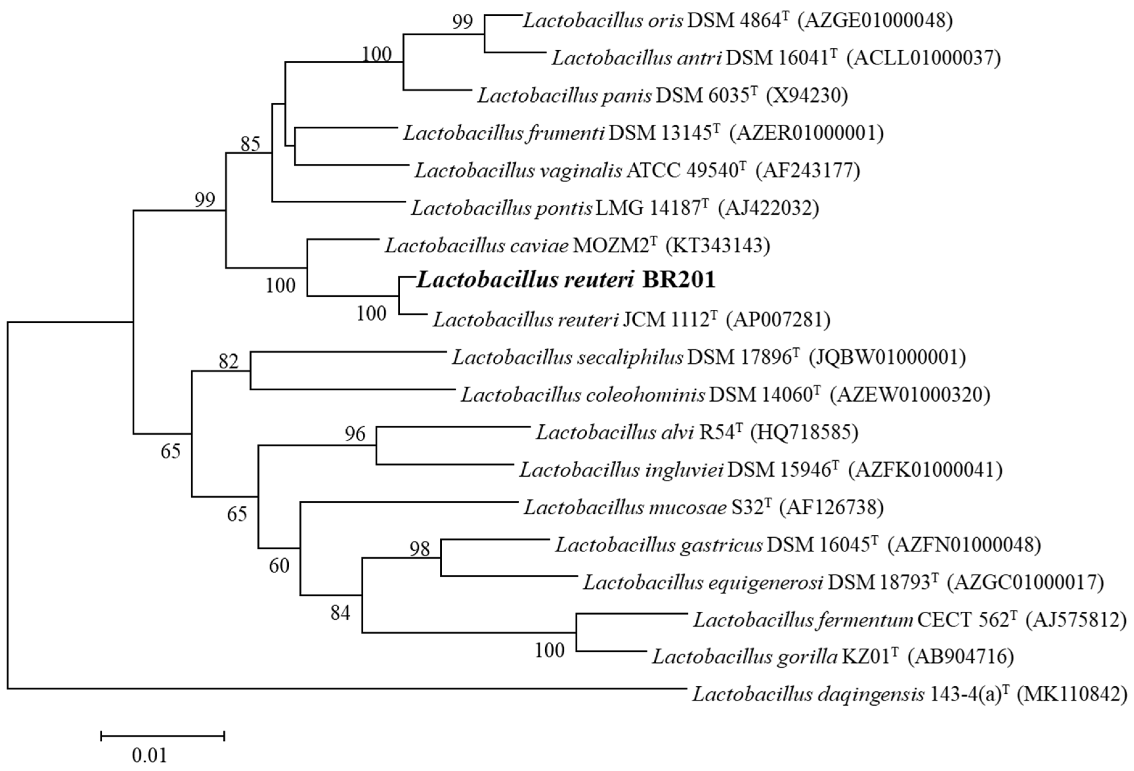
2.1. Isolation and Identification L. reuteri BR201

L. reuteri is known to produce 3-HPA, but there are differences among strains [7]. Thus, we attempted to isolate a new strain of this species that more efficiently produced 3-HPA from glycerol. We identified several potential LAB isolates from infant feces based on color, using MRS agar containing 20 g/L glycerol and 0.3 g/L bromocresol purple. We analyzed these isolates using the matrix-assisted laser desorption ionization (MALDI) Biotyper system (Bruker Daltonik, Bremen, Germany), and tested them for growth and glycerol utilization in MRS medium containing 20 g/L glycerol at anaerobic conditions. L. reuteri BR201 grew faster than all the other isolates and produced more 3-HPA. The identity of this isolate was confirmed using 16S rRNA analysis. The NCBI BLAST results showed high similarity (>99%) to L. reuteri JCM 1112T (AP007281). The 16S rRNA sequence of L. reuteri BR201 into the NCBI nucleotide sequence database (accession No. MZ269211) and L. reuteri BR201 strain also deposited it at the Korea Collection for Type Cultures (accession No. KCTC 14343BP). Figure 1 shows the relationship with other Lactobacillus strains using the neighbor-joining method. The results shown that L. reuteri BR201 was closest to L. reuteri JCM 1112T (bootstrap value: 100%), and the phylogenetic tree indicated that L. reuteri BR201 was included in a monophyletic group containing other Lactobacillus sp. strains.

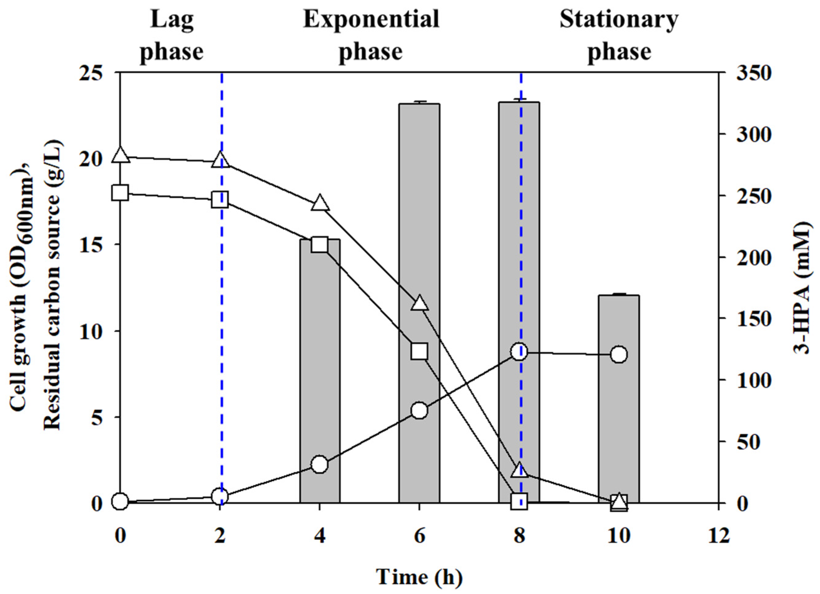
2.2. The Effect of Cell Growth for 3-HPA Bioconversion

We used a two-step procedure to produce 3-HPA using L. reuteri. The first step was promotion of cell growth using batch culture, and the second step was conversion of glycerol into 3-HPA using resting cells as biocatalytic.
Thus, we first grew cells by batch cultivation in MRS medium containing 20 g/L glycerol and 18 g/L glucose to activate genes that function in glycerol metabolism [3]. These results indicated that cells consumed all of the carbon source after 8 h, and the cell growth at this time reached OD600 nm 8.8 (Figure 2). The bioconversion glycerol to 3-HPA had a similar tendency, in that it increased during the exponential phase (6 h, 324.1 mM) and the late exponential phase (8 h, 325.6 mM). However, because cell proliferation during the late exponential phase was about 1.62-fold greater than during the exponential phase, it was possible to produce a 1.62-fold more 3-HPA using the same amount of cultivation broth. Cells were in the stationary phase at 10 h, and the bioconversion of 3-HPA was 20% (w/w) lower at that time. As shown in Table 1, the same amount of cells were present at the late exponential phase (8 h) and the stationary phase (10 h), but less glycerol was consumed at the stationary phase. This appears to be due to a decrease in cell activity from 8 h to 10 h. Similarly, Lüthi-Peng et al. reported that the L. reuteri ATCC 53608 strain produced the most 3-HPA during the late exponential/early stationary phase [3]. They also found that as cell age increased, the amount of viable cells and cell activity decreased [3]. Because cells most effectively produced 3-HPA during the late exponential phase, our subsequent experiments used cells harvested at this time.

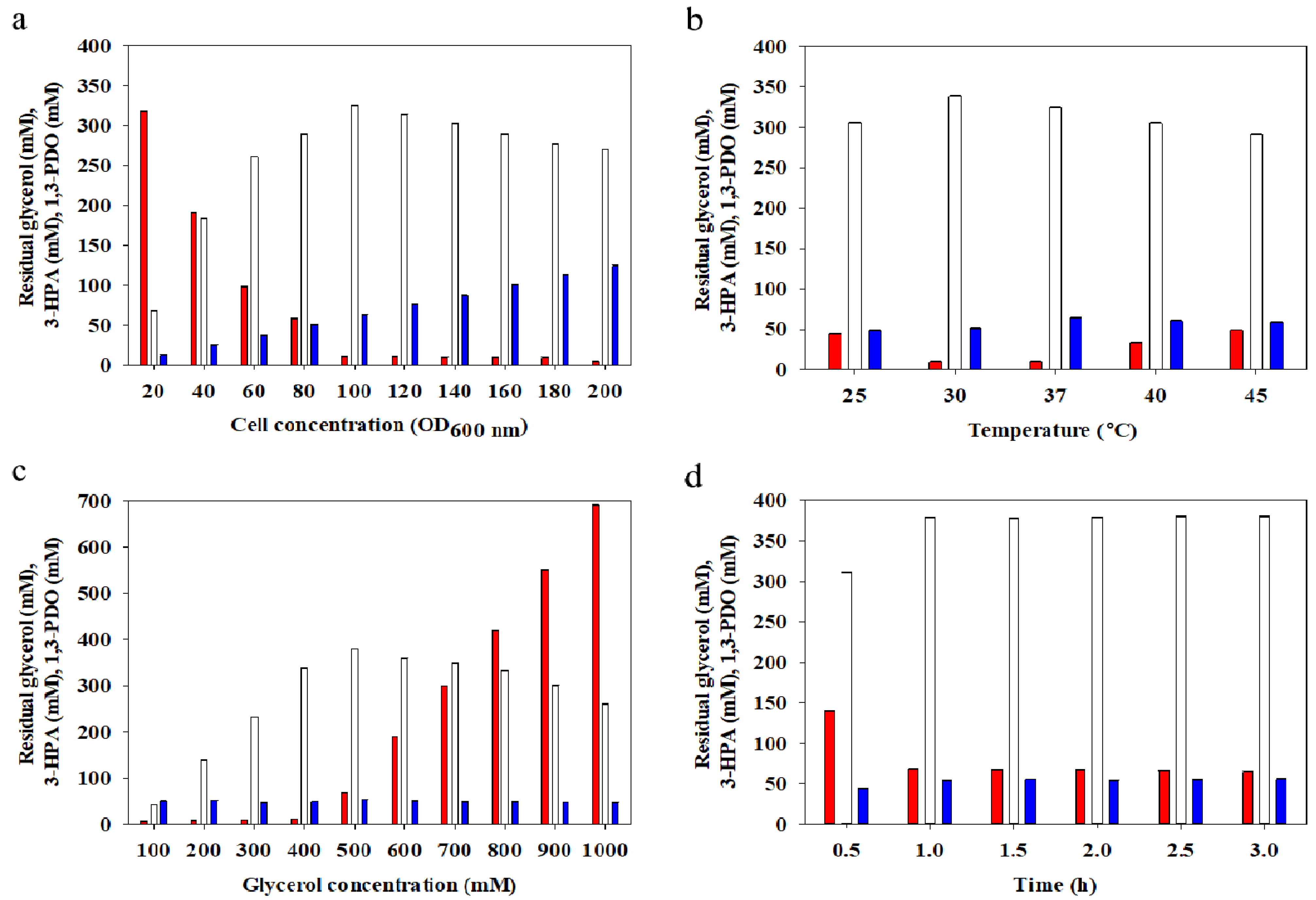
2.3. The Effect of Cell Density

We first examined the effect of different cell density on the production of 3-HPA under basic bioconversion conditions (Figure 3a). The results indicated that 3-HPA bioconversion occurred at all tested cell density (OD600 nm 20~200) under the basic bioconversion conditions. The bioconversion of 3-HPA was the highest (325.1 mM) when the cell density was OD600 nm 100. As cell density increased above this level, the production of 1,3-PDO increased and there was no change in the amount of residual glycerol. In agreement, Lüthi-Peng et al. also reported that 3-HPA production decreased when the cell density increased above certain level [3]. L. reuteri metabolizes glycerol into 3-HPA and 1,3-PDO using vitamin B12-dependent glycerol dehydratase (dhaB) and 1,3-propanediol oxidoreductase (dhaT) [20]. In the case of dhaT, NADH is used as a coenzyme. The NADH generated during the cell growth step of LAB is used as a coenzyme to convert 3-HPA to 1,3-PDO by dhaT during 3-HPA bioconversion. Therefore, a higher cell density leads to greater total NADH and an increased level of 1,3-PDO. Our results demonstrated that when the cell density was high, the amount of 3-HPA decreased because the bioconversion of 1,3-PDO increased. We therefore used a cell density of OD600 nm 100 for subsequent experiments.

2.4. The Effect of Temperature

We next examined the effect of different temperatures on the bioconversion of 3-HPA (Figure 3b). The results indicated a temperature of 30 °C led to the greatest bioconversion of 3-HPA (338.3 mM). On the other hand, the production of 1,3-PDO was the highest (64.2 mM) at 37 °C. Similarly, Doleyres et al., studied the L. reuteri ATCC 53608 strain and reported that the optimal temperature for 3-HPA production was 30 °C [14].
To confirm the effect of temperature on the bioconversion of 3-HPA and 1,3-PDO, we also measured the enzymatic activities of dhaB and dhaT at different temperatures (Figure 4). The results indicated the activities of both enzymes were highest at 37 °C. Furthermore, dhaB activity was only 3% lower at 30 °C, but dhaT activity was approximately 14% lower at 30 °C. This lower dhaT activity at 30 °C corresponds to the reduced production of 1,3-PDO at this temperature and corresponds to our measurements of these metabolites (Figure 3b). The effect of temperature on dhaT in L. panis PM1 strain was similar [21], in that the specific activity was greater at 37 °C than at 30 °C regardless of pH [21]. We used a temperature of 30 °C for subsequent experiments.

2.5. The Effect of Substrate Concentration

We next examined the effect of substrate concentration on the bioconversion of 3-HPA (Figure 3c). The results indicated that 3-HPA bioconversion was the highest (378 mM) when the glycerol concentration was 500 mM. The consumption of glycerol was also the greatest (431.8 mM) when the glycerol concentration was 500 mM, with reduced levels at higher concentrations presumably due to substrate inhibition. Previous studies of different strains of L. reuteri reported that a glycerol concentration above 250~400 mM, decreased 3-HPA due to substrate inhibition [3,7,14]. However, our results indicated that L. reuteri BR201 had stable enzyme activity even at a glycerol concentration of 500 mM. Therefore, L. reuteri BR201 is highly suitable for producing high concentrations of 3-HPA. We used a glycerol concentration of 500 mM for subsequent experiments.

2.6. The Effect of Conversion Time

We next examined the effect of conversion time on the bioconversion of 3-HPA (Figure 3d). The results indicated that a bioconversion time of 1.0 h led to the highest bioconversion of 3-HPA (378.4 mM). There was no significant difference in 3-HPA as the bioconversion time increased further. The 3-HPA bioconversion was very rapid, in that about 90% of this product was produced within 0.5 h. The dhaB enzyme, which converts glycerol to 3-HPA and uses vitamin B12 as a cofactor, is eventually inactivated because the cells cannot regenerate coenzyme B12; instead the catalytic cycle is interrupted by the formation of 5’-deoxyadenosine and the catalytically inactive cobalamin [22], and the altered cofactor remains tightly bound to the active site [23]. A dhaB-reactivating factor in Klebsiella reactivates the inactivated enzyme-bound cyanocobalamin in the presence of free vitamin B12, adenosine triphosphate (ATP), and Mg2+ by mediating the exchange of the tightly bound altered cofactor for a free intact cofactor [24]. A previous study also reported reactivation of dhaB by vitamin B12 and ATP in L. reuteri [25]. In particular, L. reuteri can produce vitamin B12 via glucose metabolism [26,27,28]. However, during the bioconversion of 3-HPA from aqueous glycerol, this species is unable to produce vitamin B12 and ATP because it lacks the ability to use glycerol as a carbon source to produce vitamin B12 and ATP [20,27]. This explains the negligible effect of increasing the bioconversion time (Figure 3d).
There are several previous studies on the bioconversion of 3-HPA using L. reuteri. Although some studies show higher 3-HPA conversion yield than L. rueteri BR201, no group reported production of more than 300 mM 3-HPA from a single batch. To the best of our knowledge, L. reuteri BR201 produced more 3-HPA than any other isolate without the use of additives (Table S1). It will be a good candidate for the industrialization of 3-HPA derived from lactic acid bacteria.

2.7. Antibacterial Activity and Stability of 3-HPA

We tested the antibacterial activity of the 3-HPA that was produced under the 378 mM 3-HPA production conditions, using criteria in the United States Pharmacopeia (USP) Chapter 51 (Table 2). The results indicated the minimal inhibitory concentration (MIC) of 3-HPA was 2~3 mM for the three bacteria, and was 4 mM for two fungi. These results are similar to those of previous studies [29,30,31].
3-HPA is widely used as a food preservative because of its high antibacterial activity [18,31,32]. When using 3-HPA as a food preservative and in other applications, it is essential to consider safety. In particular, 3-HPA undergoes spontaneous dehydration in aqueous solutions into acrolein, a toxic substance [1] Therefore, we measured the effect of temperature and time on the amount of 3-HPA degradation into acrolein using an initial 3-HPA concentration of 4 mM and a pH below 4.0 (Table 3). The initial 4 mM 3-HPA aqueous solution contained 11.6 µM (0.65 mg/L) acrolein. At 4 °C, there was very little production of acrolein after 4 weeks. At 25 °C, there was a small amount of acrolein increasing after 4 weeks, but this increase was not statistically significant. However, at 37 °C and 50 °C, there was a significant acrolein production by week-2, and this level then remained relatively constant until week-4. Similarly, Engels et al. reported no acrolein production at pH 4.0 and 4 °C, and that the conversion of 3-HPA to acrolein was reversible, with 3-HPA being favored [17].
3-HPA is an aldol with hydroxyl and aldehyde functional groups. The process of 3-HPA dehydration and conversion to acrolein is an aldol condensation reaction, a reaction that is more rapid at high temperatures [33,34]. This explains our finding of little or no production of acrolein at 4 °C, but significant production at 37 °C and 50 °C. Therefore, storage of 3-HPA at a concentration where it functions as an antibacterial (4 mM) should be below room temperature to prevent degradation into acrolein.

3. Material and Methods

3.1. Materials and Growth Media

De Man, Rogosa, and Sharpe (MRS; Difco, Sparks, MD, USA) agar and broth were used for cultivation of L. reuteri; tryptic soy broth (TSB; Difco) was used for cultivation of bacteria; and Sabouraud dextrose broth (SDB; Difco) was used for cultivation of fungi. Acrolein was purchased from Fluka (Buchs, Switzerland); 2,4-dinitrophenylhydrazine (DNPH), acrolein-2,4-DNPH, D,L-tryptophan, bromocresol purple, and sulfuric acid were purchased from Sigma (St. Louis, MO, USA); And HPLC water, acetonitrile, hydrochloric acid, and glycerol were purchased from Daejung Chemicals (Siheung, Korea). All materials were used as received, without pretreatment.

3.2. Isolation of L. reuteri Strain

Lactobacillus reuteri BR201 was isolated from infant feces. First, 1 g of infant feces samples were serially diluted in 10 mL of 0.22 μm filtered PBS buffer (pH 6.8). Then, 100 µL samples diluted in 105~107 were spread on MRS agar containing 20 g/L glycerol and 0.3 g/L bromocresol purple. The agar plates were incubated at 37 °C for 24 h in an anaerobic chamber (Shellab, Cornelius, OR, USA). The primary isolation was performed by colorimetric method using bromocresol purple. The identification of the Lactobacillus reuteri strains among the primary screening strains were conducted using the MALDI Biotyper system [35].

3.3. Identification of L. reuteri BR201 by 16S rRNA Analysis

Genomic DNA of L. reuteri BR201 was extracted using a genomic DNA mini kit (Invitrogen, Waltham, MA, USA). The 16S rRNA gene of L. reuteri BR201 was amplified using two universal primers, 27F (5′-AGAGTTTGATCMTGGCTCAG-3′) and 1492R (5′-GGTTACCTTGTTACGACTT-3′) by PCR. The nucleotide sequence of amplified PCR fragment was confirmed by Solgent (Daejeon, Korea). The result was compared with available 16S rRNA gene sequences on NCBI GenBank and EzTaxon [36]. Phylogenetic analysis was conducted by MEGA6 using the neighbor-joining method [37,38]. The bootstrap value for 1000 replicates was calculated for each branch [39].

3.4. Cultivation of L. reuteri BR201

Seed cells for batch cultivation were prepared using 50 mL conical tube containing 30 mL MRS broth with 20 g/L glycerol. The conical tube was statically incubated at 37 °C for 12 h, and subsequent cultivation was performed in 5-L fermentor (Kobiotech, Incheon, Korea) at an inoculation concentration of 1% (v/v). Batch cultivations were conducted in a 5-L fermentor that contained 3 L MRS broth with 20 g/L glycerol. All cultivation experiments were conducted with 100 rpm and without aeration at 37 °C. The pH was maintained at 5.5 ± 0.1 using 6 M NaOH. Cell growth was measured by absorbance at OD600 nm. All results are presented as averages from three independent experiments.

3.5. Cultivation Metabolite Analysis

The concentrations of residual carbon sources and metabolites were determined using an Agilent 1200 series high-performance liquid chromatography (HPLC) system (Agilent Technologies, Santa Clara, CA, USA) that had an Aminex HPX-87H column (300 × 7.8 mm; Bio-Rad, Hercules, CA, USA) and a refractive index detector (RID). As the mobile phase, 2.5 mM H2SO4 was used, and the flow rate was 0.6 mL/min. The temperatures of the column oven and the RID cell were 65 °C and 45 °C, respectively [40].

3.6. 3-HPA Bioconversion

Cells resulting from 8 h of cultivation in a 5 L fermentor were used for 3-HPA bioconversion. The cultivation broth was first centrifuged to obtain resting cells. Then, the resting cells were washed with 1/2 volume DW, resuspended in an aqueous glycerol solution, and transferred to a constant temperature incubator. Unless otherwise noted, the conditions used for of 3-HPA bioconversion were cell density OD600 nm 100, glycerol concentration of 400 mM, bioconversion temperature of 37 °C, and bioconversion time of 1 h. For 3-HPA bioconversion, the effects of cell density, bioconversion temperature, glycerol concentration, and bioconversion time were then determined. After different experimental treatments, the bioconversion solution was centrifuged to remove the cells and passed through a 0.22 µm filter. Quantification of the 3-HPA was determined using the acrolein coloring method [41]. In particular, a 100 µL of sample was thoroughly mixed with 75 µL of a 10 mM DL-tryptophan solution (dissolved in 0.05 mM HCl), and 300 µL of HCl 37% (w/v). Mixtures containing samples and standards were incubated for 20 min in a water bath at 37 °C, and OD560 nm was then measured [14]. The enzymatic activities of dhaB and dhaT were measured as previously described [28].

3.7. Measurement of Antimicrobial Activity

Five microbes were used for antibacterial activity tests, as described in Chapter 51 of the USP (Antimicrobial Effectiveness Testing). There were threee bacteria (Escherichia coli ATCC 8739, Pseudomonas aeruginosa ATCC 9027, Staphylococcus aureus ATCC 6538), one yeast (Candida albicans ATCC 10231), and one mold (Aspergillus niger ATCC 16404). These experiments were performed after subculturing twice in TSB (bacteria) or SDB (fungi). The minimum inhibitory concentration (MIC) was used as an indication of antimicrobial activity [42], and the concentration of 3-HPA in these tests ranged from 0 to 5 mM. For bacteria, 1 × 10⁷ CFU/mL were inoculated into TSB medium containing 3-HPA and incubated at 37 °C for 3 days. For fungi, 1 × 10⁵ CFU/mL were inoculated into SDB medium containing 3-HPA and incubated at 30 °C for 3 days.

3.8. Acrolein Analysis

The concentration of acrolein in the 3-HPA solution was analyzed using the modified 2,4-DNPH derivatization method [43,44,45]. A 20 µL sample was added to 100 µL of citric acid/sodium citrate buffer (8:2), 400 µL of acetonitrile (ACN), 280 µL of DW, and 200 µL of 10 mM 2,4-DNPH in ACN at 25 °C. This solution was placed on a shaker (250 rpm) and derivatized for 1 h. The derivatization solution was passed through a 0.45 µm Teflon filter, and the concentration of acrolein-2,4-DNPH was determined using an Agilent 1200 series HPLC system (Agilent Technologies) that was equipped with a diode array detector and a ZORBAX eclipse plus C18 column (250 × 4.6 mm, 5 µm; Agilent Technologies). The mobile phase was a water/acetonitrile mixture (4:6, v/v), the flow rate was 1.0 mL/min, the mobile phase was degassed for 1 h before use, the column temperature was 30 °C, and the diode array detector measured OD360 nm (Figure S1).

4. Conclusions

We isolated L. reuteri BR201, an excellent biocatalyst for 3-HPA bioconversion, and determined effect of bioconversion conditions (cell density, temperature, substrate concentration, time) for the bioconversion of 3-HPA without additives. This strain produced 378 mM of 3-HPA under confirmed conditions. Our assessment of the antimicrobial activity of the resulting 3-HPA indicated it was effective against bacteria and fungi at low concentrations (4 mM). Moreover, storage of the resulting 3-HPA aqueous solution at a low temperature led to minimal degradation into acrolein. These results suggest that LAB-derived 3-HPA has potential for use in various industrial applications.

Supplementary Materials

The following are available online at https://www.mdpi.com/article/10.3390/catal11101139/s1.

Author Contributions

Conceptualization, J.-H.J.; methodology, S.-G.J. and S.-Y.H.; investigation, J.-H.J. and S.-G.J.; resources, S.-Y.H. and M.-S.K.; writing—original draft preparation, J.-H.J., K.M.L. and B.-R.O.; writing—review and editing, J.-H.J., C.-H.K. and B.-R.O.; supervision, C.-H.K.; project administration, M.-S.K., B.-R.O., funding acquisition, K.M.L. and B.-R.O. All authors have read and agreed to the published version of the manuscript.

Funding

This work was supported by the Korea Research Institute of Bioscience and Biotechnology (KRIBB) Research Initiative Program (KGM5482113). This research was financially supported by the Ministry of Trade, Industry and Energy (MOTIE) and Korea Institute for Advancement of Technology (KIAT) through the International Cooperative R&D program. (Project No. P0014932).

Data Availability Statement

The datasets supporting the conclusions of this article are included within the article.

Conflicts of Interest

The authors declare no conflict of interest.

References

  1. Vollenweider, S.; Lacroix, C. 3-Hydroxypropionaldehyde: Applications and Perspectives of Biotechnological Production. Appl. Microbiol. Biotechnol. 2004, 64, 16–27. [Google Scholar] [CrossRef] [PubMed]
  2. Bauer, R.; du Toit, M.; Kossmann, J. Influence of Environmental Parameters on Production of the Acrolein Precursor 3-Hydroxypropionaldehyde by Lactobacillus reuteri DSMZ 20016 and Its Accumulation by Wine Lactobacilli. Int. J. Food Microbiol. 2010, 137, 28–31. [Google Scholar] [CrossRef] [PubMed]
  3. Lüthi-Peng, Q.; Schärer, S.; Puhan, Z. Production and Stability of 3-Hydroxypropionaldehyde in Lactobacillus reuteri. Appl. Microbiol. Biotechnol. 2002, 60, 73–80. [Google Scholar] [PubMed]
  4. Arqués, J.L.; Fernández, J.; Gaya, P.; Nuñez, M.; Rodríguez, E.; Medina, M. Antimicrobial Activity of Reuterin in Combination with Nisin Against Food-Borne Pathogens. Int. J. Food Microbiol. 2004, 95, 225–229. [Google Scholar] [CrossRef]
  5. Arqués, J.L.; Rodríguez, E.; Nuñez, M.; Medina, M. Inactivation of Gram-Negative Pathogens in Refrigerated Milk by Reuterin in Combination with Nisin or the Lactoperoxidase System. Eur. Food Res. Technol. 2008, 227, 77–82. [Google Scholar] [CrossRef]
  6. Sobolov, M.; Smiley, K.L. Metabolism of Glycerol by an Acrolein-Forming Lactobacillus. J. Bacteriol. 1960, 79, 261–266. [Google Scholar] [CrossRef] [Green Version]
  7. Talarico, T.L.; Casas, I.A.; Chung, T.C.; Dobrogosz, W.J. Production and Isolation of Reuterin, a Growth Inhibitor Produced by Lactobacillus reuteri. Antimicrob. Agents Chemother. 1988, 32, 1854–1858. [Google Scholar] [CrossRef] [Green Version]
  8. Talarico, T.L.; Dobrogosz, W.J. Chemical Characterization of an Antimicrobial Substance Produced by Lactobacillus reuteri. Antimicrob. Agents Chemother. 1989, 33, 674–679. [Google Scholar] [CrossRef] [Green Version]
  9. Hayek, S.A.; Ibrahim, S.A. Current Limitations and Challenges with Lactic Acid Bacteria: A Review. Food Nutr. Sci. 2013, 4, 40133. [Google Scholar] [CrossRef] [Green Version]
  10. Kumar, V.; Ashok, S.; Park, S. Recent Advances in Biological Production of 3-Hydroxypropionic Acid. Biotechnol. Adv. 2013, 31, 945–961. [Google Scholar] [CrossRef]
  11. U.S. Food and Drug Administration. Generally Recognized as Safe (GRAS) Determination of Lactobacillus reuteri Strain DSM 17938; FDA: Silver Sping, MD, USA, 2008.
  12. Lüthi-Peng, Q.; Dileme, F.; Puhan, Z. Effect of Glucose on Glycerol Bioconversion by Lactobacillus reuteri. Appl. Microbiol. Biotechnol. 2002, 59, 289–296. [Google Scholar] [PubMed]
  13. Vieira, P.B.; Kilikian, B.V.; Bastos, R.V.; Perpetuo, E.A.; Nascimento, C.A.O. Process Strategies for Enhanced Production of 1,3-Propanediol by Lactobacillus reuteri Using Glycerol as a Co-Substrate. Biochem. Eng. J. 2015, 94, 30–38. [Google Scholar] [CrossRef]
  14. Doleyres, Y.; Beck, P.; Vollenweider, S.; Lacroix, C. Production of 3-Hydroxypropionaldehyde Using a Two-Step Process with Lactobacillus reuteri. Appl. Microbiol. Biotechnol. 2005, 68, 467–474. [Google Scholar] [CrossRef] [PubMed] [Green Version]
  15. Stevens, M.J.A.; Vollenweider, S.; Mertes, P.; Lacroix, C. Bisulfite as Scavenger for Enhanced Biotechnological Production of 3-Hydroxypropionaldehyde by Lactobacillus reuteri. Biochem. Eng. J. 2013, 79, 239–245. [Google Scholar] [CrossRef]
  16. Sardari, R.R.R.; Dishisha, T.; Pyo, S.-H.; Hatti-Kaul, R. Semicarbazide-Functionalized Resin as a New Scavenger for In Situ Recovery of 3-Hydroxypropionaldehyde during Biotrans-Formation of Glycerolby Lactobacillus reuteri. J. Biotechnol. 2014, 192, 223–230. [Google Scholar] [CrossRef] [PubMed] [Green Version]
  17. Engels, C.; Schwab, C.; Zhang, J.; Stevens, M.J.A.; Bieri, C.; Ebert, M.-O.; McNeill, K.; Sturla, S.J.; Lacroix, C. Acrolein Contributes Strongly to Antimicrobial and Heterocyclic Amine Transformation Activities of Reuterin. Sci. Rep. 2016, 6, 36246. [Google Scholar] [CrossRef] [Green Version]
  18. Stevens, M.; Vollenweider, S.; Lacroix, C.; Zurich, E.T.H. 5-The Potential of Reuterin Produced by Lactobacillus reuteri as a Broad Spectrum Preservative in Food. In Protective Cultures, Antimicrobial Metabolites and Bacteriophages for Food and Beverage Biopreservation; Lacroix, C., Ed.; Woodhead Publishing: Sawston, UK, 2011; pp. 129–160. [Google Scholar]
  19. Abraham, K.; Andres, S.; Palavinskas, R.; Berg, K.; Appel, K.E.; Lampen, A. Toxicology and Risk Assessment of Acrolein in Food. Mol. Nutr. Food Res. 2011, 55, 1277–1290. [Google Scholar] [CrossRef]
  20. Ju, J.-H.; Heo, S.-Y.; Choi, S.-W.; Kim, Y.-M.; Kim, M.-S.; Kim, C.-H.; Oh, B.-R. Effective Bioconversion of 1,3-Propanediol from Biodiesel-Derived Crude Glycerol Using Organic Acid Resistance-Enhanced Lactobacillus reuteri JH83. Bioresour. Technol. 2021, 337, 125361. [Google Scholar] [CrossRef]
  21. Kang, T.S.; Korber, D.R.; Tanaka, T. Glycerol and Environmental Factors: Effects on 1,3-Propanediol Production and NAD+ Regenerationin Lactobacillus panis PM1. J. Appl. Microbiol. 2013, 115, 1003–1011. [Google Scholar] [CrossRef]
  22. Daniel, R.; Bobik, T.A.; Gottschalk, G. Biochemistry of Coenzyme B12-Dependent Glycerol and Diol Dehydratases and Organization of the Encoding Genes. FEMS Microbiol. Rev. 1998, 22, 553–566. [Google Scholar] [CrossRef]
  23. Mori, K.; Hosokawa, Y.; Yoshinaga, T.; Toraya, T. Diol Dehydratase-Reactivating Factor Is a Reactivase-Evidence for Multiple Turnovers and Subunit Swapping with Diol Dehy-Dratase. FEBS J. 2010, 227, 4931–4943. [Google Scholar] [CrossRef] [PubMed]
  24. Toraya, T.; Mori, K. A Reactivating Factor for Coenzyme B12-Dependent Diol Dehydratase. J. Biol. Chem. 1999, 274, 3372–3377. [Google Scholar] [CrossRef] [PubMed] [Green Version]
  25. Liu, F.; Yu, B. Efficient Production of Reuterin from Glycerol by Magnetically Immobilized Lactobacillus reuteri. Appl. Microbiol. Biotechnol. 2015, 99, 4659–4666. [Google Scholar] [CrossRef] [PubMed]
  26. Taranto, M.P.; Vera, J.L.; Hugenholtz, J.; De Valdez, G.F.; Sesma, F. Lactobacillus reuteri CRL1098 Produces Cobalamin. J. Bacteriol. 2003, 185, 5643–5647. [Google Scholar] [CrossRef] [Green Version]
  27. Santos, F.; Teusink, B.; Molenaar, D.; van Heck, M.; Wels, M.; Sieuwerts, S.; de Vos, W.M.; Hugenholtz, J. Effect of Amino Acid Availability on Vitamin B12 Production in Lactobacillus reuteri. Appl. Environ. Microbiol. 2009, 75, 3930. [Google Scholar] [CrossRef] [Green Version]
  28. Ju, J.-H.; Wang, D.; Heo, S.-Y.; Kim, M.-S.; Seo, J.-W.; Kim, Y.-M.; Kim, D.-H.; Kang, S.-A.; Kim, C.-H.; Oh, B.-R. Enhancement of 1, 3-Propanediol Production from Industrial By-Productby Lactobacillus reuteri CH53. Microb. Cell Fact. 2020, 19, 1–10. [Google Scholar] [CrossRef] [Green Version]
  29. Rasch, M. The Influence of Temperature, Salt and pH on the Inhibitory Effect of Reuterin on Escherichia coli. Int. J. Food Microbiol. 2002, 72, 225–231. [Google Scholar] [CrossRef]
  30. Yunmbam, M.K.; Roberts, J.F. The In Vitro Efficacy of Reuterin on the Culture and Bloodstream Forms of Trypanosoma brucei brucei. Comp. Biochem. Physiol. C Comp. Pharmacol. Toxicol. 1992, 101, 235–238. [Google Scholar] [CrossRef]
  31. Vimont, A.; Fernandez, B.; Ahmed, G.; Fortin, H.-P.; Fliss, I. Quantitative Antifungal Activity of Reuterin Against Food Isolates of Yeasts and Moulds and Its Potential Application in Yogurt. Int. J. Food Microbiol. 2019, 289, 182–188. [Google Scholar] [CrossRef]
  32. El-Ziney, M.G.; van den Tempel, T.; Debevere, J.; Jakobsen, M. Application of Reuterin Produced by Lactobacillus reuteri 12002 for Meat Decontamination and Preservation. J. Food Prot. 1999, 62, 257–261. [Google Scholar] [CrossRef]
  33. Crouch, R.D.; Richardson, A.; Howard, J.L.; Harker, R.L.; Barker, K.H. The Aldol Addition and Condensation: The Effect of Conditions on Reaction Pathway. J. Chem. Educ. 2007, 84, 475. [Google Scholar] [CrossRef]
  34. Josephson, D.B.; Lindsay, R.C. Retro-Aldol Degradations of Unsaturated Aldehydes: Role in the Formation of c4-Heptenal from t2, c6-Nonadienal in Fish, Oyster and Other Flavors. J. Am. Oil Chem. Soc. 1987, 64, 132–138. [Google Scholar] [CrossRef]
  35. Buchan, B.W.; Riebe, K.M.; Ledeboer, N.A. Comparison of the MALDI Biotyper System Using Sepsityper Specimen Processing to Routine Microbiological Methods for Identification of Bacteria from Positive Blood Culture Bottles. J. Clin. Microbiol. 2012, 50, 346–352. [Google Scholar] [CrossRef] [Green Version]
  36. Kim, O.-S.; Cho, Y.-J.; Lee, K.; Yoon, S.-H.; Kim, M.; Na, H.; Park, S.-C.; Jeon, Y.S.; Lee, J.-H.; Yi, H.; et al. Introducing EzTaxon-E: A Prokaryotic 16S rRNA Gene Sequence Database with Phylotypes that Represent Uncultured Species. Int. J. Syst. Evol. Microbiol. 2012, 62, 716–721. [Google Scholar] [CrossRef] [PubMed]
  37. Tamura, K.; Stecher, G.; Peterson, D.; Filipski, A.; Kumar, S. MEGA6: Molecular Evolutionary Genetics Analysis Version 6.0. Mol. Biol. Evol. 2013, 30, 2725–2729. [Google Scholar] [CrossRef] [PubMed] [Green Version]
  38. Saitou, N.; Nei, M. The Neighbor-Joining Method: A New Method for Reconstructing Phylogenetic Trees. Mol. Biol. Evol. 1987, 4, 406–425. [Google Scholar]
  39. Felsenstein, J. Confidence Limits on Phylogenies: An Approach Using the Bootstrap. Evolution 1985, 39, 783–791. [Google Scholar] [CrossRef]
  40. Ju, J.-H.; Ko, D.-J.; Heo, S.-Y.; Lee, J.-J.; Kim, Y.-M.; Lee, B.-S.; Kim, M.-S.; Kim, C.-H.; Seo, J.-W.; Oh, B.-R. Regulation of Lipid Accumulation Using Nitrogen for Microalgae Lipid Production in Schizochytrium sp. ABC101. Renew. Energy 2020, 153, 580–587. [Google Scholar] [CrossRef]
  41. Circle, S.J.; Stone, L.; Boruff, C.S. Acrolein Determination by Means of Tryptophane. A Colorimetric Micromethod. Ind. Eng. Chem. Anal. Ed. 1945, 17, 259–262. [Google Scholar] [CrossRef]
  42. Asare, P.T.; Zurfluh, K.; Greppi, A.; Lynch, D.; Schwab, C.; Stephan, R.; Lacroix, C. Reuterin Demonstrates Potent Antimicrobial Activity Against a Broad Panel of Human and Poultry Meat Campylobacter spp. Isolates. Microorganisms 2020, 8, 78. [Google Scholar] [CrossRef] [Green Version]
  43. Hanoune, B.; LeBris, T.; Allou, L.; Marchand, C.; Le Calve, S.J.A.E. Formaldehyde Measurements in Libraries: Comparison between Infrared Diode Laser Spectroscopy and a DNPH-Derivatization Method. Atmos. Environ. 2006, 40, 5768–5775. [Google Scholar] [CrossRef]
  44. Possanzini, M.; Di Palo, V.; Petricca, M.; Fratarcangeli, R.; Brocco, D.J.A.E. Measurements of Lower Carbonyls in Rome Ambient Air. Atmos. Environ. 1996, 30, 3757–3764. [Google Scholar] [CrossRef]
  45. Slemr, J.; Junkermann, W.; Volz-Thomas, A.J.A.E. Temporal Variations in Formaldehyde, Acetaldehyde and Acetone and Budget of Formaldehyde at a Rural Site in Southern Germany. Atmos. Environ. 1996, 30, 3667–3676. [Google Scholar] [CrossRef]
Figure 1. The neighbor-joining phylogenetic tree of Lactobacillus reuteri BR201 based on 16S rRNA gene sequences. The number at each branch represents the bootstrap value (%) for the node calculated from 1000 replicates. Bar: 0.01 substitutions per nucleotide.
Catalysts 11 01139 g001
Figure 2. Batch cultivation of Lactobacillus reuteri BR201 and bioconversion of 3-hydroxypropionaldehyde during different growth phases. Open triangles, residual glycerol; open squares, residual glucose; open circles, cell growth; gray bars, 3-hydroxypropionaldehyde concentration. All results are averages from three independent experiments.
Catalysts 11 01139 g002
Figure 3. 3-hydroxypropionaldehyde bioconversion at a different cell density (a), bioconversion temperatures (b), glycerol concentrations (c), and bioconversion times (d). Red bars, residual glycerol; white bars, 3-hydroxypropionaldehyde; blue bars, 1,3-propanediol. All results are averages from three independent experiments.
Catalysts 11 01139 g003
Figure 4. The Effect of temperature on the relative enzyme activity of vitamin B12-dependent glycerol dehydratase, dhaB (a) and 1,3-propanediol oxidoreductase, dhaT (b). All results are averages from three independent experiments.
Catalysts 11 01139 g004
Table 1. The Effect of cultivation time on the levels of residual glycerol, 3-hydroxypropionaldehyde, and 1,3-propanediol by L. reuteri BR201.
Cultivation Time (h)Regidual Glycerol (mM)3-HPA (mM)1,3-PDO (mM)
4137.5 ± 2.1214.2 ± 2.637.8 ± 0.2
614.1 ± 0.1324.1 ± 3.859.9 ± 0.6
811.4 ± 0.1325.6 ± 4.361.8 ± 0.5
10195.9 ± 1.2168.5 ± 2.734.2 ± 0.4
Table 2. The Effect of 3-hydroxypropionaldehyde concentration on antibiotic activity.
PathogenResponse * at Different 3-HPA Concentration (mM)
01234MIC (mM)
E. coli ATCC 8739++++3
S. aureus ATCC 6538+++2
P. aeruginosa ATCC 9027+++2
C. albicans ATCC 10231+++++4
A. niger ATCC 16404+++++4
* ++: growth; +: inhibition of growth; −: no growth.
Table 3. The Effect of temperature and time on the production of acrolein from 3-hydroxypropionaldehyde in aqueous solution.
Temperature (°C)Acrolein (mg/L)
0 Days7 Days14 Days21 Days28 Days
40.65 ± 0.020.67 ± 0.040.68 ± 0.050.69 ± 0.040.69 ± 0.03
251.04 ± 0.111.42 ± 0.081.54 ± 0.061.66 ± 0.12
3711.36 ± 0.3714.43 ± 0.3215.82 ± 0.1415.96 ± 0.18
5012.77 ± 0.4215.19 ± 0.2816.54 ± 0.2416.58 ± 0.11
Publisher’s Note: MDPI stays neutral with regard to jurisdictional claims in published maps and institutional affiliations.

