Article

Microwave-Assisted Synthesis and Characterization of Solar-Light-Active Copper–Vanadium Oxide: Evaluation of Antialgal and Dye Degradation Activity

1
Department of Marine Convergence Design, Pukyong National University, Busan 48513, Korea
2
Department of Chemistry, Pukyong National University, Busan 48513, Korea
*
Author to whom correspondence should be addressed.
Catalysts 2021, 11(1), 36; https://doi.org/10.3390/catal11010036
Submission received: 3 December 2020 / Revised: 24 December 2020 / Accepted: 28 December 2020 / Published: 30 December 2020
(This article belongs to the Section Environmental Catalysis)

Abstract

:
In this work, solar-light-active copper–vanadium oxide (Cu-VO) was synthesized by a simple microwave method and characterized by FESEM, EDS, XRD, XPS, UV–Vis/near-infrared (NIR), and FT-IR spectroscopy. Antialgal and dye degradation activities of Cu-VO were investigated against Microcystis aeruginosa and methylene blue dye (MB), respectively. The mechanism of action of Cu-VO was examined regarding the production of hydroxyl radical (·OH) in the medium and intracellular reactive oxygen species (ROS) in M. aeruginosa. FESEM and XRD analyses of Cu-VO disclosed the formation of monoclinic crystals with an average diameter of 132 nm. EDX and XPS analyses showed the presence of Cu, V, and O atoms on the surface of Cu-VO. Furthermore, FT-IR analysis of Cu-VO exposed the presence of tetrahedral VO4 and octahedral CuO6. Cu-VO effectively reduced the algal growth and degraded methylene blue under solar light. A total of 4 mg/L of Cu-VO was found to be effective for antialgal activity. Cu-VO degraded 93% of MB. The investigation of the mechanism of action of Cu-VO showed that ·OH mediated antialgal and dye degradation of M. aeruginosa and MB. Cu-VO also triggered the production of intracellular ROS in M. aeruginosa, leading to cell death. Thus, Cu-VO could be an effective catalyst for wastewater treatment.

Graphical Abstract

1. Introduction

Clean and mineral-rich water is critically important to preserve life on earth [1]. Increased industrialization, global warming, and high living standards contribute to water pollution and pose continuing risks to human health. Water pollutants, such as blooming harmful cyanobacteria and dye, limit the access to fresh drinking water and sanitation, which necessitates the remediation of these pollutants [1]. Several strains of bloom-forming cyanobacteria, such as Anabaena spp., Microcystis spp., and Oscillatoria spp., are found in the aquatic system [2]. Out of these strains, Microcystis aeruginosa is a major bloom-forming freshwater blue-green algae [3,4]. Textile, paper, cosmetic, and dye industries discharge various unused dye into water [5]. Methylene blue (MB) is a major industrial waste discharged into water [6]. M. aeruginosa contaminates freshwater by releasing cyanotoxins such as neurotoxins and hepatotoxins, whereas MB is toxic for aquatic animals and humans. These issues could be countered by applying an efficient, cheap, and easy technique.
Semiconductor photocatalysts that are active under a sustainable solar energy could be a promising and attractive one to deal with blooming M. aeruginosa [7] and MB pollutants [6]. Irradiation of photocatalysts with higher light energy than their band gap generates electrons and holes in the conduction and valence bands, respectively [8]. These exited photocatalysts can efficiently oxidize or reduce surrounding pollutants, which arouse their substantial interest in degrading environmental pollutants. Vanadium pentoxide (V2O5) is an efficient n-type semiconductor photocatalyst for the degradation of organic pollutants due to its benign nature, high chemical and light stability, and visible light activity [9]. V2O5 shows less degradation efficiency towards MB dye [9]. Fabrication of metal oxide composites of V2O5 is an effective strategy for improving degradation efficiency towards MB dye [10]. Recently, visible light-active transition of metal vanadium oxides has been intensively researched for various applications, such as antimicrobial activity [11], dye degradation [12], optical devices [13], and lithium batteries [14]. Out of several transition metal vanadates, copper–vanadium oxide has been extensively studied for photocatalytic performances and electrochemical applications [15]. Copper–vanadium oxide is a natural mineral identified as pseudolyonsite [16] and has been extensively used for O2 production from water [15], batteries [17], and dye degradation [18]. Jain et al. [19] found that the precursor of copper–vanadium oxide, i.e., Cu3V2O7(OH)2·2H2O, behaves like a peroxidase enzyme. Peroxidases mimicking metal oxides have a high potential for oxidative degradation of the microbial cell membranes [20]. The literature survey showed that, to date, none investigated the antialgal activity of Cu-VO. Therefore, it is important to investigate the antialgal activity of copper–vanadium oxide.
Semiconductor photocatalysts oxidize water under light to produce reactive oxygen species such as hydroxyl free radicals (·OH) [21]. ·OH is a highly reactive species with high oxidation potential and can degrade organic pollutants [22] and microorganisms in an aqueous medium [23]. Several studies have indicated that photocatalysts can also cause oxidative stress in algal cells by increasing the intracellular ROS level, which could damage DNA, RNA, proteins, and lipids, leading to cell death [24,25,26]. Thus, the water oxidation property of semiconductors is a critical mechanism for environmental abatement.
Here, we attempted to synthesize Cu-VO using a simple microwave method. Cu-VO was characterized by a field-emission scanning electron microscope (FESEM), energy dispersive X-ray analysis (EDX), X-ray diffraction (XRD), X-ray photoelectron spectroscopy (XPS), ultraviolet–visible/near-IR spectroscopy (UV–Vis/NIR), and FT-IR spectroscopy. The antialgal and dye degradation efficiency of Cu-VO was inspected under natural solar light. The stability of Cu-VO was determined by performing the recyclability experiment. The concentration of leached copper and vanadium into the reaction medium was investigated using inductively coupled plasma optical emission spectroscopy (ICP-OES). Furthermore, the mechanisms of action of Cu-VO were investigated by ·OH and ROS assays.

2. Results

2.1. Synthesis

The reaction of blue-colored CuSO4·5H2O with white-colored KVO3 under microwave led to the formation of bright yellow-colored Cu-VO precipitate. The color of Cu-VO changed to brown after annealing at 400 °C up to 2 h.

2.2. Characterization

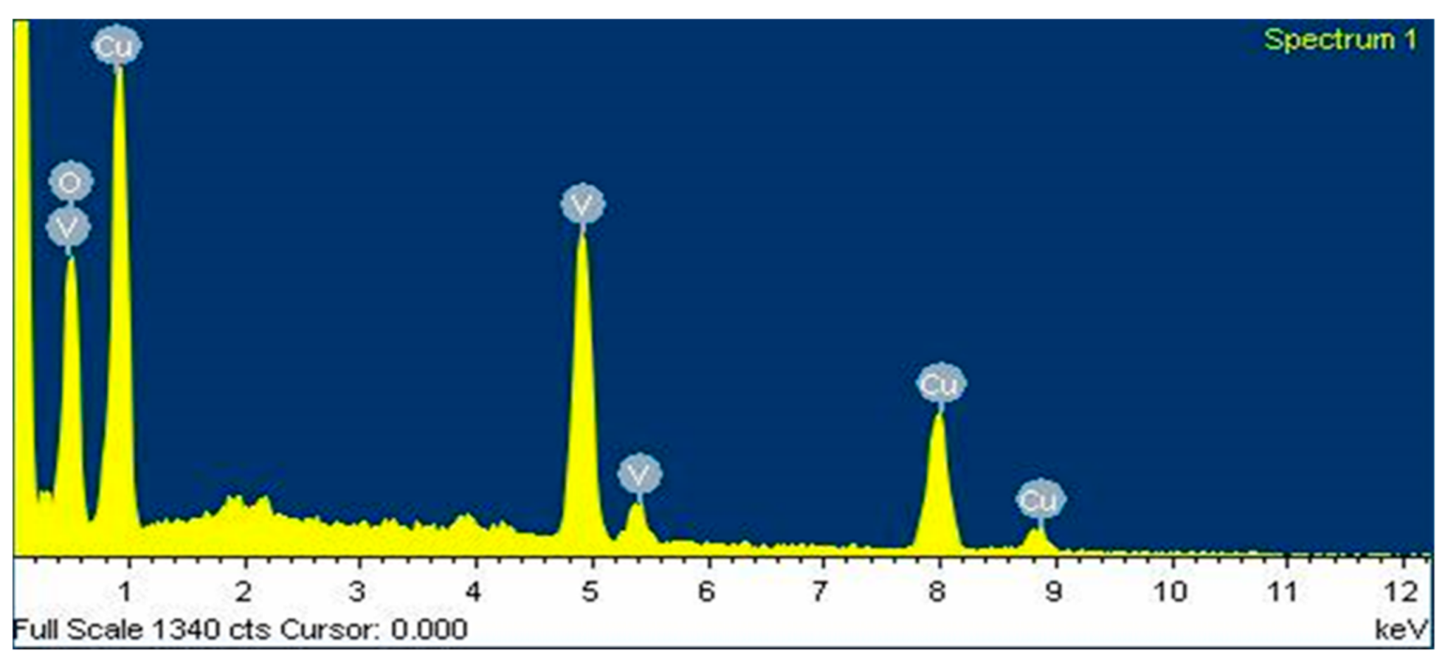
The FESEM image of Cu-VO showed the presence of well-dispersed spheroid-like particles (Figure 1) [27]. Cu-VO had particle sizes between 25 and 350 nm. The average particle size of Cu-VO was 133 nm. The EDX spectrum of Cu-VO showed the presence of Cu, V, and O elements in the single crystal of Cu-VO (Figure 2). The weight and atomic percentages of the elements present in Cu-VO are shown in Table 1. The atomic ratio of Cu:V:O in Cu-VO using EDX data was 3.13:2:7.25. It was observed that the product Cu-VO could have a single phase of Cu3V2O8.
The XRD plot of Cu-VO showed the presence of (100), (−110), (020), (021), (111), (−121), (−211), (021), (121), (−212), (130), (−131), (102), (211), (040), (031), (−123), (212), (−142), (042), (−233), and (−330) peaks (Figure 3) [27]. All these diffraction peaks corresponded to the monoclinic Cu3V2O8 phase with space group P21/c (JCPDS card no. 26-0567) [27]. These results showed the presence of single-phase crystals of Cu3V2O8 in Cu-VO. These results matched well with the EDS analysis of Cu-VO.
The crystallinity of Cu-VO was analyzed using an X-ray diffractometer (Rigaku Ultima IV, Rigaku, Japan), with Cu Kα radiation (wavelength = 15.4 nm) operated at 40 kV and 40 mA. The crystallite size D of Cu-VO was calculated using the Scherrer equation (Table 2) [18]:
D = /βcosθ
where D is the crystalline size; k is the shape factor (0.9); λ is the wavelength of X-ray (1.54056 Å); β is the full width at half maximum (FWHM) of the diffraction peak (in radians); θ is the diffraction angle of the reflection.
The lattice constants were measured by applying the following equation (Table 2) [28]:
1/d2 = 1/sin2β ((h/a)2 + (k/b)2sin2β + (l/c)2 − (2hl cosβ/ac))
where a, b, and c are the lattice parameters, and d is the spacing between the (hkl) planes. β was the unique interaxial angle, reported to be 111.49° for the monoclinic Cu3V2O8 crystal system [29].
The lattice strain (ε) was measured using the tangent formula (Table 2) [30]:
ε = β/4tanθ
where β is the full width at half maximum (FWHM) of the diffraction peak (in radians); θ is the diffraction angle of the reflection.
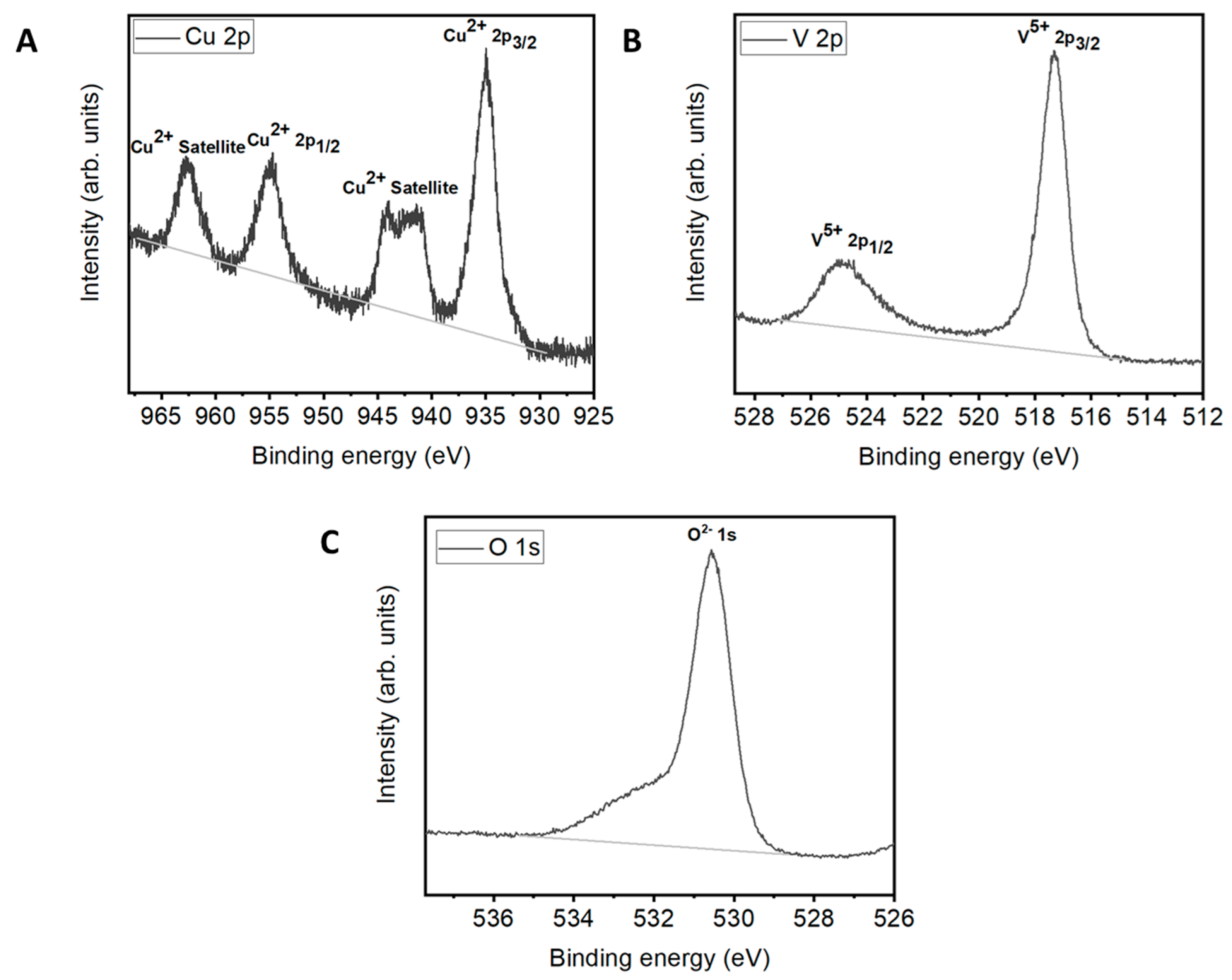
The high resolution XPS spectrum of Cu 2p showed the main characteristic doublet peaks at 955 and 935 eV, corresponding to Cu2+ 2p3/2 and Cu2+ 2p1/2 (Figure 4A) [31]. Two additional strong satellite peaks other than the main peaks of Cu2+ 2p appeared at 962.5 and 942 eV. The binding energy (BE) difference of the spin orbits of Cu2+ 2p3/2 and Cu2+ 2p1/2 was approximately 20 eV, which matched well with the values reported earlier [31]. The multiple excitations of CuO generated satellite peaks toward the higher BE side of the core level Cu 2p XPS. These satellite peaks were attributed to the 2p03d9 configuration of Cu2+ in the CuO phase [32]. The high-resolution XPS spectrum of V 2p showed the main characteristic doublet peaks V5+ 2p3/2 and V5+ 2p1/2 at 517.3 and 525.1 eV, respectively (Figure 4B). The difference between the BE of V5+ 2p3/2 and V5+ 2p1/2 was 7.8 eV, which matched well with the values reported earlier [27]. The XPS spectrum of O 1s showed a main peak at 530.5 eV, which corresponded to O2− 1s (Figure 4C).
The UV–Vis/NIR spectrum of Cu-VO was shown in Figure 5A. Cu-VO showed the broad absorption spectrum from the UV region to the visible region (~200 to ~600 nm) [33]. The broad absorption spectrum indicated that the Cu-VO could have excellent solar-energy-harvesting capacity. The direct and indirect band gaps of Cu-VO were calculated using Tauc’s plot (Figure 5B). Cu-VO had one indirect band gap of ~1.84 eV and two direct band gaps of 1.18 and 3.2 eV. The band gap values of Cu-VO revealed that Cu-VO could be a good solar light-active photocatalyst. The results are in good agreement with the photocatalytic activity of Cu-VO obtained against M. aeruginosa and MB, as discussed below.
The FT-IR spectrum of Cu-VO was shown in Figure 6. The absorption band in the range of 3750–3250 cm−1 could be assigned to the stretching vibration of hydroxyl groups of the water adsorbed on the surface of Cu-VO [18]. The bending vibration of the hydroxyl group appeared at around 1630 cm−1. The FT-IR spectrum of Cu-VO showed the characteristic bands for VO43− at 900–700 cm−1 [34]. The vibration of the Cu-O bond appeared at around 420 cm−1 [35]. The bands in the range of 960–450 cm−1 indicated the presence of tetrahedral VO4 and octahedral CuO6 crystals in Cu-VO. FT-IR spectra of Cu-VO matched well with the XRD and EDS results.

2.3. Antialgal Assay

The antialgal activity of Cu-VO was shown in Figure 6. A change in the pigment of M. aeruginosa was observed after incubating with various concentrations of Cu-VO (Figure 7A). Cu-VO showed concentration-dependent growth inhibition of M. aeruginosa. The culture of M. aeruginosa incubated with 8 mg/L, 12 mg/L, and 24 mg/L of Cu-VO displayed change of pigment (dark green to faint green) within 3 h of incubation, whereas 4 mg/L of Cu-VO exhibited a change in pigment within 4 h of incubation. The lowest concentration of Cu-VO (0.4 mg/L) was found inefficient for algal growth inhibition.
OD680 value of M. aeruginosa was decreased in a concentration-dependent manner (Figure 7B). The kinetics of algal growth inhibition was initially slow, whereas the sudden increase was observed after a specific time interval. The algal cells incubated with 0.4 mg/L of Cu-VO showed growth of algae similar to control (C). Overall, the suppression of M. aeruginosa growth by Cu-VO was justified in this work. Four mg/L was found to be the minimum effective concentration of Cu-VO for the antialgal activity.

2.4. Dye Degradation Assay

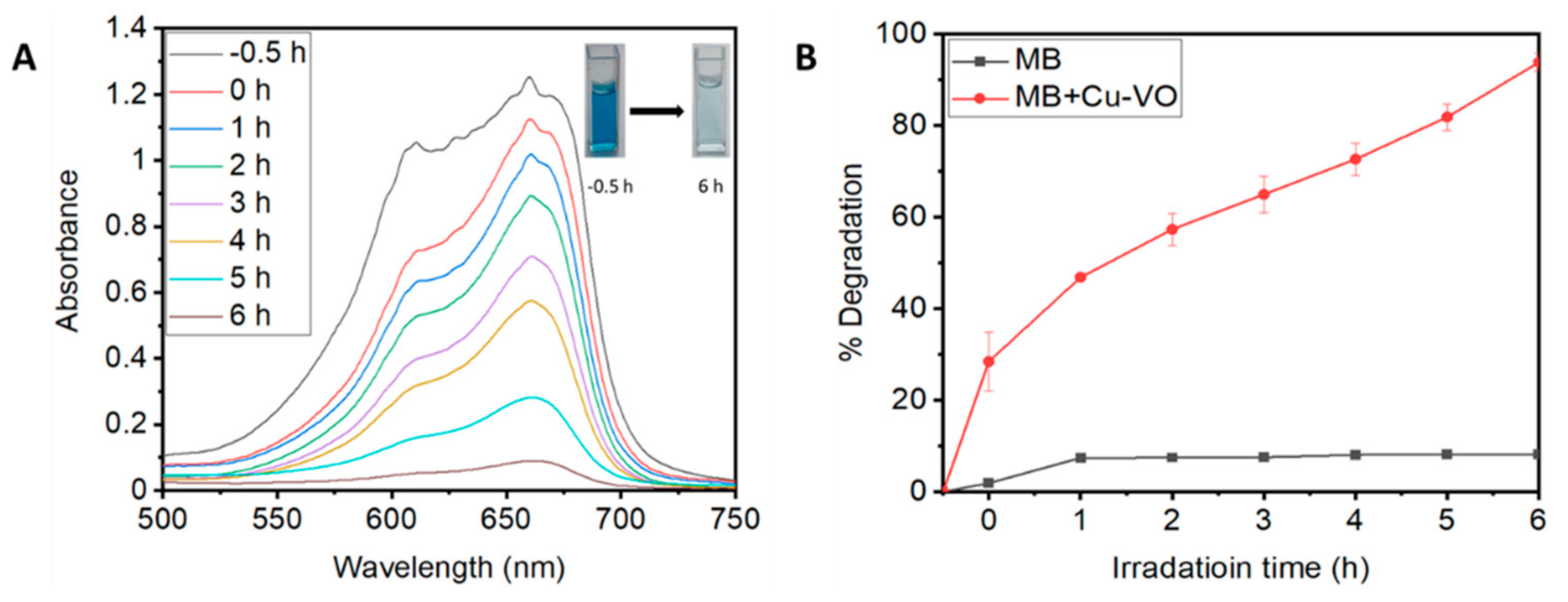
The UV–Vis spectrum of MB treated with Cu-VO was shown in Figure 8A. The absorbance of MB decreased with time, indicating the decrease in concentration in the presence of Cu-VO. The color of the MB dye solution almost disappeared within 6 h in the presence of Cu-VO. The degradation percentage of MB was shown in Figure 8B. Cu-VO degraded almost 93% of MB within 6 h of incubation under natural solar light.
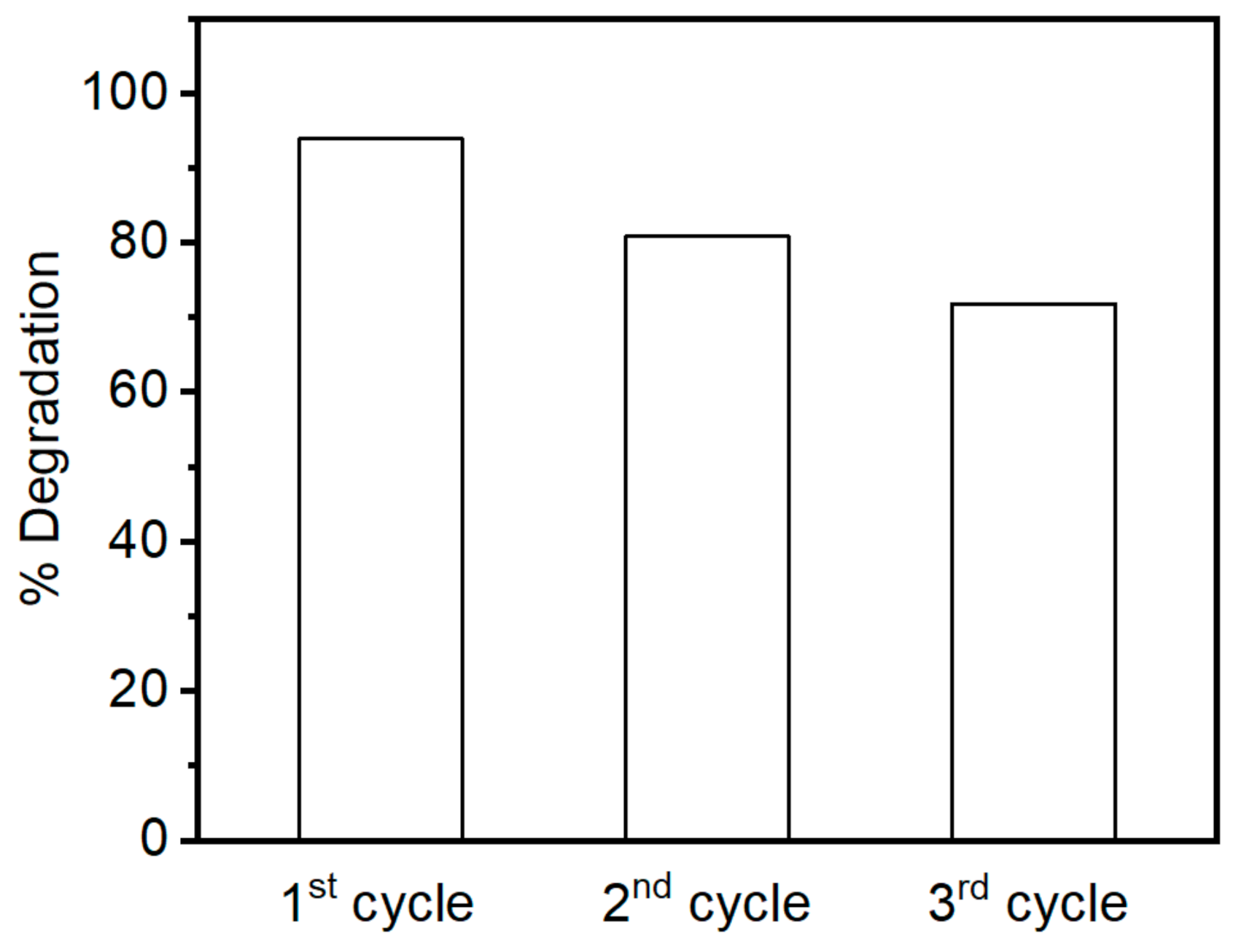
The stability and reusability were critical factors for industrial applications of the photocatalyst. The stability of photocatalyst is proportionally correlated with reusability. The dye degradation experiments were performed to determine the stability and reusability of Cu-VO (Figure 9). Similar experimental conditions were maintained throughout the experiment, given in Section 4.3.2. After each experiment, the solution was centrifuged to collect the Cu-VO. The harvested Cu-VO was washed with distilled water, dried, and resuspended in a new MB solution. Cu-VO still showed significant dye degradation activity even up to three cycles we performed. The photocatalytic activity of Cu-VO decreased by 20% in the third cycle compared to the first cycle. The decreased photocatalytic activity of Cu-VO could be due to its aggregation in the reaction medium, as shown in Figure S1 [36]. In other words, Cu-VO could be reusable at least up to three cycles
Copper [37] and vanadium [38] are common toxic heavy metals that contaminate drinking water, causing harmful effects on aquatic animals. The use of copper and vanadium photocatalysts for industrial purposes could cause their leaching into the wastewater bodies. Therefore, to ensure the safety of synthesized Cu-VO, leaching of copper and vanadium into the medium was analyzed using ICP-OES. Eight mg/L of Cu-VO was added to the distilled water and sonicated for 30 min. A resulting solution was kept under the sun light for up to 6 h (sunny day; clear sky). After 6 h of incubation, the solution was centrifuged at 4000 rpm for 5 min and filtered using a 0.22 μm membrane filter. The obtained filtrate was used for ICP-OES analysis. An intensity versus concentration graph of the standard solution (see supporting information Figure S2) of copper and vanadium was used to calculate the concentration of leached copper and vanadium into the medium. The concentration of leached copper and vanadium in the filtrate was 0.088 mg/L (1.1%) and 0.157 mg/L (1.9%), respectively.

2.5. Mechanism of Action

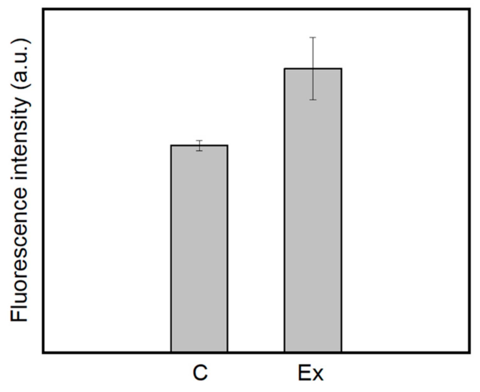
Generation of extracellular ·OH by Cu-VO in BG11 medium under natural solar light was investigated using ·OH assay (Figure 10). The fluorescence intensity of the control groups (C) was lower than that of the experimental groups (Ex). Isopropanol, an ·OH scavenger, could decrease the number of ·OH generated by Cu-VO. Thus, the fluorescence intensity was decreased in the control group in the presence of isopropanol [39]. The fluorescence intensity of the experimental group was 1.42-fold higher than that of the control group.
The plausible mechanism of ·OH free radicals generated by Cu-VO was given as follows:
Cu-VO e C B + h + V B
h + + H 2 O O H + H +
The absorption of photon energy by Cu-VO was equal to or greater than the energy of the band gap, which led to the transition of electrons from the valence band to the conduction band (e (CB)), leaving behind positively charged valence band or holes (h+ (VB)) [40]. Holes can remove electrons from nearby water molecules, producing ·OH radicals. The highly reactive ·OH radicals readily degraded harmful cyanobacteria and dye.
Intracellular ROS induced by different concentrations of Cu-VO in the M. aeruginosa cells was measured up to 4 h (Figure 11). M. aeruginosa cells showed concentration-dependent intracellular ROS production.

3. Discussion

In this study, we successfully synthesized Cu-VO using a simple microwave method. The synthesis of Cu-VO with this method was easy and fast (10 min) compared to other methods, such as thermal decomposition [29], simple heating [17], chemical precipitation [41], and hydrothermal [42]. These results indicate that microwave-assisted synthesis of Cu-VO is a highly efficient method. FESEM, XRD, and XPS results of Cu-VO matched well with the results obtained by Li et al. [27]. The size of Cu-VO obtained in this study was twice that obtained by Li et al. [27]. Cu-VO had a spheroid shape with a monoclinic crystal structure. The direct and indirect band gap values of Cu-VO were 1.8 eV (visible region), 3.2 eV (UV region), and 1.84 eV (visible region), indicating the ability to show efficient photocatalytic activity under solar light [43].
Cu-VO showed better growth inhibition against M. aeruginosa at a minimum concentration of 4 mg/L within 4 h under natural solar light than other metal oxides [7,44,45]. CuO nanoparticles inhibited algal growth within 96 h at a concentration of 50 mg/L [44]. Cu-MOF-74 inhibited the growth of algal cells within 120 h at an effective concentration of 5 mg/L [45]. Cu-g-C3N4 showed algal growth inhibition within 96 h at an effective concentration of 100 mg/L [7]. These results indicate that Cu-VO is a highly efficient antialgal agent compared to CuO, Cu-MOF-74, and Cu-g-C3N4. Additionally 200 mg/L of Cu-VO degraded 93% MB (50 μM) within 6 h under solar light irradiation. A total of 200 mg/L of V2O5 itself showed poor MB (50 μM) degradation efficiency (24%) within 3 h [9]. The UV/Vis-active hollow Cu3V2O8 (1000 mg/L) degraded 78% of methyl orange (MO) (20 mg/L) within 2 h [17]. In situ- and ex situ-prepared Cu3V2O8-Cu2V2O7 (250 mg/L) degraded 51% and 83% of MB (20 mg/L) within 2 h under UV light, respectively [18]. A total of 500 mg/L of the visible light-active V2O5/ZnO composite efficiently degraded the 500 mg/L of MB within 2 h [46]. The quantum dots of vanadium oxide were found to be inefficient for the degradation of rhodamine B (25 mg/L) or MB, but the same quantum dots were highly efficient in combination with H2O2 or magnetic Fe3O4@SiO2 + H2O2 [47]. All of these results indicate that Cu-VO is highly efficient for MB degradation compared to V2O5, moderately efficient compared to hollow Cu3V2O8 or Cu3V2O8-Cu2V2O7, and poorly efficient compared to V2O5/ZnO, V2O5 + H2O2, and V2O5 + magnetic Fe3O4@SiO2 + H2O2.
The stability and reusability of Cu-VO was comparable to other photocatalysts [48,49,50]. VS4/CP (CP, carbon powder) and BaTiO3@g-C3N4 showed 90% and 96.6% degradation of MO and phenyl red in the fourth cycle, respectively [48,49]. BaTiO3@g-C3N4 showed 67–76% degradation of MO in the third cycle [50]. Cu-VO showed 72% degradation of MB in the third cycle, indicating moderate stability compared to VS4/CP (CP, carbon powder) and BaTiO3@g-C3N4. The stability of Cu-VO was similar to BaTiO3@g-C3N4. Zhang et al. [51] and Cai et al. [48] found that the leaching of vanadium in water from VS4/CP was <1%, which was considered stable and safer to use [38]. The leaching of copper and vanadium was less than 2% of 8 mg/L of Cu-VO, indicating that it could be stable and safer for practical use.
Metal oxides generated reactive oxygen species (ROS) such as ·OH, ·O2−, h+, and HO2· in the presence of light [52]. ROS has a high oxidation potential to oxidize organic contaminants such as microorganisms, dye, and other pollutants. Regmi et al. [53] found that nickel-doped bismuth vanadate generated ·OH, ·O2−, and h+ under visible light. Zhang et al. [54] found that copper vanadium oxide rapidly dissociated H2O2 to produce ·OH radicals, which could easily degrade fluconazole. The enhanced dye degradation efficiency of V2O5 in the presence of H2O2, indicating the generation of reactive ·OH, played a crucial role in dye degradation [47]. Zinc-doped copper oxide nanoparticles generated reactive ·OH for the growth inhibition of multidrug-resistant bacteria [55]. We hypothesized that Cu-VO could generate ·OH in the medium, which can degrade algal cells and dye. The increased fluorescence intensity of terephthalic acid in the presence of Cu-VO indicated the production of ·OH in the medium. Additionally, to support this result, we performed the same experiment in the presence of isopropanol [56], a well-known ·OH scavenger. The fluorescence intensity of the terephthalic acid was decreased in the presence of isopropanol, which supported our hypothesis. The presence of ·OH in the algal medium induced oxidative stress in the algal cells [56]. The antitumor activity of vanadium dioxide nanocoated quartz glass was due to the induction of intracellular ROS [57,58]. It was found that ZnO-V2O5 triggered the upsurge of ROS in bacteria, causing oxidative stress in bacteria [59]. The ROS assay showed an increase in the fluorescence intensity of nonfluorescent 2’,7’-dichlorofluorescein diacetate (DCFH-DA) incubated with Cu-VO-treated algal cells. An increased fluorescence of DCFH-DA indicated the production of intracellular ROS in M. aeruginosa. Intracellular ROS was a highly reactive species, degrading important intracellular biomolecules such as DNA, RNA, proteins, and lipids, leading to cell death. Thus, the main photocatalytic mechanism of Cu-VO was the generation of extracellular ·OH and intracellular ROS.

4. Materials and Methods

4.1. Synthesis of Cu-VO

Copper–vanadium oxide (Cu-VO) was synthesized in the aqueous medium using a microwave oven (Samsung, Korea, Model number RE-MC20G). In a typical process, 3 mmol of CuSO4·5H2O, 2 mmol of KVO3, and 1 g urea were added in 50 mL distilled water with vigorous stirring in a 250 mL conical flask [41]. The conical flask was covered with aluminum foil near the neck. After 15 min, the conical flask was irradiated with a 700 W microwave for 10 min. The reaction was stopped after every 10 s, and the reaction mixture was stirred for 10 s. The solution was evaporated during the reaction. The temperature of the reaction was monitored using a thermometer. After 10 min, the solution temperature reached 80 °C, and the color of the reaction mixture turned colorless to yellow. The yellow-colored product was cooled and centrifuged at 4000 rpm for 20 min. The product was washed four times with distilled water and three times with absolute ethanol, followed by drying at 60 °C for 6 h in an oven. The dry product was annealed at 400 °C for 2 h at the heating rate of 5 °C/min. Afterward, the product was stored in a sealed glass tube.

4.2. Characterization

The morphology of Cu-VO was analyzed by a field-emission scanning electron microscopy (FESEM, JSM-6700F, JEOL, Tokyo, Japan). Atomic percentage of Cu, V, and O in Cu-VO was analyzed using an energy dispersive X-ray spectrometer (EDX, INCAx-sight, Oxford, UK). The crystalline nature of Cu-VO was analyzed by XRD analysis (Rigaku Ultima IV, Rigaku, Japan; Cu Kα X-ray radiation). XPS measurement was carried out on a THERMO VG SCIENTIFIC (MultiLab 2000, Spring Lake, MI, USA) X-ray photoelectron spectroscopes with a monochromatic Al-Ka source to explore the elements on the surface of Cu-VO. The band gap of Cu-VO was determined by performing UV–Vis/NIR analysis. FT-IR analysis was performed to find the type of bond present in Cu-VO (JASCO FT/IR-4100, Tokyo, Japan).

4.3. Photocatalytic Experiments

The photocatalytic experiments were performed under natural solar light on sunny days [51]. The sky was clear, with slight fluctuations in solar intensity. Experiments were performed between 10 a.m. and 5 p.m.

4.3.1. Algae Culture

A strain of M. aeruginosa (No. FBCC000002) was provided by the Nakdonggang National Institute of Biological Resources (Sangju, Korea). The M. aeruginosa was cultured according to the condition given in our previous paper [7].

4.3.2. Algae Growth Inhibition Assay

The growth inhibition assay was performed using the same procedure mentioned in our previous paper [7] with slight changes. Different concentrations of Cu-VO (0, 0.4, 4, 8, 12, and 24 mg/L) were incubated with M. aeruginosa under natural solar light.

4.3.3. Dye Degradation Assay

Cu-VO was applied for the degradation of methylene blue (MB) solution under solar light. The photocatalytic activity was carried out in a conical flask. A total of 200 mg/L of Cu-VO was added to 50 μM solutions of MB. The resulting mixture was sonicated for 30 min to maintain adsorption–desorption equilibrium. Then, the conical flask was kept under natural solar light. The aliquots of the reaction mixture were filtered at every 1 h interval using a 0.22 μm filter. UV–Vis absorbance (BioDrop Duo, England, Cambridge, UK) of the filtered solution was measured, and degradation percentage was calculated as follows:
%Degradation = ((AiAf)/Ai) × 100%
where Ai and Af are the absorbance value (λmax = 660 nm) of MB solution at time 0 and t min, respectively.

4.4. Photocatalysis Mechanism

4.4.1. ·OH Assay

·OH assay was performed using the same procedure as mentioned in our previous paper [56] with slight changes. The terephthalic acid solution was incubated with 4 mg/L of Cu-VO under natural solar light.

4.4.2. ROS Assay

ROS assay was performed using the same procedure as mentioned in our previous paper [56].

5. Conclusions

Cu-VO nanoparticles were synthesized using a rapid and facile microwave method. The characterization of Cu-VO using FESEM, EDX, XRD, XPS, and FT-IR measurements indicated that Cu-VO contained typical monoclinic nanocrystals of approximately 58 nm. The calculated values of lattice constants from XRD data were matched well with the values given in the literature. XPS measurement showed the presence of Cu2+2p1/2, Cu2+2p3/2, V5+2p1/2, V5+2p3/2, and O2−1s in Cu-VO. FESEM analysis showed that the average particle size of Cu-VO was 132 nm. EDX analysis of Cu-VO showed the presence of Cu, V, and O elements in Cu-VO with atomic percentages of 3.13%, 2%, and 7.25%, respectively. XRD and FT-IR analysis revealed the presence of tetrahedral VO4 and octahedral CuO6 crystals in Cu-VO. The investigation of the antialgal activity showed that Cu-VO inhibited the growth of algae in a concentration-dependent manner. The superior antialgal activity was found at a minimum effective concentration value of 4 mg/L under solar light. Cu-VO degraded 93% of methylene blue dye within 6 h. It was stable and safer to reuse in up to three consecutive cycles. The generation of highly reactive ·OH radicals was indicated by the increase in the fluorescence of terephthalic acid under solar light. The ·OH radical efficiently reacted with algae and dye, causing the degradation of algae and dye. The presence of the ·OH radical in the algal medium produced intracellular ROS in algae. The oxidative stress developed by intracellular ROS could destroy cell organelle, ultimately causing algal death. Furthermore, our study suggested that a solar-light-active Cu-VO could be used as an efficient nanoparticle for wastewater treatment.

Supplementary Materials

The following are available online at https://www.mdpi.com/2073-4344/11/1/36/s1, Figure S1: Optical microscopic image of 200 mg/L of Cu-VO. Figure S2: Calibration curves for (A) Cu and (B) V using ICP-OES.

Author Contributions

Conceptualization: S.N., and H.J.K. methodology: S.N., S.B., and H.J.K. validation: S.N., J.H.C., and H.J.K. formal analysis: S.N. and H.J.K. investigation: S.N. and S.B. data curation: S.N. and H.J.K. writing—original draft preparation: S.N. writing—review and editing: S.N., and H.J.K. visualization: S.N. and H.J.K. supervision: H.J.K. funding acquisition: H.J.K. All authors have read and agreed to the published version of the manuscript.

Funding

This research was funded by Pukyong National University (2019).

Conflicts of Interest

The authors declare no conflict of interest.

References

  1. Zhang, C.; Li, Y.; Shuai, D.; Shen, Y.; Xiong, W.; Wang, L. Graphitic carbon nitride (g-C3N4)-based photocatalysts for water disinfection and microbial control: A review. Chemosphere 2019, 214, 462–479. [Google Scholar] [CrossRef] [PubMed]
  2. Dai, R.; Wang, P.; Jia, P.; Zhang, Y.; Chu, X.; Wang, Y. A review on factors affecting microcystins production by algae in aquatic environments. World J. Microbiol. Biotechnol. 2016, 32, 51. [Google Scholar] [CrossRef] [PubMed]
  3. Halbus, A.F.; Horozov, T.S.; Paunov, V.N. Surface-Modified Zinc Oxide Nanoparticles for Antialgal and Antiyeast Applications. ACS Appl. Nano Mater. 2020, 3, 440–451. [Google Scholar] [CrossRef] [Green Version]
  4. El-Sheekh, M.M.; Haroon, A.; Sabae, S. Activity of some Nile River aquatic macrophyte extracts against the cyanobacterium Microcystis aeruginosa. Afr. J. Aquat. Sci. 2017, 42, 271–277. [Google Scholar] [CrossRef]
  5. Begum, R.; Najeeb, J.; Sattar, A.; Naseem, K.; Irfan, A.; Al-Sehemi, A.G.; Farooqi, Z.H. Chemical reduction of methylene blue in the presence of nanocatalysts: A critical review. Rev. Chem. Eng. 2020, 36, 749–770. [Google Scholar] [CrossRef]
  6. Khaki, M.R.D.; Shafeeyan, M.S.; Raman, A.A.A.; Daud, W.M.A.W. Application of doped photocatalysts for organic pollutant degradation—A review. J. Environ. Manag. 2017, 198, 78–94. [Google Scholar] [CrossRef]
  7. Cao, X.; Yue, L.; Lian, F.; Wang, C.; Cheng, B.; Lv, J.; Wang, Z.; Xing, B. CuO nanoparticles doping recovered the photocatalytic antialgal activity of graphitic carbon nitride. J. Hazard. Mater. 2021, 403, 123621. [Google Scholar] [CrossRef]
  8. Lebedev, A.; Anariba, F.; Tan, J.C.; Li, X.; Wu, P. A review of physiochemical and photocatalytic properties of metal oxides against Escherichia coli. J. Photochem. Photobiol. A Chem. 2018, 360, 306–315. [Google Scholar] [CrossRef]
  9. Jayaraj, S.K.; Sadishkumar, V.; Thirumurugan, A.; Thangadurai, P. Enhanced photocatalytic activity of V2O5 nanorods for the photodegradation of organic dyes: A detailed understanding of the mechanism and their antibacterial activity. Mater. Sci. Semicond. Process. 2018, 85, 122–133. [Google Scholar] [CrossRef]
  10. Zeleke, M.A.; Kuo, D.-H. Synthesis and application of V2O5-CeO2 nanocomposite catalyst for enhanced degradation of methylene blue under visible light illumination. Chemosphere 2019, 235, 935–944. [Google Scholar] [CrossRef]
  11. Simo, A.; Drah, M.; Sibuyi, N.; Nkosi, M.; Meyer, M.; Maaza, M. Hydrothermal synthesis of cobalt-doped vanadium oxides: Antimicrobial activity study. Ceram. Int. 2018, 44, 7716–7722. [Google Scholar] [CrossRef]
  12. Suresh, R.; Giribabu, K.; Manigandan, R.; Munusamy, S.; Kumar, S.P.; Muthamizh, S.; Stephen, A.; Narayanan, V. Doping of Co into V2O5 nanoparticles enhances photodegradation of methylene blue. J. Alloys Compd. 2014, 598, 151–160. [Google Scholar] [CrossRef]
  13. He, P.; Zhang, G.; Liao, X.; Yan, M.; Xu, X.; An, Q.; Liu, J.; Mai, L. Sodium Ion Stabilized Vanadium Oxide Nanowire Cathode for High-Performance Zinc-Ion Batteries. Adv. Energy Mater. 2018, 8, 1702463. [Google Scholar] [CrossRef]
  14. Ni, S.; Ma, J.; Zhang, J.; Yang, X.; Zhang, L. Electrochemical performance of cobalt vanadium oxide/natural graphite as anode for lithium ion batteries. J. Power Sources 2015, 282, 65–69. [Google Scholar] [CrossRef]
  15. Seabold, J.A.; Neale, N.R. All First Row Transition Metal Oxide Photoanode for Water Splitting Based on Cu3V2O8. Chem. Mater. 2015, 27, 1005–1013. [Google Scholar] [CrossRef]
  16. Zelenski, M.E.; Zubkova, N.V.; Pekov, I.V.; Boldyreva, M.M.; Pushcharovsky, D.Y.; Nekrasov, A.N. Pseudolyonsite, Cu3(VO4)2, a new mineral species from the Tolbachik volcano, Kamchatka Peninsula, Russia. Eur. J. Miner. 2011, 23, 475–481. [Google Scholar] [CrossRef]
  17. Zhang, S.; Sun, Y.; Li, C.; Ci, L. Cu3V2O8 hollow spheres in photocatalysis and primary lithium batteries. Solid State Sci. 2013, 25, 15–21. [Google Scholar] [CrossRef]
  18. Ghiyasiyan-Arani, M.; Masjedi-Arani, M.; Salavati-Niasari, M. Facile synthesis, characterization and optical properties of copper vanadate nanostructures for enhanced photocatalytic activity. J. Mater. Sci. Mater. Electron. 2016, 27, 4871–4878. [Google Scholar] [CrossRef]
  19. Jain, S.; Sharma, B.; Thakur, N.; Mishra, S.; Sarma, T.K. Copper Pyrovanadate Nanoribbons as Efficient Multienzyme Mimicking Nanozyme for Biosensing Applications. ACS Appl. Nano Mater. 2020, 3, 7917–7929. [Google Scholar] [CrossRef]
  20. Karim, N.; Singh, M.; Weerathunge, P.; Bian, P.; Zheng, R.; Dekiwadia, C.; Ahmed, T.; Walia, S.; Della Gaspera, E.; Singh, S.; et al. Visible-Light-Triggered Reactive-Oxygen-Species-Mediated Antibacterial Activity of Peroxidase-Mimic CuO Nanorods. ACS Appl. Nano Mater. 2018, 1, 1694–1704. [Google Scholar] [CrossRef]
  21. Jing, L.; Xu, Y.; Liu, J.; Zhou, M.; Xu, H.; Xie, M.; Li, H.; Xie, J. Direct Z-scheme red carbon nitride/rod-like lanthanum vanadate composites with enhanced photodegradation of antibiotic contaminants. Appl. Catal. B Environ. 2020, 277, 119245. [Google Scholar] [CrossRef]
  22. Kadam, R.L.; Kim, Y.; Gailkwad, S.; Chang, M.; Tarte, N.H.; Han, S. Catalytic Decolorization of Rhodamine B, Congo Red, and Crystal Violet Dyes, with a Novel Niobium Oxide Anchored Molybdenum (Nb–O–Mo). Catalysts 2020, 10, 491. [Google Scholar] [CrossRef]
  23. Zhang, X.; Zhang, J.; Yu, J.; Zhang, Y.; Yu, F.; Jia, L.; Tan, Y.; Zhu, Y.; Hou, B. Enhancement in the photocatalytic antifouling efficiency over cherimoya-like InVO4/BiVO4 with a new vanadium source. J. Colloid Interface Sci. 2019, 533, 358–368. [Google Scholar] [CrossRef] [PubMed]
  24. Yan, X.; Sun, S.; Hu, B.; Wang, X.; Lu, W.; Shi, W. Enhanced photocatalytic activity induced by surface plasmon resonance on Ag-loaded strontium titanate nanoparticles. Micro Nano Lett. 2013, 8, 504–507. [Google Scholar] [CrossRef]
  25. Suman, T.Y.; Radhika, S.R.R.; Kirubagaran, R. Evaluation of zinc oxide nanoparticles toxicity on marine algae chlorella vulgaris through flow cytometric, cytotoxicity and oxidative stress analysis. Ecotoxicol. Environ. Saf. 2015, 113, 23–30. [Google Scholar] [CrossRef]
  26. Brynildsen, M.P.; Winkler, J.A.; Spina, C.S.; MacDonald, I.C.; Collins, J.J. Potentiating antibacterial activity by predictably enhancing endogenous microbial ROS production. Nat. Biotechnol. 2013, 31, 160–165. [Google Scholar] [CrossRef] [PubMed] [Green Version]
  27. Li, M.; Gao, Y.; Chen, N.; Meng, X.; Wang, C.; Zhang, Y.; Zhang, D.; Wei, Y.; Du, F.; Chen, G. Cu3V2O8 Nanoparticles as Intercalation-Type Anode Material for Lithium-Ion Batteries. Chem. A Eur. J. 2016, 22, 11405–11412. [Google Scholar] [CrossRef]
  28. Saud, S.N.; Hamzah, E.; Abu Bakar, T.A.; Farahany, S. Structure-Property Relationship of Cu-Al-Ni-Fe Shape Memory Alloys in Different Quenching Media. J. Mater. Eng. Perform. 2014, 23, 255–261. [Google Scholar] [CrossRef]
  29. Shannon, R.D.; Calvo, C. Crystal Structure of a New Form of Cu3V2O8. Can. J. Chem. 1972, 50, 3944–3949. [Google Scholar] [CrossRef] [Green Version]
  30. Boukhachem, A.; Ouni, B.; Karyaoui, M.; Madani, A.; Chtourou, R.; Amlouk, M. Structural, opto-thermal and electrical properties of ZnO:Mo sprayed thin films. Mater. Sci. Semicond. Process. 2012, 15, 282–292. [Google Scholar] [CrossRef]
  31. Akgul, F.A.; Akgul, G.; Yildirim, N.; Unalan, H.E.; Turan, R. Influence of thermal annealing on microstructural, morphological, optical properties and surface electronic structure of copper oxide thin films. Mater. Chem. Phys. 2014, 147, 987–995. [Google Scholar] [CrossRef]
  32. Svintsitskiy, D.; Kardash, T.Y.; Boronin, A.I. Surface dynamics of mixed silver-copper oxide AgCuO2 during X-ray photoelectron spectroscopy study. Appl. Surf. Sci. 2019, 463, 300–309. [Google Scholar] [CrossRef]
  33. Sivakumar, V.; Suresh, R.; Giribabu, K.; Manigandan, R.; Munusamy, S.; Kumar, S.P.; Muthamizh, S.; Narayanan, V. Copper vanadate nanoparticles: Synthesis, characterization and its electrochemical sensing property. J. Mater. Sci. Mater. Electron. 2014, 25, 1485–1491. [Google Scholar] [CrossRef]
  34. Liu, G.; Duan, X.; Li, H.; Dong, H. Hydrothermal synthesis, characterization and optical properties of novel fishbone-like LaVO4:Eu3+ nanocrystals. Mater. Chem. Phys. 2009, 115, 165–171. [Google Scholar] [CrossRef]
  35. Lin, Z.; Li, W.; Yao, J. Hydrogen-interstitial CuWO4 nanomesh: A single-component full spectrum-active photocatalyst for hydrogen evolution. Appl. Catal. B Environ. 2018, 227, 35–43. [Google Scholar] [CrossRef]
  36. Ahmed, S.; Rasul, M.G.; Martens, W.N.; Brown, R.; Hashib, M.A. Advances in Heterogeneous Photocatalytic Degradation of Phenols and Dyes in Wastewater: A Review. Water Air Soil Pollut. 2011, 215, 3–29. [Google Scholar] [CrossRef] [Green Version]
  37. Al-Saydeh, S.A.; El-Naas, M.H.; Zaidi, S.J. Copper removal from industrial wastewater: A comprehensive review. J. Ind. Eng. Chem. 2017, 56, 35–44. [Google Scholar] [CrossRef]
  38. Schiffer, S.; Liber, K. Estimation of vanadium water quality benchmarks for the protection of aquatic life with relevance to the Athabasca Oil Sands region using species sensitivity distributions. Environ. Toxicol. Chem. 2017, 36, 3034–3044. [Google Scholar] [CrossRef]
  39. Phongarthit, K.; Amornpitoksuk, P.; Suwanboon, S. Synthesis, characterization, and photocatalytic properties of ZnO nanoparticles prepared by a precipitation-calcination method using a natural alkaline solution. Mater. Res. Express 2019, 6, 045501. [Google Scholar] [CrossRef]
  40. Ali, I.; Alharbi, O.M.L.; Alothman, Z.A.; Badjah, A.Y. Kinetics, Thermodynamics, and Modeling of Amido Black Dye Photo-degradation in Water Using Co/TiO2 Nanoparticles. Photochem. Photobiol. 2018, 94, 935–941. [Google Scholar] [CrossRef]
  41. Ghiyasiyan-Arani, M.; Masjedi-Arani, M.; Ghanbari, D.; Bagheri, S.; Salavati-Niasari, M. Novel chemical synthesis and characterization of copper pyrovanadate nanoparticles and its influence on the flame retardancy of polymeric nanocomposites. Sci. Rep. 2016, 6, 25231. [Google Scholar] [CrossRef] [PubMed] [Green Version]
  42. Leblanc, M.; Ferey, G. Room-temperature structures of oxocopper(II) vanadate(V) hydrates, Cu3V2O8(H2O) and CuV2O6(H2O)2. Acta Crystallogr. Sect. C Cryst. Struct. Commun. 1990, 46, 15–18. [Google Scholar] [CrossRef]
  43. Harish, K.N.; Naik, H.B.; Kumar, P.N.P.; Viswanath, P. Optical and Photocatalytic Properties of Solar Light Active Nd-Substituted Ni Ferrite Catalysts: For Environmental Protection. ACS Sustain. Chem. Eng. 2013, 1, 1143–1153. [Google Scholar] [CrossRef]
  44. Sankar, R.; Prasath, B.B.; Nandakumar, R.; Santhanam, P.; Shivashangari, K.S.; Ravikumar, V. Growth inhibition of bloom forming cyanobacterium Microcystis aeruginosa by green route fabricated copper oxide nanoparticles. Environ. Sci. Pollut. Res. 2014, 21, 14232–14240. [Google Scholar] [CrossRef]
  45. Fan, G.; Bao, M.; Zheng, X.; Hong, L.; Zhan, J.; Chen, Z.; Qu, F. Growth inhibition of harmful cyanobacteria by nanocrystalline Cu-MOF-74: Efficiency and its mechanisms. J. Hazard. Mater. 2019, 367, 529–538. [Google Scholar] [CrossRef] [PubMed]
  46. Rajendran, S.; Gupta, V.; Mosquera, E.; Gracia, F. Preparation and characterization of V2O5/ZnO nanocomposite system for photocatalytic application. J. Mol. Liq. 2014, 198, 409–412. [Google Scholar] [CrossRef]
  47. Zhang, M.; Niu, Y.; Xu, Y. Heterogeneous Fenton-like magnetic nanosphere coated with vanadium oxide quantum dots for enhanced organic dyes decolorization. J. Colloid Interface Sci. 2020, 579, 269–281. [Google Scholar] [CrossRef]
  48. Cai, R.; Zhang, Z.; Shi, J.; Li, M.; He, Z. Rapid Photocatalytic Decolorization of Methyl Orange under Visible Light Using VS4/Carbon Powder Nanocomposites. ACS Sustain. Chem. Eng. 2017, 5, 7690–7699. [Google Scholar] [CrossRef]
  49. Vattikuti, S.P.; Police, A.K.R.; Shima, J.; Byonb, C. In Situ fabrication of the Bi2O3-V2O5 hybrid embedded with graphitic carbon nitride nanosheets: Oxygen vacancies mediated enhanced visible-light—Driven photocatalytic degradation of organic pollutants and hydrogen evolution. Appl. Surf. Sci. 2018, 447, 740–756. [Google Scholar] [CrossRef]
  50. Xian, T.; Yang, H.; Di, L.; Dai, J. Enhanced photocatalytic activity of BaTiO3@ g-C3N4 for the degradation of methyl orange under simulated sunlight irradiation. J. Alloys Compd. 2015, 622, 1098–1104. [Google Scholar] [CrossRef]
  51. Zhang, Z.; Zou, S.; Cai, R.; Li, M.; He, Z. Highly-efficient photocatalytic disinfection of Escherichia coli under visible light using carbon supported Vanadium Tetrasulfide nanocomposites. Appl. Catal. B Environ. 2018, 224, 383–393. [Google Scholar] [CrossRef]
  52. Habibi-Yangjeh, A.; Asadzadeh-Khaneghah, S.; Feizpoor, S.; Rouhi, A. Review on heterogeneous photocatalytic disinfection of waterborne, airborne, and foodborne viruses: Can we win against pathogenic viruses? J. Colloid Interface Sci. 2020, 580, 503–514. [Google Scholar] [CrossRef] [PubMed]
  53. Regmi, C.; Kshetri, Y.K.; Kim, T.-H.; Pandey, R.P.; Ray, S.K.; Lee, S.W. Fabrication of Ni-doped BiVO4 semiconductors with enhanced visible-light photocatalytic performances for wastewater treatment. Appl. Surf. Sci. 2017, 413, 253–265. [Google Scholar] [CrossRef]
  54. Zhang, N.; Xue, C.; Wang, K.; Fang, Z. Efficient oxidative degradation of fluconazole by a heterogeneous Fenton process with Cu-V bimetallic catalysts. Chem. Eng. J. 2020, 380, 122516. [Google Scholar] [CrossRef]
  55. Malka, E.; Perelshtein, I.; Lipovsky, A.; Shalom, Y.; Naparstek, L.; Perkas, N.; Patick, T.; Lubart, R.; Nitzan, Y.; Banin, E.; et al. Eradication of Multi-Drug Resistant Bacteria by a Novel Zn-doped CuO Nanocomposite. Small 2013, 9, 4069–4076. [Google Scholar] [CrossRef]
  56. Nandanwar, S.K.; Lee, M.W.; Borkar, S.B.; Cho, J.H.; Tarte, N.H.; Kim, H.J. Synthesis, Characterization, and Anti-Algal Activity of Molybdenum-Doped Metal Oxides. Catalysts 2020, 10, 805. [Google Scholar] [CrossRef]
  57. Li, J.; Jiang, M.; Zhou, H.; Jin, P.; Cheung, K.M.C.; Chu, P.K.; Yeung, K.W. Vanadium Dioxide Nanocoating Induces Tumor Cell Death through Mitochondrial Electron Transport Chain Interruption. Glob. Chall. 2018, 3, 1800058. [Google Scholar] [CrossRef]
  58. Xi, W.-S.; Tang, H.; Liu, Y.-Y.; Liu, C.-Y.; Gao, Y.; Cao, A.; Liu, Y.; Chen, Z.; Wang, H.-F. Cytotoxicity of vanadium oxide nanoparticles and titanium dioxide-coated vanadium oxide nanoparticles to human lung cells. J. Appl. Toxicol. 2019, 40, 567–577. [Google Scholar] [CrossRef]
  59. Sun, H.; Yang, Z.; Pu, Y.; Dou, W.; Wang, C.; Wang, W.; Hao, X.; Chen, S.; Shao, Q.; Dong, M.; et al. Zinc oxide/vanadium pentoxide heterostructures with enhanced day-night antibacterial activities. J. Colloid Interface Sci. 2019, 547, 40–49. [Google Scholar] [CrossRef]
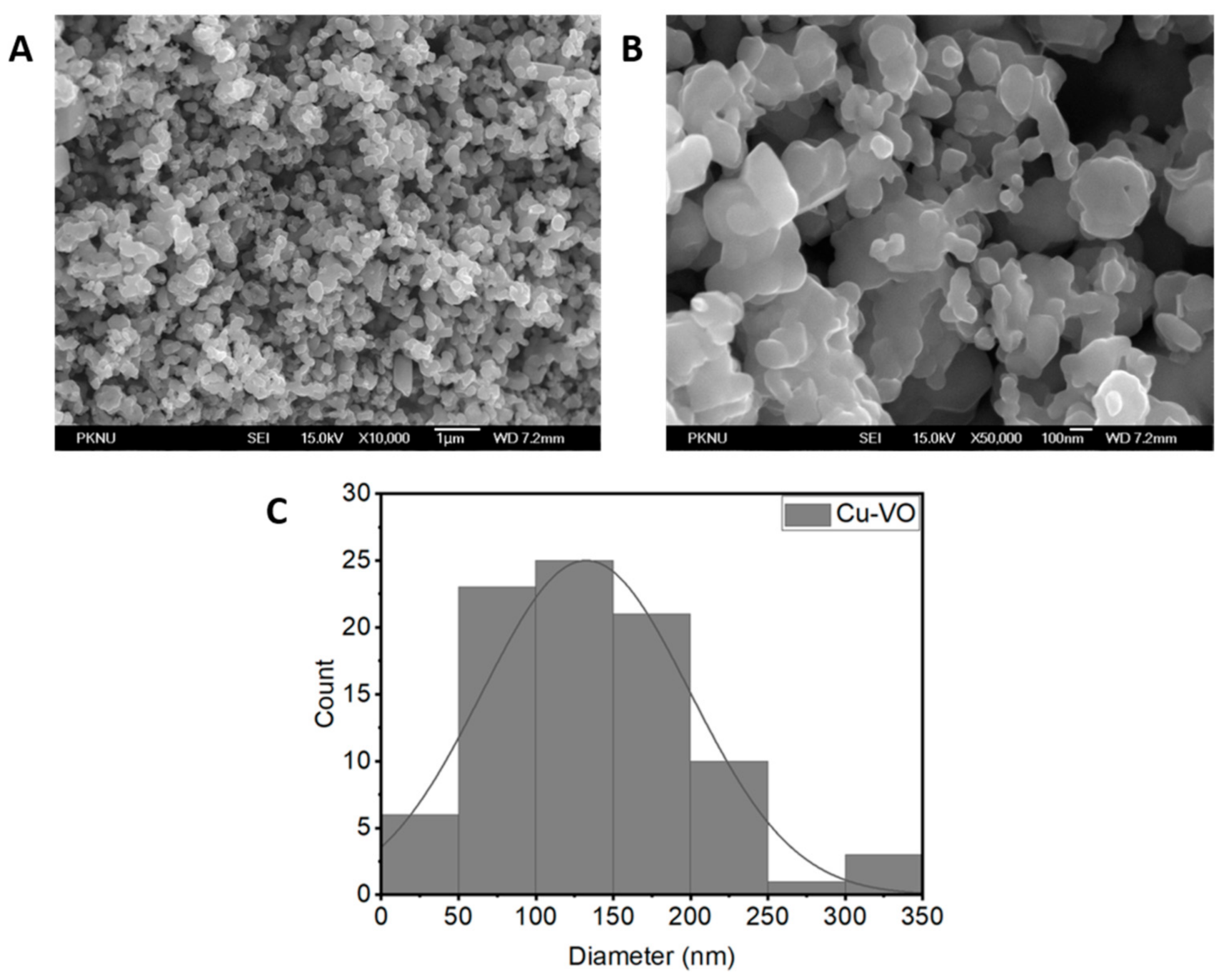
Figure 1. FESEM image of Cu-VO: (A) low magnification, (B) high magnification, and (C) particle size distribution. The particle size distribution plot of Cu-VO was obtained using a high-magnification FESEM image (B).
Catalysts 11 00036 g001
Figure 2. EDX spectrum of Cu-VO. The EDX spectrum showed the peaks of copper, vanadium, and oxygen.
Catalysts 11 00036 g002
Figure 3. X-ray diffraction patterns of Cu-VO.
Catalysts 11 00036 g003
Figure 4. XPS spectra of Cu-VO (A) Cu, (B) V, and (C) O. XPS spectra showed the peaks for 2p3/2 and 2p1/2 orbitals of Cu2+ and V5+ and 1s orbital of O2−.
Catalysts 11 00036 g004
Figure 5. Optical characterization of Cu-VO (A) UV–Vis/near-infrared (NIR) absorption spectrum of the Cu-VO and (B) Tauc’s plot for indirect and direct band gap transition derived from the UV–Vis absorption.
Catalysts 11 00036 g005
Figure 6. FT-IR spectra of Cu-VO.
Catalysts 11 00036 g006
Figure 7. Growth inhibition of Microcystis aeruginosa by Cu-VO with time. (A) Change in the pigment of M. aeruginosa suspension; (B) change in OD680 of M. aeruginosa. C was a control group containing M. aeruginosa alone. The antialgal assay was performed three times, and the OD680 plot is the average of three independent results.
Catalysts 11 00036 g007
Figure 8. UV–Vis absorbance spectra (A) and degradation percentage of 50 μM MB treated with Cu-VO catalyst at a different time interval (B) The dye degradation assay was performed three times, and the degradation percentage plot is the average of three independent results.
Catalysts 11 00036 g008
Figure 9. Reusability of Cu-Vo for the degradation of MB under natural solar light.
Catalysts 11 00036 g009
Figure 10. Fluorescence intensity of extracellular ·OH generated at 4 h. C was a control group containing isopropyl alcohol (·OH scavenger), terephthalic acid, and Cu-VO. Ex was an experimental group containing terephthalic acid and Cu-VO. ·OH assay was performed three times, and the plot was the average of three independent results.
Catalysts 11 00036 g010
Figure 11. Intracellular reactive oxygen species (ROS) induced by Cu-VO in the M. aeruginosa cells. ROS assay was performed three times, and the plot was the average of three independent results.
Catalysts 11 00036 g011
Table 1. Element proportion analysis of Cu-VO.
ElementWeight%Atomic%
O27.9058.64
V24.4116.12
Cu47.6925.24
Table 2. Calculated crystallite size, lattice constants, and microstrain values of Cu-VO.
MaterialCrystallite Size (nm)Atomic%Lattice Constant (Å)Lattice Strain (ε)
abc
Cu-VO58 ± 1058.646.287.996.390.13 ± 0.03
Publisher’s Note: MDPI stays neutral with regard to jurisdictional claims in published maps and institutional affiliations.