Abstract
One of the challenges of using nanoparticles as catalysts is the presence of reaction-disturbing stabilizers that surround the nanoparticle surface. In this report, we demonstrate a method to synthesize stabilizer-free bismuth oxychloride (BiOCl) nanoparticles to increase photocatalytic activity. This synthesis method is remarkably simple, involving only BiCl3 and deionized water. After heating an aqueous solution containing BiCl3, plate-shaped BiOCl nanoparticles were formed. The stabilizer-free BiOCl nanoplates exhibited higher photocatalytic activities compared to polyvinylpyrrolidone- and polyethyleneimine-stabilized nanoplates for the degradation of methylene blue.
1. Introduction
Controlling size, shape, and composition can modulate the optical, electronic, magnetic, and physicochemical properties of nanoparticles. [1]. By tuning these properties, nanoparticles have been utilized in various industries, such as biomedicine, bioengineering, catalytic reactions, energy conversion, and storage [2,3,4,5]. In addition, nanoparticles can be used as catalysts for various organic chemical, electrochemical, and photochemical reactions due to their high surface-area-to-volume ratio and specific surface structures [6]. Nanoparticles synthesized using wet-chemical methods are typically coated with stabilizers composed of organic molecules or polymers to prevent agglomeration by coating the nanoparticle surface with an organic layer [7,8,9,10]. This organic layer is one of the major factors that hinder the catalytic activity of nanoparticles by blocking the interaction between reactants and active metal sites [11,12,13]. Furthermore, it is difficult to completely detach the stabilizer from the nanoparticles due to their strong bond to the surface of the nanoparticles. The development of a new synthetic method to make stabilizer-free nanoparticles would contribute to the goal of obtaining more active catalysts.
The field of solar photocatalysis is a promising area for the application of nanoparticles. Semiconductor-based photocatalysts have received global attention and research is underway to harness their properties, such as their photoreaction activity, physicochemical stability, high activity in the UV region, economic efficiency, and high surface area [14,15]. Recently, many studies have utilized photocatalytic nanoparticles for pollutant decomposition, CO2 reduction, and nitrogen fixation [16,17,18,19,20]. Among the various semiconductor-based photocatalysts, bismuth oxychloride (BiOCl) has attracted considerable attention due to its characteristic structure, low toxicity, and low cost [15,21]. BiOCl is ternary complex semiconductor comprised of V-VI-VII main components, showing an open-layer structure in which Bi-O-Bi-Cl units alternate along the C-axis with strong Bi-O bonds and weak Van der waals forces. This unique structure of BiOCl induces an internal electrostatic field along the [001] direction by generating a larger space for useful polarizing atoms and atomic orbitals. The internal electrostatic fields facilitate interlayer electronic transmission and enhance electron and hole separation efficiency [15,22,23,24]. These properties greatly increase the photocatalytic efficiency of BiOCl. The chemical and physical properties of BiOCl are better than TiO2, which is mainly used as a photocatalyst. For example, the photocatalytic decomposition of dyes under UV light is higher with BiOCl compared to TiO2.
In this research, we developed a method to synthesize stabilizer-free BiOCl nanoparticles to increase their photocatalytic activities. In the case of semiconductor nanoparticles, if the size is smaller than a critical threshold, the band gap increases and the wavelength range of light that can be absorbed narrows. Therefore, our objective was to synthesize BiOCl nanoparticles larger than 100 nm in diameter to avoid the band gap enlargement. The synthesis was remarkably simple and only required BiCl3 and deionized water. After heating BiCl3 dispersed aqueous solution, plate-shaped BiOCl nanoparticles were formed. In previous syntheses, pH adjustment using the addition of acids were needed to form BiOCl and surfactants were required to form nanosized particles. [25,26,27,28,29]. As predicted, the stabilizer-free BiOCl nanoparticles showed that photocatalytic activity for methylene blue degradation was improved compared to BiOCl nanoparticles stabilized by polyvinylpyrrolidone (PVP) or polyethyleneimine (PEI).
2. Results
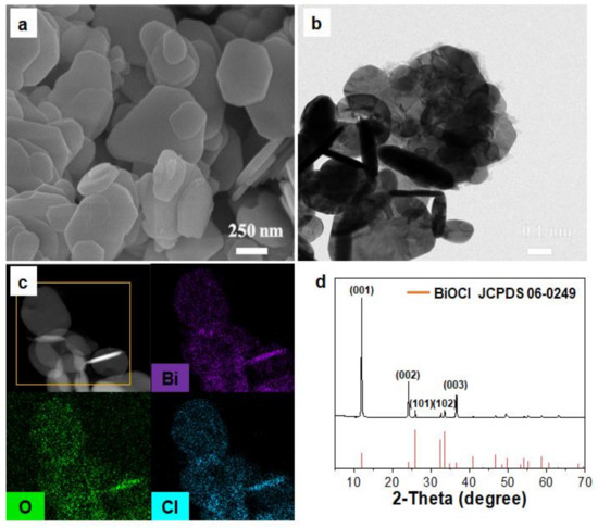
Synthesizing BiOCl nanoplates was conducted by heating a 0.05 M aqueous solution of BiCl3 at 95 °C for 2 h in the absence of a stabilizer. In the reaction, the Bi cation forms [Bi2O2]2+ by rapidly hydrating in the aqueous solution, which then reacts with Cl− to form BiOCl [30]. Scanning electron microscopy (SEM) and transmission electron microscopy (TEM) images of the resulting material reveal that the synthesized nanoplates had sizes of 100–800 nm and thicknesses of 35–112 nm (Figure 1a,b). The elemental distribution of the nanoplates was measured using energy-dispersive X-ray spectrometry (EDS) mapping and indicated that Bi, O, and Cl were evenly distributed throughout the nanoplates (Figure 1c). Figure 1d shows the powder X-ray diffractometry (XRD) patterns of the nanoplates, displaying diffraction peaks that corresponded well with the peaks of known BiOCl (JCPDS 06-0249). However, the intensity of the XRD peaks was slightly different from the previously reported XRD patterns of BiOCl. In previous reports on the synthesis of anisotropic nanoplates, the intensity of the XRD peaks was found to vary depending on the direction [31,32]. The strongest (001) peaks of the tetragonal BiOCl phase at 12° indicated that the nanoplate mainly grew in the (001) direction. To better understand the crystal structure of BiOCl nanoplates, the oxidation states of Bi and Cl in the nanoplates were characterized using X-ray photoelectron spectroscopy (XPS) (Figure S1). The binding energies of Bi 4f7/2 and Bi 4f5/2 were 163.28 and 157.98 eV, respectively, which are similar to literature values for Bi3+ (Figure S1a) [33,34]. The Cl 2p core level spectrum composed of Cl 2p3/2 and Cl 2p1/3 peaks at 198.18 and 196.58 eV, respectively, correspond to Cl– (Figure S1b). These results match with the XRD measurements that indicated that the crystal structure of the nanoplates was BiOCl. The ratio of Bi to Cl in the nanoplates was determined to be 1:1.13 from the XPS measurements. It has been reported that anions, such as halide ions, will adhere to the surface of nanoparticles in the absence of typical stabilizers [35]. In the current study, it is likely that Cl− anions act as stabilizers by attaching to the surface of the BiOCl nanoparticles, supported by the observation that there were more Cl− ions than Bi atoms detected in the XPS analysis. The zeta potential analysis showed that the surface of BiOCl had a negative charge (Figure S2), which can be attributed to the adsorption of chloride ions on its surface. The negative charges on the surface give rise to repulsive forces that provide the nanoplates with electrostatic stabilization in aqueous solutions. The UV–Vis extinction spectrum, measured from an aqueous suspension of the BiOCl nanoplates, displays a broad band around 500 nm.

Figure 1.
(a) Scanning electron microscopy (SEM) image, (b) transmission electron microscopy (TEM) image, (c) Scanning transmission electron microcopy (STEM) and energy-dispersive X-ray spectrometry (EDS) mapping images, and (d) X-ray diffractometry (XRD) patterns of the bismuth oxychloride (BiOCl) nanoplates.
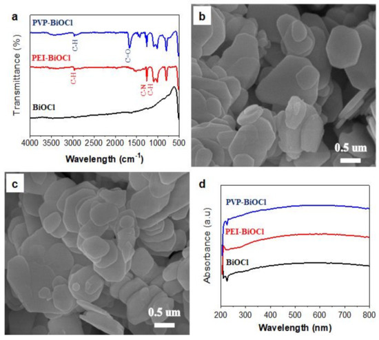
In the synthesis described above, the BiOCl nanoplates were synthesized in the absence of a stabilizer. BiOCl nanoplates were used to investigate the effect of stabilizers on photocatalytic reactions, so polyvinylpyrrolidone (PVP) and polyethyleneimine (PEI) were introduced to coat the surface of the nanoplates. PVP- and PEI-stabilized BiOCl nanoplates were prepared by heating BiOCl dispersed aqueous solution in the existence of PVP or PEI. The Fourier-transform infrared spectroscopy (FT-IR) spectra of PVP-stabilized BiOCl nanoplates exhibited distinct peaks at 2900 cm−1, assigned to a C–H bond, and 1660 cm−1, assigned to a C=O bond, indicating that PVP was successfully attached to the nanoplate surface (Figure 2a). For PEI-stabilized BiOCl nanoplates, peaks in the FT-IR spectra corresponding to PEI were observed (1250 cm−1, assigned to a C-N bond), displaying the existence of PEI on the nanoplate surface (Figure 2a). Stabilization of the nanoplates with PVP and PEI was also confirmed using a zeta potential analysis (Figure S3). The zeta potential of PVP-stabilized BiOCl nanoplates was slightly less negatively charged (−10.7 mV) than the bare BiOCl nanoplates (−20.1 mV), while PEI-stabilized nanoplates were positively charged (33.1 mV). Figure 2b,c show SEM images of the PVP- and PEI-stabilized nanoplates, confirming that there were no size or thickness changes after the stabilizer attachment. Additionally, the optical properties of the nanoparticles did not change according to the UV–Vis analysis (Figure 2d).

Figure 2.
(a) Fourier-transform infrared spectroscopy (FT-IR) spectra of BiOCl nanoplates. SEM images of (b) polyvinylpyrrolidone (PVP)-stabilized and (c) polyethyleneimine (PEI)-stabilized BiOCl nanoplates. (d) UV-Vis spectra of BiOCl nanoplates.
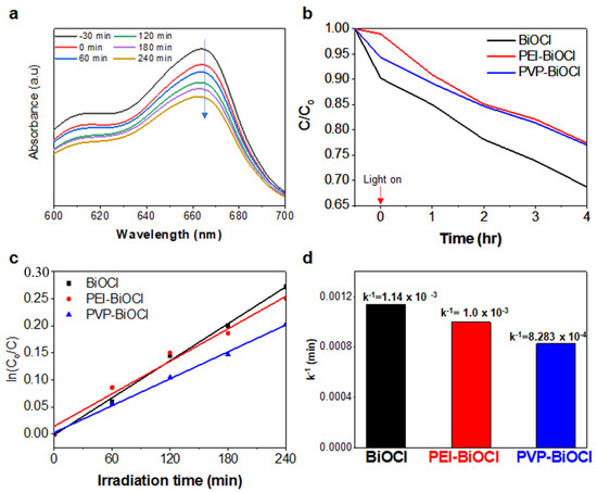
The photocatalytic properties of the BiOCl nanoplates were estimated for the decomposition of organic pollutants under visible light irradiation. To evaluate the photocatalytic performance of BiOCl, methylene blue (MB) was chosen as the organic pollutant. The photolysis process was investigated by checking the intensity of the major absorption peak at 668 nm associated with MB. The photocatalyst (BiOCl, PVP-stabilized BiOCl, or PEI-stabilized BiOCl) was added to a solution of MB and the intensity of the MB absorption peak according to the photocatalytic reaction rapidly decreased, and the results showed that the MB molecules were degraded effectively under visible light illumination in the existence of BiOCl nanoplates. (Figure 3a and Figure S4). Figure 3b displays the concentration of MB (C) normalized against the concentration at the start of the reaction (C0) for the photocatalytic reaction with the nanoplates. A plot of the normalized concentration on a logarithmic scale versus the reaction time is shown in Figure 3c, demonstrating that the reaction of MB degradation has first order kinetics, as in expressed in Equations (1) and (2):
and
where k is the rate constant. The rate constant of stabilizer-free BiOCl nanoplates was estimated to be 1.14 × 10⁻³ min−1, which was faster than PVP-stabilized BiOCl nanoplates and PEI-stabilized BiOCl nanoplates (Figure 3d, 8.283 × 10⁻⁴ min−1 and 1.0 × 10⁻³ min−1, respectively.) These differences in rate indicate the importance of the surface condition of the nanoplates in the catalytic reaction.
dC/dt = −kC
ln(C/C0) = −kt

Figure 3.
(a) UV–Vis absorption spectra for the photocatalytic degradation of methylene blue (MB) molecules using BiOCl under visible light irradiation. (b) Normalized concentration of MB on a linear scale. (c) Normalized concentration of MB on a logarithmic scale. (d) Bar diagram showing the degradation percentage of MB molecules after irradiation under visible light.
3. Materials and Methods
3.1. Materials
All reagents were commercially available and were used without further purification. Bismuth (III) chloride (BiCl3, trace metal basis), polyvinylpyrrolidone (PVP, ACS reagent, MW ~ 55,000), and polyethyleneimine (PEI, ACS reagent, MW ~ 750,000) were purchased from Sigma-Aldrich.
3.2. Synthesis of BiOCl Nanoplates
In total, 10 mL of an aqueous solution of BiCl3 0.5 mmol was heated at 95 °C for 2 h. After the reaction, the products were collected using centrifugation, washed twice with a mixed solution of water and acetone and then dried in oven at 80 °C for 1 h.
3.3. Attachment of Stabilizer to the Surface of BiOCl Nanoplates
In total, 0.1 g of BiOCl nanoplates was well dissolved in 5 mL of water (solution A) and 0.02 g of each stabilizer was dissolved in 5 mL of water (solution B). Solution B was added to solution A and the mixed solution was stirred at 50 °C for 1 h. After the reaction, the products were collected using centrifugation and washed using a mixed solution of water and acetone.
3.4. Characterization
The crystal structures of the photocatalysts were characterized by using a D8 Advance X-ray diffractometer (XRD). The sizes and morphologies of the nanoparticles were measured using scanning electron microscopy (SEM, Merlin, Carl Zeiss). The elemental distribution of the nanoparticles was obtained using transmission electron microscopy (TEM, JEM-2100F, JEOL) and energy-dispersive X-ray spectrometry (EDS, JEM-2100F). The optical properties were estimated using a Cary 60 UV–Vis spectrophotometer (Agilent Technologies). X-ray photoelectron spectroscopy (XPS) analysis was conducted using a K-Alpha spectrometer (Thermo Electron) to determine the oxidation state of the elements in the nanoparticles. The Brunauer–Emmett–Teller (BET) surface area of the nanoparticle was estimated using a nitrogen adsorption method with a BELSORP-max (MP). Zeta potential tests were conducted on a zeta potential analyzer (Zetasizer Nano ZS, Malvern, pH = 6.19). Fourier-transform infrared spectroscopy (FT-IR) was measured by using a Spectrum One system (PerkinElmer).
3.5. Evaluation of Photocatalytic Activity
In total, 5.0 mg of catalyst was added to a 50 mL aqueous solution of methylene blue (MB) (10 mg/L). To establish the adsorption–deposition equilibrium of MB molecules on the catalyst, the solution was stirred for 30 min in the absence of light. A 200 W mercury lamp (Raynics, Korea) was used to evaluate photocatalytic reaction under visible light irradiation. During the reaction, 3 mL aliquots were removed from the solution and centrifuged to remove the catalysts (1350 rpm, 5 min). The supernatant was transferred to a cuvette and UV–Vis absorption spectra were analyzed in the range of 300~800 nm.
4. Conclusions
In summary, we have synthesized stabilizer-free BiOCl nanoplates that demonstrate enhanced photocatalytic activity. The synthesis is simple, involving only the heating of an aqueous solution containing BiCl3. The stabilizer-free BiOCl nanoplates exhibited a higher photocatalytic activity compared to PVP- and PEI-stabilized nanoplates. We anticipate that this method to easily synthesize BiOCl nanoparticles can be expanded to the preparation of other oxyhalide nanoparticles, which can then be used as catalysts for many reactions.
Supplementary Materials
The following are available online at https://www.mdpi.com/2073-4344/11/1/111/s1. Figure S1: XPS spectra of the BiOCl nanoplate. Figure S2: The zeta potential value of the BiOCl nanoplates measured at pH 6.19. Figure S3: The zeta potential value of (a) PVP- and (b) PEI-stabilized BiOCl nanoplates measured at pH 6.19. Figure S4: UV–Vis absorption spectra for photocatalytic degradation of MB molecules using PVP-BiOCl and PEI-BiOCl under visible light irradiation.
Author Contributions
Conceptualization, synthesis, characterization, catalytic activity assay, and writing—original draft preparation, H.L.; reviewing and editing, S.-S.K.; supervision, reviewing and editing, S.H.B.; supervision, methodology, further data analysis, and writing—review and editing, T.Y. All authors have read and agreed to the published version of the manuscript.
Funding
This research was supported by the National Research Foundation of Korea (NRF-2014R1A5A1009799, NRF-2016M3D1A1021140, and NRF-2020R1A2C1003885). This research was supported by Basic Science Research Capacity Enhancement Project through Korea Basic Science Institute (Core Facility Center for Analysis of Optoelectronic Materials and Devices) grant funded by the Ministry of Education (NRF- 2019R1A6C1010052).
Data Availability Statement
This article is an open access article distributed under the terms and conditions of the Creative Commons Attribution (CC BY) license.
Conflicts of Interest
The authors declare no conflict of interest.
References
- Gentile, A.; Ruffino, F.; Grimaldi, M.G. Complex-morphology metal-based nanostructures: Fabrication, characterization, and applications. Nanomaterials 2016, 6, 110. [Google Scholar] [CrossRef]
- Pankhurst, Q.A.; Connolly, J.; Jones, S.K.; Dobson, J. Applications of magnetic nanoparticles in biomedicine. J. Phys. D Appl. Phys. 2003, 36, R167–R181. [Google Scholar] [CrossRef]
- Kim, E.Y.; Kumar, D.; Khanga, G.; Lim, D.K. Recent advances in gold nanoparticle-based bioengineering applications. J. Mater. Chem. B 2015, 3, 8433–8444. [Google Scholar] [CrossRef] [PubMed]
- Park, J.; Kwon, T.; Kim, J.; Jin, H.; Kim, H.Y.; Kim, B.; Joo, S.H.; Lee, K. Hollow nanoparticles as emerging electrocatalysts for renewable energy conversion reactions. Chem. Soc. Rev. 2018, 47, 8173–8202. [Google Scholar] [CrossRef]
- Wessells, C.D.; McDowell, M.T.; Peddada, S.V.; Pasta, M.; Huggins, R.A.; Cui, Y. Tunable reaction potentials in open framework nanoparticle battery electrodes for grid-scale energy storage. ACS Nano 2012, 6, 1688–1694. [Google Scholar] [CrossRef] [PubMed]
- Polshettiwar, V.; Luque, R.; Fihri, A.; Zhu, H.; Bouhrara, M.; Basset, J.-M. Magnetically recoverable nanocatalysts. Chem. Rev. 2011, 111, 3036–3075. [Google Scholar] [CrossRef] [PubMed]
- Yamamoto, K.; Imaoka, T.; Chun, W.-J.; Enoki, O.; Katoh, H.; Takenaga, M.; Sonoi, A. Size-specific catalytic activity of platinum clusters enhances oxygen reduction reactions. Nat. Chem. 2009, 1, 397–402. [Google Scholar]
- Li, Y.; Huang, Y. Morphology-Controlled Synthesis of Platinum Nanocrystals with Specific Peptides. Adv. Mater. 2010, 22, 1921–1925. [Google Scholar] [CrossRef]
- San, B.H.; Kim, S.; Moh, S.H.; Lee, H.; Jung, D.Y.; Kim, K.K. Platinum nanoparticles encapsulated by aminopeptidase: A multifunctional bioinorganic nanohybrid catalyst. Angew. Chem. Int. Ed. 2011, 50, 11924–11929. [Google Scholar] [CrossRef]
- Tanaka, S.I.; Miyazaki, J.; Tiwari, D.K.; Jin, T.; Inouye, Y. Fluorescent platinum nanoclusters: Synthesis, purification, characterization, and application to bioimaging. Angew. Chem. Int. Ed. 2011, 50, 451–455. [Google Scholar] [CrossRef]
- Stowell, C.A.; Korgel, B.A. Iridium nanocrystal synthesis and surface coating-dependent catalytic activity. Nano Lett. 2005, 5, 1203–1207. [Google Scholar] [CrossRef] [PubMed]
- Borodko, Y.; Habas, S.E.; Koebel, M.; Yang, P.; Frei, H.; Somorjai, G.A. Charge-transfer interaction of poly(vinylpyrrolidone) with platinum and rhodium nanoparticles. J. Phys. Chem. B 2006, 110, 23052–23059. [Google Scholar] [CrossRef] [PubMed]
- Lee, H.; Habas, S.E.; Kweskin, S.; Butcher, D.; Somorjai, G.A.; Yang, P. Morphological control of catalytically active platinum nanocrystals. Angew. Chem. Int. Ed. 2006, 45, 7824–7828. [Google Scholar] [CrossRef] [PubMed]
- Zhang, P.; Zhang, J.; Gong, J. Tantalum-based semiconductors for solar water splitting. Chem. Soc. Rev. 2014, 43, 4395–4422. [Google Scholar] [CrossRef] [PubMed]
- Hisatomi, T.; Kubota, J.; Domen, K. Recent advances in semiconductors for photocatalytic and photoelectrochemical water splitting. Chem. Soc. Rev. 2014, 43, 7520–7535. [Google Scholar] [CrossRef] [PubMed]
- Lee, G.J.; Zheng, Y.-C.; Wu, J.J. Fabrication of hierarchical bismuth oxyhalides (BiOX, X = Cl, Br, I) materials and application of photocatalytic hydrogen production from water splitting. Catal. Today 2018, 307, 197–204. [Google Scholar] [CrossRef]
- Zhang, X.; Li, B.; Wang, J.; Yuan, Y.; Zhang, Q.; Gao, Z.; Liu, L.M.; Chen, L. The stabilities and electronic structures of single-layer bismuth oxyhalides for photocatalytic water splitting. Phys. Chem. Chem. Phys. 2014, 16, 25854–25861. [Google Scholar] [CrossRef]
- Gao, X.; Zhang, X.; Wang, Y.; Peng, S.; Yue, B.; Fan, C. Photocatalytic degradation of carbamazepine using hierarchical BiOCl microspheres: Some key operating parameters, degradation intermediates and reaction pathway. Chem. Eng. J. 2015, 273, 156–165. [Google Scholar] [CrossRef]
- Ye, L.; Jin, X.; Liu, C.; Ding, C.; Xie, H.; Chu, K.H.; Wong, P.K. Oxygen vacancies induced exciton dissociation of flexible BiOCl nanosheets for effective photocatalytic CO2 conversion. Appl. Catal. B. 2016, 187, 281–290. [Google Scholar] [CrossRef]
- Li, H.; Shang, J.; Shi, J.; Zhao, K.; Zhang, L. Facet-dependent solar ammonia synthesis of BiOCl nanosheets via a proton-assisted electron transfer pathway. Nanoscale 2016, 8, 1986–1993. [Google Scholar] [CrossRef]
- Di, J.; Xia, J.; Li, H.; Guo, S.; Dai, S. Bismuth oxyhalide layered materials for energy and environmental applications. Nano Energy 2017, 41, 172–192. [Google Scholar] [CrossRef]
- Bai, S.; Li, X.; Kong, Q.; Long, R.; Wang, C.; Jiang, J.; Xiong, Y. Toward enhanced photocatalytic oxygen evolution: Synergetic utilization of plasmonic effect and Schottky junction via interfacing facet selection. Adv. Mater. 2015, 27, 3444–3452. [Google Scholar] [CrossRef]
- Cao, S.; Guo, C.; Lv, Y.; Guo, Y.; Liu, Q. A novel BiOCl film with flowerlike hierarchical structures and its optical properties. Nanotechnology 2009, 20, 275702. [Google Scholar] [CrossRef]
- Sharma, K.; Dutta, V.; Sharma, S.; Raizada, P.; Hosseini-Bandegharaei, A.; Thakur, P.; Singh, P. Recent advances in enhanced photocatalytic activity of bismuth oxyhalides for efficient photocatalysis of organic pollutants in water: A review. J. Ind. Eng. Chem. 2019, 78, 1–20. [Google Scholar] [CrossRef]
- Zhang, K.L.; Liu, C.M.; Huang, F.Q.; Zheng, C.; Wang, W.D. Study of the electronic structure and photocatalytic activity of the BiOCl photocatalyst. Appl. Catal. B Environ. 2006, 68, 125–129. [Google Scholar] [CrossRef]
- Ye, P.; Xie, J.; He, Y.; Zhang, L.; Wu, T.; Wu, Y. Hydrolytic synthesis of flower like BiOCl and its photocatalytic performance under visible light. Mater. Lett. 2013, 108, 168–171. [Google Scholar] [CrossRef]
- Chen, L.; Yin, S.F.; Huang, R.; Zhou, Y.; Luo, S.L.; Au, C.T. Facile synthesis of BiOCl nano-flowers of narrow band gap and their visible-light-induced photocatalytic property. Catal. Commun. 2012, 23, 54–57. [Google Scholar] [CrossRef]
- Armelao, L.; Bottaro, G.; Maccatoc, C.; Tondello, E. Bismuth oxychloride nanoflakes: Interplay between composition-structure and optical properties. Dalton Trans. 2012, 41, 5480. [Google Scholar] [CrossRef]
- Wu, Y.; Yuan, B.; Li, M.; Zhang, W.H.; Liu, Y.; Li, C. Well-defined BiOCl colloidal ultrathin nanosheets: Synthesis, characterization, and application in photocatalytic aerobic oxidation of secondary amines. Chem. Sci. 2015, 6, 1873. [Google Scholar] [CrossRef]
- Li, J.; Yu, Y.; Zhang, L. Bismuth oxyhalide nanomaterials: Layered structures meet photocatalysis. Nanoscale 2014, 6, 8473–8488. [Google Scholar] [CrossRef]
- Shi, W.; Sahoo, Y.; Zeng, H.; Ding, Y.; Swihart, M.; Prasad, P. Anisotropic growth of PbSe nanocrystals on Au–Fe3O4 hybrid nanoparticles. Adv. Mater. 2006, 18, 1889–1894. [Google Scholar] [CrossRef]
- Hoefelmeyer, J.D.; Niesz, K.; Somorjai, G.A.; Tilley, T.D. Radial Anisotropic Growth of Rhodium Nanoparticles. Nano Lett. 2005, 5, 435–438. [Google Scholar] [CrossRef] [PubMed]
- Zhang, X.; Wang, L.W.; Wang, C.Y.; Wang, W.K.; Chen, Y.L.; Huang, Y.X.; Li, W.W.; Feng, Y.J.; Yu, H.Q. Synthesis of BiOClxBr1−x nanoplate solid solutions as a robust photocatalyst with tunable band structure. Chem. Eur. J. 2015, 21, 11872–11877. [Google Scholar] [CrossRef] [PubMed]
- Yang, J.; Liang, Y.; Li, K.; Zhu, Y.; Liu, S.; Xu, R.; Zhou, W. Design of 3D flowerlike BiOClxBr1-x nanostructure with high surface area for visible light photocatalytic activitie. J. Alloys Compd. 2017, 725, 1144–1157. [Google Scholar] [CrossRef]
- Lim, G.H.; Yu, T.; Koh, T.; Lee, J.H.; Jeong, U.; Lim, B. Reduction by water for eco-friendly, capping agent-free synthesis of ultrasmall platinum nanocrystals. Chem. Phys. Lett. 2014, 595, 77–82. [Google Scholar] [CrossRef]
Publisher’s Note: MDPI stays neutral with regard to jurisdictional claims in published maps and institutional affiliations. |
© 2021 by the authors. Licensee MDPI, Basel, Switzerland. This article is an open access article distributed under the terms and conditions of the Creative Commons Attribution (CC BY) license (http://creativecommons.org/licenses/by/4.0/).