Article

Insights into Interface Charge Extraction in a Noble-Metal-Free Doped Z-Scheme NiO@BiOCl Heterojunction

1
Chongqing Key Laboratory of Environmental Materials and Remediation Technologies, College of Chemical and Environmental Engineering, Chongqing University of Arts and Sciences, Yongchuan 402160, China
2
Chongqing Key Laboratory of Extraordinary Bond Engineering and Advanced Materials Technology (EBEAM), Yangtze Normal University, Chongqing 408100, China
3
State Key Laboratory of Coal Mine Disaster Dynamics and Control, Chongqing University, Chongqing 400044, China
4
College of Chemistry and Chemical Engineering, Chongqing University, Chongqing 401331, China
*
Authors to whom correspondence should be addressed.
The two authors contributed equally to this work.
Catalysts 2020, 10(9), 958; https://doi.org/10.3390/catal10090958
Submission received: 3 July 2020 / Revised: 14 August 2020 / Accepted: 14 August 2020 / Published: 21 August 2020
(This article belongs to the Special Issue Active Sites in Heterogeneous Catalysis)

Abstract

:
It is of great significance to thoroughly explore the interface charge extraction and migration in heterojunction systems, which could guide us to synthesize higher-efficiency photocatalytic materials. A novel noble-metal-free doped Z-scheme NiO@BiOCl heterojunction was found in this work. The corresponding heterostructure, interface electron extraction, and electron migration were investigated via first-principles calculation. 5,5′-dimethyl-1-pyrroline-N-oxide (DMPO) spin-trapping electron spin resonance (ESR) and time-resolved photoluminescence (TRPL) tests were implemented to confirm the calculation results, which showed that electrons and holes stayed in the NiO (100) facet and BiOCl (110) facet, respectively. Owing to the large chemical potential of 2.40 V (vs ENHE) for the BiOCl valence-band hole, it possessed super activity to oxidize water into hydroxyl radicals or molecular oxygen. We hope this promising multifunctional photocatalytic material, therefore, NiO@BiOCl can be applied in advanced treatment of organic wastewater and oxygen production from photolysis water.

1. Introduction

Photocatalysis and photocatalytic technology using light irradiation, especially inexhaustible solar light, are the most environmentally friendly methods for treatment of environmental pollutants, water photolysis, CO2 reduction, and other high-energy barrier chemical reactions. In the photodegradation organic wastewater process, hydroxyl radicals (·OH, Eθ = 2.8 V) have a strong ability to absorb electrons that can easily break covalent bonds in organic molecules. It is very important, therefore, to understand formation routes of hydroxyl radicals. Adsorbed oxygen converts into hydroxyl radicals via a five-electron pathway. Water molecules and hydroxyls (OH) are oxidized by holes to generate hydroxyl radicals via a one-hole pathway (See Figure 1a). Considering the quantum efficiency and yield of hydroxyl radicals, the desired one-hole pathway can generate the photodegradation process. The same is true with water photolysis for hydrogen and oxygen production (Figure 1b,c). In order to ensure that there are more photoinduced charges participating in the desired reactions, it is of great significance to explore the migration of photoinduced charges and the extraction of interface electrons in heterojunction systems.
Over the past decade, some visible-light-driven photocatalytic materials have been synthesized and applied into the field of organic wastewater photodegradation. For example, Fe(0)-doped g-C3N4/MoS2, fluorinated Bi2WO6, and TiO2 with interface defects [1,2,3,4] were prepared specifically for rhodamine B (RhB) dye photodegradation due to its aquatic environmental pollution and huge discharge from various industries. However, the corresponding photocatalytic reactions were very slow, taking several hours to degrade less than 98% of RhB, which impeded their practical application as a result of the high time and production costs. The challenges faced were figuring out how to enhance the photocatalytic reaction kinetics, how to shorten the photodegradation reaction time, and how to boost the photocatalytic efficiency and quantum efficiency under identical visible light irradiation [5].
Bi-based photocatalysts are important visible-light-driven photocatalytic materials that have also attracted a huge amount of attention. Considering the stability of Bi3+, Bi3+-containing compounds, such as Bi2O3, BiVO4, Bi2WO6, BiPO4, BiFeO3, and BiOX (X = Cl, Br, I) [5,6], have been prepared for photocatalytic reactions. The majority of these compounds, especially BiOX (X = Cl, Br, I), have a layered structure and plate-like appearance that can produce an internal electric field [7,8], which facilitates the migration of photoinduced carriers to some extent. Among these Bi3+-based photocatalytic materials, various structures and properties of BiOCl have been reported. However, the photogenerated electrons and holes of BiOCl are not easily exploited and utilized under solar light irradiation [9,10].
Constructing a heterojunction is a very effective approach in order to enlighten photocatalytic activity, owing to the high separation efficiency for photoinduced charges and high quantum efficiency of heterojunctions. Some reports on the migration of photoinduced charges and the extraction of interface electrons in heterojunctions were not deep enough and only gave the possible mechanisms [9]. In this work, experiment and theoretical calculation were performed to assess if NiO@BiOCl possesses highly photocatalytic activity. The noble-metal-free doped Z-scheme heterojunction was found in the NiO@BiOCl heterojunction system by optimizing the heterojunction interface and calculating the charge density difference. Thus, this heterojunction system has higher potential holes and lower potential electrons, which was confirmed by DMPO spin-trapping electron spin resonance (ESR) and time-resolved photoluminescence (TRPL) tests.

2. Results and Discussions

Primary analysis of photodegradation reactions revealed that 4% NiO@BiOCl possessed the highest photodegradation efficiency for RhB dye under visible light irradiation. The components and structure of 4% NiO@BiOCl were, therefore, investigated.

2.1. Identification of Phases and Components

X-ray diffraction patterns of the samples are shown in Figure 2a. The diffraction peaks of BiOCl and 4% NiO@BiOCl could be indexed to the BiOCl of the tetragonal system (JCPDS No. 06–0248), indicating that NiO introduction did not enter the crystal lattice of BiOCl and altered the diffraction peak position of BiOCl. No diffraction peaks for NiO appeared due to the low concentration of Ni2+. It is worth noting that the intensity of the (101) crystal surface diffraction peak of NiO@BiOCl was obviously lower than that of the BiOCl sample. The intensity, however, of the (110) surface diffraction peak of NiO@BiOCl was higher than that of the BiOCl sample. Thus, more of the (110) surface of BiOCl was exposed in the NiO@BiOCl system. These results revealed that the incorporation of NiO possibly affects the preferred orientation of the BiOCl crystal [9]. Namely, the introduction of NiO restrained the (101) growth orientation of BiOCl caused by the NiO deposited on the surface of BiOCl. The closely contact between the two components provided the possibility for the heterojunction formation. The average crystallite size was calculated via Scherrer equation using the (110) peak. The average crystallite sizes were 8.3 nm and 4.8 nm for BiOCl and NiO@BiOCl, respectively. The NiO addition reduced the average crystallite size of BiOCl.
As shown in Figure 2b, the bands at 143.3 cm−1 and 199.6 cm−1 in the Raman spectra were assignable to symmetrical stretching vibration of the Bi-Cl bond and symmetric expansion vibration of the Bi-Cl bond, respectively [11]. The wide band at 398.0 cm−1 was attributable to the oxygen atom vibration in the BiOCl crystal system. Meanwhile, the Raman bands of BiOCl at 84.0 cm−1 could be observed, further confirming the presence of BiOCl crystal. The Raman spectrum exhibited a strong, broad band at 410.0 cm−1, which was attributed to the Ni-O stretching mode [12]. Moderately intense Raman bands in the range of 184.0~189.0 cm−1 in the NiO@BiOCl sample stemmed from lattice vibrations of NiO [12]. In addition, it was worth noting that the band at 143.3 cm−1 became flatter and wider, indicating that NiO NPs were highly dispersed on the surface of BiOCl [13,14,15,16].
The X-ray photoelectron spectroscopy (XPS) images and high-resolution spectra of all elements of the 4% NiO@BiOCl sample are exhibited in Figure 2c and Figure S1 (Supplementary Materials), respectively. It was found that the as-prepared sample contained Bi, O, Cl, and Ni, which illustrated the existence of Ni in this sample. In addition, C1s and N1s signals stemmed from the polyvinylpyrrolidone chemical compound. The peaks at 158.9 eV and 164.1 eV corresponded to Bi 4f 7/2 and Bi 4f 5/2, respectively, showing the presence of Bi3+ on the sample surface [17]. O1s had a main peak at 529.6 eV and a shoulder peak at 531.0 eV, which were assignable to the Bi-O bond in the BiOCl system and O species from NiO, respectively. The peaks for Cl 2p appeared at 197.4 eV and 198.9 eV, corresponding to Cl 2p3/2 and Cl 2p1/2, respectively. The peaks at 854.6 eV and 859.8 eV were ascribable to Ni 2p3/2 [18,19], while the peak at 873.1 eV resulted from Ni 2p1/2 [19,20]. Thus, the presence of NiO was further confirmed.
Scanning electron microscope (SEM) and transmission electron microscope (TEM) images of BiOCl and 4% NiO@BiOCl are presented in Figure S2 (Supplementary Materials). It can be seen that BiOCl was mainly composed of microspheres formed by crisscross nano-sheets. When 4% NiO was loaded, the morphology became irregular. The nano-sheet was randomly stacked, except for the formation of microspheres, which was ascribable to the exposure of more (110) crystal surfaces. The generation of more (110) crystal surfaces thickened the BiOCl nano-sheet, giving rise to the formation of less microspheres. It was also seen from Figure S2b (Supplementary Materials) that there were nano-particles on the surface of the nano-sheet, revealing that NiO nano-particles lie on the BiOCl surface. Meanwhile, the presence of NiO was also verified by EDS detection. As shown in Figure S2c, the existence of Ni, Bi, and O elements in the EDS spectrum was affirmed. The corresponding TEM and HRTEM graphs are illustrated in Figure S2d,e (Supplementary Materials).
Apart from the existence of microspheres, the 4% NiO@BiOCl sample had a randomly stacked nano-sheet, which was consistent with the SEM test results. The inset is shown as a magnifying map of the edge of the sample. This close contact would contribute to the formation of NiO and BiOCl heterostructures and promote the photoelectron conduction. Clear lattice stripes can be seen in Figure S2e (Supplementary Materials). The calculated lattice spacing was approximately 0.275 nm, which corresponded to the lattice spacing of the BiOCl (110) surface with the JCPDS (No. 06–0248). The lattice spacing of the other region was approximately 0.20 nm, which was in accordance with the NiO (200) surface of the JCPDS (No. 78–0423) [17], indicating the existence of NiO again.

2.2. Heterostructure Fabrication and Interface Electron Extraction

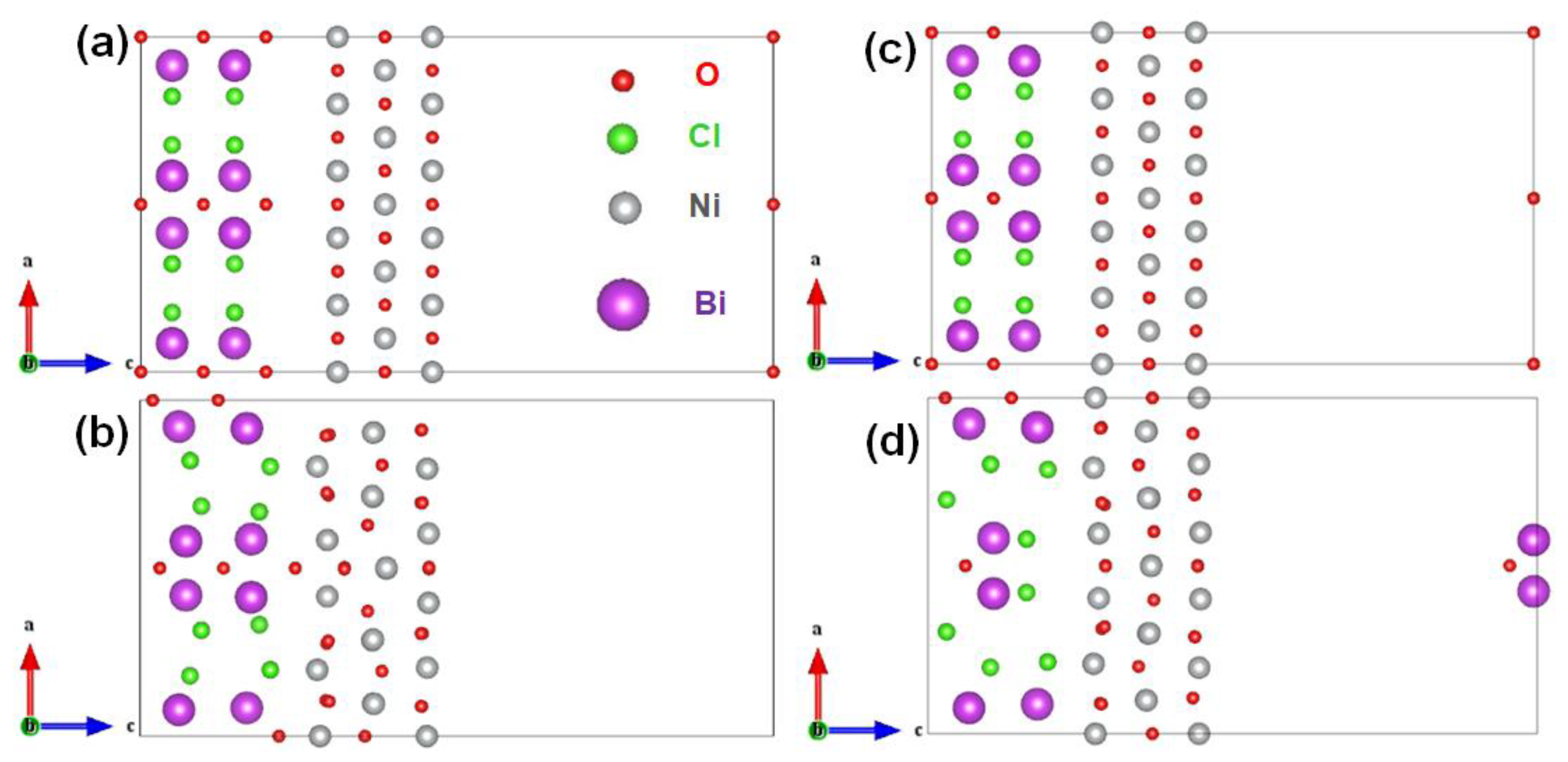
The BiOCl crystal had a tetragonal structure of space group P4/nmm (No. 129) with the lattice parameters a 0   =   b 0   =   3.891   , c 0   =   7.369   [17], while NiO crystal had a simple rock salt structure with the lattice parameters a 0   =   4.178   [21]. Based on the XRD test and the HRTEM image results, NiO (100) deposited on the BiOCl (110) substrate was used to construct the NiO@BiOCl heterostructures; that is, the former lattice structure self-adapted to the latter lattice structure. Specifically, for the density functional theory (DFT) calculation, the heterostructure slab model was constructed as follows: the BiOCl (110) surface had lengths of 2 c 0 and 2 a 0 along the [001] and [ 1 1 ¯ 0 ] directions, with five-atomic-layer thickness, while the NiO (100) surface had lengths of 5 2 2 a 0 and 2 a 0 along the [ 01 1 ¯ ] and [011] directions, with three-atomic-layer thickness. Accordingly, this heterostructure possessed a small lattice mismatch (7.3%). As the BiOCl (110) surface had two terminated situations, the BiCl-polar surface and the O-polar surface, and therefore the NiO(100)-BiCl(BiOCl-110) heterojunction and NiO(100)-O(BiOCl-110) heterojunction, were considered, as shown in Figure 3. Through DFT calculation, it was found that the former case had interfacial adhesion energy of 3.44 eV, while the latter case had larger interfacial adhesion energy of 13.50 eV. Thus, the NiO(100)-O(BiClO-110) heterojunction was much more stable than the other case. The subsequent calculation and data analysis were all based on this heterostructure.
When two surface layers with different work functions form a heterostructure, this would induce charge redistribution around the contact region, while an electric field would simultaneously be established at the interface. This built-in electric field could significantly affect the photogenerated charge transfer process. Consequently, to analyze the surface electronic properties after the contact of BiOCl(110)and NiO(100), which were related to the photocatalytic performance, the surface work function (Φ), charge density difference, and the band alignment diagram were analyzed.
The calculated work functions (Φ) were 4.39 eV and 7.20 eV for NiO-100 and BiOCl-110(O-polar) surfaces, respectively (See Figure 4a). Thus, it could be deduced that electrons would transfer from the surface with lower work function, namely the higher fermi level surface (NiO-100 surface) to the other (BiOCl-110). In fact, the charge density difference results strongly confirmed that electrons migrate from the NiO-100 surface to the BiOCl-110 surface, as shown in Figure 4b. As a consequence, an electric field pointing from the NiO-100 surface to BiOCl-110 surface was established at the interface. Moreover, as a reference to the potential of normal hydrogen electrode (ENHE), which equaled −4.44 eV with respect to the absolute vacuum scale (EAVS = 0 eV), the band-edge potentials of the minimum conduction band (CBM) and maximum valence band (VBM) of NiO were approximately −1.91 V and 1.79 V, respectively; whereas those of BiOCl were approximately −1.10 V and 2.40 V, respectively [6,21,22,23]. The band alignment diagram is represented in Figure 4c. Under visible light irradiation, it could be seen that the charge transfer processes (e-e or h+-h+) would not be allowed, while the photogenerated e-h+ recombination between BiOCl (110) and Ni (100) surfaces was accelerated by the built-in electric field, hence forming a Z-scheme charge transfer route. Compared to the conventional heterojunction system, this Z-scheme heterojunction had a wider range of light harvesting and stronger redox ability, because the photogenerated electrons (holes) retained higher CBM (lower VBM) energy levels. The photocatalytic efficiency, therefore, would be remarkably improved by this heterostructure. It is worth noting that the noble-metal-free doped Z-scheme heterojunction was found in the NiO@BiOCl heterojunction system.
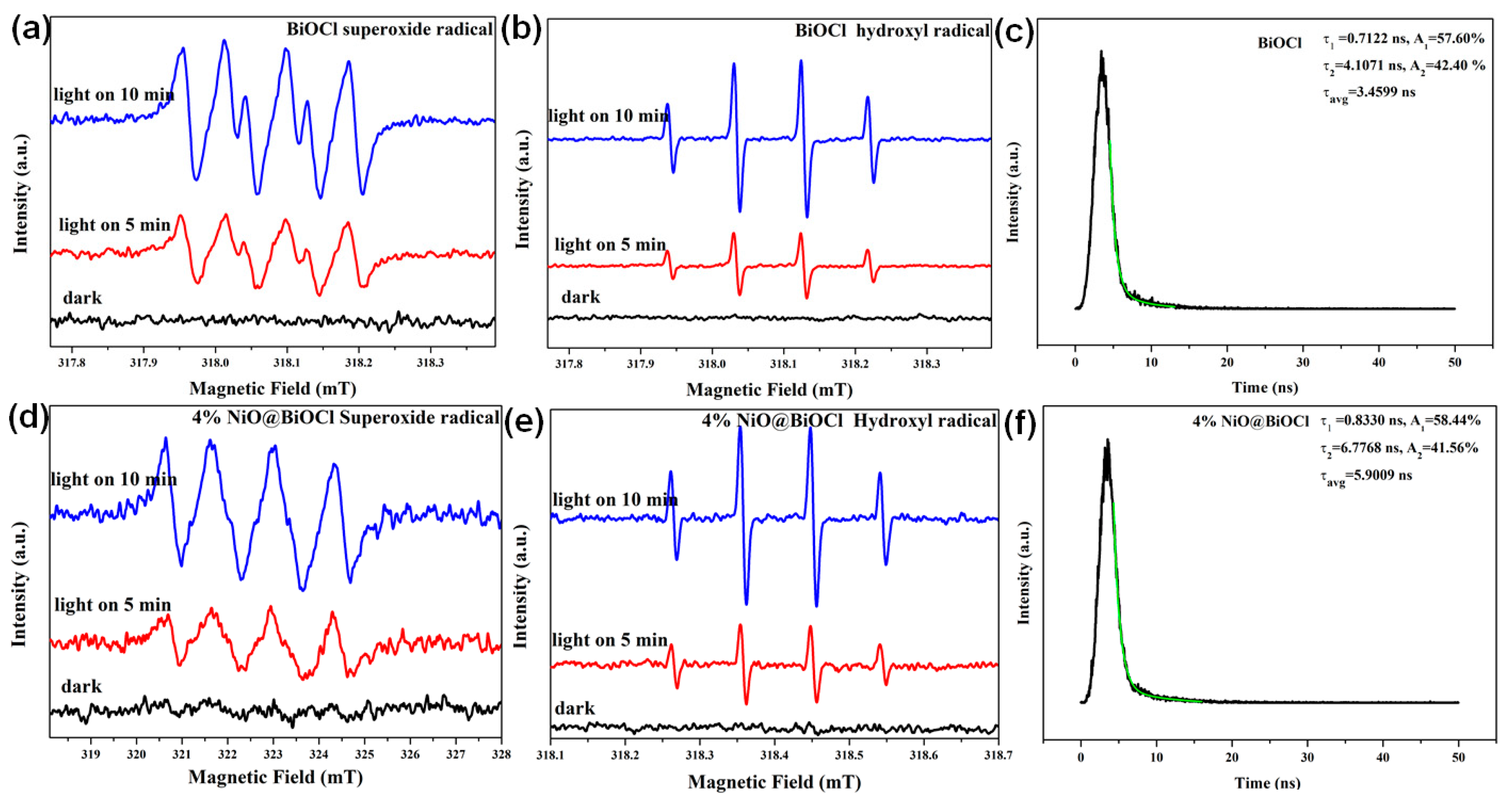
DMPO spin-trapping ESR technique was employed to verify the active radicals produced on condition that the photocatalyst was under light irradiation. As shown in Figure 5, no peaks could be observed in the darkness. Clearly, characteristic peaks for ·OH and O2•− emerged once the as-obtained samples were irradiated by visible light. Both intensities for ·OH and O2•− became stronger with increasing irradiation time.
The number of reactive species was exactly proportional to the square of the intensity of the signal detected via ESR test [24], while the ·OH and ·O2 amounts for NiO@BiOCl were calculated to be ~6.5 and 2.2 times that of BiOCl (See Table S1 (Supplementary Materials)). The composite could generate more reactive radicals due to more photoinduced carries being produced by the NiO/BiOCl system under the same conditions. The NiO(100)-O(BiClO-110) heterostructure was formed, facilitating NiO (111) crystal surface exposure. A previous investigation [25] indicated that NiO (111) exhibited a much lower energy barrier for the dissociation of H2O into Hads/OHads. The photoproduced holes could easily oxidize OHads to generate ·OHads via a one-hole pathway. The photoinduced electron could react with adsorbed or dissolved oxygen in a water environment to generate ·OH via a five-electron pathway. Meanwhile, the photoinduced electron could directly generate O2•− via a one-electron transfer pathway. Besides, in this NiO@BiOCl system, the photoinduced holes not oxidized water to obtain oxygen via a four-hole pathway or two-hole pathway, but also broke the chemical bonds in organic molecules [26,27].
Time-resolved photoluminescence (TRPL) spectra were carried out to assess recombination kinetics of photoinduced electron–hole pairs. The TRPL decay spectra waves were fitted by exponential decay kinetics function. The corresponding results are listed in Figure 5c,f. The average lifetime of photogenerated carries in the 4% NiO@BiOCl system (5.9009 ns) was longer than that of photoinduced carries for pure BiOCl (3.4599 ns) under the same conditions, which was consistent with analog computation results. The Z-scheme-type heterojunction prolonged the lifetime of photoinduced carriers and enhanced their redox capacity. Thus, the longer lifetime boosted the likelihood of photogenerated pairs participating in photochemical reactions and further enhanced their photocatalytic activity.

2.3. Optical Properties and Photocatalytic Activity

Figure S3 shows the UV-Vis diffuse reflectance spectroscopy (UV-Vis DRS) spectra of NiO@BiOCl with different NiO contents of 0%, 1%, 2%, 3%, 4%, 5%, and 6%. Compared with the pure BiOCl, all the samples loaded with NiO could increase the light harvesting to some extent in the wavelength range of 400–800 nm. With the increase of the NiO content, the light absorption increased gradually, revealing that the NiO NPs loaded on the surface of BiOCl can enhance the light absorption capacity.
On the one hand, the black color of NiO itself naturally strengthens the light absorption. On the other hand, the Ni (II) d-d transition in the heterojunction system occurred [4]. Although the light absorption capacity of BiOCl was enhanced, the absorption edge was not altered. The band gap width of the samples was almost unchanged and the band gap energy was approximately 3.47 eV, indicating that the Ni species was not incorporated into the crystal lattice of BiOCl and did not change its electronic structure [6]. From SEM and TEM diagrams, NiO only deposited on the surface of BiOCl to form the heterostructure. No Ni3+ entered into the BiOCl crystal lattice or replaced Bi3+ sites.
To determine the relationship between the incorporation amount of NiO and the photocatalytic performance of NiO@BiOCl, the NiO@BiOCl heterojunctions with NiO mass ratios of 0%, 1%, 2%, 3%, 4%, 5%, and 6% were prepared in this work. The photocatalytic activity of NiO@BiOCl was examined by RhB dye photodegradation. Because 100.0 mg (conventional dosage) of the photocatalyst was used to photodegrade 10.0 mg/L of RhB solution in our previous tests, the photodegradation reaction was too fast. The RhB dye could be completely degraded after only 5 min under visible light irradiation, meaning that the photocatalytic performance could not be compared to optimize the ratio of NiO. Thus, the dosage of the photocatalyst was reduced to 25.0 mg in this section, while the volume and concentration of the RhB dye remained unchanged.
The UV-Vis absorption spectra of the RhB photodegradation using NiO@BiOCl as the photocatalyst are shown in Figure S4a–g. Figure S4h,I (Supplementary Materials) displays the photocatalytic performance comparison and degradation ratio. The NiO@BiOCl with 4% NiO possessed the best photocatalytic activity. The corresponding photocatalytic efficiency of NiO@BiOCl was 1.86 times than that of pure BiOCl. An interesting phenomenon can be observed in Figure S4i. As the content of NiO increased in the range of 0~4%, the photocatalytic activity of NiO@BiOCl increased. When the NiO contents were 5% and 6%, the RhB degradation ratios decreased, indicating that the excessive NiO content would reduce the photocatalytic activity of NiO@BiOCl. The excessive NiO in NiO@BiOCl would abate the concentration of photoinduced holes. In addition, the NiO@BiOCl could maintain high photocatalytic activity after being reused four times, indicating that it had good photostability (See Figure S4j (Supplementary Materials)).
The p-p type heterojunction NiO@BiOCl was formed when the p-type NiO contacted the p-type BiOCl [6,28]. Meanwhile, the noble-metal-free doped Z-scheme heterojunction was found in NiO@BiOCl, which could generate photoinduced carriers with stronger redox capability and prolong the lifetime of photoinduced carriers. NiO (111) had a much lower energy barrier for water molecule dissociation of water molecules into Hads/OHads [29]. Thus, ·OH was easily obtained by oxidizing OHads through a one-hole pathway (See Figure 1).
To the best of our knowledge, under visible light irradiation the photocatalytic ratio for the RhB dye reached 100% after only 5 min of photocatalytic reaction using the NiO@BiOCl heterojunction, whose efficiency was far better than the values seen in the existing literature reports over the last two years (See Table S2 (Supplementary Materials)). The ultrafast photochemical reaction facilitated its practical application. In fact, this noble-metal-free doped Z-scheme heterojunction could serve in water photolysis for oxygen production.

3. Experimental Procedures

3.1. Materials Synthesis

Here, 1.0 g polyvinylpyrrolidone K30 (PVPK30) was completely dissolved in 100.0 mL distilled water via agitation to form a homogeneous solution that was divided into two parts. Then, 5.0 mmol Bi(NO3)3·5H2O was totally dissolved in the above homogeneous solution through stirring for 30 min. Next, 5.0 mmol KCl was also dissolved in the other part of the homogeneous solution by stirring. The KCl solution was slowly added into the Bi(NO3)3·5H2O suspension under magnetic stirring conditions. The Ni(NO3)2·6H2O solutions with different mass ratios (1%, 2%, 3%, 4%, 5%, and 6%) were slowly added into the above mixed solution of KCl and Bi(NO3)3·5H2O, then stirring continued for 4 h. The whole mixed solution was filtered. The obtained filter cake was washed several times using water and absolute ethyl alcohol. The washed filter cake was dried at 80 °C for 10 h to obtain the resultant NiO@BiOCl heterojunction.

3.2. Material Characterization

The phases and structures of samples were identified using an X-ray diffractometer (Shimadzu, XRD-6000, Kyoto, Japan) with Cu Kα radiation source at a wavelength of 0.154056 nm (40 kV, 30 mA). An INVIA Raman microprobe (Renishaw Instruments, Wotton-under-Edge, UK) and X-ray photoelectron spectrometer (Kratos, Manchester, UK) with a base pressure of 2 × 10⁻⁷ Pa were used with an X-ray gun at 180 W (12 kV, 15 mA). The samples’ morphologies were observed by scanning electron microscopy (SEM, FEI, F50, ZEISS, Oberkochen, Germany) and transmission electron microscopy (TEM, FEI, Tecnai G2 F20, Hillsboro, OR, USA). The optical properties of samples were examined by an ultraviolet-visible diffuse reflectance spectrophotometer (UV-Vis DRS, TU1901, Beijing, China). Time-resolved photoluminescence (TRPL) spectra were recorded using a fully functional steady transient fluorescence spectrometer (FLS980, Edinburgh, UK). Electron spin resonance (ESR) signals of radical species trapped by 5,5-dimethyl-1-pyrroline N-oxide (DMPO) were detected on a JES FA200 (JEOL, Japan) spectrometer.

3.3. First-Principles Calculation Method

First-principles calculations based on DFT were performed by using the Vienna Ab initio Simulation Package (VASP) [30,31]. The projector-augmented wave (PAW) method was used for the treatment of electronic core interactions [31]. Electron exchange and correlation interactions were treated by the generalized gradient approximation with Perdew–Burke–Ernzerh (PBE)-type functionality [32]. To avoid interactions between the periodic slabs along the vertical direction, a vacuum layer thickness of 2.0 nm with was used with the dipole correction method [33]. The energy cutoff was set to 500.0 eV and a force tolerance of 0.01 eV/Å on each atom was used for the convergence criterion. The Monkhorst–Pack K-points sampling of 2 × 5 × 1 was employed in the DFT calculations. The interfacial adhesion energy (Eadh) was calculated as Eadh = ENiO + EBiOCl − Ehetero, in which ENiO, EBiOCl, and Ehetero represent the total energies of the NiO (100) surface, the BiOCl (110) surface, and the corresponding heterojunction formed by them, respectively. The surface work function calculation is based on the following equation: Φ = Evac − Ef. where Φ, Evac, and Ef represent the work function, vacuum level, and Fermi level, respectively. The plane-averaged charge density difference ( Δ ρ z ) is given by:
Δ ρ z   =   Δ ρ d x d y ,
where Δ ρ   =   ρ A B ρ A ρ B , ρ A B ,   ρ A ,   ρ B represent the charge density of the heterostructure, the NiO (100) surface, and the BiOCl (110) surface, respectively.

3.4. Evaluation of Photocatalytic Activity

First, 100.0 mg of NiO@BiOCl photocatalyst powder was put into 10.0 mg/L of RhB dye solution (100.0 mL). The solution was stirred for 1 h in dark to reach adsorption–desorption equilibrium. A 500 W Xe lamp equipped with an ultraviolet (UV) light cut-off filter (λ ≥ 400 nm) was used as the visible light source. At given irradiation time intervals, a series of the reaction solution was sampled and the ultraviolet-visible absorption spectrum was measured.

4. Conclusions and Outlooks

The noble-metal-free doped Z-scheme NiO@BiOCl heterojunction was confirmed via experiments and theoretical calculations. Under visible light irradiation, the charge transfer processes (e-e or h+-h+) was not allowed, while the photogenerated e-h+ recombination between the BiOCl (110) and Ni (100) surfaces was accelerated, hence forming a Z-scheme charges transfer pathway. The Z-scheme NiO@BiOCl heterojunction generated photoinduced carriers with stronger redox capabilities, prolonged the lifetime of photoinduced carriers, and boosted the photocatalytic activity. In fact, 100.0 mL and 10.0 mg/L RhB could be completely photodegraded using 100.0 mg photocatalyst (conventional dosage) after only 5 min under visible light irradiation. We sincerely hope this heterojunction will be applied in the actual industrial field.

Supplementary Materials

The following are available online at https://www.mdpi.com/2073-4344/10/9/958/s1, Figure S1: The high-resolution XPS spectra of each elements in 4% NiO@BiOCl, Figure S2: SEM images of pure BiOCl and 4%NiO@BiOCl (a,b); EDS energy spectra of 4% NiO@BiOCl (c); TEM and HRTEM image of 4%NiO@BiOCl (d,e), Figure S3: UV-vis diffuses reflectance spectra NiO@BiOCl photocatalysts (a,b) and the corresponding band gap energy (c), Figure S4:Time-dependent UV-vis absorption spectra of the RhB in the presence of various composite NiO@BiOCl (wtNiO = 0~6%) photocatalysts (a~g), the corresponding degradation rate (h,i) and recycling use (j), Figure S5 Electrochemical impedance spectroscopy patterns of samples, Table S1 The intensity value of free radical signal and corresponding square value from ESR test, Table S2 Comparison of photodegradation ratio using different photocatalyst under visible light irradiation reported in the last three years.

Author Contributions

J.Y., Q.Z. Experimental and DFE calculation, First draft writing. J.W., Y.P., Y.W., Method, data discussion and First draft writing. C.L., Proofreading of first draft and provision of instruments. T.X., L.X., Supervision and final proofreading. All authors have read and agreed to the published version of the manuscript.

Funding

This research was funded by Scientific and Technological Research Program of Chongqing Municipal Education Commission KJQN201901401/KJZD-K201901305) and Chongqing Basic Science and Advanced Technology Research Program (CSTC2015jcyjBX0015 and CSTC2018jcyjAX 0426). And the APC was funded by Scientific and Technological Research Program of Chongqing Municipal Education Commission KJZD-K201901305.

Acknowledgments

We want to thank the financial support from the Scientific and Technological Research Program of Chongqing Municipal Education Commission KJQN201901401/KJZD-K201901305) and Chongqing Basic Science and Advanced Technology Research Program (CSTC2015jcyjBX0015 and CSTC2018jcyjAX 0426).

Conflicts of Interest

The authors declare that they have no known competing financial interests or personal relationships that could have appeared to influence the work reported in this paper.

References

  1. Wang, X.; Hong, M.Z.; Zhang, F.W.; Zhuang, Z.Y.; Yu, Y. Recyclable Nanoscale Zero Valent Iron Doped g-C3N4/MoS2 for Efficient Photocatalytic of RhB and Cr (VI) Driven by Visible Light. ACS Sustain. Chem. Eng. 2016, 4, 4055–4063. [Google Scholar] [CrossRef]
  2. Fu, H.B.; Zhang, S.C.; Xu, T.G.; Zhu, Y.F.; Chen, J.M. Photocatalytic Degradation of RhB by Fluorinated Bi2WO6 and Distributions of the Intermediate Products. Environ. Sci. Technol. 2008, 42, 2085–2091. [Google Scholar] [CrossRef] [PubMed]
  3. Zhuang, J.D.; Dai, W.X.; Tian, Q.F.; Li, Z.H.; Xie, L.Y.; Wang, J.X.; Liu, P. Photocatalytic Degradation of RhB over TiO2 Bilayer Films: Effect of Defects and Their Location. Langmuir 2010, 26, 9686–9694. [Google Scholar] [CrossRef] [PubMed]
  4. Yang, H.; Yu, J.H.; Seo, H.J.; Jeong, R.H.; Boo, J.H. Improved electrochromic properties of nanoporous NiO film by NiO flake with thickness controlled by aluminum. Appl. Surf. Sci. 2018, 461, 88–92. [Google Scholar] [CrossRef]
  5. Yang, J.; Xie, T.P.; Zhu, Q.X.; Wang, J.K.; Xu, L.J.; Liu, C.L. Boosting the photocatalytic activity of BiOX under solar light via selective crystal facet growth. J. Mater. Chem. C 2020, 8, 2579–2588. [Google Scholar] [CrossRef]
  6. Yang, J.; Xie, T.P.; Liu, C.L.; Xu, L.J. Dy(III) Doped BiOCl Powder with Superior Highly Visible-Light-Driven Photocatalytic Activity for Rhodamine B Photodegradation. Nanomaterials 2018, 8, 697. [Google Scholar] [CrossRef] [Green Version]
  7. He, R.G.; Xu, D.F.; Cheng, B.; Yu, J.G.; Ho, W.K. Review on Nanoscale Bi-based Photocatalysts. Nanoscale Horiz. 2018, 3, 464–504. [Google Scholar] [CrossRef]
  8. Jiang, J.; Zhao, K.; Xiao, X.Y.; Zhang, L.Z. Synthesis and Facet-Dependent Photoreactivity of BiOCl Single Crystalline Nanosheets. J. Am. Chem. Soc. 2012, 134, 4473–4476. [Google Scholar] [CrossRef]
  9. Xie, T.P.; Xu, L.J.; Liu, C.L.; Yang, J.; Wang, M. Magnetic composite BiOCl–SrFe12O19: A novel p-n type heterojunction with enhanced photocatalytic activity. Dalton Trans. 2014, 43, 2211–2220. [Google Scholar] [CrossRef]
  10. Li, Y.W.; Zhao, Y.; Wu, G.J.; Ma, H.M.; Zhao, J.Z. Bi superlattice nanopolygons at BiOCl (001) nanosheet assembled architectures for visible-light photocatalysis. Mater. Res. Bull. 2018, 101, 39–47. [Google Scholar] [CrossRef]
  11. Li, H.; Li, J.; Ai, Z.H.; Jia, F.L.; Zhang, L.Z. Oxygen Vacancy-Mediated Photocatalysis of BiOCl: Reactivity, Selectivity, and Perspectives. Angew. Chem. Int. Ed. 2018, 57, 122–138. [Google Scholar] [CrossRef] [PubMed]
  12. Prabhu, V.G.; Paloly, A.R.; Divya, N.G.; Bushiri, M.J. Photocatalytic and ferromagnetic properties of electrically conducting multifunctional Ni/NiO nanocomposites in amorphous carbon matrix. Mater. Sci. Eng. B 2018, 228, 132–141. [Google Scholar] [CrossRef]
  13. Ghosh, C.; Majumder, S.; Bhattachatjee, S. NiO/Ag heterostructure: Enhanced UV emission intensity, exchange interaction and photocatalytic activity. RSC Adv. 2016, 6, 56503–56510. [Google Scholar]
  14. Fechete, I.; Ivan, S.B.; Popescu, I.; Garin, F.; Parvulescu, V.I.; Marcu, I.C. The effect of phosphorus on the catalytic performance of nickel oxide in ethane oxidative dehydrogenation. Catal. Sci. Technol. 2016, 6, 6953–6964. [Google Scholar]
  15. Faisal, M.; Harraz Farid, A.; Ismail Adel, A.; El-Tonib, A.M.; Al-Sayari, S.A.; Al-Hajry, A.; Al-Assiri, M.S. Novel mesoporous NiO/TiO2 nanocomposites with enhanced photocatalytic activity under visible light illumination. Ceram. Int. 2018, 44, 7047–7056. [Google Scholar] [CrossRef]
  16. Varshney, D.; Dwivedi, S. Synthesis, structural, Raman spectroscopic and paramagnetic properties of Sn doped NiO nanoparticles. Superlattices Microstruct. 2015, 86, 430–437. [Google Scholar] [CrossRef]
  17. Hu, J.L.; Fan, W.J.; Ye, W.Q.; Huang, C.J.; Qiu, X.Q. Insights into the photosensitivity activity of BiOCl under visible light irradiation. Appl. Catal. B 2014, 158–159, 182–189. [Google Scholar] [CrossRef]
  18. Wang, M.G.; Han, J.; Hu, Y.M.; Guo, R.; Yin, Y.D. Carbon-Incorporated NiO/TiO2 Mesoporous Shells with p-n Heterojunctions for Efficient Visible Light Photocatalysis. ACS Appl. Mater. Interfaces 2016, 8, 29511–29521. [Google Scholar] [CrossRef]
  19. Xing, P.X.; Zhang, W.Q.; Chen, L.; Dai, X.Q.; Zhang, J.L.; Zhao, L.H.; He, Y.M. Preparation of a NiO/KNbO3 nanocomposite via a photodeposition method and its superior performance in photocatalytic N2 fixation. Sustain. Energy Fuels 2020, 4, 1112–1117. [Google Scholar] [CrossRef]
  20. Mallows, J.; Planells, M.; Thakare, V.; Bhosale, R.; Ogale, S.; Robertson, N. p-Type NiO Hybrid Visible Photodetector. ACS Appl. Mater. Interfaces 2015, 7, 27597–27601. [Google Scholar] [CrossRef]
  21. Fu, G. Tuning the Electronic Structure of NiO via Li Doping for the Fast Oxygen Evolution Reaction. Chem. Mater. 2019, 31, 419–428. [Google Scholar] [CrossRef]
  22. He, Z.; Shi, Y.; Gao, C.; Wen, L.; Chen, J.; Song, S. BiOCl/BiVO4 P–N Heterojunction with Enhanced Photocatalytic Activity under Visible-Light Irradiation. J. Phys. Chem. C 2014, 118, 389–398. [Google Scholar] [CrossRef]
  23. Zhang, X.; Fan, C.; Wang, Y.; Wang, Y.; Liang, Z.; Han, P. Dft+U Predictions: The Effect of Oxygen Vacancy on the Structural, Electronic and Photocatalytic Properties of Mn-Doped BiOCl. Comp. Mater. Sci. 2013, 71, 135–145. [Google Scholar] [CrossRef]
  24. Huang, H.; Tu, S.; Zeng, C.; Zhang, T.; Reshak, A.H.; Zhang, Y. Macroscopic Polarization Enhancement Promoting Photo- and Piezoelectric-Induced Charge Separation and Molecular Oxygen Activation. Angew. Chem. Int. Ed. 2017, 56, 11860–11864. [Google Scholar] [CrossRef] [PubMed]
  25. Chai, S.Y.; Kim, Y.J.; Jung, M.H.; Chakraborty, A.K.; Jung, D.; Lee, W.I. Heterojunctioned BiOCl/Bi2O3, a New Visible Light Photocatalyst. J. Catal. 2009, 262, 144–149. [Google Scholar] [CrossRef]
  26. Muñoz-Batista, M.; Gómez-Cerezo, M.; Kubacka, A.; Tudela, D.; Fernández-García, M. Role of Interface Contact in CeO2–TiO2 Photocatalytic Composite Materials. ACS Catal. 2014, 4, 63–72. [Google Scholar]
  27. Muñoz-Batista, M.; Ballari, M.; Kubacka, A.; Alfano, O.; Fernández-García, M. Braiding kinetics and spectroscopy in photo-catalysis: The spectro-kinetic approach. Chem. Soc. Rev. 2019, 48, 637–682. [Google Scholar]
  28. Huang, S.Y.; Wu, H.; Matsubara, K.; Cheng, J.; Pan, W. Facile assembly of n-SnO2 nanobelts p-NiO heterojunctions with enhanced ultraviolet photoresponse. Chem. Commun. 2014, 50, 2847–2850. [Google Scholar] [CrossRef] [Green Version]
  29. Zhao, L.; Zhang, Y.; Zhao, Z.L.; Zhang, Q.H.; Huang, L.B.; Gu, L.; Lu, G.; Hu, J.S.; Wan, L.J. Steering Elementary Steps towards Efficient Alkaline Hydrogen Evolution via Size-Dependent Ni/NiO Nanoscale Heterosurfaces. Nat. Sci. Rev. 2020, 7, 27–36. [Google Scholar] [CrossRef] [Green Version]
  30. Kresse, G.; Furthmuller, J. Efficient Iterative Schemes for Ab Initio Total-Energy Calculations Using a Plane-Wave Basis Set. Phys. Rev. B 1996, 54, 11169–11186. [Google Scholar] [CrossRef]
  31. Kresse, G.; Joubert, D. From Ultrasoft Pseudopotentials to the Projector Augmented-Wave Method. Phys. Rev. B 1999, 59, 1758–1775. [Google Scholar] [CrossRef]
  32. Perdew, J.P.; Burke, K.; Ernzerhof, M. Generalized Gradient Approximation Made Simple. Phys. Rev. Lett. 1996, 77, 3865–3868. [Google Scholar] [CrossRef] [PubMed] [Green Version]
  33. Neugebauer, J.; Scheffler, M. Adsorbate-Substrate and Adsorbate-Adsorbe Interactions of Na and K Adlayers on Al(111). Phys. Rev. B 1992, 46, 16067–16080. [Google Scholar] [CrossRef] [PubMed] [Green Version]
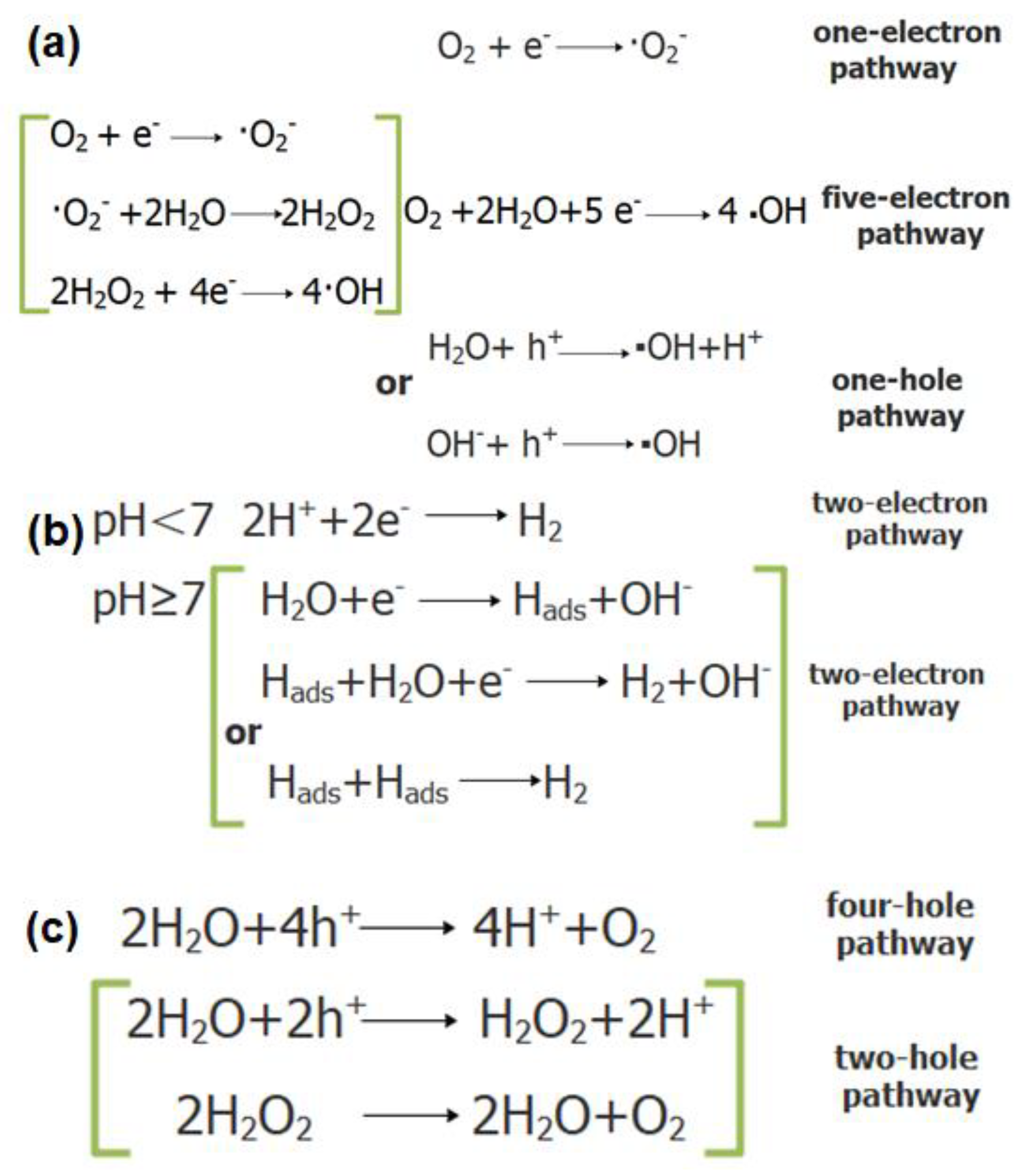
Figure 1. Hydroxyl radical obtained via two pathways (a), hydrogen production via photocatalysis of water (b), and oxygen production via photocatalysis of water (c).
Catalysts 10 00958 g001
Figure 2. XRD patterns (a) and Raman spectra (b) of samples and XPS total survey of 4% NiO@BiOCl (c).
Catalysts 10 00958 g002
Figure 3. The heterojunction structures of the NiO-100 and BiOCl-110 surfaces. The NiO(100)-O(BiOCl-110) heterostructure: (a) initial structure, (b) DFT-optimized structure. The NiO(100)-BiCl(BiOCl-110) heterostructure: (c) the initial structure, (d) DFT-optimized structure.
Catalysts 10 00958 g003
Figure 4. First-principles calculations for the plane-averaged electrostatic potential energy profile (a), the charge density difference for the NiO(100)-O(BiClO-110) heterostructure (b), and schematic illustration of the band alignment and the photogenerated charge carrier transfer process in Z-scheme mode for the NiO@BiOCl heterojunction (c). CBM, minimum conduction band; VBM, maximum valence band.
Catalysts 10 00958 g004
Figure 5. DMPO spin-trapping ESR spectra in BiOCl and 4% NiO@BiOCl in dark or under visible light irradiation: (a,d) in methanol dispersion for DMPO-O2•−; (b,e) in aqueous dispersion for DMPO-OH. Time-resolved photoluminescence (TRPL) spectra of BiOCl (c) and 4% NiO@BiOCl (f).
Catalysts 10 00958 g005