Article

Development of Specific Inhibitors for Oncogenic Phosphatase PPM1D by Using Ion-Responsive DNA Aptamer Library

Department of Chemistry, Faculty of Science, Niigata University, 8050 2-no-cho, Ikarashi, Nishi-ku, Niigata 950-2181, Japan
*
Author to whom correspondence should be addressed.
Catalysts 2020, 10(10), 1153; https://doi.org/10.3390/catal10101153
Submission received: 17 September 2020 / Revised: 1 October 2020 / Accepted: 2 October 2020 / Published: 7 October 2020
(This article belongs to the Section Biocatalysis)

Abstract

:
(1) Background: Ser/Thr protein phosphatase PPM1D is an oncogenic protein. In normal cells, however, PPM1D plays essential roles in spermatogenesis and immune response. Hence, it is necessary to develop novel PPM1D inhibitors without side effects on normal cells. Stimuli-responsive molecules are suitable for the spatiotemporal regulation of inhibitory activity. (2) Methods: In this study, we designed an ion-responsive DNA aptamer library based on G-quadruplex DNA that can change its conformation and function in response to monovalent cations. (3) Results: Using this library, we identified the PPM1D specific inhibitor M1D-Q5F aptamer. The M1D-Q5F aptamer showed anti-cancer activity against breast cancer MCF7 cells. Interestingly, the induction of the structural change resulting in the formation of G-quadruplex upon stimulation by monovalent cations led to the enhancement of the inhibitory activity and binding affinity of M1D-Q5F. (4) Conclusions: These data suggest that the M1D-Q5F aptamer may act as a novel stimuli-responsive anti-cancer agent.

1. Introduction

Protein Ser/Thr phosphatase PPM1D (Wip1), composed of 605 amino acids, is an oncogenic protein that dephosphorylates tumor-suppressor proteins such as p53, ATM, and Chk2 [1]. PPM1D acts as a negative regulator of the tumor suppressor signaling pathway and its overexpression results in tumorigenesis [1]. Gene amplification and overexpression of PPM1D have been observed in many human tumors, including breast cancer, ovarian cancer, and gastric cancer [2]. In addition, we previously developed a homology model of PPM1D using metal-dependent protein phosphatases (PPM), namely PPM1A, PPM1B, and PPM1K, whose crystal structures have been resolved, as templates [3]. Using this homology model, we reported that the catalytic domain of PPM1D consists of a characteristic basic rich loop (B-loop) in the proximity of the active site of PPM1D [3]. Since the B-loop is essential for substrate recognition and cellular localization of PPM1D, it is expected that the molecules that bind to the B-loop will act as specific inhibitors of PPM1D. Recently, C-terminal truncated mutants of PPM1D were reported to function as gain-of-function mutants and were determined as the risk factors for breast cancer and ovarian cancer [4]. Previously, we identified a novel splicing variant, namely, PPM1D430, which is composed of 430 amino acids and has a similar structure to the gain-of-function mutants, namely, C-terminal truncated PPM1Ds [5]. These observations suggest that specific inhibitors for PPM1D430, which bind especially to the B-loop, will be applicable for other PPM1D variants comprising C-terminal truncated mutants as lead compounds of anti-cancer agents.
Choi et al. reported that mice with Ppm1d gene knockout demonstrated not only cancer resistance but also defects in spermatogenesis and immune response [6,7]. In addition, we previously reported that PPM1D430 is expressed significantly in the testes and leukocytes, whereas PPM1D605 shows ubiquitous expression in normal tissues [5]. These observations demonstrate that PPM1D functions not only as an oncogenic protein but also as a regulator of the reproduction and immune system under normal conditions. Hence, specific inhibitors of PPM1D may lead to serious side effects in normal tissues. Therefore, it is necessary to develop molecules that can inhibit PPM1D activity in cancer cells but not in normal cells. To date, certain PPM1D inhibitors have been reported by several groups. We reported specific inhibitors of PPM1D derived from small compounds and peptides [3,8,9]. Hayashi et al. reported cyclic peptide inhibitor that can bind to active center of PPM1D [10]. In addition, Gilmartin et al. reported that the PPM1D inhibitor GSK2830371 exerts anti-cancer effects in tumor-bearing mice upon oral administration [11]. These studies demonstrated that PPM1D inhibitors exhibit anti-cancer activity against cancer cells and tumor tissues. However, no functional PPM1D inhibitors have been reported that act only in cancer cells but not in normal cell types, such as blood cells and testis cells.
Stimuli-responsive functional molecules have been garnering interest in various fields such as drug development and functional biomaterials [12]. These molecules change their conformation and function in response to various stimuli such as light, ion, pH, and temperature. For instance, elastin-like polypeptide (ELP), which is used in drug delivery systems, shows temperature-induced conformational changes that exhibit a sharp phase transition (soluble to insoluble) in a temperature-dependent manner [13]. Azobenzene, a light-responsive molecule, undergoes cis-trans isomerization in response to UV or visible light [14]. This ability of stimuli-responsive molecules to undergo a structural switch can be used to regulate the inhibition of target proteins with low side effects.
Recently, RNA/DNA aptamers have been the focus of many studies as molecule-targeting drugs. RNA/DNA aptamers are special single-stranded RNA or DNA molecules that can bind to target proteins with high specificity and affinity. Aptamers are typically selected using the Systematic Evolution of Ligands by EXponential enrichment (SELEX) method from a randomized oligonucleotide library [15,16,17,18]. Since aptamers exhibit high affinity and high specificity for their individual targets, they have been used as diagnostic tools and therapeutic agents [19,20]. RNA aptamer-based “Macugen”, which is a vascular endothelial growth factor (VEGF) antagonist, was approved by the Food and Drug Administration (FDA) [21]. As a cancer therapeutic agent, AS1411 DNA aptamer binds to the nucleolin on the cell surface and shows anti-cancer activity against various types of cancers, such as breast cancer [22]. An aptamer that binds to the protein MUC1, whose overexpression is observed specifically in cancers, acts as an anti-cancer agent [23]. Currently, most aptamer-based drugs that can be used in clinical therapy are RNA aptamers [19]. Since RNA oligonucleotides can form more diverse and complex 3D structures than DNA oligonucleotides owing to the presence of non-Watson–Crick base pairs that interact with each other via hydroxyl group (–OH) at the 2’ position, RNA libraries are suitable for selecting aptamers with high affinity and specificity. In contrast, RNA aptamers are unstable in serum. It is difficult to use intact RNA aptamers as therapeutic agents without chemical modifications. Although the pharmacokinetics of RNA aptamers can be improved by chemical modifications such as the F− or NH2− modified the sugar backbone and PEG conjugation, DNA aptamers show higher resistance to nuclease digestion and stability in the serum than that by RNA aptamers [21,24,25]. These observations indicate that DNA aptamers specific to disease-related proteins can be used as aptamer-based drugs with low cost and high stability in vivo.
To date, few studies have examined screening methods to identify stimuli-responsive aptamer-based molecules that can modulate the activity or function of target proteins. For instance, Hu et al. reported a pH-responsive aptamer that can regulate Taq polymerase activity in a pH-dependent manner [26]. Among DNA molecules, guanine-rich DNA can form a four-stranded G-quadruplex structure in response to cationic ions [27]. The G-quadruplex DNA structure is formed by cyclic Hoogsteen base pairs between four guanines in guanine rich DNA. In the G-quadruplex, four guanines are arranged in a square planar conformation with two or more G-quartets stacked on top of each other [28,29,30]. The G-quadruplex structure can be stabilized by the intercalation of monovalent cations such as potassium ion and sodium ion [31]. The 3D structure of G-quadruplex DNA has been observed in the human telomeric and promoter regions [32,33]. In addition, the structure and ion-responsiveness of G-quadruplex-forming sequences have been analyzed by X-ray crystallography, circular dichroism (CD) spectroscopy, NMR, and mass spectrometry (MS) [34,35]. The property of dynamic structural transition between linear DNA (w/o ion) and G-quadruplex DNA (with ion) has been used in the development of effective biosensors or imaging tools in many studies [35,36]. These observations suggest that the G-quadruplex DNA aptamer can be used for the development of functional drugs that can switch their structure and activity in response to cationic species and their concentration.
In this study, we designed an ion-responsive DNA aptamer (designated as IRDAptamer) library based on the G-quadruplex DNA sequence and screened the library for PPM1D specific aptamer, whose inhibitory activity can be regulated by external cationic stimuli. One of the identified aptamers demonstrated the regulation of the inhibitory activity and binding affinity toward PPM1D via structural changes in response to monovalent cationic species. In addition, this aptamer showed anti-cancer activity in breast cancer MCF7 cells, which demonstrate the overexpression of PPM1D. These results suggested that the aptamer identified using our IRDAptamer library may act as anti-cancer drugs targeting PPM1D.

2. Results

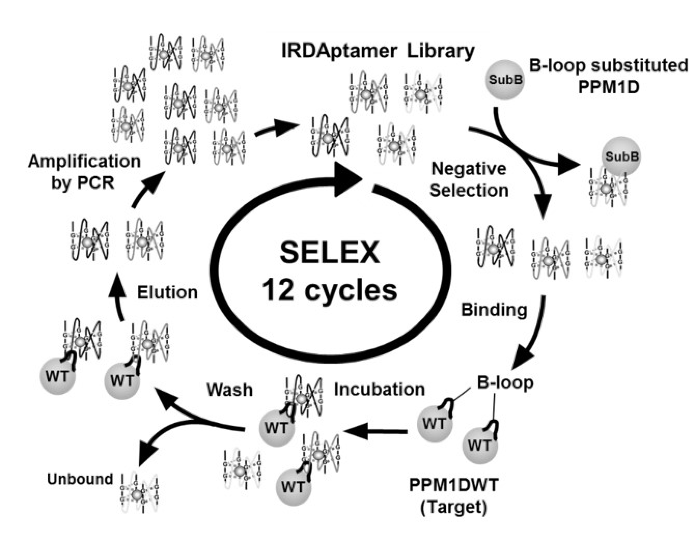
2.1. Screening of Ion-Responsive G-Quadruplex DNA Aptamer Targeting PPM1D

In order to prepare a DNA aptamer library with components that can regulate their binding and inhibitory activity in response to stimuli, we designed an ion-responsive DNA aptamer library (IRDAptamer library) consisting of a 40-mer G-quadruplex DNA aptamer region flanked by two 20-mer primer regions for PCR amplification. In the G-quadruplex DNA aptamer region, three randomized oligonucleotide regions (N6) were located between four G-stretches (AGG or TTAGGG sequences). These randomized oligonucleotide regions will recognize the individual target molecules. We previously identified PPM1D specific basic rich loop, also known as the B-loop, in the catalytic domain of PPM1D, which is associated with substrate recognition and cellular localization in PPM1D. Since the B-loop is a characteristic of PPM1D compared to other phosphatases of the PPM family, inhibitors targeting the B-loop will act as the specific inhibitors of PPM1D. Therefore, we screened IRDAptamers from the designed library targeting the B-loop of PPM1D430 using the SELEX method (Figure 1).
Aptamers bound to PPM1D, except for the B-loop, were removed by preincubation with PPM1D430SubB, in which the B-loop was substituted with the corresponding loop in PPM1A before 1st, 4th, 8th, and 12th cycles in SELEX screening. After 12 rounds of selection, we recovered and sequenced 48 clones and identified 4 individual sequences, namely M1D-Q1F, M1D-Q4F, M1D-Q5F, and M1D-Q6F, as candidates for aptamers targeting the B-loop (Table 1).
To confirm the inhibitory activity of these aptamers against PPM1D430, in vitro phosphatase assays were performed using p53(15pS) phosphorylated peptide as the physiological substrate. Among the isolated aptamers, the M1D-Q5F aptamer significantly inhibited the phosphatase activity of PPM1D430 at a concentration of 10 μM. Other aptamers showed little inhibition against PPM1D (Figure 2a).
Next, we measured the half-maximal (50%) inhibitory concentration (IC50) value of M1D-Q5F in addition to the IC50 value of M1D-Q4F and M1D-Q5F-Scrambled, which consists of M1D-Q5F sequence that is scrambled but has the same nucleotide content. The IC50 value of the M1D-Q5F aptamer was 2.9 μM. Neither M1D-Q4F nor M1D-Q5F-Scrambled aptamer showed distinct inhibitory activities (Figure 2b). Little inhibition of M1D-Q5F-Scrambled indicated that the inhibitory activity of M1D-Q5F against PPM1D430 can be attributed to the structure-dependent mechanism. Lineweaver–Burk analysis by using an in vitro phosphatase assay was also performed to analyze the inhibition mode of the M1D-Q5F aptamer. A double-reciprocal plot showing an intersection on the x-axis indicated that M1D-Q5F inhibited PPM1D430 activity in a non-competitive manner (Figure 2c).
We analyzed the enzyme specificity of the M1D-Q5F aptamer using pNPP as a common substrate for protein phosphatases. The M1D-Q5F aptamer inhibited PPM1D430. However, both PPM1A phosphatases, a PPM-type phosphatase without B-loop and FCP/SCP type phosphatase Scp1, which demonstrates phosphatase activity owing to the presence of conserved aspartic acid residues (DXDXT) motif in the active center were not inhibited by M1D-Q5F (Figure 2d). These data indicate that the M1D-Q5F aptamer exhibits a high specificity toward PPM1D among Ser/Thr protein phosphatases.

2.2. Binding Property of M1D-Q5F Aptamer on PPM1D Inhibition

Next, we analyzed the binding properties of M1D-Q5F toward PPM1D. The 5′-biotinylated M1D-Q5F aptamer was synthesized and subjected to an ELISA test to examine the binding site of M1D-Q5F in PPM1D. The results of the ELISA demonstrated that the biotinylated M1D-Q5F aptamer was bound to PPM1D430WT but not to PPM1D430SubB, indicating that the M1D-Q5F aptamer directly recognizes the B-loop of PPM1D430 (Figure 3a).
This observation strongly supported the result of Lineweaver–Burk plot analysis demonstrating a non-competitive inhibition of PPM1D by M1D-Q5F (Figure 2c). Next, we analyzed the binding affinity of the M1D-Q5F aptamer for PPM1D430 using the BLItzTM system. In this experiment, the dissociation constant of M1D-Q5F was calculated as (KD = 11 ± 4 nM) in the solution containing Na+ (Figure 3b). Interestingly, the binding affinity of M1D-Q5F was higher in the Na+ solution than in the K+ solution (KD = 55 ± 15 nM) (Figure 3c). These results suggest that M1D-Q5F recognizes the B-loop of PPM1D430 and that the binding ability of M1D-Q5F can be regulated by monovalent cationic species.

2.3. Ion-Responsibility of M1D-Q5F Aptamer in the Conformation and Inhibitory Activity

In order to elucidate the ion-responsiveness of M1D-Q5F in the conformational change and inhibitory activity, we performed CD spectrum analyses and in vitro phosphatase assays, respectively. First, we analyzed the ion-responsiveness of the IRDAptamer library. The spectra of the IRDAptamer library showed an increase in molar ellipticity at 270 nm in the presence of potassium and sodium ion solutions (Figure 4a).
These results indicated that the G-quadruplex DNA aptamer library formed a parallel (propeller) type G-quadruplex structure in potassium and sodium ion-dependent manner and suggested that the designed G-quadruplex DNA library consists of stimuli-responsive compounds [37]. CD spectra of M1D-Q5F showed significant increases in molar ellipticity at an approximate wavelength of 264 nm, indicating the formation of a parallel G-quadruplex structure in the presence of potassium ion (Figure 4b). In addition, the G-quadruplex structure was also induced by sodium ion. However, the conformational change was weaker than that induced by potassium ion (Figure 4c) [38,39]. Therefore, we measured the dynamic range of ion-induced structural switch between the normal conformation and G-quadruplex structure using CD spectroscopy. Interestingly, the G-quadruplex formation of the M1D-Q5F aptamer was induced significantly in the dynamic concentration range of potassium ion and sodium ion under physiological conditions (4–140 mM) [40]. Moreover, the apparent dissociation constants of M1D-Q5F determined by CD spectra were 27 ± 2 mM and 14 ± 1 mM in the presence of potassium and sodium ion, respectively (Figure 4d). These results suggest that the inhibitory activity of the M1D-Q5F aptamer may be controlled by monovalent cations in their physiological concentration range. To confirm this hypothesis, we performed in vitro phosphatase assay with potassium and sodium ion solutions. The M1D-Q5F aptamer showed stronger inhibitory activity in the Na+ solution than in the K+ solution (Figure 4e). These effects of cations on the inhibitory activity coincided with those on the binding property of M1D-Q5F. Collectively, these results suggest that the structure, binding affinity, and inhibitory activity of the M1D-Q5F aptamer may be controlled by monovalent cationic species under physiological conditions.

2.4. Stability of M1D-Q5F Aptamer

The stability of aptamers under in vivo conditions is essential for their use as anti-cancer agents. To estimate the resistance to degradation by the M1D-Q5F aptamer in vivo, M1D-Q5F was analyzed for resistance against nuclease and stability in the serum. The nuclease assay demonstrated that M1D-Q5F acquired nuclease resistance in the presence of both sodium and potassium ions (Figure 5a,b).
Although M1D-Q5F was digested completely by treatment with DNase I for 10 min in a non-ionic solution, more than 20% of M1D-Q5F remained in the solution with Na+ or K+ after 60 min of incubation. Moreover, serum stability analyses were also performed with M1D-Q5F and M1D-Q5FC oligonucleotide, in which the guanine residues of the M1D-Q5F sequence were substituted with cytosine residues (Figure 5c). We observed the presence of M1D-Q5F after 5 days of incubation. However, the M1D-Q5FC oligonucleotide sequence was digested within 2 days in the serum. Quantitative analyses demonstrated that the half-lives of M1D-Q5F and M1D-Q5FC were 2.0 days and 0.52 days in the serum, respectively, suggesting that the high stability of M1D-Q5F in the serum can be attributed to the formation of G-quadruplex structure (Figure 5d). These results indicate that M1D-Q5F demonstrates a high resistance to degradation by nuclease via ion-dependent formation of G-quadruplex structure.

2.5. Effect of M1D-Q5F Aptamer on Cancer Cells

We examined whether M1D-Q5F can function as a PPM1D inhibitor in cancer cells. We measured the effect of M1D-Q5F aptamer on the tumor-suppressor p53 pathway in PPM1D-overexpressed MCF7 cells by western blotting. Treatment with 3 μM M1D-Q5F aptamer increased not only the phosphorylation level of Ser15 in p53 but also the protein levels of p53. However, M1D-Q1F, which was used as a negative control, demonstrated little inhibition against PPM1D in vitro and demonstrated a weaker effect on the p53 pathway (Figure 6a).
In addition, an increase in p21 levels, which is a downstream protein of the p53 pathway, was also observed following treatment with M1D-Q5F but not by the negative control M1D-Q1F. These results indicated that the M1D-Q5F aptamer inhibited the phosphatase activity of PPM1D in MCF7 cells and induced the activation of the tumor suppressor p53 pathway (Figure 6a). To confirm whether M1D-Q5F exerts anti-cancer activity, we examined the effect of M1D-Q5F on the proliferation of cancer cells. Cell proliferation analyses also demonstrated significant inhibition of MCF7 cell proliferation upon treatment with M1D-Q5F compared to that in the A549 lung cancer cell line, in which intact PPM1D was not detected (Figure 6b,c). These data suggest that M1D-Q5F exerts an anti-cancer effect on MCF7 cells overexpressing PPM1D.

3. Discussion

In this study, we designed an ion-responsive G-quadruplex DNA aptamer library and performed in vitro screening against oncogenic Ser/Thr phosphatase PPM1D430. We identified M1D-Q5F aptamer as a specific inhibitor of PPM1D430 demonstrating ion-responsiveness. M1D-Q5F is the first nucleic acid aptamer targeting PPM1D. The M1D-Q5F aptamer was capable of binding to the basic rich loop (B-loop) of PPM1D430. Interestingly, the structure and inhibitory activity of M1D-Q5F were altered by monovalent cationic species. Treatment with M1D-Q5F activated the tumor suppressor p53 pathway and suppressed cell proliferation in the MCF7 breast cancer cell line with PPM1D overexpression. These data suggest that M1D-Q5F may be a lead compound of an ion-responsive anti-cancer drug.
To date, several PPM1D inhibitors have been reported [8,9,10]. We also reported substrate-mimicking peptides that inhibit PPM1D activity in an uncompetitive mode [3]. Gilmartin et al. reported that the allosteric PPM1D inhibitor GSK2830371 binds to the flap domain of PPM1D [11]. In contrast, the development of small-molecule inhibitors targeting the active site of PPM-type phosphatase may be difficult because of the small, shallow pocket of the enzyme [41]. M1D-Q5F, the PPM1D inhibitor identified from the IRDAptamer library designed by us, exhibits a relatively large contact surface against the binding site of the target protein and directly binds to the PPM1D specific B-loop. Double reciprocal plot analysis showed that M1D-Q5F acts as a non-competitive inhibitor of PPM1D. These observations suggest that the combination treatment of M1D-Q5F with known PPM1D inhibitors, such as small molecule drugs or peptide inhibitors, may enhance PPM1D inhibition via additive or synergistic effects. Garrigos et al. reported that a mixture of two P-glycoprotein ATPase binding activator, namely, progesterone and verapamil, which bind to the independent site of P-glycoprotein ATPase, demonstrated a synergistic effect on P-glycoprotein ATPase activity [42]. Thus, a competitive PPM1D inhibitor may be suitable for combination with M1D-Q5F, a non-competitive inhibitor of PPM1D, which binds to the B-loop of PPM1D.
PPM1D functions as a negative regulator of the tumor suppressor. It also plays an important role in the function of several organ systems, including testis and lymphoid cells. For instance, in undifferentiated cells, PPM1D regulates B cell development in a p53-dependent manner and also regulates the development of germ cells via dephosphorylation of NLK in the Wnt signaling pathway [43,44,45]. In addition, PPM1D regulates genotoxic stress-induced autophagy via dephosphorylation of Ulk1. It also affects adipogenesis via the dephosphorylation of Ser112 residue in PPARγ [46,47]. Moreover, PPM1D deficient mice exhibit immunological and spermatozoa defects [7]. These evidences shows that PPM1D plays a critical role in the life cycle of an organism. To date, functional molecules that can regulate PPM1D activity in response to various stimuli have not been reported. In this study, a stimuli-responsive inhibition of PPM1D by M1D-Q5F was demonstrated, suggesting that it may function not only as an anticancer agent but also as a molecule for the analysis of PPM1D function in the aforementioned pathways in normal cells [8,9,10,11]. Moreover, we also identified M1D-Q1F, M1D-Q4F, and M1D-Q6F as PPM1D binding aptamers with little inhibitory activity. These aptamers may also be used as molecular tools or sensors that can detect PPM1D in vitro or in vivo.
To develop functional molecules that can work as ion-responsive molecules in vivo, it is necessary to regulate the conformation of these molecules in the physiological concentration range of Na+/K+ ions. Takenaka et al. reported 57.7 mM and 0.28 mM as the dissociation constants of the telomeric G-quadruplex DNA sequence 5′-GGGTTAGGGTTAGGGTTAGGG-3′ in the presence of Na+ and K+ ions, respectively, which was determined by fluorescence detection [31]. Moreover, the dissociation constants of the thrombin binding aptamer 5′-GGTTGGTGTGGTTGG-3′ in the presence of Na+ and K+ were 104 mM and 0.29 mM, respectively [31]. In this study, M1D-Q5F altered its conformation in the physiological concentration range of Na+/K+ ions, and the dissociation constants of M1D-Q5F in the presence of Na+ and K+ determined by CD spectra were 14 ± 1 mM and 27 ± 2 mM, respectively. These data suggest that M1D-Q5F is a stimuli-responsive inhibitor in human cells, and its inhibitory activity can be regulated by physiological ion stimuli. The structural analyses using X-ray crystallography or NMR will help to clarify the importance of the G-quadruplex structure of M1D-Q5F on inhibitory activity against PPM1D.
We also demonstrated a strong inhibitory activity by M1D-Q5F aptamer in the presence of Na+ than K+. The differences of M1D-Q5F on binding ability and inhibitory activity between in the solution of Na+ and K+ will be revealed by the co-crystallization of M1D-Q5F and PPM1D430 in the solution with each cation. Higher levels of Na+ and lower levels of K+ have been reported in cancer cells compared to those in normal cells [48,49]. In addition, the overexpression of voltage-gated sodium channels (VGSCs) has been observed in many human tumors. These observations suggest that more dynamic changes in ion concentration might occur in cancer cells compared to those in normal cells [50]. Treatment with both the M1D-Q5F aptamer and the activator/inhibitor of VGSCs might impart M1D-Q5F a high ability to alter its anticancer activity.
In the development of nucleic acid drugs, the instability of aptamers in vivo is a major problem. It is necessary to overcome this difficulty by chemical modification or PEG modification [51,52,53]. In contrast, G-quadruplex-forming oligonucleotides are more resistant to serum nuclease and thermostability than unstructured oligonucleotides [54]. Bishop, JS. et al. showed that G-quadruplex-forming anti-HIV DNA oligonucleotide demonstrated high stability in serum (t1/2: 5 h ~ 4 days), whereas randomized oligonucleotides exhibited low stability (t1/2: 3 ~ 7 min) [55]. Moreover, Bates et al. reported that G-quadruplex forming aptamer AS1411, which is an anti-cancer molecule and the first DNA aptamer to reach clinical trials, showed high stability in the serum along with high cellular uptake in cancer cells [22]. Ng et al. reported that Macugen, an anti-VEGF drug approved by the FDA, showed high stability in plasma (t1/2: 18 h) [21]. These observations suggest that M1D-Q5F, which exhibits a G-quadruplex scaffold, shows high stability in vivo.
The crystal structure of PPM1D has not been resolved. Hence, it is difficult to develop potent PPM1D inhibitors based on its structure. Among 22 PPM family Ser/Thr phosphatases, the crystal structures have been solved for PPM1A, PPM1B, and PPM1K phosphatase, all of which exhibit very short and compact loops in the catalytic domain, unlike the B-loop in PPM1D [56,57]. These results suggest that the unstable structure of the B-loop may complicate the crystallization of PPM1D. The crystal structure of the GPCR protein, which has a flexible loop, was solved by co-crystallization with the Fab fragment of an antibody that can recognize the loop of GPCR [58]. Some aptamer-protein co-crystal structures have also been reported [59]. Thus, the B-loop binding aptamer M1D-Q5F may be used as a ligand for the crystallization of PPM1D. The crystal structure of PPM1D will give us valuable information for the development of PPM1D inhibitors and elucidate novel disease mechanisms associated with PPM1D.

4. Materials and Methods

4.1. Preparation of Recombinant Protein

6×His-PPM1D430WT and 6×His-PPM1D430SubB in pCold (TaKaRa, Otsu, Japan) were transformed into E. coli Rosetta (Merck Millipore, Burlington, MA, USA). 4 L of culture in LB medium with 0.1 mg/mL ampicillin was grown at 37 °C until OD600 was reached to 0.5–1.0 and then the culture was incubated at 15 °C for 30 min. After the addition of final 0.1 mM of isopropyl–β–D–thiogalactopyranoside, the culture was incubated at 15 °C for 20 h. E. coli cells were harvested by centrifugation and the pellet was suspended with 30 mL of lysis buffer (25 mM HEPES–NaOH pH6.8, 500 mM NaCl, 1 mM MgCl2, 10% Glycerol, 0.2% TritonX-100, 1×Protease inhibitor (Nacalai Tesque, Kyoto, Japan). After sonication, insoluble fraction and soluble fraction were separated by centrifugation at 4 °C, 10,000 rpm for 60 min. Soluble fraction was filtrated out using filtstar SLNY2522N (Starlab scientific, Milton Keynes, UK) and added to 10 mL of TALON metal affinity resin (TaKaRa) equilibrated by lysis buffer. This mixture was incubated with rotation at 4 °C for 60 min. Unbound proteins were washed with wash buffer (25 mM HEPES–NaOH pH6.8, 500 mM NaCl, 1 mM MgCl2, 5 mM imidazole, 10% Glycerol, 0.005% TritonX-100) and His-tagged protein was eluted with elution buffer (25 mM HEPES–NaOH pH6.8, 200 mM NaCl, 1 mM MgCl2, 150 mM imidazole, 10% Glycerol, 0.005% TritonX-100). Eluted PPM1D430 was diluted with dilution buffer (25 mM HEPES–NaOH pH6.8, 75 mM NaCl, 1 mM MgCl2, 10% Glycerol, 0.005% TritonX-100). This solution was further purified by cation exchange chromatography using HiTrap SP FF column (GE Healthcare, Chicago, IL, USA). His-PPM1D430SubB was eluted by 300 mM NaCl buffer (25 mM HEPES–NaOH pH6.8, 300 mM NaCl, 1 mM MgCl2, 10% Glycerol, 0.005% TritonX-100) and His-PPM1D430WT was eluted by 500 mM NaCl buffer (25 mM HEPES–NaOH pH6.8, 500 mM NaCl, 1 mM MgCl2, 10% Glycerol, 0.005% TritonX-100). These refined liquids were dialyzed against same volume of 80% glycerol (final 45%) and stored at −80 °C.

4.2. DNA Library Design

DNA library and primer were purchased from Integrated DNA technologies (Coralville, IA, USA). As a DNA library, we designed ion-responsive G-quadruplex DNA aptamer library that can change its conformation response to monovalent cations. Library: 5′-ATGACCATGACCCTCCACAC-NAGG-N6-TTAGGG-N6-TTAGGG-N6-TTAGGG-TCAGACTGTGGCAGGGAAAC-3′, forward primer: 5′-ATGACCATGACCCTCCACAC-3′, reverse primer: 5′-GTTTCCCTGCCACAGTCTGA-3′, biotinylated reverse primer: 5′-biotin-GTTTCCCTGCCACAGTCTGA-3′.

4.3. Screening of DNA Aptamer Using SELEX Method

As protein-bound beads, we used TALON metal affinity resin equilibrated by PBS-T (phosphate-buffered saline containing 0.05% Tween 20). 10 μg of His-PPM1D430WT or His-PPM1D430SubB were bound to 5 μL of TALON metal affinity resin in 200 μL of PBS-T. These solutions were incubated with rotation for 30 min at 4 °C. After binding of proteins, beads were washed three times with 1 mL of PBS-T. G-quadruplex DNA aptamer library (1 nmol) was denatured at 95 °C for 3 min and then cooled down slowly (−1 °C/min) to 25 °C in 100 μL of PBS-T. In the 1st, 4th, 8th, 12th round of selection, DNA library and His-PPM1D430SubB-bound TALON beads were added to PBS-T-BSA (PBS-T containing 1 μg/mL of BSA) and incubated with rotation at room temperature for 30 min. This solution was centrifugated and the supernatant was added to His-PPM1D430WT-bound TALON beads. Except for 1st, 4th, 8th, 12th round of selection, DNA library and His-PPM1D430WT-bound TALON beads were added to 10 mL of PBS-T-BSA. After incubation with rotation at room temperature for 30 min, the solution was centrifugated and the beads were washed three times with 1 mL of PBS-T and protein-aptamer complexes were eluted from the beads with 100 μL of 0.5 M imidazole-HCl pH7.4. After phenol-chloroform extraction, eluted ssDNA was precipitated with ethanol, and washed with 70% ethanol. Purified ssDNA was dissolved in 100 μL of PCR mixture (1×PCR buffer, 1 mM MgSO4, 0.2 μM forward primer, 0.2 μM biotinylated reverse primer, 0.2 mM dNTPs, 0.02 U/μL KOD plus ver.2 (TOYOBO, Osaka, Japan)) and bound DNA were amplified by PCR reaction. Amplification conditions were 3 min at 95 °C and 20 cycles of 30 s at 95 °C, 30 s at 55 °C, and 15 s at 68 °C, and a final step of 2 min at 68 °C after the last cycle. Successful amplification of correct size of DNA was monitored by electrophoresis on 2% agarose gel. 18.5 μL of 5 M NaCl and 5 μL of NeutrAvidin beads (Thermo Fisher Scientific K.K., Tokyo, Japan) were added to 80 μL of PCR product to capture biotinylated dsDNA and incubated with rotation at room temperature for 10 min. After centrifugation, NeutrAvidin beads were washed three times with 1 mL of PBS-T. To separate sense strand of the dsDNA from immobilized biotinylated anti-sense strand, 40 μL of 0.1 M NaOH was added to the beads and incubated with rotation for 5 min. This mixture was centrifugated and supernatant was neutralized with 4 μL of 1 M HCl. Sense strand DNA was precipitated with ethanol, washed with 70% ethanol, and dissolved in 110 μL of PBS-T. 100 μL of this solution was used as DNA library of next round of selection. Amount of proteins bound to TALON metal affinity resin were reduced to 5 μg since second round of selection. The selected DNA pool from round 12 was amplified with forward primer and non-biotinylated reverse primer and cloned into pTA2 vector (TOYOBO) and transformed into E. coli DH5α (TaKaRa). The plasmid DNA was sequenced and we obtained 32 sequences.

4.4. Malachite Green Assay Using Phosphorylated Peptide

Inhibitory activity of identified aptamers against PPM1D430 were measured by malachite green assay using p53(15pS) peptide as substrate. Amino acid sequence of p53(15pS) substrate peptide was Ac-VEPPLpSQETFSDLW-NH2 (pS indicated phosphorylated serine residue). This assay carried out in 50 μL of 50 mM Tris-HCl pH7.5, 30 mM MgCl2, 0.1 mM EGTA, 0.02% 2-mercaptoethanol, 4 nM His-PPM1D430WT, 10 μM of p53(15pS) peptide with various concentration of aptamers and NaCl or KCl at 30 °C for 10 min. The reactions were quenched by 20 μL of 0.2 M Glycine-HCl pH2.2 and centrifugated (2000×g, 2 min). 50 μL of this supernatant were poured into another tube and added 100 μL of BIOMOL GREEN reagent (Enzo Life Sciences, Plymouth, PA, USA). The released free inorganic phosphate was determined by measuring absorbance at 620 nm. IC50 value of aptamer was determined by fitting software KaleidaGraph 4.0 (Synergy Software, Reading, PA, USA). Ki value and inhibition mode were determined by GraphPad Prism8 (GraphPad Software, Inc., San Diego, CA, USA).

4.5. pNPPase Assay

Enzyme specificity of identified aptamer against PPM1D430 were measured by pNPP assay using p-nitrophenyl phosphate (pNPP) as substrate. This assay was carried out in 60 μL of 50 mM Tris-HCl pH7.5, 10 mM MnCl2, 50 mM NaCl, 0.1 mM EGTA, 0.02% 2-mercaptoethanol, 20 nM His-PPM1D430WT or His-PPM1A, 1 mM pNPP with various concentration of aptamers at 30 °C for 10 min. For Scp1, the assay was carried out in 60 μL of 50 mM Tris-acetate pH5.5, 10 mM MgCl2, 50 mM NaCl, 0.1 mM EGTA, 0.02% 2-mercaptoethanol, 20 nM His-Scp1, 1 mM pNPP with various concentration of aptamers at 37 °C for 10 min. The reactions were quenched by 60 μL of stop solution (0.1 M Tris, 2% SDS). The released p-nitrophenol was determined by measuring absorbance at 410 nm.

4.6. ELISA Binding Assay

5′-biotinylated M1D-Q5F was purchased from Integrated DNA Technologies (Coralville, IA, USA). 50 μL (0-1.0 μg) of His-PPM1D430WT and His-PPM1D430SubB in PBS were incubated overnight at 4 °C. 200 μL of blocking buffer (4% BSA in PBS) was added to the wells at room temperature for 2 h. After six times of wash with 200 μL of PBS-T, 50 μL of 0.5 μM 5′-biotinylated M1D-Q5F in PBS was added to each well at room temperature for 2 h. After six times of wash with 200 μL of PBS-T, then 1/1000 diluted avidin-HRP (B7786-2ML, Sigma-Aldrich, Saint Louis, MO, USA) in 50 μL of PBS containing 2% BSA was added to the wells at room temperature for 1 h. To detect bound biotinylated M1D-Q5F, 200 μL of ABTS (2, 2’-Azinobis [3-ethylbenzothazoline-6-sulfonicacid]-diammonium salt)/H2O2 solution was added to each well and the absorbance at 405 nm was measured using microplate reader ChroMate 4300 (Awareness Technology Chromate, Palm City, FL, USA).

4.7. Intermolecular Interaction Analysis Using BLItzTM System

We analyzed binding affinity of M1D-Q5F against PPM1D430 using BLItzTM system (ForteBio, NY, USA). In this system, dissociation constant of M1D-Q5F and PPM1D430 can be measured by difference of reflected wavelengths caused by binding. Streptavidin modified sensor tip (ForteBio) was incubated with biotinylated aptamer solution (1 μM Biotin-M1D-Q5F in Na+ buffer (10 mM phosphate pH7.5, 140 mM NaCl) or K+ buffer (10 mM phosphate pH7.5, 140 mM KCl)) for 120 s and Biotin-M1D-Q5F was loaded onto streptavidin modified sensor tip (ForteBio). This sensor tip was washed by wash buffer (Na+ buffer or K+ buffer) for 30 s. Next, biotin-M1D-Q5F modified sensor tip was incubated with PPM1D solution (250, 225, 200, 150 nM PPM1D430WT in Na+ buffer or K+ buffer) for 120 s (association phase). After that, sensor tip was washed with wash buffer for 120 s (dissociation phase). KD values were calculated from detection of intermolecular interaction in association phase (kon) and dissociation phase (koff) using 1:1 binding model (ForteBio).

4.8. Circular Dichroism Spectroscopy

Circular dichroism spectroscopy was performed using a Jasco CD J-720WI spectro-polarimeter (JASCO, Tokyo, Japan). DNA solutions were prepared to a concentration of 5 μM in H2O with various concentration of NaCl or KCl. Sample solution were denatured 95 °C for 3 min and then cooled slowly (−1 °C/min) to 25 °C. CD spectra were obtained by using a 1 nm band width and a scanning step of 0.5 nm from 320 to 200 nm at 20 °C. Each spectrum was an average of 5 scans with the buffer subtracted. To determine the apparent dissociation constants (KD), molar ellipticity (θ) data at 264 nm for M1D-Q5F were fit with KaleidaGraph 4.0 according to the two-state Hill equation (Equation (1))
θ = θmax + (θmin - θmax)/{1 + ([M+]/[KD])n}
where θmax is the CD signal corresponding to the fully folded M1D-Q5F; θmin is signal of M1D-Q5F CD spectrum in non-ionic solution; [M+] is the ion concentration and n is the Hill coefficient.

4.9. Serum Stability and Nuclease Resistance of Aptamer

Nuclease resistance of identified aptamer were measured using DNase I. Aptamers were incubated with 10 μg/mL of DNase I (GE healthcare), 5 mM MgCl2, 140 mM NaCl or 140 mM KCl for 0, 10, 20, 30, 60 min at 37 °C. After each time point, samples were incubated 95 °C for 10 min to quench DNase I digestion. After that DNase I resistance of aptamers were evaluated by 12% polyacrylamide gel electrophoresis. To analyze serum stability of identified aptamer, aptamers were incubated with DMEM (Nacalai Tesque) with 10% FBS for 0, 1, 2, 3, 5 days at 37 °C. After each time point, samples were treated with phenol-chloroform solution and quenched nuclease digestion. Serum stability of aptamers were determined by 12% polyacrylamide gel electrophoresis as previously described [60].

4.10. Cell Lines

MCF7 (PPM1D-overexpressed) and A549 (Normal level of PPM1D) cells were obtained from ATCC (Rockville, MD, USA) and cultured in Dulbecco’s modified Eagles medium (DMEM) (Nacalai Tesque) containing 10% fetal bovine serum and 100 Units/mL of penicillin and 100 μg/mL of streptomycin (Nacalai Tesque).

4.11. Antibodies and Western Blotting Analysis

Mouse monoclonal antibody for p53(15pS) was purchased from Cell Signaling Technology Inc (Beverly, MA, USA). Mouse monoclonal anti-p53 (DO-1), anti-p21 (F-5) and anti-actin (C-2) were purchased from Santa Cruz (Paso Robles, CA, USA). For secondary antibody, Anti-Mouse IgG, HRP-Linked Whole Ab Sheep NA931 was purchased from GE healthcare. Rabbit polyclonal antibody specific for B-loop of PPM1D was produced by Sigma-Aldrich. The antibody raised against KLH-CVNRVVWKRPRLTHNGPVRR-OH. Obtained serum from immunized rabbits was purified by affinity column conjugated with B-loop peptide. 2×10⁵ cells of MCF7 cells were plated in 35 mm dish with 2 mL of medium and incubated for 24 h. Then, medium was replaced 0.75 mL DMEM, 0.75 mL Opti-MEM (Thermo Fisher Scientific K.K., Tokyo, Japan) and M1D-Q5F was transfected into cells in the concentration of 3 μM using X-treme GENE HP DNA Transfection Reagent (Roche, Basel, Swiss) according to manufacturer’s protocol. After 48 h incubation, cells were resuspended in lysis buffer (1×phosphatase inhibitor (Nacalai Tesque), 1×protease inhibitor (Nacalai Tesque), 50 mM Tris-HCl pH7.5, 500 mM NaCl, 1% TritonX-100). This mixture was incubated with rotation at 4 °C for 30 min, and centrifugated. The supernatant was poured into new tubes and concentration of protein was measured using Bradford’s method. Protein concentrations were normalized between samples and these solutions were adjusted to 1×sample buffer (50 mM Tris-HCl pH6.8, 2% SDS, 100 mM DTT, 10% Glycerol). Samples were denatured at 95 °C for 10 min, cooled to room temperature. 20 μL of sample solution were loaded into 8% polyacrylamide gels and electrophoresis was carried out at 20 mA for 85 min. Proteins were transferred to membranes at 30 V for 75 min. The membranes were blocked with 5% skim milk in TBS-T (TBS containing 0.05% Tween-20). The solutions of primary antibodies (1:1000 diluted anti-p53 in 2% milk in TBST; anti-p21 in Canget Signal 1 (TOYOBO); anti-actin in Canget Signal 1; anti-p53(15pS) in Canget Signal 1) were added to membranes and incubated with rotation at 4 °C for overnight. After three time of wash with TBS-T, the solutions of secondary antibody (1:1000 diluted anti-mouse-HRP in Canget Signal solution 2 (TOYOBO)) were added to membranes and incubated at room temperature for 30 min. The membranes were visualized with ECL reagent (GE healthcare).

4.12. Cell Proliferation Assay

1×10⁴ cells of MCF7 cells and A549 cells were plated onto 96-well plate with 100 μL of DMEM and incubated for 24 h. Then, medium was replaced 75 μL DMEM, 25 μL Opti-MEM and M1D-Q5F was transfected into cells in the concentration of 0-5 μM using Lipofectamine3000 transfection reagent (Thermo Fisher Scientific) according to manufacturer’s protocol. After 72 h of incubation, cells were trypsinized and cell viability was measured with a Burker-Turk line.

4.13. Statistical Analysis

Results are showed as mean ± S.D. Data shown in the study were obtained from at least three independent experiments. Statistical analyses were performed using an unpaired, two-tailed Student’s t-test. Data were considered statistically significant when the value of p < 0.05.

5. Conclusions

In summary, we identified the PPM1D inhibitor M1D-Q5F aptamer from a stimuli-responsive aptamer library designed by us. M1D-Q5F is the first aptamer based PPM1D inhibitor. The inhibitory activity of M1D-Q5F was altered with monovalent cationic species. The M1D-Q5F aptamer may act as a lead compound for anti-cancer drugs. Our IRDAptamer library can be applied to a variety of target proteins for the development of stimuli-responsive functional molecules.

Author Contributions

Conceptualization, A.K. and Y.C.; methodology, A.K. and Y.C.; validation, A.K., M.W. and Y.C.; formal analysis, A.K.; investigation, A.K., M.W., M.M. and H.S.; resources, A.K., K.F. and Y.C.; data curation, A.K., M.W.; writing—original draft preparation, A.K.; writing—review and editing, A.K., M.W., M.M., H.S. and Y.C.; visualization, A.K.; supervision, K.F, Y.C.; project administration, Y.C.; funding acquisition, A.K., Y.C. All authors have read and agreed to the published version of the manuscript.

Funding

This work was supported in part by a Grant-in-Aid for Scientific Research (C) (No. 15K05560) and (B) (No. 19H03512) (to Y.C.) from Japanese Society for the Promotion of Sciences, Research Fellowships for Young Scientists from JSPS (No. 18J20422 to A.K.) and U-go grant from Niigata University (to Y.C.).

Conflicts of Interest

The authors declare no conflict of interest.

References

  1. Lu, X.; Nguyen, T.A.; Moon, S.H.; Darlington, Y.; Sommer, M.; Donehower, L.A. The type 2C phosphatase Wip1: An oncogenic regulator of tumor suppressor and DNA damage response pathways. Cancer Metastasis Rev. 2008, 27, 123–135. [Google Scholar] [CrossRef] [PubMed] [Green Version]
  2. Lambros, M.B.; Natrajan, R.; Geyer, F.C.; Lopez-Garcia, M.A.; Dedes, K.J.; Savage, K.; Lacroix-Triki, M.; Jones, R.L.; Lord, C.J.; Linardopoulos, S.; et al. PPM1D gene amplification and overexpression in breast cancer: A qRT-PCR and chromogenic in situ hybridization study. Mod. Pathol. 2010, 23, 1334–1345. [Google Scholar] [CrossRef] [Green Version]
  3. Chuman, Y.; Yagi, H.; Fukuda, T.; Nomura, T.; Matsukizono, M.; Shimohigashi, Y.; Sakaguchi, K. Characterization of the active site and a unique uncompetitive inhibitor of the PPM1-type protein phosphatase PPM1D. Protein Pept. Lett. 2008, 15, 938–948. [Google Scholar] [CrossRef] [PubMed]
  4. Ruark, E.; Snape, K.; Humburg, P.; Loveday, C.; Bajrami, I.; Brough, R.; Rodrigues, D.N.; Renwick, A.; Seal, S.; Ramsay, E.; et al. Breast and Ovarian Cancer Susceptibility Collaboration; Wellcome Trust Case Control Consortium. Mosaic PPM1D mutations are associated with predisposition to breast and ovarian cancer. Nature 2013, 493, 406–410. [Google Scholar] [CrossRef] [PubMed] [Green Version]
  5. Chuman, Y.; Kurihashi, W.; Mizukami, Y.; Nashimoto, T.; Yagi, H.; Sakaguchi, K. PPM1D430, a novel alternative splicing variant of the human PPM1D, can dephosphorylate p53 and exhibits specific tissue expression. J. Biochem. 2009, 145, 1–12. [Google Scholar] [CrossRef] [PubMed]
  6. Nannenga, B.; Lu, X.; Dumble, M.; Van Maanen, M.; Nguyen, T.A.; Sutton, R.; Kumar, T.R.; Donehower, L.A. Augmented cancer resistance and DNA damage response phenotypes in PPM1D null mice. Mol. Carcinog. 2006, 45, 594–604. [Google Scholar] [CrossRef] [PubMed]
  7. Choi, J.; Nannenga, B.; Demidov, O.N.; Bulavin, D.V.; Cooney, A.; Brayton, C.; Zhang, Y.; Mbawuike, I.N.; Bradley, A.; Appella, E.; et al. Mice deficient for the wild-type p53-induced phosphatase gene (Wip1) exhibit defects in reproductive organs, immune function, and cell cycle control. Mol. Cell Biol. 2002, 22, 1094–1105. [Google Scholar] [CrossRef] [Green Version]
  8. Yagi, H.; Chuman, Y.; Kozakai, Y.; Imagawa, T.; Takahashi, Y.; Yoshimura, F.; Tanino, K.; Sakaguchi, K. A small molecule inhibitor of p53-inducible protein phosphatase PPM1D. Bioorg. Med. Chem. Lett. 2012, 22, 729–732. [Google Scholar] [CrossRef]
  9. Ogasawara, S.; Kiyota, Y.; Chuman, Y.; Kowata, A.; Yoshimura, F.; Tanino, K.; Kamada, R.; Sakaguchi, K. Novel inhibitors targeting PPM1D phosphatase potently suppress cancer cell proliferation. Bioorg. Med. Chem. 2015, 23, 6246–6249. [Google Scholar] [CrossRef]
  10. Hayashi, R.; Tanoue, K.; Durell, S.R.; Chatterjee, D.K.; Jenkins, L.M.; Appella, D.H.; Appella, E. Optimization of a cyclic peptide inhibitor of Ser/Thr phosphatase PPM1D (Wip1). Biochemistry 2011, 50, 4537–4549. [Google Scholar] [CrossRef] [Green Version]
  11. Gilmartin, A.G.; Faitg, T.H.; Richter, M.; Groy, A.; Seefeld, M.A.; Darcy, M.G.; Peng, X.; Federowicz, K.; Yang, J.; Zhang, S.Y.; et al. Allosteric Wip1 phosphatase inhibition through flap-subdomain interaction. Nat. Chem. Biol. 2014, 10, 181–187. [Google Scholar] [CrossRef] [PubMed]
  12. Bian, Y.; Guo, D. Targeted Therapy for Hepatocellular Carcinoma: Co-Delivery of Sorafenib and Curcumin Using Lactosylated pH-Responsive Nanoparticles. Drug Des. Devel. Ther. 2020, 14, 647–659. [Google Scholar] [CrossRef] [PubMed] [Green Version]
  13. Shi, P.; Gustafson, J.A.; MacKay, J.A. Genetically engineered nanocarriers for drug delivery. Int. J. Nanomed. 2014, 9, 1617–1626. [Google Scholar]
  14. Deiana, M.; Pokladek, Z.; Olesiak-Banska, J.; Młynarz, P.; Samoc, M.; Matczyszyn, K. Photochromic switching of the DNA helicity induced by azobenzene derivatives. Sci. Rep. 2016, 6, 28605. [Google Scholar] [CrossRef] [Green Version]
  15. Gold, L. Oligonucleotides as research, diagnostic, and therapeutic agents. J. Biol. Chem. 1995, 270, 13581–13584. [Google Scholar] [CrossRef] [Green Version]
  16. Antipova, O.M.; Zavyalova, E.G.; Golovin, A.V.; Pavlova, G.V.; Kopylov, A.M.; Reshetnikov, R.V. Advances in the application of modified nucleotides in SELEX technology. Biochemistry 2018, 83, 1161–1172. [Google Scholar] [CrossRef]
  17. Bayat, P.; Nosrati, R.; Alibolandi, M.; Rafatpanah, H.; Abnous, K.; Khedri, M.; Ramezani, M. SELEX methods on the road to protein targeting with nucleic acid aptamers. Biochimie 2018, 154, 132–155. [Google Scholar] [CrossRef]
  18. Wu, Y.X.; Kwon, Y.J. Aptamers: The “evolution” of SELEX. Methods 2016, 106, 21–28. [Google Scholar] [CrossRef]
  19. Hori, S.; Herrera, A.; Rossi, J.J.; Zhou, J. Current Advances in Aptamers for Cancer Diagnosis and Therapy. Cancers 2018, 10, 9. [Google Scholar] [CrossRef] [Green Version]
  20. Chandola, C.; Kalme, S.; Casteleijn, M.G.; Urtti, A.; Neerathilingam, M. Application of aptamers in diagnostics, drug-delivery and imaging. J. Biosci. 2016, 41, 535–561. [Google Scholar] [CrossRef] [Green Version]
  21. Ng, E.W.; Shima, D.T.; Calias, P.; Cunningham, E.T., Jr.; Guyer, D.R.; Adamis, A.P. Pegaptanib, a targeted anti-VEGF aptamer for ocular vascular disease. Nat. Rev. Drug Discov. 2006, 5, 123–132. [Google Scholar] [CrossRef] [PubMed]
  22. Bates, P.J.; Reyes-Reyes, E.M.; Malik, M.T.; Murphy, E.M.; O’Toole, M.G.; Trent, J.O. G-quadruplex oligonucleotide as a cancer-targeting agent: Uses and mechanisms. Biochimica Biophysica Acta 2017, 1861, 1414–1428. [Google Scholar] [CrossRef] [PubMed]
  23. Nabavinia, M.S.; Gholoobi, A.; Charbgoo, F.; Nabavinia, M.; Ramezani, M.; Abnous, K. Anti-MUC1 aptamer: A potential opportunity for cancer treatment. Med. Res. Rev. 2017, 37, 1518–1539. [Google Scholar] [CrossRef] [PubMed]
  24. Zhu, Q.; Liu, G.; Kai, M. DNA Aptamers in the Diagnosis and Treatment of Human Diseases. Molecules 2015, 20, 20979–20997. [Google Scholar] [CrossRef]
  25. Chen, Z.; Liu, H.; Jain, A.; Zhang, L.; Liu, C.; Cheng, K. Discovery of Aptamer Ligands for Hepatic Stellate Cells Using SELEX. Theranostics 2017, 7, 2982–2995. [Google Scholar] [CrossRef] [Green Version]
  26. Hu, Y.; Wang, Z.; Chen, Z.; Pan, L. Switching the activity of Taq polymerase using clamp-like triplex aptamer structure. Nucleic Acids Res. 2020, 48, 8591–8600. [Google Scholar] [CrossRef]
  27. Catherine, A.T.; Shishido, S.N.; Robbins-Welty, G.A.; Diegelman-Parente, A. Rational design of a structure-switching DNA aptamer for potassium ions. FEBS Open Bio. 2014, 4, 788–795. [Google Scholar] [CrossRef] [Green Version]
  28. Lech, C.J.; Heddi, B.; Phan, A.T. Guanine base stacking in G-quadruplex nucleic acids. Nucleic Acids Res. 2013, 41, 2034–2046. [Google Scholar] [CrossRef] [Green Version]
  29. Roxo, C.; Kotkowiak, W.; Pasternak, A. G-Quadruplex-Forming Aptamers-Characteristics, Applications, and Perspectives. Molecules 2019, 24, 3781. [Google Scholar] [CrossRef] [Green Version]
  30. Kwok, C.K.; Merrick, C.J. G-Quadruplexes: Prediction, characterization, and biological application. Trends Biotechnol. 2017, 35, 997–1013. [Google Scholar] [CrossRef]
  31. Takenaka, S.; Juskowiak, B. Fluorescence detection of potassium ion using the G-quadruplex structure. Anal. Sci. 2011, 27, 1167–1172. [Google Scholar] [CrossRef] [PubMed] [Green Version]
  32. Fujii, T.; Podbevšek, P.; Plavec, J.; Sugimoto, N. Effects of metal ions and cosolutes on G-quadruplex topology. J. Inorg. Biochem. 2017, 166, 190–198. [Google Scholar] [CrossRef] [PubMed]
  33. Parkinson, G.N.; Lee, M.P.; Neidle, S. Crystal structure of parallel quadruplexes from human telomeric DNA. Nature 2002, 417, 876–880. [Google Scholar] [CrossRef] [PubMed]
  34. Hong, E.S.; Yoon, H.J.; Kim, B.; Yim, Y.H.; So, H.Y.; Shin, S.K. Mass spectrometric studies of alkali metal ion binding on thrombin-binding aptamer DNA. J. Am. Soc. Mass. Spectrom. 2010, 21, 1245–1255. [Google Scholar] [CrossRef] [PubMed] [Green Version]
  35. Deng, B.; Lin, Y.; Wang, C.; Li, F.; Wang, Z.; Zhang, H.; Li, X.F.; Le, X.C. Aptamer binding assays for proteins: The thrombin example–A review. Analytica Chimica Acta 2014, 837, 1–15. [Google Scholar] [CrossRef] [PubMed]
  36. Hamaguchi, N.; Ellington, A.; Stanton, M. Aptamer beacons for the direct detection of proteins. Anal. Biochem. 2001, 294, 126–131. [Google Scholar] [CrossRef] [PubMed] [Green Version]
  37. Zhou, J.; Fleming, A.M.; Averill, A.M.; Burrows, C.J.; Wallace, S.S. The NEIL glycosylases remove oxidized guanine lesions from telomeric and promoter quadruplex DNA structures. Nucleic Acids Res. 2015, 43, 4039–4054. [Google Scholar] [CrossRef] [Green Version]
  38. Riccardi, C.; Meyer, A.; Vasseur, J.; Krauss, I.R.; Paduano, L.; Morvan, F.; Montesarchio, D. Fine-tuning the properties of the thrombin binding aptamer through cyclization: Effect of the 5′-3′ connecting linker on the aptamer stability and anticoagulant activity. Bioorg. Chem. 2020, 94, 103379. [Google Scholar] [CrossRef]
  39. Hud, N.V.; Smith, F.W.; Anet, F.A.; Feigon, J. The selectivity for K+ versus Na+ in DNA quadruplexes is dominated by relative free energies of hydration: A thermodynamic analysis by 1H NMR. Biochemistry 1996, 35, 15383–15390. [Google Scholar] [CrossRef]
  40. Lewis, D.L.; Lechleiter, J.D.; Kim, D.; Nanavati, C.; Clapham, D.E. Intracellular regulation of ion channels in cell membranes. Mayo. Clin. Proc. 1990, 65, 1127–1143. [Google Scholar] [CrossRef] [Green Version]
  41. Harrison, M.; Li, J.; Degenhardt, Y.; Hoey, T.; Powers, S. Wip1-deficient mice are resistant to common cancer genes. Trends Mol. Med. 2004, 10, 351–361. [Google Scholar] [CrossRef] [PubMed]
  42. Garrigos, M.; Orlowski, S.; Mir, L.M. Competitive and non-competitive inhibition of the multidrug-resistance-associated P-glycoprotein ATPase–Further experimental evidence for a multisite model. Eur. J. Biochem. 1997, 244, 664–673. [Google Scholar] [CrossRef] [PubMed]
  43. Yi, W.; Hu, X.; Chen, Z.; Liu, L.; Tian, Y.; Chen, H.; Cong, Y.S.; Yang, F.; Zhang, L.; Rudolph, K.L.; et al. Phosphatase Wip1 controls antigen-independent B-cell development in a p53-dependent manner. Blood 2015, 126, 620–628. [Google Scholar] [CrossRef] [Green Version]
  44. Zhu, Y.; Demidov, O.N.; Goh, A.M.; Virshup, D.M.; Lane, D.P.; Bulavin, D.V. Phosphatase WIP1 regulates adult neurogenesis and WNT signaling during aging. J. Clin. Invest. 2014, 124, 3263–3273. [Google Scholar] [CrossRef]
  45. Cho, S.J.; Cha, B.S.; Kwon, O.S.; Lim, J.; Shin, D.M.; Han, D.W.; Ishitani, T.; Jho, E.H.; Fornace, A.J.; Cha, H.J. Wip1 directly dephosphorylates NLK and increases Wnt activity during germ cell development. Biochimica Biophysica Acta 2017, 1863, 1013–1022. [Google Scholar] [CrossRef] [PubMed]
  46. Torii, S.; Yoshida, T.; Arakawa, S.; Honda, S.; Nakanishi, A.; Shimizu, S. Identification of PPM1D as an essential Ulk1 phosphatase for genotoxic stress-induced autophagy. EMBO Rep. 2016, 17, 1552–1564. [Google Scholar] [CrossRef]
  47. Li, D.; Zhang, L.; Xu, L.; Liu, L.; He, Y.; Zhang, Y.; Huang, X.; Zhao, T.; Wu, L.; Zhao, Y.; et al. WIP1 phosphatase is a critical regulator of adipogenesis through dephosphorylating PPARγ serine 112. Cell Mol. Life Sci. 2017, 74, 2067–2079. [Google Scholar] [CrossRef]
  48. Marino, A.A.; Iliev, I.G.; Schwalke, M.A.; Gonzalez, E.; Marler, K.C.; Flanagan, C.A. Association between cell membrane potential and breast cancer. Tumour Biol. 1994, 15, 82–89. [Google Scholar] [CrossRef]
  49. Frajese, G.V.; Benvenuto, M.; Fantini, M.; Ambrosin, E.; Sacchetti, P.; Masuelli, L.; Giganti, M.G.; Modesti, A.; Bei, R. Potassium increases the antitumor effects of ascorbic acid in breast cancer cell lines in vitro. Oncol. Lett. 2016, 11, 4224–4234. [Google Scholar] [CrossRef] [Green Version]
  50. Fairhurst, C.; Martin, F.; Watt, I.; Doran, T.; Bland, M.; Brackenbury, W.J. Sodium channel-inhibiting drugs and cancer survival: Protocol for a cohort study using the CPRD primary care database. BMJ Open 2016, 6, e011661. [Google Scholar] [CrossRef] [Green Version]
  51. Riccardi, C.; Napolitano, E.; Platella, C.; Musumeci, D.; Montesarchio, D. G-quadruplex-based aptamers targeting human thrombin: Discovery, chemical modifications and antithrombotic effects. Pharmacol. Ther. 2020, 7, 107649. [Google Scholar] [CrossRef] [PubMed]
  52. Odeh, F.; Nsairat, H.; Alshaer, H.; Ismail, M.A.; Esawi, E.; Qaqish, B.; Bawab, A.A.; Ismail, S.I. Aptamers chemistry: Chemical modifications and conjugation strategies. Molecules 2020, 25, 3. [Google Scholar] [CrossRef] [PubMed] [Green Version]
  53. Adachi, T.; Nakamura, Y. Aptamers: A Review of Their Chemical Properties and Modifications for Therapeutic Application. Molecules 2019, 24, 4229. [Google Scholar] [CrossRef] [PubMed] [Green Version]
  54. Bates, P.J.; Laber, D.A.; Miller, D.M.; Thomas, S.D.; Trent, J.O. Discovery and development of the G-rich oligonucleotide AS1411 as a novel treatment for cancer. Exp. Mol. Pathol. 2009, 86, 151–164. [Google Scholar] [CrossRef] [Green Version]
  55. Bishop, J.S.; Guy-Caffey, J.K.; Ojwang, J.O.; Smith, S.R.; Hogan, M.E.; Cossum, P.A.; Rando, R.F.; Chaudhary, N. Intramolecular G-quartet motifs confer nuclease resistance to a potent anti-HIV oligonucleotide. J. Biol. Chem. 1996, 271, 5698–5703. [Google Scholar] [CrossRef] [PubMed] [Green Version]
  56. Das, A.K.; Helps, N.R.; Cohen, P.T.; Barford, D. Crystal structure of the protein serine/threonine phosphatase 2C at 2.0 A resolution. EMBO J. 1996, 15, 6798–6809. [Google Scholar] [CrossRef]
  57. Almo, S.C.; Bonanno, J.B.; Sauder, J.M.; Emtage, S.; Dilorenzo, T.P.; Malashkevich, V.; Wasserman, S.R.; Swaminathan, S.; Eswaramoorthy, S.; Agarwal, R.; et al. Structural genomics of protein phosphatases. J. Struct. Funct. Genom. 2007, 8, 121–140. [Google Scholar] [CrossRef] [Green Version]
  58. Rasmussen, S.G.; Choi, H.J.; Rosenbaum, D.M.; Kobilka, T.S.; Thian, F.S.; Edwards, P.C.; Burghammer, M.; Ratnala, V.R.; Sanishvili, R.; Fischetti, R.F.; et al. Crystal structure of the human beta2 adrenergic G-protein-coupled receptor. Nature 2007, 450, 383–387. [Google Scholar] [CrossRef]
  59. Gelinas, A.D.; Davies, D.R.; Edwards, T.E.; Rohloff, J.C.; Carter, J.D.; Zhang, C.; Gupta, S.; Ishikawa, Y.; Hirota, M.; Nakaishi, Y.; et al. Crystal structure of interleukin-6 in complex with a modified nucleic acid ligand. J. Biol. Chem. 2014, 289, 8720–8734. [Google Scholar] [CrossRef] [Green Version]
  60. Ahirwar, R.; Nahar, S.; Aggarwal, S.; Ramachandran, S.; Maiti, S.; Nahar, P. In silico selection of an aptamer to estrogen receptor alpha using computational docking employing estrogen response elements as aptamer-alike molecules. Sci. Rep. 2016, 6, 21285. [Google Scholar] [CrossRef] [Green Version]
Figure 1. Scheme of screening of ion-responsive DNA aptamers targeting B-loop of PPM1D from the designed IRDAptamer library.
Catalysts 10 01153 g001
Figure 2. Inhibitory activity of identified IRDAptamer against PPM1D. (a) The inhibitory activity of identified aptamers at 10 μM. In vitro phosphatase analyses were performed with 4 nM recombinant His-PPM1D430 using 10 μM p53(15pS) peptide as substrate. Data represent the mean ± S.D. (n = 6). (b) Dose-dependency of aptamers against PPM1D430 (filled circle, M1D-Q5F; triangle, M1D-Q4F; opened circle, M1D-Q5F-Scrambled). Data represent the mean ± S.D. (n = 6). (c) Analysis of inhibition mode of M1D-Q5F aptamer. Lineweaver–Burk plot showed non-competitive inhibition (diamond, absent; square, 1.0 μM; triangle, 2.0 μM; circle, 3.3 μM). Data represent the mean ± S.D. (n = 6). (d) Specificity of M1D-Q5F aptamer. Phosphatase analyses were performed with 20 nM recombinant His-PPM1D430 (filled circle), His-PPM1A (opened circle), His-Scp1 (triangle) using 1 mM pNPP as substrate. Data represent the mean ± S.D. (n = 6).
Catalysts 10 01153 g002
Figure 3. Analysis of binding properties of M1D-Q5F aptamer against PPM1D430. (a) ELISA binding assay that measured binding of biotinylated M1D-Q5F aptamer against PPM1D430WT (circle) and PPM1D430SubB (triangle). Data represent the mean ± S.D. (n = 9). (b) Kinetics analysis of M1D-Q5F against PPM1D using BLItzTM system in the solution with Na+. Binding curves of M1D-Q5F were detected at 250, 225, 200, 150 nM PPM1D. (c) Kinetics analysis of M1D-Q5F against PPM1D using BLItzTM system in the solution with K+. Binding curves of M1D-Q5F were detected at 250, 225, 200, 150 nM PPM1D.
Catalysts 10 01153 g003
Figure 4. Ion-responsibility of M1D-Q5F aptamer. (a) Circular dichroism (CD) spectra of IRDAptamer library (0 mM ion, 140 mM Na+ and 140 mM K+). (b) CD spectra of M1D-Q5F aptamer (K+ dependency). (c) CD spectra of M1D-Q5F aptamer (Na+ dependency). (d) Monitoring ion-responsive structure switching of M1D-Q5F aptamer by CD spectra at 264 nm (circle, K+; triangle, Na+). (e) Effects of monovalent cations on PPM1D inhibition by M1D-Q5F. In vitro phosphatase analyses were performed with 4 nM recombinant His-PPM1D430 using 10 μM p53(15P) peptide as substrate. Assay was performed in the condition of 100 mM Na+ or K+. Data represent the mean ± S.D. (n = 9) asterisk indicates p < 0.005.
Catalysts 10 01153 g004aCatalysts 10 01153 g004b
Figure 5. Stability of M1D-Q5F aptamer. (a) Ion dependency of DNase I resistance of M1D-Q5F in the solution with Na+ or K+. (b) Quantitation of the data in (a). Data show relative band signal to 0 min and represent mean ± S.D. (n = 3) (triangle, w/o ion; square, Na+; circle, K+). (c) Stability of M1D-Q5F aptamer and M1D-Q5FC oligo in DMEM, 10% fatal bovine serum at 37 °C for indicated days. (d) Quantitation of the data in (c). Data show relative band signal to 0 day and represent mean ± S.D. (n = 3) (circle, M1D-Q5F; triangle, M1D-Q5FC).
Catalysts 10 01153 g005
Figure 6. Effects of M1D-Q5F on cancer cells. (a) Effect of M1D-Q5F aptamer on p53 pathway in breast cancer MCF7 cells. Phosphorylation level of p53 at Ser15, protein level of p53 and p21 were analyzed by western blotting method. (b) The effects of M1D-Q5F aptamer on cell proliferation of PPM1D-overexpressed MCF7 cells (circle) or A549 cells with normal level of PPM1D (triangle). Cell numbers were measured after 3 days. Data represent the mean ± S.D. (n = 6) asterisk indicates p < 0.005. (c) Expression level of PPM1D430 analyzed by western blotting method.
Catalysts 10 01153 g006
Table 1. Sequences of identified aptamer targeting B-loop of PPM1D430 from the IRDAptamer library.
NameSequencesFrequency
M1D-Q1F5′Primer-AAGGATAGTTTTAGGGTGTGCGTTAGGGTCCGG-TTAGGG-3′Primer13
M1D-Q4F5′Primer-GAGGAAAAGCTTAGGGTAGACCTTAGG-TGGTTTTTAGGG-3′Primer4
M1D-Q5F5′Primer-GAGGTAATTGTTAGGGGCGTTGTTAGGGTGGGACTTAGGG-3′Primer3
M1D-Q6F5′Primer-AAGGGCTTGCTTAGGGAGGTGGTTAGGGAGGTTATTAGGG-3′Primer2

