Review

A Systematic Investigation of Models for Color Image Processing in Wound Size Estimation

1
R&D Unit in Digital Services, Applications and Content, Polytechnic Institute of Castelo Branco, 6000-767 Castelo Branco, Portugal
2
Instituto de Telecomunicações, Universidade da Beira Interior, 6200-001 Covilhã, Portugal
3
Computer Science Department, Polytechnic Institute of Viseu, 3504-510 Viseu, Portugal
4
UICISA: E Research Centre, School of Health, Polytechnic Institute of Viseu, 3504-510 Viseu, Portugal
5
Global Delivery Center (GDC), Altranportugal, 1990-096 Lisbon, Portugal
6
Faculty of Computer Science and Engineering, University Ss Cyril and Methodius, 1000 Skopje, North Macedonia
7
Laboratory for Open Systems and Networks, Jozef Stefan Institute, 1000 Ljubljana, Slovenia
*
Author to whom correspondence should be addressed.
Computers 2021, 10(4), 43; https://doi.org/10.3390/computers10040043
Submission received: 5 March 2021 / Revised: 28 March 2021 / Accepted: 30 March 2021 / Published: 1 April 2021
(This article belongs to the Special Issue Artificial Intelligence for Health)

Abstract

:
In recent years, research in tracking and assessing wound severity using computerized image processing has increased. With the emergence of mobile devices, powerful functionalities and processing capabilities have provided multiple non-invasive wound evaluation opportunities in both clinical and non-clinical settings. With current imaging technologies, objective and reliable techniques provide qualitative information that can be further processed to provide quantitative information on the size, structure, and color characteristics of wounds. These efficient image analysis algorithms help determine the injury features and the progress of healing in a short time. This paper presents a systematic investigation of articles that specifically address the measurement of wounds’ sizes with image processing techniques, promoting the connection between computer science and health. Of the 208 studies identified by searching electronic databases, 20 were included in the review. From the perspective of image processing color models, the most dominant model was the hue, saturation, and value (HSV) color space. We proposed that a method for measuring the wound area must implement different stages, including conversion to grayscale for further implementation of the threshold and a segmentation method to measure the wound area as the number of pixels for further conversion to metric units. Regarding devices, mobile technology is shown to have reached the level of reliable accuracy.

1. Introduction

The treatment, care, and prevention of wounds represent high costs for health services [1]. In [2], Landi et al. showed that mortality rates associated with specific wound pathologies have increased in the last few years. This has significant consequences for health, especially for populations at risk, such as older adults.
Accurate wound assessment is critical to determine the correct diagnosis and assign treatment. Ordinarily, this evaluation is performed by visual inspection, using standardized scales or indexes. Nevertheless, this approach has been shown to be an inaccurate method to deal with wound diagnosis [3,4].
Information and communications technology (ICT) advancements, particularly in mobile technology, have led to new applications in health and medicine [5,6]. Mobile development has led to various applications in health [7], such as teaching and learning in clinical medicine [8], documenting medical findings using eyewear devices [9], and applying markerless augmented reality in forensic medicine [10], among others. Within this scope, a specific class of novel applications related to wound analysis has emerged that leverages the capabilities of mobile devices’ cameras. By utilizing the continuous improvements in processing power and battery capacity, these applications are used to assess and evaluate the wound’s different characteristics.
Novel approaches in wound diagnosis are critical, as wound healing can be highly demanding in many diseases such as diabetes, tuberculosis, allergies, or ulcers [11,12]. The challenges of working with wound images lie in their very heterogeneous colorations related to the patient’s skin color and anomalies such as erythema and striations. Additionally, the tissue segmentation process’s complexity is increased further, as the boundaries between different tissue regions are often vague and highly irregular. Hence, image processing and computational intelligence techniques have been applied in several studies to address various aspects of wound diagnosis.
The main focus of wound-related approaches is on non-intrusive or less-intrusive telematic procedures for measurement and monitoring [13]. In [14], Pal et al. proposed early detection and monitoring of chronic wounds using low-cost, omniphobic, paper-based smart bandages. Alternatively, the authors proposed predicting and monitoring the chronic dermal wounds’ therapeutic responses [15]. Another class of problems is related to wound area identification. It has been tackled with different techniques such as contour detection with histogram segmentation [16], active contours modeling [17], clustering approaches [18], and skin texture models [19]. It is further extended to the classification and triage of identified wounds [20]. The applications rely on optical character recognition to characterize and measure injuries, depending on multispectral images capturing the color, temperature, and geometry of the wounds [21].
With the exponential growth of smartphone and tablet devices, these processes have become even more reliable and agile, especially in techniques that utilize mobile devices for wound measurement [22] and wound area assessment [23]. Furthermore, in [24], Gupta presented a method for providing real-time mobile wound segmentation and management. Additionally, mobile devices have been used for performing 3D wound imaging [25] or using machine learning within a pipeline relying on mobile images [26]. Within the context of machine learning, deep learning approaches for image segmentation have been proven to be successful [27] and versatile, as well as applicable to different learning tasks with adaptable learning rates [28].
It is essential to perform research in which the color model qualities of image processing techniques are evaluated to enable adequate wound size estimation methods in the future. Furthermore, it would provide information on whether the particular technique and subsequent results can be trusted. Although other systematic reviews were performed previously, these reviews did not consider the application of image processing techniques in wound size estimation. Therefore, this review aims to analyze the technological advances and the dissemination of new solutions that allow for the automatic identification of wound sizes using image processing techniques. In addition, mathematical formulas enable establishing a border between the healthy skin and the part that contains the wound area. The main contribution is providing a summary and best evidence synthesis on each technique’s properties by presenting different levels of research.
The structure of this paper follows with Section 2, where we present the strategy used to conduct this systematic review, the description of the research questions, and the literature selection criteria. Subsequently, in Section 3, we present the results with a detailed discussion and classification of the retrieved studies. The results are discussed and placed within Section 4 before concluding the paper with Section 5.

2. Methods

2.1. Research Questions

This review’s leading research questions were the following: (RQ1) Which are the techniques that can be applied in a mobile application to measure a wound’s area? (RQ2) What are the most significant features to define a method for the automatic calculation of a wound’s size? (RQ3) What are the benefits that this kind of study can bring to the medical profession?

2.2. Inclusion Criteria

The inclusion criteria of studies and assessing methods for this review were as follows: (1) studies that focus on measuring the size of a wound; (2) studies using a mobile application and image processing to detect and calculate the area of a wound; (3) studies that present methods of segmentation and the use of color to identify the area of a wound; (4) studies that seek to present the medical evolution for the detection of features from a chronic wound through a photo; (5) studies that utilize at least motion or magnetic sensors; (6) studies that were published between 2010 and 2020; and (7) studies written in English.

2.3. Search Strategy

The team searched for studies meeting the inclusion criteria on the following electronic databases: IEEE Xplore, ScienceDirect, Google Scholar, and PubMed Central. The research terms used to write this systematic review were as follows: wound, measurement, size, image processing, and mobile device. Four reviewers independently evaluated every study, and their suitability was determined with all parties’ agreement. The studies were examined to identify the different approaches relative to measuring a wound’s size using mobile devices and image processing methods.

2.4. Extraction of Study Characteristics

After examining the different studies, the following data were presented in Table 1: year of publication, population, purpose, devices, methods, and analyzed diseases. As some studies did not show some data, we contacted the different corresponding authors of each study by email, asking about the implemented methods. The contact with the various authors was also related to the source code’s request in the different studies. All of the studies were analyzed to measure the different methods used for the measurement of wound size.

3. Results

As illustrated in Figure 1, our review identified 208 articles that included nine duplicates, which were removed. The remaining 199 studies were evaluated in terms of title, abstract, and keywords, resulting in the exclusion of 161 papers. The remaining 38 studies were analyzed in terms of the purpose of the study. Full-text evaluation was performed, resulting in 18 studies that did not match the inclusion criteria. The remaining 20 papers were included in the qualitative synthesis and quantitative synthesis. In summary, we examined 20 scientific articles.
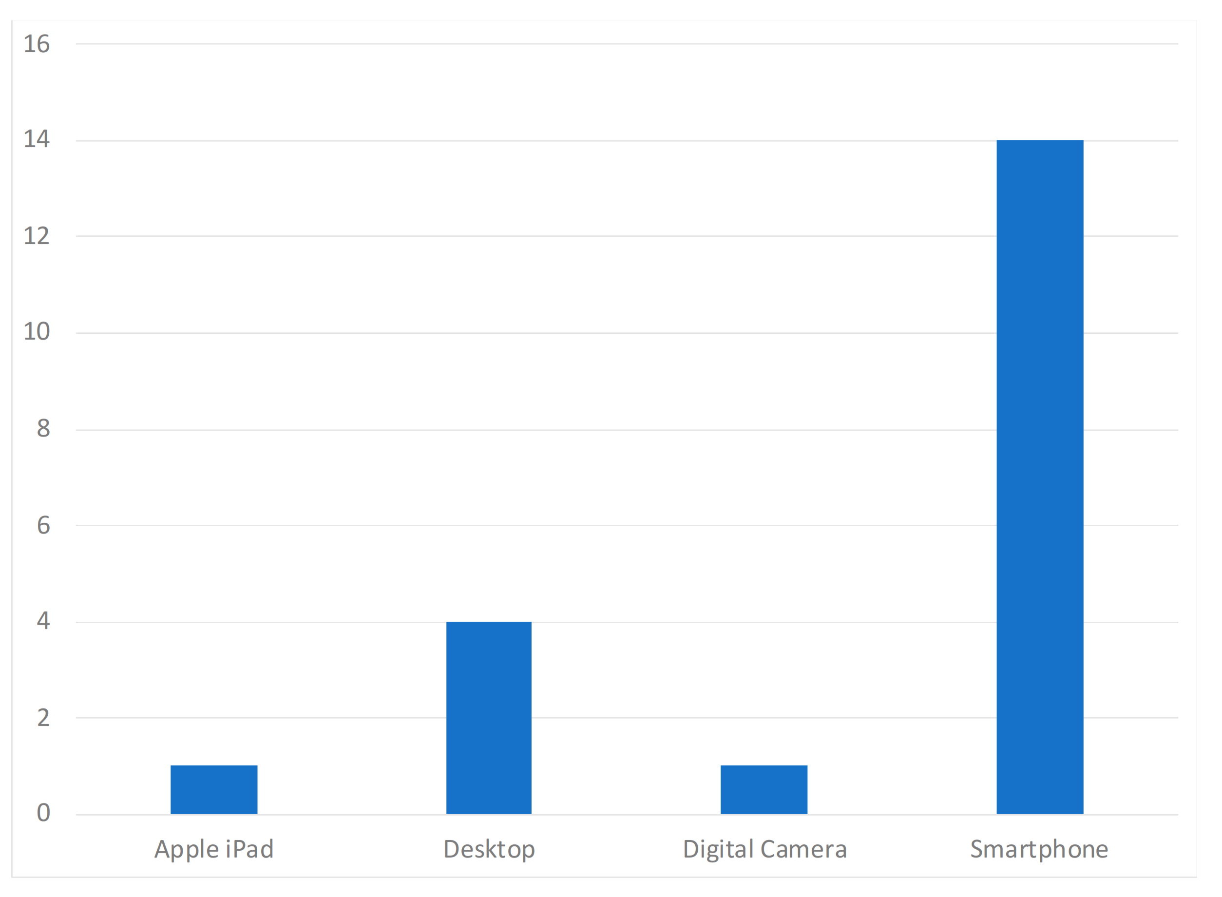
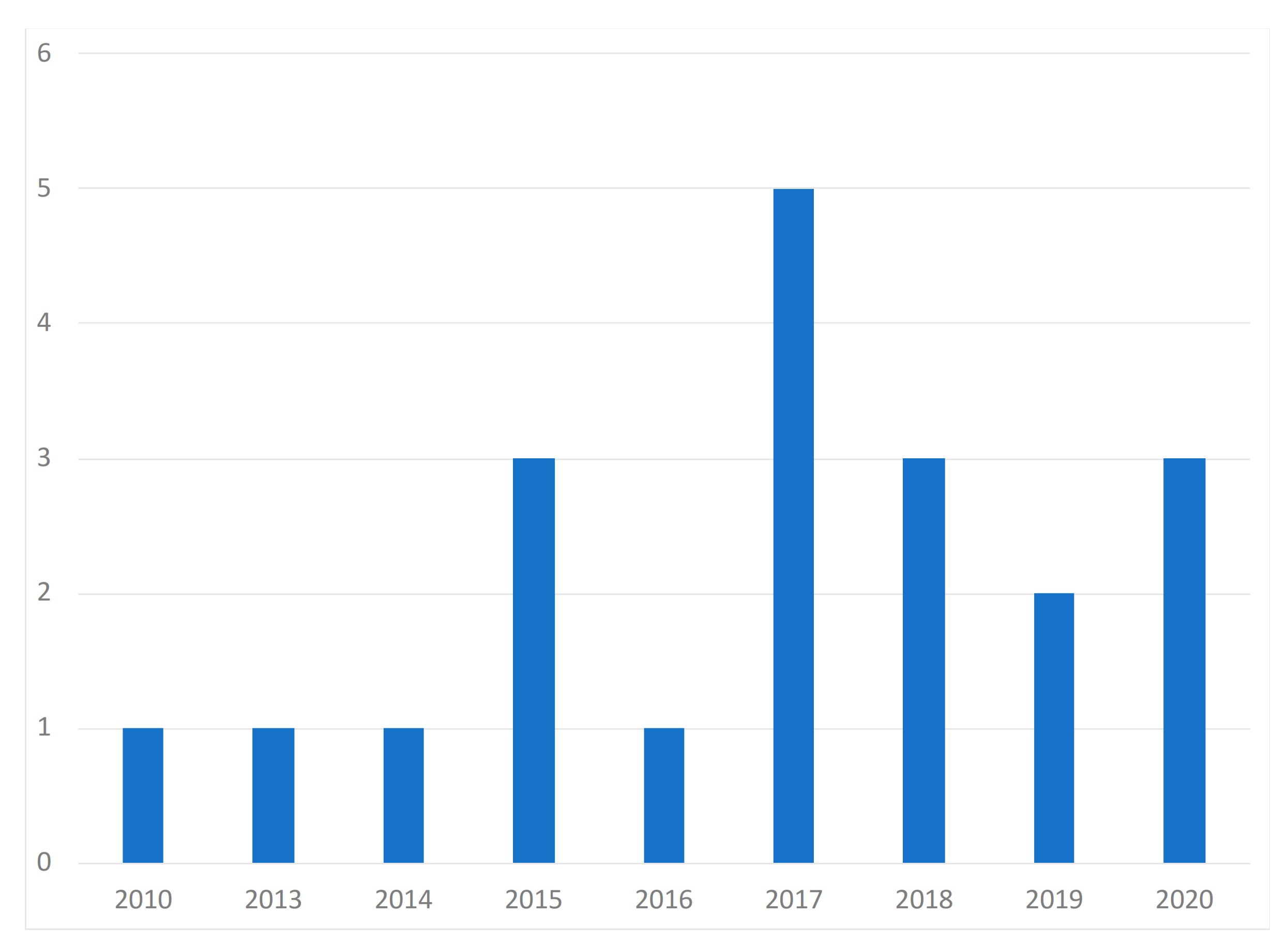
To analyze the different studies and get more relevant information regarding wound measurement in the different studies, the interested reader must read the original works. Based on the results presented in Table 1, the analyzed studies were published between 2010 and 2020, reporting three studies in 2020 (15%), two studies in 2019 (10%), three studies in 2018 (15%), five studies in 2017 (25%), one study in 2015 (5%), three studies in 2016 (15%), three studies in 2015 (15%), one study in 2014 (5%), one study in 2013 (5%), and one study in 2010 (5%). Regarding the studies that reported the number of images used in the study, the average reported was approximately 27 images. For the devices used, five studies used desktop computers (25%), and fifteen studies used mobile devices (75%). None of the studies published a proprietary dataset regarding the dataset availability of the images used for the different experiments. Three studies (15%) used datasets already published in other studies, as mentioned in Table 1. The studies considered different types of diseases that corresponded to the different types of wounds. All studies referenced in Table 1 used supervised learning and were model-based approaches. The presented analyses only summarize the results of each research paper. The interested readers must read the full text for more detailed information.
Casal-Guisande et al. [29] analyzed pressure ulcers to measure the size and evolution of wounds using the hue, saturation, and value (HSV) model. The population studied had chronic wounds. The proposed system used MATLAB software (R2020a, MathWorks’, Natick, MA, USA), which implemented different image processing algorithms as a decision support system with concurrent fuzzy inference engines. After analyzing the various points, the decision factor’s growth was reversed, reporting that the corrections prevented abnormal behaviors. For the measurement of the wound, the steps performed were selecting the saturation plan, the inversion and filtering of saturation values, and the performance of segmentation with a threshold.
Zahia et al. [30] also calculated the size of a wound. They used the mask recurrent convolutional neural network (RCNN) model to classify chronic wounds, reporting results with an accuracy of around 87%. The authors used Python 3.6 with Keras 2.0 [50] and TensorFlow 1.3 [51] for the segmentation of the images and measurement of the different features, which were processed with MATLAB software (R2018b, MathWorks’, Natick, MA, USA). Internally, the proposed system extracted all quantitative information by matching the 2D image with its 3D mesh. Finally, they captured the depth, volume, area, and major and minor axes of each wound. The final method was achieved by applying the mask recurrent convolutional neural network (RCNN) model to segment the injury. In continuation, the 3D mesh was rasterized, generating a top view image and the matrix of face indices. The expectation–maximization (EM) approach was implemented, and a projective transform matrix of the image was calculated. Next, the image was segmented, and the RANdom SAmple Consensus (RANSAC) algorithm was used to calculate the best fitting hyperplane. Finally, the authors calculated the faces and the wound’s boundary, computing its depth, area, volume, and axes. For example, one of the images available in the authors’ dataset was tested in grayscale and is presented in Figure 2.
In [33], Cazzolato et al. proposed the rule-based ulcer segmentation and measurement (URule) framework for the segmentation and measurement of the wound area. The results obtained reported an F-measure of 0.8 in the measurements performed with ulcer wounds. The method was implemented in the URule app, which included capturing and processing the images with a mobile device. The method starts with dividing a mobile application into a matrix, continuing with the interior seed region’s estimation considering the user’s annotation. Next, the threshold is applied to the areas, and the pixels are filled. Sequentially, the different mathematical models are used for the measurement of the wound area. Thus, the implemented method is composed of the segmentation of RGB images into the foreground and background. After that, the minimum bounding rectangle (MBR) of the region of interest is cropped. Next, the image is binarized and converted to grayscale. After that, the ISODATA algorithm was used to find a threshold for the image, and the line segment detector (LSD) approach was implemented to find the ticks of the measurement tool. Initially, the ticks were grouped by angle. Next, the authors considered the segments with angles greater than or equal to five degrees of difference for the group with more elements. Finally, the wound area was measured by the computation of the distance between ticks in pixels, which were converted to centimeters. For example, one of the images available in the authors’ dataset was tested in grayscale, and it is presented in Figure 3.
Wu et al. [36] also used the HSV color model to detect the wound area in images captured by a Redmi6A smartphone with volunteers. The authors also used the grabCut algorithm in four images, reporting an accuracy of 50%, and the color threshold approach in the same images, reporting an accuracy of 100%. The color threshold approach was also more efficient than the other algorithms. Thus, the OpenCV library was used to apply a coin detection algorithm. After that, the image was segmented, and a rectangle was created around the wound. Next, the grabCut algorithm was used to measure the wound area. Still, the intensity values of colors with the OpenCV histogram were calculated. Finally, the coin detection algorithm was implemented to improve the wound area measurement.
Liu et al. [37] proposed a system to perform 3D measurement of wounds with mobile device images. The smartphone collected 2D images, and a 3D model was constructed with these images. With these images, the authors unwrapped the texture coordinates, transforming the 3D model to a 2D plane with the least squares conformal map (LSCM) algorithm, segmenting and scaling it to extract and measure the wound area with the conversion between pixels and the actual length. After analyzing 118 wounds on 54 patients, the implemented method reported an accuracy of 97%. The implemented method was constituted by the 3D reconstruction of the body’s wound part, and they mapped the 3D model to the 2D plane. After applying the image segmentation techniques, the scale conversion algorithm was implemented to measure the wound area.
Huang et al. [38] implemented a python-based user interface to measure a wound’s size with the enhance local contrast (CLAHE) algorithm’s implementation. The system reported a reliable accuracy in estimating the size of chronic wounds, but it needed the correct definition of each pixel’s size. The proposed system implemented different techniques, including white balance, anti-glare, the enhance local contrast (CLAHE) algorithm, the level set algorithm to find the wound’s boundary, and the snakes model algorithm to define an energy function of the image and detect the size of an injury.
Naraghi et al. [39] proposed photogrammetric 3D reconstruction, induration segmentations, and segmented depth maps to measure the wound size with a mobile application to measure tuberculosis infections. The authors started with the scaling of the image and photogrammetric 3D reconstruction. Next, the position and orientation of each image in the 3D space were estimated. With these features, the authors performed the feature matching process. In continuation, the authors evaluated the surface of the object by a dense point cloud. After applying histogram equalization, the Otsu’s thresholding algorithm was used to enhance the image’s contrast. The induration was cropped for the identification of the margin of the rough. Finally, the elliptical approximation of the induration margins was calculated, and the wound area was measured. In general, the proposed system reported reliable accuracy.
Chen et al. [40] also used the HSV color space when measuring the wound size in surgical wounds, reporting an accuracy of 91%. The system was implemented with a non-professional system that identified the injury in an image, distinguished the state of the surgical wound, and assessed the symptoms. It combined machine learning techniques with image processing techniques to compare the textures of the skin and wounds. Thus, the authors normalized the images. After that, they applied the SEED algorithm for superpixel segmentation to identify the superpixels that were skin. The skin area was reconstructed with superpixels, and the area was detected.
In [24], Gupta converted the image to the HSV color model while measuring a chronic wound area. Next, they performed the segmentation. Next, they extracted the saturation space from the color space and implemented the threshold to enhance the image’s contrast with Otsu’s thresholding algorithm and the dilation operation. After that, the implemented method found the contours from the binary image using the Suzuki85 algorithm. The number of black pixels inside the segmented image was calculated to design a healing curve plot. Thus, the results reported an accuracy of 70% in the tests performed with 20 photos from various healthcare centers.
Sirazitdinova and Deserno [25] measured the size, depth, volume, rate of healing, color, presence of necrosis, and types of a skin lesion with several techniques. Initially, the authors started with color correction and color calibration on the original 2D images to measure the wound area. Next, 3D reconstruction, color correction, tissue segmentation, and HSV were applied to compute the wound perimeter. Finally, the authors designed the contour in a 2D plane, calculated the area as the number of pixels between the outlines, and translated the measured pixels to real-world measurement units. The system reported a reliable accuracy.
Tang et al. [42] used a scaling method to measure chronic wounds’ sizes with a mobile device, reporting reliable results. The scaling method included calculating the approximate size of an injury and inserting and adjusting a rectangular box in the image.
In [43], Garcia-Zapirain et al. mainly implemented the linear combination of discrete Gaussians (LCDG) model to measure the sizes of the wounds, reporting an accuracy of 90.4% in the tests with 24 clinical images of pressure ulcers. Thus, the authors’ images of injuries were segmented in the ulcer region, with image decomposition using the parametric equations that defined the toroidal geometry. After that, the resulting image was decomposed in different contrast levels. Next, the threshold technique was performed to enhance the contrast of the image with Otsu’s thresholding algorithm, and the contours of the wound were detected. In continuation, the resulting image was transformed from RGB to grayscale, creating an appearance model using linear combinations of discrete Gaussians (LCDG) and minimizing the noise with a generalized Gauss–Markov random field (GGMRF) image model for the calculation of the areas of the wounds.
Dendere et al. [44] proposed a mobile system to evaluate the sizes of wounds from tuberculosis infection with the Mantoux tuberculin skin test (TST), reporting an accuracy of 96.5%. The TST method performs the round-off of the induration size to the nearest millimeter. It starts with the application of a mask to select the relevant part. Next, the matching of common points to the input images is found, estimating each image’s camera positions. In continuation, the camera calibration parameters are refined, building the point cloud model, a polygonal mesh, and the texture. Finally, the 3D reconstruction is performed for the measurement of the wound area.
Yee et al. [45] implemented a mobile application for remote wound measurement, tracking, and diagnosis which implemented the Seymour wound model 0910 with granulating, necrotic, and slough, reporting an accuracy of 99.13%. The core of the method includes the extraction of images from videos, and they compute the absolute scale of the wound with optics equations. Finally, the methods trace the contour with wound boundary detection for the calculation of the wound area. The authors tested different known areas from a constant distance of 20 cm from the wound to the smartphone, various specialized tissue types within wounds with known areas, and actual wound simulations.
Saathesha et al. [46] implemented PH2 dermoscopy image information and a D-quick Fourier rework to measure shape geometry asymmetry, border irregularity, color, and the diameter of a melanoma, reporting reliable accuracy. Thus, the authors started with the extraction of RGB channels from images, extracting the blue channel. Next, histogram equalization was performed. Before the application of segmentation techniques, the picture was converted from RGB to grayscale. Next, the average low pass filter was applied, and the threshold was calculated. In continuation, the degree intensity image was converted to a binary image, and Otsu’s thresholding algorithm was used to enhance the contrast of the picture. After that, the asymmetry, border irregularity, color, and diameter were extracted, and the image was denoised for the extraction of the region of interest (ROI) of the wound. Finally, the support vector machine (SVM) was implemented for the classification of the wounds.
Furthermore, Cheung et al. [47] estimated the wound size. They classified the tissue with color variation, asymmetry, maximum distance, texture, and border irregularity, based on a 3D model using structure from light, photogrammetry, or motion. The implemented methods were for feature extraction and segmentation.
Pires et al. [23] used the OpenCV library for the measurement of the sizes of wounds. The implemented method consisted of three stages, including preprocessing, segmentation, and wound area measurement, implementing the HSV color model with a Gaussian filter 31 × 31 in size, reporting reliable results. The OpenCV library was used for the implementation of segmentation, threshold, and color detection techniques.
Bulan [48] used a method for detecting wounds in 36 patients with allergic diseases that included the HSV color space and linear discriminant analysis (LDA) with a reported accuracy of 94%. This paper presented the detection of a wound. Initially, the authors inserted calibration marks to identify the localization of the region of the injury. Next, the image was converted to grayscale, and it was transformed from an RGB to YCbCr color space. After that, principal component analysis (PCA) was performed on the Cb and Cr color channels. Thus, it was possible to find the fly’s subspace with the maximal contrast between the wheel and the surrounding erythema. The authors performed the dimensionality reduction by finding an orthogonal projection of the high-dimensional data into a lower-dimensional subspace. The implementation finished with the median filtering, the threshold to enhance the contrast of the image with Otsu’s thresholding algorithm, the erosion operation on the binarized image, and the suppression of the structures connected to the image border dilation operation.
Hettiarachchi et al. [22] also implemented the HSV color space while measuring the wound size based on segmentation, camera distance, angle, and lighting conditions. It implemented preprocessing techniques to reduce errors from artifacts and lighting conditions, reporting an accuracy of 90%. The method started with the crop of the center of the wound and removed the unnecessary artifacts. Next, the image was resized and resampled to extract the saturation plane of the HSV color model. In continuation, the authors calculated the contour with the contrast between infected and normal skin, and the authors smoothed the image with a Gaussian filter. After that, the authors applied the snakes model algorithm to define the image’s energy function, and they transformed it into a grayscale image. The grayscale image was segmented, building the contour of the wound for further measurement of the wound size.
Kanade et al. [49] measured the cell population, cell cycle, mitotic cell rate, and tree synchrony with microscopic images and performed segmentation of the images. Thus, they performed several actions, including the detection of small, bright rectangular regions in each image using thresholding and convolution, overlapped areas combined into one patch, the detection of candidate patch sequences based on intensity change, and segmentation to analyze the wound images.

4. Discussion

This systematic analysis shows the importance of using technology in the health sector. Once again, and in the wake of what we have witnessed, the emergence of new methods, systems, and applications for the health sector has taken advantage of the development of mobile systems exponentially in recent years, as shown in Figure 4.
With the improvement of the camera resolution’s processing capacity and quality, there is an evident increase in the number of studies and applications related to this integration of health and informatics, especially in recent years, as shown in Figure 5.
The analysis of chronic wounds and their calculation and monitoring appear in this context. According to the analysis of this systematic analysis’s different studies, they have made firm steps because they present a very high accuracy at 90%. The most-used method in this set of studies was the HSV method, used in 7 of the 20 selected studies, demonstrating its usability and usefulness in these studies. As other recent studies [27,28] in image segmentation show, deep learning has great potential to improve feature extraction. Adaptative learning rates can help speed up the convergence process while achieving better predictive performance [28]. In turn, these approaches might overcome traditional issues in object segmentation related to variable lighting conditions, which might be particularly common when attempting to segment wounds [52].
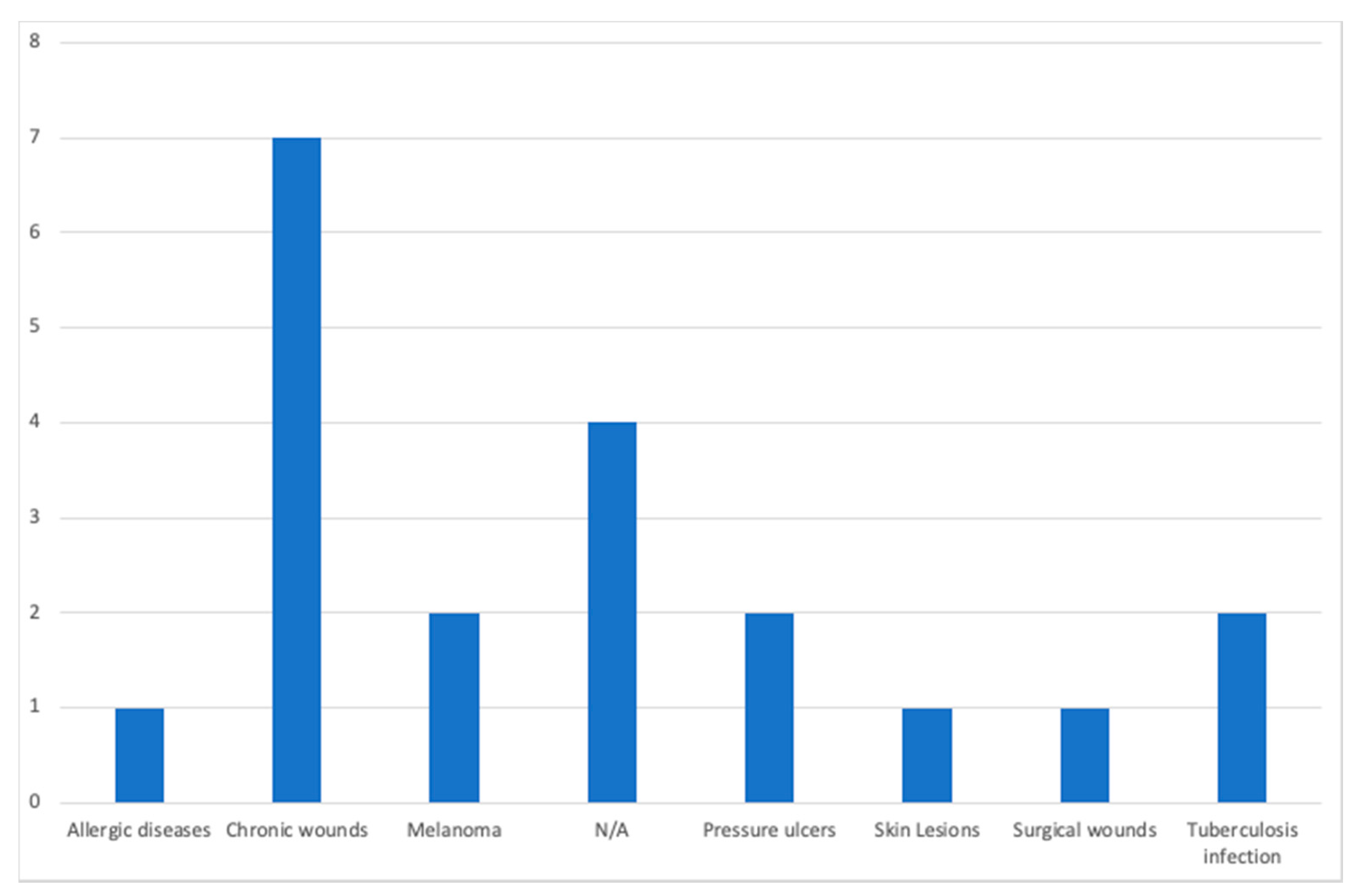
The number of articles that consider different diseases is shown in Figure 6, and apparently, chronic wounds are in the focus of seven studies.
The use of cameras embedded in mobile devices gives a more commodity-based measurement in different places. However, it may report less accuracy than other measurements performed with more powerful cameras. As this review is more focused on mobile devices, Table 2 and Table 3 present the features with more relevance in various studies to measure the wound area and contours with mobile devices and desktop techniques. The use of a desktop computer was not the main focus of this research, and other methods may be applied for the measurement of the wound area, as presented in Table 3. Initially, due to the more high-power processing, we can think that the desktop techniques may allow the more accurate detection of wounds. As presented in Table 2, the mobile devices include other sensors that may increase the accuracy of the wound area’s measurement. The mobile devices currently embed more powerful cameras to capture the different wound images with quality to measure the wound area. These devices now include high capabilities for various measurements’ performance, and they can perform more accurate measurements. Table 4 complements this analysis with the presentation of the comparison of mobile devices and desktop computers.
Based on the methods applied in the different studies analyzed, the ranking of the most-applied methods is presented in Table 5 to check the most-implemented strategies and further propose of a method to be implemented as future work.
The use of deep learning methods to improve the feature extraction of the different images’ image segmentation was used in two of the studies analyzed. This subject is recent and exploiting [27,30]. Other studies in the literature have been about using deep learning techniques for these steps that will improve the results [53,54]. However, using these techniques requires more hardware resources that may not be available in standard mobile devices.
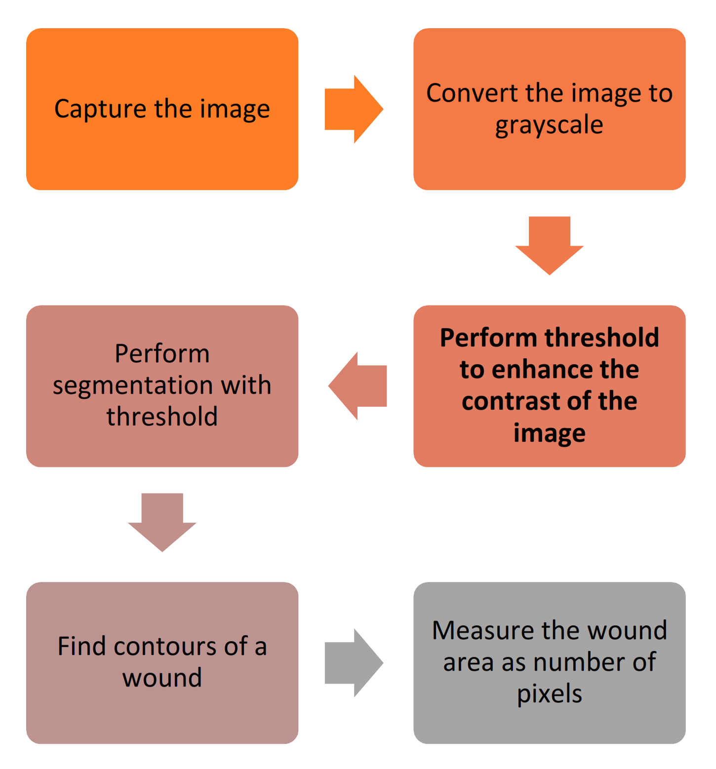
From the analysis of the methods for wound area measurement with a mobile device, one important finding is that they need to be more lightweight to have practical applications. Finally, when considering implementing a mobile application for Android devices, the OpenCV library allows the implementation of the different methods locally in the Java programming language. As presented in Figure 7, it must start with the image’s capture, and it must be converted to grayscale for further implementation of the threshold. Next, a segmentation method is to be implemented to measure the wound area as the number of pixels for the conversion to metric units.
The most critical challenge of this work was the conversion of the measured pixels to metric units. Thus, the mobile devices’ proximity sensors can be used to increase the accuracy, measuring the distance to the wound and extrapolating the real area of the injury. The comparative analysis of the studies shown in Table 1 revealed that the smartphone with the operating system was predominant.
No incompatibility was found in the studies analyzed for confidentiality or data protection in the different experiences. We performed a rigorous evaluation of each study to verify the existence of a validation of the study protocol by a human subject research ethics committee, but the information was not conclusive. Thus, we contacted the authors and research groups to obtain more clarification about each study’s data protection, but we have not yet received the responses. For the different features in this context, the most common were studies that sought to calculate the wound area, size, and color.

5. Conclusions

In conclusion, this systematic review analyzed, verified, and identified studies that aimed to investigate the size, structure, and area of wounds using open-source systems and most mobile devices. The use of image processing methods for this type of study has grown with the development of smartphones. Still, for the methods to have practical application, the accuracy and robustness of the methods need to be improved. Twenty studies were examined, and the main findings are summarized as follows:
  •  (RQ1) Which are the techniques that can be applied in a mobile application to measure a wound area? A mobile application can capture different pictures related to different situations, including wounds. The mobile application commonly applies preprocessing techniques, segmentation, threshold, and other methods to measure the wound area;
  •  (RQ2) What are the most significant features to define a method for the automatic calculation of a wound’s size? The most notable feature related to the wound’s size is measuring the different pixels and the different points of each wound’s contour. The processing techniques and artificial intelligence techniques may be powerful in the measurement of the wound’s size;
  •  (RQ3) What are the benefits that this kind of study can bring to the medical sector? This kind of study’s benefits consist of the correct measurement of the evolution of a wound’s treatment and the medicine’s adaptation according to its changes. It is especially important in patients with diabetes.
For future work, we intend to develop a mobile system to measure the wound size anywhere for a non-specialist. It must have acceptable accuracy for implementing the segmentation, threshold, and identification of the wound size, measuring the distance between the camera and the wound for the accurate measurement of the wound size.

Funding

This work was funded by FCT/MEC through national funds and co-funded by the FEDER—PT2020 partnership agreement under the project UIDB/50008/2020 (Este trabalho é financiado pela FCT/MEC através de fundos nacionais e cofinanciado pelo FEDER, no âmbito do Acordo de Parceria PT2020 no âmbito do projeto UIDB/50008/2020). This work was also funded by national funds through the Foundation for Science and Technology (FCT), I.P., within the scope of the project UIDB/00742/2020.

Institutional Review Board Statement

Not applicable.

Informed Consent Statement

Not applicable.

Data Availability Statement

Authors can confirm that all relevant data are included in the article.

Acknowledgments

This work was funded by FCT/MEC through national funds and co-funded by the FEDER—PT2020 partnership agreement under the project UIDB/50008/2020 (Este trabalho é financiado pela FCT/MEC através de fundos nacionais e cofinanciado pelo FEDER, no âmbito do Acordo de Parceria PT2020 no âmbito do projeto UIDB/50008/2020). This work was also funded by national funds through the Foundation for Science and Technology (FCT), I.P., within the scope of the project UIDB/00742/2020. This article is based upon work from COST Action IC1303–AAPELE–Architectures, Algorithms and Protocols for Enhanced Living Environments and COST Action CA16226–SHELD-ON–Indoor living space improvement: Smart Habitat for the Elderly, supported by the European Cooperation in Science and Technology (COST). More information can be found at www.cost.eu (accessed on 31 March 2021). Furthermore, we would like to thank the Politécnico de Viseu for their support.

Conflicts of Interest

The authors declare no conflict of interest.

References

  1. Camilo, M.E. Disease-Related Malnutrition: An Evidence-Based Approach to Treatment. Clin. Nutr. 2003, 22, 585. [Google Scholar] [CrossRef]
  2. Landi, F.; Onder, G.; Russo, A.; Bernabei, R. Pressure Ulcer and Mortality in Frail Elderly People Living in Community. Arch. Gerontol. Geriatr. 2007, 44, 217–223. [Google Scholar] [CrossRef] [PubMed]
  3. Beeckman, D.; Schoonhoven, L.; Fletcher, J.; Furtado, K.; Gunningberg, L.; Heyman, H.; Lindholm, C.; Paquay, L.; Verdú, J.; Defloor, T. EPUAP Classification System for Pressure Ulcers: European Reliability Study. J. Adv. Nurs. 2007, 60, 682–691. [Google Scholar] [CrossRef] [PubMed] [Green Version]
  4. Edsberg, L.E. Pressure Ulcer Tissue Histology: An Appraisal of Current Knowledge. Ostomy Wound Manag. 2007, 53, 40–49. [Google Scholar]
  5. Pires, I.M.; Marques, G.; Garcia, N.M.; Flórez-Revuelta, F.; Ponciano, V.; Oniani, S. A Research on the Classification and Applicability of the Mobile Health Applications. JPM 2020, 10, 11. [Google Scholar] [CrossRef] [Green Version]
  6. Pires, I.M.; Garcia, N.M.; Pombo, N.; Flórez-Revuelta, F.; Rodríguez, N.D. Validation Techniques for Sensor Data in Mobile Health Applications. J. Sens. 2016, 2016. [Google Scholar] [CrossRef] [Green Version]
  7. Sim, I. Mobile Devices and Health. N. Engl. J. Med. 2019, 381, 956–968. [Google Scholar] [CrossRef]
  8. Lumsden, C.J.; Byrne-Davis, L.M.T.; Mooney, J.S.; Sandars, J. Using Mobile Devices for Teaching and Learning in Clinical Medicine. Arch. Dis. Child Educ. Pract. 2015, 100, 244–251. [Google Scholar] [CrossRef] [Green Version]
  9. Albrecht, U.-V.; von Jan, U.; Kuebler, J.; Zoeller, C.; Lacher, M.; Muensterer, O.J.; Ettinger, M.; Klintschar, M.; Hagemeier, L. Google Glass for Documentation of Medical Findings: Evaluation in Forensic Medicine. J. Med. Internet Res. 2014, 16, e53. [Google Scholar] [CrossRef]
  10. Kilgus, T.; Heim, E.; Haase, S.; Prüfer, S.; Müller, M.; Seitel, A.; Fangerau, M.; Wiebe, T.; Iszatt, J.; Schlemmer, H.-P.; et al. Mobile Markerless Augmented Reality and Its Application in Forensic Medicine. Int. J. Comput. Assist. Radiol. Surg. 2015, 10, 573–586. [Google Scholar] [CrossRef]
  11. CDC. Disability and Health Data System (DHDS)|CDC. Available online: https://www.cdc.gov/ncbddd/disabilityandhealth/dhds/index.html (accessed on 5 August 2020).
  12. Yin, H.; Jha, N.K. A Health Decision Support System for Disease Diagnosis Based on Wearable Medical Sensors and Machine Learning Ensembles. IEEE Trans. Multi-Scale Comp. Syst. 2017, 3, 228–241. [Google Scholar] [CrossRef]
  13. Mehmood, N.; Hariz, A.; Templeton, S.; Voelcker, N.H. A Flexible and Low Power Telemetric Sensing and Monitoring System for Chronic Wound Diagnostics. Biomed. Eng. Online 2015, 14, 17. [Google Scholar] [CrossRef] [Green Version]
  14. Pal, A.; Goswami, D.; Cuellar, H.E.; Castro, B.; Kuang, S.; Martinez, R.V. Early Detection and Monitoring of Chronic Wounds Using Low-Cost, Omniphobic Paper-Based Smart Bandages. Biosens. Bioelectron. 2018, 117, 696–705. [Google Scholar] [CrossRef]
  15. Moore, K.; McCallion, R.; Searle, R.J.; Stacey, M.C.; Harding, K.G. Prediction and Monitoring the Therapeutic Response of Chronic Dermal Wounds. Int. Wound J. 2006, 3, 89–98. [Google Scholar] [CrossRef]
  16. Karkanis, S.A.; Iakovidis, D.K.; Maroulis, D.E.; Karras, D.A.; Tzivras, M. Computer-Aided Tumor Detection in Endoscopic Video Using Color Wavelet Features. IEEE Trans. Inform. Technol. Biomed. 2003, 7, 141–152. [Google Scholar] [CrossRef] [Green Version]
  17. Jones, T.D.; Plassmann, P. An Active Contour Model for Measuring the Area of Leg Ulcers. IEEE Trans. Med. Imaging 2000, 19, 1202–1210. [Google Scholar] [CrossRef]
  18. Veredas, F.; Mesa, H.; Morente, L. Binary Tissue Classification on Wound Images With Neural Networks and Bayesian Classifiers. IEEE Trans. Med. Imaging 2010, 29, 410–427. [Google Scholar] [CrossRef]
  19. Cula, O.G.; Dana, K.J.; Murphy, F.P.; Rao, B.K. Skin Texture Modeling. Int. J. Comput. Vis. 2005, 62, 97–119. [Google Scholar] [CrossRef]
  20. Neidrauer, M.; Papazoglou, E.S. Optical Non-invasive Characterization of Chronic Wounds. In Bioengineering Research of Chronic Wounds; Gefen, A., Ed.; Studies in Mechanobiology, Tissue Engineering and Biomaterials; Springer: Berlin/Heidelberg, Germany, 2009; Volume 1, pp. 381–404. ISBN 978-3-642-00533-6. [Google Scholar]
  21. Barone, S.; Paoli, A.; Razionale, A.V. Assessment of Chronic Wounds by Three-Dimensional Optical Imaging Based on Integrating Geometrical, Chromatic, and Thermal Data. Proc. Inst. Mech. Eng. 2011, 225, 181–193. [Google Scholar] [CrossRef]
  22. Hettiarachchi, N.D.J.; Mahindaratne, R.B.H.; Mendis, G.D.C.; Nanayakkara, H.T.; Nanayakkara, N.D. Mobile Based Wound Measurement. In Proceedings of the 2013 IEEE Point-of-Care Healthcare Technologies (PHT), Bangalore, India, 16–18 January 2013; pp. 298–301. [Google Scholar]
  23. Pires, I.M.; Garcia, N.M. Wound Area Assessment Using Mobile Application. BIODEVICES 2015, 1, 271–282. [Google Scholar]
  24. Gupta, A. Real Time Wound Segmentation/Management Using Image Processing on Handheld Devices. JCM 2017, 17, 321–329. [Google Scholar] [CrossRef]
  25. Sirazitdinova, E.; Deserno, T.M. System Design for 3D Wound Imaging Using Low-Cost Mobile Devices; Cook, T.S., Zhang, J., Eds.; SPIE: Orlando, FL, USA, 2017; p. 1013810. [Google Scholar]
  26. Deserno, T.M.; Kamath, S.; Sirazitdinova, E. Machine Learning for Mobile Wound Assessment. In Medical Imaging 2018: Imaging Informatics for Healthcare, Research, and Applications; Zhang, J., Chen, P.-H., Eds.; SPIE: Houston, TX, USA, 2018; p. 42. [Google Scholar]
  27. Petrovska, B.; Zdravevski, E.; Lameski, P.; Corizzo, R.; Štajduhar, I.; Lerga, J. Deep Learning for Feature Extraction in Remote Sensing: A Case-Study of Aerial Scene Classification. Sensors 2020, 20, 3906. [Google Scholar] [CrossRef] [PubMed]
  28. Petrovska, B.; Atanasova-Pacemska, T.; Corizzo, R.; Mignone, P.; Lameski, P.; Zdravevski, E. Aerial Scene Classification through Fine-Tuning with Adaptive Learning Rates and Label Smoothing. Appl. Sci. 2020, 10, 5792. [Google Scholar] [CrossRef]
  29. Casal-Guisande, M.; Comesaña-Campos, A.; Cerqueiro-Pequeño, J.; Bouza-Rodríguez, J.-B. Design and Development of a Methodology Based on Expert Systems, Applied to the Treatment of Pressure Ulcers. Diagnostics 2020, 10, 614. [Google Scholar] [CrossRef]
  30. Zahia, S.; Garcia-Zapirain, B.; Elmaghraby, A. Integrating 3D Model Representation for an Accurate Non-Invasive Assessment of Pressure Injuries with Deep Learning. Sensors 2020, 20, 2933. [Google Scholar] [CrossRef]
  31. COCO—Common Objects in Context. Available online: https://cocodataset.org/#home (accessed on 5 March 2021).
  32. ImageNet. Available online: http://www.image-net.org/ (accessed on 5 March 2021).
  33. Cazzolato, M.T.; Ramos, J.S.; Rodrigues, L.S.; Scabora, L.C.; Chino, D.Y.T.; Jorge, A.E.S.; Azevedo-Marques, P.M.; Traina, C.; Traina, A.J.M. Semi-Automatic Ulcer Segmentation and Wound Area Measurement Supporting Telemedicine. In Proceedings of the 2020 IEEE 33rd International Symposium on Computer-Based Medical Systems (CBMS), Rochester, MN, USA, 28–30 July 2020; pp. 356–361. [Google Scholar]
  34. Dorileo, E.A.; Frade, M.A.C.; Roselino, A.M.F.; Rangayyan, R.M.; Azevedo-Marques, P.M. Color Image Processing and Content-Based Image Retrieval Techniques for the Analysis of Dermatological Lesions. In Proceedings of the 2008 30th Annual International Conference of the IEEE Engineering in Medicine and Biology Society, Vancouver, BC, Canada, 20–25 August 2008; pp. 1230–1233. [Google Scholar]
  35. Chino, D.Y.T.; Scabora, L.C.; Cazzolato, M.T.; Jorge, A.E.S.; Traina, C., Jr.; Traina, A.J.M. Segmenting Skin Ulcers and Measuring the Wound Area Using Deep Convolutional Networks. Comput. Methods Programs Biomed. 2020, 191, 105376. [Google Scholar] [CrossRef]
  36. Wu, W.-L.; Yong, K.Y.-W.; Federico, M.A.J.; Gan, S.K.-E. The APD Skin Monitoring App for Wound Monitoring: Image Processing, Area Plot, and Colour Histogram. Sci. Phone Apps Mob. Device 2019. [Google Scholar] [CrossRef]
  37. Liu, C.; Fan, X.; Guo, Z.; Mo, Z.; Chang, E.I.-C.; Xu, Y. Wound Area Measurement with 3D Transformation and Smartphone Images. BMC Bioinform. 2019, 20, 724. [Google Scholar] [CrossRef] [Green Version]
  38. Huang, C.-H.; Jhan, S.-D.; Lin, C.-H.; Liu, W.-M. Automatic Size Measurement and Boundary Tracing of Wound on a Mobile Device. In Proceedings of the 2018 IEEE International Conference on Consumer Electronics-Taiwan (ICCE-TW), Taichung, Taiwan, 19–21 May 2018; pp. 1–2. [Google Scholar]
  39. Naraghi, S.; Mutsvangwa, T.; Goliath, R.; Rangaka, M.X.; Douglas, T.S. Mobile Phone-Based Evaluation of Latent Tuberculosis Infection: Proof of Concept for an Integrated Image Capture and Analysis System. Comput. Biol. Med. 2018, 98, 76–84. [Google Scholar] [CrossRef]
  40. Chen, Y.-W.; Hsu, J.-T.; Hung, C.-C.; Wu, J.-M.; Lai, F.; Kuo, S.-Y. Surgical Wounds Assessment System for Self-Care. IEEE Trans. Syst. Man Cybernsyst. 2019, 1–16. [Google Scholar] [CrossRef] [Green Version]
  41. UCI. Machine Learning Repository: Skin Segmentation Data Set. Available online: https://archive.ics.uci.edu/ml/datasets/Skin+Segmentation (accessed on 5 March 2021).
  42. Tang, M.; Gary, K.; Guler, O.; Cheng, P. A Lightweight App Distribution Strategy to Generate Interest in Complex Commercial Apps: Case Study of an Automated Wound Measurement System. In Proceedings of the Hawaii International Conference on System Sciences 2017, Big Island, HI, USA, 3–7 January 2017. [Google Scholar]
  43. Garcia-Zapirain, B.; Shalaby, A.; El-Baz, A.; Elmaghraby, A. Automated Framework for Accurate Segmentation of Pressure Ulcer Images. Comput. Biol. Med. 2017, 90, 137–145. [Google Scholar] [CrossRef]
  44. Dendere, R.; Mutsvangwa, T.; Goliath, R.; Rangaka, M.X.; Abubakar, I.; Douglas, T.S. Measurement of Skin Induration Size Using Smartphone Images and Photogrammetric Reconstruction: Pilot Study. JMIR Biomed. Eng. 2017, 2, e3. [Google Scholar] [CrossRef]
  45. Yee, A.; Patel, M.; Wu, E.; Yi, S.; Marti, G.; Harmon, J. IDr: An Intelligent Digital Ruler App for Remote Wound Assessment. In Proceedings of the 2016 IEEE First International Conference on Connected Health: Applications, Systems and Engineering Technologies (CHASE), Washington, DC, USA, 27–29 June 2016; pp. 380–381. [Google Scholar]
  46. Satheesha, T.Y.; Satyanarayana, D.; Prasad, G. Early Detection of Melanoma Using Color and Shape Geometry Feature. JBEMi 2015, 2. [Google Scholar] [CrossRef] [Green Version]
  47. Cheung, N.-M.; Pomponiu, V.; Toan, D.; Nejati, H. Mobile Image Analysis for Medical Applications. Spie Newsroom 2015. [Google Scholar] [CrossRef]
  48. Bulan, O. Improved Wheal Detection from Skin Prick Test Images. In Proceedings of the IS&T/SPIE ELECTRONIC IMAGING, San Francisco, CA, USA, 2–6 February 2014. [Google Scholar]
  49. Kanade, T.; Yin, Z.; Bise, R.; Huh, S.; Eom, S.; Sandbothe, M.F.; Chen, M. Cell Image Analysis: Algorithms, System and Applications. In Proceedings of the 2011 IEEE Workshop on Applications of Computer Vision (WACV), Kona, HI, USA, 5–7 January 2011; pp. 374–381. [Google Scholar]
  50. Brownlee, J. Deep Learning with Python: Develop Deep Learning Models on Theano and TensorFlow Using Keras; Machine Learning Mastery: San Francisco, CA, USA, 2016. [Google Scholar]
  51. Holdroyd, T. TensorFlow 2.0 Quick Start Guide: Get up to Speed with the Newly Introduced Features of TensorFlow 2.0; Packt Publishing Ltd.: Birmingham, UK, 2019; ISBN 978-1-78953-696-6. [Google Scholar]
  52. Lameski, P.; Zdravevski, E.; Trajkovik, V.; Kulakov, A. Weed Detection Dataset with RGB Images Taken Under Variable Light Conditions. In ICT Innovations 2017; Trajanov, D., Bakeva, V., Eds.; Communications in Computer and Information Science; Springer International Publishing: Cham, Switzerland, 2017; Volume 778, pp. 112–119. ISBN 978-3-319-67596-1. [Google Scholar]
  53. Long, J.; Shelhamer, E.; Darrell, T. Fully Convolutional Networks for Semantic Segmentation. In Proceedings of the 2015 IEEE Conference on Computer Vision and Pattern Recognition (CVPR), Boston, MA, USA, 7–15 June 2015; pp. 3431–3440. [Google Scholar]
  54. Yu, L.; Fan, G. DrsNet: Dual-Resolution Semantic Segmentation with Rare Class-Oriented Superpixel Prior. Multimed. Tools Appl. 2021, 80, 1687–1706. [Google Scholar] [CrossRef]
Figure 1. Flow diagram of the identification and inclusion of papers.
Computers 10 00043 g001
Figure 2. Foot ulcer image available in the MS-COCO dataset [31].
Computers 10 00043 g002
Figure 3. Fibrin skin ulcer available in [34].
Computers 10 00043 g003
Figure 4. Number of studies per device.
Computers 10 00043 g004
Figure 5. Trend of the selected papers per year.
Computers 10 00043 g005
Figure 6. Number of studies per disease.
Computers 10 00043 g006
Figure 7. Proposed method to be implemented in a mobile application.
Computers 10 00043 g007
Table 1. Study analysis.
PaperYear of PublicationPopulationPurpose of the StudyDevicesDataset AvailabilityMethodsDiseases
Casal-Guisande et al. [29]2020N/AMonitor and analyze the chronic wound treatment processDesktopNot availableSegmentation, threshold, color detectionPressure ulcers
Zahia et al. [30]2020N/AMeasure the depth, area, volume, main axis, and secondary axis of chronic woundsSmartphoneMS COCO [31] and ImageNet [32] datasetsMask recurrent convolutional neural networks (RCNN) modelChronic wounds
Cazzolato et al. [33]2020People with chronic woundsSegment and measure skin ulcersSmartphoneDatasets from [34,35]Rule-based ulcer segmentation and measurement (URule) frameworkPressure ulcers
Wu et al. [36]2019Voluntary peopleDetection of wounds with image processing techniquesSmartphoneNot availableSegmentation, threshold, color detectionN/A
Liu et al. [37]201954 patientsDetection and measurement of the wound areaSmartphoneAvailable by requestLeast squares conformal map (LSCM) algorithmN/A
Huang et al. [38]2018N/AMeasurement of wound sizeSmartphoneNot availableEnhance local contrast (CLAHE) algorithmChronic wounds
Naraghi et al. [39]2018People with tuberculosisDetection of wounds in people with tuberculosisSmartphoneNot availablePhotogrammetric reconstructionTuberculosis infection
Chen et al. [40]2018N/AEvaluation of a surgical woundSmartphoneDataset available in [41]Segmentation, threshold, color detectionSurgical wounds
Gupta et al. [24]201720 wound imagesMobile system for the segmentation and identification of a woundSmartphoneNot availableSegmentation, threshold, color detectionChronic wounds
Sirazitdinova et al. [25]2017N/AAutomatic wound reconstructionSmartphoneNot availableColor correction, tissue segmentationSkin Lesions
Tang et al. [42]2017N/AAnalysis of the evolution of woundsSmartphoneNot availableScaling methodChronic wounds
Zapirain et al. [43]201724 clinical images of pressure ulcersSegmentation and identification of chronic woundsDesktopNot publicly availableLinear combination of discrete gaussians (LCDG) modelChronic wounds
Dendere et al. [44]201710 subjectsMeasure the size of a woundSmartphoneNot availableTuberculin skin test (TST)Tuberculosis infection
Yee et al. [45]2016N/AMeasurement, tracking, and diagnosis of woundsSmartphoneNot availableSeymour wound model 0910Chronic wounds
Satheesha et al. [46]2015People with skin cancerSegmentation and analysis techniques of wounds for the detection of the shapeSmartphoneNot availablePH2 dermoscopy image information, D-quick Fourier reworkMelanoma
Cheung et al. [47]2015N/ADiagnosis and treatment of chronic woundsSmartphoneNot availablePhotogrammetric reconstructionMelanoma
Pires et al. [23]2015N/ACalculation of the wound areaDesktopNot availableSegmentation, threshold, color detectionN/A
Bulan et al. [48]201436 patients with allergic diseasesIdentification of a wound in imagesDesktopNot availableLinear discriminant analysis (LDA)Allergic diseases
Hettiarachchi et al. [22]201320 patientsMeasurement of wound area with segmentation techniquesSmartphoneNot availableSegmentation, threshold, color detectionChronic wounds
Kanade et al. [49]2010Wide range of peopleRestore, detect, and track cells and cellular tissuesDesktopNot availableHCRF modelN/A
Table 2. Implemented methods in mobile devices.
ActionOccurrences
Perform segmentation with threshold11
Measure the wound area as the number of pixels5
Convert image to grayscale4
Perform 3D reconstruction4
Crop the center of the wound3
Extract saturation space from color space3
Perform the histogram equalization3
Perform threshold to enhance the contrast of the image 3
Adjust the size of the rectangle to the wound2
Apply snakes model algorithm to define an energy function of the image2
Apply the level set algorithm to find the boundary of the wound2
Convert associate degree intensity image to a binary image2
Extract asymmetry, border irregularity, color, and diameter2
Extract superpixels which are skin2
Find contours of the wound2
Implement support vector machine (SVM) to classify the skin2
Insert rectangular box in the image2
Perform dilation operation2
Table 3. Implemented methods in desktop computers.
ActionOccurrences
Measure the wound area as the number of pixels4
Perform segmentation with threshold4
Find contours of the wound3
Perform threshold to enhance the contrast of the image3
Convert image to grayscale2
Detect the wound2
Table 4. Mobile devices vs. desktop computers.
Mobile DevicesDesktop Computers
Measure the wound area anywhere at anytimeMeasure the wound area in a static place
Mobile devices are currently embedding high-quality camerasThe measurement depends on the external cameras that are dispendious
Mobile devices embed other sensors that may allow the calibration of the camerasThe calibration of the cameras depends on other external devices
The resources available are not unlimitedThe resources available can be expanded as needed with costs.
Table 5. Implemented methods.
ActionOccurrences
Perform segmentation with threshold15
Measure the wound area as the number of pixels13
Convert image to grayscale6
Perform threshold to enhance the contrast of the image 6
Find contours of the wound5
Perform 3D reconstruction4
Perform the histogram equalization3
Crop the center of the wound3
Perform dilation operation2
Detect the wound2
Extract saturation space from color space3
Insert rectangular box in the image2
Adjust the size of the rectangle to the wound2
Apply the level set algorithm to find the boundary of the wound2
Apply snakes model algorithm to define an energy function of the image2
Convert associate degree intensity image to a binary image2
Extract asymmetry, border irregularity, color, and diameter2
Implement support vector machine (SVM) to classify the skin2
Extract superpixels which are skin2
Publisher’s Note: MDPI stays neutral with regard to jurisdictional claims in published maps and institutional affiliations.