Systematic Review

Regional Lymph Node Metastasis in Sebaceous Carcinoma of the Head and Neck: A Systematic Review and Meta-Analysis


1
Keck School of Medicine, University of Southern California, Los Angeles, CA 90033, USA
2
Department of Otolaryngology-Head and Neck Surgery, The Mount Sinai Hospital, New York, NY 10029, USA
3
Department of Otolaryngology-Head and Neck Surgery, University of Chicago, Chicago, IL 60637, USA
4
Department of Otolaryngology-Head and Neck Surgery, University of California-Irvine, Orange, CA 92868, USA
*
Author to whom correspondence should be addressed.
Cancers 2026, 18(9), 1424; https://doi.org/10.3390/cancers18091424
Submission received: 4 March 2026 / Revised: 20 April 2026 / Accepted: 22 April 2026 / Published: 29 April 2026
(This article belongs to the Special Issue Precision Oncology for Rare Skin Cancers)

Simple Summary

Sebaceous carcinoma (SC) is a rare cutaneous malignancy with reported rates of locoregional metastasis of 4–33% among tumors of the head and neck. This broad range makes decisions regarding lymph node management difficult. The aim of our study was to determine a pooled rate of occult and evident lymph node metastasis among adults with SC of the head and neck to inform clinical guidelines relating to management of the lymph node basins. In a pooled cohort of 2371 patients, 16% had regional lymph node involvement, and patients with higher T stage and recurrent disease were more likely to have lymph node metastases. Seven percent of patients had occult metastases. Given this high risk of lymph node involvement, patients with SC of the head and neck should receive care from a multidisciplinary team that can provide an appropriate head and neck exam, imaging, consideration for sentinel lymph node biopsy, and treatment recommendations.

Abstract

Background/Objectives: Sebaceous carcinoma (SC) is a rare and aggressive malignancy most often arising in the head and neck. The reported rate of lymph node metastasis is variable and current clinical guidelines surrounding pre-treatment imaging and management of lymph nodes are not well defined. The aim of our systematic review and meta-analysis was to determine a pooled rate of clinically apparent and occult lymph node metastases for SC of the head and neck to inform clinical guidelines. Methods: Per PRISMA guidelines, systematic search of the Pubmed/MEDLINE and EMBASE databases identified studies published before October 2023 reporting regional lymph node status in adults with SC of the head and neck. Meta analysis using the random-effects model was applied to calculate the pooled proportion of subjects with lymph node metastasis. Clinical characteristics of subjects were further analyzed using chi square tests and univariate logistic regression. Results: Thirty-eight studies met inclusion criteria with a total of 2371 patients. The pooled prevalence of regional lymph node involvement, including clinically apparent and occult disease, was 16% (95% CI 13–18%, I2 65%), with increased risk with increasing T stage. The pooled rate of occult lymph node metastases was 7% (95% CI 4–9%, I2 68%). Conclusions: There is a high rate of lymph node involvement in SC of the head and neck, much of which goes undetected during initial workup and treatment. Initial workup should reflect this risk and include appropriate physical exam, imaging, consideration for sentinel lymph node biopsy, and involvement of a multi-disciplinary team.

1. Introduction

Sebaceous carcinoma (SC) is a rare but aggressive cutaneous carcinoma arising from sebaceous adnexal structures of the skin [1]. SC usually arises in the head and neck, most commonly around the eyelids [2,3,4,5,6,7]. SC is rare, but with increasing incidence in the United States [8]. The rate of metastasis of SC of the head and neck is variable with reports from 4 to 33% and is most commonly to the regional lymph nodes [9,10,11,12,13,14,15]. Metastasis is associated with poor clinical outcomes; involvement of preauricular or cervical lymph nodes is associated with a 50 to 67% 5-year disease-specific mortality rate in periocular SC [16,17]. The mainstay of treatment of the primary site is surgery, either via wide local excision or Mohs micrographic surgery, though some patients require radiation and complex facial reconstruction for more advanced disease [18,19,20,21]. Lymph node metastases are often treated with surgery and adjuvant radiation, but a clinical question remains about the appropriate workup and management of lymph nodes without clinically apparent metastases.
As SC is rare and data regarding outcomes are limited, recommendations for initial work up are vague. Current clinical guidelines developed by the Committee on Invasive Skin Tumor Evidence-Based Recommendations (CISTERN) recommend to consider imaging in patients presenting with palpable lymphadenopathy or a tumor with high-risk features and to consider sentinel lymph node biopsy (SLNB) for periocular tumors stage T2c or higher [19]. Prior reviews have focused on treatment modalities for the primary tumor, but have not established the rate or treatment of lymph node metastasis, which is a major contributor to morbidity and mortality in SC [22]. In practice, imaging is variable and SNLB is not routinely done. While uncommon, even early-stage SC have been known to metastasize to head and neck lymph nodes [23]. Few large-volume studies exploring the rate of lymph node involvement in SC exist, thus hindering clinical decision-making ability. Similarly, there are no data regarding the rate of occult metastases, cancer cells that have spread form the primary site to lymph nodes but are too small for detection by standard routine methods. SNLB is often utilized for tumors with a high rate of occult metastasis to aid in early detection and treatment of metastatic disease, but due to limited data, is rarely utilized for SC. Understanding the rate of occult metastases would inform the potential survival benefit of a more aggressive and systematic initial workup for SC.
We conducted a systematic review and meta-analysis to determine the pooled rate of lymph node involvement in patients with SC of the head and neck based on T stage, disease time course, and other clinical factors. We aim to determine the frequency of lymph node involvement in SC patients, and in which clinical scenarios nodal metastasis is most common to inform clinical guidelines with regard to head and neck imaging, lymph nodes biopsies, and inclusion of an otolaryngologist in patient management.

2. Materials and Methods

2.1. Design and Search Strategy

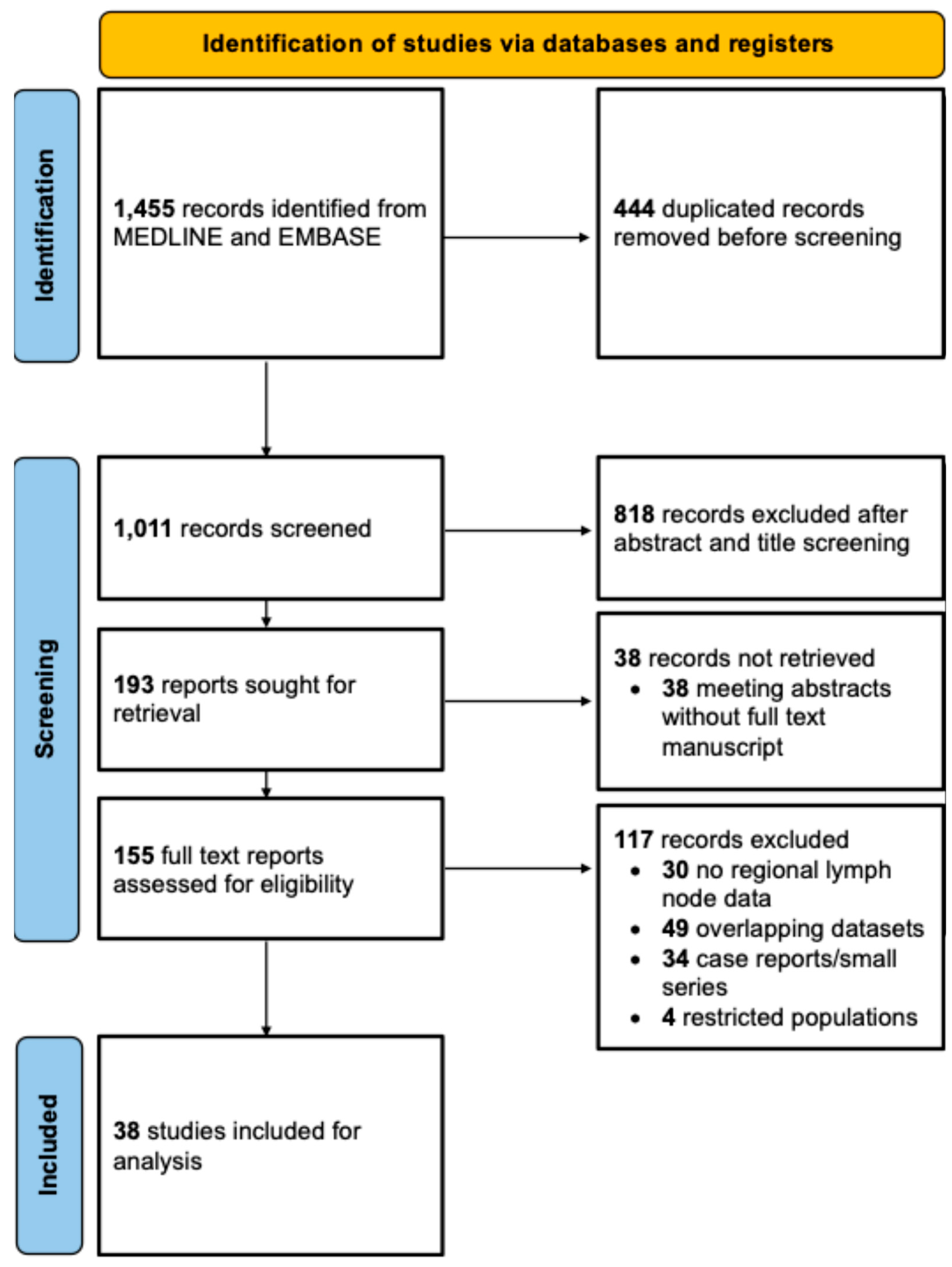
This study was conducted according to the Preferred Reporting Items for a Systematic Review and Meta-analysis (PRISMA) guidelines depicted in Figure 1. The PRISMA checklist is available in Supplementary Table S1. This study was registered with the international prospective register of systematic reviews, PROSPERO [24].
A systematic search was done using PubMed/MEDLINE and EMBASE databases to identify English language studies published before 1 October 2023 reporting regional lymph node status in adults with SC of the head and neck. Search terms used are presented in Table 1.

2.2. Study Selection

After removal of duplicates and screening of titles/abstracts, two authors (MK and MN) independently performed full-text reviews to assess for inclusion eligibility. Disagreements were resolved by a third independent reviewer. The following exclusion criteria was used: no regional lymph node data, same subject pool as other included studies, limited populations (such as non-representative subgroups), non-head-and-neck primary site of SC, registry or database studies (to avoid double counting of cases previously reported in case reports and series), case reports or small series < 10 subjects, and conference abstracts and letters. Study characteristics are available in Supplementary Table S2.

2.3. Statistical Analysis

Meta-analysis using the random-effects model was applied to calculate the pooled proportion of subjects with lymph node metastasis using thirty-eight studies. Sixteen studies that included patients with lymph node metastases at the time of initial presentation were eligible for subgroup analyses for pooled proportions. Twenty-five studies documented the presence of lymph node metastases either at presentation or at the time of disease recurrence and were analyzed via subgroup analyses for pooled proportions. Twenty-four studies documented location of recurrence (primary site versus nodal) and were used for subgroup analysis to determine the proportion of occult metastases. In this analysis, occult metastasis is defined as recurrence in regional lymph node(s) only with no recurrence at the primary site following treatment of the primary disease. Pooled proportions were calculated using a random-effects meta-analysis of binomial data applying the Freeman–Tukey double arcsine transformation with between-study variance estimated using restricted maximum likelihood (REML). Heterogeneity was assessed using Cochran’s Q test and quantified using I2 statistic. Publication bias and small-study effects were evaluated using funnel plot and Egger’s test. Galbraith plot was used to identify outlier studies (Supplementary Figure S1). Moderate heterogeneity was observed (I2 57.3%, Q = 89.63, p < 0.001). Egger’s test did not demonstrate significant small-study publication bias (p = 0.27). Categorical variables (T stage vs. nodal metastasis) were further analyzed using a chi square test. The association between T stage and nodal metastasis was evaluated using logistic regression. T stage was analyzed both as an ordinal variable (T1–T4, modeled as a continuous predictor to estimate the odds ratio per one-stage increase) and as a categorical variable with T1 as the reference group. Odds ratios (ORs) with 95% confidence intervals (CIs) were reported. Statistical significance was defined as p < 0.05. All data analysis was carried out using Stata V17.0 (College Station, TX, USA).

3. Results

Study demographics are presented in Table 2. A total of thirty-eight studies met the criteria for inclusion in the meta-analysis, with a wide geographic distribution of study cohorts [15,25,26,27,28,29,30,31,32,33,34,35,36,37,38,39,40,41,42,43,44,45,46,47,48,49,50,51,52,53,54,55,56,57,58,59,60]. Most studies that were included were retrospective in nature. A total of 2371 patients were included with weighted mean age of 64.8 (SD 5.04) years. Of those studies specifying the primary site of the disease (2337 patients), 97.3% of sebaceous carcinoma originated in the periorbital region, while 2.7% were extraocular. A total of 13 studies included tumor and nodal staging distributions, and most patients presented with T2 (58.2%) or T3 (26.4%) stage disease. The pooled percentage of patients all types of node metastases is stratified by primary disease site and tumor staging in Table 3.
The proportion of patients with nodal metastasis differed significantly across T stages (χ2(3) = 214.7, p < 0.0001). Among patients with T1, T2, T3, and T4 tumors, 4.35%, 8.18%, 36.0%, and 58.1% had lymph node metastases, respectively. In univariable analysis, each incremental increase in T stage conferred a 4.39-fold increase in odds of nodal metastasis (95% CI 3.48–5.54; p < 0.001). Table 4 presents univariate logistic regression analysis. When modeled categorically with T1 as the reference, T3 and T4 disease were significantly associated with increased odds of nodal metastasis (T3 vs. T1: OR = 12.4, 95% CI 5.31–28.9; T4 vs. T1: OR = 30.5, 95% CI 11.7–79.6). T2 did not reach statistical significance (OR 1.86, 95% CI 0.831–4.62).
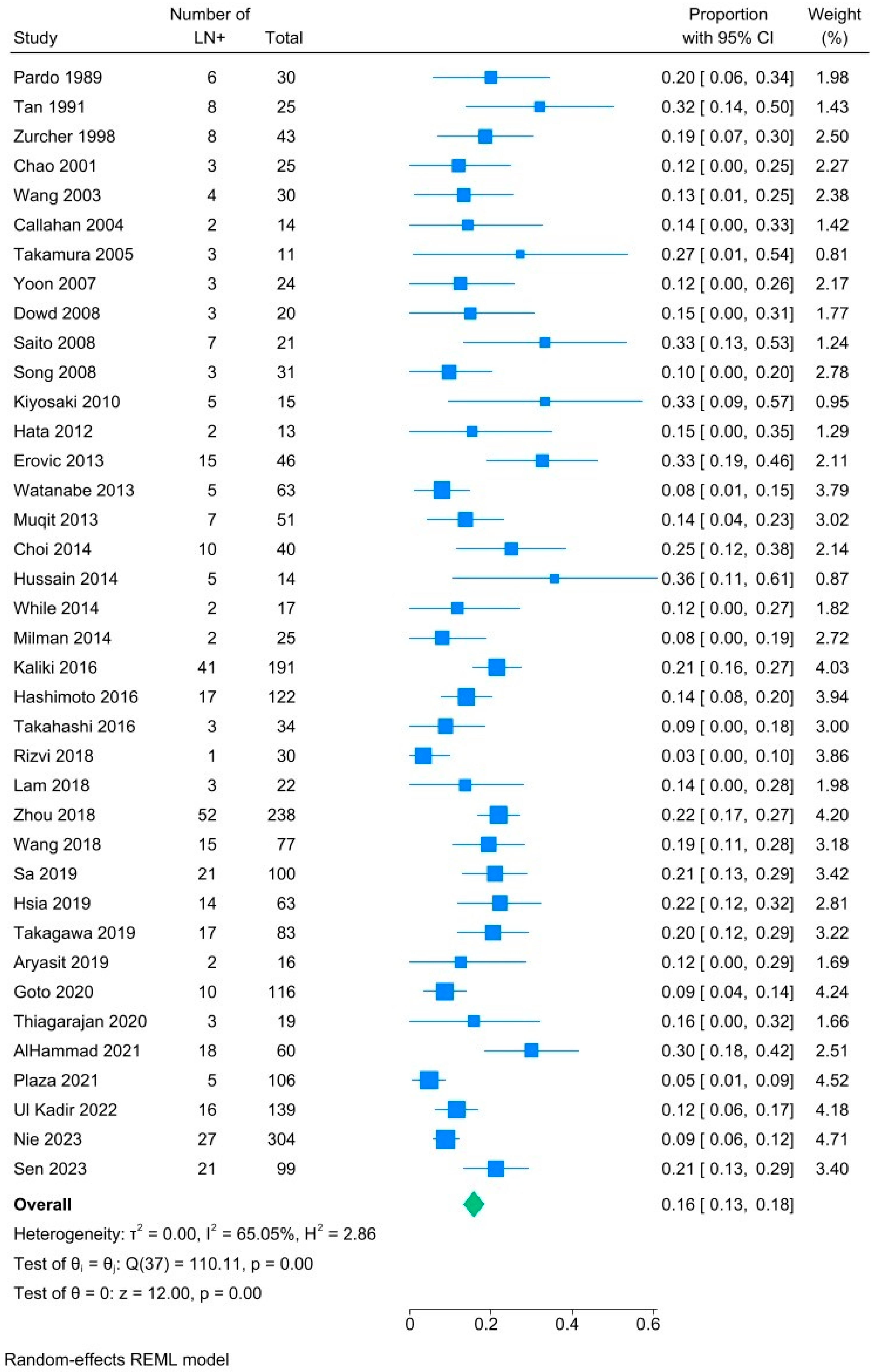
A total of thirty-eight studies reported regional nodal metastases in patients with SC. The overall rate of regional lymph node involvement is assessed via forest plot in Figure 2. The pooled prevalence of nodal metastases, including those at initial presentation and upon recurrence, was 16% (95% CI, 13–18%, I2 65%).
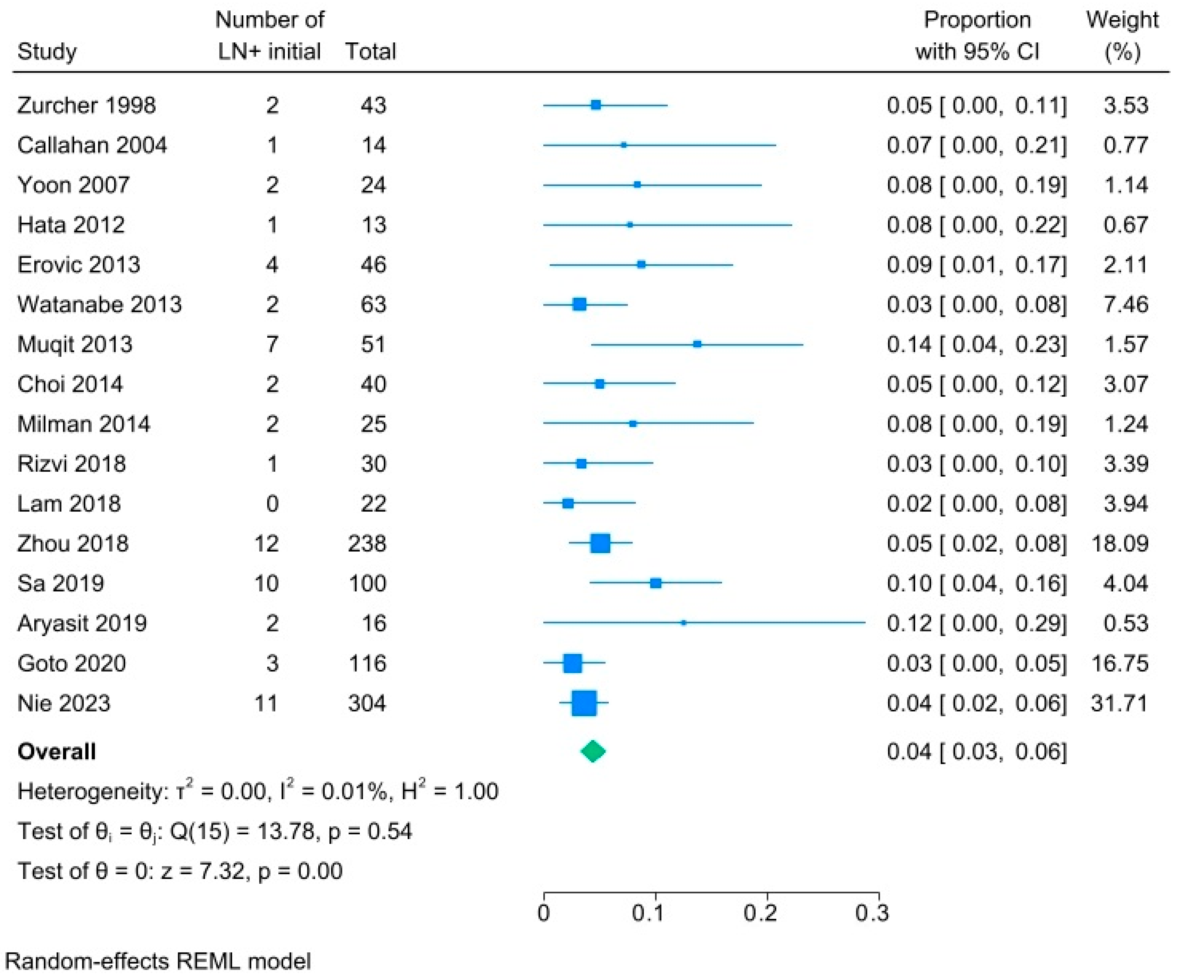
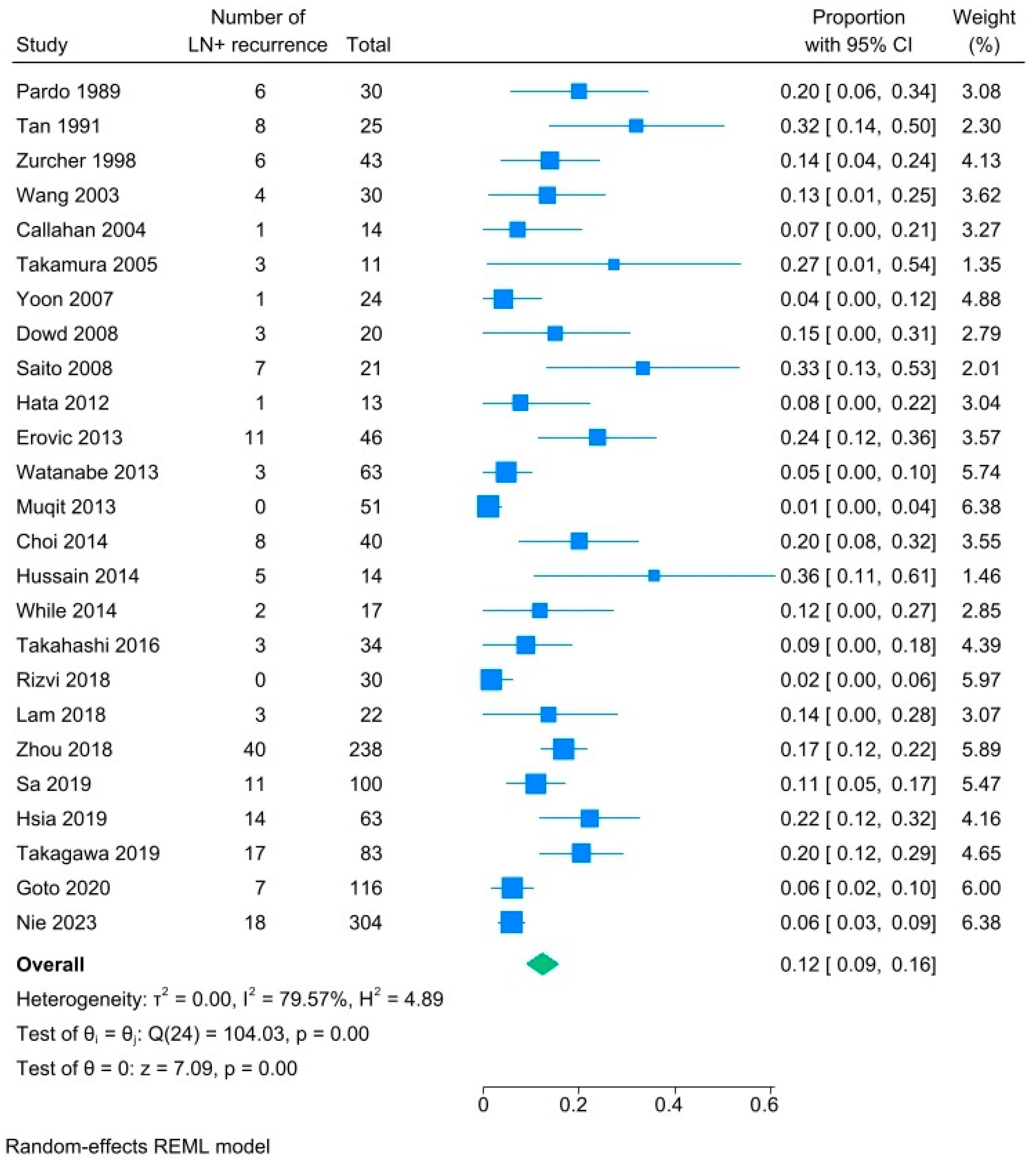
Subgroup analyses were performed for those studies that specified the time course of regional nodal metastases. Figure 3 demonstrates that the pooled prevalence of regional nodal metastases at the time of initial diagnosis of SC was only 4% (95% CI, 3–6%, I2. 0.01%). Figure 4 demonstrates that the pooled prevalence of regional nodal metastases among patients who presented with any type of recurrence, including at the primary site and/or in the lymph nodes, following treatment of the primary tumor was 12% (95% CI 9–16%, I2 79%).
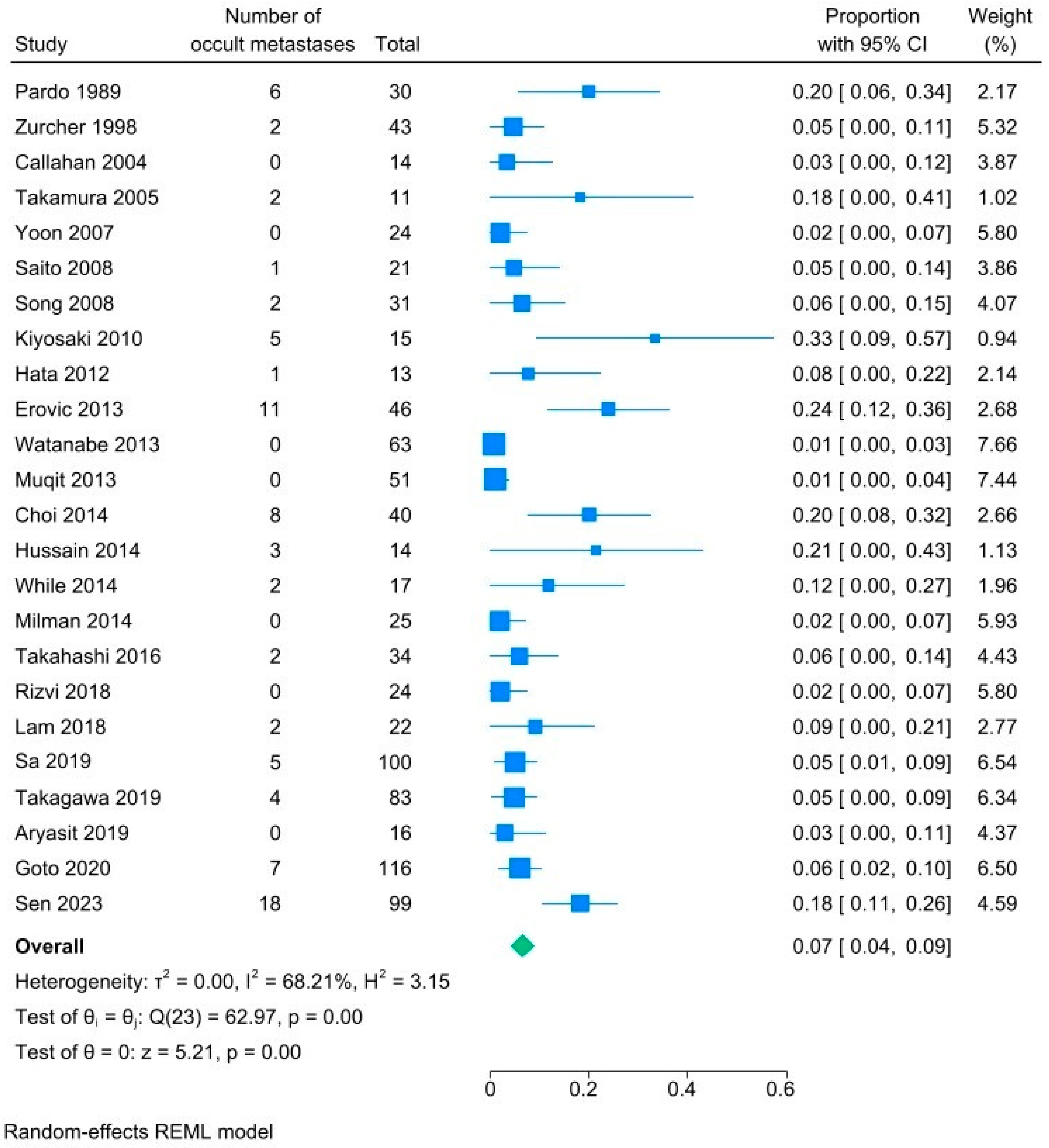
An additional subgroup analysis was performed for those studies that specified the location of recurrence: primary site, regional lymph node, or both. Figure 5 demonstrates that the pooled prevalence of occult metastases was 7% (95% CI 4–9%, I2. 68%). This represents patients that presented with recurrence only in the regional lymph nodes, without recurrence at the primary site. These patients had occult metastases—cancer cells in the regional lymph nodes that went undetected at the time of primary work up and treatment but later became clinically evident. Among patients with occult metastases, only 11 had initial T-stage available. Of these, 7 (63.6%) were T2, 3 (27.3%) were T3, and one was T4 (9.10%).

4. Discussion

This systematic review and meta-analysis is, to the best of our knowledge, the largest volume study of pooled data regarding the rate of lymph node involvement in sebaceous carcinoma of the head and neck. Of the 38 articles reviewed with a total of 2371 patients, lymph node involvement was found to occur in 16% of all cases, with increasing risk for advanced T stage. The reported rates of recurrence in sebaceous carcinoma are highly variable ranging from 4 to 33% with only 5.7% of patients presenting at initial presentation with nodal metastasis [11,16,21,26,40,62,63]. High rates of recurrence indicate an opportunity to improve initial work up to ensure detection of the full scope of disease.
While this study found that most cases with lymph node metastasis were in patients with high stage tumors (T3 and T4), and that increasing T stage is associated with increased risk of lymph node metastasis, T1 and T2 tumors also had small but clinically meaningful levels of nodal involvement (4% and 8%, respectively). This is almost certainly an underestimate due to diagnostic bias, as imaging of the neck is not currently recommended for most patients. Multiple studies have found that T1 and T2 tumors carry a small but significant risk of nodal metastasis [47,64,65]. Morawala et al. [64] found a 5% and 9%, respectively, 5-year risk of lymph node metastasis in T1 and T2 sebaceous carcinomas. A 2016 case series by Takahashi et al. [47] revealed that a patient with a T2 tumor without high risk factors and a complete resection with clear margins proceeded to have metastasis to the parotid. Even low-grade tumors carry the risk for nodal metastasis, further highlighting the importance of lymph node screening for all patients with sebaceous carcinoma. Notably, there is heterogeneity in the AJCC edition used for TNM staging data in this meta-analysis, as studies utilized different guidelines, which may limit the generalizability of this finding. Such heterogeneity, and that many studies did not report T-stage, limited this analysis.
Most notably in this study, 7% of patients had occult metastases. This suggests a significant risk of occult nodal disease, as these patients did not have local recurrence but still presented with nodal disease following initial treatment. These patients likely had lymph node metastases that went undetected, and thus untreated, in the initial work up. Had the nodal involvement been detected earlier, these patients may have undergone more aggressive initial treatment with potential for improved outcomes. As many patients do not receive neck imaging or SLNB as part of their initial work up for sebaceous carcinoma, it is impossible to know if these occult metastases were too small to detect by clinical exam and imaging, or if these screening tools were never utilized at all. The definition of occult disease used in this study is recurrence-based, rather than relying upon SLNB data which can show microscopic nodal disease that is not apparent clinically or on imaging. As such, this definition is narrower and is likely an underestimate of the true rate of occult disease.
Current guidelines for management of sebaceous carcinoma were developed via a consensus-based multidisciplinary conference of experts [19]. These guidelines recommend considering imaging for periocular tumors stage T2c and higher or tumors with high-risk features, such as poorly differentiated pathology, pagetoid spread, or perineural invasion. Lymph node sampling via fine-needle aspiration or core needle biopsy is recommended only in the case of enlarged lymph nodes identified on exam. Such recommendations are based on a paucity of data regarding rates of regional lymph node metastasis and occult metastases. There is little guidance regarding SNLB to detect occult metastatic disease.
Nevertheless, metastasis to the lymph nodes is a poor prognostic factor in sebaceous carcinoma [1,15,66]. Zhou et al. found that for patients with lymph node involvement upon initial presentation, survival rate was just 50% with a median survival time of 28.0 months [25]. Given the high rates of recurrence, poor outcomes associated with recurrence, increasing risk of lymph node metastasis with T stage, and the finding that 7% of patients have occult metastases, all patients with sebaceous carcinoma of the head and neck should receive a thorough head and neck lymph node exam and be offered neck imaging. Patients with T3 and T4 tumors, and some patients with T2 tumors, may benefit from SNLB upon initial treatment. Risks and benefits should be discussed with patients individually, with the understanding that there is a significant risk of occult metastasis in sebaceous carcinoma and that lymph node involvement is associated with poorer prognosis.
Though early case studies support the use of SNLB in periocular sebaceous carcinoma, the effect of positive sentinel lymph node biopsy on survival is unknown [67,68,69]. Thus, SNLB is not routinely recommended in management of sebaceous carcinoma, though it can be considered for tumors stage T2c and higher. Among sebaceous carcinoma, SNLB has a positivity rate of 7.4–16% and a false negative rate of 7.14–26.7% [15,70,71,72]. In melanoma of the head and neck, in which SNLB is recommended for all tumors stage T1b and higher, the rate of lymph node positivity is 12.9–20% and the rate of false negatives is 9.4–34.5% [73,74]. Despite high rates of false negative, SNLB provides a significant survival benefit to patients with melanoma [75]. Given the efficacy of sentinel lymph node biopsy in melanoma patients, and the similarity in rate of positivity and false negatives, it is possible that SNLB could have a similarly significant survival benefit in patients with sebaceous carcinoma. In this current study, patients with T3 and T4 disease have a 36.0%, and 58.1% risk of presenting with lymph node metastases. Due to the high risk of nodal recurrence, these patients are reasonable candidates for pre-operative PET/CT and SNLB to accurately stage their disease and escalate treatment as necessary. Given the lack of data showing survival benefit of SLNB in sebaceous carcinoma, these decisions should be individualized given a patient’s risk, health status, and preferences. Additionally, 8.18% of patients with T2 disease present with lymph node metastasis, and though the rate of occult metastasis per T stage cannot be determined, at least some of that risk is attributed to T2 disease. These patients, especially those with high-risk features, may be considered for SNLB as well. In these patients especially, the potential benefits of SNLB should be weighed against the limitations, such as the rate of false negatives and the need for appropriate imaging and/or multidisciplinary team to effectively identify the sentinel lymph node. Additionally, the risk of complications, such as lymphedema, seroma or hematoma, infection, nerve injury, or anaphylaxis to blue dyes should be considered [76].
This study is limited by its retrospective nature and study-level heterogeneity. Study heterogeneity in this meta-analysis was moderate and significant due to differences in design, staging, and reported data among included studies. This may limit the generalizability of our findings. Subgroup analyses were required as staging and recurrence data were limited in some studies. Additionally, staging data is mixed as they were pulled directly from papers which utilized different AJCC and TNM staging criteria. Future research should focus on developing a prospective study of lymph node involvement in patients with sebaceous carcinoma of the head and neck, which would allow for less diagnostic bias in the results. A prospective study delineating the efficacy of early imaging and SLNB in improving survival or preventing recurrence would be especially valuable in clarifying imaging and treatment guidelines. Such studies may be particularly valuable in patients with stage T2 sebaceous carcinoma, where the risk of occult disease is more nebulous. Given the rarity of SC, multicenter cohorts or prospective registries are reasonable approaches to ensure adequate study size. Such study designs may also ensure a diverse and more representative patient cohort studied under similar circumstances, as one of the major limitations in this study is the heterogeneity of study populations and methods.

5. Conclusions

Our meta-analysis found a significant rate of lymph node involvement in sebaceous carcinoma, with significantly increased risk for T3 and T4 tumors. While the risk of nodal metastasis is greatest in high stage tumors, there is a notable risk among all tumor stages. Given the poor prognosis associated with nodal metastasis and the need for aggressive treatment, diagnosis of sebaceous carcinoma of any T stage warrants evaluation of the lymph nodes through physical exam and head and neck imaging. Given the high rate of occult metastasis and increasing risk of lymph node metastasis with increasing T stage observed in this study, SLNB should be strongly considered in patients with T3 and T4 tumors and may be appropriate in those with T2 tumors with high-risk features. Patients would benefit from a multi-disciplinary team to ensure full evaluation of lymph node involvement via physical exam, imaging, and SLNB and to promote best possible outcomes.

Supplementary Materials

The following supporting information can be downloaded at: https://www.mdpi.com/article/10.3390/cancers18091424/s1, Table S1: PRISMA Checklist, Table S2: Study Characteristics, Figure S1: Funnel and Galbraith plot for sensitivity analysis.

Author Contributions

Conceptualization, M.S.S. and M.K.; methodology, M.K., M.N. and M.S.S.; software, M.K., M.N. and T.A.W.; validation, M.K., M.N. and M.S.S.; formal analysis, M.N., M.K. and T.A.W.; investigation, M.N., M.K., M.S.S. and T.A.W.; resources, M.N., M.K., M.S.S. and T.A.W.; data curation, M.N. and M.K.; writing—original draft preparation, M.N., T.A.W. and M.K.; writing—review and editing, M.N., M.K., M.S.S. and T.A.W.; supervision, M.N. and M.S.S.; project administration, M.N. and M.K. All authors have read and agreed to the published version of the manuscript.

Funding

This study did not receive funding from any internal or external sources.

Data Availability Statement

No new data were created or analyzed in this study. Data sharing is not applicable to this article.

Acknowledgments

The authors would like to thank Alison Yu (The Medical University of South Carolina, Charleston, South Carolina) and Daniel Kwon (University of Southern California) for their advice and input on paper conceptualization. The authors would also like to thank Daniel Palmieri (University of Southern California) for his contribution resolving disagreements in study selection.

Conflicts of Interest

The authors declare no conflicts of interest.

Abbreviations

The following abbreviations are used in this manuscript:
SCSebaceous carcinoma
LNLymph node
PRISMAPreferred Reporting Items for Systemic reviews and Meta-Analyses
SLNBSentinel lymph node biopsy
AJCCAmerican Joint Committee on Cancer
CISTERNCommittee on Invasive Skin Tumor Evidence-Based Recommendations
SDStandard deviation

References

  1. Tryggvason, G.; Bayon, R.; Pagedar, N.A. Epidemiology of sebaceous carcinoma of the head and neck: Implications for lymph node management. Head Neck 2012, 34, 1765–1768. [Google Scholar] [CrossRef]
  2. Nelson, B.R.; Hamlet, K.R.; Gillard, M.; Railan, D.; Johnson, T.M. Sebaceous carcinoma. J. Am. Acad. Dermatol. 1995, 33, 1–15. [Google Scholar] [CrossRef]
  3. Sukumaran, Y.; Lynn, J.C.S.; Baba, F.B. Nasal Sebaceous Carcinoma—A Rare Presentation. Indian J. Otolaryngol. Head Neck Surg. 2024, 76, 2151–2154. [Google Scholar] [CrossRef] [PubMed]
  4. Moreno, C.; Jacyk, W.K.; Judd, M.J.; Requena, L. Highly Aggressive Extraocular Sebaceous Carcinoma. Am. J. Dermatopathol. 2001, 23, 450. [Google Scholar] [CrossRef]
  5. Castle, M.S.; Shah, S.; Roditi, R.E.; Compton, M.L.; Giampoli, E.J.; Dutcher, P.O. Sebaceous Carcinoma of the Middle Ear: A Case Report. Laryngoscope 2024, 134, 3769–3772. [Google Scholar] [CrossRef]
  6. Sindhura, M.; Santosh, T.; Kumar, J.R.; Shankaralingappa, A.; Pannerselvam, N.K.; Balakrishna, P. Extra-ocular sebaceous carcinoma—A rare case report. Indian J. Pathol. Microbiol. 2024, 67, 169. [Google Scholar] [CrossRef]
  7. Pearl, K.; Zielinski, P.; Zaragoza, B.J. Extra-Ocular Sebaceous Carcinoma Unveiled: A Case Study of Dermal Challenges in an Elderly African American Patient. Cureus 2024, 16, e51934. [Google Scholar] [CrossRef] [PubMed]
  8. Tripathi, R.; Chen, Z.; Li, L.; Bordeaux, J.S. Incidence and survival of sebaceous carcinoma in the United States. J. Am. Acad. Dermatol. 2016, 75, 1210–1215. [Google Scholar] [CrossRef] [PubMed]
  9. Bailet, J.W.; Zimmerman, M.C.; Arnstein, D.P.; Wollman, J.S.; Mickel, R.A. Sebaceous carcinoma of the head and neck. Case report and literature review. Arch. Otolaryngol. Head Neck Surg. 1992, 118, 1245–1249. [Google Scholar] [CrossRef]
  10. Kaliki, S.; Ayyar, A.; Dave, T.V.; Ali, M.J.; Mishra, D.K.; Naik, M.N. Sebaceous gland carcinoma of the eyelid: Clinicopathological features and outcome in Asian Indians. Eye 2015, 29, 958–963. [Google Scholar] [CrossRef]
  11. Desiato, V.M.; Byun, Y.J.; Nguyen, S.A.; Thiers, B.H.; Day, T.A. Sebaceous Carcinoma of the Eyelid: A Systematic Review and Meta-Analysis. Dermatol. Surg. 2021, 47, 104–110. [Google Scholar] [CrossRef] [PubMed]
  12. Khan, J.A.; Grove, A.S.; Joseph, M.P.; Goodman, M. Sebaceous carcinoma. Diuretic use, lacrimal system spread, and surgical margins. Ophthalmic Plast. Reconstr. Surg. 1989, 5, 227–234. [Google Scholar] [CrossRef] [PubMed]
  13. Xu, M.; Chen, Q.; Luo, Y.; Chai, P.; He, X.; Huang, H.; Tan, J.; Ye, J.; Zhou, C. Recurrence in Eyelid Sebaceous Carcinoma: A Multicentric Study of 418 Patients. Investig. Ophthalmol. Vis. Sci. 2024, 65, 4. [Google Scholar] [CrossRef]
  14. Utikal, J.; Nagel, P.; Müller, V.; Becker, J.C.; Dippel, E.; Frisman, A.; Gschnell, M.; Griewank, K.; Hadaschik, E.; Helbig, D.; et al. S1-Guideline Sebaceous Carcinoma. J. Der Dtsch. Dermatol. Ges. 2024, 22, 730–747. [Google Scholar] [CrossRef] [PubMed]
  15. Sa, H.-S.; Rubin, M.L.; Xu, S.; Ning, J.; Tetzlaff, M.; Sagiv, O.; Kandl, T.J.; Esmaeli, B. Prognostic factors for local recurrence, metastasis and survival for sebaceous carcinoma of the eyelid: Observations in 100 patients. Br. J. Ophthalmol. 2019, 103, 980–984. [Google Scholar] [CrossRef]
  16. Rao, N.A.; Hidayat, L.C.A.A.; McLean, L.C.I.W.; Zimmerman, L.E. Sebaceous carcinomas of the ocular adnexa: A clinicopathologic study of 104 cases, with five-year follow-up data. Hum. Pathol. 1982, 13, 113–122. [Google Scholar] [CrossRef]
  17. Dasgupta, T.; Wilson, L.D.; Yu, J.B. A retrospective review of 1349 cases of sebaceous carcinoma. Cancer 2009, 115, 158–165. [Google Scholar] [CrossRef]
  18. Yadlapati, S.; Rosa-Nieves, P.M.; Mehta, N.; Merritt, B.G.; Carrasquillo, O.Y. Treatment of sebaceous carcinoma with Mohs micrographic surgery versus wide local excision: A systematic review. Int. J. Dermatol. 2024, 63, 1357–1362. [Google Scholar] [CrossRef]
  19. Owen, J.L.; Kibbi, N.; Worley, B.; Kelm, R.C.; Wang, J.V.; Barker, C.A.; Behshad, R.; Bichakjian, C.K.; Bolotin, D.; Bordeaux, J.S.; et al. Sebaceous carcinoma: Evidence-based clinical practice guidelines. Lancet Oncol. 2019, 20, e699–e714. [Google Scholar] [CrossRef]
  20. Vempuluru, V.S.; Sinha, P.; Tanna, V.; Maniktala, Y.; Palkonda, V.A.R.; Kaliki, S. Neoadjuvant chemotherapy for advanced eyelid and periocular sebaceous gland carcinoma: A study of 25 cases. Int. Ophthalmol. 2024, 44, 341. [Google Scholar] [CrossRef]
  21. Shields, J.A.; Demirci, H.; Marr, B.P.; Eagle, R.C.; Shields, C.L. Sebaceous carcinoma of the eyelids: Personal experience with 60 cases. Ophthalmology 2004, 111, 2151–2157. [Google Scholar] [CrossRef]
  22. Ribero, S.; Baduel, E.S.; Brizio, M.; Picciotto, F.; Dika, E.; Fierro, M.T.; Macripò, G.; Quaglino, P. Metastatic sebaceous cell carcinoma, review of the literature and use of electrochemotherapy as possible new treatment modality. Radiol. Oncol. 2016, 50, 308–312. [Google Scholar] [CrossRef] [PubMed]
  23. Ioannidis, A.; Kyrodimos, E.; Riga, D.; Nikolopoulos, I.; Giotakis, A.I. Parotid Metastasis of Early-Stage Upper Eyelid Sebaceous Carcinoma: A Case Presentation and a Literature Review. Cureus 2024, 16, e56838. [Google Scholar] [CrossRef]
  24. Kulich, M.; Swanson, M. Regional Lymph Node Metastasis in Sebaceous Carcinoma. 2024. Available online: https://www.crd.york.ac.uk/PROSPERO/view/CRD42023474325 (accessed on 20 April 2026).
  25. Zhou, C.; Shi, Y.; Chai, P.; Wu, F.; Xia, W.; He, X.; Shi, Y.; Huang, H.; Jia, R.; Fan, X. Contemporary update of overall prognosis and nomogram to predict individualized survival for Chinese patients with eyelid sebaceous carcinoma. eBioMedicine 2018, 36, 221–228. [Google Scholar] [CrossRef]
  26. Kaliki, S.; Gupta, A.; Ali, M.H.; Ayyar, A.; Naik, M.N. Prognosis of eyelid sebaceous gland carcinoma based on the tumor (T) category of the American Joint Committee on Cancer (AJCC) classification. Int. Ophthalmol. 2016, 36, 681–690. [Google Scholar] [CrossRef]
  27. Pardo, F.S.; Wang, C.C.; Albert, D.; Stracher, M.A. Sebaceous carcinoma of the ocular adnexa: Radiotherapeutic management. Int. J. Radiat. Oncol. Biol. Phys. 1989, 17, 643–647. [Google Scholar] [CrossRef]
  28. Tan, K.C.; Lee, S.T.; Cheah, S.T. Surgical treatment of sebaceous carcinoma of eyelids with clinico-pathological correlation. Br. J. Plast. Surg. 1991, 44, 117–121. [Google Scholar] [CrossRef] [PubMed]
  29. Zürcher, M.; Hintschich, C.R.; Garner, A.; Bunce, C.; Collin, J.R. Sebaceous carcinoma of the eyelid: A clinicopathological study. Br. J. Ophthalmol. 1998, 82, 1049–1055. [Google Scholar] [CrossRef] [PubMed]
  30. Wang, J.K.; Liao, S.L.; Jou, J.R.; Lai, P.-C.; Kao, S.C.S.; Hou, P.-K.; Chen, M.-S. Malignant eyelid tumours in Taiwan. Eye 2003, 17, 216–220. [Google Scholar] [CrossRef]
  31. Callahan, E.F.; Appert, D.L.; Roenigk, R.K.; Bartley, G.B. Sebaceous carcinoma of the eyelid: A review of 14 cases. Dermatol. Surg. 2004, 30, 1164–1168. [Google Scholar] [CrossRef]
  32. Takamura, H.; Yamashita, H. Clinicopathological Analysis of Malignant Eyelid Tumor Cases at Yamagata University Hospital: Statistical Comparison of Tumor Incidence in Japan and in Other Countries. Jpn. J. Ophthalmol. 2005, 49, 349–354. [Google Scholar] [CrossRef] [PubMed]
  33. Yoon, J.S.; Kim, S.H.; Lee, C.S.; Lew, H.; Lee, S.Y. Clinicopathological analysis of periocular sebaceous gland carcinoma. Ophthalmologica 2007, 221, 331–339. [Google Scholar] [CrossRef] [PubMed]
  34. Dowd, M.B.; Kumar, R.J.; Sharma, R.; Murali, R. Diagnosis and management of sebaceous carcinoma: An Australian experience. ANZ J. Surg. 2008, 78, 158–163. [Google Scholar] [CrossRef]
  35. Saito, A.; Tsutsumida, A.; Furukawa, H.; Saito, N.; Yamamoto, Y. Sebaceous carcinoma of the eyelids: A review of 21 cases. J. Plast. Reconstr. Aesthetic Surg. 2008, 61, 1328–1331. [Google Scholar] [CrossRef]
  36. Song, A.; Carter, K.D.; Syed, N.A.; Song, J.; Nerad, J.A. Sebaceous cell carcinoma of the ocular adnexa: Clinical presentations, histopathology, and outcomes. Ophthalmic Plast. Reconstr. Surg. 2008, 24, 194–200. [Google Scholar] [CrossRef] [PubMed]
  37. Kiyosaki, K.; Nakada, C.; Hijiya, N.; Tsukamoto, Y.; Matsuura, K.; Nakatsuka, K.; Daa, T.; Yokoyama, S.; Imaizumi, M.; Moriyama, M. Analysis of p53 mutations and the expression of p53 and p21WAF1/CIP1 protein in 15 cases of sebaceous carcinoma of the eyelid. Investig. Ophthalmol. Vis. Sci. 2010, 51, 7–11. [Google Scholar] [CrossRef]
  38. Hata, M.; Koike, I.; Omura, M.; Maegawa, J.; Ogino, I.; Inoue, T. Noninvasive and curative radiation therapy for sebaceous carcinoma of the eyelid. Int. J. Radiat. Oncol. Biol. Phys. 2012, 82, 605–611. [Google Scholar] [CrossRef]
  39. Erovic, B.M.; Goldstein, D.P.; Kim, D.; Al Habeeb, A.; Waldron, J.; Ghazarian, D.; Irish, J.C. Sebaceous gland carcinoma of the head and neck: The Princess Margaret Hospital experience. Head Neck 2013, 35, 316–320. [Google Scholar] [CrossRef]
  40. Watanabe, A.; Sun, M.T.; Pirbhai, A.; Ueda, K.; Katori, N.; Selva, D. Sebaceous carcinoma in Japanese patients: Clinical presentation, staging and outcomes. Br. J. Ophthalmol. 2013, 97, 1459–1463. [Google Scholar] [CrossRef]
  41. Muqit, M.M.K.; Foot, B.; Walters, S.J.; Mudhar, H.S.; Roberts, F.; Rennie, I.G. Observational prospective cohort study of patients with newly-diagnosed ocular sebaceous carcinoma. Br. J. Ophthalmol. 2013, 97, 47–51. [Google Scholar] [CrossRef]
  42. Choi, Y.J.; Jin, H.C.; Lee, M.J.; Kim, N.; Choung, H.-K.; Khwarg, S.I. Prognostic value of clinical and pathologic T stages defined by the American Joint Committee on Cancer for eyelid sebaceous carcinoma in Korea. Jpn. J. Ophthalmol. 2014, 58, 327–333. [Google Scholar] [CrossRef] [PubMed]
  43. Hussain, R.M.; Matthews, J.L.; Dubovy, S.R.; Thompson, J.M.; Wang, G. UV-independent p53 mutations in sebaceous carcinoma of the eyelid. Ophthalmic Plast. Reconstr. Surg. 2014, 30, 392–395. [Google Scholar] [CrossRef]
  44. While, B.; Salvi, S.; Currie, Z.; Mudhar, H.S.; Tan, J.H.Y. Excision and delayed reconstruction with paraffin section histopathological analysis for periocular sebaceous carcinoma. Ophthalmic Plast. Reconstr. Surg. 2014, 30, 105–109. [Google Scholar] [CrossRef]
  45. Milman, T.; Schear, M.J.; Eagle, R.C. Diagnostic utility of adipophilin immunostain in periocular carcinomas. Ophthalmology 2014, 121, 964–971. [Google Scholar] [CrossRef]
  46. Hashimoto, K.; Yasumatsu, R.; Toh, S.; Shiratsuchi, H.; Yoshida, T.; Nishiyama, K.; Yoshikawa, H.; Nakashima, T.; Nakagawa, T. Patterns of lymphatic spread and the management of eyelid carcinomas. Auris Nasus Larynx 2016, 43, 666–671. [Google Scholar] [CrossRef] [PubMed]
  47. Takahashi, Y.; Takahashi, E.; Nakakura, S.; Kitaguchi, Y.; Mupas-Uy, J.; Kakizaki, H. Risk Factors for Local Recurrence or Metastasis of Eyelid Sebaceous Gland Carcinoma After Wide Excision With Paraffin Section Control. Am. J. Ophthalmol. 2016, 171, 67–74. [Google Scholar] [CrossRef]
  48. Raza Rizvi, S.A.; Alam, M.S.; Akhtar, K. Eyelid sebaceous gland carcinoma: Varied presentations and reconstruction outcome. Oman J. Ophthalmol. 2018, 11, 21. [Google Scholar] [CrossRef]
  49. Lam, S.C.; Li, E.Y.M.; Yuen, H.K.L. 14-year case series of eyelid sebaceous gland carcinoma in Chinese patients and review of management. Br. J. Ophthalmol. 2018, 102, 1723–1727. [Google Scholar] [CrossRef]
  50. Wang, A.; Wang, X.; Wang, Y.; Wang, D.; Zhou, L. The clinical significance of CD133, MACC1, and KAI1 expression in sebaceous gland carcinoma of the eyelid. Int. J. Clin. Exp. Pathol. 2018, 11, 5079–5085. [Google Scholar]
  51. Hsia, Y.; Yeh, C.-Y.; Wei, Y.-H.; Chen, L.-W.; Liao, S.-L. Eyelid sebaceous carcinoma: Validation of the 8th edition of the American Joint Committee on cancer T staging system and the prognostic factors for local recurrence, nodal metastasis, and survival. Eye 2019, 33, 887–895. [Google Scholar] [CrossRef] [PubMed]
  52. Takagawa, Y.; Tamaki, W.; Suzuki, S.; Inaba, K.; Murakami, N.; Takahashi, K.; Igaki, H.; Nakayama, Y.; Shigematsu, N.; Itami, J. Radiotherapy for localized sebaceous carcinoma of the eyelid: A retrospective analysis of 83 patients. J. Radiat. Res. 2019, 60, 622–629. [Google Scholar] [CrossRef]
  53. Aryasit, O.; Preechawai, P.; Hajeewaming, N. Clinicopathologic Characteristics and Predictors Affecting Survival Outcome of Eyelid Malignancy. J. Craniofacial Surg. 2019, 30, 1516–1519. [Google Scholar] [CrossRef]
  54. Goto, H.; Tsubota, K.; Nemoto, R.; Ueda, S.; Umazume, K.; Usui, Y.; Matsumura, H. Clinical features and prognosis of sebaceous carcinoma arising in the eyelid or conjunctiva. Jpn. J. Ophthalmol. 2020, 64, 549–554. [Google Scholar] [CrossRef]
  55. Thiagarajan, S.; Bahani, A.; Chaukar, D.; Dcruz, A.K. Eyelid carcinoma: An experience from a tertiary cancer center. J. Cancer Res. Ther. 2020, 16, S48–S52. [Google Scholar] [CrossRef]
  56. AlHammad, F.; Edward, D.P.; Alkatan, H.M.; Elkhamary, S.; Iuliano, A.; Maktabi, A.; Al-Horani, S.E.-D.; Al-Sheikh, O.; Al Hussain, H.; Strianese, D. Eyelid sebaceous gland carcinoma: An assessment of the T classification of the American Joint Committee of Cancer TNM staging system 8th versus 7th edition. Eur. J. Ophthalmol. 2021, 31, 2055–2063. [Google Scholar] [CrossRef]
  57. Plaza, J.A.; Chung, C.; Salim, S.; Gru, A.; Sangueza, M. Sebaceous Carcinomas: A Clinicopathological Comparison of Ocular and Extraocular Variants. Am. J. Dermatopathol. 2021, 43, 763–772. [Google Scholar] [CrossRef] [PubMed]
  58. Ul Kadir, S.M.; Rani Mitra, M.; Rashid, R.; Nuruddin, M.; Hassan Khan, M.K.; Haider, G.; Nessa, M.S. Clinicopathological Analysis and Surgical Outcome of Eyelid Malignancies: A Study of 332 Cases. J. Ski. Cancer 2022, 2022, 4075668. [Google Scholar] [CrossRef]
  59. Nie, Z.; Geng, J.; Xu, X.; Zhang, R.; Li, D. Development and validation of a nomogram to predict the recurrence of eyelid sebaceous gland carcinoma. Cancer Med. 2023, 12, 14912–14921. [Google Scholar] [CrossRef]
  60. Sen, S.; Bhardwaj, M.; Jayaraj, P.; Bakhshi, S.; Pushker, N.; Kashyap, S.; Bajaj, M.S.; Khandpur, S. Eyelid sebaceous gland carcinoma, validation of AJCC 8th edition T staging—A retrospective study from North India. Acta Ophthalmol. 2023, 101, 49–56. [Google Scholar] [CrossRef]
  61. Chao, A.N.; Shields, C.L.; Krema, H.; Shields, J.A. Outcome of patients with periocular sebaceous gland carcinoma with and without conjunctival intraepithelial invasion. Ophthalmology 2001, 108, 1877–1883. [Google Scholar] [CrossRef] [PubMed]
  62. Doxanas, M.T.; Green, W.R. Sebaceous Gland Carcinoma: Review of 40 Cases. Arch. Ophthalmol. 1984, 102, 245–249. [Google Scholar] [CrossRef]
  63. Muqit, M.M.K.; Roberts, F.; Lee, W.R.; Kemp, E. Improved survival rates in sebaceous carcinoma of the eyelid. Eye 2004, 18, 49–53. [Google Scholar] [CrossRef]
  64. Morawala, A.; Mohamed, A.; Krishnamurthy, A.; Jajapuram, S.D.; Kaliki, S. Sebaceous gland carcinoma: Analysis based on the 8th edition of American Joint Cancer Committee classification. Eye 2023, 37, 714–719. [Google Scholar] [CrossRef] [PubMed]
  65. Esmaeli, B.; Nasser, Q.J.; Cruz, H.; Fellman, M.; Warneke, C.L.; Ivan, D. American Joint Committee on Cancer T Category for Eyelid Sebaceous Carcinoma Correlates with Nodal Metastasis and Survival. Ophthalmology 2012, 119, 1078–1082. [Google Scholar] [CrossRef]
  66. Husain, A.; Blumenschein, G.; Esmaeli, B. Treatment and outcomes for metastatic sebaceous cell carcinoma of the eyelid. Int. J. Dermatol. 2008, 47, 276–279. [Google Scholar] [CrossRef]
  67. Wilson, M.W.; Fleming, J.C.; Fleming, R.M.; Haik, B.G. Sentinel node biopsy for orbital and ocular adnexal tumors. Ophthalmic Plast. Reconstr. Surg. 2001, 17, 338–344; discussion 344–345. [Google Scholar] [CrossRef]
  68. Savar, A.; Oellers, P.; Myers, J.; Prieto, V.G.; Torres-Cabala, C.; Frank, S.J.; Ivan, D.; Esmaeli, B. Positive sentinel node in sebaceous carcinoma of the eyelid. Ophthalmic Plast. Reconstr. Surg. 2011, 27, e4–e6. [Google Scholar] [CrossRef] [PubMed]
  69. Vuthaluru, S.; Pushker, N.; Lokdarshi, G.; Kumar, R.; Bajaj, M.S.; Kashyap, S.; Mathur, S.; Chawla, B.; Khurana, S.; Ghose, S. Sentinel lymph node biopsy in malignant eyelid tumor: Hybrid single photon emission computed tomography/computed tomography and dual dye technique. Am. J. Ophthalmol. 2013, 156, 43–49.e2. [Google Scholar] [CrossRef] [PubMed]
  70. Maloney, N.J.; Nguyen, K.A.; So, N.A.; Zaba, L.C. Yield of sentinel lymph node biopsy in sebaceous carcinoma and predictors of advanced disease: A retrospective analysis of the National Cancer Database. J. Am. Acad. Dermatol. 2023, 88, 504–506. [Google Scholar] [CrossRef]
  71. Kibbi, N.; Worley, B.; Owen, J.L.; Kelm, R.C.; Bichakjian, C.K.; Chandra, S.; Demirci, H.; Kim, J.; Nehal, K.S.; Thomas, J.R.; et al. Sebaceous carcinoma: Controversies and their evidence for clinical practice. Arch. Dermatol. Res. 2020, 312, 25–31. [Google Scholar] [CrossRef]
  72. Ho, V.H.; Ross, M.I.; Prieto, V.G.; Khaleeq, A.; Kim, S.; Esmaeli, B. Sentinel Lymph Node Biopsy for Sebaceous Cell Carcinoma and Melanoma of the Ocular Adnexa. Arch. Otolaryngol. Head Neck Surg. 2007, 133, 820–826. [Google Scholar] [CrossRef] [PubMed]
  73. Passmore-Webb, B.; Gurney, B.; Yuen, H.M.; Sloane, J.; Lee, J.; Proctor, M.; Sundram, F.; Newlands, C.; Sharma, S. Sentinel lymph node biopsy for melanoma of the head and neck: A multicentre study to examine safety, efficacy, and prognostic value. Br. J. Oral Maxillofac. Surg. 2019, 57, 891–897. [Google Scholar] [CrossRef] [PubMed]
  74. Pasha, T.; Arain, Z.; Buscombe, J.; Aloj, L.; Durrani, A.; Patel, A.; Roshan, A. Association of Complex Lymphatic Drainage in Head and Neck Cutaneous Melanoma With Sentinel Lymph Node Biopsy Outcomes: A Cohort Study and Literature Review. JAMA Otolaryngol. Head. Neck Surg. 2023, 149, 416–423. [Google Scholar] [CrossRef]
  75. Multicenter Selective Lymphadenectomy Trials Study Group; Crystal, J.S.; Thompson, J.F.; Hyngstrom, J.; Caracò, C.; Zager, J.S.; Jahkola, T.; Bowles, T.L.; Pennacchioli, E.; Beitsch, P.D.; et al. Therapeutic Value of Sentinel Lymph Node Biopsy in Patients With Melanoma: A Randomized Clinical Trial. JAMA Surg. 2022, 157, 835–842. [Google Scholar] [CrossRef] [PubMed]
  76. Brănişteanu, D.E.; Cozmin, M.; Porumb-Andrese, E.; Brănişteanu, D.; Toader, M.P.; Iosep, D.; Sinigur, D.; Brănişteanu, C.I.; Brănişteanu, G.; Porumb, V.; et al. Sentinel Lymph Node Biopsy in Cutaneous Melanoma, a Clinical Point of View. Medicina 2022, 58, 1589. [Google Scholar] [CrossRef]
Figure 1. PRISMA flow diagram for study selection.
Cancers 18 01424 g001
Figure 2. Summary forest plot analysis of overall rate of lymph node metastases for patients with sebaceous carcinoma [15,25,26,27,28,29,30,31,32,33,34,35,36,37,38,39,40,41,42,43,44,45,46,47,48,49,50,51,52,53,54,55,56,57,58,59,60,61].
Cancers 18 01424 g002
Figure 3. Subgroup forest plot analyses of rates of lymph node metastases at time of initial presentation for patients with sebaceous gland carcinoma [15,25,29,31,33,38,39,40,41,42,45,48,49,53,54,59].
Cancers 18 01424 g003
Figure 4. Subgroup forest plot analyses of rates of lymph node metastases at time of disease recurrence for patients with sebaceous carcinoma [15,25,27,28,29,30,31,32,33,34,35,38,39,40,41,42,43,44,47,48,49,51,52,54,59].
Cancers 18 01424 g004
Figure 5. Subgroup forest plot analyses of rates of occult lymph node metastases at time of disease recurrence for patients with sebaceous carcinoma [15,27,29,31,32,33,35,36,37,38,39,40,41,42,43,44,45,47,48,49,52,53,54,60].
Cancers 18 01424 g005
Table 1. Study search criteria utilized in the meta-analysis.
DatabaseSearch Criteria
PubMed(“sebaceous gland neoplasms”[MeSH Terms]) OR “sebaceous carcinoma” OR “sebaceous gland carcinoma” OR “sebaceous adenocarcinoma” OR “meibomian gland carcinoma”) AND ((lymph node*) OR (lymph*) OR (metastas*)) AND (“english”[Language])
Embase(“sebaceous carcinoma” OR “sebaceous gland carcinoma” OR “sebaceous adenocarcinoma” OR “meibomian gland carcinoma”) AND ((Lymph nod*) OR (lymph*) OR (metastas*)) AND [english]/lim
Table 2. Summary of patient demographics from studies included in meta-analysis.
Variablen%
Study Region
 North America923.7
 Southeast Asia25.3
 Europe37.9
 East Asia1744.7
 Australia12.6
 South Asia410.5
 West Asia25.3
 Total38
Study Design
 Retrospective37
 Prospective1
 Total38
Age (weighted mean, SD)64.8 (5.04)
Sex
 Male99241.8
 Female135357.1
 Not Specified261.10
 Total2371
Primary Site
 Periorbital *227495.9
 Extraocular632.65
 Not specified341.43
 Total2371
Tumor Stage
 T11385.82
 T275832.0
 T334414.5
 T4622.61
 Not specified106945.1
 Total2371
* Periorbital includes eyelid, caruncle, brow, and periorbital adnexa.
Table 3. Proportions of patients with sebaceous carcinoma and lymph node metastasis compared to all patients, stratified by primary site and by tumor stage.
Total PatientsWith Lymph Node Metastases
n%n% of Total
Primary Site
 Periorbital *227497.3%38116.8%
 Extraorbital632.7%57.9%
 Total2337 38616.5%
Tumor Stage
 T113810.6%62.6%
 T275858.2%6227.2%
 T334426.4%12454.4%
 T4624.8%3615.8%
 Total1302 228
* Periorbital includes eyelid, caruncle, brow, and periorbital adnexa.
Table 4. Univariate logistic regression analysis for association between tumor stage and lymph node metastasis.
Total Patients (n)% of n with MetastasisOR (95% CI)p-Value
Tumor Stage
 T11384.35%(ref)
 T27588.18%1.86 (0.831–4.62)0.125
 T334436.0%12.4 (5.31–28.9)<0.0001
 T46258.1%30.5 (11.7–79.6)<0.0001
Disclaimer/Publisher’s Note: The statements, opinions and data contained in all publications are solely those of the individual author(s) and contributor(s) and not of MDPI and/or the editor(s). MDPI and/or the editor(s) disclaim responsibility for any injury to people or property resulting from any ideas, methods, instructions or products referred to in the content.