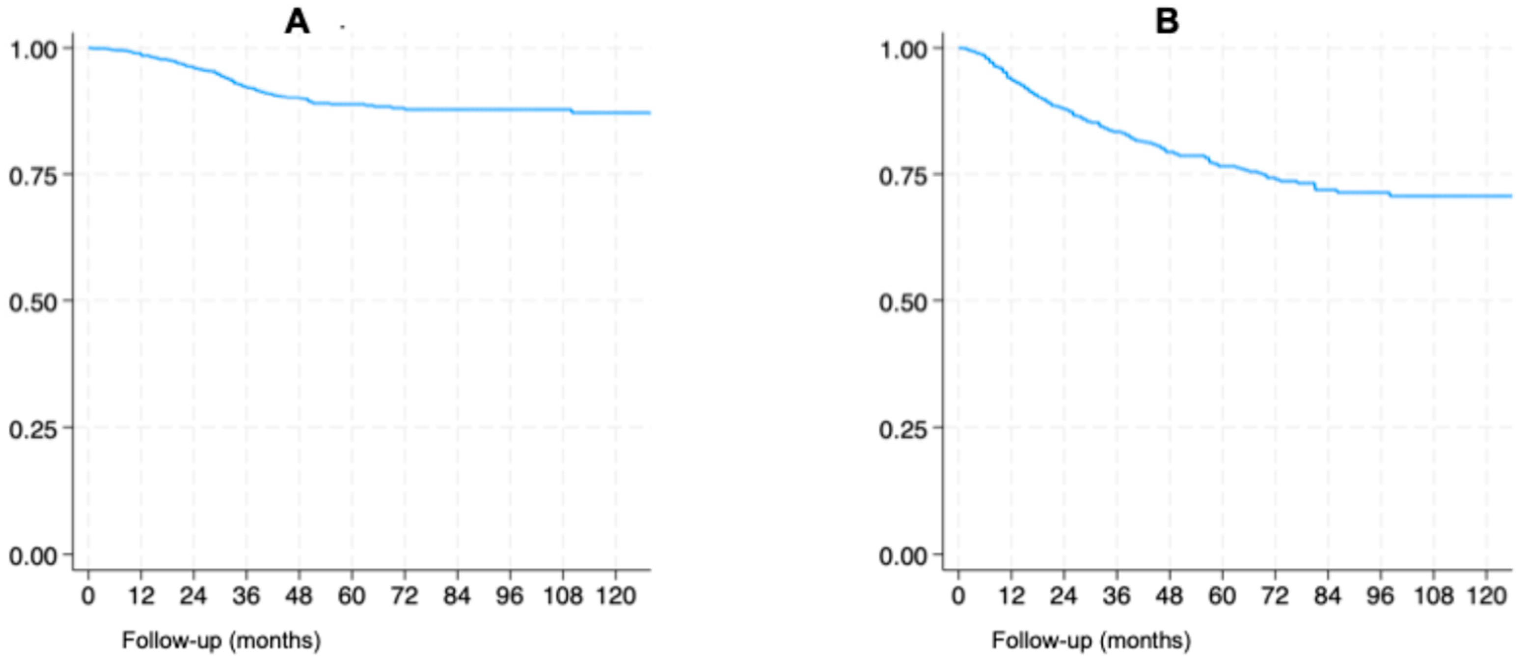
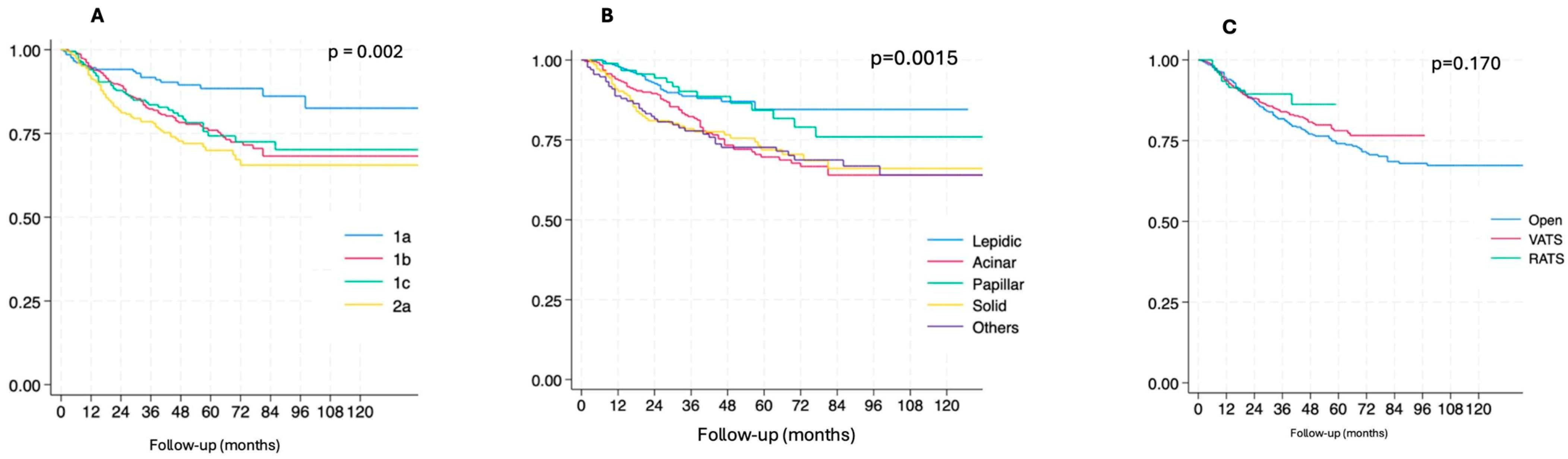
Predictors of Recurrence After Surgery in Patients with Stage I Non-Small Cell Lung Cancer

 , , , , ,
Simple Summary
Abstract
1. Introduction
2. Materials and Methods
Statistical Analysis
3. Results
3.1. Recurrences
3.2. Survival Analysis
3.3. Cox Regression
4. Discussion
Limitations
5. Conclusions
Supplementary Materials
Author Contributions
Funding
Institutional Review Board Statement
Informed Consent Statement
Data Availability Statement
Conflicts of Interest
Abbreviations
| NSCLC | Non-Small Cell Lung Cancer |
| VATS | Video-Assisted Thoracic Surgery |
| RATS | Robotic-Assisted Thoracic Surgery |
| TNM | Tumors, Node and Metastasis |
| CT | Computed Tomography |
| FDG-PET | Fluorodeoxyglucose Positron Emission Tomography |
| DFS | Disease-Free Survival |
| OS | Overall Survival |
| STAS | Spread Through Airspaces |
References
- Postmus, P.E.; Kerr, K.M.; Oudkerk, M.; Senan, S.; Waller, D.A.; Vansteenkiste, J.; Escriu, C.; Peters, S.; ESMO Guidelines Committee. Early and locally advanced non-small-cell lung cancer (NSCLC): ESMO Clinical Practice Guidelines for diagnosis, treatment and follow-up. Ann. Oncol. 2017, 28, iv1–iv21. [Google Scholar] [CrossRef]
- Howington, J.A.; Blum, M.G.; Chang, A.C.; Balekian, A.A.; Murthy, S.C. Treatment of stage I and II non-small cell lung cancer: Diagnosis and management of lung cancer, 3rd ed: American College of Chest Physicians evidence-based clinical practice guidelines. Chest 2013, 143, e278S–e313S. [Google Scholar] [CrossRef] [PubMed]
- Al-Kattan, K.; Sepsas, E.; Fountain, S.W.; Townsend, E.R. Disease recurrence after resection for stage I lung cancer. Eur. J. Cardiothorac. Surg. 1997, 12, 380–384. [Google Scholar] [CrossRef]
- Carnio, S.; Novello, S.; Papotti, M.; Loiacono, M.; Scagliotti, G.V. Prognostic and predictive biomarkers in early stage non-small cell lung cancer: Tumor based approaches including gene signatures. Transl. Lung Cancer Res. 2013, 2, 372–381. [Google Scholar]
- Thornblade, L.W.; Mulligan, M.S.; Odem-Davis, K.; Hwang, B.; Waworuntu, R.L.; Wolff, E.M.; Kessler, L.; Wood, D.E.; Farjah, F. Challenges in Predicting Recurrence After Resection of Node-Negative Non-Small Cell Lung Cancer. Ann. Thorac. Surg. 2018, 106, 1460–1467. [Google Scholar] [CrossRef] [PubMed]
- Wei, W.; Zhou, J.; Zhang, Q.; Liao, D.; Liu, Q.; Zhong, B.; Liang, Z.; Zhang, Y.; Jiang, R.; Liu, G.; et al. Postoperative intensity-modulated radiation therapy reduces local recurrence and improves overall survival in III-N2 non-small-cell lung cancer: A single-center, retrospective study. Cancer Med. 2020, 9, 2820–2832. [Google Scholar] [CrossRef]
- Martini, N.; Bains, M.S.; Burt, M.E.; Zakowski, M.F.; McCormack, P.; Rusch, V.W.; Ginsberg, R.J. Incidence of local recurrence and second primary tumors in resected stage I lung cancer. J. Thorac. Cardiovasc. Surg. 1995, 109, 120–129. [Google Scholar] [CrossRef]
- Maeda, R.; Yoshida, J.; Ishii, G.; Aokage, K.; Hishida, T.; Nishimura, M.; Nishiwaki, Y.; Nagai, K. Long-term outcome and late recurrence in patients with completely resected stage IA non-small cell lung cancer. J. Thorac. Oncol. 2010, 5, 1246–1250. [Google Scholar] [CrossRef]
- Liang, W.; Zhang, L.; Jiang, G.; Wang, Q.; Liu, L.; Liu, D.; Wang, Z.; Zhu, Z.; Deng, Q.; Xiong, X.; et al. Development and validation of a nomogram for predicting survival in patients with resected non-small-cell lung cancer. J. Clin. Oncol. 2015, 33, 861–869. [Google Scholar] [CrossRef]
- Yatabe, Y.; Dacic, S.; Borczuk, A.C.; Warth, A.; Russell, P.A.; Lantuejoul, S.; Beasley, M.B.; Thunnissen, E.; Pelosi, G.; Rekhtman, N.; et al. Best Practices Recommendations for Diagnostic Immunohistochemistry in Lung Cancer. J. Thorac. Oncol. 2019, 14, 377–407. [Google Scholar] [CrossRef]
- Travis, W.D.; Asamura, H.; Bankier, A.A.; Beasley, M.B.; Detterbeck, F.; Flieder, D.B.; Goo, J.M.; MacMahon, H.; Naidich, D.; Nicholson, A.G.; et al. The IASLC Lung Cancer Staging Project: Proposals for Coding T Categories for Subsolid Nodules and Assessment of Tumor Size in Part-Solid Tumors in the Forthcoming Eighth Edition of the TNM Classification of Lung Cancer. J. Thorac. Oncol. 2016, 11, 1204–1223. [Google Scholar] [CrossRef] [PubMed]
- Cuschieri, S. The STROBE guidelines. Saudi J. Anaesth. 2019, 13, S31. [Google Scholar] [CrossRef]
- Kiankhooy, A.; Taylor, M.D.; LaPar, D.J.; Isbell, J.M.; Lau, C.L.; Kozower, B.D.; Jones, D.R. Predictors of early recurrence for node-negative t1 to t2b non-small cell lung cancer. Ann. Thorac. Surg. 2014, 98, 1175–1183. [Google Scholar] [CrossRef]
- Chen, Y.-Y.; Huang, T.-W.; Tsai, W.-C.; Lin, L.-F.; Cheng, J.-B.; Chang, H.; Lee, S.-C. Risk factors of postoperative recurrences in patients with clinical stage I NSCLC. World J. Surg. Oncol. 2014, 12, 10. [Google Scholar] [CrossRef]
- Fick, C.N.; Dunne, E.G.; Vanstraelen, S.; Toumbacaris, N.; Tan, K.S.; Rocco, G.; Molena, D.; Huang, J.; Park, B.J.; Rekhtman, N.; et al. High-risk features associated with recurrence in stage I lung adenocarcinoma. J. Thorac. Cardiovasc. Surg. 2025, 169, 436–444. [Google Scholar] [CrossRef]
- Moreira, A.L.; Ocampo, P.S.; Xia, Y.; Zhong, H.; Russell, P.A.; Minami, Y.; Cooper, W.A.; Yoshida, A.; Bubendorf, L.; Papotti, M.; et al. A Grading System for Invasive Pulmonary Adenocarcinoma: A Proposal from the International Association for the Study of Lung Cancer Pathology Committee. J. Thorac. Oncol. 2020, 15, 1599–1610. [Google Scholar] [CrossRef] [PubMed]
- Kadota, K.; Colovos, C.; Suzuki, K.; Rizk, N.P.; Dunphy, M.P.S.; Zabor, E.C.; Sima, C.S.; Yoshizawa, A.; Travis, W.D.; Rusch, V.W.; et al. FDG-PET SUVmax combined with IASLC/ATS/ERS histologic classification improves the prognostic stratification of patients with stage I lung adenocarcinoma. Ann. Surg. Oncol. 2012, 19, 3598–3605. [Google Scholar] [CrossRef]
- Caso, R.; Sanchez-Vega, F.; Tan, K.S.; Mastrogiacomo, B.; Zhou, J.; Jones, G.D.; Nguyen, B.; Schultz, N.; Connolly, J.G.; Brandt, W.S.; et al. The Underlying Tumor Genomics of Predominant Histologic Subtypes in Lung Adenocarcinoma. J. Thorac. Oncol. 2020, 15, 1844–1856. [Google Scholar] [CrossRef]
- Jiang, L.; Liang, W.; Shen, J.; Chen, X.; Shi, X.; He, J.; Yang, C.; He, J. The impact of visceral pleural invasion in node-negative non-small cell lung cancer: A systematic review and meta-analysis. Chest 2015, 148, 903–911. [Google Scholar] [CrossRef]
- Woo, W.; Cha, Y.J.; Kim, B.J.; Moon, D.H.; Lee, S. Validation Study of New IASLC Histology Grading System in Stage I Non-Mucinous Adenocarcinoma Comparing with Minimally Invasive Adenocarcinoma. Clin. Lung Cancer 2022, 23, e435–e442. [Google Scholar] [CrossRef] [PubMed]
- Tuminello, S.; Veluswamy, R.; Lieberman-Cribbin, W.; Gnjatic, S.; Petralia, F.; Wang, P.; Flores, R.; Taioli, E. Prognostic value of immune cells in the tumor microenvironment of early-stage lung cancer: A meta-analysis. Oncotarget 2019, 10, 7142–7155. [Google Scholar] [CrossRef] [PubMed]
- Shahid, M.; Choi, T.G.; Nguyen, M.N.; Matondo, A.; Jo, Y.H.; Yoo, J.Y.; Nguyen, N.N.Y.; Yun, H.R.; Kim, J.; Akter, S.; et al. An 8-gene signature for prediction of prognosis and chemoresponse in non-small cell lung cancer. Oncotarget 2016, 7, 86561–86572. [Google Scholar] [CrossRef]
- Merritt, R.E.; Abdel-Rasoul, M.; Fitzgerald, M.; D’Souza, D.M.; Kneuertz, P.J. Nomograms for Predicting Overall and Recurrence-free Survival from Pathologic Stage IA and IB Lung Cancer After Lobectomy. Clin. Lung Cancer 2021, 22, e574–e583. [Google Scholar] [CrossRef]
- Lou, F.; Huang, J.; Sima, C.S.; Dycoco, J.; Rusch, V.; Bach, P.B. Patterns of recurrence and second primary lung cancer in early-stage lung cancer survivors followed with routine computed tomography surveillance. J. Thorac. Cardiovasc. Surg. 2013, 145, 75–82. [Google Scholar] [CrossRef]
- Zhao, Z.-R.; Xi, S.-Y.; Li, W.; Situ, D.-R.; Chen, K.-M.; Yang, H.; Su, X.-D.; Lin, Y.-B.; Long, H. Prognostic impact of pattern-based grading system by the new IASLC/ATS/ERS classification in Asian patients with stage I lung adenocarcinoma. Lung Cancer Amst. Neth. 2015, 90, 604–609. [Google Scholar] [CrossRef]
- Woo, W.; Yang, Y.H.; Cha, Y.J.; Moon, D.H.; Shim, H.S.; Cho, A.; Kim, B.J.; Kim, H.E.; Park, B.J.; Lee, J.G.; et al. Prognosis of resected invasive mucinous adenocarcinoma compared with the IASLC histologic grading system for invasive nonmucinous adenocarcinoma: Surgical database study in the TKIs era in Korea. Thorac. Cancer 2022, 13, 3310–3321. [Google Scholar] [CrossRef]
- Kneuertz, P.J.; D’Souza, D.M.; Richardson, M.; Abdel-Rasoul, M.; D Moffatt-Bruce, S.; Merritt, R.E. Long-term oncological outcomes after robotic lobectomy for eraly-stage no-small-cell lung cancer versu video-assisted thoracoscopic and opern thoracotomy apprach. Clin. Lung Cancer 2020, 21, 214–224. [Google Scholar] [CrossRef]
- Whitson, B.A.; Groth, S.S.; Duval, S.J.; Swanson, S.J.; Maddaus, M.A. Surgery for early-stage non-small cell lung cancer: A systematic review of the video-assisted thoracoscopic surgery versus thoracotomy approaches to lobectomy. Ann. Thorac. Surg. 2008, 86, 2008–2018. [Google Scholar] [CrossRef] [PubMed]
- Lee, P.C.; Nasar, A.; Port, J.L.; Paul, S.; Stiles, B.; Chiu, Y.-L.; Andrews, W.G.; Altorki, N.K. Long-term survival after lobectomy for non-small cell lung cancer by video-assisted thoracic surgery versus thoracotomy. Ann. Thorac. Surg. 2013, 96, 951–961. [Google Scholar] [CrossRef] [PubMed]
- Yang, H.-X.; Woo, K.M.; Sima, C.S.; Bains, M.S.; Adusumilli, P.S.; Huang, J.; Finley, D.J.; Rizk, N.P.; Rusch, V.W.; Jones, D.R.; et al. Long-term Survival Based on the Surgical Approach to Lobectomy For Clinical Stage I Nonsmall Cell Lung Cancer: Comparison of Robotic, Video-assisted Thoracic Surgery, and Thoracotomy Lobectomy. Ann. Surg. 2017, 265, 431–437. [Google Scholar] [CrossRef]
- Kozu, Y.; Maniwa, T.; Takahashi, S.; Isaka, M.; Ohde, Y.; Nakajima, T. Risk factors for both recurrence and survival in patients with pathological stage I non-small-cell lung cancer. Eur. J. Cardio-Thorac. Surg. 2013, 44, e53–e58. [Google Scholar] [CrossRef] [PubMed][Green Version]
- Altorki, N.; Wang, X.; Kozono, D.; Watt, C.; Landrenau, R.; Wigle, D.; Port, J.; Jones, D.R.; Conti, M.; Ashrafi, A.S.; et al. Lobar or Sublobar Resection for Peripheral Stage IA Non-Small-Cell Lung Cancer. N. Engl. J. Med. 2023, 388, 489–498. [Google Scholar] [CrossRef] [PubMed]
- Saji, H.; Okada, M.; Tsuboi, M.; Nakajima, R.; Suzuki, K.; Aokage, K.; Aoki, T.; Okami, J.; Yoshino, I.; Ito, H.; et al. Segmentectomy versus lobectomy in small-sized peripheral non-small-cell lung cancer (JCOG0802/WJOG4607L): A multicentre, open-label, phase 3, randomised, controlled, non-inferiority trial. Lancet 2022, 399, 1607–1617. [Google Scholar] [CrossRef] [PubMed]
- Varlotto, J.M.; Medford-Davis, L.N.; Recht, A.; Flickinger, J.; Yao, N.; Hess, C.; Reed, M.F.; Toth, J.; Zander, D.S.; DeCamp, M.M. Identification of Stage I Non-small Cell Lung Cancer Patients at High Risk for Local Recurrence Following Sublobar Resection. Chest 2013, 143, 1365–1377. [Google Scholar] [CrossRef]
- El-Sherif, A.; Gooding, W.E.; Santos, R.; Pettiford, B.; Ferson, P.F.; Fernando, H.C.; Urda, S.J.; Luketich, J.D.; Landreneau, R.J. Outcomes of sublobar resection versus lobectomy for stage I non-small cell lung cancer: A 13-year analysis. Ann. Thorac. Surg. 2006, 82, 408–416. [Google Scholar] [CrossRef]
- Kadota, K.; Nitadori, J.; Sima, C.S.; Ujiie, H.; Rizk, N.P.; Jones, D.R.; Adusumilli, P.S.; Travis, W.D. Tumor spread through air spaces is an important pattern of invasion and impacts the frequency and location of recurrences after limited resection for small stage I lung adenocarcinomas. J. Thorac. Oncol. 2015, 10, 806–814. [Google Scholar] [CrossRef]



| Variables | Value | % |
|---|---|---|
| Age at surgery (mean ± SD) | 68 ± 8.8 | |
| Gender (male) | 629 | 55.5% |
| Body mass index (kg/m2) * | 24.6 ± 5.4 | |
| Comorbidity (none) | 300 | 26.5% |
| Smoke | ||
| Never | 400 | 35.3% |
| Ex | 495 | 43.7% |
| Current | 227 | 20.1% |
| Unknown | 10 | 0.9% |
| CT appearance | ||
| Solid | 758 | 67% |
| Ground-glass | 164 | 14.5% |
| Mixed | 147 | 13% |
| Unknown | 63 | 5.5% |
| Surgical approach | ||
| Open | 474 | 41.9% |
| VATS | 552 | 48.7% |
| RATS | 106 | 9.4% |
| Resection volume | ||
| Lobectomy/bilobectomy | 995 | 87.9% |
| Segmentectomy | 56 | 4.9% |
| Wedge | 81 | 7.2% |
| pT status (8th TNM) | ||
| 1a | 212 | 18.7% |
| 1b | 402 | 35.5% |
| 1c | 223 | 19.7% |
| 2a | 295 | 26.1% |
| Histology | ||
| Adenocarcinoma | 920 | 81.3% |
| Squamous carcinoma | 141 | 12.5% |
| Other | 71 | 6.2% |
| Subtype of adenocarcinoma | ||
| Lepidic | 285 | 25.2% |
| Acinar | 264 | 23.3% |
| Papillar | 95 | 8.4% |
| Solid | 147 | 13% |
| Other | 134 | 11.8% |
| NA | 207 | 18.3% |
| Univariate Analysis | |||
|---|---|---|---|
| Early Recurrence | Late Recurrence | p | |
| N | 72 | 152 | |
| Age | 73 (60–77) | 70 (61–77) | 0.015 |
| Gender (male) | 52 (72.2%) | 89 (58.5%) | 0.048 |
| Surgical approach | 0.005 | ||
| Open | 29 (40.3%) | 89 (58.6%) | |
| VATS | 35 (48.6%) | 59 (38.8%) | |
| RATS | 8 (11.1%) | 4 (2.6%) | |
| Resection volume | 0.469 | ||
| Lobectomy/bilobectomy | 59 (81.9%) | 132 (86.9) | |
| Segmentectomy | 3 (4.2%) | 7 (4.6%) | |
| Wedge | 10 (13.9%) | 13 (8.5) | |
| pT status (8th TNM) | 0.246 | ||
| 1a | 11 (15.3%) | 11 (7.2%) | |
| 1b | 23 (31.9%) | 60 (39.5%) | |
| 1c | 13 (18.1%) | 31 (20.4%) | |
| 2a | 25 (34.7%) | 50 (32.9%) | |
| Histology | 0.029 | ||
| Adenocarcinoma | 54 (75%) | 132 (86.8%) | |
| Squamous carcinoma | 13 (18.1%) | 10 (6.6%) | |
| Other | 5 (6.9%) | 10 (6.6%) | |
| Subtype of adenocarcinoma | 0.118 | ||
| Lepidic | 6 (8.3%) | 25 (16.3%) | |
| Acinar | 16 (22.2%) | 50 (32.7%) | |
| Papillar | 2 (2.8%) | 12 (7.8%) | |
| Solid | 15 (20.8%) | 24 (16.3%) | |
| Other | 15 (20.8%) | 23 (15%) | |
| NA | 18 (25.1%) | 18 (11.9%) | |
| Variable | Adjusted OR | 95% CI | p-Value |
|---|---|---|---|
| Surgical Approach | |||
| VATS vs Open | 1.59 | 0.79–3.20 | 0.190 |
| RATS vs Open | 4.81 | 1.16–19.87 | 0.030 |
| pT Status | |||
| 1b vs 1a | 0.20 | 0.07–0.59 | 0.004 |
| 1c vs 1a | 0.19 | 0.06–0.65 | 0.008 |
| 2a vs 1a | 0.23 | 0.08–0.71 | 0.010 |
| Histology | |||
| Squamous vs Adeno | 2.72 | 1.05–7.03 | 0.039 |
| Other vs Adeno | 1.32 | 0.41–4.32 | 0.642 |
| Age | 1.03 | 0.99–1.07 | 0.114 |
| Female vs Male | 0.57 | 0.30–1.10 | 0.094 |
| Year | 1.08 | 0.94–1.24 | 0.265 |
| Univariate Analysis | |||
|---|---|---|---|
| Local Recurrence | Distant * Recurrence | p | |
| N | 86 | 138 | |
| Age | 73 (60–77) | 70 (61–77) | 0.059 |
| Gender (male) | 57 (66.2%) | 84 (60.8%) | 0.425 |
| Surgical approach | 0.007 | ||
| Open | 53 (61.6%) | 65 (47.1%) | |
| VATS | 33 (38.4%) | 61 (44.2%) | |
| RATS | 0 (0%) | 12 (8.7%) | |
| Resection volume | 0.0001 | ||
| Lobectomy/bilobectomy | 63 (73.3%) | 128 (92.7%) | |
| Segmentectomy | 7 (8.1%) | 3 (2.2%) | |
| Wedge | 16 (18.6%) | 7 (5.1%) | |
| pT status (8th TNM) | 0.008 | ||
| 1a | 14 (16.3%) | 8 (5.8%) | |
| 1b | 35 (40.7%) | 48 (34.8%) | |
| 1c | 18 (20.9%) | 26 (18.8%) | |
| 2a | 19 (22.1%) | 56 (40.6%) | |
| Histology | 0.198 | ||
| Adenocarcinoma | 76 (88.4%) | 110 (79.7%) | |
| Squamous carcinoma | 7 (8.1%) | 16 (11.6%) | |
| Other | 3 (3.5%) | 12 (8.7%) | |
| Subtype of adenocarcinoma | 0.084 | ||
| Lepidic | 6 (7%) | 25 (18.1%) | |
| Acinar | 28 (32.5%) | 38 (27.5%) | |
| Papillar | 8 (9.3%) | 6 (4.3%) | |
| Solid | 16 (18.6%) | 23 (16.7%) | |
| Other | 18 (20.9%) | 20 (14.5%) | |
| NA | 10 (11.7%) | 26 (18.9%) | |
| Univariate Analysis | Multivariate Analysis | ||||
|---|---|---|---|---|---|
| Recurrence | No Recurrence | p | HR (95%CI) | ||
| N | 224 | 908 | |||
| Age | 69.52 ± 9.44 | 68.25 ± 8.76 | 0.057 | 1.01 (0.99–1.03) | 0.184 |
| Gender (male) | 141 (62.9%) | 488 (53.7%) | 0.013 | 0.67 (0.49–0.91) | 0.010 |
| Body mass index (kg/m2) * | 24.7 ± 3.6 (N = 124) | 24.6 ± 3.1 (N = 637) | 0.802 | ||
| Comorbidity | 53 (34.7%) | 247 (37.0%) | 0.315 | ||
| Smoke | 0.482 | ||||
| Never | 78 (35.3%) | 322 (35.7%) | |||
| Ex | 104 (47.1%) | 391 (43.4%) | |||
| Current | 39 (17.7%) | 188 (20.9%) | |||
| CT appearance | 0.285 | ||||
| Solid | 166 (74.7%) | 592 (69.9%) | |||
| Ground-glass | 32 (14.4%) | 132 (15.6%) | |||
| Mixed | 24 (10.8%) | 123 (14.5%) | |||
| Surgical approach | <0.001 | ||||
| Open | 118 (52.7%) | 356(39.0%) | 1 (ref) | ||
| VATS | 94 (42.0%) | 458 (50.4%) | 0.71 (0.52–0.97) | 0.032 | |
| RATS | 12 (5.4%) | 94 (10.3%) | 0.57 (0.29–1.17) | 0.169 | |
| Resection volume | 0.127 | ||||
| Lobectomy/bilobectomy | 191 | 804 | |||
| Segmentectomy | 23 | 58 | |||
| Wedge | 10 | 46 | |||
| Intraoperative complications | 63 (28.1%) | 227 (25.1%) | 0.508 | ||
| pT status (8th TNM) | <0.001 | ||||
| 1a | 22(9.8%) | 190 (20.9%) | 1 (ref) | ||
| 1b | 83 (37.0%) | 319 (35.1%) | 1.68 (1.03–2.72) | 0.036 | |
| 1c | 44 (19.6%) | 179 (19.7%) | 1.70 (1.00–2.89) | 0.052 | |
| 2a | 75 (33.5%) | 220 (24.2%) | 2.16 (1.31–3.55) | 0.003 | |
| Histology | 0.557 | ||||
| Adenocarcinoma | 186 (83.0%) | 735 (80.9%) | |||
| Squamous carcinoma | 23 (10.3%) | 117 (12.9%) | |||
| other | 15 (6.7%) | 56 (6.2%) | |||
| Subtype of adenocarcinoma | <0.001 | ||||
| Lepidic | 31 (13.8%) | 254 (28.0%) | 1 (ref) | ||
| Acinar | 66 (29.5%) | 198 (21.8%) | 1.76 (1.13–2.73) | 0.012 | |
| Papillar | 14 (6.2%) | 81 (8.9%) | 0.97 (0.51–1.85) | 0.931 | |
| Solid | 39 (17.4%) | 108 (11.9%) | 1.81 (1.12–2.93) | 0.016 | |
| Not applicable | 38 (17.0%) | 96 (10.6%) | 1.97 (1.19–3.25) | 0.008 | |
Disclaimer/Publisher’s Note: The statements, opinions and data contained in all publications are solely those of the individual author(s) and contributor(s) and not of MDPI and/or the editor(s). MDPI and/or the editor(s) disclaim responsibility for any injury to people or property resulting from any ideas, methods, instructions or products referred to in the content. |
© 2026 by the authors. Licensee MDPI, Basel, Switzerland. This article is an open access article distributed under the terms and conditions of the Creative Commons Attribution (CC BY) license.
Share and Cite
Voulaz, E.; Brascia, D.; Giudici, V.; Margaritora, S.; Lucchi, M.; Aprile, V.; Chiappetta, M.; Patirelis, A.; Ambrogi, V.; Marulli, G. Predictors of Recurrence After Surgery in Patients with Stage I Non-Small Cell Lung Cancer. Cancers 2026, 18, 1152. https://doi.org/10.3390/cancers18071152
Voulaz E, Brascia D, Giudici V, Margaritora S, Lucchi M, Aprile V, Chiappetta M, Patirelis A, Ambrogi V, Marulli G. Predictors of Recurrence After Surgery in Patients with Stage I Non-Small Cell Lung Cancer. Cancers. 2026; 18(7):1152. https://doi.org/10.3390/cancers18071152
Chicago/Turabian StyleVoulaz, Emanuele, Debora Brascia, Veronica Giudici, Stefano Margaritora, Marco Lucchi, Vittorio Aprile, Marco Chiappetta, Alexandro Patirelis, Vincenzo Ambrogi, and Giuseppe Marulli. 2026. "Predictors of Recurrence After Surgery in Patients with Stage I Non-Small Cell Lung Cancer" Cancers 18, no. 7: 1152. https://doi.org/10.3390/cancers18071152
APA StyleVoulaz, E., Brascia, D., Giudici, V., Margaritora, S., Lucchi, M., Aprile, V., Chiappetta, M., Patirelis, A., Ambrogi, V., & Marulli, G. (2026). Predictors of Recurrence After Surgery in Patients with Stage I Non-Small Cell Lung Cancer. Cancers, 18(7), 1152. https://doi.org/10.3390/cancers18071152