Geospatial and Cell Density Analysis Using Multiplex Immunofluorescence Reveals an Important Role of Clustering Patterns of Immunosuppressive Macrophages in Survival Outcomes of Penile Squamous Cell Carcinoma


 , ,
, ,  ,
,  ,
,  and
and
Simple Summary
Abstract
1. Introduction
2. Materials and Methods
2.1. Multiplex Immunofluorescence and Image Analysis
2.2. Cell Density Analysis, Phenotyping and Compartmentalization
2.3. Geospatial Analysis
2.4. Statistical Analysis
3. Results
3.1. Study Population
3.2. Survival Outcomes and Prognostic Factors
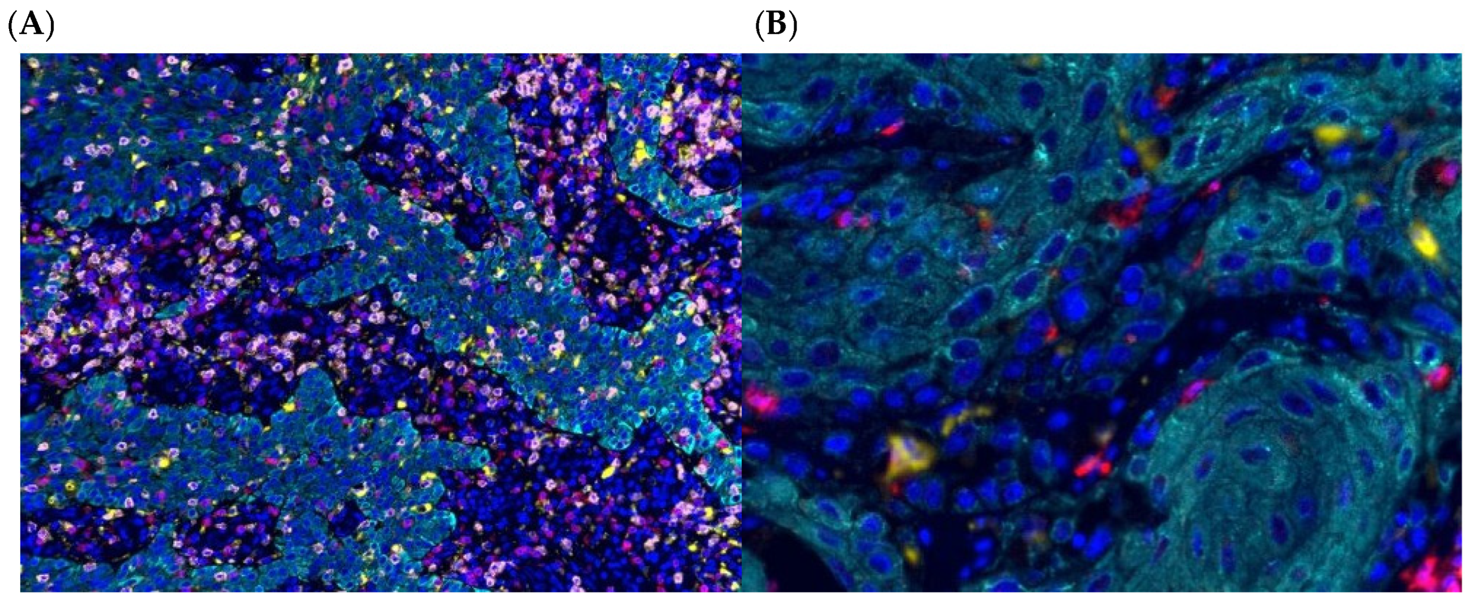
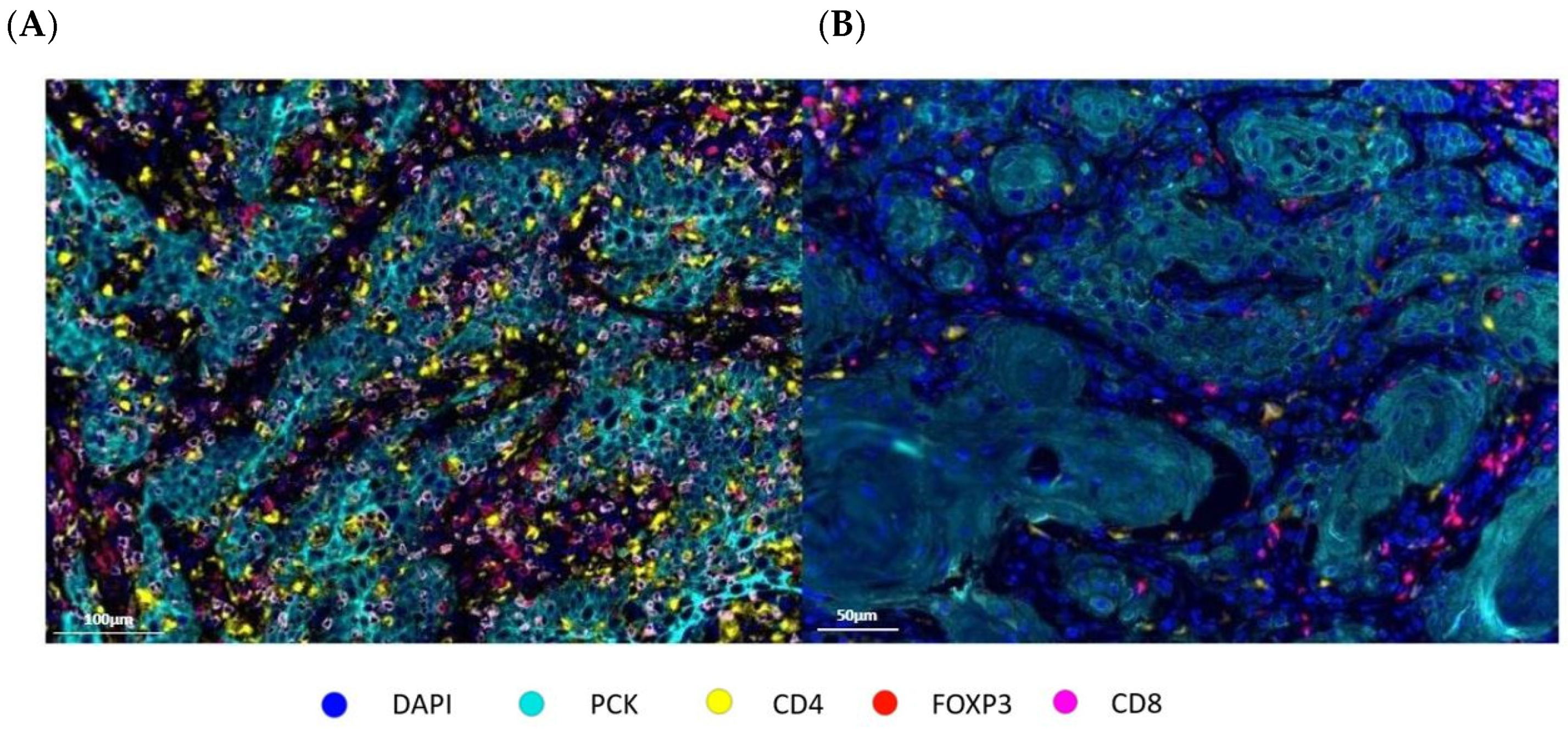
3.3. Immune Phenotypes in mIF Panels
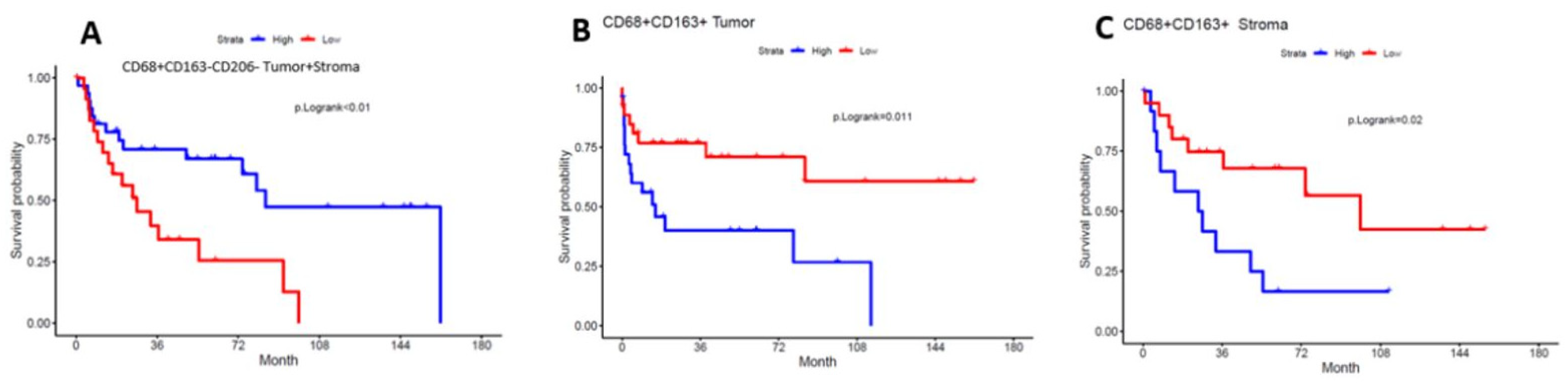
3.4. High Clustering of M2 Macrophages to One Another and Tumor Cells Is Associated with Worse Overall Survival
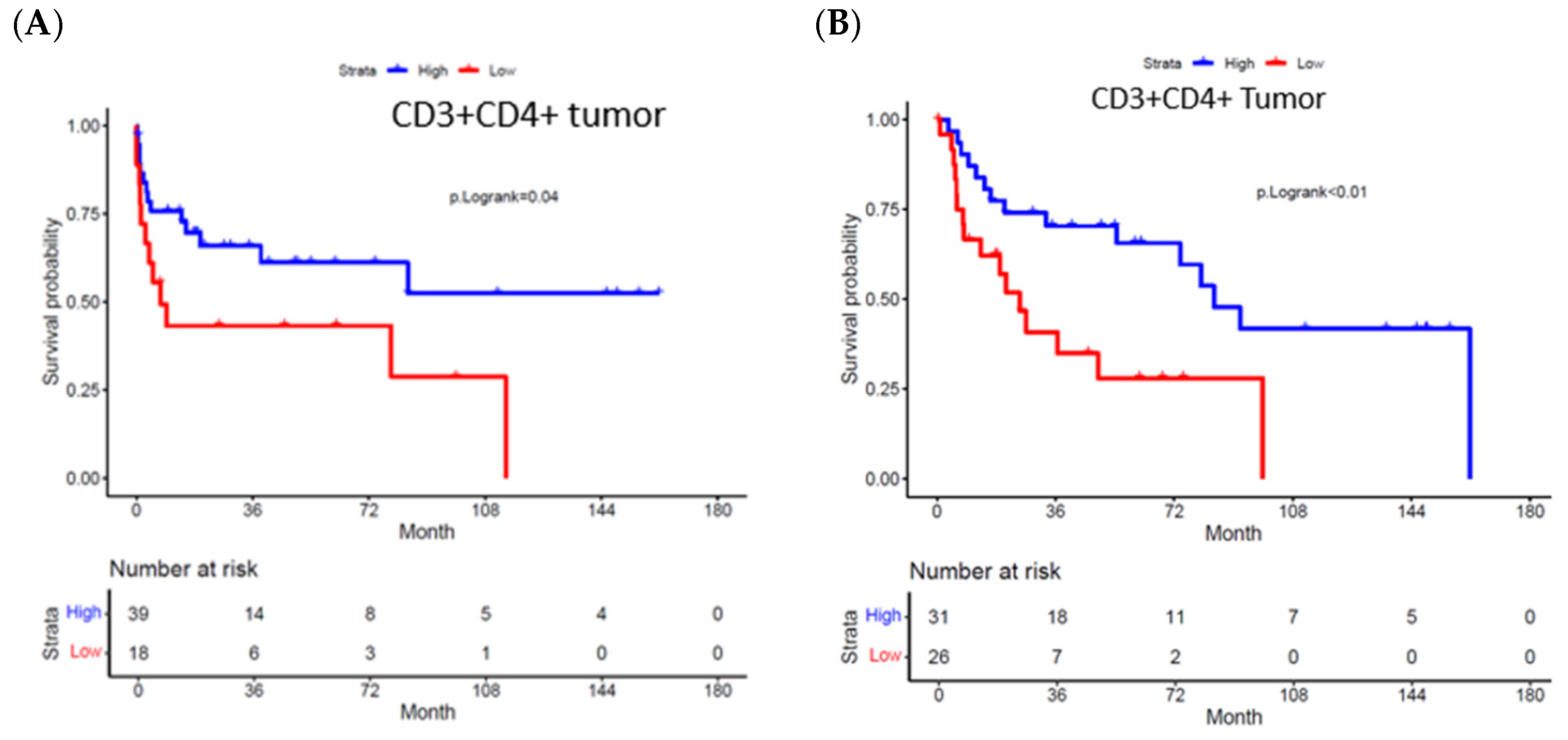
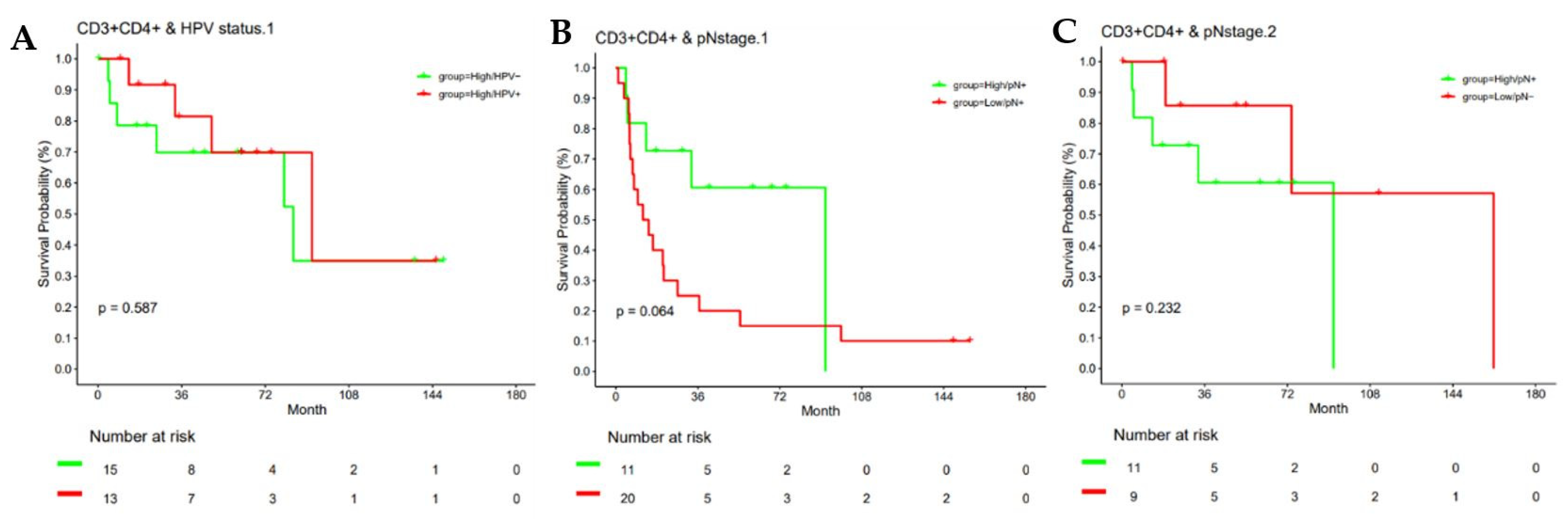
3.5. High Cell Density and Geospatial Clustering of Tumor-Infiltrating Helper T Cells Is Associated with Improved Overall Survival
4. Discussion
5. Conclusions
Author Contributions
Funding
Institutional Review Board Statement
Informed Consent Statement
Data Availability Statement
Conflicts of Interest
References
- American Cancer Society. Cancer Facts & Figures 2024; American Cancer Society: Atlanta, GA, USA, 2024. [Google Scholar]
- Fu, L.; Tian, T.; Yao, K.; Chen, X.-F.; Luo, G.; Gao, Y.; Lin, Y.-F.; Wang, B.; Sun, Y.; Zheng, W.; et al. Global Pattern and Trends in Penile Cancer Incidence: Population-Based Study. JMIR Public Health Surveill. 2022, 8, e34874. [Google Scholar] [CrossRef]
- Cardona, C.E.M.; García-Perdomo, H.A. Incidence of penile cancer worldwide: Systematic review and meta-analysis. Rev. Panam. Salud Publica Am. J. Public Health 2017, 41, e117. [Google Scholar] [CrossRef]
- Menon, S.; Moch, H.; Berney, D.; Cree, I.; Srigley, J.; Tsuzuki, T.; Compérat, E.; Hartmann, A.; Netto, G.; Rubin, M.; et al. WHO 2022 classification of penile and scrotal cancers: Updates and evolution. Histopathology 2022, 82, 508–520. [Google Scholar] [CrossRef] [PubMed]
- Morrison, B. Risk Factors and Prevalence of Penile Cancer. West Indian Med. J. 2018, 63, 559–560. [Google Scholar] [CrossRef][Green Version]
- Emmanuel, A.; Watkin, N. Update on organ preserving surgical strategies for penile cancer. Urol. Oncol. Semin. Orig. Investig. 2022, 40, 179–183. [Google Scholar] [CrossRef]
- Crook, J. Organ preserving radiation strategies for penile cancer. Urol. Oncol. Semin. Orig. Investig. 2022, 40, 184–190. [Google Scholar] [CrossRef]
- Alnajjar, H.M.; Lam, W.; Bolgeri, M.; Rees, R.W.; Perry, M.J.; Watkin, N.A. Treatment of Carcinoma In Situ of the Glans Penis with Topical Chemotherapy Agents. Eur. Urol. 2012, 62, 923–928. [Google Scholar] [CrossRef]
- Chahoud, J.; Kohli, M.; Spiess, P.E. Management of Advanced Penile Cancer. Mayo Clin. Proc. 2021, 96, 720–732. [Google Scholar] [CrossRef]
- Djajadiningrat, R.S.; Graafland, N.M.; van Werkhoven, E.; Meinhardt, W.; Bex, A.; van der Poel, H.G.; van Boven, H.H.; Olmos, R.A.V.; Horenblas, S. Contemporary Management of Regional Nodes in Penile Cancer—Improvement of Survival? J. Urol. 2014, 191, 68–73. [Google Scholar] [CrossRef] [PubMed]
- Pagliaro, L.C.; Williams, D.L.; Daliani, D.; Williams, M.B.; Osai, W.; Kincaid, M.; Wen, S.; Thall, P.F.; Pettaway, C.A. Neoadjuvant Paclitaxel, Ifosfamide, and Cisplatin Chemotherapy for Metastatic Penile Cancer: A Phase II Study. J. Clin. Oncol. 2010, 28, 3851–3857. [Google Scholar] [CrossRef] [PubMed]
- Azizi, M.; Aydin, A.M.; Hajiran, A.; Lai, A.; Kumar, A.; Peyton, C.C.; Minhas, S.; Sonpavde, G.P.; Chahoud, J.; Pagliaro, L.C.; et al. Systematic Review and Meta-Analysis—Is there a Benefit in Using Neoadjuvant Systemic Chemotherapy for Locally Advanced Penile Squamous Cell Carcinoma? J. Urol. 2020, 203, 1147–1155. [Google Scholar] [CrossRef]
- Djajadiningrat, R.S.; Bergman, A.M.; van Werkhoven, E.; Vegt, E.; Horenblas, S. Neoadjuvant Taxane-Based Combination Chemotherapy in Patients With Advanced Penile Cancer. Clin. Genitourin. Cancer 2015, 13, 44–49. [Google Scholar] [CrossRef]
- Cohen, E.E.W.; Le Tourneau, C.; Licitra, L.; Ahn, M.-J.; Soria, A.; Machiels, J.-P.; Mach, N.; Mehra, R.; Burtness, B.; Zhang, P.; et al. Pembrolizumab versus methotrexate, docetaxel, or cetuximab for recurrent or metastatic head-and-neck squamous cell carcinoma (KEYNOTE-040): A randomised, open-label, phase 3 study. Lancet 2019, 393, 156–167. [Google Scholar] [CrossRef] [PubMed]
- El Zarif, T.; Nassar, A.H.; Pond, G.R.; Zhuang, T.Z.; Master, V.; Nazha, B.; Niglio, S.; Simon, N.; Hahn, A.W.; Pettaway, C.A.; et al. Safety and efficacy of immune checkpoint inhibitors in advanced penile cancer: Report from the Global Society of Rare Genitourinary Tumors. JNCI J. Natl. Cancer Inst. 2023, 115, 1605–1615. [Google Scholar] [CrossRef] [PubMed]
- de Vries, H.M.; Rafael, T.S.; Gil-Jimenez, A.; de Feijter, J.M.; Bekers, E.; van der Laan, E.; Lopez-Yurda, M.; Hooijberg, E.; Broeks, A.; Peters, D.; et al. Atezolizumab With or Without Radiotherapy for Advanced Squamous Cell Carcinoma of the Penis (The PERICLES Study): A Phase II Trial. J. Clin. Oncol. 2023, 41, 4872–4880. [Google Scholar] [CrossRef] [PubMed]
- Lu, S.; Stein, J.E.; Rimm, D.L.; Wang, D.W.; Bell, J.M.; Johnson, D.B.; Sosman, J.A.; Schalper, K.A.; Anders, R.A.; Wang, H.; et al. Comparison of Biomarker Modalities for Predicting Response to PD-1/PD-L1 Checkpoint Blockade: A Systematic Review and Meta-analysis. JAMA Oncol. 2019, 5, 1195–1204. [Google Scholar] [CrossRef]
- Ionescu, F.; Nguyen, J.; Segura, C.M.; Paravathaneni, M.; Grass, G.D.; Johnstone, P.; Zacharias, N.M.; Pettaway, C.A.; Lu, X.; Kim, Y.; et al. Multiplex Immunofluorescence Captures Progressive Immune Exhaustion with Advancing Penile Squamous Cell Cancer Stage. Cancers 2024, 16, 303. [Google Scholar] [CrossRef]
- Miyagi, H.; Yu, X.; Peak, T.; Dhillon, J.; Le, C.; Wang, X.; Yoder, S.; Marchion, D.; Lu, X.; Pettaway, C.; et al. Progressive T cell exhaustion and predominance of aging tissue associated macrophages with advancing disease stage in penile squamous cell carcinoma. Sci. Rep. 2025, 15, 7703. [Google Scholar] [CrossRef]
- Parra, E.R.; Uraoka, N.; Jiang, M.; Cook, P.; Gibbons, D.; Forget, M.-A.; Bernatchez, C.; Haymaker, C.; Wistuba, I.I.; Rodriguez-Canales, J. Validation of multiplex immunofluorescence panels using multispectral microscopy for immune-profiling of formalin-fixed and paraffin-embedded human tumor tissues. Sci. Rep. 2017, 7, 13380. [Google Scholar] [CrossRef]
- Parra, E.R. Methods to Determine and Analyze the Cellular Spatial Distribution Extracted From Multiplex Immunofluorescence Data to Understand the Tumor Microenvironment. Front. Mol. Biosci. 2021, 8, 668340. [Google Scholar] [CrossRef]
- Barnes, D.; Harris, W.; Smith, P.; Millis, R.; Rubens, R. Immunohistochemical determination of oestrogen receptor: Comparison of different methods of assessment of staining and correlation with clinical outcome of breast cancer patients. Br. J. Cancer 1996, 74, 1445–1451. [Google Scholar] [CrossRef]
- Nuovo, G.J.; Hochman, H.A.; Eliezri, Y.D.; Lastarria, D.; Comite, S.L.; Silvers, D.N. Detection of human papillomavirus DNA in penile lesions histologically negative for condylomata. Analysis by in situ hybridization and the polymerase chain reaction. Am. J. Surg. Pathol. 1990, 14, 829–836. [Google Scholar] [CrossRef]
- Kerr, D.A.; Sweeney, B.; Arpin, R.N.; Ring, M.; Pitman, M.B.; Wilbur, D.C.; Faquin, W.C. Automated Extraction of Formalin-Fixed, Paraffin-Embedded Tissue for High-Risk Human Papillomavirus Testing of Head and Neck Squamous Cell Carcinomas Using the Roche Cobas 4800 System. Arch. Pathol. Lab. Med. 2016, 140, 844–848. [Google Scholar] [CrossRef] [PubMed]
- Bruna, F.; Scodeller, P. Pro-Tumorigenic Macrophage Infiltration in Oral Squamous Cell Carcinoma and Possible Macrophage-Aimed Therapeutic Interventions. Front. Oncol. 2021, 11, 675664. [Google Scholar] [CrossRef]
- Najafi, M.; Hashemi Goradel, N.; Farhood, B.; Salehi, E.; Nashtaei, M.S.; Khanlarkhani, N.; Khezri, Z.; Majidpoor, J.; Abouzaripour, M.; Habibi, M.; et al. Macrophage polarity in cancer: A review. J. Cell. Biochem. 2019, 120, 2756–2765. [Google Scholar] [CrossRef] [PubMed]
- Sumitomo, R.; Menju, T.; Shimazu, Y.; Toyazaki, T.; Chiba, N.; Miyamoto, H.; Hirayama, Y.; Nishikawa, S.; Tanaka, S.; Yutaka, Y.; et al. M2-like tumor-associated macrophages promote epithelial–mesenchymal transition through the transforming growth factor β/Smad/zinc finger e-box binding homeobox pathway with increased metastatic potential and tumor cell proliferation in lung squamous cell carcinoma. Cancer Sci. 2023, 114, 4521–4534. [Google Scholar] [CrossRef]
- Zhang, P.; Zhang, Y.; Wang, L.; Lou, W. Tumor-regulated macrophage type 2 differentiation promotes immunosuppression in laryngeal squamous cell carcinoma. Life Sci. 2021, 267, 118798. [Google Scholar] [CrossRef]
- Ottenhof, S.R.; Djajadiningrat, R.S.; Thygesen, H.H.; Jakobs, P.J.; Jóźwiak, K.; Heeren, A.M.; de Jong, J.; Sanders, J.; Horenblas, S.; Jordanova, E.S. The Prognostic Value of Immune Factors in the Tumor Microenvironment of Penile Squamous Cell Carcinoma. Front. Immunol. 2018, 9, 1253. [Google Scholar] [CrossRef]
- Song, H.; Tong, Z.; Xie, G.; Li, Y.; Zhao, Y.; Fan, F.; Yang, Z.; Shi, Q.; Zhang, Q.; Wen, F.; et al. Single-Cell and Spatial Transcriptomic Profiling of Penile Squamous Cell Carcinoma Reveals Dynamics of Tumor Differentiation and Immune Microenvironment. Adv. Sci. 2025, 12, e00216. [Google Scholar] [CrossRef]
- Pirilä, E.; Väyrynen, O.; Sundquist, E.; Päkkilä, K.; Nyberg, P.; Nurmenniemi, S.; Pääkkönen, V.; Pesonen, P.; Dayan, D.; Vered, M.; et al. Macrophages Modulate Migration and Invasion of Human Tongue Squamous Cell Carcinoma. PLoS ONE 2015, 10, e0120895. [Google Scholar] [CrossRef] [PubMed]
- Summer, M.; Riaz, S.; Ali, S.; Noor, Q.; Ashraf, R.; Khan, R.R.M. Understanding the Dual Role of Macrophages in Tumor Growth and Therapy: A Mechanistic Review. Chem. Biodivers. 2025, 22, e202402976. [Google Scholar] [CrossRef] [PubMed]
- Pierini, S.; Gabbasov, R.; Oliveira-Nunes, M.C.; Qureshi, R.; Worth, A.; Huang, S.; Nagar, K.; Griffin, C.; Lian, L.; Yashiro-Ohtani, Y.; et al. Chimeric antigen receptor macrophages (CAR-M) sensitize HER2+ solid tumors to PD1 blockade in pre-clinical models. Nat. Commun. 2025, 16, 706. [Google Scholar] [CrossRef]
- Lohneis, P.; Boral, S.; Kaufmann, A.M.; Lehmann, A.; Schewe, C.; Dietel, M.; Anagnostopoulos, I.; Jöhrens, K. Human papilloma virus status of penile squamous cell carcinoma is associated with differences in tumour-infiltrating T lymphocytes. Virchows Arch. 2014, 466, 323–331. [Google Scholar] [CrossRef]
- Wei, L.; Li, Z.; Guo, S.; Ma, H.; Shi, Y.; An, X.; Huang, K.; Xiong, L.; Xue, T.; Zhang, Z.; et al. Human papillomavirus infection affects treatment outcomes and the immune microenvironment in patients with advanced penile squamous cell carcinoma receiving programmed cell death protein 1 inhibitor–based combination therapy. Cancer 2023, 130, 1650–1662. [Google Scholar] [CrossRef]
- Nazha, B.; Zhuang, T.; Wu, S.; Brown, J.T.; Magee, D.; Carthon, B.C.; Kucuk, O.; Nabhan, C.; Barata, P.C.; Heath, E.I.; et al. Comprehensive genomic profiling of penile squamous cell carcinoma and the impact of human papillomavirus status on immune-checkpoint inhibitor-related biomarkers. Cancer 2023, 129, 3884–3893. [Google Scholar] [CrossRef]
- Guimarães, S.J.A.; Vale, A.A.M.; Rocha, M.C.B.; Butarelli, A.L.d.A.; da Silva, J.M.; de Deus, A.J.S.; Nogueira, L.; Coelho, R.W.P.; Pereira, S.R.; Azevedo-Santos, A.P.S. Human papillomavirus infection affects the immune microenvironment and antigen presentation in penile cancer. Front. Oncol. 2024, 14, 1463445. [Google Scholar] [CrossRef] [PubMed]
- Kortekaas, K.E.; Santegoets, S.J.; Abdulrahman, Z.; van Ham, V.J.; van der Tol, M.; Ehsan, I.; van Doorn, H.C.; Bosse, T.; van Poelgeest, M.I.E.; van der Burg, S.H. High numbers of activated helper T cells are associated with better clinical outcome in early stage vulvar cancer, irrespective of HPV or p53 status. J. Immunother. Cancer 2019, 7, 236. [Google Scholar] [CrossRef] [PubMed]
- Elst, L.; Philips, G.; Vandermaesen, K.; Bassez, A.; Lodi, F.; Vreeburg, M.T.; Brouwer, O.R.; Schepers, R.; Van Brussel, T.; Mohanty, S.K.; et al. Single-cell Atlas of Penile Cancer Reveals TP53 Mutations as a Driver of an Aggressive Phenotype, Irrespective of Human Papillomavirus Status, and Provides Clues for Treatment Personalization. Eur. Urol. 2024, 86, 114–127. [Google Scholar] [CrossRef]
- Chu, C.; Yao, K.; Lu, J.; Zhang, Y.; Chen, K.; Lu, J.; Zhang, C.Z.; Cao, Y. Immunophenotypes Based on the Tumor Immune Microenvironment Allow for Unsupervised Penile Cancer Patient Stratification. Cancers 2020, 12, 1796. [Google Scholar] [CrossRef]
- Tang, Y.; Hu, X.; Wu, K.; Li, X. Immune landscape and immunotherapy for penile cancer. Front. Immunol. 2022, 13, 1055235. [Google Scholar] [CrossRef]
- Joshi, V.B.; Spiess, P.E.; Necchi, A.; Pettaway, C.A.; Chahoud, J. Immune-based therapies in penile cancer. Nat. Rev. Urol. 2022, 19, 457–474. [Google Scholar] [CrossRef] [PubMed]




| Characteristic (n = 57) | N (%) |
|---|---|
| Age at diagnosis (y), median (IQR) | 60 (51–76) |
| Race | |
| Caucasian | 44 (77) |
| Hispanic | 9 (15.8) |
| African American | 3 (5.3) |
| Other | 1 (1.8) |
| Pathologic Grade | |
| Grade 1 | 38 (66.7) |
| Grade 2 | 16 (28.1) |
| Grade 3 | 3 (5.3) |
| HPV status (ISH) | |
| HPV − | 34 (59.6) |
| HPV + | 23 (40.4) |
| Pathologic T staging | |
| pT1 | 17 (29.8) |
| pT2 | 16 (28.1) |
| pT3 | 22 (38.6) |
| pT4 | 2 (3.5) |
| Pathologic N stage | |
| pN0 | 26 (45.6) |
| pN1 | 6 (10.5) |
| pN2 | 20 (35.1) |
| pN3 | 5 (8.8) |
| Variable | N = 57 | % | |
|---|---|---|---|
| Recurrence | Yes | 26 | 45.6 |
| No | 31 | 54.4 | |
| Recurrence type | Local | 5 | 8.8 |
| Regional | 15 | 26.3 | |
| Distant | 6 | 10.5 | |
| Vital status | Alive | 26 | 45.6 |
| Dead from disease | 21 | 36.8 | |
| Died of other | 10 | 17.5 | |
| Adjuvant therapy | Yes | 26 | 45.6 |
| No | 21 | 36.8 | |
| Time to recurrence | 6.3 (3.1–12.7) | ||
| Median OS | 36.3 (18.9–52.31) | ||
| Median follow up | 45.5 (29.3–93.6) |
| Covariate | N | HR (95% CI) | p Value | |
|---|---|---|---|---|
| Race | Non-white | 13 | 0.76 (0.26–2.26) | 0.62 |
| White | 44 | − | ||
| pTstage | 1 | 17 | − | 0.302 |
| 2 | 16 | 1.7 (0.7–4.46) | ||
| 3–4 | 24 | 2.08 (0.79–5.48) | ||
| pTgrade | G1 | 38 | − | |
| G2–3 | 19 | 1.75 (0.84–3.65) | 0.134 | |
| LVI | Yes | 32 | 1.32 (0.62–2.8) | 0.469 |
| No | 23 | |||
| HPV (ISH) | + | 23 | 0.34 (0.12–0.92) | 0.034 |
| − | 34 | − | ||
| PDL1 status | + | 33 | 1.23 (0.78–3.39) | 0.127 |
| − | 24 | − | ||
| pNstage | 2–3 | 25 | 7.70 (2.58–22.96) | <0.001 |
| 0–1 | 32 | |||
| Adjuvant therapy | Yes | 17 | 1.77 (0.82–3.82) | 0.146 |
| No | 40 |
| Marker Co-Expression | Phenotype | Median Cell Density Total (Cell/mm2) | Median Cell Density Tumor | Median Cell Density Stroma |
|---|---|---|---|---|
| CD68+ | Total macrophage | 322.95 | 253.94 | 428.88 |
| CD68+CD163−CD206− | M1 Macrophage | 157.64 | 128.03 | 170.88 |
| CD68+CD163+ | M2 Macrophage | 58.79 | 45.3 | 92.9 |
| CD68+CD206+ | M2 Macrophage | 48.09 | 31.27 | 65.24 |
| CD20+ | B cell activation | 4.82 | 2.13 | 7.81 |
| NKp46+ | Natural Killer | 6.7 | 4.59 | 7.99 |
| CD3+ | Total T cells | 45.45 | 27.96 | 103.16 |
| CD3+CD8+ | Cytotoxic T cells | 10.11 | 7.28 | 15.7 |
| CD3+CD4+ | Helper T cells | 12.2 | 4.28 | 33.36 |
| FOXP3+ | Regulatory T cells | 12.19 | 7.58 | 36.19 |
| CD45RO+ | Memory T cells | 519.24 | 295.92 | 782.21 |
Disclaimer/Publisher’s Note: The statements, opinions and data contained in all publications are solely those of the individual author(s) and contributor(s) and not of MDPI and/or the editor(s). MDPI and/or the editor(s) disclaim responsibility for any injury to people or property resulting from any ideas, methods, instructions or products referred to in the content. |
© 2026 by the authors. Licensee MDPI, Basel, Switzerland. This article is an open access article distributed under the terms and conditions of the Creative Commons Attribution (CC BY) license.
Share and Cite
Fazili, A.; Gullapalli, K.; Roman Souza, G.; Hatoum, F.; Miller, J.; Kim, Y.; Whiting, J.; Johnson, J.S.; Dhillon, J.; Nguygen, J.; et al. Geospatial and Cell Density Analysis Using Multiplex Immunofluorescence Reveals an Important Role of Clustering Patterns of Immunosuppressive Macrophages in Survival Outcomes of Penile Squamous Cell Carcinoma. Cancers 2026, 18, 257. https://doi.org/10.3390/cancers18020257
Fazili A, Gullapalli K, Roman Souza G, Hatoum F, Miller J, Kim Y, Whiting J, Johnson JS, Dhillon J, Nguygen J, et al. Geospatial and Cell Density Analysis Using Multiplex Immunofluorescence Reveals an Important Role of Clustering Patterns of Immunosuppressive Macrophages in Survival Outcomes of Penile Squamous Cell Carcinoma. Cancers. 2026; 18(2):257. https://doi.org/10.3390/cancers18020257
Chicago/Turabian StyleFazili, Adnan, Keerthi Gullapalli, Gabriel Roman Souza, Firas Hatoum, Justin Miller, Youngchul Kim, Junmin Whiting, Jeffrey S. Johnson, Jasreman Dhillon, Jonathan Nguygen, and et al. 2026. "Geospatial and Cell Density Analysis Using Multiplex Immunofluorescence Reveals an Important Role of Clustering Patterns of Immunosuppressive Macrophages in Survival Outcomes of Penile Squamous Cell Carcinoma" Cancers 18, no. 2: 257. https://doi.org/10.3390/cancers18020257
APA StyleFazili, A., Gullapalli, K., Roman Souza, G., Hatoum, F., Miller, J., Kim, Y., Whiting, J., Johnson, J. S., Dhillon, J., Nguygen, J., Segura, C. M., Spiess, P. E., & Chahoud, J. (2026). Geospatial and Cell Density Analysis Using Multiplex Immunofluorescence Reveals an Important Role of Clustering Patterns of Immunosuppressive Macrophages in Survival Outcomes of Penile Squamous Cell Carcinoma. Cancers, 18(2), 257. https://doi.org/10.3390/cancers18020257