1. Introduction
Epithelial ovarian cancer (EOC) continues to represent one of the most challenging malignancies in gynecologic oncology. Despite significant improvements in surgical techniques, perioperative management, molecular characterization, and maintenance therapies, the majority of patients with advanced-stage disease eventually experience recurrence and die from progressive cancer [
1,
2]. Over the last decade, considerable efforts have been directed toward understanding the interactions between ovarian cancer and the immune system. The presence of tumor-infiltrating lymphocytes, the prognostic significance of immune signatures, and the development of immune checkpoint inhibitors have all highlighted the potential importance of antitumor immunity in ovarian cancer [
3]. Nevertheless, clinical responses to immunotherapy remain substantially lower than those observed in malignancies such as melanoma, non-small-cell lung cancer, or renal cell carcinoma [
4,
5,
6,
7,
8].
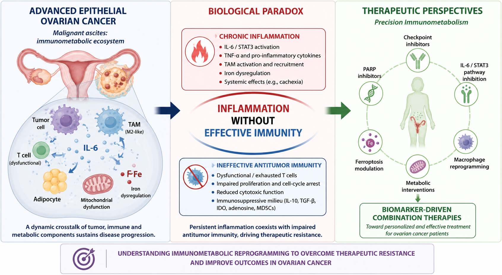
This discrepancy suggests that ovarian cancer may not simply be characterized by insufficient immune recognition. Instead, the ovarian tumor microenvironment may contain abundant immune populations that are unable to generate effective and durable antitumor responses. Recent advances have introduced several concepts that help explain this phenomenon, including immune exhaustion, chronic inflammatory activation, macrophage polarization, metabolic competition, ferro-metabolic signaling, and immunometabolic reprogramming [
3,
9]. These mechanisms are increasingly recognized as interconnected components of a complex biological network that regulates tumor progression and therapeutic resistance.
Several biological roots of this framework can be traced to translational studies performed during the 1990s and early 2000s on malignant ascites and neoplastic effusions from ovarian cancer patients [
10,
11]. Although the language of modern immuno-oncology was not yet available, those investigations identified abnormalities involving immune-cell activation, cytokine signaling, macrophage biology, iron metabolism, and systemic inflammatory responses [
11,
12,
13]. The central hypothesis of this review is that ovarian cancer may represent a condition of inflammation without effective immunity. In this model, chronic inflammatory activation coexists with impairment of immune competence, generating a tumor-promoting immunometabolic ecosystem that favors progression, cachexia, and resistance to therapy [
10].
Importantly, epithelial ovarian cancer should not be treated as a single immunological entity. High-grade serous, clear-cell, endometrioid, mucinous, and low-grade serous carcinomas arise from distinct molecular backgrounds and display different patterns of immune infiltration, checkpoint expression, stromal organization, and macrophage composition. Spatially resolved profiling across 254 ovarian cancers has confirmed subtype-specific immune architectures and immunoregulatory targets, including distinct patterns in high-grade serous, clear-cell, mucinous, endometrioid, and low-grade serous carcinomas. Accordingly, “inflammation without effective immunity” is proposed here as an overarching framework that may arise through distinct histotype- and context-dependent immunometabolic configurations rather than as a uniform phenotype shared by all ovarian cancers [
14,
15].
2. Narrative Scope and Interpretive Framework
This manuscript is presented as a narrative and interpretive review rather than as a systematic review or meta-analysis. The synthesis is based on the peer-reviewed literature cited in this manuscript, including historical translational studies on ovarian cancer ascites and tumor-associated lymphomonocytes, contemporary reviews and mechanistic studies on cancer immunometabolism, and selected clinical or translational literature addressing immunotherapy resistance, macrophage biology, iron metabolism, ferroptosis, cachexia, and PARP biology.
The purpose of the review is not to claim that early studies directly demonstrated modern molecular entities such as T-cell exhaustion, macrophage polarization, or ferroptosis in their current definitions. Rather, the objective is to re-examine functional and inflammatory observations from ovarian cancer effusions through contemporary biological frameworks. A strict distinction is therefore maintained throughout this review between (i) findings directly demonstrated in historical ovarian cancer samples; (ii) mechanisms subsequently demonstrated in ovarian cancer using contemporary molecular or spatial approaches; and (iii) mechanistic interpretations extrapolated from broader cancer or preclinical literature. Terms such as exhaustion-like dysfunction and ferro-metabolic signaling are used only at the interpretive level unless the cited study directly measured the defining molecular, transcriptional, epigenetic, or functional features.
3. A Historical Hypothesis Revisited
The development of modern immuno-oncology has profoundly changed our understanding of cancer biology. Concepts such as T-cell exhaustion, immune checkpoint regulation, metabolic reprogramming, and macrophage polarization now occupy a central role in explaining tumor progression and resistance to therapy. However, scientific paradigms rarely emerge suddenly. In many cases, biological observations precede the conceptual frameworks required to interpret them [
16,
17,
18,
19,
20,
21]. This phenomenon appears particularly evident in ovarian cancer.
Long before the introduction of immune checkpoint inhibitors and before the emergence of the term “immunometabolism”, translational studies performed on ovarian cancer ascites had identified a striking biological paradox. Immune cells recovered from neoplastic effusions were surrounded by a highly inflammatory milieu rich in cytokines and activation signals, yet they displayed functional impairment [
10,
11,
12]. At that time, these findings were interpreted primarily within the context of cytokine biology and tumor-induced immunosuppression. Today, however, they can be re-examined through a broader framework integrating inflammation, metabolism, immune dysfunction, and host adaptation. Viewed retrospectively, malignant ascites represented an in vivo biological compartment in which several mechanisms currently included within immunometabolic models were functionally suggested, although not molecularly defined [
11]. Rather than isolated observations, these findings may represent early elements of a biological continuum linking chronic inflammation, metabolic adaptation, macrophage reprogramming, immune dysfunction, and therapeutic resistance [
8,
9,
10].
4. Early Translational Evidence of Immune Dysfunction in Ovarian Cancer
One of the earliest indications that ovarian cancer might induce profound local immune dysregulation emerged from studies investigating tumor-associated lymphomonocytes isolated from malignant effusions (TALMs). These investigations demonstrated that TALMs exhibited markedly impaired proliferative responses when compared with autologous peripheral blood mononuclear cells [
13]. The impairment persisted despite exposure to an inflammatory microenvironment characterized by abundant cytokine production and apparent immune activation. Cell-cycle analyses revealed accumulation within the G0/G1 phase of the cell cycle, indicating quiescent or early gap-phase arrest, suggesting an inability to progress toward effective immune activation and proliferation. Functionally, these findings indicated immune paralysis rather than simple immune absence [
13].
Particularly relevant was the observation that this proliferative defect coexisted with elevated concentrations of multiple inflammatory mediators, including IL-6, TNF-α, IL-10, IFN-γ, and soluble IL-2 receptor [
10,
11]. Such coexistence of activation signals and functional impairment constitutes a central feature of dysfunctional immune activation. From a contemporary perspective, these findings resemble aspects of exhaustion-like immune phenotypes, because immune cells remain present within the tumor microenvironment and retain evidence of activation, yet fail to generate effective antitumor responses [
19,
20,
21]. This interpretation should remain cautious, because the original studies were not designed to test modern molecular definitions of T-cell exhaustion.
Additional investigations further strengthened this hypothesis. Elevated circulating levels of inflammatory cytokines and soluble IL-2 receptor correlated with impaired T-cell responsiveness and markers of systemic inflammation [
11]. These observations suggested that immune dysfunction was not restricted to the local tumor environment but reflected a broader host response involving both immune and metabolic compartments. The analysis of Fas receptor/CD95 and CD25/interleukin-2 receptor alpha-chain signaling pathways provided further insight into this paradox. Increased expression of activation markers coexisted with defective immune-cell function, suggesting that chronic stimulation may promote dysfunction rather than effective activation [
12]. Collectively, these studies established a conceptual foundation for understanding ovarian cancer as a disease characterized not by immune silence, but by biologically ineffective immunity [
10].
Table 1 summarizes the historical evidence of inflammatory and immunological processes that establish in advanced ovarian cancer and are associated with systemic disease.
5. The Ascitic Microenvironment as an Immunometabolic Niche
Malignant ascites represents one of the most distinctive biological features of advanced epithelial ovarian cancer. Traditionally considered a consequence of peritoneal dissemination and altered vascular permeability, ascites is now recognized as a highly active biological compartment capable of profoundly influencing tumor behavior, host responses, and therapeutic outcomes [
33,
34,
35]. Rather than functioning as a passive reservoir of tumor cells, malignant ascites constitutes a dynamic ecosystem composed of neoplastic cells, tumor-associated macrophages (TAMs), lymphocytes, mesothelial cells, adipocytes, fibroblasts, extracellular vesicles, cytokines, chemokines, growth factors, and metabolic mediators [
10,
33,
34,
35].
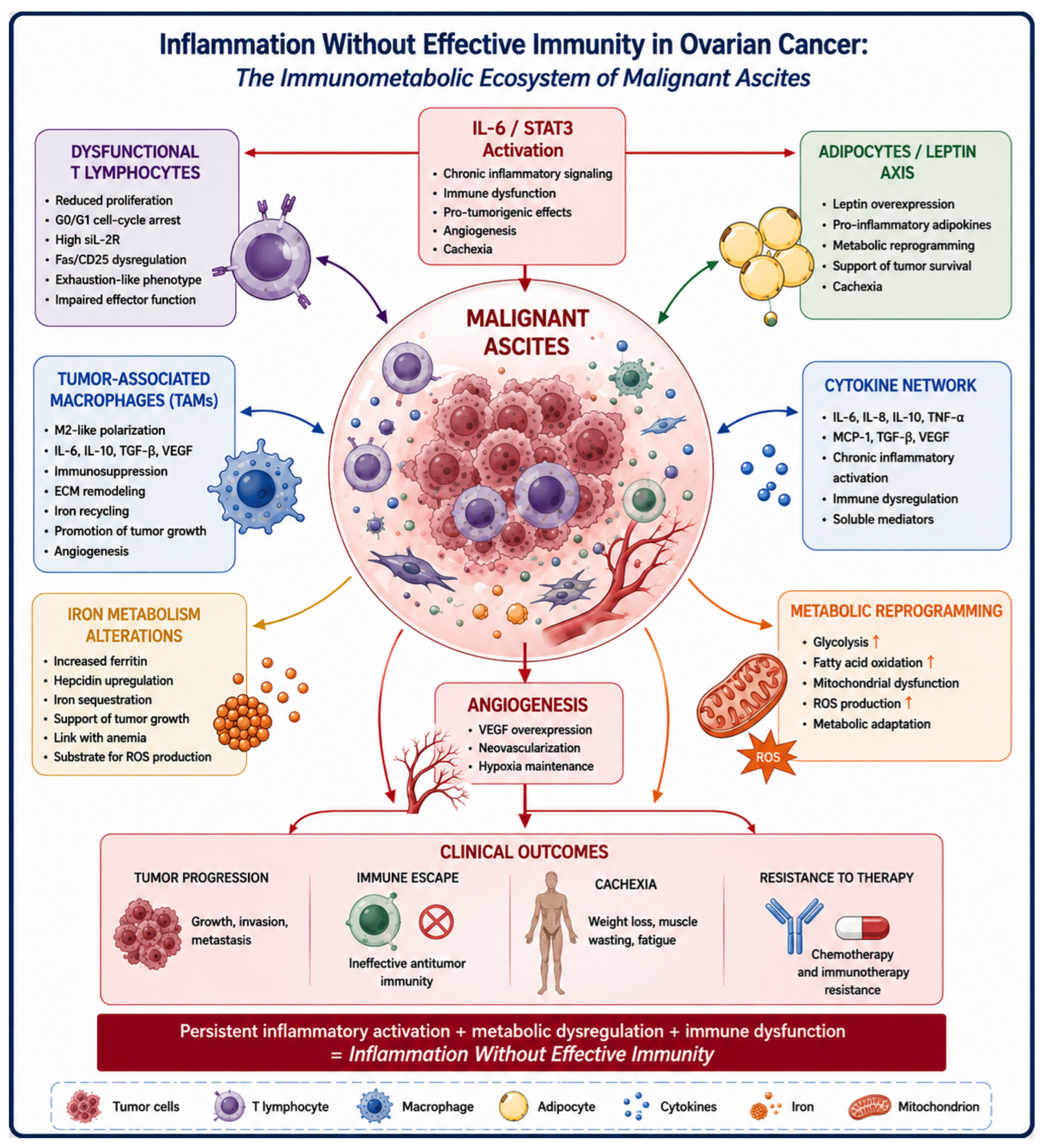
Continuous interactions among these components generate a complex network that simultaneously promotes tumor progression and suppresses effective antitumor immunity. The biological relevance of ascites became evident through early translational studies demonstrating elevated concentrations of inflammatory mediators, particularly IL-6, TNF-α, IL-10, and soluble IL-2 receptor [
10,
11]. These observations suggested that ovarian cancer develops within a state of persistent inflammatory activation extending beyond the tumor itself. Subsequent investigations progressively expanded this inflammatory model toward a broader immunometabolic framework. The ascitic compartment emerged as a site where immune regulation, metabolic adaptation, angiogenesis, and host systemic responses converge [
33,
34,
35] (
Figure 1).
Contemporary single-cell and spatial profiling have now provided direct support for this ecosystem view. These approaches demonstrate that malignant ascites and metastatic ovarian cancer contain heterogeneous tumor, T-cell, myeloid, fibroblast, and stromal populations linked by site-specific cell–cell communication networks. Recent spatial studies further show that immune organization differs not only among histotypes but also among anatomical compartments within high-grade serous ovarian cancer. These data strengthen the interpretation of ascites as an active tumor–host interface while also cautioning against extrapolating one immune configuration to all ovarian cancers [
14,
15,
37]. Independent ascites-focused single-cell studies identify heterogeneous macrophages and ligand–receptor interactions involving T/NK cells and malignant cells, while large spatial analyses of high-grade serous tubo-ovarian cancer demonstrate that tumor-cell states and local tissue architecture predict lymphocyte infiltration and immune evasion [
14,
15,
37,
38,
39].
5.1. IL-6 as a Central Regulatory Hub
Among the numerous cytokines identified within ovarian cancer ascites, IL-6 occupies a particularly relevant position. Accumulating evidence indicates that IL-6 contributes to multiple hallmarks of ovarian cancer progression, including tumor proliferation, angiogenesis, thrombopoiesis, cachexia, immune dysregulation, cancer-related anemia, and resistance to therapy [
40]. Through activation of the Janus kinase/signal transducer and activator of transcription 3 (JAK/STAT3) pathway, IL-6 orchestrates extensive transcriptional programs affecting both malignant and non-malignant cellular populations [
41].
The centrality of IL-6 is especially relevant because many of its biological effects simultaneously involve immune and metabolic regulation. Persistent STAT3 activation influences macrophage polarization, promotes tumor survival, facilitates angiogenesis, and contributes to immune dysfunction [
41,
42,
43]. Consequently, IL-6 may be viewed not simply as an inflammatory cytokine but as a regulatory mediator linking inflammation, metabolism, iron handling, anemia, and immune competence [
44].
5.2. Tumor-Associated Macrophages and Metabolic Reprogramming
Macrophages represent one of the most abundant and functionally diverse immune populations within ovarian cancer ascites. Earlier studies frequently described tumor-associated macrophages using an M1/M2 framework, which remains useful for interpreting selected functional observations but does not capture the full diversity revealed by contemporary single-cell and spatial analyses. Current data identify multiple macrophage states with distinct inflammatory, antigen-presenting, lipid-metabolic, iron-handling, angiogenic, and immunosuppressive programs. Their abundance and functional state vary with anatomical site, treatment context, molecular background, and ovarian cancer histotype. Thus, macrophage biology in ovarian cancer is better considered as a continuum of context-dependent states rather than a fixed binary polarization. Recent ovarian cancer studies further identify ascites-enriched lipid-loaded PLIN2-high macrophages and HRD-associated macrophage states with distinct spatial distributions, illustrating why a fixed M1/M2 classification is insufficient [
45,
46].
Glycolysis, oxidative phosphorylation, lipid metabolism, and iron handling collectively influence macrophage behavior and contribute to the formation of immunosuppressive microenvironments [
28,
29,
30,
31]. Several of these concepts were biologically foreshadowed by earlier investigations of ovarian cancer ascites. Alterations in macrophage-associated inflammatory pathways, cytokine production, and iron metabolism suggested that metabolic reprogramming was already an integral component of tumor–host interactions [
28,
29,
30,
31].
5.3. Iron Metabolism and Ferro-Metabolic Signaling
Iron metabolism has emerged as an important determinant of both tumor biology and immune regulation. Macrophages play a central role in iron recycling and storage, and disturbances in iron trafficking may influence oxidative stress, cytokine production, mitochondrial function, cellular proliferation, and cancer-related anemia [
22]. Studies investigating ovarian cancer-associated inflammation demonstrated that alterations in iron homeostasis are closely linked to systemic inflammation, nutritional status, anemia, and disease progression [
23].
Contemporary evidence supports a role for iron availability and ferroptosis susceptibility in ovarian cancer biology, but the level of evidence differs across compartments. Ovarian cancer-specific experimental studies demonstrate that tumor-cell ferroptosis sensitivity can be modified by iron availability and lipid/redox pathways. By contrast, direct demonstration that ferroptotic programs within human malignant ascites determine macrophage states or clinical immune escape remains limited. Accordingly, the proposed connection among ascitic iron handling, macrophage plasticity, and ferroptosis should be regarded as biologically plausible and hypothesis-generating rather than as an established mechanism in patients [
24,
25,
47,
48].
5.4. Adipocytes, Leptin, and Metabolic Crosstalk
The peritoneal cavity provides a unique biological context characterized by intimate interactions between ovarian cancer cells and adipose tissue. Adipocytes serve not only as energy reservoirs but also as active endocrine cells capable of producing adipokines, cytokines, and metabolic mediators [
33,
34,
35]. Leptin, in particular, has attracted attention because of its involvement in inflammation, angiogenesis, energy homeostasis, and immune regulation [
36]. The interaction between adipocytes, macrophages, and tumor cells contributes to a complex metabolic network that supports tumor growth and facilitates adaptation to environmental stress. Clinical evidence linking IL-6 and leptin to energy-metabolic alterations in advanced ovarian cancer further supports the relevance of this axis [
36]. Such crosstalk reinforces the concept that ovarian cancer progression is driven not only by genetic alterations but also by dynamic interactions within an integrated immunometabolic ecosystem.
6. From Inflammation to Immunometabolism and Exhaustion-like Immune Dysfunction
The evolution of ovarian cancer biology over the last three decades has progressively transformed our understanding of inflammation. Initially viewed primarily as a host response to malignancy, inflammation is now recognized as a central biological driver capable of shaping tumor progression, immune competence, and metabolic adaptation [
8]. In ovarian cancer, this transition from inflammation-centered models toward immunometabolic paradigms appears particularly evident. Cancer-associated cachexia illustrates this evolution. Historically considered a consequence of advanced disease, cachexia is now understood as a systemic syndrome driven by coordinated interactions among cytokines, immune cells, neuroendocrine pathways, and metabolic regulators [
49]. IL-6, TNF-α, leptin dysregulation, and macrophage activation collectively contribute to profound alterations in protein turnover, lipid metabolism, mitochondrial function, and energy expenditure [
50]. These same mediators simultaneously influence immune-cell function, thereby creating an integrated biological network linking inflammation and immune dysfunction [
50].
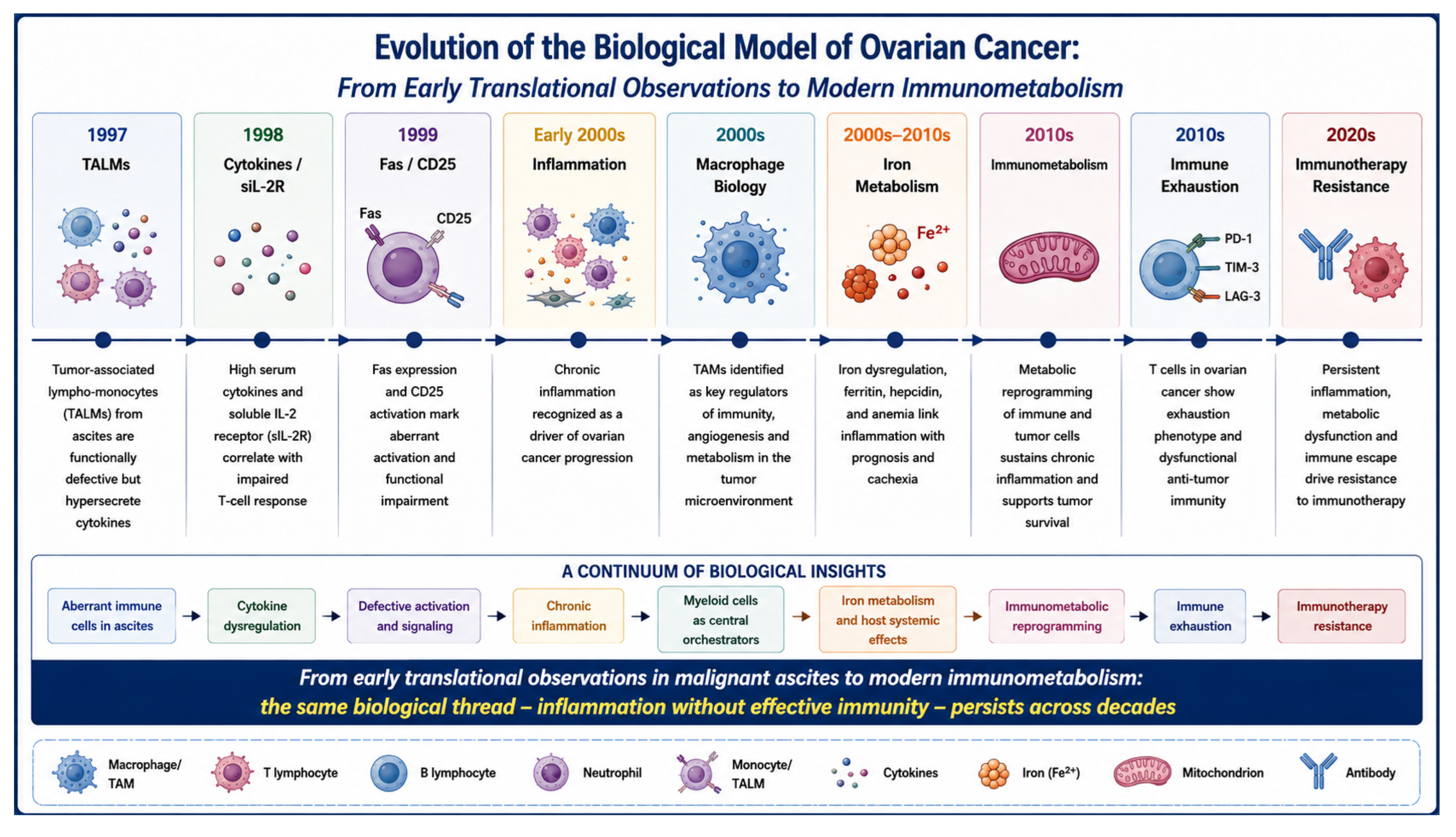
The historical TALM findings should be interpreted as evidence of dysfunctional immune activation, not as a retrospective molecular diagnosis of T-cell exhaustion. Reduced proliferative responses, G0/G1 accumulation, altered Fas/CD25 signaling, and cytokine release demonstrated that immune cells could remain activated yet functionally ineffective within malignant effusions. These observations overlap phenomenologically with selected features seen in chronically stimulated T cells, but they did not assess the transcriptional, epigenetic, clonotypic, inhibitory-receptor, or differentiation programs that now define exhausted T-cell states. Contemporary single-cell and spatial studies in ovarian cancer can therefore be used to connect these early functional observations to directly characterized T-cell states, while preserving the distinction between historical evidence and modern mechanistic interpretation [
19,
20,
21,
37]. Modern ovarian cancer studies now directly characterize spatially confined exhausted T-cell states and their myeloid context, particularly after chemotherapy, providing the contemporary molecular evidence that was unavailable in the original TALM studies [
51].
The interpretation proposed here does not imply that early investigators identified immune exhaustion in its modern molecular definition. Rather, it highlights how careful translational observations recognized fundamental biological patterns whose significance became clearer after later advances in immunology and metabolism [
52,
53]. In this sense, ovarian cancer ascites may be considered an early functional model of inflammation associated with ineffective immunity. This framework also intersects with previous concepts linking chronic inflammation, regulatory T-cell (Treg) biology, macrophage activation, oxidative stress, IL-6 production, altered iron metabolism, and metabolic dysfunction. Persistent inflammatory activation may progressively impair T-cell competence and promote immune suppression [
19,
20,
21]. Thus, chronic inflammation and immune dysfunction should not be considered opposing processes but interconnected manifestations of the same pathological ecosystem (
Figure 2).
7. Implications for Immunotherapy Resistance
Clinical experience with immune-checkpoint inhibition in ovarian cancer illustrates the distinction between immune-cell presence and therapeutically effective immunity. Early single-agent PD-1/PD-L1 studies produced reproducible but generally low response rates, and several large randomized trials incorporating checkpoint blockade into frontline or recurrent-disease regimens failed to improve outcomes in unselected populations [
54,
55,
56,
57,
58]. These findings argue against checkpoint signaling alone as a sufficient explanation for immune dysfunction. Importantly, the 2026 ENGOT-ov65/KEYNOTE-B96 trial demonstrated that pembrolizumab combined with weekly paclitaxel, with or without bevacizumab, improved clinical outcomes in platinum-resistant recurrent disease, with a clinically relevant effect in the PD-L1 CPS ≥ 1 population [
59]. Immune resistance in ovarian cancer should therefore not be regarded as absolute: efficacy appears dependent on biological context, therapeutic combination, and patient selection. Histotype composition must also be considered when interpreting aggregate trial results because different epithelial ovarian cancer subtypes have distinct immune microenvironments [
54,
55,
56,
57,
58,
59,
60,
61].
Table 2 shows the clinical trials that evaluated immunotherapy in the treatment of patients with ovarian cancer, both as monotherapy and in combination with chemotherapy.
At present, immune-checkpoint inhibition has the strongest direct clinical evidence among the therapeutic concepts discussed here, although benefit is context- and biomarker-dependent. Macrophage reprogramming, IL-6/JAK/STAT3 modulation, ferroptosis targeting, and manipulation of iron or lipid metabolism remain biologically compelling but are not established standards for ovarian cancer ascites. Their inclusion in this review is intended to define testable translational hypotheses and biomarker strategies rather than to recommend clinical use outside appropriate trials.
Persistent IL-6 signaling may represent one driver of this process. Chronic activation of the JAK/STAT3 pathway promotes tumor survival while simultaneously impairing effective antitumor immunity [
40,
41,
42,
43,
44]. Sustained inflammatory stimulation may further contribute to exhaustion-like phenotypes characterized by reduced proliferative capacity and diminished effector function. TAMs constitute another major source of resistance through cytokine production, angiogenic signaling, extracellular matrix remodeling, metabolic regulation, and modulation of nutrient availability, iron metabolism, and mitochondrial function [
28,
29,
30,
32].
Metabolic competition represents an additional layer of complexity. Tumor cells, activated macrophages, and dysfunctional lymphocytes coexist within an environment characterized by limited nutrient availability and altered metabolic fluxes [
50]. Glucose deprivation, amino acid depletion, lactate accumulation, oxidative stress, and mitochondrial dysfunction may contribute to progressive immune impairment [
32]. Hypoxia further amplifies these mechanisms by promoting angiogenesis, metabolic adaptation, and immune suppression. The resulting biological landscape favors tumor persistence despite the presence of immune-cell infiltration [
62,
63,
64]. Viewed through this integrated framework, ovarian cancer should not be regarded simply as immunologically cold. Rather, it may represent a biologically active example of inflammation-associated immune dysfunction. Strategies based solely on immune checkpoint inhibition may therefore be insufficient because they target only one component of a broader inflammatory–immunometabolic network [
65].
8. Future Therapeutic Perspectives: Beyond Tumor-Centered Therapy
The therapeutic evolution of ovarian cancer has been largely driven by tumor-centered strategies aimed at directly targeting malignant cells. Cytoreductive surgery, chemotherapy, anti-angiogenic agents, PARP inhibitors, antibody–drug conjugates, and platinum-based therapy have all contributed to improved outcomes [
1,
2,
66,
67]. Nevertheless, durable disease control remains elusive for many patients. The immunometabolic model proposed in this review suggests that future progress may depend on a broader therapeutic vision in which both tumor cells and their biological ecosystem become treatment targets [
66].
Table 3 shows a summary of the biological axes involved in generalized inflammation, proposed as potential targets for translational medicine, discussed below.
8.1. Targeting the IL-6/STAT3 Axis
Among inflammatory pathways, IL-6 occupies an attractive position because of its central role in connecting inflammation, immune dysfunction, thrombopoiesis, cachexia, angiogenesis, anemia, and tumor progression [
26,
36]. Persistent activation of STAT3 signaling has been implicated in multiple mechanisms of therapeutic resistance. Consequently, inhibition of the IL-6/STAT3 pathway may provide benefits extending beyond direct tumor effects by simultaneously modulating immune and metabolic compartments [
41,
42,
43]. Future studies should evaluate whether inflammatory biomarkers can identify subsets of patients most likely to benefit from cytokine-directed interventions.
8.2. Macrophage Reprogramming
TAMs have emerged as critical orchestrators of the ovarian cancer microenvironment [
23]. Their contribution extends across disease progression, immune suppression, angiogenesis, extracellular matrix remodeling, iron regulation, anemia, and metabolic adaptation [
28,
29,
30,
31]. Rather than eliminating macrophages, emerging strategies increasingly focus on functional reprogramming. Converting tumor-promoting macrophages into immune-supportive populations could enhance antitumor immunity while disrupting mechanisms of tumor progression [
29]. The ascitic microenvironment may represent an attractive setting for evaluating macrophage-directed interventions because of the abundance and accessibility of these cellular populations [
33,
34,
35].
8.3. Iron Metabolism, Ferroptosis, and Macrophage Plasticity
The growing recognition of iron metabolism as a regulator of tumor biology opens additional therapeutic opportunities. Iron availability influences oxidative stress, mitochondrial activity, macrophage function, anemia, and susceptibility to ferroptosis [
22,
23,
24,
25,
27]. Because ovarian cancer frequently develops within an inflammatory environment characterized by altered iron handling, modulation of iron-metabolic pathways may represent a rational therapeutic vulnerability [
68].
Ferroptosis should not be considered solely as a direct tumor-cell killing mechanism. Ferroptotic stress may also influence the tumor microenvironment through lipid peroxidation products, oxidized phospholipids, damage-associated molecular patterns, macrophage recruitment, and functional polarization [
47,
48,
69,
70]. In ovarian cancer ascites and metastatic niches, macrophages are exposed to oxidative stress, iron flux alterations, and lipid-rich microenvironments, making the interaction between ferroptosis and macrophage plasticity biologically plausible [
71]. However, the immunological consequences of ferroptosis are context dependent. Extensive ferroptosis may support immunogenic signaling, whereas incomplete or sublethal ferroptotic programs may favor adaptive responses that support tumor persistence. Effective ferroptosis-based strategies will therefore likely require simultaneous modulation of macrophage reprogramming and immune suppression pathways to convert oxidative stress into productive antitumor immunity rather than tumor adaptation [
71,
72,
73].
8.4. Metabolic Therapy
Cancer metabolism has traditionally focused on tumor-cell energetics. However, contemporary evidence increasingly demonstrates that metabolism regulates the behavior of virtually every component of the tumor microenvironment [
18,
63,
64,
65]. T lymphocytes, macrophages, fibroblasts, adipocytes, and endothelial cells all undergo metabolic adaptations that influence therapeutic response [
66]. Interventions targeting glucose utilization, mitochondrial function, lipid metabolism, amino acid pathways, oxidative stress, or inflammation may therefore exert effects extending beyond direct tumor inhibition [
50,
66]. Within this framework, metabolic therapy may also function as immune therapy and inflammatory therapy.
8.5. PARP Inhibition as a Metabolic and Immunomodulatory Intervention
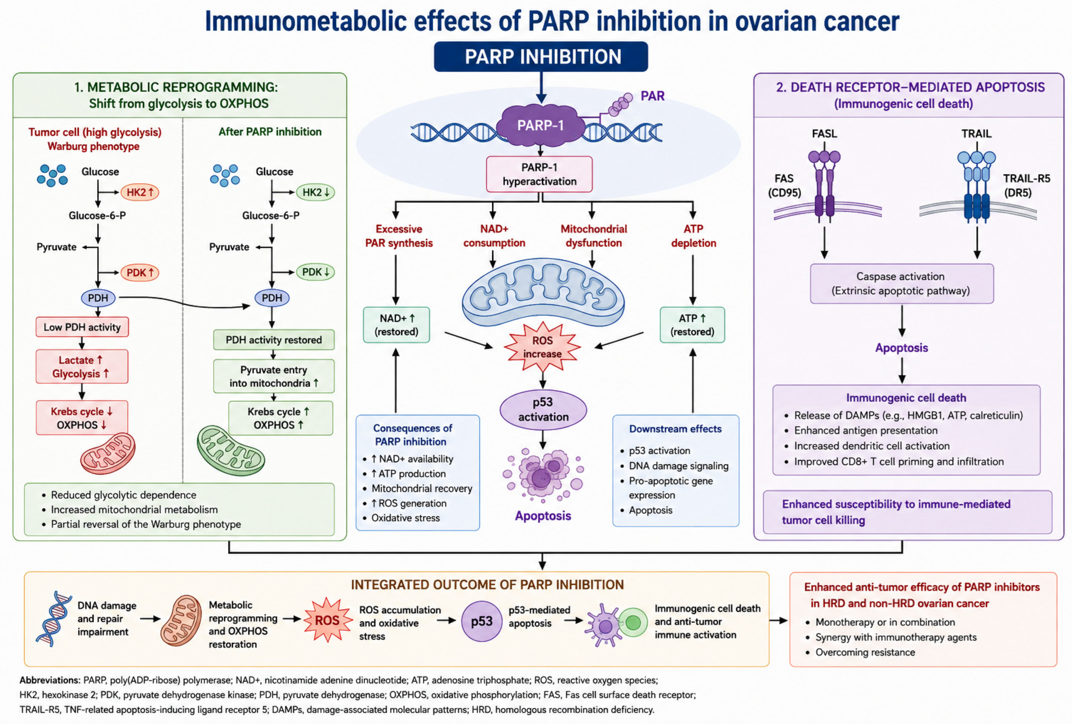
PARP inhibitors are established primarily through synthetic-lethality and DNA-repair mechanisms, particularly in homologous recombination-deficient tumors. Experimental literature also links PARP activity to NAD+ consumption, mitochondrial function, redox balance, innate immune signaling, and interactions with checkpoint blockade. However, these metabolic and immunomodulatory effects are not yet sufficiently demonstrated in human ovarian cancer ascites to constitute a central pillar of the present framework. PARP inhibition is therefore considered here as a potentially relevant modifier of the immunometabolic ecosystem rather than as evidence for the model itself; mechanistic claims should be supported by original experimental studies rather than commentary alone [
74].
Figure 3 shows the immunometabolic and immunomodulatory mechanisms induced by PARP inhibitors in ovarian cancer.
8.6. Combinatorial Immunotherapy
The modest activity of single-agent checkpoint inhibitors suggests that ovarian cancer may require combinatorial therapeutic strategies [
5]. Future regimens may integrate immune checkpoint inhibition, IL-6/STAT3 targeting, macrophage reprogramming, anti-angiogenic therapy, ferroptosis-based approaches, metabolic modulation, antibody–drug conjugates, PARP inhibitors, and platinum-based chemotherapy. The challenge will not simply be identifying effective combinations but selecting biologically appropriate combinations for specific immunometabolic phenotypes [
3,
65,
66].
8.7. Precision Immunometabolism
The next generation of ovarian cancer treatment may depend on the development of precision immunometabolism. Rather than classifying patients exclusively according to histology, stage, or genomic alterations, future models may incorporate inflammatory signatures, metabolic profiles, cytokine networks, macrophage phenotypes, ascitic biomarkers, iron-related parameters, and sarcopenia/cachexia indicators. Candidate features of inflammation without effective immunity may include high inflammatory cytokine activity, abundant TAMs, impaired lymphocyte responsiveness, altered iron homeostasis, anemia of inflammation, cachexia or sarcopenia, hypoxia, lactate accumulation, mitochondrial stress, and ascites enriched in suppressive immune populations. Such approaches could allow therapeutic strategies to be tailored according to the dominant biological drivers operating within each patient’s tumor ecosystem [
14,
15,
37,
65,
75].
9. Future Perspectives
The convergence of immunology, metabolism, inflammation, and systems biology is progressively transforming our understanding of ovarian cancer. Several priorities appear particularly relevant for future research. First, the identification of reproducible inflammatory phenotypes may improve biological stratification and facilitate personalized therapeutic approaches [
75,
76]. Second, comprehensive profiling of malignant ascites should be further developed as a source of biomarkers capable of integrating immune, metabolic, and inflammatory information [
33,
34,
35]. Third, advances in spatial biology and single-cell technologies are providing increasingly direct insight into cellular interactions occurring within the ovarian cancer microenvironment [
14,
15,
37]. Fourth, ferro-metabolic pathways, macrophage heterogeneity, and cytokine-network architecture require further investigation to clarify their contribution to disease progression and therapeutic resistance [
69,
71]. Finally, integrative computational approaches may facilitate the interpretation of increasingly complex biological datasets, allowing dynamic modeling of tumor–host interactions and identification of previously unrecognized therapeutic vulnerabilities. Collectively, these developments support a transition from reductionist models toward ecosystem-based interpretations of ovarian cancer biology.
10. Limitations of the Proposed Framework
Several limitations should be acknowledged. First, the historical studies discussed in this review were performed before the availability of contemporary single-cell, spatial, transcriptomic, and epigenetic technologies; therefore, their findings cannot be directly equated with modern molecular definitions of immune exhaustion, macrophage polarization, or ferroptosis-related immune regulation. Second, the concept of inflammation without effective immunity remains an integrative interpretive framework rather than a validated biomarker-defined clinical state. Third, several mechanistic links among IL-6 signaling, iron metabolism, ferroptosis, macrophage plasticity, adipocyte-derived signaling, PARP-related metabolic effects, and immunotherapy resistance remain incompletely established in ovarian cancer. Fourth, the therapeutic implications discussed here require prospective validation in biomarker-selected clinical studies. These limitations do not weaken the biological relevance of the proposed framework, but they indicate that it should be interpreted as a translational hypothesis requiring further mechanistic and clinical validation.
11. Conclusions
The history of ovarian cancer research provides an example of biological observations preceding the conceptual frameworks later used to interpret them. Studies performed on malignant effusions and tumor-associated lymphomonocytes in the 1990s demonstrated impaired proliferative responses, altered activation signaling, and substantial cytokine release despite the presence of immune cells. These findings constitute direct historical evidence of dysfunctional immune activation; they should not be equated with modern molecular definitions of T-cell exhaustion, macrophage states, or ferroptosis [
11,
12,
13].
Author Contributions
Conceptualization, A.M.; methodology, A.M.; literature review, A.M., M.N., V.V., G.S. and P.A.F.; writing—original draft preparation, A.M.; writing—review and editing, A.M., M.N., V.V., G.S. and P.A.F.; supervision, A.M. All authors have read and agreed to the published version of the manuscript.
Funding
This research received no external funding.
Institutional Review Board Statement
Not applicable.
Informed Consent Statement
Not applicable.
Data Availability Statement
No new data were created or analyzed in this study. Data sharing is not applicable to this article.
Acknowledgments
An AI-assisted tool (ChatGPT—OpenAI, GPT-5.5 Thinking version) was used exclusively for English-language grammar, syntax, and language polishing during manuscript preparation. No generative artificial intelligence tool was used to generate scientific content, perform literature searches, select references, analyze data, draw conclusions, or create figures or tables. The authors reviewed and edited all AI-assisted language suggestions and take full responsibility for the final content of this publication.
Conflicts of Interest
The authors declare no conflicts of interest.
References
- Matulonis, U.A.; Sood, A.K.; Fallowfield, L.; Howitt, B.E.; Sehouli, J.; Karlan, B.Y. Ovarian cancer. Nat. Rev. Dis. Primers 2016, 2, 16061. [Google Scholar] [PubMed]
- Lheureux, S.; Gourley, C.; Vergote, I.; Oza, A.M. Epithelial ovarian cancer. Lancet 2019, 393, 1240–1253. [Google Scholar] [CrossRef] [Scilit] [PubMed]
- Kandalaft, L.E.; Odunsi, K.; Coukos, G. Immunotherapy in Ovarian Cancer: Are We There Yet? J. Clin. Oncol. 2019, 37, 2460–2471. [Google Scholar] [CrossRef] [Scilit] [PubMed]
- Odunsi, K. Immunotherapy in ovarian cancer. Ann. Oncol. 2017, 28, viii1–viii7. [Google Scholar] [CrossRef] [Scilit] [PubMed]
- Peng, Z.; Li, M.; Li, H.; Gao, Q. PD-1/PD-L1 immune checkpoint blockade in ovarian cancer: Dilemmas and opportunities. Drug Discov. Today 2023, 28, 103666. [Google Scholar] [CrossRef] [Scilit] [PubMed]
- Hanahan, D.; Weinberg, R.A. Hallmarks of cancer: The next generation. Cell 2011, 144, 646–674. [Google Scholar] [CrossRef] [Scilit] [PubMed]
- Hanahan, D. Hallmarks of Cancer: New Dimensions. Cancer Discov. 2022, 12, 31–46. [Google Scholar] [CrossRef] [Scilit] [PubMed]
- Grivennikov, S.I.; Greten, F.R.; Karin, M. Immunity, inflammation, and cancer. Cell 2010, 140, 883–899. [Google Scholar] [CrossRef] [Scilit] [PubMed]
- Mantovani, A.; Allavena, P.; Sica, A.; Balkwill, F. Cancer-related inflammation. Nature 2008, 454, 436–444. [Google Scholar] [CrossRef] [Scilit] [PubMed]
- Macciò, A.; Madeddu, C. Inflammation and ovarian cancer. Cytokine 2012, 58, 133–147. [Google Scholar] [CrossRef] [Scilit] [PubMed]
- Macciò, A.; Lai, P.; Santona, M.C.; Pagliara, L.; Melis, G.B.; Mantovani, G. High serum levels of soluble IL-2 receptor, cytokines, and C reactive protein correlate with impairment of T cell response in patients with advanced epithelial ovarian cancer. Gynecol. Oncol. 1998, 69, 248–252. [Google Scholar] [CrossRef] [Scilit] [PubMed]
- Mantovani, G.; Macciò, A.; Massa, E.; Lai, P.; Manca, G.; Mudu, C.; Versace, R.; Pusceddu, G. Relationships between Fas expression, activation molecule CD25, and functional activity of tumor-associated lymphomonocytes from neoplastic effusions. Oncol. Rep. 1999, 6, 235–244. [Google Scholar] [CrossRef] [Scilit] [PubMed]
- Mantovani, G.; Macciò, A.; Pisano, M.; Versace, R.; Lai, P.; Esu, S.; Massa, E.; Ghiani, M.; Dessì, D.; Melis, G.B.; et al. Tumor-associated lympho-monocytes from neoplastic effusions are immunologically defective in comparison with patient autologous PBMCs but are capable of releasing high amounts of various cytokines. Int. J. Cancer 1997, 71, 724–731. [Google Scholar] [CrossRef] [Scilit]
- Mateiou, C.; Lokhande, L.; Diep, L.H.; Knulst, M.; Carlsson, E.; Ek, S.; Sundfeldt, K.; Gerdtsson, A. Spatial tumor immune microenvironment phenotypes in ovarian cancer. npj Precis. Oncol. 2024, 8, 148. [Google Scholar] [CrossRef] [Scilit] [PubMed]
- Frank, A.M.; Carlsson, E.; Ouyang, H.; Olsson, L.; Mateoiu, C.C.; Ek, S.; Sundfeldt, K.; Gerdtsson, A. Distinct immune microenvironments in ovarian cancer subtypes indicate potential for immunotherapies. J. Transl. Med. 2026, 24, 337. [Google Scholar] [CrossRef] [Scilit] [PubMed]
- Biswas, S.K. Metabolic Reprogramming of Immune Cells in Cancer Progression. Immunity 2015, 43, 435–449. [Google Scholar] [CrossRef] [Scilit] [PubMed]
- Steinert, E.M.; Vasan, K.; Chandel, N.S. Mitochondrial Metabolism Regulation of T Cell-Mediated Immunity. Annu. Rev. Immunol. 2021, 39, 395–416. [Google Scholar] [CrossRef] [Scilit] [PubMed]
- Pavlova, N.N.; Thompson, C.B. The Emerging Hallmarks of Cancer Metabolism. Cell Metab. 2016, 23, 27–47. [Google Scholar] [CrossRef] [Scilit] [PubMed]
- Wherry, E.J.; Kurachi, M. Molecular and cellular insights into T cell exhaustion. Nat. Rev. Immunol. 2015, 15, 486–499. [Google Scholar] [CrossRef] [Scilit] [PubMed]
- Blank, C.U.; Haining, W.N.; Held, W.; Hogan, P.G.; Kallies, A.; Lugli, E.; Lynn, R.C.; Philip, M.; Rao, A.; Restifo, N.P.; et al. Defining ‘T cell exhaustion’. Nat. Rev. Immunol. 2019, 19, 665–674. [Google Scholar] [CrossRef] [Scilit] [PubMed]
- McLane, L.M.; Abdel-Hakeem, M.S.; Wherry, E.J. CD8 T Cell Exhaustion During Chronic Viral Infection and Cancer. Annu. Rev. Immunol. 2019, 37, 457–495. [Google Scholar] [CrossRef] [Scilit] [PubMed]
- Macciò, A.; Madeddu, C.; Gramignano, G.; Mulas, C.; Tanca, L.; Cherchi, M.C.; Floris, C.; Omoto, I.; Barracca, A.; Ganz, T. The role of inflammation, iron, and nutritional status in cancer-related anemia: Results of a large, prospective, observational study. Haematologica 2015, 100, 124–132. [Google Scholar] [CrossRef] [Scilit] [PubMed]
- Madeddu, C.; Gramignano, G.; Kotsonis, P.; Coghe, F.; Atzeni, V.; Scartozzi, M.; Macciò, A. Microenvironmental M1 tumor-associated macrophage polarization influences cancer-related anemia in advanced ovarian cancer: Key role of interleukin-6. Haematologica 2018, 103, e388–e391. [Google Scholar] [CrossRef] [Scilit] [PubMed]
- Stockwell, B.R. Ferroptosis turns 10: Emerging mechanisms, physiological functions, and therapeutic applications. Cell 2022, 185, 2401–2421. [Google Scholar] [CrossRef] [Scilit] [PubMed]
- Jiang, X.; Stockwell, B.R.; Conrad, M. Ferroptosis: Mechanisms, biology and role in disease. Nat. Rev. Mol. Cell Biol. 2021, 22, 266–282. [Google Scholar] [CrossRef] [Scilit] [PubMed]
- Macciò, A.; Madeddu, C.; Massa, D.; Mudu, M.C.; Lusso, M.R.; Gramignano, G.; Serpe, R.; Melis, G.B.; Mantovani, G. Hemoglobin levels correlate with interleukin-6 levels in patients with advanced untreated epithelial ovarian cancer: Role of inflammation in cancer-related anemia. Blood 2005, 106, 362–367. [Google Scholar] [CrossRef] [Scilit] [PubMed]
- Torti, S.V.; Torti, F.M. Iron and cancer: More ore to be mined. Nat. Rev. Cancer 2013, 13, 342–355. [Google Scholar] [CrossRef] [Scilit] [PubMed]
- Vitale, I.; Manic, G.; Coussens, L.M.; Kroemer, G.; Galluzzi, L. Macrophages and Metabolism in the Tumor Microenvironment. Cell Metab. 2019, 30, 36–50. [Google Scholar] [CrossRef] [Scilit] [PubMed]
- Mantovani, A.; Marchesi, F.; Malesci, A.; Laghi, L.; Allavena, P. Tumour-associated macrophages as treatment targets in oncology. Nat. Rev. Clin. Oncol. 2017, 14, 399–416. [Google Scholar] [CrossRef] [Scilit] [PubMed]
- DeNardo, D.G.; Ruffell, B. Macrophages as regulators of tumour immunity and immunotherapy. Nat. Rev. Immunol. 2019, 19, 369–382. [Google Scholar] [CrossRef] [Scilit] [PubMed]
- Cassetta, L.; Pollard, J.W. Tumor-associated macrophages. Curr. Biol. 2020, 30, R246–R248. [Google Scholar] [CrossRef] [Scilit] [PubMed]
- Macciò, A.; Gramignano, G.; Cherchi, M.C.; Tanca, L.; Melis, L.; Madeddu, C. Role of M1-polarized tumor-associated macrophages in the prognosis of advanced ovarian cancer patients. Sci. Rep. 2020, 10, 6096. [Google Scholar] [CrossRef] [Scilit] [PubMed]
- Kipps, E.; Tan, D.S.; Kaye, S.B. Meeting the challenge of ascites in ovarian cancer: New avenues for therapy and research. Nat. Rev. Cancer 2013, 13, 273–282. [Google Scholar] [CrossRef] [Scilit] [PubMed]
- Ahmed, N.; Stenvers, K.L. Getting to know ovarian cancer ascites: Opportunities for targeted therapy-based translational research. Front. Oncol. 2013, 3, 256. [Google Scholar] [CrossRef] [Scilit] [PubMed]
- Ford, C.E.; Werner, B.; Hacker, N.F.; Warton, K. The untapped potential of ascites in ovarian cancer research and treatment. Br. J. Cancer 2020, 123, 9–16. [Google Scholar] [CrossRef] [Scilit] [PubMed]
- Macciò, A.; Madeddu, C.; Massa, D.; Astara, G.; Farci, D.; Melis, G.B.; Mantovani, G. Interleukin-6 and leptin as markers of energy metabolic changes in advanced ovarian cancer patients. J. Cell. Mol. Med. 2009, 13, 3951–3959. [Google Scholar] [CrossRef] [Scilit] [PubMed]
- Zheng, X.; Wang, X.; Cheng, X.; Liu, Z.; Yin, Y.; Li, X.; Huang, Z.; Wang, Z.; Guo, W.; Ginhoux, F.; et al. Single-cell analyses implicate ascites in remodeling the ecosystems of primary and metastatic tumors in ovarian cancer. Nat. Cancer 2023, 4, 1138–1156. [Google Scholar] [CrossRef] [Scilit] [PubMed]
- Kim, S.; Han, Y.; Kim, S.I.; Lee, J.; Jo, H.; Wang, W.; Cho, U.; Park, W.; Rando, T.A.; Dhanasekaran, D.N.; et al. Computational modeling of malignant ascites reveals CCL5-SDC4 interaction in the immune microenvironment of ovarian cancer. Mol. Carcinog. 2021, 60, 297–312. [Google Scholar] [CrossRef] [Scilit] [PubMed]
- Yeh, C.Y.; Aguirre, K.; Laveroni, O.; Kim, S.; Wang, A.; Liang, B.; Zhang, X.; Han, L.M.; Valbuena, R.; Bassik, M.C.; et al. Mapping spatial organization and genetic cell-state regulators to target immune evasion in ovarian cancer. Nat. Immunol. 2024, 25, 1943–1958. [Google Scholar] [CrossRef] [Scilit] [PubMed]
- Macciò, A.; Madeddu, C. The role of interleukin-6 in the evolution of ovarian cancer: Clinical and prognostic implications—A review. J. Mol. Med. 2013, 91, 1355–1368. [Google Scholar] [CrossRef] [Scilit] [PubMed]
- Johnson, D.E.; O’Keefe, R.A.; Grandis, J.R. Targeting the IL-6/JAK/STAT3 signalling axis in cancer. Nat. Rev. Clin. Oncol. 2018, 15, 234–248. [Google Scholar] [CrossRef] [Scilit] [PubMed]
- Yu, H.; Pardoll, D.; Jove, R. STATs in cancer inflammation and immunity: A leading role for STAT3. Nat. Rev. Cancer 2009, 9, 798–809. [Google Scholar] [CrossRef] [Scilit] [PubMed]
- Sansone, P.; Bromberg, J. Targeting the interleukin-6/Jak/stat pathway in human malignancies. J. Clin. Oncol. 2012, 30, 1005–1014. [Google Scholar] [CrossRef] [Scilit] [PubMed]
- Taniguchi, K.; Karin, M. IL-6 and related cytokines as the critical lynchpins between inflammation and cancer. Semin. Immunol. 2014, 26, 54–74. [Google Scholar] [CrossRef] [Scilit] [PubMed]
- Lan, H.; Xu, F.; Li, L.; Wei, X.; Li, M. HRD-Driven Reprogramming of Macrophage Function and Spatial Architecture in High-Grade Serous Ovarian Cancer. Genet. Res. 2026, 2026, 7364793. [Google Scholar] [CrossRef] [Scilit] [PubMed]
- Luo, H.; She, X.; Zhang, Y.; Xie, B.; Zhang, S.; Li, Q.; Zhou, Y.; Guo, S.; Zhang, S.; Jiang, Y.; et al. PLIN2 Promotes Lipid Accumulation in Ascites-Associated Macrophages and Ovarian Cancer Progression by HIF1α/SPP1 Signaling. Adv. Sci. 2025, 12, e2411314. [Google Scholar] [CrossRef] [Scilit] [PubMed]
- Zhai, X.; Lin, Y.; Zhu, L.; Wang, Y.; Zhang, J.; Liu, J.; Li, L.; Lu, X. Ferroptosis in cancer immunity and immunotherapy: Multifaceted interplay and clinical implications. Cytokine Growth Factor Rev. 2024, 75, 101–109. [Google Scholar] [CrossRef] [Scilit] [PubMed]
- Dixon, S.J.; Lemberg, K.M.; Lamprecht, M.R.; Skouta, R.; Zaitsev, E.M.; Gleason, C.E.; Patel, D.N.; Bauer, A.J.; Cantley, A.M.; Yang, W.S.; et al. Ferroptosis: An iron-dependent form of nonapoptotic cell death. Cell 2012, 149, 1060–1072. [Google Scholar] [CrossRef] [Scilit] [PubMed]
- Argilés, J.M.; Busquets, S.; Stemmler, B.; López-Soriano, F.J. Cancer cachexia: Understanding the molecular basis. Nat. Rev. Cancer 2014, 14, 754–762. [Google Scholar] [CrossRef] [Scilit] [PubMed]
- Macciò, A.; Madeddu, C.; Lai, E.; Scartozzi, M. Cancer cachexia and chronic inflammation: An unbreakable bond. Br. J. Cancer 2023, 128, 1609–1610. [Google Scholar] [CrossRef] [Scilit] [PubMed]
- Launonen, I.M.; Niemiec, I.; Hincapié-Otero, M.; Erkan, E.P.; Junquera, A.; Afenteva, D.; Falco, M.M.; Liang, Z.; Salko, M.; Chamchougia, F.; et al. Chemotherapy induces myeloid-driven spatially confined T cell exhaustion in ovarian cancer. Cancer Cell 2024, 42, 2045–2063.e10. [Google Scholar] [CrossRef] [Scilit] [PubMed]
- O’Neill, L.A.; Kishton, R.J.; Rathmell, J. A guide to immunometabolism for immunologists. Nat. Rev. Immunol. 2016, 16, 553–565. [Google Scholar] [CrossRef] [Scilit] [PubMed]
- Buck, M.D.; Sowell, R.T.; Kaech, S.M.; Pearce, E.L. Metabolic Instruction of Immunity. Cell 2017, 169, 570–586. [Google Scholar] [CrossRef] [Scilit] [PubMed]
- Hamanishi, J.; Mandai, M.; Ikeda, T.; Minami, M.; Kawaguchi, A.; Murayama, T.; Kanai, M.; Mori, Y.; Matsumoto, S.; Chikuma, S.; et al. Safety and Antitumor Activity of Anti-PD-1 Antibody, Nivolumab, in Patients with Platinum-Resistant Ovarian Cancer. J. Clin. Oncol. 2015, 33, 4015–4022. [Google Scholar] [CrossRef] [Scilit] [PubMed]
- Matulonis, U.A.; Shapira-Frommer, R.; Santin, A.D.; Lisyanskaya, A.S.; Pignata, S.; Vergote, I.; Raspagliesi, F.; Sonke, G.S.; Birrer, M.; Provencher, D.M.; et al. Antitumor activity and safety of pembrolizumab in patients with advanced recurrent ovarian cancer: Results from the phase II KEYNOTE-100 study. Ann. Oncol. 2019, 30, 1080–1087. [Google Scholar] [CrossRef] [Scilit] [PubMed]
- Zamarin, D.; Burger, R.A.; Sill, M.W.; Powell, D.J., Jr.; Lankes, H.A.; Feldman, M.D.; Zivanovic, O.; Gunderson, C.; Ko, E.; Mathews, C.; et al. Randomized Phase II Trial of Nivolumab Versus Nivolumab and Ipilimumab for Recurrent or Persistent Ovarian Cancer: An NRG Oncology Study. J. Clin. Oncol. 2020, 38, 1814–1823. [Google Scholar] [CrossRef] [Scilit] [PubMed]
- Pujade-Lauraine, E.; Fujiwara, K.; Ledermann, J.A.; Oza, A.M.; Kristeleit, R.; Ray-Coquard, I.L.; Richardson, G.E.; Sessa, C.; Yonemori, K.; Banerjee, S.; et al. Avelumab alone or in combination with chemotherapy versus chemotherapy alone in platinum-resistant or platinum-refractory ovarian cancer (JAVELIN Ovarian 200): An open-label, three-arm, randomised, phase 3 study. Lancet Oncol. 2021, 22, 1034–1046. [Google Scholar] [CrossRef] [Scilit] [PubMed]
- Hamanishi, J.; Takeshima, N.; Katsumata, N.; Ushijima, K.; Kimura, T.; Takeuchi, S.; Matsumoto, K.; Ito, K.; Mandai, M.; Nakai, H.; et al. Nivolumab Versus Gemcitabine or Pegylated Liposomal Doxorubicin for Patients with Platinum-Resistant Ovarian Cancer: Open-Label, Randomized Trial in Japan (NINJA). J. Clin. Oncol. 2021, 39, 3671–3681. [Google Scholar] [CrossRef] [Scilit] [PubMed]
- Colombo, N.; Zsiros, E.; Parma, G.; Rulli, E.; Sebastianelli, A.; Bidzinski, M.; Gallardo, C.; Matanes, E.; Hasegawa, K.; Kose, F.; et al. Pembrolizumab plus weekly paclitaxel in platinum-resistant recurrent ovarian cancer (ENGOT-ov65/KEYNOTE-B96): A multicentre, randomised, double-blind, phase 3 study. Lancet 2026, 407, 1525–1537. [Google Scholar] [CrossRef] [Scilit] [PubMed]
- Moore, K.N.; Bookman, M.; Sehouli, J.; Miller, A.; Anderson, C.; Scambia, G.; Myers, T.; Taskiran, C.; Robison, K.; Mäenpää, J.; et al. Atezolizumab, Bevacizumab, and Chemotherapy for Newly Diagnosed Stage III or IV Ovarian Cancer: Placebo-Controlled Randomized Phase III Trial (IMagyn050/GOG 3015/ENGOT-OV39). J. Clin. Oncol. 2021, 39, 1842–1855. [Google Scholar] [CrossRef] [Scilit] [PubMed]
- Kurtz, J.E.; Pujade-Lauraine, E.; Oaknin, A.; Belin, L.; Leitner, K.; Cibula, D.; Denys, H.; Rosengarten, O.; Rodrigues, M.; de Gregorio, N.; et al. Atezolizumab Combined with Bevacizumab and Platinum-Based Therapy for Platinum-Sensitive Ovarian Cancer: Placebo-Controlled Randomized Phase III ATALANTE/ENGOT-ov29 Trial. J. Clin. Oncol. 2023, 41, 4768–4778. [Google Scholar] [CrossRef] [Scilit] [PubMed]
- Reina-Campos, M.; Moscat, J.; Diaz-Meco, M. Metabolism shapes the tumor microenvironment. Curr. Opin. Cell Biol. 2017, 48, 47–53. [Google Scholar] [CrossRef] [Scilit] [PubMed]
- Hiam-Galvez, K.J.; Allen, B.M.; Spitzer, M.H. Systemic immunity in cancer. Nat. Rev. Cancer 2021, 21, 345–359. [Google Scholar] [CrossRef] [Scilit] [PubMed]
- Schreiber, R.D.; Old, L.J.; Smyth, M.J. Cancer immunoediting: Integrating immunity’s roles in cancer suppression and promotion. Science 2011, 331, 1565–1570. [Google Scholar] [CrossRef] [Scilit] [PubMed]
- Bader, J.E.; Voss, K.; Rathmell, J.C. Targeting Metabolism to Improve the Tumor Microenvironment for Cancer Immunotherapy. Mol. Cell 2020, 78, 1019–1033. [Google Scholar] [CrossRef] [Scilit] [PubMed]
- Macciò, A.; Madeddu, C. Blocking inflammation to improve immunotherapy of advanced cancer. Immunology 2020, 159, 357–364. [Google Scholar] [CrossRef] [Scilit] [PubMed]
- Macciò, A.; Madeddu, C. Cisplatin: An old drug with a newfound efficacy—From mechanisms of action to cytotoxicity. Expert Opin. Pharmacother. 2013, 14, 1839–1857. [Google Scholar] [CrossRef] [Scilit] [PubMed]
- Macciò, A.; Madeddu, C.; Gramignano, G.; Mulas, C.; Sanna, E.; Mantovani, G. Efficacy and safety of oral lactoferrin supplementation in combination with rHuEPO-β for the treatment of anemia in advanced cancer patients undergoing chemotherapy: Open-label, randomized controlled study. Oncologist 2010, 15, 894–902. [Google Scholar] [CrossRef] [Scilit] [PubMed]
- Battaglia, A.M.; Sacco, A.; Perrotta, I.D.; Faniello, M.C.; Scalise, M.; Torella, D.; Levi, S.; Costanzo, F.; Biamonte, F. Iron Administration Overcomes Resistance to Erastin-Mediated Ferroptosis in Ovarian Cancer Cells. Front. Oncol. 2022, 12, 868351. [Google Scholar] [CrossRef] [Scilit] [PubMed]
- Cui, K.; Wang, K.; Huang, Z. Ferroptosis and the tumor microenvironment. J. Exp. Clin. Cancer Res. 2024, 43, 315. [Google Scholar] [CrossRef] [Scilit] [PubMed]
- Zhou, J.; Zeng, T.; Liu, Y.; Ye, M.; Li, M.; Zhang, Z.; Meng, Y. FCGR2B+ Macrophages as a Critical Node Linking Ferroptosis and Immunosuppression: A Multiomics Framework for Prognosis and Therapy in High-Grade Serous Ovarian Cancer. Hum. Mutat. 2026, 2026, 8027584. [Google Scholar] [CrossRef] [Scilit] [PubMed]
- Wang, W.; Green, M.; Choi, J.E.; Gijón, M.; Kennedy, P.D.; Johnson, J.K.; Liao, P.; Lang, X.; Kryczek, I.; Sell, A.; et al. CD8+ T cells regulate tumour ferroptosis during cancer immunotherapy. Nature 2019, 569, 270–274. [Google Scholar] [CrossRef] [Scilit] [PubMed]
- Bersuker, K.; Hendricks, J.M.; Li, Z.; Magtanong, L.; Ford, B.; Tang, P.H.; Roberts, M.A.; Tong, B.; Maimone, T.J.; Zoncu, R.; et al. The CoQ oxidoreductase FSP1 acts parallel to GPX4 to inhibit ferroptosis. Nature 2019, 575, 688–692. [Google Scholar] [CrossRef] [Scilit] [PubMed]
- Ding, L.; Kim, H.J.; Wang, Q.; Kearns, M.; Jiang, T.; Ohlson, C.E.; Li, B.B.; Xie, S.; Liu, J.F.; Stover, E.H.; et al. PARP Inhibition Elicits STING-Dependent Antitumor Immunity in Brca1-Deficient Ovarian Cancer. Cell Rep. 2018, 25, 2972–2980.e5. [Google Scholar] [CrossRef] [Scilit] [PubMed]
- Binnewies, M.; Roberts, E.W.; Kersten, K.; Chan, V.; Fearon, D.F.; Merad, M.; Coussens, L.M.; Gabrilovich, D.I.; Ostrand-Rosenberg, S.; Hedrick, C.C.; et al. Understanding the tumor immune microenvironment (TIME) for effective therapy. Nat. Med. 2018, 24, 541–550. [Google Scholar] [CrossRef] [Scilit] [PubMed]
- Miyata, K.; Yotsumoto, F. Metabolism-driven Immune Escape Defines Therapeutic Vulnerability in Type I and Type II Ovarian Cancer. Anticancer Res. 2026, 46, 2851–2861. [Google Scholar] [CrossRef] [Scilit] [PubMed]
| Disclaimer/Publisher’s Note: The statements, opinions and data contained in all publications are solely those of the individual author(s) and contributor(s) and not of MDPI and/or the editor(s). MDPI and/or the editor(s) disclaim responsibility for any injury to people or property resulting from any ideas, methods, instructions or products referred to in the content. |




 ,
,