

Gastric Cancer Biomarkers: From Cellular Identity to Precision Oncology



 ,
,
Simple Summary
Abstract
1. Introduction
2. Materials and Methods
2.1. Study Design
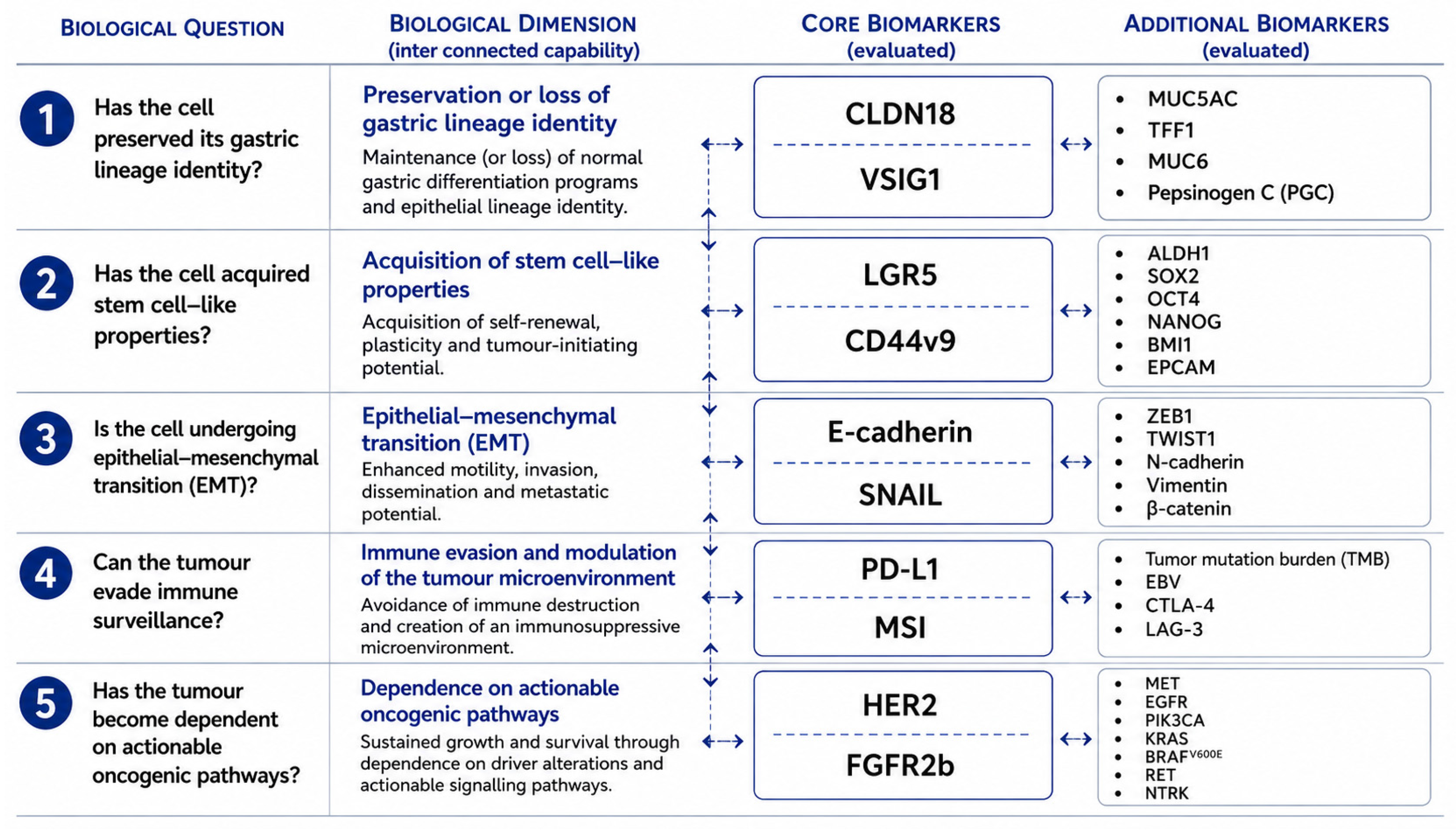
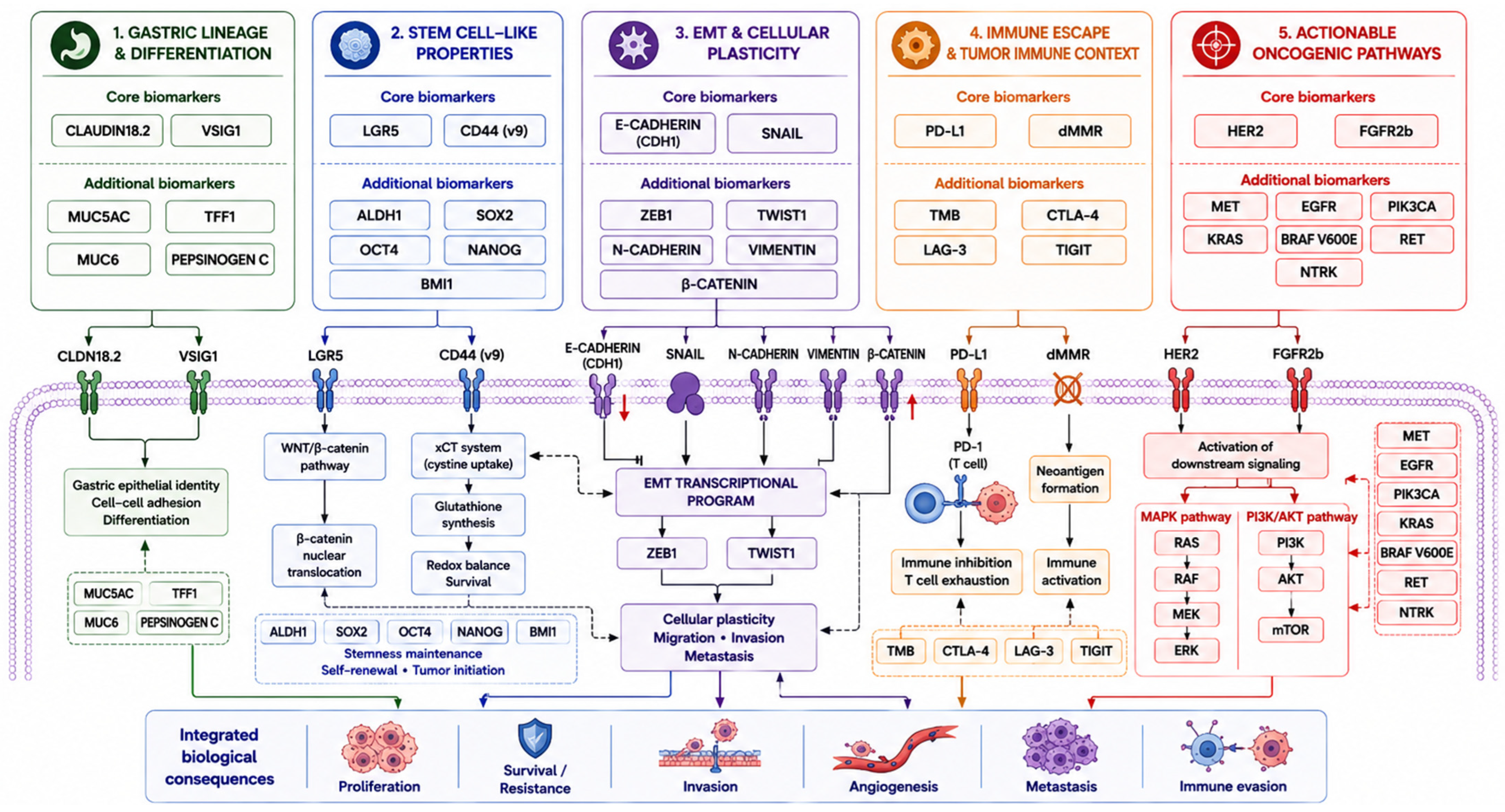
2.2. Development of the Biological Framework
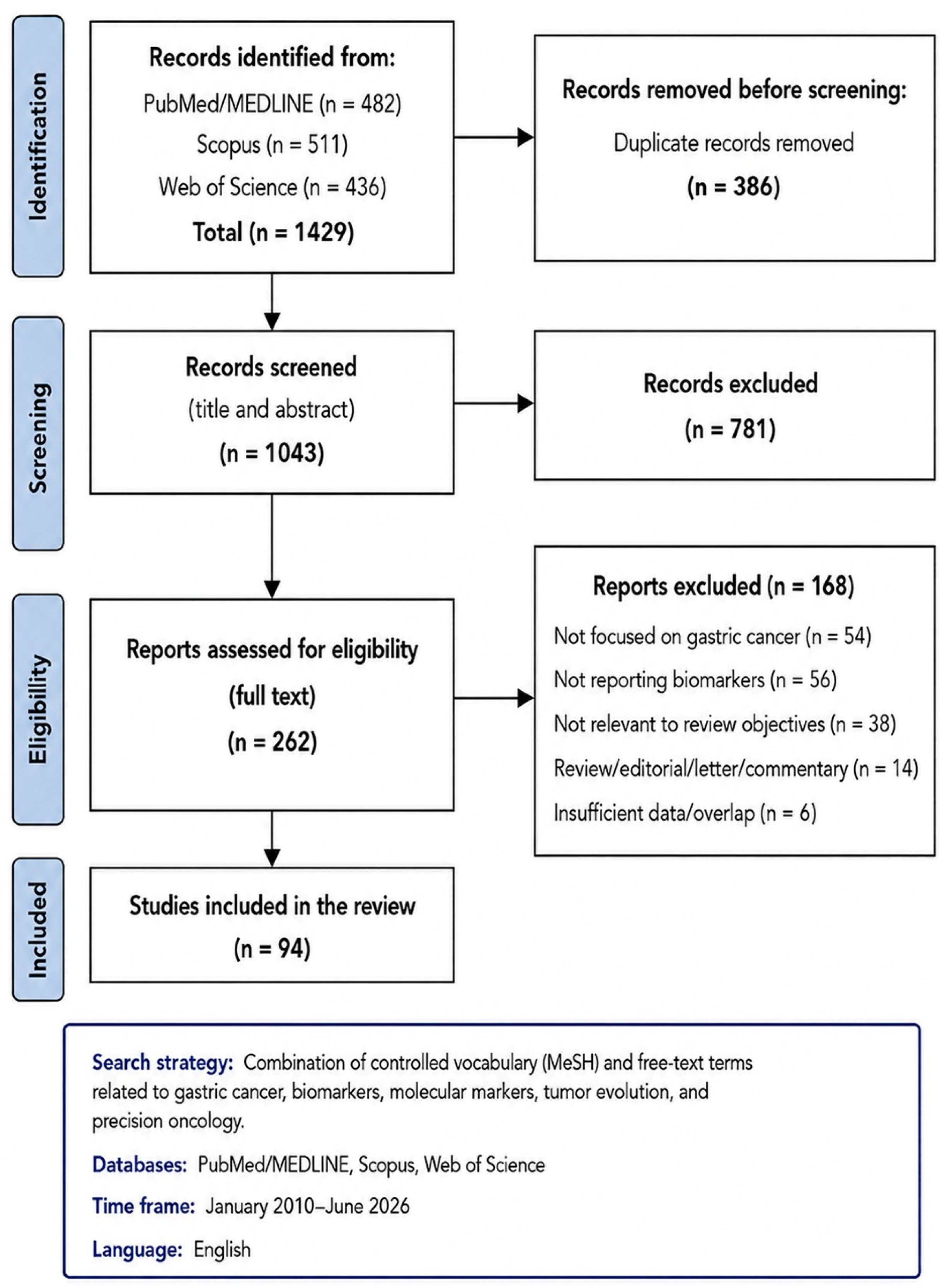
2.3. Literature Search Strategy
2.4. Study Selection
2.5. Evidence Assessment and Risk of Bias Considerations
2.6. Biomarker Selection Strategy
2.7. Methodological Considerations
3. Has the Cell Preserved Its Gastric Lineage Identity?
3.1. Claudin-18 (CLDN18)
3.2. V-Set and Immunoglobulin Domain Containing 1 (VSIG1)
3.3. Additional Markers of Gastric Epithelial Differentiation
4. Has the Cell Acquired Stem Cell-like Properties?
4.1. Leucine-Rich Repeat-Containing G-Protein-Coupled Receptor 5 (LGR5)
4.2. CD44v9
4.3. Additional Stemness-Associated Biomarkers
5. Is the Cell Undergoing Epithelial–Mesenchymal Transition?
5.1. E-Cadherin
5.2. SNAIL
5.3. Additional Biomarkers of Epithelial–Mesenchymal Transition
6. Can the Cell Escape Immune Surveillance?
6.1. PD-L1
6.2. Mismatch Repair Deficiency (dMMR) and Microsatellite Instability (MSI)
6.3. Additional Biomarkers
7. Has the Tumor Become Dependent on Actionable Oncogenic Pathways?
7.1. Human Epidermal Growth Factor Receptor 2 (HER2)
7.2. Fibroblast Growth Factor Receptor 2b (FGFR2b)
7.3. Emerging Actionable Biomarkers: Broadening the Landscape of Precision Oncology
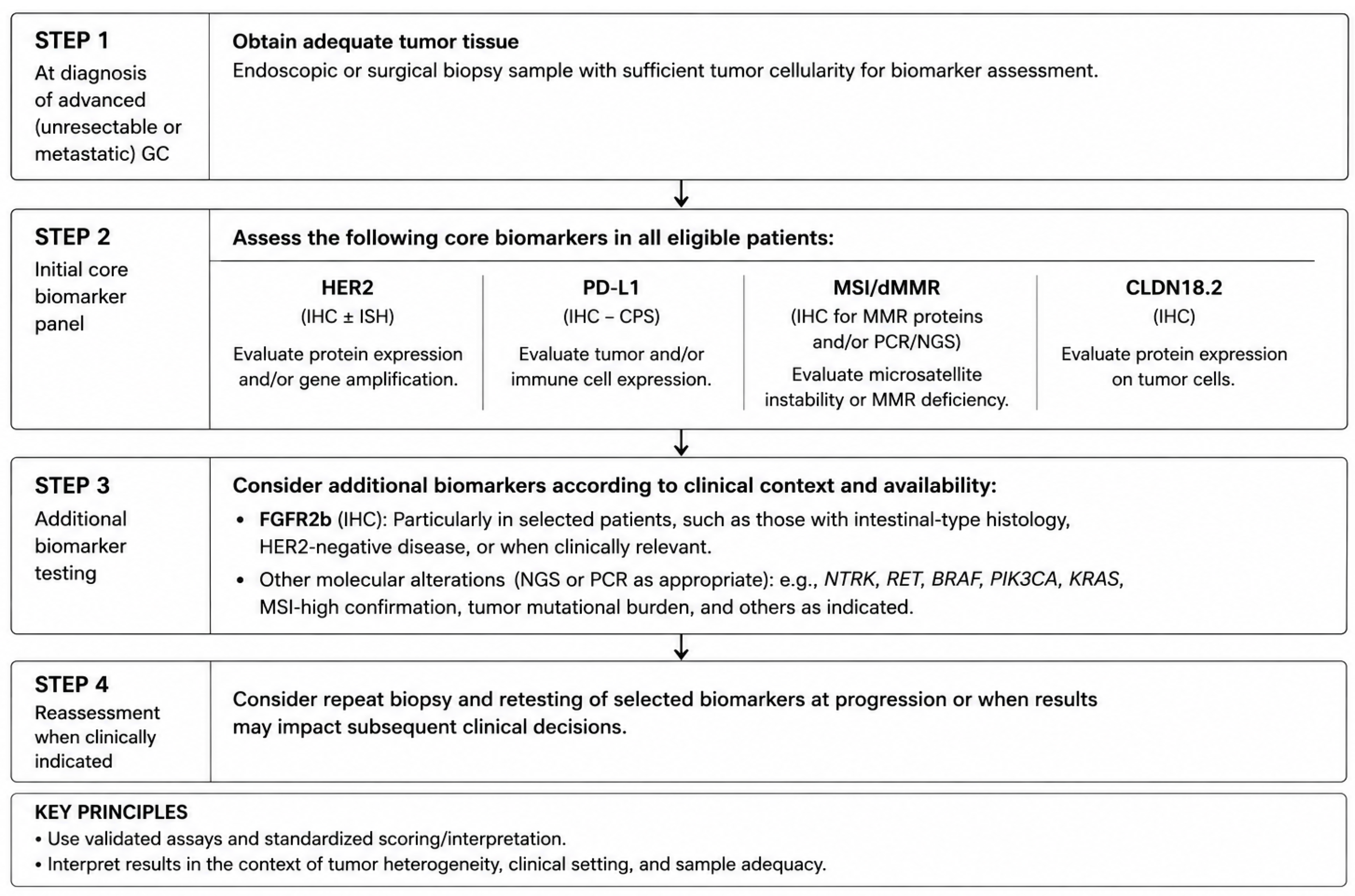
7.4. Practical Biomarker Testing Strategy in Gastric Cancer
8. Conclusions
Supplementary Materials
Author Contributions
Funding
Institutional Review Board Statement
Informed Consent Statement
Data Availability Statement
Acknowledgments
Conflicts of Interest
References
- Jang, E.; Shin, M.-K.; Kim, H.; Lim, J.Y.; Lee, J.E.; Park, J.; Kim, J.; Kim, H.; Shin, Y.; Son, H.-Y.; et al. Clinical Molecular Subtyping Reveals Intrinsic Mesenchymal Reprogramming in Gastric Cancer Cells. Exp. Mol. Med. 2023, 55, 974–986. [Google Scholar] [CrossRef] [Scilit]
- Yun, H.; Dong, F.; Wei, X.; Yan, X.; Zhang, R.; Zhang, X.; Wang, Y. Role and Value of the Tumor Microenvironment in the Progression and Treatment Resistance of Gastric Cancer (Review). Oncol. Rep. 2025, 53, 14. [Google Scholar] [CrossRef] [Scilit] [PubMed]
- Sun, Y.; Puspanathan, P.; Lim, T.; Lin, D. Advances and Challenges in Gastric Cancer Testing: The Role of Biomarkers. Cancer Biol. Med. 2025, 22, 212–230. [Google Scholar] [CrossRef] [Scilit]
- Sato, Y.; Okamoto, K.; Kawano, Y.; Kasai, A.; Kawaguchi, T.; Sagawa, T.; Sogabe, M.; Miyamoto, H.; Takayama, T. Novel Biomarkers of Gastric Cancer: Current Research and Future Perspectives. J. Clin. Med. 2023, 12, 4646. [Google Scholar] [CrossRef] [Scilit] [PubMed]
- Kim, J.; Park, C.; Kim, K.H.; Kim, E.H.; Kim, H.; Woo, J.K.; Seong, J.K.; Nam, K.T.; Lee, Y.C.; Cho, S.Y. Single-Cell Analysis of Gastric Pre-Cancerous and Cancer Lesions Reveals Cell Lineage Diversity and Intratumoral Heterogeneity. npj Precis. Oncol. 2022, 6, 9. [Google Scholar] [CrossRef] [Scilit]
- Kumar, V.; Ramnarayanan, K.; Sundar, R.; Padmanabhan, N.; Srivastava, S.; Koiwa, M.; Yasuda, T.; Koh, V.; Huang, K.K.; Tay, S.T.; et al. Single-Cell Atlas of Lineage States, Tumor Microenvironment, and Subtype-Specific Expression Programs in Gastric Cancer. Cancer Discov. 2022, 12, 670–691. [Google Scholar] [CrossRef] [Scilit] [PubMed]
- Satala, C.B.; Kovacs, Z.; Bara, T.; Jung, I.; Gurzu, S. Signet-Ring Cell Squamous Cell Carcinoma: A Biphenotypic Neoplasm of the Gastro-Esophageal Junction with Uncertain Biological Potential: Case Report and Literature Review. Int. J. Mol. Sci. 2023, 24, 9535. [Google Scholar] [CrossRef] [Scilit]
- Shitara, K.; Xu, R.-H.; Ajani, J.A.; Moran, D.; Guerrero, A.; Li, R.; Pavese, J.; Matsangou, M.; Bhattacharya, P.; Ueno, Y.; et al. Global Prevalence of Claudin 18 Isoform 2 in Tumors of Patients with Locally Advanced Unresectable or Metastatic Gastric or Gastroesophageal Junction Adenocarcinoma. Gastric Cancer 2024, 27, 1058–1068. [Google Scholar] [CrossRef] [Scilit]
- Satala, C.-B.; Jung, I.; Kovacs, Z.; Stefan-Van Staden, R.-I.; Molnar, C.; Bara, T.; Patrichi, A.-I.; Gurzu, S. V-Set and Immunoglobulin Domain Containing 1 (VSIG1) as an Emerging Target for Epithelial-Mesenchymal Transition of Gastric Cancer. Sci. Rep. 2022, 12, 16241. [Google Scholar] [CrossRef] [Scilit]
- Arai, J.; Hayakawa, Y.; Tateno, H.; Fujiwara, H.; Kasuga, M.; Fujishiro, M. The Role of Gastric Mucins and Mucin-related Glycans in Gastric Cancers. Cancer Sci. 2024, 115, 2853–2861. [Google Scholar] [CrossRef] [Scilit]
- Sahin, U.; Koslowski, M.; Dhaene, K.; Usener, D.; Brandenburg, G.; Seitz, G.; Huber, C.; Türeci, O. Claudin-18 Splice Variant 2 Is a Pan-Cancer Target Suitable for Therapeutic Antibody Development. Clin. Cancer Res. 2008, 14, 7624–7634. [Google Scholar] [CrossRef] [Scilit]
- Dottermusch, M.; Krüger, S.; Behrens, H.-M.; Halske, C.; Röcken, C. Expression of the Potential Therapeutic Target Claudin-18.2 Is Frequently Decreased in Gastric Cancer: Results from a Large Caucasian Cohort Study. Virchows Arch. 2019, 475, 563–571. [Google Scholar] [CrossRef] [Scilit]
- Cao, W.; Xing, H.; Li, Y.; Tian, W.; Song, Y.; Jiang, Z.; Yu, J. Claudin18.2 Is a Novel Molecular Biomarker for Tumor-Targeted Immunotherapy. Biomark. Res. 2022, 10, 38. [Google Scholar] [CrossRef] [Scilit]
- Kwak, Y.; Kim, T.-Y.; Nam, S.K.; Hwang, H.J.; Han, D.; Oh, H.J.; Kong, S.-H.; Park, D.J.; Oh, D.-Y.; Lee, H.-J.; et al. Clinicopathologic and Molecular Characterization of Stages II-IV Gastric Cancer with Claudin 18.2 Expression. Oncologist 2025, 30, oyae238. [Google Scholar] [CrossRef] [Scilit]
- Pellino, A.; Brignola, S.; Riello, E.; Niero, M.; Murgioni, S.; Guido, M.; Nappo, F.; Businello, G.; Sbaraglia, M.; Bergamo, F.; et al. Association of CLDN18 Protein Expression with Clinicopathological Features and Prognosis in Advanced Gastric and Gastroesophageal Junction Adenocarcinomas. J. Pers. Med. 2021, 11, 1095. [Google Scholar] [CrossRef] [Scilit]
- Sahin, U.; Türeci, Ö.; Manikhas, G.; Lordick, F.; Rusyn, A.; Vynnychenko, I.; Dudov, A.; Bazin, I.; Bondarenko, I.; Melichar, B.; et al. FAST: A Randomised Phase II Study of Zolbetuximab (IMAB362) plus EOX versus EOX Alone for First-Line Treatment of Advanced CLDN18.2-Positive Gastric and Gastro-Oesophageal Adenocarcinoma. Ann. Oncol. 2021, 32, 609–619. [Google Scholar] [CrossRef] [Scilit]
- Shitara, K.; Lordick, F.; Bang, Y.-J.; Enzinger, P.; Ilson, D.; Shah, M.A.; Van Cutsem, E.; Xu, R.-H.; Aprile, G.; Xu, J.; et al. Zolbetuximab plus mFOLFOX6 in Patients with CLDN18.2-Positive, HER2-Negative, Untreated, Locally Advanced Unresectable or Metastatic Gastric or Gastro-Oesophageal Junction Adenocarcinoma (SPOTLIGHT): A Multicentre, Randomised, Double-Blind, Phase 3 Trial. Lancet 2023, 401, 1655–1668. [Google Scholar] [CrossRef] [Scilit]
- Shah, M.A.; Shitara, K.; Ajani, J.A.; Bang, Y.-J.; Enzinger, P.; Ilson, D.; Lordick, F.; Van Cutsem, E.; Gallego Plazas, J.; Huang, J.; et al. Zolbetuximab plus CAPOX in CLDN18.2-Positive Gastric or Gastroesophageal Junction Adenocarcinoma: The Randomized, Phase 3 GLOW Trial. Nat. Med. 2023, 29, 2133–2141. [Google Scholar] [CrossRef] [Scilit]
- Kubota, Y.; Kawazoe, A.; Mishima, S.; Nakamura, Y.; Kotani, D.; Kuboki, Y.; Bando, H.; Kojima, T.; Doi, T.; Yoshino, T.; et al. Comprehensive Clinical and Molecular Characterization of Claudin 18.2 Expression in Advanced Gastric or Gastroesophageal Junction Cancer. ESMO Open 2023, 8, 100762. [Google Scholar] [CrossRef] [Scilit]
- Scanlan, M.J.; Ritter, G.; Yin, B.W.T.; Williams, C.; Cohen, L.S.; Coplan, K.A.; Fortunato, S.R.; Frosina, D.; Lee, S.-Y.; Murray, A.E.; et al. Glycoprotein A34, a Novel Target for Antibody-Based Cancer Immunotherapy. Cancer Immun. 2006, 6, 2. [Google Scholar]
- Oidovsambuu, O.; Nyamsuren, G.; Liu, S.; Göring, W.; Engel, W.; Adham, I.M. Adhesion Protein VSIG1 Is Required for the Proper Differentiation of Glandular Gastric Epithelia. PLoS ONE 2011, 6, e25908. [Google Scholar] [CrossRef] [Scilit]
- Chen, Y.; Pan, K.; Li, S.; Xia, J.; Wang, W.; Chen, J.; Zhao, J.; Lü, L.; Wang, D.; Pan, Q.; et al. Decreased Expression of V-Set and Immunoglobulin Domain Containing 1 (VSIG1) Is Associated with Poor Prognosis in Primary Gastric Cancer. J. Surg. Oncol. 2012, 106, 286–293. [Google Scholar] [CrossRef] [Scilit]
- Inoue, Y.; Matsuura, S.; Yoshimura, K.; Iwashita, Y.; Kahyo, T.; Kawase, A.; Tanahashi, M.; Maeda, M.; Ogawa, H.; Inui, N.; et al. Characterization of V-Set and Immunoglobulin Domain Containing 1 Exerting a Tumor Suppressor Function in Gastric, Lung, and Esophageal Cancer Cells. Cancer Sci. 2017, 108, 1701–1714. [Google Scholar] [CrossRef] [Scilit]
- Satala, C.-B.; Jung, I.; Gurzu, S. Mucin-Phenotype and Expression of the Protein V-Set and Immunoglobulin Domain Containing 1 (VSIG1): New Insights into Gastric Carcinogenesis. Int. J. Mol. Sci. 2023, 24, 8697. [Google Scholar] [CrossRef] [Scilit]
- Satala, C.-B.; Gurau, A.-M.; Mihalache, D.; Patrichi, G.; Mehedinti, R.-C.; Leibovici, A.R.; Gurău, G. From Primary Tumor to Peritoneal Niche: Microenvironmental Divergence in Gastric Cancer Peritoneal Metastasis. Cells 2026, 15, 1055. [Google Scholar] [CrossRef] [Scilit]
- Satala, C.-B.; Patrichi, G.; Gurau, A.-M.; Onofrei Popa, A.; Mihalache, D. Beyond Gastric Specificity: V-Set and Immunoglobulin Domain-Containing 1 (VSIG1) in Digestive Tract Tumors. Cancers 2026, 18, 867. [Google Scholar] [CrossRef] [Scilit]
- Lutz, F.; Han, S.-Y.; Büyücek, S.; Möller, K.; Viehweger, F.; Schlichter, R.; Menz, A.; Luebke, A.M.; Bawahab, A.A.; Reiswich, V.; et al. Expression of Trefoil Factor 1 (TFF1) in Cancer: A Tissue Microarray Study Involving 18,878 Tumors. Diagnostics 2024, 14, 2157. [Google Scholar] [CrossRef] [Scilit]
- E, Y.; Yu, Q.; Sun, T.; Xue, H.; Zhao, X.-R.; Zheng, H.-C. The Relationship between Pepsinogen C and Gastric Carcinogenesis: A Transgene and Population Study. BMC Cancer 2023, 23, 520. [Google Scholar] [CrossRef] [Scilit]
- Zaarour, R.F.; Ribeiro, M.; Azzarone, B.; Kapoor, S.; Chouaib, S. Tumor Microenvironment-Induced Tumor Cell Plasticity: Relationship with Hypoxic Stress and Impact on Tumor Resistance. Front. Oncol. 2023, 13, 1222575. [Google Scholar] [CrossRef] [Scilit]
- Yang, Y.; Meng, W.-J.; Wang, Z.-Q. Cancer Stem Cells and the Tumor Microenvironment in Gastric Cancer. Front. Oncol. 2022, 11, 803974. [Google Scholar] [CrossRef] [Scilit]
- Razmi, M.; Ghods, R.; Vafaei, S.; Sahlolbei, M.; Saeednejad Zanjani, L.; Madjd, Z. Clinical and Prognostic Significances of Cancer Stem Cell Markers in Gastric Cancer Patients: A Systematic Review and Meta-Analysis. Cancer Cell Int. 2021, 21, 139. [Google Scholar] [CrossRef] [Scilit]
- Fatehullah, A.; Terakado, Y.; Sagiraju, S.; Tan, T.L.; Sheng, T.; Tan, S.H.; Murakami, K.; Swathi, Y.; Ang, N.; Rajarethinam, R.; et al. A Tumour-Resident Lgr5+ Stem-Cell-like Pool Drives the Establishment and Progression of Advanced Gastric Cancers. Nat. Cell Biol. 2021, 23, 1299–1313. [Google Scholar] [CrossRef] [Scilit]
- Lau, W.M.; Teng, E.; Chong, H.S.; Lopez, K.A.P.; Tay, A.Y.L.; Salto-Tellez, M.; Shabbir, A.; So, J.B.Y.; Chan, S.L. CD44v8-10 Is a Cancer-Specific Marker for Gastric Cancer Stem Cells. Cancer Res. 2014, 74, 2630–2641. [Google Scholar] [CrossRef] [Scilit]
- Barker, N.; Huch, M.; Kujala, P.; van de Wetering, M.; Snippert, H.J.; van Es, J.H.; Sato, T.; Stange, D.E.; Begthel, H.; van den Born, M.; et al. Lgr5(+ve) Stem Cells Drive Self-Renewal in the Stomach and Build Long-Lived Gastric Units in Vitro. Cell Stem Cell 2010, 6, 25–36. [Google Scholar] [CrossRef] [Scilit]
- Leushacke, M.; Barker, N.; Pin, C. Quantifying Lgr5-Positive Stem Cell Behaviour in the Pyloric Epithelium. Sci. Rep. 2016, 6, 21923. [Google Scholar] [CrossRef] [Scilit]
- Li, X.-B.; Yang, G.; Zhu, L.; Tang, Y.-L.; Zhang, C.; Ju, Z.; Yang, X.; Teng, Y. Gastric Lgr5(+) Stem Cells Are the Cellular Origin of Invasive Intestinal-Type Gastric Cancer in Mice. Cell Res. 2016, 26, 838–849. [Google Scholar] [CrossRef] [Scilit]
- Simon, E.; Petke, D.; Böger, C.; Behrens, H.-M.; Warneke, V.; Ebert, M.; Röcken, C. The Spatial Distribution of LGR5+ Cells Correlates with Gastric Cancer Progression. PLoS ONE 2012, 7, e35486. [Google Scholar] [CrossRef] [Scilit]
- Koo, B.-K.; Clevers, H. Stem Cells Marked by the R-Spondin Receptor LGR5. Gastroenterology 2014, 147, 289–302. [Google Scholar] [CrossRef] [Scilit]
- Jang, B.G.; Lee, B.L.; Kim, W.H. Distribution of LGR5+ Cells and Associated Implications during the Early Stage of Gastric Tumorigenesis. PLoS ONE 2013, 8, e82390. [Google Scholar] [CrossRef] [Scilit]
- Zheng, Z.-X.; Sun, Y.; Bu, Z.-D.; Zhang, L.-H.; Li, Z.-Y.; Wu, A.-W.; Wu, X.-J.; Wang, X.-H.; Cheng, X.-J.; Xing, X.-F.; et al. Intestinal Stem Cell Marker LGR5 Expression during Gastric Carcinogenesis. World J. Gastroenterol. 2013, 19, 8714–8721. [Google Scholar] [CrossRef] [Scilit]
- Takaishi, S.; Okumura, T.; Tu, S.; Wang, S.S.W.; Shibata, W.; Vigneshwaran, R.; Gordon, S.A.K.; Shimada, Y.; Wang, T.C. Identification of Gastric Cancer Stem Cells Using the Cell Surface Marker CD44. Stem Cells 2009, 27, 1006–1020. [Google Scholar] [CrossRef] [Scilit]
- Ishimoto, T.; Nagano, O.; Yae, T.; Tamada, M.; Motohara, T.; Oshima, H.; Oshima, M.; Ikeda, T.; Asaba, R.; Yagi, H.; et al. CD44 Variant Regulates Redox Status in Cancer Cells by Stabilizing the xCT Subunit of System Xc(-) and Thereby Promotes Tumor Growth. Cancer Cell 2011, 19, 387–400. [Google Scholar] [CrossRef] [Scilit]
- Hirata, K.; Suzuki, H.; Imaeda, H.; Matsuzaki, J.; Tsugawa, H.; Nagano, O.; Asakura, K.; Saya, H.; Hibi, T. CD44 Variant 9 Expression in Primary Early Gastric Cancer as a Predictive Marker for Recurrence. Br. J. Cancer 2013, 109, 379–386. [Google Scholar] [CrossRef] [Scilit]
- Go, S.-I.; Ko, G.H.; Lee, W.S.; Kim, R.B.; Lee, J.-H.; Jeong, S.-H.; Lee, Y.-J.; Hong, S.C.; Ha, W.S. CD44 Variant 9 Serves as a Poor Prognostic Marker in Early Gastric Cancer, But Not in Advanced Gastric Cancer. Cancer Res. Treat. 2016, 48, 142–152. [Google Scholar] [CrossRef] [Scilit][Green Version]
- Kodama, H.; Murata, S.; Ishida, M.; Yamamoto, H.; Yamaguchi, T.; Kaida, S.; Miyake, T.; Takebayashi, K.; Kushima, R.; Tani, M. Prognostic Impact of CD44-Positive Cancer Stem-like Cells at the Invasive Front of Gastric Cancer. Br. J. Cancer 2017, 116, 186–194. [Google Scholar] [CrossRef] [Scilit]
- Jogo, T.; Oki, E.; Nakanishi, R.; Ando, K.; Nakashima, Y.; Kimura, Y.; Saeki, H.; Oda, Y.; Maehara, Y.; Mori, M. Expression of CD44 Variant 9 Induces Chemoresistance of Gastric Cancer by Controlling Intracellular Reactive Oxygen Spices Accumulation. Gastric Cancer 2021, 24, 1089–1099. [Google Scholar] [CrossRef] [Scilit]
- Wang, L.; Wang, L.; Yu, Y.; Su, R.; Zhang, Y.; Li, Y.; Zang, M. Aldehyde Dehydrogenase 1 in Gastric Cancer. J. Oncol. 2022, 2022, 5734549. [Google Scholar] [CrossRef] [Scilit]
- Pádua, D.; Barros, R.; Amaral, A.L.; Mesquita, P.; Freire, A.F.; Sousa, M.; Maia, A.F.; Caiado, I.; Fernandes, H.; Pombinho, A.; et al. A SOX2 Reporter System Identifies Gastric Cancer Stem-Like Cells Sensitive to Monensin. Cancers 2020, 12, 495. [Google Scholar] [CrossRef] [Scilit]
- Li, N.; Pang, Y.; Sang, J.; Sun, Y.; Hou, W. The Controversial Expression of SOX2 in Gastric Cancer and Its Correlation with Helicobacter Pylori Infection: A Meta-Analysis. Medicine 2022, 101, e30886. [Google Scholar] [CrossRef] [Scilit]
- Zhao, H.; Hu, H.; Chen, B.; Xu, W.; Zhao, J.; Huang, C.; Xing, Y.; Lv, H.; Nie, C.; Wang, J.; et al. Overview on the Role of E-Cadherin in Gastric Cancer: Dysregulation and Clinical Implications. Front. Mol. Biosci. 2021, 8, 689139. [Google Scholar] [CrossRef] [Scilit]
- Chen, X.; Li, J.; Hu, L.; Yang, W.; Lu, L.; Jin, H.; Wei, Z.; Yang, J.Y.; Arabnia, H.R.; Liu, J.S.; et al. The Clinical Significance of Snail Protein Expression in Gastric Cancer: A Meta-Analysis. Hum. Genom. 2016, 10, 22. [Google Scholar] [CrossRef] [Scilit]
- Becker, K.F.; Atkinson, M.J.; Reich, U.; Becker, I.; Nekarda, H.; Siewert, J.R.; Höfler, H. E-Cadherin Gene Mutations Provide Clues to Diffuse Type Gastric Carcinomas. Cancer Res. 1994, 54, 3845–3852. [Google Scholar]
- Machado, J.C.; Oliveira, C.; Carvalho, R.; Soares, P.; Berx, G.; Caldas, C.; Seruca, R.; Carneiro, F.; Sobrinho-Simöes, M. E-Cadherin Gene (CDH1) Promoter Methylation as the Second Hit in Sporadic Diffuse Gastric Carcinoma. Oncogene 2001, 20, 1525–1528. [Google Scholar] [CrossRef] [Scilit]
- Machado, J.C.; Carneiro, F.; Beck, S.; Rossi, S.; Lopes, J.; Taveira-Gomes, A.; Cardoso-Oliveira, M. E-Cadherin Expression Is Correlated with the Isolated Cell/Diffuse Histotype and with Features of Biological Aggressiveness of Gastric Carcinoma. Int. J. Surg. Pathol. 1998, 6, 135–144. [Google Scholar] [CrossRef] [Scilit]
- Corso, G.; Carvalho, J.; Marrelli, D.; Vindigni, C.; Carvalho, B.; Seruca, R.; Roviello, F.; Oliveira, C. Somatic Mutations and Deletions of the E-Cadherin Gene Predict Poor Survival of Patients with Gastric Cancer. J. Clin. Oncol. 2013, 31, 868–875. [Google Scholar] [CrossRef] [Scilit]
- Cancer Genome Atlas Research Network. Comprehensive Molecular Characterization of Gastric Adenocarcinoma. Nature 2014, 513, 202–209. [Google Scholar] [CrossRef] [Scilit]
- Figueiredo, J.; Ferreira, R.M.; Xu, H.; Gonçalves, M.; Barros-Carvalho, A.; Cravo, J.; Maia, A.F.; Carneiro, P.; Figueiredo, C.; Smith, M.L.; et al. Integrin Β1 Orchestrates the Abnormal Cell-Matrix Attachment and Invasive Behaviour of E-Cadherin Dysfunctional Cells. Gastric Cancer 2022, 25, 124–137. [Google Scholar] [CrossRef] [Scilit]
- Rosivatz, E.; Becker, I.; Specht, K.; Fricke, E.; Luber, B.; Busch, R.; Höfler, H.; Becker, K.-F. Differential Expression of the Epithelial-Mesenchymal Transition Regulators Snail, SIP1, and Twist in Gastric Cancer. Am. J. Pathol. 2002, 161, 1881–1891. [Google Scholar] [CrossRef] [Scilit]
- Rosivatz, E.; Becker, K.-F.; Kremmer, E.; Schott, C.; Blechschmidt, K.; Höfler, H.; Sarbia, M. Expression and Nuclear Localization of Snail, an E-Cadherin Repressor, in Adenocarcinomas of the Upper Gastrointestinal Tract. Virchows Arch. 2006, 448, 277–287. [Google Scholar] [CrossRef] [Scilit]
- He, H.; Chen, W.; Wang, X.; Wang, C.; Liu, F.; Shen, Z.; Xu, J.; Gu, J.; Sun, Y. Snail Is an Independent Prognostic Predictor for Progression and Patient Survival of Gastric Cancer. Cancer Sci. 2012, 103, 1296–1303. [Google Scholar] [CrossRef] [Scilit]
- Shin, N.R.; Jeong, E.H.; Choi, C.I.; Moon, H.J.; Kwon, C.H.; Chu, I.S.; Kim, G.H.; Jeon, T.Y.; Kim, D.H.; Lee, J.H.; et al. Overexpression of Snail Is Associated with Lymph Node Metastasis and Poor Prognosis in Patients with Gastric Cancer. BMC Cancer 2012, 12, 521. [Google Scholar] [CrossRef] [Scilit]
- Wu, H.; Chen, Y.; Lin, H.; Xu, Y.; Guo, Z.; Li, Z. The Clinical Significance of SNAIL, TWIST, and E-Cadherin Expression in Gastric Mesentery Tumor Deposits of Advanced Gastric Cancer. Indian J. Pathol. Microbiol. 2024, 67, 21–28. [Google Scholar] [CrossRef] [Scilit]
- Xue, Y.; Zhang, L.; Zhu, Y.; Ke, X.; Wang, Q.; Min, H. Regulation of Proliferation and Epithelial-to-Mesenchymal Transition (EMT) of Gastric Cancer by ZEB1 via Modulating Wnt5a and Related Mechanisms. Med. Sci. Monit. 2019, 25, 1663–1670. [Google Scholar] [CrossRef] [Scilit]
- Hou, W.; Zhao, Y.; Zhu, H. Predictive Biomarkers for Immunotherapy in Gastric Cancer: Current Status and Emerging Prospects. Int. J. Mol. Sci. 2023, 24, 15321. [Google Scholar] [CrossRef] [Scilit]
- Fuchs, C.S.; Doi, T.; Jang, R.W.; Muro, K.; Satoh, T.; Machado, M.; Sun, W.; Jalal, S.I.; Shah, M.A.; Metges, J.-P.; et al. Safety and Efficacy of Pembrolizumab Monotherapy in Patients With Previously Treated Advanced Gastric and Gastroesophageal Junction Cancer: Phase 2 Clinical KEYNOTE-059 Trial. JAMA Oncol. 2018, 4, e180013. [Google Scholar] [CrossRef] [Scilit]
- Janjigian, Y.Y.; Shitara, K.; Moehler, M.; Garrido, M.; Salman, P.; Shen, L.; Wyrwicz, L.; Yamaguchi, K.; Skoczylas, T.; Campos Bragagnoli, A.; et al. First-Line Nivolumab plus Chemotherapy versus Chemotherapy Alone for Advanced Gastric, Gastro-Oesophageal Junction, and Oesophageal Adenocarcinoma (CheckMate 649): A Randomised, Open-Label, Phase 3 Trial. Lancet 2021, 398, 27–40. [Google Scholar] [CrossRef] [Scilit]
- Rha, S.Y.; Oh, D.-Y.; Yañez, P.; Bai, Y.; Ryu, M.-H.; Lee, J.; Rivera, F.; Alves, G.V.; Garrido, M.; Shiu, K.-K.; et al. Pembrolizumab plus Chemotherapy versus Placebo plus Chemotherapy for HER2-Negative Advanced Gastric Cancer (KEYNOTE-859): A Multicentre, Randomised, Double-Blind, Phase 3 Trial. Lancet Oncol. 2023, 24, 1181–1195. [Google Scholar] [CrossRef] [Scilit]
- Shitara, K.; Van Cutsem, E.; Bang, Y.-J.; Fuchs, C.; Wyrwicz, L.; Lee, K.-W.; Kudaba, I.; Garrido, M.; Chung, H.C.; Lee, J.; et al. Efficacy and Safety of Pembrolizumab or Pembrolizumab Plus Chemotherapy vs Chemotherapy Alone for Patients With First-Line, Advanced Gastric Cancer: The KEYNOTE-062 Phase 3 Randomized Clinical Trial. JAMA Oncol. 2020, 6, 1571–1580. [Google Scholar] [CrossRef] [Scilit]
- Smyth, E.C.; Wotherspoon, A.; Peckitt, C.; Gonzalez, D.; Hulkki-Wilson, S.; Eltahir, Z.; Fassan, M.; Rugge, M.; Valeri, N.; Okines, A.; et al. Mismatch Repair Deficiency, Microsatellite Instability, and Survival: An Exploratory Analysis of the Medical Research Council Adjuvant Gastric Infusional Chemotherapy (MAGIC) Trial. JAMA Oncol. 2017, 3, 1197–1203. [Google Scholar] [CrossRef] [Scilit]
- Pietrantonio, F.; Miceli, R.; Raimondi, A.; Kim, Y.W.; Kang, W.K.; Langley, R.E.; Choi, Y.Y.; Kim, K.-M.; Nankivell, M.G.; Morano, F.; et al. Individual Patient Data Meta-Analysis of the Value of Microsatellite Instability As a Biomarker in Gastric Cancer. J. Clin. Oncol. 2019, 37, 3392–3400. [Google Scholar] [CrossRef] [Scilit]
- Pietrantonio, F.; Randon, G.; Di Bartolomeo, M.; Luciani, A.; Chao, J.; Smyth, E.C.; Petrelli, F. Predictive Role of Microsatellite Instability for PD-1 Blockade in Patients with Advanced Gastric Cancer: A Meta-Analysis of Randomized Clinical Trials. ESMO Open 2021, 6, 100036. [Google Scholar] [CrossRef] [Scilit]
- Wang, D.; Wang, N.; Li, X.; Chen, X.; Shen, B.; Zhu, D.; Zhu, L.; Xu, Y.; Yu, Y.; Shu, Y. Tumor Mutation Burden as a Biomarker in Resected Gastric Cancer via Its Association with Immune Infiltration and Hypoxia. Gastric Cancer 2021, 24, 823–834. [Google Scholar] [CrossRef] [Scilit]
- Satala, C.-B.; Gurău, G.; Patrichi, G.; Gurau, A.-M.; Mehedinti, R.-C.; Leibovici, A.R.; Mihalache, D. FGFR2b in Gastric Cancer: Translating a Therapeutic Target into a Reliable Biomarker. Cancers 2026, 18, 1863. [Google Scholar] [CrossRef] [Scilit] [PubMed]
- Tanner, M.; Hollmén, M.; Junttila, T.T.; Kapanen, A.I.; Tommola, S.; Soini, Y.; Helin, H.; Salo, J.; Joensuu, H.; Sihvo, E.; et al. Amplification of HER-2 in Gastric Carcinoma: Association with Topoisomerase IIα Gene Amplification, Intestinal Type, Poor Prognosis and Sensitivity to Trastuzumab. Ann. Oncol. 2005, 16, 273–278. [Google Scholar] [CrossRef] [Scilit]
- Bang, Y.-J.; Van Cutsem, E.; Feyereislova, A.; Chung, H.C.; Shen, L.; Sawaki, A.; Lordick, F.; Ohtsu, A.; Omuro, Y.; Satoh, T.; et al. Trastuzumab in Combination with Chemotherapy versus Chemotherapy Alone for Treatment of HER2-Positive Advanced Gastric or Gastro-Oesophageal Junction Cancer (ToGA): A Phase 3, Open-Label, Randomised Controlled Trial. Lancet 2010, 376, 687–697. [Google Scholar] [CrossRef] [Scilit]
- Hofmann, M.; Stoss, O.; Shi, D.; Büttner, R.; van de Vijver, M.; Kim, W.; Ochiai, A.; Rüschoff, J.; Henkel, T. Assessment of a HER2 Scoring System for Gastric Cancer: Results from a Validation Study. Histopathology 2008, 52, 797–805. [Google Scholar] [CrossRef] [Scilit]
- Hecht, J.R.; Bang, Y.-J.; Qin, S.K.; Chung, H.C.; Xu, J.M.; Park, J.O.; Jeziorski, K.; Shparyk, Y.; Hoff, P.M.; Sobrero, A.; et al. Lapatinib in Combination With Capecitabine Plus Oxaliplatin in Human Epidermal Growth Factor Receptor 2-Positive Advanced or Metastatic Gastric, Esophageal, or Gastroesophageal Adenocarcinoma: TRIO-013/LOGiC—A Randomized Phase III Trial. J. Clin. Oncol. 2016, 34, 443–451. [Google Scholar] [CrossRef] [Scilit]
- Satoh, T.; Xu, R.-H.; Chung, H.C.; Sun, G.-P.; Doi, T.; Xu, J.-M.; Tsuji, A.; Omuro, Y.; Li, J.; Wang, J.-W.; et al. Lapatinib plus Paclitaxel versus Paclitaxel Alone in the Second-Line Treatment of HER2-Amplified Advanced Gastric Cancer in Asian Populations: TyTAN—A Randomized, Phase III Study. J. Clin. Oncol. 2014, 32, 2039–2049. [Google Scholar] [CrossRef] [Scilit]
- Tabernero, J.; Hoff, P.M.; Shen, L.; Ohtsu, A.; Shah, M.A.; Cheng, K.; Song, C.; Wu, H.; Eng-Wong, J.; Kim, K.; et al. Pertuzumab plus Trastuzumab and Chemotherapy for HER2-Positive Metastatic Gastric or Gastro-Oesophageal Junction Cancer (JACOB): Final Analysis of a Double-Blind, Randomised, Placebo-Controlled Phase 3 Study. Lancet Oncol. 2018, 19, 1372–1384. [Google Scholar] [CrossRef] [Scilit]
- Thuss-Patience, P.C.; Shah, M.A.; Ohtsu, A.; Van Cutsem, E.; Ajani, J.A.; Castro, H.; Mansoor, W.; Chung, H.C.; Bodoky, G.; Shitara, K.; et al. Trastuzumab Emtansine versus Taxane Use for Previously Treated HER2-Positive Locally Advanced or Metastatic Gastric or Gastro-Oesophageal Junction Adenocarcinoma (GATSBY): An International Randomised, Open-Label, Adaptive, Phase 2/3 Study. Lancet Oncol. 2017, 18, 640–653. [Google Scholar] [CrossRef] [Scilit]
- Shitara, K.; Van Cutsem, E.; Gümüş, M.; Lonardi, S.; de la Fouchardière, C.; Coutzac, C.; Dekervel, J.; Hochhauser, D.; Shen, L.; Mansoor, W.; et al. Trastuzumab Deruxtecan or Ramucirumab plus Paclitaxel in Gastric Cancer. N. Engl. J. Med. 2025, 393, 336–348. [Google Scholar] [CrossRef] [Scilit]
- Kunii, K.; Davis, L.; Gorenstein, J.; Hatch, H.; Yashiro, M.; Di Bacco, A.; Elbi, C.; Lutterbach, B. FGFR2-Amplified Gastric Cancer Cell Lines Require FGFR2 and Erbb3 Signaling for Growth and Survival. Cancer Res. 2008, 68, 2340–2348. [Google Scholar] [CrossRef] [Scilit]
- Matsumoto, K.; Arao, T.; Hamaguchi, T.; Shimada, Y.; Kato, K.; Oda, I.; Taniguchi, H.; Koizumi, F.; Yanagihara, K.; Sasaki, H.; et al. FGFR2 Gene Amplification and Clinicopathological Features in Gastric Cancer. Br. J. Cancer 2012, 106, 727–732. [Google Scholar] [CrossRef] [Scilit]
- Pearson, A.; Smyth, E.; Babina, I.S.; Herrera-Abreu, M.T.; Tarazona, N.; Peckitt, C.; Kilgour, E.; Smith, N.R.; Geh, C.; Rooney, C.; et al. High-Level Clonal FGFR Amplification and Response to FGFR Inhibition in a Translational Clinical Trial. Cancer Discov. 2016, 6, 838–851. [Google Scholar] [CrossRef] [Scilit]
- Catenacci, D.V.T.; Rasco, D.; Lee, J.; Rha, S.Y.; Lee, K.-W.; Bang, Y.J.; Bendell, J.; Enzinger, P.; Marina, N.; Xiang, H.; et al. Phase I Escalation and Expansion Study of Bemarituzumab (FPA144) in Patients With Advanced Solid Tumors and FGFR2b-Selected Gastroesophageal Adenocarcinoma. J. Clin. Oncol. 2020, 38, 2418–2426. [Google Scholar] [CrossRef] [Scilit]
- Wainberg, Z.A.; Enzinger, P.C.; Kang, Y.-K.; Qin, S.; Yamaguchi, K.; Kim, I.-H.; Saeed, A.; Oh, S.C.; Li, J.; Turk, H.M.; et al. Bemarituzumab in Patients with FGFR2b-Selected Gastric or Gastro-Oesophageal Junction Adenocarcinoma (FIGHT): A Randomised, Double-Blind, Placebo-Controlled, Phase 2 Study. Lancet Oncol. 2022, 23, 1430–1440. [Google Scholar] [CrossRef] [Scilit]
- Wainberg, Z.A.; Kang, Y.-K.; Lee, K.-W.; Qin, S.; Yamaguchi, K.; Kim, I.-H.; Saeed, A.; Oh, S.C.; Li, J.; Turk, H.M.; et al. Bemarituzumab as First-Line Treatment for Locally Advanced or Metastatic Gastric/Gastroesophageal Junction Adenocarcinoma: Final Analysis of the Randomized Phase 2 FIGHT Trial. Gastric Cancer 2024, 27, 558–570. [Google Scholar] [CrossRef] [Scilit]
- Amgen. A Randomized, Multi-Center, Double-Blind, Placebo-Controlled Phase 3 Study of Bemarituzumab Plus Chemotherapy Versus Placebo Plus Chemotherapy in Subjects with Previously Untreated Advanced Gastric or Gastroesophageal Junction Cancer with FGFR2b Overexpression. 2026. Available online: https://clinicaltrials.gov (accessed on 9 August 2026).
- Satala, C.-B.; Gurau, G.; Gurau, A.-M.; Patrichi, G.; Mihalache, D. Tumor Budding in Gastric Carcinoma: Beyond Counting Cells at the Invasive Front—A Review of Current Evidence and Biological Perspectives. Int. J. Mol. Sci. 2026, 27, 3787. [Google Scholar] [CrossRef] [Scilit]
- Waddell, T.; Chau, I.; Cunningham, D.; Gonzalez, D.; Okines, A.F.C.; Okines, C.; Wotherspoon, A.; Saffery, C.; Middleton, G.; Wadsley, J.; et al. Epirubicin, Oxaliplatin, and Capecitabine with or without Panitumumab for Patients with Previously Untreated Advanced Oesophagogastric Cancer (REAL3): A Randomised, Open-Label Phase 3 Trial. Lancet Oncol. 2013, 14, 481–489. [Google Scholar] [CrossRef] [Scilit]
- Lordick, F.; Kang, Y.-K.; Chung, H.-C.; Salman, P.; Oh, S.C.; Bodoky, G.; Kurteva, G.; Volovat, C.; Moiseyenko, V.M.; Gorbunova, V.; et al. Capecitabine and Cisplatin with or without Cetuximab for Patients with Previously Untreated Advanced Gastric Cancer (EXPAND): A Randomised, Open-Label Phase 3 Trial. Lancet Oncol. 2013, 14, 490–499. [Google Scholar] [CrossRef] [Scilit]
- Pozzi, V.; Campagna, R.; Sartini, D.; Emanuelli, M. Nicotinamide N-Methyltransferase as Promising Tool for Management of Gastrointestinal Neoplasms. Biomolecules 2022, 12, 1173. [Google Scholar] [CrossRef] [Scilit] [PubMed] [PubMed Central]
- Ulanovskaya, O.A.; Zuhl, A.M.; Cravatt, B.F. NNMT promotes epigenetic remodeling in cancer by creating a metabolic methylation sink. Nat. Chem. Biol. 2013, 9, 300–306. [Google Scholar] [CrossRef] [Scilit] [PubMed] [PubMed Central]
- Hanrahan, A.J.; Chen, Z.; Rosen, N.; Solit, D.B. BRAF—A Tumour-Agnostic Drug Target with Lineage-Specific Dependencies. Nat. Rev. Clin. Oncol. 2024, 21, 224–247. [Google Scholar] [CrossRef] [Scilit]
- Subbiah, V.; Wolf, J.; Konda, B.; Kang, H.; Spira, A.; Weiss, J.; Takeda, M.; Ohe, Y.; Khan, S.; Ohashi, K.; et al. Tumour-Agnostic Efficacy and Safety of Selpercatinib in Patients with RET Fusion-Positive Solid Tumours Other than Lung or Thyroid Tumours (LIBRETTO-001): A Phase 1/2, Open-Label, Basket Trial. Lancet Oncol. 2022, 23, 1261–1273. [Google Scholar] [CrossRef] [Scilit] [PubMed]
- Drilon, A.; Laetsch, T.W.; Kummar, S.; DuBois, S.G.; Lassen, U.N.; Demetri, G.D.; Nathenson, M.; Doebele, R.C.; Farago, A.F.; Pappo, A.S.; et al. Efficacy of Larotrectinib in TRK Fusion-Positive Cancers in Adults and Children. N. Engl. J. Med. 2018, 378, 731–739. [Google Scholar] [CrossRef] [Scilit]
- Dong, C.; Wang, Y.; Gu, X.; Lv, X.; Ren, S.; Wang, Z.; Dai, Z. Differential diagnostic value of tumor markers and contrast-enhanced computed tomography in gastric hepatoid adenocarcinoma and gastric adenocarcinoma. Front. Oncol. 2023, 13, 1222853. [Google Scholar] [CrossRef] [Scilit] [PubMed] [PubMed Central]
- Zhang, L.; Zhao, L.; Lin, X.; Zhao, S.; Pan, W.; Wang, D.; Sun, Z.; Li, J.; Liang, Z.; Zhang, R.; et al. Comparison of Tumor Non-specific and PD-L1 Specific Imaging by Near-Infrared Fluorescence/Cerenkov Luminescence Dual-Modality In-situ Imaging. Mol. Imaging 2024, 23, 15353508241261473. [Google Scholar] [CrossRef] [Scilit] [PubMed] [PubMed Central]

| Biological Question | Biological Process Evaluated | Purpose |
|---|---|---|
| Has the cell preserved its gastric lineage identity? | Gastric differentiation and lineage commitment | Identify biomarkers reflecting preservation or loss of gastric epithelial identity |
| Has the cell acquired stem cell-like properties? | Stemness and cellular plasticity | Identify biomarkers associated with tumor plasticity and stem cell-like features |
| Is the cell undergoing epithelial–mesenchymal transition? | Epithelial–mesenchymal transition (EMT) | Evaluate biomarkers reflecting epithelial plasticity, loss of epithelial integrity, and acquisition of invasive potential |
| Can the tumor evade host immune surveillance? | Tumor–immune interaction | Evaluate biomarkers associated with immune escape mechanisms |
| Has the tumor become dependent on actionable oncogenic pathways? | Targetable oncogenic signaling and therapeutic vulnerabilities | Identify predictive biomarkers that guide targeted therapy and precision oncology |
| Criterion | Definition |
|---|---|
| Biological specificity | Evidence supporting a direct role within the biological domain under investigation |
| Human evidence | Validation in gastric adenocarcinoma cohorts |
| Evidence consistency | Reproducible findings across independent studies |
| Clinical relevance | Association with diagnosis, prognosis, prediction or therapy |
| Technical feasibility | Assessment using established pathological or molecular techniques |
| (A) Landmark Studies Underpinning the Selection of CLDN18.2 as a Core Biomarker of Preserved Gastric Lineage Identity | ||||
| Reference | Study Design | Cohort | Principal Finding | Biological Milestone |
| Sahin et al., 2008 [11] | Discovery/translational study | Experimental models and human gastric tissues | Identified CLDN18.2 as a gastric-restricted tight-junction protein with highly selective physiological expression. | Established the biological identity of CLDN18. |
| Dottermusch et al., 2019 [12] | Whole-slide immunohistochemical cohort | 481 gastric adenocarcinomas | Comprehensively characterized CLDN18.2 prevalence, expression patterns, and marked intratumoral heterogeneity. | Validated pathological assessment of CLDN18. |
| Kwak et al., 2024 [14] | Multicentre clinicopathological study | 1000 gastric adenocarcinomas (stage II–IV) | Defined CLDN18.2 prevalence and analyzed associations with HER2, FGFR2, PD-L1, MSI, and EBV status. | Expanded the molecular and clinicopathological landscape of CLDN18. |
| Pellino et al., 2021 [15] | Multicentre molecular pathology study | 350 oesophagogastric adenocarcinomas | Evaluated CLDN18.2 prevalence together with molecular characteristics and clinically relevant scoring criteria. | Integrated CLDN18 into molecular pathology. |
| FAST Trial (Sahin et al., 2021) [16] | Phase II randomised clinical trial | Patients with advanced CLDN18.2-positive gastric/GEJ adenocarcinoma | First prospective demonstration that zolbetuximab improved clinical outcomes in CLDN18.2-positive disease. | Established predictive clinical utility. |
| Kubota et al., 2023 [19] | Contemporary clinicopathological validation | 408 gastric/GEJ adenocarcinomas | Validated CLDN18.2 prevalence using the VENTANA companion diagnostic assay and contemporary scoring criteria. | Supported standardized biomarker assessment. |
| SPOTLIGHT (Shitara et al., 2023) [17] | Global Phase III randomised trial | 565 patients with HER2-negative advanced gastric/GEJ adenocarcinoma | Demonstrated significant improvements in progression-free and overall survival with zolbetuximab plus mFOLFOX6. | Changed clinical practice. |
| GLOW (Shah et al., 2023) [18] | Global Phase III randomised trial | 507 patients with HER2-negative advanced gastric/GEJ adenocarcinoma | Independently confirmed the efficacy of zolbetuximab combined with CAPOX. | Provided independent Phase III validation. |
| (B) Landmark Studies Underpinning the Selection of VSIG1 as a Core Biomarker of Preserved Gastric Lineage Identity | ||||
| Reference | Study Design | Cohort | Principal Finding | Biological Milestone |
| Scanlan et al., 2006 [20] | Gene discovery study | Human tissue expression profiling | First identified VSIG1 as a tissue-restricted immunoglobulin superfamily protein with predominant gastric epithelial expression. | Discovery of VSIG1 as a gastric lineage-associated molecule. |
| Oidovsambuu et al., 2011 [21] | Experimental functional study | Murine gastric epithelium | Demonstrated that VSIG1 is required for normal gastric epithelial differentiation and glandular organization. | Established the biological role of VSIG1 in gastric epithelial differentiation. |
| Chen et al., 2012 [22] | Clinicopathological study | 232 gastric adenocarcinomas | Demonstrated reduced VSIG1 expression in GC and identified loss of expression as an adverse prognostic factor. | First evidence linking VSIG1 loss to tumor progression and poor prognosis. |
| Inoue et al., 2017 [23] | Functional and prognostic study | 362 GCs and experimental models | Demonstrated tumor suppressor-like activity and identified preserved VSIG1 expression as an independent favorable prognostic factor. | Defined the functional significance of VSIG1 in GC. |
| Satala et al., 2022 [9] | Translational pathology study | 94 gastric adenocarcinomas | Correlated VSIG1 expression with epithelial–mesenchymal transition, tumor stage and lymph-node metastasis, highlighting marked intratumoral heterogeneity. | Expanded the pathological interpretation of VSIG1 during tumor progression. |
| Satala et al., 2023 [24] | Clinicopathological study | 60 gastric adenocarcinomas | Integrated VSIG1 with gastric and intestinal mucin phenotypes, demonstrating that VSIG1 reflects preservation of the gastric differentiation program rather than simple anatomical origin. | Positioned VSIG1 as a marker of gastric-enriched epithelial differentiation. |
| (A) Landmark Studies Underpinning the Selection of LGR5 as a Core Biomarker of Stem Cell-like Properties in GC | ||||
| Reference | Study Design | Cohort | Principal Finding | Biological Milestone |
| Barker et al., 2010 [34] | Experimental lineage-tracing study | Mouse stomach and gastric organoid model | Identified LGR5 as the first bona fide adult gastric stem-cell marker and demonstrated its capacity for long-term self-renewal and multipotency. | Established LGR5 as the well-established marker of normal gastric stem cells. |
| Simon et al., 2012 [37] | Translational clinicopathological study | Gastrointestinal tumors, including gastric adenocarcinomas | Demonstrated increased LGR5 expression during gastric carcinogenesis and confirmed its association with stem cell-related molecular programs in human GC. | Provided one of the earliest clinicopathological validations of LGR5 in human GC. |
| Leushacke et al., 2016 [35] | Experimental stem-cell dynamics | Mouse pyloric epithelium | Demonstrated that LGR5+ gastric stem cells maintain epithelial homeostasis through symmetric division and neutral drift. | Defined the biological behavior of LGR5-positive gastric stem cells during tissue homeostasis. |
| Jang et al., 2013 [39] | Human tissue-based translational study | Normal gastric mucosa, intestinal metaplasia, gastric adenomas and early gastric carcinoma | Demonstrated expansion of LGR5-positive cells during intestinal metaplasia and early gastric neoplasia, supporting their involvement in early gastric carcinogenesis. | Linked LGR5-positive stem-cell expansion with early stages of human gastric tumorigenesis. |
| Zheng et al., 2013 [40] | Clinicopathological study | Gastric adenocarcinomas and precursor lesions | Demonstrated progressive LGR5 expression across the gastric carcinogenesis sequence and its association with adverse clinicopathological features. | Validated the clinicopathological relevance of LGR5 during GC progression. |
| Li et al., 2016 [36] | Genetic lineage-tracing mouse model | Conditional mouse model of gastric carcinogenesis | Demonstrated that LGR5-positive gastric stem cells can function as the cell of origin for invasive intestinal-type gastric adenocarcinoma. | Provided direct experimental evidence that LGR5-positive stem cells can initiate GC. |
| (B) Landmark Studies Underpinning the Selection of CD44v9 as a Core Biomarker of Stem Cell-like Properties in GC | ||||
| Reference | Study Design | Cohort | Principal Finding | Biological Milestone |
| Takaishi et al., 2009 [41] | Experimental and translational study | Human GC cell lines and primary GCs | Identified CD44-positive cells as tumor-initiating GC stem cells capable of self-renewal and tumor formation. | Foundation of the GC stem-cell concept. |
| Ishimoto et al., 2011 [42] | Mechanistic experimental study | Experimental cancer models | Demonstrated that CD44 variant isoforms stabilize xCT, suppress reactive oxygen species accumulation and promote survival of stem-like cancer cells. | Discovery of the CD44 variant–xCT–redox resistance pathway. |
| Hirata et al., 2013 [43] | Clinicopathological study | 88 patients with early GC treated by ESD | Demonstrated that CD44v9 expression predicts metachronous recurrence after curative endoscopic resection. | First clinical validation of CD44v9 as a recurrence biomarker in GC. |
| Go et al., 2016 [44] | Large clinicopathological study | 333 GCs | Demonstrated progressive CD44v9 expression during gastric carcinogenesis and established its prognostic significance in early GC. | Large-scale clinicopathological validation of CD44v9. |
| Kodama et al., 2017 [45] | Clinicopathological study | 123 resected GCs | Demonstrated that CD44-positive stem-like cells, including CD44v9-positive populations, at the invasive tumor front are associated with poor survival. | Established the biological significance of stem-like cells at the invasive front. |
| Jogo et al., 2021 [46] | Translational study | GC patients and cell-line models | Demonstrated that CD44v9 promotes chemoresistance through regulation of intracellular ROS and independently predicts poor clinical outcome. | Functional validation of CD44v9-mediated therapy resistance. |
| (A) Landmark Studies Underpinning the Selection of E-Cadherin as a Core Biomarker of Epithelial–Mesenchymal Transition in GC | ||||
| Reference | Study Design | Cohort | Principal Finding | Biological Milestone |
| Becker et al., 1994 [52] | Molecular and immunohistochemical analysis | 53 gastric carcinomas | First demonstration of somatic CDH1 mutations predominantly in diffuse-type gastric carcinoma, linking E-cadherin dysfunction to loss of epithelial cohesion. | Established CDH1 as a tumor suppressor gene involved in diffuse gastric carcinogenesis. |
| Machado et al., 1998 [54] | Immunohistochemical clinicopathological study | 50 gastric carcinomas | Abnormal E-cadherin expression correlated with diffuse histology, lymph node metastasis, advanced stage, and aggressive tumor behavior. | First clinicopathological validation of E-cadherin loss as a marker of tumor aggressiveness. |
| Machado et al., 2001 [53] | Molecular epigenetic study | Sporadic diffuse gastric carcinomas | Demonstrated promoter hypermethylation as a major mechanism of CDH1 inactivation in sporadic GC. | Established epigenetic silencing as a key mechanism of E-cadherin loss. |
| Corso et al., 2013 [55] | Integrated molecular and clinicopathological analysis | 246 GCs | Combined assessment of CDH1 mutations, deletions, and protein expression demonstrated significant prognostic value of CDH1 alterations. | Validated the prognostic significance of CDH1 alterations in GC. |
| The Cancer Genome Atlas (TCGA) Research Network, 2014 [56] | Comprehensive genomic characterization | 295 primary gastric adenocarcinomas | Identified frequent CDH1 alterations within the genomically stable molecular subtype, linking E-cadherin dysfunction to diffuse GC biology. | Integrated CDH1 into the molecular classification of gastric adenocarcinoma. |
| Figueiredo et al., 2022 [57] | Functional and mechanistic experimental study | Cell models and GC experimental systems | Demonstrated that E-cadherin dysfunction promotes abnormal cell–matrix interactions and invasive behavior through integrin β1 signaling. | Provided contemporary mechanistic evidence linking E-cadherin dysfunction to tumor invasion and EMT-associated plasticity. |
| (B) Landmark Studies Underpinning the Selection of SNAIL as a Core Biomarker of Epithelial–Mesenchymal Transition in GC | ||||
| Reference | Study Design | Cohort | Principal Finding | Biological Milestone |
| Rosivatz et al., 2002 [58] | Quantitative RT-PCR and molecular analysis | 48 primary gastric carcinomas (28 diffuse, 20 intestinal) with matched non-neoplastic tissues | First study to investigate EMT regulators in GC, demonstrating that increased SNAIL expression is associated with reduced E-cadherin expression in diffuse-type gastric carcinoma. | First evidence establishing SNAIL as a principal transcriptional regulator of EMT in GC. |
| Rosivatz et al., 2006 [59] | Immunohistochemical study | Upper gastrointestinal adenocarcinomas (including gastric carcinomas) | Demonstrated that nuclear localization of SNAIL, rather than expression alone, is associated with E-cadherin repression and tumor progression. | Supported nuclear SNAIL expression as the biologically active form linked to EMT. |
| He et al., 2012 [60] | Clinicopathological and functional study | 103 gastric carcinomas, 10 normal gastric tissues, and GC cell lines | Showed that SNAIL promotes dedifferentiation, stem cell-like phenotype, migration and invasion, and independently predicts poor overall survival. | Supported SNAIL as an independent prognostic biomarker and functional driver of GC progression. |
| Shin et al., 2012 [61] | Immunohistochemistry, tissue microarray, functional assays and gene-expression profiling | 314 gastric adenocarcinomas | Demonstrated that SNAIL overexpression correlates with lymph node metastasis, lymphovascular invasion, advanced stage and poor prognosis, while functionally enhancing tumor invasiveness. | Large-scale clinicopathological and functional validation of SNAIL as a marker of aggressive GC. |
| Wu et al., 2024 [62] | Immunohistochemical prognostic study | 190 patients with advanced GC | Confirmed that SNAIL overexpression in primary tumors and tumor deposits is associated with lymph node metastasis, tumor deposits and shorter disease-free survival. | Contemporary clinical validation of SNAIL as an EMT-associated biomarker in advanced GC. |
| (A) Landmark Studies Underpinning the Selection of PD-L1 as a Core Biomarker of Immune Evasion in GC | ||||
| Reference | Study Design | Cohort | Principal Finding | Biological/Clinical Milestone |
| The Cancer Genome Atlas (TCGA) Research Network, 2014 [56] | Integrated genomic and molecular analysis | 295 primary gastric adenocarcinomas | Identified the EBV-positive molecular subtype characterized by recurrent CD274 (PD-L1) and PDCD1LG2 (PD-L2) amplification, establishing immune checkpoint activation as a defining biological feature of a subset of GCs. | Established the molecular basis of PD-L1 activation in GC and integrated it into the TCGA molecular classification. |
| KEYNOTE-059 (Fuchs et al., 2018) [65] | Phase II clinical trial | 259 patients with previously treated advanced gastric/GEJ adenocarcinoma | Demonstrated durable responses to pembrolizumab, particularly in PD-L1-positive (CPS ≥ 1) tumors, leading to the first regulatory approval of pembrolizumab in advanced GC. | First clinical validation of PD-L1 as a predictive biomarker for anti-PD-1 therapy. |
| KEYNOTE-062 (Shitara et al., 2020) [68] | Phase III randomized trial | 763 untreated advanced gastric/GEJ adenocarcinomas with PD-L1 CPS ≥ 1 | Showed that clinical benefit from pembrolizumab depends on PD-L1 expression level, with greater benefit observed in tumors with higher CPS values, firmly establishing the clinical importance of CPS scoring. | Validated PD-L1 Combined Positive Score (CPS) as the principal biomarker for patient stratification. |
| CheckMate 649 (Janjigian et al., 2021) [66] | Phase III randomized trial | 1581 patients with untreated HER2-negative advanced gastric/GEJ/oesophageal adenocarcinoma | Nivolumab plus chemotherapy significantly improved overall and progression-free survival, particularly in patients with PD-L1 CPS ≥ 5, establishing chemo-immunotherapy as the first-line standard of care. | Confirmed PD-L1 CPS as the key predictive biomarker guiding first-line immunotherapy. |
| KEYNOTE-859 (Rha et al., 2023) [67] | Global phase III randomized trial | 1579 patients with HER2-negative advanced gastric/GEJ adenocarcinoma | Pembrolizumab plus chemotherapy significantly improved overall survival, progression-free survival and response rate, with increasing benefit according to higher PD-L1 CPS expression. | Contemporary confirmation of PD-L1 CPS as a robust predictive biomarker in first-line GC immunotherapy. |
| (B) Landmark Studies Underpinning the Selection of dMMR/MSI as a Core Biomarker of Immune Responsiveness in GC | ||||
| Reference | Study Design | Cohort | Principal Finding | Biological/Clinical Milestone |
| The Cancer Genome Atlas (TCGA) Research Network, 2014 [56] | Integrated genomic and molecular analysis | 295 primary gastric adenocarcinomas | Identified MSI as one of the four molecular subtypes of GC, characterized by a hypermutated phenotype, abundant immune-cell infiltration, and increased immunogenicity. | Established the biological foundation of MSI as a distinct molecular subtype and provided the rationale for immune checkpoint inhibition in GC. |
| Smyth et al., 2017 (MAGIC exploratory analysis) [69] | Translational analysis of the phase III MAGIC trial | 303 resectable gastric and gastro-oesophageal adenocarcinomas | Demonstrated that patients with MSI-H/dMMR tumors had a favorable prognosis and derived little or no benefit from perioperative chemotherapy. | First evidence establishing MSI as both a prognostic biomarker and a potential predictor of chemotherapy response in resectable GC. |
| Pietrantonio et al., 2019 [70] | Individual patient data meta-analysis (MAGIC, CLASSIC, ARTIST and ITACA-S trials) | 1556 patients with resectable GC | Confirmed the favorable prognosis of MSI-H GC and demonstrated the limited benefit of perioperative or adjuvant chemotherapy in this molecular subgroup. | Validation of MSI as a robust prognostic biomarker and a clinically relevant predictor of chemotherapy benefit. |
| Pietrantonio et al., 2021 [71] | Meta-analysis of randomized immunotherapy trials (including KEYNOTE-061, KEYNOTE-062, CheckMate 649 and JAVELIN Gastric 100) | Patients with advanced gastric/gastro-oesophageal adenocarcinoma enrolled in randomized phase III trials | Demonstrated that MSI-H/dMMR tumors derive the greatest survival benefit from PD-1 blockade compared with microsatellite-stable tumors, irrespective of the specific treatment regimen. | Established MSI as one of the strongest predictive biomarkers for immune checkpoint inhibition in advanced GC. |
| KEYNOTE-859 (Rha et al., 2023) [67] | Global phase III randomized clinical trial | 1579 patients with HER2-negative advanced gastric/gastro-oesophageal junction adenocarcinoma | Confirmed the efficacy of pembrolizumab plus chemotherapy in first-line treatment and reinforced the exceptional responsiveness of the MSI-H subgroup in exploratory biomarker analyzes. | Contemporary clinical validation supporting the role of MSI in selecting patients most likely to benefit from immunotherapy. |
| (A) Landmark Studies Underpinning the Selection of HER2 as a Core Actionable Biomarker in GC | ||||
| Reference | Study Design | Cohort | Principal Finding | Biological/Clinical Milestone |
| Tanner et al., 2005 [74] | Clinicopathological study | 131 gastric carcinomas | Demonstrated HER2 amplification and overexpression in a biologically distinct subset of GCs and associated HER2 positivity with adverse clinicopathological features. | First large clinicopathological evidence establishing HER2 as a clinically relevant biomarker in GC. |
| Hofmann et al., 2008 [76] | International pathology validation study | Gastric and gastro-oesophageal junction adenocarcinomas | Developed the GC-specific HER2 immunohistochemical scoring system and validated HER2 assessment using IHC and ISH. | Standardized HER2 testing and established the pathological criteria adopted in clinical practice. |
| Bang et al. (ToGA), 2010 [75] | International Phase III randomized trial | 594 patients with HER2-positive advanced gastric/GEJ adenocarcinoma | Demonstrated that adding trastuzumab to chemotherapy significantly improved overall survival compared with chemotherapy alone. | Established HER2 as the first predictive biomarker and introduced precision medicine into GC. |
| The Cancer Genome Atlas (TCGA), 2014 [56] | Comprehensive molecular characterization | 295 primary gastric adenocarcinomas | Identified HER2 (ERBB2) amplification as a defining feature of the chromosomal instability (CIN) molecular subtype. | Defined the molecular biological context of HER2-positive GC. |
| LOGiC (Hecht et al., 2016) [77] | International Phase III randomized trial | 545 patients with HER2-positive advanced gastric/GEJ adenocarcinoma | Lapatinib combined with chemotherapy failed to significantly improve overall survival despite activity in selected subgroups. | Demonstrated that not all HER2-targeted strategies translate into clinical benefit. |
| TyTAN (Satoh et al., 2014) [78] | Phase III randomized trial | 261 Asian patients with HER2-amplified advanced GC | Lapatinib plus paclitaxel did not significantly improve overall survival in the overall population, although benefit was observed in patients with strong HER2 overexpression (IHC3+). | Highlighted HER2 heterogeneity and the importance of accurate patient selection. |
| GATSBY (Thuss-Patience et al., 2017) [80] | Adaptive Phase II/III randomized trial | 415 previously treated HER2-positive advanced gastric/GEJ adenocarcinomas | Trastuzumab emtansine (T-DM1) failed to improve survival compared with taxane chemotherapy. | Demonstrated the limitations of first-generation HER2 antibody–drug conjugates in GC. |
| JACOB (Tabernero et al., 2018) [79] | International Phase III randomized trial | 780 HER2-positive metastatic gastric/GEJ adenocarcinomas | Addition of pertuzumab to trastuzumab and chemotherapy numerically improved survival but did not meet the primary endpoint. | Refined the concept of dual HER2 blockade in GC. |
| DESTINY-Gastric01 (Shitara et al., 2020) [68] | Phase II randomized trial | 187 patients with previously treated HER2-positive gastric/GEJ adenocarcinoma | Trastuzumab deruxtecan significantly improved objective response rate and overall survival after trastuzumab failure. | Established next-generation HER2 antibody–drug conjugates as an effective therapeutic strategy. |
| DESTINY-Gastric04 (phase III randomized trial) [81] | Global Phase III randomized trial | 494 patients with HER2-positive unresectable/metastatic gastric or GEJ adenocarcinoma after progression on trastuzumab-containing first-line therapy | Trastuzumab deruxtecan significantly improved overall survival compared with ramucirumab plus paclitaxel (median OS, 14.7 vs. 11.4 months; HR 0.70, 95% CI 0.55–0.90; p = 0.004), with improved progression-free survival (HR 0.74) and objective response rate (44.3% vs. 29.1%). | Provided randomized Phase III evidence supporting trastuzumab deruxtecan as an effective second-line treatment for HER2-positive advanced gastric/GEJ adenocarcinoma after trastuzumab-based first-line therapy. |
| (B) Landmark Studies Underpinning the Development of FGFR2b as an Actionable Biomarker in GC | ||||
| Reference | Study Design | Cohort | Principal Finding | Biological/Clinical Milestone |
| Kunii et al., 2008 [82] | Experimental and translational study | GC cell lines and xenograft models | Demonstrated that FGFR2 amplification functions as an oncogenic driver and that inhibition of FGFR2 suppresses tumor growth. | First study identifying FGFR2 amplification as a therapeutically relevant molecular alteration in GC. |
| Matsumoto et al., 2012 [83] | Clinicopathological study | Primary gastric adenocarcinomas | Showed that FGFR2 amplification and protein overexpression are associated with aggressive clinicopathological features and poor prognosis. | Established the biological and prognostic significance of FGFR2 alterations in GC. |
| Pearson et al., 2016 [84] | Comprehensive genomic analysis | GC cohorts | Demonstrated marked intratumoral heterogeneity of FGFR2 amplification and highlighted its implications for biomarker assessment and therapeutic resistance. | Defined the complexity of FGFR2 as a predictive biomarker and emphasized sampling considerations. |
| FPA144-001 (Catenacci et al., 2020) [85] | First-in-human Phase I study | Advanced FGFR2b-positive gastric/GEJ adenocarcinoma | Demonstrated the feasibility, safety and preliminary antitumor activity of the FGFR2b-specific monoclonal antibody bemarituzumab. | First clinical proof-of-concept for selective FGFR2b inhibition. |
| FIGHT (Wainberg et al., 2022) [86] | Randomized double-blind Phase II trial | 155 patients with HER2-negative FGFR2b-positive advanced gastric/GEJ adenocarcinoma | Bemarituzumab plus mFOLFOX significantly improved progression-free survival, overall survival and objective response rate compared with chemotherapy alone, with greatest benefit in tumors showing high FGFR2b expression. | Established FGFR2b as the second clinically actionable biomarker in GC after HER2. |
| FIGHT final analysis (Wainberg et al., 2024) [87] | Final efficacy analysis of Phase II trial | FGFR2b-positive advanced gastric/GEJ adenocarcinoma | Confirmed durable survival benefit, particularly in patients with ≥10% FGFR2b-positive tumor cells, supporting biomarker enrichment strategies. | Validated FGFR2b expression level as a clinically meaningful predictive biomarker. |
| FORTITUDE-101 [88] | Global randomized Phase III trial | First-line FGFR2b-overexpressing advanced gastric/GEJ adenocarcinoma | Ongoing confirmatory trial evaluating bemarituzumab plus chemotherapy and nivolumab versus standard therapy. | Expected to establish FGFR2b-directed therapy as a first-line standard of care. |
| Biomarker | Biological Domain | Method/Assay | Cut-Off/Interpretation | Clinical Role | Therapy/Level of Evidence |
|---|---|---|---|---|---|
| HER2 | Actionable pathways | IHC ± ISH; HERCEPTEST | IHC 3+ or IHC 2+/ISH+ | Predictive | Trastuzumab; T-DXd; Clinical implementation |
| PD-L1 | Immune evasion | IHC; CPS; 22C3/28-8 | CPS (treatment-specific thresholds) | Predictive | PD-1 inhibition; Clinical implementation |
| MSI/dMMR | Immune evasion | MMR IHC ± PCR/NGS | MSI-H or MMR protein loss | Predictive/molecular classification | Immune-checkpoint inhibitors; Clinical implementation |
| CLDN18.2 | Lineage/actionable | IHC; VENTANA 43-14A | ≥75% tumor cells with 2+/3+ membranous staining | Predictive | Zolbetuximab; Clinical implementation |
| FGFR2b | Actionable pathways | IHC; clinical-trial assays | ≥10% tumor cells with 2+/3+ staining | Emerging predictive | Bemarituzumab; Clinical evidence |
| NTRK fusions | Genomic alterations | DNA/RNA NGS | NTRK1/2/3 fusion present | Predictive | TRK inhibitors; Clinical implementation |
| BRAF V600E | Genomic alterations | NGS/mutation assay | BRAF V600E present | Predictive | BRAF/MEK inhibition; Clinical implementation |
| RET fusions | Genomic alterations | DNA/RNA NGS | RET fusion present | Predictive | RET inhibitors; Clinical implementation |
| TMB-H | Genomic/immune | NGS | ≥10 mutations/Mb | Predictive | Pembrolizumab in eligible tumors; Clinical implementation |
| VSIG1 | Lineage identity | IHC ± molecular | No validated cutoff | Biological/research | No established therapy; Research |
| LGR5 | Stemness | IHC/molecular | No validated cutoff | Biological/prognostic research | No established therapy; Research |
| CD44v9 | Stemness/plasticity | IHC | No validated cutoff | Biological/prognostic research | No established therapy; Research |
| NNMT | Metabolic/epigenetic | IHC/molecular | No validated cutoff | Emerging research | No established therapy; Preclinical |
Disclaimer/Publisher’s Note: The statements, opinions and data contained in all publications are solely those of the individual author(s) and contributor(s) and not of MDPI and/or the editor(s). MDPI and/or the editor(s) disclaim responsibility for any injury to people or property resulting from any ideas, methods, instructions or products referred to in the content. |
© 2026 by the authors. Licensee MDPI, Basel, Switzerland. This article is an open access article distributed under the terms and conditions of the Creative Commons Attribution (CC BY) license.
Share and Cite
Satala, C.-B.; Gurau, A.-M.; Patrichi, G.; Gurau, G.; Mehedinti, R.-C.; Mihalache, D. Gastric Cancer Biomarkers: From Cellular Identity to Precision Oncology. Cancers 2026, 18, 2757. https://doi.org/10.3390/cancers18172757
Satala C-B, Gurau A-M, Patrichi G, Gurau G, Mehedinti R-C, Mihalache D. Gastric Cancer Biomarkers: From Cellular Identity to Precision Oncology. Cancers. 2026; 18(17):2757. https://doi.org/10.3390/cancers18172757
Chicago/Turabian StyleSatala, Catalin-Bogdan, Alina-Mihaela Gurau, Gabriela Patrichi, Gabriela Gurau, Roxana-Cristina Mehedinti, and Daniela Mihalache. 2026. "Gastric Cancer Biomarkers: From Cellular Identity to Precision Oncology" Cancers 18, no. 17: 2757. https://doi.org/10.3390/cancers18172757
APA StyleSatala, C.-B., Gurau, A.-M., Patrichi, G., Gurau, G., Mehedinti, R.-C., & Mihalache, D. (2026). Gastric Cancer Biomarkers: From Cellular Identity to Precision Oncology. Cancers, 18(17), 2757. https://doi.org/10.3390/cancers18172757

