Tumor Progression, Parallel Mechanisms and Therapeutic Targets


Simple Summary
Abstract
1. Introduction
1.1. Cancer Progression
1.2. Primary and Second-Generation Immunoregulatory Mechanisms
1.3. Parallel Regulatory Mechanisms
2. Tumor-Associated Antigens—Immune Complexes—Immunosuppression
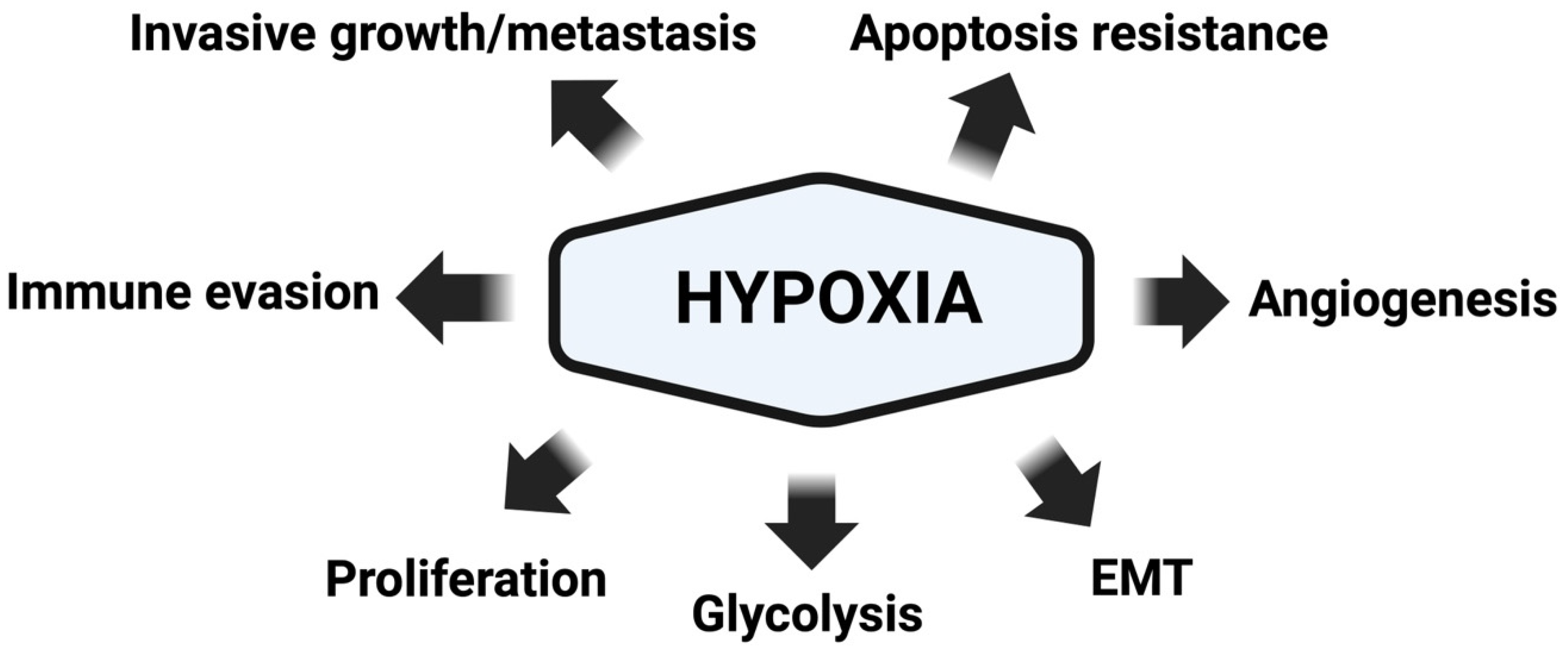
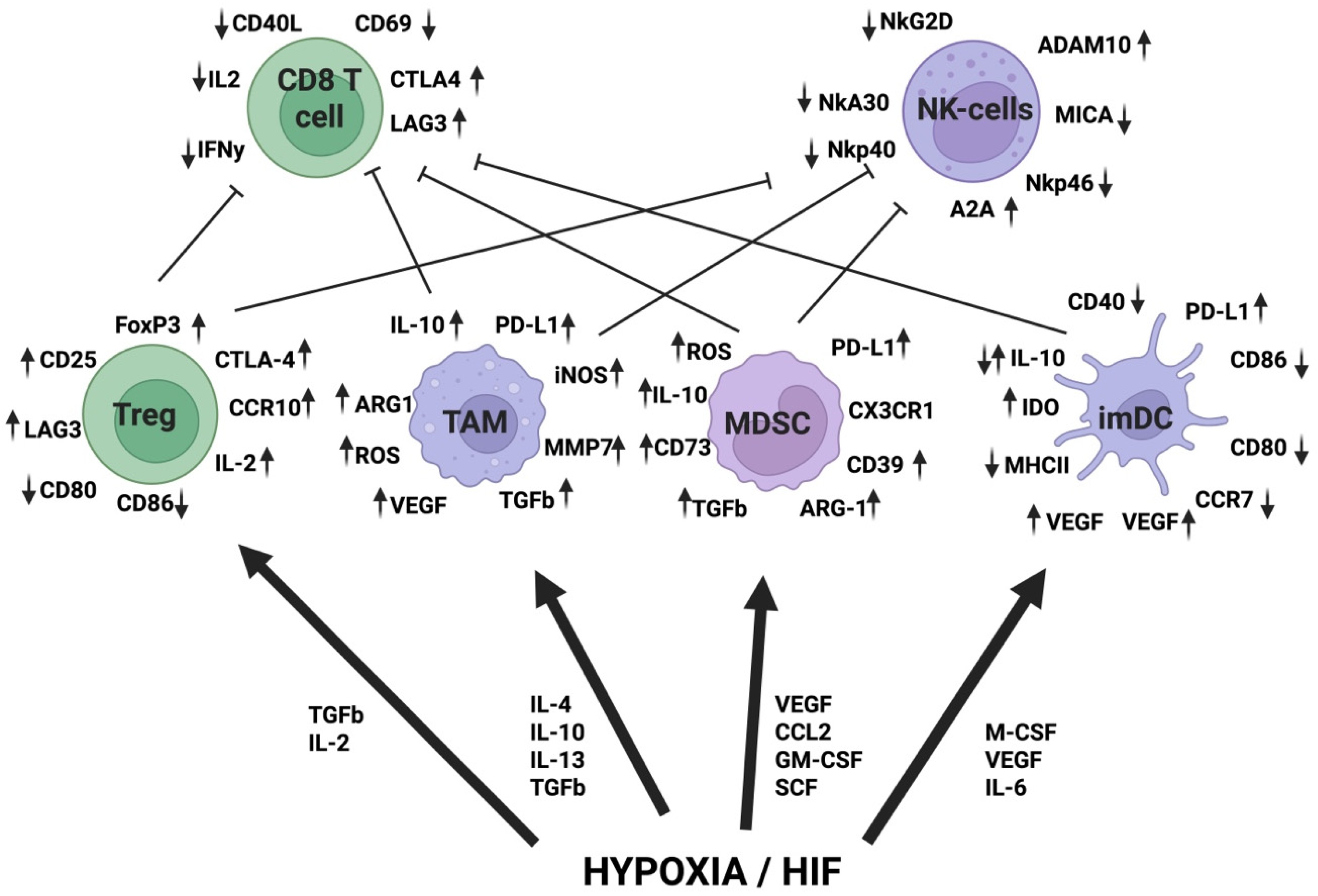
3. Hypoxia
4. Lactate
4.1. Protein Lactylation
4.2. Proteases in Inflammation and Cancer
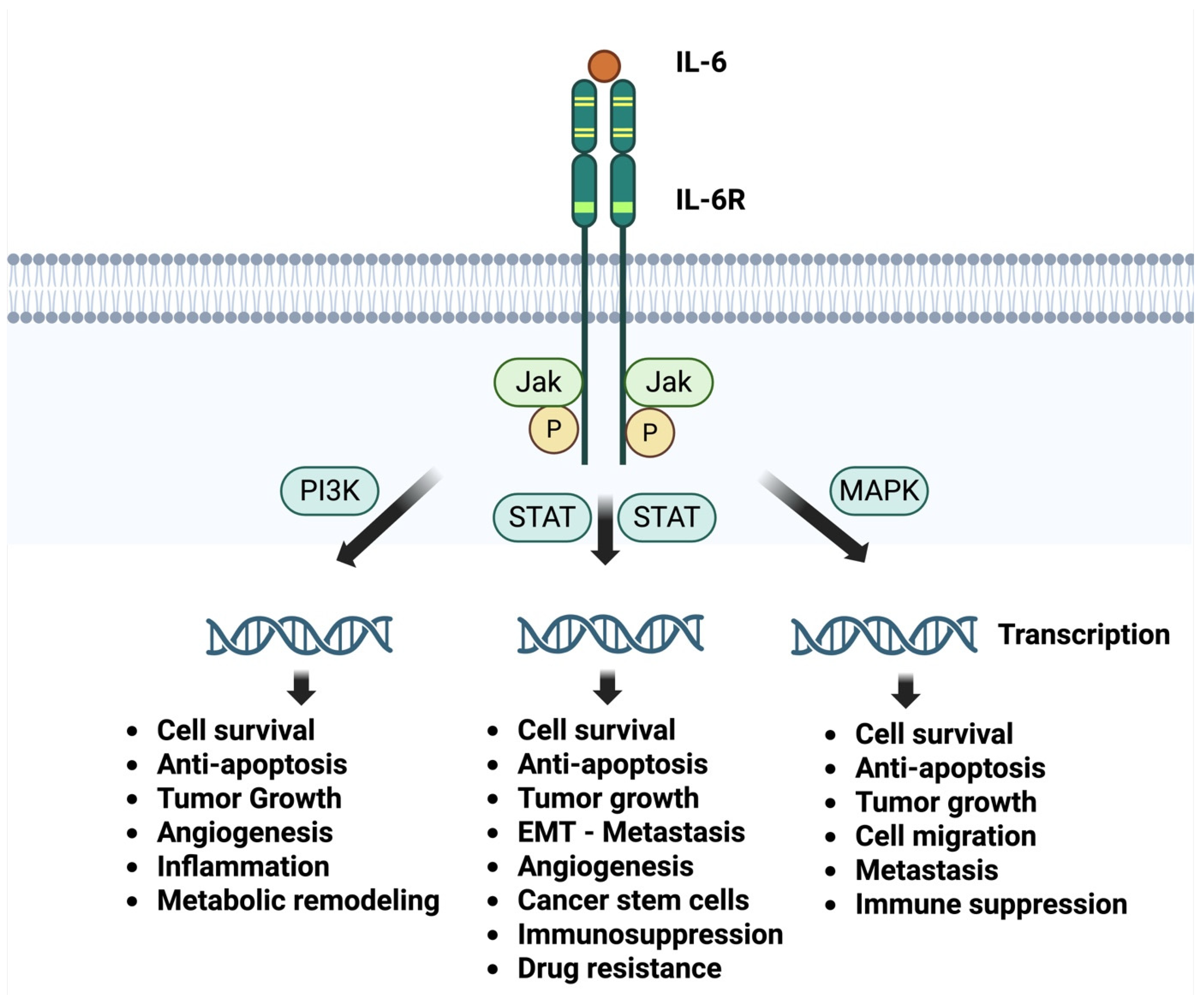
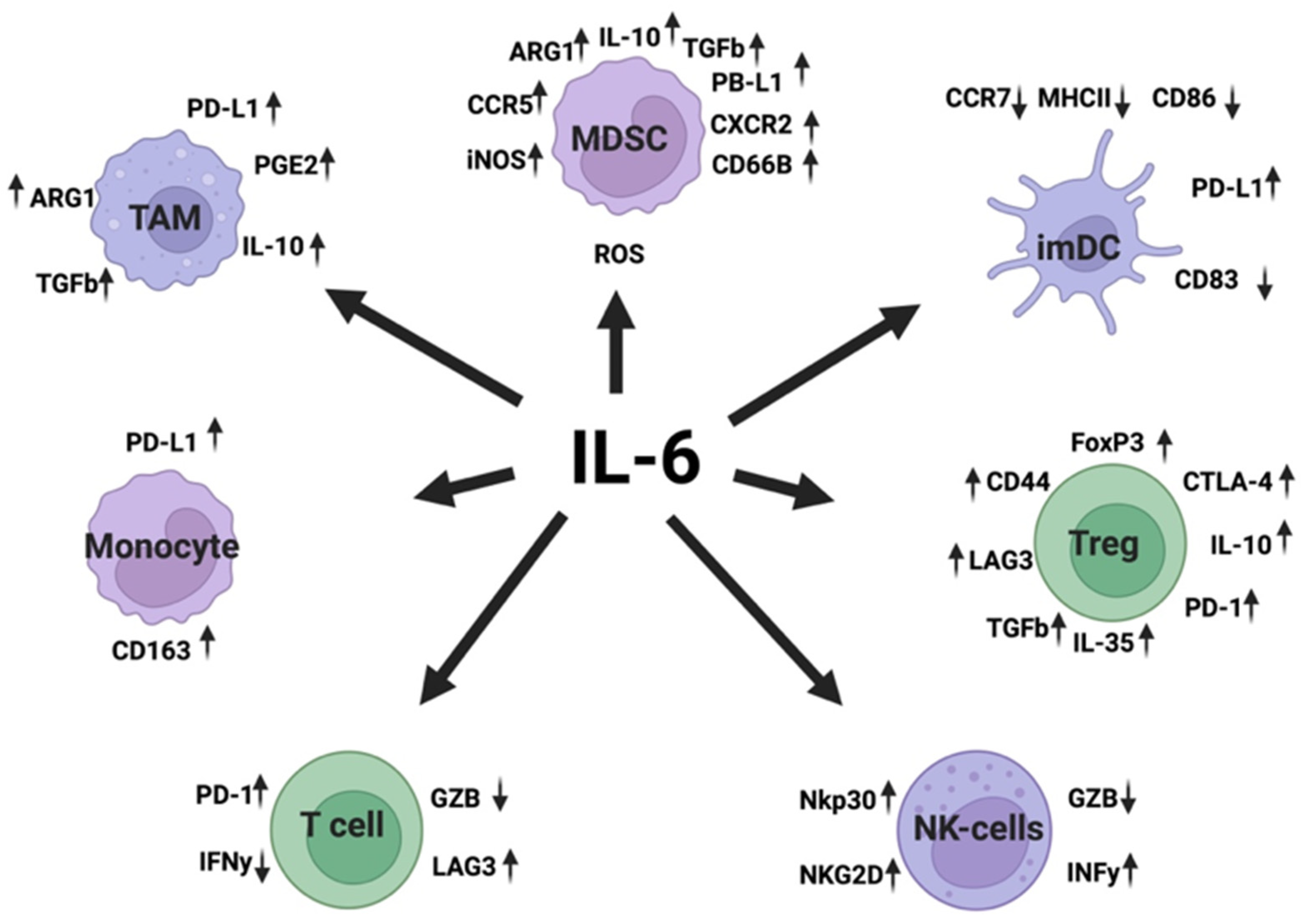
4.3. Interleukin-6
5. Stat3 Cancer
6. Synergy Between Hypoxia, IL-6 and Stat3 in Tumor Progression

| Immunoregulatory Molecules | Hypoxia References | IL-6 References |
|---|---|---|
| ARG1 | [112] | [113] |
| ROS | [114,115] | [116] |
| PD-L1 | [117,118] | [119] |
| TGFβ | [120,121] | [122,123] |
| IL-10 | [124] | [125,126] |
| VEGF | [127,128] | [129,130] |
| Adenosine | [131,132] | [133,134] |
| CD47 | [135,136] | [137,138] |
| CD73 | [139,140] | [141,142] |
| HLA-DR | [143] | [144] |
| CD80 | [143] | [145] |
| CD86 | [143,146] | [147] |
| EGFR | [148,149] | [150,151] |
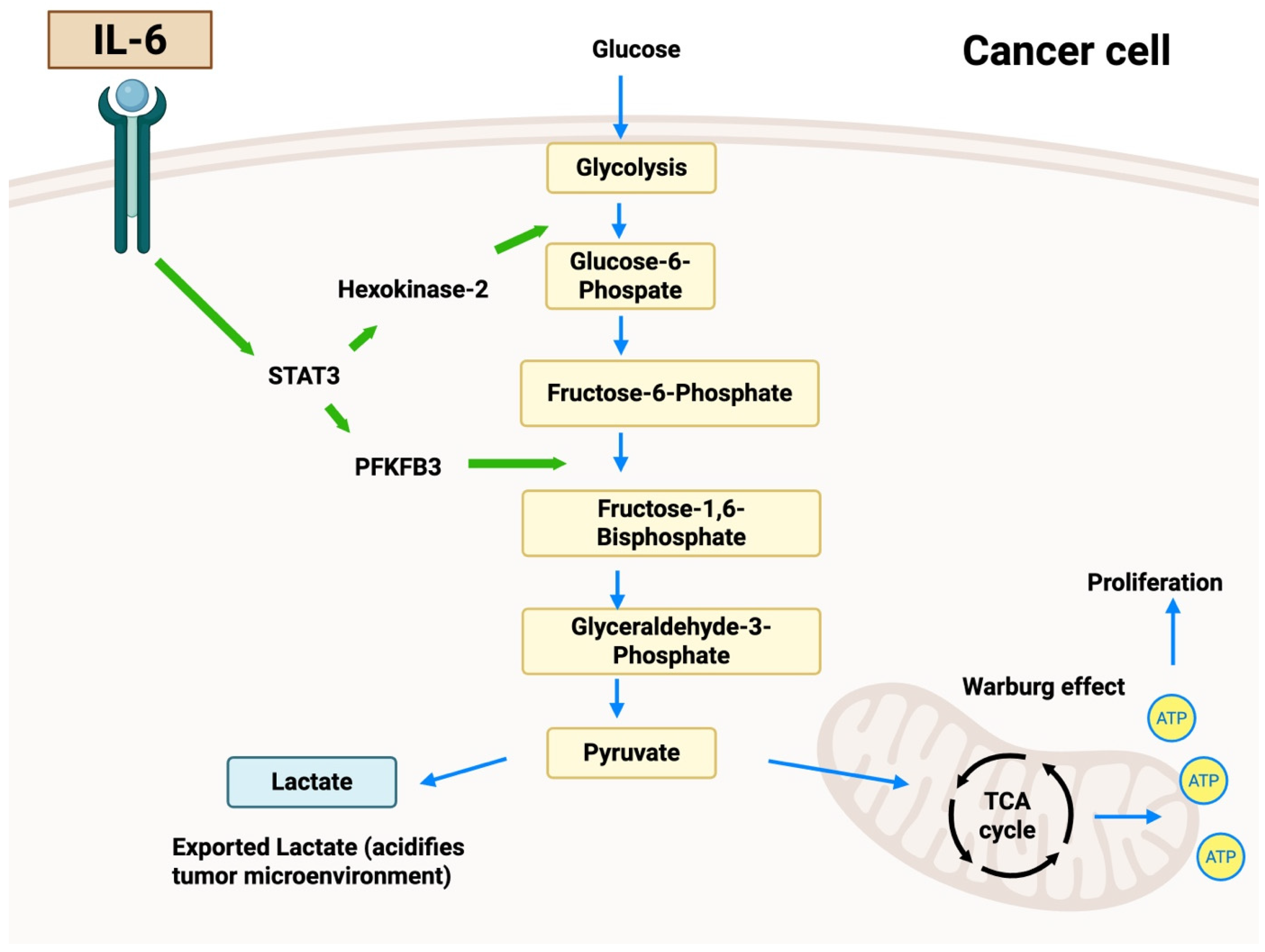
7. IL-6 Regulation of Glycolytic Enzymes
8. Discussion
9. Conclusions
Author Contributions
Funding
Data Availability Statement
Conflicts of Interest
References
- Goldmann, O.; Nwofor, O.V.; Chen, Q.; Medina, E. Mechanisms underlying immunosuppression by regulatory cells. Front. Immunol. 2024, 15, 1328193. [Google Scholar] [CrossRef] [PubMed] [PubMed Central]
- Mundhara, N.; Sadhukhan, P. Cracking the Codes behind Cancer Cells’ Immune Evasion. Int. J. Mol. Sci. 2024, 25, 8899. [Google Scholar] [CrossRef] [PubMed] [PubMed Central]
- Liu, Y.; Liu, Z.; Yang, Y.; Cui, J.; Sun, J.; Liu, Y. The prognostic and biology of tumour-infiltrating lymphocytes in the immunotherapy of cancer. Br. J. Cancer 2023, 129, 1041–1049. [Google Scholar] [CrossRef] [PubMed] [PubMed Central]
- Yang, J.; Kang, H.; Lyu, L.; Xiong, W.; Hu, Y. A target map of clinical combination therapies in oncology: An analysis of clinicaltrials.gov. Discov. Oncol. 2023, 14, 151. [Google Scholar] [CrossRef] [PubMed] [PubMed Central]
- Nair, N.U.; Greninger, P.; Zhang, X.; Friedman, A.A.; Amzallag, A.; Cortez, E.; Sahu, A.D.; Lee, J.S.; Dastur, A.; Egan, R.K.; et al. A landscape of response to drug combinations in non-small cell lung cancer. Nat. Commun. 2023, 14, 3830. [Google Scholar] [CrossRef] [PubMed]
- Liao, D.; Johnson, R.S. Hypoxia: A key regulator of angiogenesis in cancer. Cancer Metastasis Rev. 2007, 26, 281–290. [Google Scholar] [CrossRef] [PubMed]
- Lv, X.; Li, J.; Zhang, C.; Hu, T.; Li, S.; He, S.; Yan, H.; Tan, Y.; Lei, M.; Wen, M.; et al. The role of hypoxia-inducible factors in tumor angiogenesis and cell metabolism. Genes Dis. 2016, 4, 19–24. [Google Scholar] [CrossRef] [PubMed]
- Middleton, K.; Jones, J.; Lwin, Z.; Coward, J.I. Interleukin-6: An angiogenic target in solid tumours. Crit. Rev. Oncol. Hematol. 2014, 89, 129–139. [Google Scholar] [CrossRef] [PubMed]
- Tzafra, C.; Nahari, D.; Weiss Cerem, L.; Neufeld, G.; Levi, B.-Z. Interleukin 6 Induces the Expression of Vascular Endothelial Growth Factor. J. Biol. Chem. 1996, 271, 736–741. [Google Scholar] [CrossRef] [PubMed]
- Chen, Z.; Han, F.; Du, Y.; Shi, H.; Zhou, W. Hypoxic microenvironment in cancer: Molecular mechanisms and therapeutic interventions. Signal Transduct. Target. Ther. 2023, 8, 70. [Google Scholar] [CrossRef] [PubMed]
- Hubbi, M.E.; Semenza, G.L. Regulation of cell proliferation by hypoxia-inducible factors. Am. J. Physiol. Cell Physiol. 2015, 309, C775–C782. [Google Scholar] [CrossRef] [PubMed]
- Fisher, D.T.; Appenheimer, M.M.; Evans, S.S. The two faces of IL-6 in the tumor microenvironment. Semin. Immunol. 2014, 26, 38–47. [Google Scholar] [CrossRef] [PubMed]
- Yang, C.; Jiang, L.; Zhang, H.; Shimoda, L.A.; DeBerardinis, R.J.; Semenza, G.L. Analysis of hypoxia-induced metabolic reprogramming. Methods Enzymol. 2014, 542, 425–455. [Google Scholar] [CrossRef] [PubMed]
- Xie, H.; Simon, M.C. Oxygen availability and metabolic reprogramming in cancer. J. Biol. Chem. 2017, 292, 16825–16832. [Google Scholar] [CrossRef] [PubMed] [PubMed Central]
- Xu, S.; Deng, K.Q.; Lu, C.; Fu, X.; Zhu, Q.; Wan, S.; Zhang, L.; Huang, Y.; Nie, L.; Cai, H.; et al. Interleukin-6 classic and trans-signaling utilize glucose metabolism reprogramming to achieve anti- or pro-inflammatory effects. Metabolism 2024, 155, 155832. [Google Scholar] [CrossRef] [PubMed]
- Yun, Z.; Lin, Q. Hypoxia and regulation of cancer cell stemness. Adv. Exp. Med. Biol. 2014, 772, 41–53. [Google Scholar] [CrossRef] [PubMed]
- Shang, T.; Jia, Z.; Li, J.; Cao, H.; Xu, H.; Cong, L.; Ma, D.; Wang, X.; Liu, J. Unraveling the triad of hypoxia, cancer cell stemness, and drug resistance. J. Hematol. Oncol. 2025, 18, 32. [Google Scholar] [CrossRef] [PubMed]
- Wang, C.Q.; Sun, H.T.; Gao, X.M.; Ren, N.; Sheng, Y.Y.; Wang, Z.; Zheng, Y.; Wei, J.W.; Zhang, K.L.; Yu, X.X.; et al. Interleukin-6 enhances cancer stemness and promotes metastasis of hepatocellular carcinoma via up-regulating osteopontin expression. Am. J. Cancer Res. 2016, 6, 1873–1889. [Google Scholar] [PubMed] [PubMed Central]
- Lee, C.H.; Park, S.Y.; Lee, J.S.; Kim, D.S.; Kim, H.Y.; Song, M.J.; Im, S.-A.; Lee, K.-H.; Lee, D.-W.; Nikas, I.P.; et al. MSN/STAT3 drives cancer stemness and chemoresistance via IL-6/LPAR1 ligand receptor complex in triple-negative breast cancer. Breast Cancer Res. 2025, 27, 136. [Google Scholar] [CrossRef] [PubMed]
- Hapke, R.Y.; Haake, S.M. Hypoxia-induced epithelial to mesenchymal transition in cancer. Cancer Lett. 2020, 487, 10–20. [Google Scholar] [CrossRef] [PubMed] [PubMed Central]
- Jolly, M.K.; Ware, K.E.; Gilja, S.; Somarelli, J.A.; Levine, H. EMT and MET: Necessary or permissive for metastasis? Mol. Oncol. 2017, 11, 755–769. [Google Scholar] [CrossRef] [PubMed] [PubMed Central]
- Abaurrea, A.; Araujo, A.M.; Caffarel, M.M. The Role of the IL-6 Cytokine Family in Epithelial-Mesenchymal Plasticity in Cancer Progression. Int. J. Mol. Sci. 2021, 22, 8334. [Google Scholar] [CrossRef] [PubMed]
- Sun, Q.; Shang, Y.; Sun, F.; Dong, X.; Niu, J.; Li, F. Interleukin-6 Promotes Epithelial-Mesenchymal Transition and Cell Invasion through Integrin β6 Upregulation in Colorectal Cancer. Oxid. Med. Cell Longev. 2020, 2020, 8032187. [Google Scholar] [CrossRef] [PubMed] [PubMed Central]
- Djagaeva, I.; Doronkin, S. Hypoxia response pathway in border cell migration. Cell Adhes. Migr. 2010, 4, 391–395. [Google Scholar] [CrossRef] [PubMed] [PubMed Central]
- Sun, W.; Liu, D.; Li, W.; Zhang, L.; Long, G.; Wang, J.; Mei, Q.; Hu, G. Interleukin-6 promotes the migration and invasion of nasopharyngeal carcinoma cell lines and upregulates the expression of MMP-2 and MMP-9. Int. J. Oncol. 2014, 44, 1551–1560. [Google Scholar] [CrossRef] [PubMed]
- Rašková, M.; Lacina, L.; Kejík, Z.; Venhauerová, A.; Skaličková, M.; Kolář, M.; Jakubek, M.; Rosel, D.; Smetana, K., Jr.; Brábek, J. The Role of IL-6 in Cancer Cell Invasiveness and Metastasis-Overview and Therapeutic Opportunities. Cells 2022, 11, 3698. [Google Scholar] [CrossRef] [PubMed] [PubMed Central]
- Gilkes, D.M.; Semenza, G.L.; Wirtz, D. Hypoxia and the extracellular matrix: Drivers of tumour metastasis. Nat. Rev. Cancer 2014, 14, 430–439. [Google Scholar] [CrossRef] [PubMed] [PubMed Central]
- Jiang, X.; Ou, L.; Fan, X.; Yang, J.; Zhang, C.; Liu, Q. Research on hypoxia and the extracellular matrix in cancer: A visualization analysis. Eurasian J. Med. Oncol. 2025, 9, 168–192. [Google Scholar] [CrossRef]
- He, Q.; Lin, Y.; Liao, B.; Zhou, L.; Ai, J.; Jin, X.; Li, H.; Wang, K. The role of interleukin-6/interleukin-6 receptor signaling in the mechanical stress-induced extracellular matrix remodeling of bladder smooth muscle. Arch. Biochem Biophys. 2021, 702, 108674. [Google Scholar] [CrossRef] [PubMed]
- Yuan, Z.; Li, Y.; Zhang, S.; Wang, X.; Dou, H.; Yu, X.; Zhang, Z.; Yang, S.; Xiao, M. Extracellular matrix remodeling in tumor progression and immune escape: From mechanisms to treatments. Mol. Cancer 2023, 22, 48. [Google Scholar] [CrossRef] [PubMed] [PubMed Central]
- Rankin, E.B.; Giaccia, A.J. Hypoxic control of metastasis. Science 2016, 352, 175–180. [Google Scholar] [CrossRef] [PubMed] [PubMed Central]
- Lu, X.; Kang, Y. Hypoxia and hypoxia-inducible factors: Master regulators of metastasis. Clin. Cancer Res. 2010, 16, 5928–5935. [Google Scholar] [CrossRef] [PubMed] [PubMed Central]
- Ara, T.; Declerck, Y.A. Interleukin-6 in bone metastasis and cancer progression. Eur. J. Cancer 2010, 46, 1223–1231. [Google Scholar] [CrossRef] [PubMed] [PubMed Central]
- Nejad, E.A.; Najafgholian, S.; Rostami, A.; Sistani, A.; Shojaeifar, S.; Esparvarinha, M.; Nedaeinia, R.; Haghjooy Javanmard, S.; Taherian, M.; Ahmadlou, M.; et al. The role of hypoxia in the tumor microenvironment and development of cancer stem cell: A novel approach to developing treatment. Cancer Cell Int. 2021, 21, 62. [Google Scholar] [CrossRef] [PubMed] [PubMed Central]
- Vaupel, P. The role of hypoxia-induced factors in tumor progression. Oncologist 2004, 9, 10–17. [Google Scholar] [CrossRef] [PubMed]
- Meng, F.; Yamagiwa, Y.; Ueno, Y.; Patel, T. Over-expression of interleukin-6 enhances cell survival and transformed cell growth in human malignant cholangiocytes. J. Hepatol. 2006, 44, 1055–1065. [Google Scholar] [CrossRef] [PubMed] [PubMed Central]
- Kumari, N.; Dwarakanath, B.S.; Das, A.; Bhatt, A.N. Role of interleukin-6 in cancer progression and therapeutic resistance. Tumour Biol. 2016, 37, 11553–11572. [Google Scholar] [CrossRef] [PubMed]
- Wu, Q.; You, L.; Nepovimova, E.; Heger, Z.; Wu, W.; Kuča, K.; Adam, V. Hypoxia-inducible factors: Master regulators of hypoxic tumor immune escape. J. Hematol. Oncol. 2022, 15, 77. [Google Scholar] [CrossRef] [PubMed]
- Mortezaee, K.; Majidpoor, J. The impact of hypoxia on immune state in cancer. Life Sci. 2021, 286, 120057. [Google Scholar] [CrossRef] [PubMed]
- Li, W.; Wu, Z.; Meng, W.; Zhang, C.; Cheng, M.; Chen, Y.; Zou, Y.; Li, K.; Lin, S.; Xiong, W.; et al. Blockade of IL-6 inhibits tumor immune evasion and improves anti-PD-1 immunotherapy. Cytokine 2022, 158, 155976. [Google Scholar] [CrossRef] [PubMed]
- Montero-Calle, A.; Garranzo-Asensio, M.; Moreno-Casbas, M.T.; Campuzano, S.; Barderas, R. Autoantibodies in cancer: A systematic review of their clinical role in the most prevalent cancers. Front. Immunol. 2024, 15, 1455602. [Google Scholar] [CrossRef] [PubMed]
- de Jonge, H.; Iamele, L.; Maggi, M.; Pessino, G.; Scotti, C. Anti-Cancer Auto-Antibodies: Roles, Applications and Open Issues. Cancers 2021, 13, 813. [Google Scholar] [CrossRef] [PubMed] [PubMed Central]
- Mkaddem, B.S.; Benhamou, M.; Monteiro, R.C. Understanding Fc Receptor Involvement in Inflammatory Diseases: From Mechanisms to New Therapeutic Tools. Front. Immunol. 2019, 10, 811. [Google Scholar] [CrossRef] [PubMed] [PubMed Central]
- Berger, S.; Balló, H.; Stutte, H.J. Immune complex-induced interleukin-6, interleukin-10 and prostaglandin secretion by human monocytes: A network of pro- and anti-inflammatory cytokines dependent on the antigen: Antibody ratio. Eur. J. Immunol. 1996, 26, 1297–1301. [Google Scholar] [CrossRef] [PubMed]
- Charab, W.; Rosenberger, M.G.; Shivram, H.; Mirazee, J.M.; Donkor, M.; Shekhar, S.R.; Gjuka, D.; Khoo, K.H.; Kim, J.E.; Iyer, V.R.; et al. IgG Immune Complexes Inhibit Naïve T Cell Proliferation and Suppress Effector Function in Cytotoxic T Cells. Front. Immunol. 2021, 12, 713704. [Google Scholar] [CrossRef] [PubMed] [PubMed Central]
- Dass, T.K.; Aziz, M.; Rattan, A. Prognostic significance of circulating immune complexes in cancer patients. Jpn. J. Cancer Res. 1991, 82, 1284–1291. [Google Scholar] [CrossRef] [PubMed] [PubMed Central]
- Argyrios, T.N. Immune Complexes in Cancer. N. Engl. J. Med. 1982, 307, 1208–1209. [Google Scholar] [CrossRef] [PubMed]
- Hellström, I.; Sjögren, H.O.; Warner, G.; Hellström, K.E. Blocking of cell-mediated tumor immunity by sera from patients with growing neoplasms. Int. J. Cancer 1971, 7, 226–237. [Google Scholar] [CrossRef] [PubMed]
- Hellström, I.; Warner, G.A.; Hellström, K.E.; Sjögren, H.O. Sequential studies on cell-mediated tumor immunity and blocking serum activity in ten patients with malignant melanoma. Int. J. Cancer 1973, 11, 280–292. [Google Scholar] [CrossRef] [PubMed]
- Hellström, I.; Hellström, K.E.; Sjögren, H.O.; Warner, G.A. Serum factors in tumor-free patients cancelling the blocking of cell-mediated tumor immunity. Int. J. Cancer 1971, 8, 185–191. [Google Scholar] [CrossRef] [PubMed]
- Bansal, S.C.; Sjögren, H.O. Counteraction of the blocking of cell-mediated tumor immunity by inoculation of unblocking sera and splenectomy: Immunotherapeutic effects on primary polyoma tumors in rats. Int. J. Cancer 1972, 9, 490–509. [Google Scholar] [CrossRef] [PubMed]
- Sjögren, H.O.; Hellström, I.; Bansal, S.C.; Hellström, K.E. Suggestive evidence that the “blocking antibodies” of tumor-bearing individuals may be antigen--antibody complexes. Proc. Natl. Acad. Sci. USA 1971, 68, 1372–1375. [Google Scholar] [CrossRef] [PubMed] [PubMed Central]
- Hakansson, L.; Dunér, P.; Broströmer, E.; Gustavsson, B.; Wettergren, Y.; Ghafouri, B.; Hakansson, A.; Clinchy, B. A New IL-6-Inducing Mechanism in Cancer with New Therapeutic Possibilities. Cancers 2024, 16, 3588. [Google Scholar] [CrossRef] [PubMed] [PubMed Central]
- Hu, J.; Li, X.; Yang, L.; Li, H. Hypoxia, a key factor in the immune microenvironment. Biomed. Pharmacother. 2022, 151, 113068. [Google Scholar] [CrossRef] [PubMed]
- Fu, Z.; Mowday, A.M.; Smaill, J.B.; Hermans, I.F.; Patterson, A.V. Tumour Hypoxia-Mediated Immunosuppression: Mechanisms and Therapeutic Approaches to Improve Cancer Immunotherapy. Cells 2021, 10, 1006. [Google Scholar] [CrossRef] [PubMed] [PubMed Central]
- Wicks, E.E.; Semenza, G.L. Hypoxia-inducible factors: Cancer progression and clinical translation. J. Clin. Investig. 2022, 132, e159839. [Google Scholar] [CrossRef] [PubMed] [PubMed Central]
- Otto, W. The Metabolism of Carcinoma Cells. J. Cancer Res. 1925, 9, 148–163. [Google Scholar] [CrossRef]
- Kierans, S.J.; Taylor, C.T. Regulation of glycolysis by the hypoxia-inducible factor (HIF): Implications for cellular physiology. J. Physiol. 2021, 599, 23–37. [Google Scholar] [CrossRef] [PubMed]
- Chen, J.; Huang, Z.; Chen, Y.; Tian, H.; Chai, P.; Shen, Y.; Yao, Y.; Xu, S.; Ge, S.; Jia, R. Lactate and lactylation in cancer. Signal Transduct. Target. Ther. 2025, 10, 38. [Google Scholar] [CrossRef] [PubMed]
- Kim, E.Y.; Abides, J.; Keller, C.R.; Martinez, S.R.; Li, W. Tumor Microenvironment Lactate: Is It a Cancer Progression Marker, Immunosuppressant, and Therapeutic Target? Molecules 2025, 30, 1763. [Google Scholar] [CrossRef] [PubMed]
- Quinn, W.J., 3rd; Jiao, J.; TeSlaa, T.; Stadanlick, J.; Wang, Z.; Wang, L.; Akimova, T.; Angelin, A.; Schäfer, P.M.; Cully, M.D.; et al. Lactate Limits T Cell Proliferation via the NAD(H) Redox State. Cell Rep. 2020, 33, 108500. [Google Scholar] [CrossRef] [PubMed] [PubMed Central]
- Llibre, A.; Kucuk, S.; Gope, A.; Certo, M.; Mauro, C. Lactate: A key regulator of the immune response. Immunity 2025, 58, 535–554. [Google Scholar] [CrossRef] [PubMed]
- Zeng, Y.; Huang, Y.; Tan, Q.; Peng, L.; Wang, J.; Tong, F.; Dong, X. Influence of lactate in resistance to anti-PD-1/PD-L1 therapy: Mechanisms and clinical applications (Review). Mol. Med. Rep. 2025, 31, 48. [Google Scholar] [CrossRef] [PubMed]
- Barbieri, L.; Veliça, P.; Gameiro, P.A.; Cunha, P.P.; Foskolou, I.P.; Rullman, E.; Bargiela, D.; Johnson, R.S.; Rundqvist, H. Lactate exposure shapes the metabolic and transcriptomic profile of CD8+ T cells. Front. Immunol. 2023, 14, 1101433. [Google Scholar] [CrossRef] [PubMed] [PubMed Central]
- Feng, Q.; Liu, Z.; Yu, X.; Huang, T.; Chen, J.; Wang, J.; Wilhelm, J.; Li, S.; Song, J.; Li, W. Lactate increases stemness of CD8 + T cells to augment anti-65tumor immunity. Nat. Commun. 2022, 13, 4981. [Google Scholar] [CrossRef] [PubMed]
- Zhang, X.; Liang, C.; Wu, C.; Wan, S.; Xu, L.; Wang, S.; Wang, J.; Huang, X.; Xu, L. A rising star involved in tumour immunity: Lactylation. J. Cell Mol. Med. 2024, 28, e70146. [Google Scholar] [CrossRef] [PubMed]
- Shi, W.; Cassmann, T.J.; Bhagwate, A.V.; Hitosugi, T.; Ip, W.K.E. Lactic acid induces transcriptional repression of macrophage inflammatory response via histone acetylation. Cell Rep. 2024, 43, 113746. [Google Scholar] [CrossRef] [PubMed]
- Li, X.M.; Yang, Y.; Jiang, F.Q.; Hu, G.; Wan, S.; Yan, W.Y.; He, X.S.; Xiao, F.; Yang, X.M.; Guo, X. Histone lactylation inhibits RARγ expression in macrophages to promote colorectal tumorigenesis through activation of TRAF6-IL-6-STAT3 signaling. Cell Rep. 2024, 43, 113688. [Google Scholar] [CrossRef] [PubMed]
- Li, W.; Zhou, C.; Yu, L.; Hou, Z.; Liu, H.; Kong, L.; Xu, Y.; He, J.; Lan, J.; Ou, Q. Tumor-derived lactate promotes resistance to bevacizumab treatment by facilitating autophagy enhancer protein RUBCNL expression through histone H3 lysine 18 lactylation (H3K18la) in colorectal cancer. Autophagy 2024, 20, 114–130. [Google Scholar] [CrossRef] [PubMed]
- Li, F.; Zhang, H.; Huang, Y.; Li, D.; Zheng, Z.; Xie, K.; Cao, C.; Wang, Q.; Zhao, X.; Huang, Z. Single-cell transcriptome analysis reveals the association between histone lactylation and cisplatin resistance in bladder cancer. Drug Resist. Updates 2024, 73, 101059. [Google Scholar] [CrossRef] [PubMed]
- Huang, G.; Chen, S.; He, J.; Li, H.; Ma, Z.; Lubamba, G.P.; Wang, L.; Guo, Z.; Li, C. Histone lysine lactylation (Kla)-induced BCAM promotes OSCC progression and cis-Platinum resistance. Oral Dis. 2024, 31, 1116–1132. [Google Scholar] [CrossRef] [PubMed]
- Wang, R.; Li, C.; Cheng, Z.; Li, M.; Shi, J.; Zhang, Z.; Jin, S.; Ma, H. H3K9 lactylation in malignant cells facilitates CD8(+) T cell dysfunction and poor immunotherapy response. Cell Rep. 2024, 43, 114686. [Google Scholar] [CrossRef] [PubMed]
- Radisky, E.S. Extracellular proteolysis in cancer: Proteases, substrates, and mechanisms in tumor progression and metastasis. J. Biol. Chem. 2024, 300, 107347. [Google Scholar] [CrossRef] [PubMed] [PubMed Central]
- Vizovisek, M.; Ristanovic, D.; Menghini, S.; Christiansen, M.G.; Schuerle, S. The Tumor Proteolytic Landscape: A Challenging Frontier in Cancer Diagnosis and Therapy. Int. J. Mol. Sci. 2021, 22, 2514. [Google Scholar] [CrossRef] [PubMed]
- Salardani, M.; Barcick, U.; Zelanis, A. Proteolytic signaling in cancer. Expert Rev. Proteom. 2023, 20, 345–355. [Google Scholar] [CrossRef] [PubMed]
- Wojtukiewicz, M.Z.; Hempel, D.; Sierko, E.; Tucker, S.C.; Honn, K.V. Protease-activated receptors (PARs)--biology and role in cancer invasion and metastasis. Cancer Metastasis Rev. 2015, 34, 775–796. [Google Scholar] [CrossRef] [PubMed] [PubMed Central]
- Nadendla, E.K.; Tweedell, R.E.; Kasof, G.; Kanneganti, T.D. Caspases: Structural and molecular mechanisms and functions in cell death, innate immunity, and disease. Cell Discov. 2025, 11, 42. [Google Scholar] [CrossRef] [PubMed] [PubMed Central]
- Wolf, M.; Albrecht, S.; Märki, C. Proteolytic processing of chemokines: Implications in physiological and pathological conditions. Int. J. Biochem Cell Biol. 2008, 40, 1185–1198. [Google Scholar] [CrossRef] [PubMed]
- Afonina, I.S.; Müller, C.; Martin, S.J.; Beyaert, R. Proteolytic Processing of Interleukin-1 Family Cytokines: Variations on a Common Theme. Immunity 2015, 42, 991–1004. [Google Scholar] [CrossRef] [PubMed]
- Jenkins, G. The role of proteases in transforming growth factor-beta activation. Int. J. Biochem Cell Biol. 2008, 40, 1068–1078. [Google Scholar] [CrossRef] [PubMed]
- Sower, L.E.; Froelich, C.J.; Carney, D.H.; Fenton, J.W., 2nd; Klimpel, G.R. Thrombin induces IL-6 production in fibroblasts and epithelial cells. Evidence for the involvement of the seven-transmembrane domain (STD) receptor for alpha-thrombin. J. Immunol. 1995, 155, 895–901. [Google Scholar] [CrossRef] [PubMed]
- Li, T.; Wang, H.; He, S. Induction of interleukin-6 release from monocytes by serine proteinases and its potential mechanisms. Scand. J. Immunol. 2006, 64, 10–16. [Google Scholar] [CrossRef] [PubMed]
- Lippitz, B.E.; Harris, R.A. Cytokine patterns in cancer patients: A review of the correlation between interleukin 6 and prognosis. OncoImmunology 2016, 5, 1093722. [Google Scholar] [CrossRef] [PubMed]
- Lu, C.-C.; Kuo, H.-C.; Wang, F.-S.; Jou, M.-H.; Lee, K.-C.; Chuang, J.-H. Upregulation of TLRs and IL-6 as a Marker in Human Colorectal Cancer. Int. J. Mol. Sci. 2015, 16, 159–177. [Google Scholar] [CrossRef] [PubMed]
- Khan, S.; Wardill, H.R.; Bowen, J.M. Role of toll-like receptor 4 (TLR4)-mediated interleukin-6 (IL-6) production in chemotherapy-induced mucositis. Cancer Chemother. Pharmacol. 2018, 82, 31–37. [Google Scholar] [CrossRef] [PubMed]
- Brahmakshatriya, V.; Kuang, Y.; Devarajan, P.; Xia, J.; Zhang, W.; Vong, A.M.; Swain, S.L. IL-6 Production by TLR-Activated APC Broadly Enhances Aged Cognate CD4 Helper and B Cell Antibody Responses In Vivo. J. Immunol. 2017, 198, 2819–2833. [Google Scholar] [CrossRef] [PubMed] [PubMed Central]
- Boyden, S. Autoimmunity and Inflammation. Nature 1964, 201, 200–201. [Google Scholar] [CrossRef] [PubMed]
- Wilkin, T.J. The primary lesion theory of autoimmunity: A speculative hypothesis. Autoimmunity 1990, 7, 225–235, Erratum in Autoimmunity 1991, 9, 187. [Google Scholar] [CrossRef] [PubMed]
- Hirano, T. IL-6 in inflammation, autoimmunity and cancer. Int. Immunol. 2021, 33, 127–148. [Google Scholar] [CrossRef] [PubMed] [PubMed Central]
- Hideshima, T.; Nakamura, N.; Chauhan, D.; Anderson, K.C. Biologic sequelae of interleukin-6 induced PI3-K/Akt signaling in multiple myeloma. Oncogene 2001, 20, 5991–6000. [Google Scholar] [CrossRef] [PubMed]
- Hilal, G. Involvement of Interleukin-6 Induced PI3K/Akt/mTor Pathway in the Regulation of Telomerase and Alpha-Fetoprotein Expression in Hepatocellular Carcinoma. Clin. Oncol. 2023, 6, 1–13. [Google Scholar]
- Ogata, A.; Chauhan, D.; Teoh, G.; Treon, S.P.; Urashima, M.; Schlossman, R.L.; Anderson, K.C. IL-6 triggers cell growth via the Ras-dependent mitogen-activated protein kinase cascade. J. Immunol. 1997, 159, 2212–2221. [Google Scholar] [CrossRef] [PubMed]
- Badache, A.; Hynes, N.E. Interleukin 6 inhibits proliferation and, in cooperation with an epidermal growth factor receptor autocrine loop, increases migration of T47D breast cancer cells. Cancer Res. 2001, 61, 383–391. [Google Scholar] [PubMed]
- Zi, Y.; Liu, L.; Gao, J.; Xu, X.; Guan, Y.; Rong, Z.; Cao, Z.; Li, M.; Zeng, Z.; Fan, Q.; et al. Phosphorylation of PPDPF via IL6-JAK2 activates the Wnt/β-catenin pathway in colorectal cancer. EMBO Rep. 2023, 24, e55060. [Google Scholar] [CrossRef] [PubMed] [PubMed Central]
- Gao, S.; Hu, J.; Wu, X.; Liang, Z. PMA treated THP-1-derived-IL-6 promotes EMT of SW48 through STAT3/ERK-dependent activation of Wnt/β-catenin signaling pathway. Biomed. Pharmacother. 2018, 108, 618–624. [Google Scholar] [CrossRef] [PubMed]
- Wongchana, W.; Palaga, T. Direct regulation of interleukin-6 expression by Notch signaling in macrophages. Cell Mol. Immunol. 2012, 9, 155–162. [Google Scholar] [CrossRef] [PubMed] [PubMed Central]
- Sansone, P.; Storci, G.; Tavolari, S.; Guarnieri, T.; Giovannini, C.; Taffurelli, M.; Ceccarelli, C.; Santini, D.; Paterini, P.; Marcu, K.B.; et al. IL-6 triggers malignant features in mammospheres from human ductal breast carcinoma and normal mammary gland. J. Clin. Investig. 2007, 117, 3988–4002. [Google Scholar] [CrossRef] [PubMed] [PubMed Central]
- Yang, L.; Wang, L.; Lin, H.K.; Kan, P.Y.; Xie, S.; Tsai, M.Y.; Wang, P.H.; Chen, Y.T.; Chang, C. Interleukin-6 differentially regulates androgen receptor transactivation via PI3K-Akt, STAT3, and MAPK, three distinct signal pathways in prostate cancer cells. Biochem Biophys. Res. Commun. 2003, 305, 462–469. [Google Scholar] [CrossRef] [PubMed]
- Tian, Y.; Qi, H.; Zhu, N.; Yan, X.; Guo, L.; Wang, L.; Teng, X.; Xiang, X.; Zhu, L.; Yuan, Y.; et al. An IL-6-induced STAT3-to-PI3K signaling switch potently drives PD-L1 transcription in cancer stem cells of colorectal cancer. Sci. Bull. 2025, 71, 273–277. [Google Scholar] [CrossRef] [PubMed]
- Libermann, T.A.; Baltimore, D. Activation of interleukin-6 gene expression through the NF-kappa B transcription factor. Mol. Cell Biol. 1990, 10, 2327–2334. [Google Scholar] [CrossRef] [PubMed] [PubMed Central]
- Wang, L.; Walia, B.; Evans, J.; Gewirtz, A.T.; Merlin, D.; Sitaraman, S.V. IL-6 induces NF-kappa B activation in the intestinal epithelia. J. Immunol. 2003, 171, 3194–3201. [Google Scholar] [CrossRef] [PubMed]
- Montgomery, A.; Tam, F.; Gursche, C.; Cheneval, C.; Besler, K.; Enns, W.; Manku, S.; Rey, K.; Hanson, P.J.; Rose-John, S.; et al. Overlapping and distinct biological effects of IL-6 classic and trans-signaling in vascular endothelial cells. Am. J. Physiol. Cell Physiol. 2021, 320, C554–C565. [Google Scholar] [CrossRef] [PubMed]
- Hu, Y.; Dong, Z.; Liu, K. Unraveling the complexity of STAT3 in cancer: Molecular understanding and drug discovery. J. Exp. Clin. Cancer Res. 2024, 43, 23. [Google Scholar] [CrossRef] [PubMed]
- Dai, Y.; Ying, Y.; Zhu, G.; Xu, Y.; Ji, K. STAT3 drives the expression of HIF1alpha in cancer cells through a novel super-enhancer. Biochem. Biophys. Res. Commun. 2024, 735, 150483. [Google Scholar] [CrossRef] [PubMed]
- Dinarello, A.; Betto, R.M.; Diamante, L.; Tesoriere, A.; Ghirardo, R.; Cioccarelli, C.; Meneghetti, G.; Peron, M.; Laquatra, C.; Tiso, N.; et al. STAT3 and HIF1α cooperatively mediate the transcriptional and physiological responses to hypoxia. Cell Death Discov. 2023, 9, 226. [Google Scholar] [CrossRef] [PubMed]
- Nishiwaki, N.; Noma, K.; Ohara, T.; Kunitomo, T.; Kawasaki, K.; Akai, M.; Kobayashi, T.; Narusaka, T.; Kashima, H.; Sato, H.; et al. Overcoming cancer-associated fibroblast-induced immunosuppression by anti-interleukin-6 receptor antibody. Cancer Immunol. Immunother. 2023, 72, 2029–2044. [Google Scholar] [CrossRef] [PubMed] [PubMed Central]
- Xu, S.; Yu, C.; Ma, X.; Li, Y.; Shen, Y.; Chen, Y.; Huang, S.; Zhang, T.; Deng, W.; Wang, Y. IL-6 promotes nuclear translocation of HIF-1α to aggravate chemoresistance of ovarian cancer cells. Eur. J. Pharmacol. 2021, 894, 173817. [Google Scholar] [CrossRef] [PubMed]
- Gao, X.; Li, Y.; Wang, H.; Li, C.; Ding, J. Inhibition of HIF-1α decreases expression of pro-inflammatory IL-6 and TNF-α in diabetic retinopathy. Acta Ophthalmol. 2017, 95, e746–e750. [Google Scholar] [CrossRef] [PubMed]
- Zhang, T.; Yang, J.; Sun, Y.; Song, J.; Gao, D.; Huang, S.; Pang, A.; Zhang, J.; Wang, J.; Wang, Y.; et al. Interleukin-6 and Hypoxia Synergistically Promote EMT-Mediated Invasion in Epithelial Ovarian Cancer via the IL-6/STAT3/HIF-1α Feedback Loop. Anal. Cell Pathol. 2023, 2023, 8334881. [Google Scholar] [CrossRef] [PubMed] [PubMed Central]
- Carter, R.; Petrik, J.J.; Moorehead, R.A. Overexpression of miR-200s inhibits proliferation and invasion while increasing apoptosis in murine ovarian cancer cells. PLoS ONE 2024, 19, e0307178. [Google Scholar] [CrossRef] [PubMed] [PubMed Central]
- Guo, Q.Y.; Song, J.N.; Chen, Y.M.; Yuan, H.N.; Xue, W.S.; Sun, Y.; Niu, X.L.; Wang, Y.; Chen, X. IL-6 regulates epithelial ovarian cancer EMT, invasion, and metastasis by modulating Let-7c and miR-200c through the STAT3/HIF-1α pathway. Med. Oncol. 2024, 41, 155. [Google Scholar] [CrossRef] [PubMed]
- Wei, C.; Liu, X.; Wang, Q.; Li, Q.; Xie, M. Identification of Hypoxia Signature to Assess the Tumor Immune Microenvironment and Predict Prognosis in Patients with Ovarian Cancer. Int. J. Endocrinol. 2021, 2021, 4156187. [Google Scholar] [CrossRef] [PubMed] [PubMed Central]
- Das, A.; Roy, S.; Bairagi, A.; Alam, N.; Chatterjee, N. IL-6 mediated CD206+ARG-1+ tumor associated macrophage polarization induces Treg infiltration in non-responder luminal A breast cancer. FEBS Lett. 2025, 599, 739–754. [Google Scholar] [CrossRef] [PubMed]
- Mendoza, E.N.; Ciriolo, M.R.; Ciccarone, F. Hypoxia-Induced Reactive Oxygen Species: Their Role in Cancer Resistance and Emerging Therapies to Overcome It. Antioxidants 2025, 14, 94. [Google Scholar] [CrossRef] [PubMed] [PubMed Central]
- Korbecki, J.; Simińska, D.; Gąssowska-Dobrowolska, M.; Listos, J.; Gutowska, I.; Chlubek, D.; Baranowska-Bosiacka, I. Chronic and Cycling Hypoxia: Drivers of Cancer Chronic Inflammation through HIF-1 and NF-κB Activation: A Review of the Molecular Mechanisms. Int. J. Mol. Sci. 2021, 22, 10701. [Google Scholar] [CrossRef] [PubMed] [PubMed Central]
- Li, J.; Lan, T.; Zhang, C.; Zeng, C.; Hou, J.; Yang, Z.; Zhang, M.; Liu, J.; Liu, B. Reciprocal activation between IL-6/STAT3 and NOX4/Akt signalings promotes proliferation and survival of non-small cell lung cancer cells. Oncotarget 2015, 6, 1031–1048. [Google Scholar] [CrossRef] [PubMed] [PubMed Central]
- Er, Ö.S.; Efil, S.C.; Çanakçı, D.; Ağaçkıran, Y.; Şener Dede, D.; Onak Kandemir, N.; Doğan, M.; Ünal, T.D.K.; Kıran, M.M.; Kayaçetin, S.; et al. Correlation of PD-L1 and HIF-1 Alpha Expression with KRAS Mutation and Clinicopathological Parameters in Non-Small Cell Lung Cancer . Curr. Issues Mol. Biol. 2025, 47, 121. [Google Scholar] [CrossRef] [PubMed] [PubMed Central]
- Yu, Y.; Sun, R.; Hu, F.; Ding, Z.; Li, X.; Han, J.; Liang, L.; Wang, T.; Xi, G.; Dong, X.; et al. Hypoxia upregulates the expression of PD-L1 via NPM1 in breast cancer. J. Immunother. Cancer 2025, 13, e010151. [Google Scholar] [CrossRef] [PubMed] [PubMed Central]
- Jeong, H.; Koh, J.; Kim, S.; Yim, J.; Song, S.G.; Kim, H.; Li, Y.; Lee, S.H.; Chung, Y.K.; Kim, H.; et al. Cell-intrinsic PD-L1 signaling drives immunosuppression by myeloid-derived suppressor cells through IL-6/Jak/Stat3 in PD-L1-high lung cancer. J. Immunother. Cancer 2025, 13, e010612. [Google Scholar] [CrossRef] [PubMed] [PubMed Central]
- Mallikarjuna, P.; Zhou, Y.; Landström, M. The Synergistic Cooperation between TGF-β and Hypoxia in Cancer and Fibrosis. Biomolecules 2022, 12, 635. [Google Scholar] [CrossRef] [PubMed] [PubMed Central]
- D’Ignazio, L.; Batie, M.; Rocha, S. Hypoxia and Inflammation in Cancer, Focus on HIF and NF-κB. Biomedicines 2017, 5, 21. [Google Scholar] [CrossRef] [PubMed] [PubMed Central]
- Shi, J.; Feng, J.; Xie, J.; Mei, Z.; Shi, T.; Wang, S.; Du, Y.; Yang, G.; Wu, Y.; Cheng, X.; et al. Targeted blockade of TGF-β and IL-6/JAK2/STAT3 pathways inhibits lung cancer growth promoted by bone marrow-derived myofibroblasts. Sci. Rep. 2017, 7, 8660. [Google Scholar] [CrossRef] [PubMed] [PubMed Central]
- Abulaiti, A.; Shintani, Y.; Funaki, S.; Nakagiri, T.; Inoue, M.; Sawabata, N.; Minami, M.; Okumura, M. Interaction between non-small-cell lung cancer cells and fibroblasts via enhancement of TGF-β signaling by IL-6. Lung Cancer 2013, 82, 204–213. [Google Scholar] [CrossRef] [PubMed]
- Xu, L.; Wang, X.; Wang, J.; Liu, D.; Wang, Y.; Huang, Z.; Tan, H. Hypoxia-induced secretion of IL-10 from adipose-derived mesenchymal stem cell promotes growth and cancer stem cell properties of Burkitt lymphoma. Tumour Biol. J. Int. Soc. Oncodevelopmental Biol. Med. 2016, 37, 7835–7842. [Google Scholar] [CrossRef] [PubMed]
- Jin, J.O.; Han, X.; Yu, Q. Interleukin-6 induces the generation of IL-10-producing Tr1 cells and suppresses autoimmune tissue inflammation. J. Autoimmun. 2013, 40, 28–44. [Google Scholar] [CrossRef] [PubMed] [PubMed Central]
- Terai, M.; Eto, M.; Young, G.D.; Berd, D.; Mastrangelo, M.J.; Tamura, Y.; Harigaya, K.; Sato, T. Interleukin 6 mediates production of interleukin 10 in metastatic melanoma. Cancer Immunol. Immunother. 2012, 61, 145–155. [Google Scholar] [CrossRef] [PubMed] [PubMed Central]
- Obeagu, E.I. Hypoxia-driven angiogenesis in breast cancer mechanisms and therapeutic targets: A narrative review. Ann. Med. Surg. 2025, 87, 4246–4254. [Google Scholar] [CrossRef] [PubMed] [PubMed Central]
- Chung, C. From oxygen sensing to angiogenesis: Targeting the hypoxia signaling pathway in metastatic kidney cancer. Am. J. Health Syst. Pharm. 2020, 77, 2064–2073. [Google Scholar] [CrossRef] [PubMed]
- Ishii, K.; Sasaki, T.; Iguchi, K.; Kajiwara, S.; Kato, M.; Kanda, H.; Hirokawa, Y.; Arima, K.; Mizokami, A.; Sugimura, Y. Interleukin-6 induces VEGF secretion from prostate cancer cells in a manner independent of androgen receptor activation. Prostate 2018, 78, 849–856. [Google Scholar] [CrossRef] [PubMed]
- Su, C.Y.; Fu, X.L.; Duan, W.; Yu, P.W.; Zhao, Y.L. High density of CD68+ tumor-associated macrophages predicts a poor prognosis in gastric cancer mediated by IL-6 expression. Oncol. Lett. 2018, 15, 6217–6224. [Google Scholar] [CrossRef] [PubMed] [PubMed Central]
- Steingold, J.M.; Hatfield, S.M. Targeting Hypoxia-A2A Adenosinergic Immunosuppression of Antitumor T Cells During Cancer Immunotherapy. Front. Immunol. 2020, 11, 570041. [Google Scholar] [CrossRef] [PubMed] [PubMed Central]
- Sitkovsky, M.V. Sufficient numbers of anti-tumor T cells is a condition of maximal efficacy of anti-hypoxia-A2-adenosinergic drugs during cancer immunotherapy. Curr. Opin. Pharmacol. 2020, 53, 98–100. [Google Scholar] [CrossRef] [PubMed]
- Chi, L.; Huan, L.; Zhang, C.; Wang, H.; Lu, J. Adenosine receptor A2b confers ovarian cancer survival and PARP inhibitor resistance through IL-6-STAT3 signalling. J. Cell Mol. Med. 2023, 27, 2150–2164. [Google Scholar] [CrossRef] [PubMed] [PubMed Central]
- Naqash, A.R.; McCallen, J.D.; Mi, E.; Iivanainen, S.; Marie, M.A.; Gramenitskaya, D.; Clark, J.; Koivunen, J.P.; Macherla, S.; Jonnalagadda, S.; et al. Increased interleukin-6/C-reactive protein levels are associated with the upregulation of the adenosine pathway and serve as potential markers of therapeutic resistance to immune checkpoint inhibitor-based therapies in non-small cell lung cancer. J. Immunother. Cancer 2023, 11, e007310. [Google Scholar] [CrossRef] [PubMed] [PubMed Central]
- Chen, X.J.; Guo, C.H.; Wang, Z.C.; Yang, Y.; Pan, Y.H.; Liang, J.Y.; Sun, M.G.; Fan, L.S.; Liang, L.; Wang, W. Hypoxia-induced ZEB1 promotes cervical cancer immune evasion by strengthening the CD47-SIRPα axis. Cell Commun. Signal 2024, 22, 15. [Google Scholar] [CrossRef] [PubMed] [PubMed Central]
- Zhang, H.; Lu, H.; Xiang, L.; Bullen, J.W.; Zhang, C.; Samanta, D.; Gilkes, D.M.; He, J.; Semenza, G.L. HIF-1 regulates CD47 expression in breast cancer cells to promote evasion of phagocytosis and maintenance of cancer stem cells. Proc. Natl. Acad. Sci. USA 2015, 112, E6215–E6223, Erratum in Proc. Natl. Acad. Sci. USA 2024, 121, e2418194121. https://doi.org/10.1073/pnas.2418194121. [Google Scholar] [CrossRef] [PubMed] [PubMed Central]
- Chen, J.; Zheng, D.X.; Yu, X.J.; Sun, H.W.; Xu, Y.T.; Zhang, Y.J.; Xu, J. Macrophages induce CD47 upregulation via IL-6 and correlate with poor survival in hepatocellular carcinoma patients. Oncoimmunology 2019, 8, e1652540. [Google Scholar] [CrossRef] [PubMed] [PubMed Central]
- Huang, C.Y.; Ye, Z.H.; Huang, M.Y.; Lu, J.J. Regulation of CD47 expression in cancer cells. Transl. Oncol. 2020, 13, 100862. [Google Scholar] [CrossRef] [PubMed] [PubMed Central]
- Cao, X.; Zhu, Z.; Cao, Y.; Hu, J.; Min, M. CD73 is a hypoxia-responsive gene and promotes the Warburg effect of human gastric cancer cells dependent on its enzyme activity. J. Cancer 2021, 12, 6372–6382. [Google Scholar] [CrossRef] [PubMed] [PubMed Central]
- Giatromanolaki, A.; Kouroupi, M.; Pouliliou, S.; Mitrakas, A.; Hasan, F.; Pappa, A.; Koukourakis, M.I. Ectonucleotidase CD73 and CD39 expression in non-small cell lung cancer relates to hypoxia and immunosuppressive pathways. Life Sci. 2020, 259, 118389. [Google Scholar] [CrossRef] [PubMed]
- Hu, G.; Cheng, P.; Pan, J.; Wang, S.; Ding, Q.; Jiang, Z.; Cheng, L.; Shao, X.; Huang, L.; Huang, J. An IL6-Adenosine Positive Feedback Loop between CD73+ γδTregs and CAFs Promotes Tumor Progression in Human Breast Cancer. Cancer Immunol. Res. 2020, 8, 1273–1286. [Google Scholar] [CrossRef] [PubMed]
- Allahyari, S.E.; Hajizadeh, F.; Zekiy, A.O.; Mansouri, N.; Gilan, P.S.; Mousavi, S.M.; Masjedi, A.; Hassannia, H.; Ahmadi, M.; Mohammadi, H.; et al. Simultaneous inhibition of CD73 and IL-6 molecules by siRNA-loaded nanoparticles prevents the growth and spread of cancer. Nanomedicine 2021, 34, 102384. [Google Scholar] [CrossRef] [PubMed]
- Liu, Y.; Shen, B.; Huang, T.; Wang, J.; Jiang, J. Construction and validation of 3-genes hypoxia-related prognostic signature to predict the prognosis and therapeutic response of hepatocellular carcinoma patients. PLoS ONE 2023, 18, e0288013. [Google Scholar] [CrossRef] [PubMed] [PubMed Central]
- Kitamura, H.; Kamon, H.; Sawa, S.-I.; Park, S.-J.; Katunuma, N.; Ishihara, K.; Murakami, M.; Hirano, T. IL-6-STAT3 Controls Intracellular MHC Class II αβ Dimer Level through Cathepsin S Activity in Dendritic Cells. Immunity 2005, 23, 491–502. [Google Scholar] [CrossRef] [PubMed]
- Jiménez-Uribe, A.P.; Valencia-Martínez, H.; Carballo-Uicab, G.; Vallejo-Castillo, L.; Medina-Rivero, E.; Chacón-Salinas, R.; Pavón, L.; Velasco-Velázquez, M.A.; Mellado-Sánchez, G.; Estrada-Parra, S.; et al. CD80 Expression Correlates with IL-6 Production in THP-1-Like Macrophages Costimulated with LPS and Dialyzable Leukocyte Extract (Transferon®). J. Immunol. Res. 2019, 2019, 2198508. [Google Scholar] [CrossRef] [PubMed] [PubMed Central]
- Zhao, J.; Zhang, H.; Zhao, Y.; Lin, Z.; Lin, F.; Wang, Z.; Mo, Q.; Lu, G.; Zhao, G.; Wang, G. Exploratory Research for HIF-1α Overexpression Tumor Antigen in the Activation of Dendritic Cells and the Potent Anti-Tumor Immune Response. Cancer Manag Res. 2024, 16, 1813–1822. [Google Scholar] [CrossRef] [PubMed] [PubMed Central]
- Ohno, Y.; Kitamura, H.; Takahashi, N.; Ohtake, J.; Kaneumi, S.; Sumida, K.; Homma, S.; Kawamura, H.; Minagawa, N.; Shibasaki, S.; et al. IL-6 down-regulates HLA class II expression and IL-12 production of human dendritic cells to impair activation of antigen-specific CD4(+) T cells. Cancer Immunol. Immunother. 2016, 65, 193–204. [Google Scholar] [CrossRef] [PubMed] [PubMed Central]
- Swinson, D.E.; O’Byrne, K.J. Interactions between hypoxia and epidermal growth factor receptor in non-small-cell lung cancer. Clin. Lung Cancer 2006, 7, 250–256. [Google Scholar] [CrossRef] [PubMed]
- Yang, Z.; Wang, R.; Zhang, T.; Dong, X. Hypoxia/lncRNA-AK123072/EGFR pathway induced metastasis and invasion in gastric cancer. Int. J. Clin. Exp. Med. 2015, 8, 19954–19968. [Google Scholar] [PubMed] [PubMed Central]
- Ray, K.; Ujvari, B.; Ramana, V.; Donald, J. Cross-talk between EGFR and IL-6 drives oncogenic signaling and offers therapeutic opportunities in cancer. Cytokine Growth Factor Rev. 2018, 41, 18–27. [Google Scholar] [CrossRef] [PubMed]
- Zhang, N.; Zeng, Y.; Du, W.; Zhu, J.; Shen, D.; Liu, Z.; Huang, J.A. The EGFR pathway is involved in the regulation of PD-L1 expression via the IL-6/JAK/STAT3 signaling pathway in EGFR-mutated non-small cell lung cancer. Int. J. Oncol. 2016, 49, 1360–1368. [Google Scholar] [CrossRef] [PubMed]
- Ando, M.; Uehara, I.; Kogure, K.; Asano, Y.; Nakajima, W.; Abe, Y.; Kawauchi, K.; Tanaka, N. Interleukin 6 enhances glycolysis through expression of the glycolytic enzymes hexokinase 2 and 6-phosphofructo-2-kinase/fructose-2,6-bisphosphatase-3. J. Nippon. Med. Sch. 2010, 77, 97–105. [Google Scholar] [CrossRef] [PubMed]
- Lu, S.; Zhao, R.; Han, Y.; Shao, S.; Ji, Y.; Zhang, J.; Pan, H.; Sun, J.; Feng, Y. Identification of PFKFB3 as a key factor in the development of colorectal cancer and immunotherapy resistance. Clin. Exp. Med. 2024, 24, 219. [Google Scholar] [CrossRef] [PubMed] [PubMed Central]
- Han, J.; Meng, Q.; Xi, Q.; Zhang, Y.; Zhuang, Q.; Han, Y.; Jiang, Y.; Ding, Q.; Wu, G. Interleukin-6 stimulates aerobic glycolysis by regulating PFKFB3 at early stage of colorectal cancer. Int. J. Oncol. 2016, 48, 215–224. [Google Scholar] [CrossRef] [PubMed]
- Vidoni, C.; Ferraresi, A.; Vallino, L.; Salwa, A.; Ha, J.H.; Seca, C.; Garavaglia, B.; Dhanasekaran, D.N.; Isidoro, C. Glycolysis Inhibition of Autophagy Drives Malignancy in Ovarian Cancer: Exacerbation by IL-6 and Attenuation by Resveratrol. Int. J. Mol. Sci. 2023, 24, 1723. [Google Scholar] [CrossRef] [PubMed]
- Chen, X.; Wei, J.; Li, C.; Pierson, C.R.; Finlay, J.L.; Lin, J. Blocking interleukin-6 signaling inhibits cell viability/proliferation, glycolysis, and colony forming activity of human medulloblastoma cells. Int. J. Oncol. 2018, 52, 571–578. [Google Scholar] [CrossRef] [PubMed]
- Vesely, M.D.; Schreiber, R.D. Cancer immunoediting: Antigens, mechanisms, and implications to cancer immunotherapy. Ann. N. Y. Acad. Sci. 2013, 1284, 1–5. [Google Scholar] [CrossRef] [PubMed] [PubMed Central]
- Rankin, E.B.; Nam, J.M.; Giaccia, A.J. Hypoxia: Signaling the Metastatic Cascade. Trends Cancer 2016, 2, 295–304. [Google Scholar] [CrossRef] [PubMed] [PubMed Central]
- Itoh, Y. Proteolytic modulation of tumor microenvironment signals during cancer progression. Front. Oncol. 2022, 12, 935231. [Google Scholar] [CrossRef] [PubMed] [PubMed Central]
- de Campos, R.P.; Wu, X.; Inal, A.; Liu, Z.; Thompson, C.B.; Palm, W. Ras promotes macropinocytic nutrient uptake by suppressing the albumin recycling receptor FcRn. EMBO Rep. 2026, 27, 3214–3230. [Google Scholar] [CrossRef] [PubMed]
- Zhang, D.; Tang, Z.; Huang, H.; Zhou, G.; Cui, C.; Weng, Y.; Liu, W.; Kim, S.; Lee, S.; Perez-Neut, M.; et al. Metabolic regulation of gene expression by histone lactylation. Nature 2019, 574, 575–580. [Google Scholar] [CrossRef] [PubMed]
- Dichtl, S.; Lindenthal, L.; Zeitler, L.; Behnke, K.; Schlösser, D.; Strobl, B.; Scheller, J.; El Kasmi, K.C.; Murray, P.J. Lactate and IL6 define separable paths of inflammatory metabolic adaptation. Sci. Adv. 2021, 7, eabg3505. [Google Scholar] [CrossRef] [PubMed] [PubMed Central]
- Devaiah, B.N.; Singer, D.S. CIITA and Its Dual Roles in MHC Gene Transcription. Front. Immunol. 2013, 4, 476. [Google Scholar] [CrossRef] [PubMed] [PubMed Central]
- Wu, X.; Kong, X.; Chen, D.; Li, H.; Zhao, Y.; Xia, M.; Fang, M.; Li, P.; Fang, F.; Sun, L.; et al. SIRT1 links CIITA deacetylation to MHC II activation. Nucleic Acids Res. 2011, 39, 9549–9558. [Google Scholar] [CrossRef] [PubMed] [PubMed Central]
- Lin, Z.; Fang, D. The Roles of SIRT1 in Cancer. Genes Cancer 2013, 4, 97–104. [Google Scholar] [CrossRef] [PubMed] [PubMed Central]
- Hashemi, M.; Zali, N.; Ghafarzadeh Dastjerdi, S.; Pakshad, B.; Aliahmadi, M.; Sharifi, N.; Esfahani, K.S.; Kohandani, F.S.; Chamanian, S.; Abbasi, F.; et al. SIRT1 as a masterful reciprocal regulator of molecular mechanisms and signaling pathways involved in tumor growth and expansion. Clin. Exp. Med. 2025, 25, 225. [Google Scholar] [CrossRef] [PubMed]
- Hu, J.; Jing, H.; Lin, H. Sirtuin inhibitors as anticancer agents. Future Med. Chem. 2014, 6, 945–966. [Google Scholar] [CrossRef] [PubMed]
- Dong, W.; Lu, J.; Li, Y.; Zeng, J.; Du, X.; Yu, A.; Zhao, X.; Chi, F.; Xi, Z.; Cao, S. SIRT1: A novel regulator in colorectal cancer. BioMed Pharmacother. 2024, 178, 117176. [Google Scholar] [CrossRef] [PubMed]
- Tang, L.; Chen, Q.; Meng, Z.; Sun, L.; Zhu, L.; Liu, J.; Hu, J.; Ni, Z.; Wang, X. Suppression of Sirtuin-1 Increases IL-6 Expression by Activation of the Akt Pathway During Allergic Asthma. Cell Physiol. Biochem. 2017, 43, 1950–1960. [Google Scholar] [CrossRef] [PubMed]



| Tumor Characteristics | References | |
|---|---|---|
| Hypoxia/HIF | Interleukin-6 | |
| Angiogenesis | [6,7] | [8,9] |
| Tumor cell proliferation | [10,11] | [12] |
| Metabolic reprogramming | [13,14] | [15] |
| Stemness | [16,17] | [18,19] |
| EMT | [20,21] | [22,23] |
| Cell invasion | [10,24] | [25,26] |
| ECM remodeling | [27,28] | [29,30] |
| Metastasis | [31,32] | [26,33] |
| Tumor cell survival | [34,35] | [36,37] |
| Immune evasion | [38,39] | [40] |
Disclaimer/Publisher’s Note: The statements, opinions and data contained in all publications are solely those of the individual author(s) and contributor(s) and not of MDPI and/or the editor(s). MDPI and/or the editor(s) disclaim responsibility for any injury to people or property resulting from any ideas, methods, instructions or products referred to in the content. |
© 2026 by the authors. Licensee MDPI, Basel, Switzerland. This article is an open access article distributed under the terms and conditions of the Creative Commons Attribution (CC BY) license.
Share and Cite
Håkansson, L.; Dunér, P.; Håkansson, A. Tumor Progression, Parallel Mechanisms and Therapeutic Targets. Cancers 2026, 18, 2518. https://doi.org/10.3390/cancers18152518
Håkansson L, Dunér P, Håkansson A. Tumor Progression, Parallel Mechanisms and Therapeutic Targets. Cancers. 2026; 18(15):2518. https://doi.org/10.3390/cancers18152518
Chicago/Turabian StyleHåkansson, Leif, Pontus Dunér, and Annika Håkansson. 2026. "Tumor Progression, Parallel Mechanisms and Therapeutic Targets" Cancers 18, no. 15: 2518. https://doi.org/10.3390/cancers18152518
APA StyleHåkansson, L., Dunér, P., & Håkansson, A. (2026). Tumor Progression, Parallel Mechanisms and Therapeutic Targets. Cancers, 18(15), 2518. https://doi.org/10.3390/cancers18152518