1. Introduction
Our understanding of neuroendocrine neoplasms has evolved significantly over the past 100 years, in terms of both their origin and their clinical behavior [
1]. Initially mischaracterized by Siegfried Oberndorfer as benign and inconsequential [
1,
2], we now understand that they exhibit a spectrum of aggressiveness ranging from slow-growing histologies to forms that closely resemble their more common epithelial counterparts [
3]. Today, we know there are various ways to characterize and predict their behavior, and currently the most helpful are histological parameters, namely differentiation and proliferation markers [
1].
Pathologists have long predicted the aggressiveness of a neoplasm based on how closely the tumor cells resemble “normal” tissue. Differentiation is a subjective histological evaluation that codifies this, with well-differentiated disease looking similar to background tissue and poorly differentiated neoplasms bearing little resemblance to the original healthy tissue [
4]. Another predictor of aggressiveness is the rate of cell division and multiplication. Proliferation markers such as mitotic count and Ki-67 are biological indicators of these rates [
5,
6].
In 2010, in an attempt to standardize nomenclature, the World Health Organization (WHO) published a classification system for neuroendocrine neoplasms that divided them by differentiation into neuroendocrine tumors (well-differentiated) and neuroendocrine carcinomas (poorly differentiated) [
1]. Proliferation, assessed by mitotic count or Ki-67 index, was then assigned a numerical grade within each of these categories (
Table 1) [
6]. Of note, under the 2010 scheme, high proliferation was treated as a feature of poorly differentiated carcinomas; a well-differentiated tumor with high proliferation had no defined category and would not be recognized until later revisions.
A series of revisions, formalized in a 2018 international consensus and adopted in the 2022 WHO classification, added a category for well-differentiated tumors with high proliferation; well-differentiated neuroendocrine tumors are now graded G1, G2, or G3 (
Table 2) [
7,
8]. Neuroendocrine carcinomas remained poorly differentiated by definition and were further subclassified into small-cell and large-cell types [
7,
8]. This analysis is nonetheless anchored to the 2010 framework, because its central division of well-differentiated tumors from poorly differentiated carcinomas is unchanged in every later edition and is the distinction most relevant to how these neoplasms are recorded in national databases.
When studying neuroendocrine neoplasms, researchers commonly use large, population-based cancer registries such as the Surveillance, Epidemiology and End Results Program (SEER) and the National Cancer Database (NCDB). As nomenclature has evolved over time, discordances have developed between how data are stored in these registries and the updated classification systems. For example, per the 2010 WHO classification framework [
9], neuroendocrine tumors are by definition well-differentiated. However, both the NCDB and SEER contain data on moderately, poorly, and undifferentiated neoplasms stored under neuroendocrine tumor histologic classifications. The limitations of population-based registries for neuroendocrine neoplasm epidemiology, including absent proliferation (Ki-67) data and lagging behind an evolving classification, have been recognized [
10]. While it is suspected that these database limitations are carried forward into the published literature, the scale and nature of this phenomenon across the gastroenteropancreatic neuroendocrine tumor literature remains uncharacterized.
In addition, both SEER and the NCDB store information on tumor differentiation as grade 1 (well-differentiated), grade 2 (moderately differentiated), grade 3 (poorly differentiated), and grade 4 (undifferentiated). These grades are completely distinct from the grading system (G1, G2, and G3) used for neuroendocrine neoplasms, which is based on proliferation (mitotic count or Ki-67 index). By definition, a neuroendocrine tumor is well-differentiated (SEER/NCDB grade 1 or possibly grade 2) and a neuroendocrine carcinoma is poorly differentiated (SEER/NCDB grade 3 or possibly grade 4). There is concern that researchers may be assuming that a neoplasm defined as grade 1 in SEER or NCDB is equivalent to a G1 neuroendocrine neoplasm as defined by WHO. This concern is supported by the observation of published articles utilizing SEER and the NCDB reporting data on G1, G2, and G3 neuroendocrine neoplasms when proliferation data necessary for making these categorizations is not available in the databases. Of note, the NCDB and SEER have updated their coding systems, enabling up-to-date WHO neuroendocrine neoplasm nomenclature, so this concern is mostly relevant for SEER data prior to 2021 and NCDB data prior to 2018 [
11,
12].
Finally, the term “carcinoid” has been discouraged by the WHO since 2000 [
1,
3], yet it remains common in recent publications. Coined in 1907 by Siegfried Oberndorfer for intestinal tumors that appeared less aggressive than adenocarcinomas [
1,
2], the use of the word for gastroenteropancreatic neuroendocrine tumors has since become not merely outdated but actively misleading. It implies an indolent, uniform behavior that misrepresents a biologically heterogeneous group of tumors which have the potential to metastasize [
13]. It is further confounded by “carcinoid syndrome,” a hormonal syndrome that arises in only a minority of these tumors, so that one word denotes both a tumor type and a clinical syndrome that most such tumors never produce. Well-differentiated neoplasms of the digestive tract are therefore now designated neuroendocrine tumors [
8], and even in the lung, where “carcinoid” has been retained, recent expert proposals advocate moving toward the same neuroendocrine tumor framework [
14].
Given that the application of the WHO 2010 neuroendocrine neoplasm definitions to population-based data appears highly heterogeneous, a scoping review is necessary to identify where conceptual drift occurs between clinical guidelines and registry-based research. Further, as a result of the rapid increase in database-driven publications and the evolving nature of neuroendocrine neoplasm nomenclature, a scoping approach is uniquely suited to provide a comprehensive overview of current reporting practices and to identify specific areas where terminology requires standardization.
Aim
The aim of this scoping review was to map the utilization of neuroendocrine neoplasm nomenclature within research articles published from 2012 to 2022 which utilized SEER and/or the NCDB. Specifically, we sought to characterize the nature and extent of nomenclature discordance with the 2010 WHO classification in studies focusing on gastroenteropancreatic neuroendocrine tumors. The starting date of 2012 was chosen to give researchers 2 years to assimilate the updated classification system. The end date of 2022 was selected to capture a complete 10-year study period and directly aligns with the execution date of our comprehensive literature search. It is important to note that this scoping review focused exclusively on neuroendocrine tumors. Research articles limited to neuroendocrine carcinomas were excluded.
2. Methods
2.1. Study Design
This study was conducted as a scoping review to map the landscape of nomenclature usage in gastroenteropancreatic neuroendocrine tumor research. We followed the methodological framework for scoping reviews. This scoping review was designed and reported in accordance with the Preferred Reporting Items for Systematic Reviews and Meta-Analyses extension for Scoping Reviews (PRISMA-ScR) [
15]; a completed PRISMA-ScR checklist is provided in
Supplementary File S1. A formal review protocol was not registered, as scoping reviews are not eligible for registration in the PROSPERO international prospective register of systematic reviews; nonetheless, the review questions, eligibility criteria, and data-charting framework were defined a priori by the study team. In accordance with scoping review methodology, a formal critical appraisal or risk of bias assessment of the included studies was not performed, as the primary objective was to characterize reporting practices rather than synthesize clinical outcomes.
2.2. Inclusion and Exclusion Criteria
To be included in this review, articles had to be primary research published between 2012 and 2022. Studies were required to utilize data from the Surveillance, Epidemiology and End Results (SEER) program and/or the National Cancer Database (NCDB) to research primary gastroenteropancreatic neuroendocrine tumors. The following types of articles were excluded:
Articles other than primary research (e.g., reviews, letters, commentaries, case reports).
Articles not published in English or abstract-only records.
Studies focused on non-neuroendocrine tumors (e.g., adenocarcinomas) or non-gastroenteropancreatic neuroendocrine tumors.
Articles including only neuroendocrine carcinomas with no neuroendocrine tumors.
2.3. Search Strategy and Study Selection
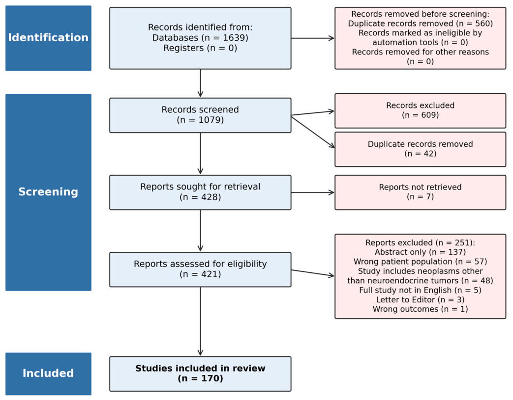
A medical librarian (LHY) searched the literature for records including the concepts of neuroendocrine neoplasms, Surveillance Epidemiology End Results (SEER), and The National Cancer Database (NCDB). The librarian created search strategies using a combination of keywords and controlled vocabulary in Embase.com 1947–, Ovid Medline 1946–, Scopus 1823–, Cochrane Central Register of Controlled Trials (CENTRAL), The Cochrane Database of Systematic Reviews (CDSR), and Clinicaltrials.gov 1997–. All search strategies were completed on 4 February 2022, with no added limits and a total of 1639 results found. Overall, 558 duplicate records were deleted after using the de-duplication processes described in “De-duplication of database search results for systematic reviews in EndNote,” [
16] and an additional 2 duplicates were removed using Covidence (Veritas Health Innovation, Melbourne, Australia) [
17], resulting in a total of 1079 citations included in the project library.
The remaining articles’ abstracts were independently screened for relevance by two authors (TH and MH) and conflicts resolved by a third author (CWH) using covidence.org. The full text of the remaining articles was assessed according to the inclusion and exclusion criteria. Fully reproducible search strategies for each database are provided in
Supplementary File S2. Inter-reviewer agreement was high at both stages. Cohen’s κ was computed from the Covidence inter-rater reliability report on the records that both primary reviewers (TH and MH) independently screened, 1029 of the 1079 records at title/abstract and 388 of the 421 full texts; records that did not receive two independent votes from this pair (those screened by a single reviewer or adjudicated by the senior author) fall outside the κ denominator. On this set, the reviewers agreed on 994 of 1029 dually screened records (96.6%; Cohen’s κ = 0.93) and, at full text, on 368 of 388 (94.8%; κ = 0.89); the 35 and 20 disagreements, respectively, were resolved by adjudication with the senior author (CWH).
2.4. Data Charting
Data charting and the assignment of each study to the nomenclature categories were performed independently and in duplicate by two authors (TH and MH), with discrepancies resolved by consensus and adjudicated by the senior author (CWH) when needed. Because category assignment followed the explicit, criterion-based definitions below (registry morphology codes and the differentiation field), most studies classified unambiguously; studies with borderline or ambiguous coding or differentiation reporting were re-examined jointly against these criteria, and any that remained unresolved were adjudicated by the senior author. To map the landscape of neuroendocrine neoplasm nomenclature usage, a standardized charting form was used to record the following information from each article:
Study Characteristics: Author, year of publication, citation title, journal, database utilized (SEER or NCDB), and study period.
Nomenclature and Classification Variables: International Classification of Diseases (ICD) topographic and morphology numbers and whether neuroendocrine neoplasm-specific taxonomy was retrieved directly from the cancer registries.
Grading and Differentiation Data: The number of tumors recorded under specific differentiation grades (well, moderately, poorly, or undifferentiated) and whether the article referred to neuroendocrine tumors as “carcinoids”.
The primary goal of this charting process was to classify the literature into a descriptive framework consisting of four categories to assess the nature and extent of nomenclature discordance, with a secondary goal of capturing the persistence of “carcinoid” terminology in a fifth category:
Insufficient Data: Articles lacking the necessary tumor grade or ICD codes to assess nomenclature accuracy.
Appropriate Interpretation: Articles where terminology was used consistently with the 2010 WHO classification.
Inclusion of Poorly/Undifferentiated Neoplasms: Articles grouping poorly or undifferentiated neoplasms under the “neuroendocrine tumor” designation. Studies that reported neuroendocrine tumors and neuroendocrine carcinomas but analyzed them as separate cohorts were classified on the basis of the well-differentiated cohort and were not counted in this category.
Conflation of Grading Systems: Articles reporting neuroendocrine neoplasm-specific grades (G1, G2, G3) derived from database differentiation grades rather than proliferation markers.
Historical Terminology: Articles utilizing the outdated term “carcinoid” to describe neuroendocrine tumors.
2.5. Forward Citation Analysis
To assess whether the nomenclature discordances identified in this review have been cited in the broader literature, we performed a forward citation analysis of the 124 unique discordant studies cataloged in
Supplementary Tables S3 and S4.
Each study was resolved to a Digital Object Identifier (DOI) using Crossref and PubMed; matches with a title-similarity score below 0.90 or with Crossref/PubMed disagreement were verified manually by the senior author, and seven incorrect DOIs were corrected.
Semantic Scholar [
18] provides an influentialCitationCount metric which algorithmically flags citations that substantively build upon the cited work rather than passingly mentioning it. This count was captured for each study.
Citing works were classified as reviews or meta-analyses if flagged as such by OpenAlex [
19], by a PubMed publication type of “Review,” “Systematic Review,” or “Meta-Analysis,” or by review-specific terminology in the title. To assess citation by clinical guidance, we intersected the reference lists of 13 major neuroendocrine tumor guideline and consensus documents (National Comprehensive Cancer Network [NCCN], European Society for Medical Oncology [ESMO], European Neuroendocrine Tumor Society [ENETS], North American Neuroendocrine Tumor Society [NANETS], UK and Ireland Neuroendocrine Tumour Society [UKINETS], and WHO classifications) with the discordant-study set; guidelines with fewer than 30 entries in their OpenAlex reference list were hand-checked against the source manuscript. For NCCN-cited discordant studies, the citing-sentence text and parent section heading were manually verified against the current edition of the NCCN Neuroendocrine and Adrenal Tumors guideline (v1.2026) [
20].
2.6. Statistical Analysis
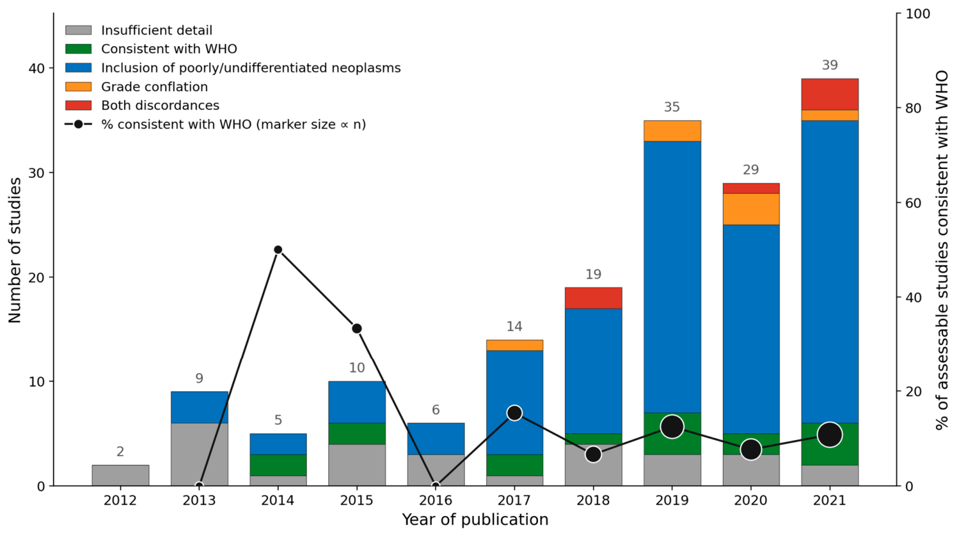
Inter-reviewer agreement at the title/abstract and full-text screening stages was quantified with Cohen’s κ. The temporal trend in the proportion of assessable studies showing nomenclature discordance was assessed with the Cochran–Armitage test for trends across annual data from 2013 to 2021 (2022 excluded as a partial year). Citation counts are summarized as medians with interquartile ranges. Analyses were performed in Python 3.13 (NumPy 2.3.4, SciPy 1.16.2).
4. Discussion
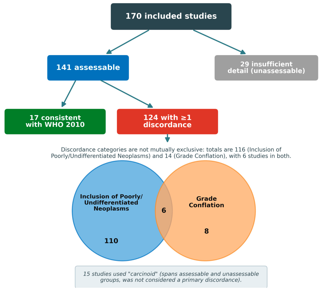
This scoping review of 170 studies published from 2012 to 2022 reveals discordance between international neuroendocrine neoplasm nomenclature guidelines and their application in research utilizing national cancer registries that was widespread and persistent across the study period. Despite the 2010 WHO update intended to standardize these definitions, 88% of 141 assessable studies failed to apply the nomenclature accurately. This suggests a significant “conceptual drift” where the limitations of database coding appear to have been systematically conflated with clinically meaningful classification.
4.1. Registry Choice and Systemic Discordance
Our mapping of the literature highlights a heavy reliance on the SEER database, which was used in over 72% of the included studies (
n = 123). This is significant because SEER data prior to 2021 lacks the proliferation markers (mitotic count and Ki-67 index) necessary for accurate WHO grading [
11]; these registry limitations for neuroendocrine neoplasm epidemiology have been noted previously [
10]. Furthermore, the fact that nomenclature discordance was identified across all topographic sites, from the frequently studied pancreas (41.8%) to rarer sites such as the esophagus and gallbladder, suggests that the conceptual drift between registry codes and clinical guidelines is a systemic issue within the field, rather than one confined to a specific organ or database. The most concrete consequence of this discordance is the composition of the cohorts themselves: many registry-based “neuroendocrine tumor” studies include patients whose tumors would not meet WHO criteria for a neuroendocrine tumor.
These difficulties are not unique to neuroendocrine neoplasms: registry coding schemes lag or blur evolving pathological classifications across oncology, and database studies performed before the coding is reconciled inherit the mismatch. A prominent example is the 2016 reclassification of the noninvasive encapsulated follicular variant of papillary thyroid carcinoma as noninvasive follicular thyroid neoplasm with papillary-like nuclear features, which removed a large group of indolent tumors from the “carcinoma” category; registry-based analyses conducted before this change had overcounted thyroid carcinoma and contributed to overtreatment [
30]. In the biliary tract, inconsistent definitions and ICD-O coding of intrahepatic, hilar, and Klatskin tumors have similarly confounded registry-based estimates of cholangiocarcinoma incidence [
31]. The general lesson, applicable well beyond neuroendocrine neoplasms, is that registry codes must be verified against the current classification before they are used to define a study cohort.
4.2. Inclusion of Poorly or Undifferentiated Neoplasms in Neuroendocrine Tumor Research
The most critical finding in our map of the literature is the systematic inclusion of poorly differentiated and undifferentiated neoplasms within neuroendocrine tumor-specific cohorts. According to the 2010 definitions, these aggressive neoplasms are neuroendocrine carcinomas, which are biologically and clinically distinct from well-differentiated neuroendocrine tumors [
32]. At the molecular level, neuroendocrine carcinomas are characteristically driven by TP53 and RB1 inactivation, whereas well-differentiated neuroendocrine tumors typically retain wild-type TP53 and RB1 and, in pancreatic primaries, often carry ATRX, DAXX, or MEN1 alterations [
33]. With an average poorly or undifferentiated neoplasm inclusion rate of 18.7% across the 107 studies that reported differentiation, much of the published literature regarding gastroenteropancreatic neuroendocrine tumors is inadvertently weighted by the far more aggressive behavior of poorly or undifferentiated neoplasms.
4.3. Grade Conflation: Proliferation Versus Differentiation
A second major theme is the conflation of the registry differentiation grade with the WHO proliferation grade. SEER (prior to 2021) and the NCDB (prior to 2018) use a 1–4 scale that describes how closely tumor cells resemble normal tissue, that is, their differentiation [
11,
12], and many researchers have treated these values as interchangeable with WHO G1, G2, and G3. They are not: WHO grading is defined strictly by proliferation (mitotic count or Ki-67 index), data that do not exist in these earlier registry versions. This is not a divergence between the AJCC and WHO systems, which are concordant, since AJCC staging for well-differentiated neuroendocrine tumors incorporates the WHO proliferation grade. The problem lies instead within the registry differentiation field itself, whose levels do not correspond to WHO G1–G3 at all; researchers simply assign such a correspondence.
The consequence is specific. When a study stratifies its cohort by the registry differentiation grade and relabels the strata G1, G2, and G3, its highest-grade arm (registry grade 3, and often grade 4) consists of poorly or undifferentiated neoplasms, that is, neuroendocrine carcinomas; a comparison of outcomes “by grade” is then actually a comparison of neuroendocrine tumors against neuroendocrine carcinomas, presented as an effect of proliferation grade. Where inclusion of poorly or undifferentiated neoplasms biases the data, grade conflation biases the interpretation, dressing a differentiation-based contrast as a proliferation-grade finding and reporting grades that were never measured. Differentiation and proliferation are, moreover, independent axes: a well-differentiated tumor may itself be highly proliferative (a Ki-67 above 20%, recognized in the 2022 WHO classification [
8] as a well-differentiated neuroendocrine tumor G3). Such a tumor is still biologically a neuroendocrine tumor, molecularly distinct from a neuroendocrine carcinoma, yet it carries the same proliferation grade as a poorly differentiated carcinoma; proliferation grade alone does not separate them, and the registry differentiation field, which records only differentiation, can neither identify it nor stand in for the WHO grade.
4.4. Downstream Citation Exposure of Discordant Studies
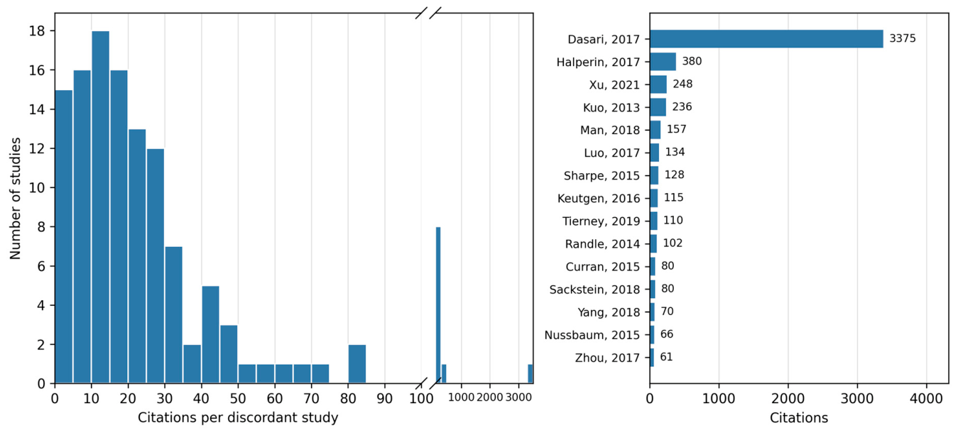
The downstream citation analysis demonstrates that the nomenclature discordances identified in this review are not confined to isolated primary studies. With 87.9% of the discordant database studies cited by subsequent reviews and meta-analyses, discordant studies have received substantial citation exposure within the broader oncology literature; whether this exposure has reproduced the underlying nomenclature error in citing works is not established by citation counts alone. This exposure is heavily skewed by a small number of highly visible publications; a single 2017 study [
22] accounted for 46.1% of all downstream citations. Crucially, 32.6% of the neoplasms included in this highly cited study were poorly or undifferentiated neoplasms, neuroendocrine carcinomas by WHO definition. However, even with this extreme outlier excluded, the remaining discordant studies still generated 3948 citations, indicating that this downstream citation exposure is broad rather than confined to a single paper. Because such heavily cited discordant studies have been incorporated into clinical practice guidelines and consensus documents, including NCCN, ESMO, and ENETS, there is a concrete mechanism by which registry-based nomenclature errors can skew the perceived aggressiveness of neuroendocrine tumors. Whether this citation exposure has influenced treatment recommendations is not established by these data; in the one guideline we examined in detail, the discordant studies were cited for staging validation and background rather than for therapy.
4.5. Potential Risk of Over-Treatment
These nomenclature errors are not merely semantic; they have the potential to bias clinical decision-making. In 2016, Tang et al. [
32] demonstrated a disease-specific survival of 55 months in patients with well-differentiated gastroenteropancreatic neuroendocrine tumors with a high-grade component, compared with only 11 months in patients with poorly differentiated neuroendocrine carcinoma. Even in patients with advanced disease, the prognostic difference between neuroendocrine tumors and carcinomas is substantial. The two are also managed differently: well-differentiated neuroendocrine tumors are treated with somatostatin analogs, everolimus, or peptide receptor radionuclide therapy, whereas neuroendocrine carcinomas receive platinum-based chemotherapy, so misclassification can translate directly into inappropriate treatment [
20,
24]. When a registry-derived “neuroendocrine tumor” cohort includes a mean of 18.7% poorly or undifferentiated neoplasms, the resulting outcomes data are likely to overestimate the adverse prognosis for true neuroendocrine tumor patients. In principle, this amplified perception of risk could bias clinicians toward more aggressive treatment for patients with relatively indolent, well-differentiated disease. We emphasize, however, that this scoping review measured nomenclature usage and citation exposure only; whether these errors have actually altered treatment recommendations or produced over-treatment was not assessed and remains a hypothesis for future study.
4.6. Persistence of “Carcinoid”
The continued use of “carcinoid” in 8.8% of the literature, decades after the WHO discouraged it, marks the slow assimilation of standardized terminology. Beyond imprecision, the term carries misleading connotations when it is applied generically to gastroenteropancreatic neuroendocrine tumors [
13]. Because “carcinoid” is bound to carcinoid syndrome, the label implies hormonal activity, yet most gastroenteropancreatic neuroendocrine tumors are non-functional [
24]; a “carcinoid” designation can therefore lead clinicians to presume a functional tumor and to pursue unnecessary biochemical testing or pre-operative somatostatin prophylaxis against carcinoid crisis. We did not measure these downstream clinical effects and present them as illustrative rather than established. This does not mean that the term is always inappropriate: it remains the current designation for pulmonary typical and atypical carcinoid, although a shift toward neuroendocrine tumor terminology has been proposed even there [
14], and for carcinoid syndrome itself. Precisely because the term retains these specific, legitimate meanings, its generic use for gastroenteropancreatic tumors imports them where they do not belong. For this review, “carcinoid” usage was recorded separately as a descriptive category rather than as one of the two primary discordances; consistent with its being a marker rather than a driver, 14 of the 15 studies using “carcinoid” also exhibited another form of nomenclature discordance or lacked sufficient detail to assess. Residual “carcinoid” usage therefore rarely occurred in isolation, tending to co-occur with, rather than independently produce, the more substantive errors.
4.7. A Reference Approach for Registry-Based Neuroendocrine Tumor Studies
The studies we classified as consistent with the 2010 WHO nomenclature (
Supplementary Table S2) share several practices that can serve as a practical benchmark for future registry-based neuroendocrine tumor research. First, they restricted morphology (ICD-O-3) selection to well-differentiated neuroendocrine tumor codes and explicitly excluded poorly differentiated, small-cell, and large-cell neuroendocrine carcinoma codes, or analyzed any carcinomas as a separate cohort. Morphology-code selection alone is not sufficient, however: in practice, cohorts retrieved with the correct well-differentiated codes can still span the full range of registry differentiation grades (1 through 4), because that field is recorded independently of the morphology code. A rigorous cohort therefore also filters the differentiation field, retaining well-differentiated cases and excluding those recorded as poorly differentiated (grade 3) or undifferentiated (grade 4), which are by definition neuroendocrine carcinomas; moderately differentiated cases (grade 2) are intermediate, and either their inclusion or their exclusion is defensible so long as the choice is stated. Used in this way, for differentiation rather than as a proxy for proliferation, the differentiation field is informative; the error lies not in using it but in mistaking it for the WHO proliferation grade (
Section 4.3). Second, none of these studies used the registry versions that carry proliferation information (NCDB from 2018; SEER from 2021); instead, they refrained from reporting proliferation-based WHO grades (G1–G3) when the underlying Ki-67 or mitotic data were unavailable, which was the appropriate choice for the data they had. As those newer versions mature, investigators will increasingly be able to assign WHO grade directly, although only for diagnosis years from the update onward (
Section 3.6). Third, they reported the differentiation composition of the final cohort, including how intermediate-grade cases were handled, so that its makeup is transparent to readers.
Table 4 summarizes the ICD-O-3 morphology codes most relevant to gastroenteropancreatic neuroendocrine neoplasms, and how they should be used to isolate neuroendocrine tumors, as a quick reference for investigators querying these registries.
4.8. Limitations of the Scoping Review
As with all scoping reviews, this study is limited by search-strategy bias: only studies indexed in the queried databases, returned by our specific search parameters, and published in English-language journals were eligible for inclusion. Furthermore, 17% of the identified literature provided such poor methodological detail that their nomenclature usage could not be assessed, which may indicate that the true rate of discordance is even higher than reported here. Finally, registry differentiation is assigned by the reporting pathologist rather than by central re-review, and the differentiation fields may be incomplete, historically variable, or subject to coding error; a poorly or undifferentiated designation nonetheless establishes that a neoplasm is not a well-differentiated neuroendocrine tumor, although we could not independently verify these pathological assessments.
Our literature search closed in February 2022, and it is possible that the discordance has resolved since. However, the registry updates are prospective: the NCDB (2018) and SEER (2021) proliferation fields populate only for diagnosis years from those updates onward, whereas the studies we identified pooled a median of 14 diagnosis years (range 5–43), with 91% spanning at least a decade. Therefore, until the updated coding has been in place for at least ten years (approximately 2028 for the NCDB and 2031 for SEER), at least a portion of every such cohort will likely still depend on the older differentiation coding, which must still be interpreted correctly. Accordingly, we have confined our claims regarding the prevalence and persistence of discordance to the 2012–2022 study period; we cannot directly characterize studies published after our search, and the structural argument above indicates only that the underlying source of discordance is unlikely to have resolved, not that its rate has necessarily remained unchanged.
Our forward citation analysis is limited by the inherent coverage variations among citation databases. OpenAlex counts were cross-validated against Semantic Scholar, with Google Scholar arbitration of divergent records, to establish a conservative lower bound of downstream exposure. Even so, certain non-journal artifacts such as the AJCC Cancer Staging Manual lack a digitally indexed reference list. Consequently, the true rate of citation by clinical guidelines may be higher than reported. Furthermore, capturing a downstream citation establishes exposure to the original nomenclature error but does not confirm whether the error was accepted uncritically by the citing authors.
4.9. Future Directions
Several operational steps follow from these findings. The most direct is that journals and guideline panels could require a brief nomenclature-compliance statement for registry-based submissions, confirming that cohort composition aligns with current WHO definitions. Beyond this, engagement with SEER and NCDB leadership could help refine grade-field instructions and surface the neuroendocrine tumor–carcinoma boundary at the query stage, and targeted educational efforts (society webinars and methods primers) could raise awareness among investigators who use these registries. Finally, a dedicated study contacting the corresponding authors of discordant studies to ascertain the rationale for their coding choices, together with a citation-context analysis of how discordant studies are actually used by citing works, would test whether the citation exposure documented here translates into propagated conceptual error. Such root-cause and citation-content investigations are distinct lines of inquiry beyond the scope of the present descriptive map. In particular, a prospective update of this review, once the post-2018 (NCDB) and post-2021 (SEER) coded literature has matured, would directly test whether the registry coding updates have improved nomenclature accuracy, an assessment that the 2012–2022 window cannot yet support.




 ,
,