Review

PET/CT Imaging for Therapy Assessment in Multiple Myeloma Including MRD

1
Service de Médecine Nucléaire, Département d’Imagerie Médicale, Institut Gustave Roussy, F-94805 Villejuif, France
2
Nuclear Medicine Division, AOU Città della Salute e della Scienza di Torino, University of Turin, 10126 Turin, Italy
3
Département d’Hematologie, Institut Gustave Roussy, F-94805 Villejuif, France
4
Department of Medical Sciences, University of Turin, 10126 Turin, Italy
*
Author to whom correspondence should be addressed.
Cancers 2026, 18(14), 2184; https://doi.org/10.3390/cancers18142184
Submission received: 22 May 2026 / Revised: 29 June 2026 / Accepted: 2 July 2026 / Published: 8 July 2026

Simple Summary

Monitoring treatment response in multiple myeloma presents challenges because bone marrow assessment techniques can miss disease due to heterogeneous distribution and cannot detect extramedullary involvement. [18F]FDG PET/CT addresses these limitations by providing a whole-body metabolic evaluation of disease activity. This review examines the published evidence on the diagnostic accuracy and prognostic value of [18F]FDG PET/CT in assessing treatment response with a particular focus on minimal residual disease detection. We examine how standardized interpretation criteria (Deauville, IMPeTUs, IMWG) have improved the reproducibility of response assessment and demonstrate how integrating metabolic imaging with bone marrow techniques optimizes patient stratification. The review also evaluates emerging radiotracers beyond FDG, including agents targeting CXCR4 and CD38, which may enhance diagnostic capabilities in specific clinical contexts and support more personalized treatment approaches.

Abstract

Background/Objectives: Multiple myeloma (MM) is a complex hematologic malignancy characterized by abnormal plasma cell proliferation in the bone marrow. [18F]FDG PET/CT has emerged as a key imaging modality, demonstrating high sensitivity and specificity for the detection of metabolically active disease compared with conventional imaging techniques. This review examines the role of PET/CT imaging in evaluating therapeutic response in symptomatic myeloma, including minimal residual disease (MRD) assessment. Methods: We surveyed the literature published between 2014 and 2024 across PubMed, Scopus, Web of Science, and the Cochrane Library for peer-reviewed articles evaluating PET imaging in the assessment of treatment response in multiple myeloma. Results: [18F]FDG PET/CT provides a whole-body assessment of disease burden, extramedullary involvement detection, and early metabolic response evaluation. Standardized response assessment criteria (Deauville, IMPeTUs, IMWG) have enhanced the reproducibility and accuracy of treatment monitoring. Semiquantitative parameters including SUVmax and metabolic tumor volume (MTV) provide useful prognostic information with PET negativity correlating with improved progression-free and overall survival. The integration of PET/CT with bone marrow assessment techniques (next-generation flow cytometry and sequencing) optimizes MRD evaluation and patient stratification. Conclusions: PET/CT represents a valuable imaging tool for improving therapeutic decision making and risk stratification in MM management. Emerging radiotracers beyond FDG, including [11C]choline, [11C]methionine, and targeted agents like [68Ga]pentixafor and CD38-targeted immunoPET tracers, hold promise for enhanced diagnostic capabilities in specific clinical contexts.

1. Introduction

Symptomatic myeloma, also known as multiple myeloma (MM), is a complex and often challenging hematologic malignancy characterized by the abnormal proliferation of plasma cells in the bone marrow. Symptomatic myeloma typically originates from an initial indolent monoclonal gammopathy, which then progresses into an asymptomatic myeloma form, and eventually into the symptomatic form requiring treatment. The prognosis can be variable and related to different clinical presentations [1].
The diagnosis of symptomatic myeloma relies on a combination of clinical evaluation, laboratory tests, imaging studies, and bone marrow examination [2]. Despite variable clinical manifestations, the primary criteria for defining symptomatic myeloma include elevated calcium, renal impairment, anemia, and the presence of bone lesions (CRAB criteria). Common symptoms and signs include fatigue related to anemia, bone pain resulting from bone involvement and the common presence of lytic lesions, and occasionally neurological symptoms. Laboratory tests allow for the detection of specific biomarkers: abnormal proteins (M-proteins) identified through serum and urine protein electrophoresis, high levels of free light chains in the blood assessed by serum free light chain assay, and cytopenia related to bone marrow infiltration observed in the complete blood count (CBC). M proteins and free light chains are also used as biomarkers for monitoring the disease during and after treatment. Finally, the examination of bone marrow aspirate and biopsy samples helps to confirm the presence of clonal plasma cells and to assess the degree of marrow involvement [3].
In MM, bone involvement primarily manifests as lytic lesions disseminated throughout the skeleton. The presence of at least one asymptomatic lytic lesion is a criterion for initiating treatment. Imaging techniques such as magnetic resonance imaging (MRI) and computed tomography (CT) are commonly employed to assess bone involvement and detect lytic lesions as well as to evaluate disease response. Whole-body acquisitions have been the recommended protocols for several years to enable a comprehensive assessment of tumor burden. In particular, MRI is a valuable technique to assess local complications of bone involvement such as medullary compression [4,5]. Over recent years, numerous studies have demonstrated that [18F]Fluorodeoxyglucose ([18F]FDG) Positron Emission Tomography (PET/CT), a molecular imaging technique, is a powerful tool in the initial assessment of MM. It enables the evaluation with a high specificity of both medullary and extramedullary involvement, the latter occurring in around 5% of patients at initial diagnosis. FDG uptake at PET can vary among patients and appears to be associated with the dedifferentiation of plasma cells rather than clonal proliferation. The main findings at PET are focal lytic lesions, diffuse bone marrow involvement and more rarely extramedullary active disease. However, it is important to note that an increased uptake at PET alone is insufficient for MM diagnosis; evidence of underlying osteolytic bone lesion is still required from the CT component of the examination [6,7].

1.1. Management and Treatment

The management of symptomatic myeloma requires a multidisciplinary approach involving hematologists, oncologists, radiologists, nuclear medicine physicians and supportive care specialists. Treatment strategies aim to control the disease, alleviate symptoms, and improve patient outcome. The first step in treatment is represented by the induction therapy. Initial treatment typically has been for many years a combination of triplet regimens: bortezomib (proteasome inhibitor), thalidomide or lenalidomide (immunomodulatory drug), and dexamethasone (VTd and VRd regimen, respectively). Autologous stem cell transplantation (ASCT) is proposed to eligible patients following induction therapy to achieve deeper and more durable responses [8]. Maintenance therapy with lenalidomide or bortezomib may be recommended to prolong remission and delay disease progression after ASCT. Consolidation treatments are recommended if complete response is not achieved after induction and intensification treatments. Supportive measures, including bisphosphonates for bone health, erythropoietin-stimulating agents for anemia, and antibiotics for infection prophylaxis, are essential components of symptoms management. The advent of targeted therapies and immunotherapy, such as monoclonal antibodies (e.g., elotuzumab, daratumumab approved by the FDA in 2015) and selective proteasome inhibitors (e.g., carfilzomib approved in 2012), has revolutionized the treatment landscape for myeloma, offering additional options for MM patients with high response rates and prolonged remission duration especially in refractory and relapsed MM [3,9]. Indeed, regardless of their cytogenetic and molecular profile, the large majority of MM patients express both SLAMF7 and CD38, making these antigens attractive therapeutic targets [10,11]. Elotuzumab targets the extracellular domain of the signaling-lymphocytic-activation-molecule F7 (SLAMF7) [11], also referred to as CS1, which plays a role in MM cell adhesion to bone marrow stromal cells; it enhances antibody-dependent cellular cytotoxicity (ADCC) and activates natural killer (NK) cells. In the phase III ELOQUENT-2 trial [12], the addition of elotuzumab to lenalidomide and dexamethasone in relapsed or refractory MM prolonged median progression-free survival (19.4 vs. 14.9 months; HR 0.70, 95% CI 0.57–0.85; p < 0.001).
Daratumumab is an anti-CD38 monoclonal antibody leading to myeloma cell killing via the direct induction of apoptosis, Fc-related mechanism of myeloma cytotoxicity, and eradication of CD38-positive regulatory T cells, regulatory B cells, and myeloid-derived suppressor cells in the BM microenvironment [10]. Daratumumab showed excellent efficacy in the treatment of relapsed or refractory MM both as monotherapy and in combination with standard therapies. In the randomized phase III POLLUX trial [13], patients with an early relapse of MM were randomized to receive lenalidomide/dexamethasone with or without daratumumab. The hazard ratio for disease progression or death in the daratumumab group (n = 286) versus the control group (n = 283) was 0.37 (95% CI: 0.27–0.52; p < 0.001 by stratified log-rank test). Patients receiving daratumumab achieved a higher overall response rate (93% vs. 76%), including 43% with a complete response or better, together with better survival and a favorable side-effect profile.
The addition of daratumumab to standard regimens such as VTd (Dara-VTd) and VRd (DaraVRd) in first-line therapy has been shown to induce superior complete response (CR) rates compared to standard regimens alone in two large randomized controlled trials. A phase III trial, the CASSIOPEIA study, randomized patients with newly diagnosed myeloma to receive standard VTd regimen with or without Daratumumab. The rate of complete response or better, assessed at day 100 post-ASCT, was higher with the addition of daratumumab to VTd (Dara-VTd: 39% versus VTd: 26%), resulting in a superior PFS (PFS at 18 months: 93% for Dara-VTd vs. 85% for VTd) [14]. Quadruplet combinations before ASCT are now becoming a new standard of care.
Selective proteasome inhibitors such as carfilzomib (in association with anti-CD38 antibodies such as daratumumab or isatuximab) have been tested in phase III trials and are indicated as therapy options in case of first relapse after one previous line of treatment in MM refractory to lenalidomide [15,16].
Currently, antibodies against BCMA (B-cell maturation antigen), bispecific antibodies, and CAR T-cell (chimeric antigen receptor T cells) therapies have been investigated in clinical trials especially for patients with MM refractory to all the previous mentioned drugs with very promising results in terms of high response rates and impact on outcome [17]. Nevertheless, these treatments are not still widely available due to their high costs, and the prognosis of patients with MM refractory to many lines of treatment still remains poor. In this setting, FDG PET/CT has also been investigated for response assessment after cellular immunotherapy. Although most of the available quantitative PET evidence in this context derives from lymphoma, preliminary data in multiple myeloma suggest that a negative FDG PET/CT following BCMA-directed CAR-T cell therapy may be associated with a favorable prognosis [18].

1.2. The Challenge of Response Evaluation in MM

Understanding the response to treatment in myeloma is crucial for guiding therapeutic decisions and optimizing patient outcomes. Monitoring this response involves a combination of clinical evaluations, laboratory tests, imaging studies, and bone marrow examinations.
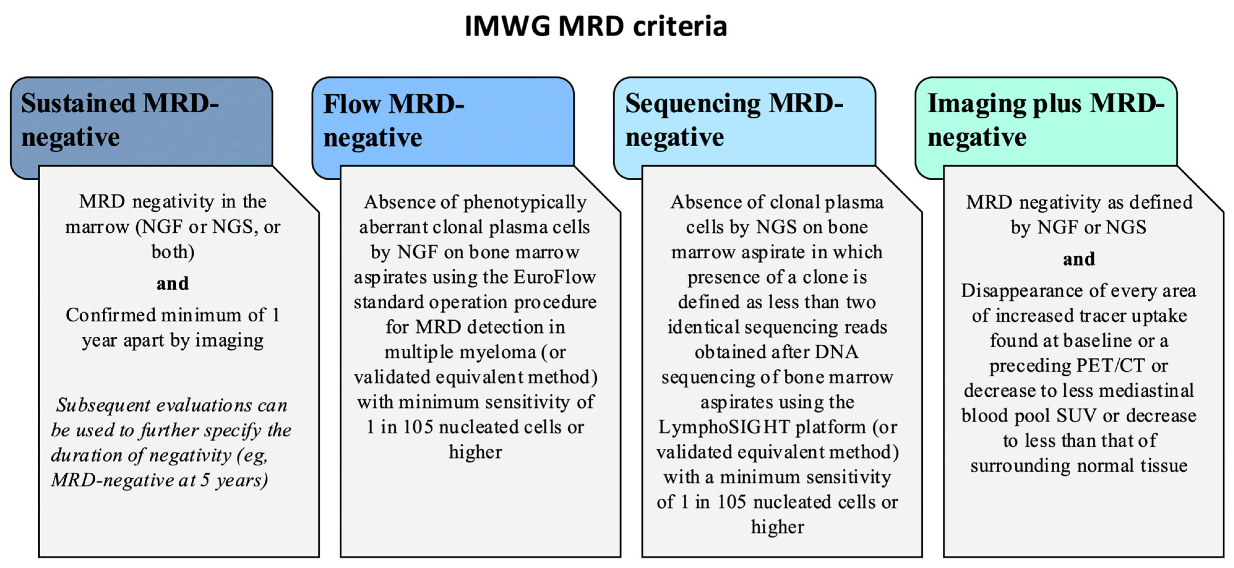
Serum and urine protein electrophoresis track changes in M-protein levels, which serve as biomarkers of disease activity. Additionally, monitoring serum-free light chain levels provides further information on disease burden and response to therapy. Bone marrow plasma cell infiltration is often heterogeneously distributed, which can increase the risk of false-negative results when using techniques that rely on bone marrow specimens limited to a small area of the body. Moreover, bone marrow evaluation does not detect extramedullary disease, which can indicate metastatic spread. Thus, comprehensive monitoring that includes advanced imaging and molecular techniques is essential for accurately assessing disease status and guiding treatment decisions. With improvements in treatment efficacy, the response of MM is currently classified as complete response, near complete response, minimal residual disease (MRD), very good partial response, or partial response. MRD detection has emerged as a pivotal concept in the management of myeloma, revolutionizing treatment strategies and prognostication. In myeloma, MRD refers to small amounts of cancer cells that persist after therapy and cannot be detected by standard methods. The significance of achieving MRD negativity post-treatment lies in its strong correlation with improved long-term outcomes, including prolonged progression-free survival and overall survival. This allows for more precise risk stratification, guiding treatment decisions to optimize patient outcomes [19]. The goal of MRD testing is to determine the depth of response to treatment and to assess the risk of disease recurrence, thus guiding treatment intensification and maintenance strategies. The detection of MRD using advanced standardized techniques at the bone marrow level, such as next-generation multiparametric flow cytometry (NGF) or next-generation sequencing (NGS), with sensitivity thresholds as low as 10−5 to 10−6, has emerged as a crucial predictor of long-term outcomes and survival. The International Myeloma Working Group (IMWG) initially established criteria for clinical evaluation primarily recommending NGF, polymerase chain reaction (PCR), or NGS on bone marrow samples collected from patients who have undergone treatment. In 2016, the IMWG updated its response criteria to integrate advanced imaging techniques like low-dose CT, which is actually the reference technique for MM diagnosis, MRI, and FDG PET/CT for a more thorough assessment of disease and monitoring of progression [20]. FDG PET/CT has emerged as a useful modality for detecting decreased tumor viability during treatment and identifying MRD in cases of initial positivity on pretreatment images. Based on its ability to distinguish between metabolically active and inactive disease, FDG PET/CT is a useful functional imaging modality for evaluating and monitoring the effect of therapy in myeloma. The 2017 IMWG consensus guidelines recommended coupling FDG PET/CT with sensitive bone marrow tools, including serum chemistry, serum β2-microglobulin levels, immunofixation, bone marrow biopsy, and serum-free light chains [6]. This comprehensive approach aids in detecting MRD both within and outside the bone marrow and identifying patients with imaging MRD negativity. Based on current evidence, the IMWG recommends also follow-up examinations using FDG PET/CT because monitoring disease activity through changes in FDG uptake is more straightforward than relying on morphological changes in osteolytic lesions. In 2019, the IMWG consensus guidelines provided specific recommendations for the optimal use of imaging in patients with multiple myeloma for initial workup and treatment response assessments [7]. The most recent European Hematology Association (EHA) and European Society for Medical Oncology (ESMO) guidelines also recommend FDG PET/CT as obligatory to confirm imaging MRD to be repeated every 12 months in bone marrow negative MRD patients during follow up [3]. These updates underscore the importance of integrating advanced imaging techniques with traditional biomarkers to achieve a more detailed and accurate assessment of treatment response and disease status.
The purpose of this review is to present the clinical data supporting the guidelines and recommendations on the role of FDG PET/CT imaging as a valuable diagnostic and prognostic tool for assessing response to treatment in multiple myeloma, including the detection of MRD. Several reviews and consensus documents on PET imaging in multiple myeloma have appeared in recent years, each with a distinct focus: Bezzi et al. provided a general update on the clinical value of FDG PET/CT across staging and therapy assessment [21]; Ishibashi et al. emphasized radiotracer development and the technical evolution of PET instrumentation, including non-FDG agents [22]; the EANM Focus 4 consensus addressed molecular imaging and therapy across hematological tumors more broadly [23]; and the 2024 EANM guideline established procedural and interpretative standards for the acquisition and reporting of FDG PET/CT in plasma cell disorders [24]. Building on but distinct from these works, the present reviewprovides an integrated, clinically oriented synthesis centered specifically on therapy response and minimal residual disease, bringing together the prognostic evidence across the different treatment phases, the standardized interpretation criteria, and the integration of FDG PET/CT with bone marrow-based MRD assessment.

2. Materials and Methods

For this narrative review, a structured but non-systematic literature search was performed across PubMed, Scopus, Web of Science, and the Cochrane Library to identify relevant studies evaluating the role of PET in assessing treatment response in multiple myeloma with a focus on articles published between 2014 and 2024. Search terms included “Positron Emission Tomography”, “PET”, “multiple myeloma”, “myeloma”, “treatment response” and “imaging”. Priority was given to peer-reviewed original articles in English involving patients with multiple myeloma and including PET imaging for treatment response assessment, while case reports and small case series were generally not considered. Reviews and meta-analyses were consulted to help identify relevant original articles. Articles were selected by the authors on the basis of their relevance to the scope of this review, and seminal studies published before 2014 were included where appropriate to provide context.

3. Results

3.1. The Role of [18F]FDG PET/CT

The most commonly used radiotracer in myeloma imaging is [18F]FDG, which reflects glucose metabolism and cell proliferation in malignant plasma cells. [18F]FDG PET/CT allows for the comprehensive evaluation of the disease burden throughout the skeleton and extramedullary sites, providing a whole-body assessment of disease activity both at baseline and during treatment.

3.1.1. Diagnostic Accuracy of [18F]FDG PET/CT

[18F]FDG PET/CT has a good sensitivity and an overall good specificity. Although FDG PET/CT may be less effective than MRI in detecting bone marrow involvement in multiple myeloma at staging, it remains the best technique to precisely define complete response. [18F]FDG PET/CT is exceptionally useful in particular due to its capacity to differentiate between metabolically active and inactive sites of disease [25], particularly in cases where conventional imaging techniques such as MRI, whole-body CT, or radiography continue to show abnormalities [26]. Whole-body MRI (WB-MRI) has also become an available tool for this purpose, but FDG PET/CT permits an earlier response assessment compared to WB-MRI with higher specificity and accuracy, as metabolic changes consistently precede morphological changes; this is of particular relevance in the evaluation after induction therapy to identify patients eligible for ASCT and in the evaluation after transplantation. In an early, small comparative study assessing the diagnostic performance of WB-MRI and FDG PET/CT for determining the remission status in 31 patients with MM after stem cell transplantation (SCT), FDG PET/CT had a sensitivity of 50.0%, a specificity of 85.7%, a positive predictive value of 62.5%, a negative predictive value of 78.3%, and an overall accuracy of 74.2% [27]. In contrast, MRI had a sensitivity of 80.0%, a specificity of 38.1%, a positive predictive value of 38.1%, a negative predictive value of 80%, and an overall accuracy of 51.6%. Basha et al. [28] in 2018 also compared the diagnostic performance of WB-MRI and FDG PET/CT in 56 patients with MM, of whom 22 were evaluated early after the end of treatment for response assessment. They demonstrated that WB-MRI and FDG PET/CT had similar sensitivity while FDG PET/CT had greater specificity than WB-MRI in detecting residual involvement in treated patients (86% vs. 43% respectively; p = 0.0081). Therefore, it should be considered more suitable than MRI to accurately determine the remission status. The retrospective study of Gómez León et al. [29] aimed to compare in 44 patients the agreement between WB-MRI, FDG PET/CT, and skeletal survey (SS) in patients with MM for diagnosis, initial staging, response evaluation, and early detection of complications. The concordance between WB-MRI and FDG PET/CT was generally good or excellent across most locations. For response evaluation, WB-MRI showed false positives results due to residual lesions without active disease, whereas FDG PET/CT was more reliable for assessing complete remission. WB-MRI was superior in detecting complications such as spinal cord or medullary compression and avascular necrosis. The concordance between clinical and radiological response was good for both WB-MRI (κ = 0.76) and FDG PET/CT (κ = 0.63) with p < 0.0001 for both. Nevertheless, functional MRI techniques like Diffusion-Weighted-Imaging MRI (DWI-MRI), which measures tissue water molecule movement, can provide insights into residual cellularity and lesion microcirculation. As demonstrated in the study by Paternain et al. [30], DWI-MRI and apparent diffusion coefficient (ADC) values provide another approach for evaluating treatment response in MM patients, showing a strong correlation with FDG PET/CT results and IMWG criteria. After treatment, the mean ADC in lesions from responders was significantly higher than in non-responders (1585.51 × 10−6 mm2/s vs. 698.17 × 10⁻⁶ mm2/s; p < 0.001). The SUVmax of the same lesions was significantly lower in responders than in non-responders (2.05 vs. 5.33; p < 0.001). Concerning specific treatment, PET can guide intensification treatment in PET-positive MM patients after upfront ASCT. One study evaluated the efficacy of four cycles of carfilzomib-lenalidomide-dexamethasone (KRd) as consolidation treatment after ASCT in patients with a positive PET, showing a 33% conversion rate into PET negativity [31].
A recognized limitation of FDG PET/CT is its potential for false-negative findings in specific settings. On MRI, myeloma marrow infiltration may present as a micronodular, or “salt-and-pepper”, pattern, with tiny foci (<5 mm) of altered signal scattered throughout otherwise preserved marrow [32], as illustrated in Figure 1; small lesions of this kind are particularly challenging for FDG PET/CT, whose spatial resolution restricts the reliable identification of lesions under approximately 10–15 mm [32]. A biologically distinct cause of false negativity is intrinsically low glucose-avid disease, which may reflect the reduced expression of hexokinase-2, which is the enzyme that traps FDG within tumor cells; together with diffuse marrow infiltration, this is among the principal causes of false-negative FDG PET/CT so that viable myeloma may fail to accumulate FDG and appear negative despite active disease detectable by MRI or alternative tracers [33]. As this phenomenon is largely independent of tumor burden, it supports the use of non-FDG radiotracers in selected patients.
Beyond these limitations, given the capability of FDG PET/CT to identify extramedullary disease (EMD), it is recommended to integrate this technique with sensitive bone marrow assessments as part of MRD evaluation to verify MRD negativity within and outside the bone marrow. Several studies have confirmed the complementarity between FDG PET/CT and blood/bone marrow assessments for MRD evaluation. The study by Rasche et al. [34] involved patients who achieved complete remission (CR) during first-line or salvage therapy. These patients underwent DWI-MRI, PET, and MRD flow cytometry at the onset of CR. The presence of residual focal lesions (FLs) and their impact on progression-free survival (PFS) were analyzed. Residual focal lesions were found in 24% of first-line patients and in 50% of patients after salvage therapy. DWI-MRI detected more residual lesions compared to PET (21% vs. 6%). Only six patients with positive DWI-MRI were also PET-positive; however, there were five patients positive only at FDG PET. By combining cytometry and imaging, residual disease was detectable in 64% of patients, improving the prediction of outcomes by defining double-negative and double-positive patients with excellent and poor outcomes, respectively.
Taken together, these data indicate that the two modalities are complementary rather than interchangeable: WB-MRI, particularly with DWI, tends to be more sensitive for diffuse marrow infiltration, whereas FDG PET/CT offers greater specificity by distinguishing metabolically active from inactive disease and provides earlier, whole-body response information. A systematic review and meta-analysis comparing the two modalities for treatment-response assessment reported a numerically higher pooled sensitivity for WB-MRI that did not reach statistical significance, together with a significantly higher specificity for FDG PET/CT, leading the authors to favor FDG PET/CT as the preferred modality for response assessment on current evidence [35]. Hybrid PET/MRI, which acquires both datasets simultaneously, is an emerging option that combines the metabolic information of PET with the marrow sensitivity of MRI; early experience in newly diagnosed disease suggests comparable focal-lesion detection by the two components, although limited scanner availability still restricts its routine use [36]. Beyond myeloma, molecular and metabolic imaging are increasingly regarded as complementary tools in the evaluation of hematological malignancies, and the integration of nuclear medicine techniques with other imaging modalities is an active field of development [37].
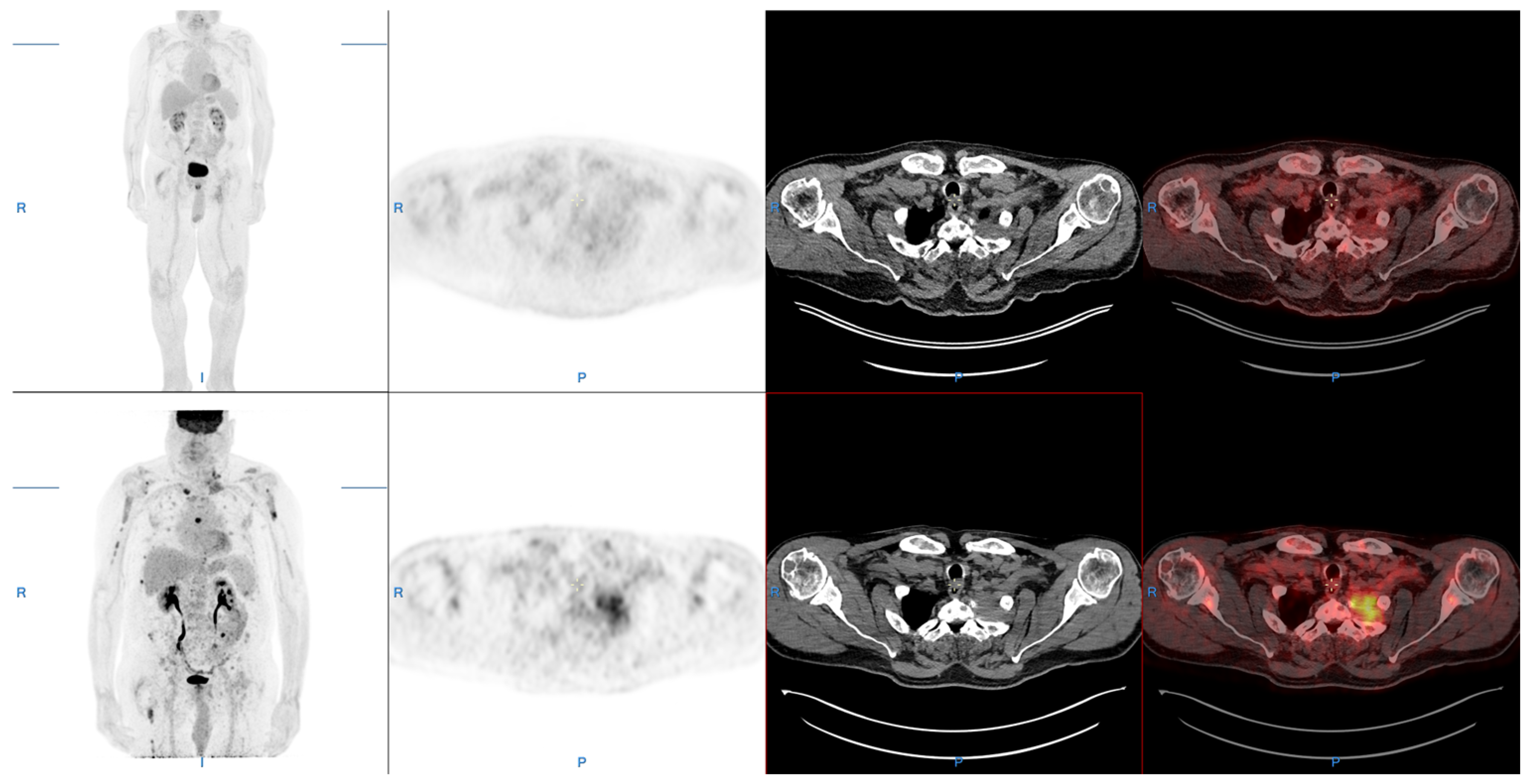
Figure 2 shows an example of complete metabolic response at FDG PET/CT after four cycles of treatment by Dara-VRd of a multiple myeloma patient with multifocal bone lytic and paramedullary involvement.

3.1.2. Prognostic Value of [18F]FDG PET/CT

As previously stated, beyond the diagnostic accuracy of FDG PET/CT, its heightened sensitivity in detecting bone and extra-skeletal lesions underscores its potential as a valuable tool for prognostic evaluation in MM.
Baseline factors derived from PET/CT are associated with PFS, such as the presence of extramedullary or paramedullary disease, the number of positive bone lesions, the metabolic tumor volume (MTV) and the tracer uptake intensity expressed by the SUVmax, supporting the role of FDG PET/CT for monitoring MM treatment response. The study by Zamagni et al. [38] has shown that MM patients with lesions exhibiting SUVmax > 4.2 at baseline presented shorter disease-free survival (DFS) and overall survival (OS). Mesguich et al. [39] demonstrated that the involvement of more than four skeleton areas on FDG PET/CT at baseline significantly impacted PFS. Interestingly, the presence of focal lesions (FLs) in the inferior limbs at baseline was also associated with shorter PFS. Moreover, MTV measured using FDG PET/CT was found to be a significant prognostic indicator, reliably predicting PFS and OS outcomes in MM patients [38,40].
Extramedullary disease (EMD)—myeloma growth independent of the bone marrow microenvironment—occurs in approximately 5% of patients at diagnosis and more frequently at relapse and consistently confers an adverse prognosis. Because EMD lies beyond the reach of bone marrow-based MRD techniques, FDG PET/CT has a distinct advantage as the only routine modality providing a whole-body detection of extramedullary and paramedullary sites. On FDG PET/CT, the presence of paramedullary or extramedullary disease at baseline has been independently associated with shorter PFS, retaining significance after adjustment for ISS stage and high-risk cytogenetics [41,42]. After therapy, the persistence of extramedullary uptake identifies a particularly high-risk subgroup. Accordingly, EMD status should be explicitly reported at both staging and response assessment, as its detection may alter risk stratification and management.
The prognostic value of FDG PET/CT has been assessed at different phases of treatment—early/after induction, after ASCT, and during maintenance—each carrying distinct prognostic and clinical implications, as detailed below. After therapy, FDG PET has a high negative predictive value, and a consistent body of evidence links post-therapy PET negativity to a favorable prognosis across different treatment phases [39,43,44,45]. Accordingly, a complete normalization of FDG PET findings after induction therapy, mainly at the premaintenance stage, has been recognized as a strong positive prognostic factor for PFS and OS in multiple large prospective studies [26,43,45,46,47,48], even in patients undergoing allogenic stem cell transplantation [49]. For newly diagnosed MM patients, treatment until complete PET normalization is an important therapeutic goal because their prognosis is comparable to PET-negative patients at diagnosis [50]. The PET negativity can be evaluated at different time points such as during induction treatment, after induction or after transplantation. The aim of the study by Davies et al. [50] was to determine the prognostic value of the decrease in PET/CT uptake at different time points post-therapy initiation: day 7, post-induction, post-transplant, and at maintenance therapy. A total of 596 patients underwent baseline PET/CT and were evaluated serially during their disease course using peak standardized uptake values above the background red marrow signal. The presence of more than three focal lesions at presentation stratified patients with worse PFS and OS (3-year PFS: 59% [95% CI 52–65] and 3-year OS: 72% [95% CI 66–78] in case of >3 lesions vs 3-year PFS: 74% [95% CI 67–81] and 3-year OS: 85% [95% CI 79–91] for patients with 1–3 lesions vs. 3-year PFS: 74% [95% CI 68–80] and 3-year OS: 89% [95% CI 84–93] for patients with 0 focal lesions). The complete disappearance of FLs uptake at various time points (day 7, post-induction, post-transplant, and during maintenance) correlated with significantly improved PFS and OS. Patients with no detectable FLs after treatment had similar outcomes to those with no FLs at diagnosis, while those with persistent FLs had significantly worse outcomes. Notably, PET at day 7 of therapy was the most prognostic: patients with complete FLs disappearance had the same prognosis as those with no lesions at diagnosis (3-year PFS: 76% [95% CI 67–86] 3-year OS: 89% [95% CI 82–96]). The study from Zamagni et al. [47] evaluated the role of FDG PET/CT in 282 patients with symptomatic MM. All patients were studied by PET/CT at baseline, during post-treatment follow-up, and at the time of relapse. After treatment, PET/CT negativity was observed in 70% of patients, while conventionally defined complete response was achieved in 53%. Achieving PET/CT negativity significantly improved both PFS and OS. PET/CT negativity was an independent predictor of prolonged PFS and OS even among patients with a conventionally defined complete response. The PFS was 44 months in patients with residual focal lesions at PET vs. 84 months in patients with completely negative PET (p = 0.0009). Sixty-three percent of patients experienced relapse or progression with skeletal progression exclusively detected by systematic PET/CT during follow-up in 12% of these cases. Multivariate analysis revealed that persistence of SUVmax > 4.2 following first-line treatment was independently associated with progression detected exclusively by PET/CT.
The occurrence of a PET response can be detected very early after starting treatment. Many different parameters have been tested to quantify the metabolic response [43]. Bailly et al. [44] evaluated patients with FDG–avid MM included in the prospective IMAJEM study after three cycles of lenalidomide, bortezomib, and dexamethasone (VRd). The change in SUVmax (cutoff of 25%) appeared to be an independent prognostic factor for PFS, allowing the identification of a patient subgroup with an improved median PFS (22.6 months vs. not reached, respectively), showing that an interim PET can be useful to guide further treatments [44]. Additionally, achieving complete FDG response in FLs before the initial transplantation was associated with significantly better outcomes, whereas the presence of more than three FDG-avid lesions emerged as an independent parameter linked to inferior overall and event-free survival. Pre-ASCT negativity on FDG PET/CT was associated with a favorable prognosis, whereas post-ASCT positivity, even in patients achieving a complete biological response, represented an independent negative prognostic factor.
A study by Kaddoura et al. [45] including 229 MM patients undergoing ASCT showed FDG PET/CT positivity around day 100 to be an independent negative prognostic factor associated with inferior time to progression (TTP) and worse OS. These results underscore the importance of incorporating PET/CT scans post-transplant, particularly in patients achieving CR, as an independent prognostic factor for stratifying myeloma disease and supporting its inclusion in the MRD definition in the IMWG response criteria. Charalampous et al. [48] in a study including 195 patients demonstrated that a negative PET/CT at 6 months after therapy was associated with significantly prolonged median conventional hematologic responses in terms of time to next treatment (TTNT) and OS compared to a positive PET/CT. This study also revealed a significant prognostic impact across different subgroups based on the hematologic response achieved at 6 months. Notably, patients that achieved very good partial response (VGPR) or better had significantly prolonged TTNT and OS if PET/CT was negative compared to positive, which was a trend similarly observed in patients with less than VGPR.
Finally, the combination of MRD assessment and imaging results is very important since the combination of negative results from multiparametric flow cytometry or NGS, along with a negative FDG PET/CT scan and a normal heavy/light chain ratio, could potentially indicate complete disease remission. In this regard, FDG PET/CT offers a further practical advantage: unlike bone marrow-based techniques, it is not affected by the interference that anti-CD38 monoclonal antibodies (e.g., daratumumab) exert on flow cytometry and serum immunofixation [51]. Conversely, there is currently no robust evidence that these agents alter FDG avidity or generate treatment-specific false-positive findings on PET/CT, although inflammatory or post-procedural uptake remains a general, non-specific pitfall of the technique. Böckle et al. [52] investigated a cohort of 102 newly diagnosed and relapsed/refractory MM patients showing that an MRD-triggered consolidation approach which integrates functional imaging (i.e., FDG PET/CT or DWI imaging) and bone marrow assessment can lead to a superior PFS and to an OS comparable to deep responders with double-negative results after standard treatment without consolidation. In particular, 45% of patients achieved MRD negativity on both NGF and imaging (double negativity), and 8% and 40% of patients were negative on either NGF or imaging, respectively. Imaging positivity despite negativity at NGF was more common in heavily pretreated disease (four or more previous lines) compared to newly diagnosed MM (p < 0.01).
The study from Davies et al. [50] also explored the relationship between imaging response and MRD assessed via flow cytometry. Some patients with FLs at the time of MRD assessment were MRD positive, while others were MRD negative, highlighting the importance of combining imaging with MRD assessment for a comprehensive evaluation.
Taken together, across the prospective and large retrospective cohorts reviewed, post-therapy PET negativity or complete metabolic response was consistently associated with improved PFS with reported hazard ratios broadly in the range of approximately 0.2–0.5 for the favorable (PET-negative) group, reinforcing the robustness of metabolic response as a prognostic marker despite differences in timing and reading criteria.

3.2. Available and Proposed [18F]FDG PET/CT Criteria for Treatment Response

Some specific PET-based response assessment criteria have been proposed for myeloma. Several standardized systems are currently available, including the Deauville criteria, the Italian Myeloma criteria for PET Use (IMPeTUs), the International Myeloma Working Group (IMWG) response criteria, and the European Association of Nuclear Medicine (EANM) Focus 4 recommendations, which are each discussed in the following subsections.

3.2.1. Deauville Criteria

The Deauville criteria, originally developed for lymphoma, have been tested in other hematological diseases [53,54]. The Deauville score (DS) originally applied for Hodgkin lymphomas emerged as a valuable tool to evaluate treatment response in patients with MM. The application of the Deauville scale has proved to be practical and relevant especially in identifying complete response to treatment. This 5-point scale is used to define treatment response according to the variation in [18F]FDG uptake during and after treatment with mediastinum and liver uptake as reference. Score 1 and 2 (no significant uptake or uptake ≤ mediastinum) and score 3 (uptake > mediastinum but ≤liver) indicate no active disease, suggesting a complete response to treatment. Score 4 and 5 (uptake moderately or markedly higher than the liver, or new lesions, respectively) suggest the persistence of active disease, indicating incomplete response or progressive disease. For myeloma, complete metabolic response has been defined as residual uptake not exceeding the liver background activity (Deauville score 1–3) in all initially involved bone marrow, focal lesions, paramedullary and extramedullary disease sites.

3.2.2. IMPeTUs Criteria

Multiple myeloma poses significant challenges for imaging interpretation due to its complexity and heterogeneity. Furthermore, the skeletal manifestations of MM can vary, including diffuse/infiltrative patterns or focal lesions. To standardize PET/CT reading in MM patients, the novel Italian Myeloma criteria for Pet Use (IMPeTUs) was proposed [55,56].
The IMPeTUs criteria, introduced by Nanni et al. [56] in 2016, provide a structured framework for baseline and therapy response FDG PET in MM patients and were developed to standardize the interpretation of FDG PET/CT scans. They provide a reproducible method to assess disease activity and response to therapy using PET imaging. First, these criteria recommend to take into account prognostic parameters including bone marrow metabolism, focal FDG-avid lesions (considering number, location, and CT morphology), and the presence of extramedullary disease (EMD) or paramedullary disease (PMD). The summary of the included parameters is presented in Table 1.
The response evaluation is based on the Deauville five-point scale. The IMPeTUs criteria define positivity cut-offs for identifying active disease. Specifically, patients with a Deauville score (DS) of 4 or higher in at least one focal lesion or in the bone marrow are considered to have still persistent disease. Deauville scores 4 and 5 are defined as target lesion uptake > liver uptake +10% and uptake >> liver uptake (twice), respectively. This scoring system has proven to be a significant prognostic indicator with higher scores correlating with shorter progression-free survival and overall survival. Additionally, the IMPeTUs criteria facilitate the identification of both bone marrow involvement and extramedullary disease, which are critical for a comprehensive evaluation of disease spread and response to treatment. Notably, because extramedullary and paramedullary disease lie beyond the reach of bone marrow-based MRD techniques, their inclusion among the IMPeTUs parameters is of particular clinical relevance, also in view of the recognized prognostic impact of EMD. By providing a standardized approach, these criteria help improve the reproducibility and accuracy of PET/CT interpretation across different clinical settings and studies. In a single-center cohort of 47 patients, Sachpekidis et al. [41] evaluated the prognostic role of the IMPeTUs criteria in MM patients undergoing high-dose chemotherapy (HDT) followed by ASCT. Forty-seven newly diagnosed MM patients underwent FDG PET/CT before treatment initiation and 34 of them after ASCT. At baseline PET/CT, the number of focal FDG-avid lesions was significantly correlated with bone marrow infiltration rate and R-ISS (International staging system), and the presence of PMD and EMD was associated to adverse PFS. Multivariate analysis confirmed that a higher number of focal lesions and the presence of EMD were associated with poorer prognosis, which was independent of ISS stage and high-risk cytogenetic abnormalities. At follow-up PET, a decrease in the number of focal lesions correlated with better PFS, while the presence/persistence of at least one focal lesion after therapy was associated with shorter PFS, confirming the association between PET-positive lesions after treatment and impaired clinical outcome. The decreased metabolic state of the bone marrow and focal lesions was useful to define the response to treatment but did not significantly affect PFS. The study supported the use of IMPeTUs criteria for standardized PET/CT evaluation in MM, highlighting their potential in patient stratification and response assessment.
These observations, derived from a limited single-center cohort, were subsequently supported by larger prospective analyses. In the multicenter study by Zamagni et al. [54], 228 patients with MM underwent FDG PET/CT at baseline and before starting maintenance treatment, and the five-point DS was applied to describe bone marrow status and focal lesion uptake and tested for its impact on clinical outcomes. At both uni- and multivariable analysis, FL and BM scores < 4 were associated with prolonged PFS and OS (OS: HR 0.6 and 0.47, respectively; PFS: HR 0.36 and 0.24, respectively). The study concluded that FL and BM FDG uptake lower than the liver background after therapy was an independent predictor for improved PFS and OS and can be proposed as the standardized criterion of PET complete metabolic response, confirming the value of the Deauville score for patients with MM as proposed by IMPETUS criteria.
More recently, a study by Zamagni et al. [25] aimed to confirm the role of metabolic response according to DS and its complementarity with bone marrow multiparameter flow cytometry (MFC) in an independent cohort of newly diagnosed, transplant-eligible MM patients previously enrolled in the phase II randomized FORTE trial. This analysis included 109 of the 474 patients enrolled in the trial who had paired PET/CT scans (performed at baseline [B] and prior to maintenance therapy [PM]) and MFC evaluations. At baseline, 93% of patients had focal lesions within the bones (FL score ≥ 4 in 89%) and 99% showed increased bone marrow uptake (BM score ≥ 4 in 61%). At the PM evaluation, complete metabolic response (CMR) was achieved in 63% of patients, which strongly predicted prolonged PFS in univariate analysis (HR 0.40, p = 0.0065) and in Cox multivariate analysis (HR 0.31, p = 0.0023). Regarding OS, there was a trend favoring CMR in univariate analysis (HR 0.44, p = 0.094) and a significant benefit in the Cox multivariate model (HR 0.17, p = 0.0037). Patients achieving both PET/CT CMR and MFC negativity at PM showed significantly extended PFS in univariate (HR 0.45, p = 0.020) and multivariate analyses (HR 0.41, p = 0.015).

3.2.3. IMWG Criteria

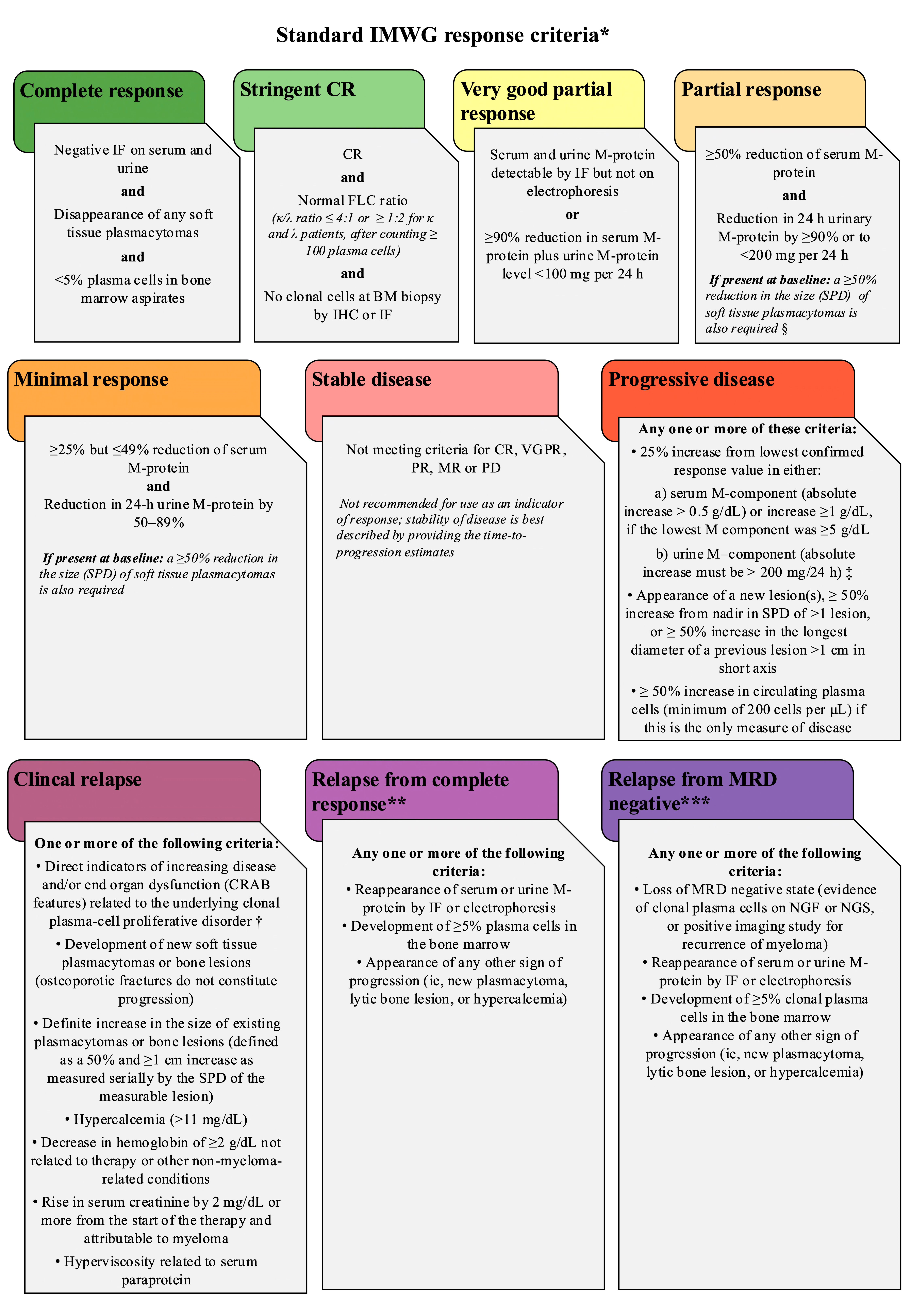
The International Myeloma Working Group (IMWG) proposed updated criteria for the diagnosis, treatment and follow-up of myeloma, and it included PET/CT within the diagnostic flow chart for both initial evaluation and therapy response assessment [7]. Notably, for the response assessment, IMWG criteria define all the parameters that need to be taken into account. In the latest version (2019) [7], responders are categorized into subgroups: complete response, near-complete response, very good partial response, or partial response. It is fundamental that the same imaging technique is used in patients’ follow-up. In case of using PET/CT to evaluate response, it is obligatory to perform PET/CT at baseline. Furthermore, no change in treatment can be recommended on the basis of imaging results except for progression. Complete response definition includes the combination of negative imaging (such as PET/CT) with an MRD-negative result from flow cytometry or sequencing (imaging + MRD-negative). In the case of persistent focal lesions at PET, yearly follow-up is recommended because these patients are at higher risk of relapse of disease. A summary of IMWG response criteria is presented in Figure 3.

3.2.4. EANM Consensus Recommendations

The European Association of Nuclear Medicine (EANM) promoted an initiative to achieve expert consensus on key issues related to the application of nuclear medicine in hematological malignancies, including multiple myeloma. The EANM Focus 4 Consensus Recommendations [23], developed through a modified Delphi method, particularly focus on aspects such as patient eligibility, imaging technique, staging, response assessment and reporting standards, thus providing guidance on the use of molecular imaging in multiple myeloma. A summary of these recommendations is presented in Table 2.
Although these standardized systems share the Deauville five-point scale as their common metabolic reference, they differ in scope and intended application and are therefore best regarded as complementary rather than competing (Table 3). The Deauville criteria provide the underlying visual scale, grading [18F]FDG uptake relative to mediastinal and hepatic background, and they are used principally to define complete metabolic response after therapy. The IMPeTUs criteria build on this scale to provide a structured, reproducible reporting framework that captures bone marrow metabolism, the number and site of focal lesions, paramedullary and extramedullary disease, and lytic lesions on CT, and they are therefore particularly suited to standardized baseline and response reporting across centers. The IMWG criteria integrate imaging with bone marrow-based MRD assessment, defining complete response as the combination of negative PET/CT and an MRD-negative result by flow cytometry or sequencing, and thus serve to anchor imaging within the broader composite response definition. Finally, the EANM Focus 4 recommendations, complemented by the more recent EANM procedural guideline, address acquisition technique, indications, and reporting standards, harmonizing how PET/CT is performed and interpreted rather than defining response per se.

3.3. Beyond FDG

The development and utilization of novel PET/CT radiotracers holds promise for further enhancing the evaluation and prognostication of MM patients in the coming decade. Metabolic tracers other than [18F]FDG have been explored in plasma cell disorders, but data on these alternatives are limited, and there is no standard guidance for their use in current clinical settings. Unlike FDG, which assesses glucose metabolism, these tracers focus on cell membrane metabolism, amino acid metabolism, DNA or other specific targets.
Choline, a component of cell membranes, shows increased uptake in cells with higher metabolic activity. [11C]choline and [18F]fluorocholine have demonstrated uptake in multiple myeloma and offer an improved detection of focal bone lesions over FDG PET/CT [57,58,59]. However, larger prospective studies with defined reference standards are needed to confirm their diagnostic effectiveness and their potential role in response assessment. Methionine, an amino acid marker of metabolism, can be radiolabeled, and [11C]methionine can be used to detect bone localizations of myeloma. Studies indicate that [11C]methionine has a higher sensitivity for detecting a greater number of lesions in most patients compared to [18F]FDG [60,61,62,63]. On the other hand, [18F]fluorothymidine, a pyrimidine deoxynucleoside imaging DNA synthesis and cell proliferation, showed lower sensitivity for bone lesions compared to [18F]FDG and limited use in disease follow-up [64] in pilot studies on multiple myeloma several years ago. [11C]acetate, a tracer for lipid metabolism, is elevated in myeloma cell lines. In a study of 35 patients (26 with symptomatic MM, 5 with SMM, and 4 with MGUS), [11C]acetate PET performed significantly better than [18F]FDG in detecting both diffuse and focal MM with higher metabolic intensity [65]. Preliminary reports [66] suggest that [11C]acetate PET may effectively assess MRD negativity after daratumumab-based induction in newly diagnosed multiple myeloma.
More experimental tracers are under evaluation—mainly chemokine receptor type 4 (CXCR4) targeted small molecules and anti-CD38 radiolabeled antibodies. CXCR4-related tracers bind the stromal cell-derived factor 1, leading to tumor growth in several cancers. This mechanism is notably overexpressed in multiple myeloma cells, and MM cells have high CXCR4 expression, which can be imaged in vivo using [68Ga]pentixafor, which is a peptidomimetic ligand with a strong affinity for CXCR4. Preliminary data suggest that CXCR4 expression represents a negative prognostic factor and a potential target for myeloma-specific treatment [67,68]. Pentixafor can also be conjugated with beta-emitting elements like [177Lu] and [90Y] for therapeutic purposes, becoming an interesting therapeutic option in the setting of radioligand therapy [69,70]. Very late antigen 4 (VLA4, also known as α4β1 integrin, CD49d/CD29) is abundantly expressed in MM and bone marrow stromal cells within the tumor microenvironment [71]. LLP2A, a peptidomimetic ligand that selectively binds to the activated form of VLA4, can be labeled with [64Cu] using the chelator CB-TE1A1P. The first human study indicated that [64Cu]LLP2A is safe with acceptable radiation dosimetry [72].
Immune-based PET imaging (immuno-PET) combines the precise targeting abilities of monoclonal antibodies (mAbs) with the high sensitivity and specificity of whole-body PET imaging. CD38 is uniformly overexpressed in MM, making it an ideal target for developing CD38-targeted PET radiopharmaceuticals. Daratumumab and Isatuximab, two anti-CD38 mAbs used for treating MM, can be directly labeled with long-life beta emitters such as [89Zr] or [64Cu]. Preliminary phase 1 studies have shown that [64Cu]Daratumumab has greater sensitivity and resolution compared to [18F]FDG for detecting multiple myeloma especially in the bone marrow [73,74]. [89Zr]Isatuximab is also in early phase evaluation, but it has shown similar sensitivity in detecting MM lesions [75]. Other radiotracers like [68Ga]Nb1053 (CD38-specific single domain antibody), [89Zr]Elotuzumab (anti-SLAMF7 antibody), and [89Zr]Bevacizumab (anti-VEGF antibody) are also still in preclinical stages [22,76].
Although these approaches are still in the early stages of clinical development, preliminary studies suggest that targeted molecular imaging may improve the diagnosis and treatment monitoring of multiple myeloma when combined with conventional methods. At present, however, all these tracers remain at an investigational stage and are not yet available for routine clinical use with evidence largely limited to small or early-phase studies.

3.4. Evaluation of Strength of Evidence

The evidence underpinning FDG PET/CT in multiple myeloma response assessment is uneven in maturity. The strongest support (Table 4), drawn from large prospective cohorts, trial sub-studies, and consensus recommendations, lies with its prognostic applications: complete metabolic response by the Deauville score, the adverse significance of residual focal lesions and persistent SUVmax > 4.2, the impact of baseline extramedullary disease and high focal-lesion burden, and the value of combining PET/CT with bone marrow MRD to identify double-negative patients. These findings are reproducible across different cohorts and reading criteria and can be regarded as relatively well established. A second tier of applications—interim or early imaging to guide therapy, IMPeTUs-based standardized reading, and treatment intensification in PET-positive patients—is promising but rests largely on single trials or single-center series and awaits broader confirmation. The weakest evidence concerns the non-FDG radiotracers: choline, methionine, acetate, FLT, CXCR4-targeted pentixafor, and CD38- or SLAMF7-targeted immunoPET agents are supported only by pilot, first-in-human, or preclinical studies with no validated role in response assessment to date. Overall, while the prognostic value of metabolic response is firmly grounded, much of the field’s more recent and technologically driven promise remains investigational.

3.5. Limitations

This review has several limitations. First, it is a narrative rather than a systematic review: although a structured search of PubMed, Scopus, Web of Science, and the Cochrane Library was performed, the work was not registered as a protocol and does not include a PRISMA flow diagram, dual independent screening, a published search string, or quantitative synthesis. Study selection and emphasis therefore reflect a degree of author judgement, and selection or citation bias cannot be excluded. Relatedly, no formal risk-of-bias or quality assessment (e.g., QUADAS-2) was applied, so the individual studies are not graded for internal validity.
Second, the underlying evidence is heterogeneous, which limits the strength of the conclusions. The included studies differ in design (retrospective single-center series versus large multicenter prospective trials), sample size (from cohorts of roughly 30–50 patients to series of several hundred), treatment era and regimen, and timing of imaging (interim, post-induction, post-transplant, premaintenance, and follow-up). Critically, there is no uniform definition of PET positivity: SUVmax thresholds, Deauville-based cut-offs, focal-lesion counts, and IMPeTUs reading have all been used. Reported performance metrics should therefore be read within the specific population, treatment context, and criteria of each study rather than as fixed values for the modality.
Beyond between-study heterogeneity, FDG PET/CT is also subject to technical and interpretative variability: differences in acquisition protocols (uptake time, reconstruction, scanner calibration) and in reader experience can affect SUV-based and visual assessments, which is precisely the variability that standardized frameworks such as IMPeTUs and the EANM recommendations aim to reduce. Furthermore, the non-FDG radiotracers discussed here face additional barriers to clinical implementation, including limited availability (notably for 11C-labeled, cyclotron-dependent agents), absence of regulatory approval for myeloma indications, and lack of standardized acquisition and interpretation protocols.
Third, the level of evidence is uneven across applications. The prognostic value of complete metabolic response, the adverse significance of residual focal lesions and extramedullary disease, and the complementarity of FDG PET/CT with bone marrow-based MRD are supported by large prospective datasets and consensus recommendations and may be regarded as relatively well established. Other observations—such as single-center conversion rates after intensification in PET-positive patients, or the optimal day-7 interim time point—derive from individual cohorts and await confirmation. Data on non-FDG radiotracers ([11C]choline, [11C]methionine, [18F]fluorothymidine, [11C]acetate, [68Ga]pentixafor, and CD38- or SLAMF7-targeted immunoPET agents) arise largely from pilot, first-in-human, or preclinical work and are best regarded as promising investigational tools rather than validated alternatives to FDG. To make these distinctions explicit, the principal applications are graded by evidence level in Table 4.
Finally, the literature is concentrated and the field is moving quickly. Much of the work on standardized interpretation originates from a limited number of expert groups, and the IMPeTUs framework in particular has been developed predominantly within Italian cohorts, so its consistency may partly reflect overlapping investigators and populations; broader international validation is still required. At the same time, treatment paradigms (anti-CD38 quadruplets, bispecific antibodies, CAR T-cell therapy) and MRD methodology are evolving faster than imaging studies can mature, so some reviewed cohorts were treated with regimens that no longer reflect current standard of care. The thresholds and response definitions summarized here will require ongoing re-evaluation.

4. Conclusions and Future Directions

Combining clinical assessment, laboratory testing, bone marrow analysis, whole-body MRI and [18F]FDG PET/CT allows a comprehensive evaluation of disease activity in multiple myeloma. With its whole-body coverage and sensitivity for metabolically active skeletal and extramedullary disease, FDG PET/CT has become an important tool for response assessment and carries prognostic value, including in MRD evaluation. It is best viewed as one component of a multimodal assessment rather than a stand-alone determinant of response, complementing rather than replacing bone marrow-based MRD techniques and other imaging modalities.
The evidence reviewed in this paper indicates that achieving complete metabolic response, as defined by the Deauville score, has been identified as an independent predictor of prolonged PFS and OS. Moreover, the integration of [18F]FDG PET/CT with bone marrow-based MRD techniques such as NGF and NGS may allow for a more comprehensive evaluation of disease status, helping to identify patients with double-negative results who appear to have the most favorable outcomes. Standardized interpretation criteria, particularly the IMPeTUs framework and the EANM Focus 4 recommendations, have contributed to improving reproducibility and to facilitating a multicenter comparison of PET-based response assessment.
Despite the growing role of FDG PET/CT in multiple myeloma, several issues remain unresolved. The optimal timing of PET/CT for response assessment is not firmly established, as imaging is variably performed after induction, before maintenance, or during follow-up, and these time points may carry different prognostic weight. In addition, PET/CT and bone marrow-based MRD assessment may yield discordant results, and the management of patients who are PET-positive but bone marrow MRD-negative remains undefined, underscoring the complementary rather than interchangeable nature of the two approaches. Finally, much of the evidence supporting PET-guided or MRD-driven treatment adaptation derives from clinical trials, and these strategies should currently be regarded as investigational rather than standard of care in routine practice.
Several avenues for future investigation are emerging. Alternative tracers (e.g., [11C]choline, [11C]methionine) each offer distinct metabolic insights, but the supporting data remain limited and none has yet shown a clear advantage over FDG in routine practice. CXCR4-targeted imaging with [68Ga]pentixafor has given encouraging early prospective results and may complement [18F]FDG, particularly in extramedullary disease, although its response-assessment role is not yet established. CD38-targeted immunoPET could in principle enable myeloma-specific imaging reflecting the target of anti-CD38 therapy, but current evidence is confined to first-in-human and preclinical studies. Hybrid PET/MRI, combining metabolic information with superior soft-tissue contrast, also warrants further study.
Artificial intelligence and radiomics represent a further emerging direction. By extracting quantitative imaging features beyond visual analysis, radiomics combined with machine learning has shown potential to refine prognostic stratification and to support minimal residual disease assessment in multiple myeloma [77,78], although these approaches remain preliminary and require prospective validation before clinical use. More broadly, future research should prioritize the prospective validation of Deauville score thresholds in the setting of novel therapy regimens, the standardization of PET/CT acquisition and timing, and trials comparing imaging- or tracer-guided response-adapted strategies with conventional approaches. Together, these approaches highlight the dynamic landscape of molecular imaging in multiple myeloma.

Author Contributions

Conceptualization, A.L.D., A.C., T.H., A.D., L.A., K.T., T.K., G.R., S.M. and D.D.; Writing—Original Draft Preparation, A.L.D., A.C., T.H., A.D., L.A., K.T., T.K., G.R., S.M. and D.D.; Writing—Review and Editing, A.L.D., A.C., T.H., A.D., L.A., K.T., T.K., G.R., S.M. and D.D.; Supervision, D.D. All authors have read and agreed to the published version of the manuscript.

Funding

This research received no external funding.

Institutional Review Board Statement

Not applicable.

Informed Consent Statement

Not applicable.

Data Availability Statement

No new data were created or analyzed in this study. Data sharing is not applicable to this article.

Conflicts of Interest

The authors declare no conflicts of interest.

Abbreviations

The following abbreviations are used in this manuscript:
ADCCAntibody-Dependent Cellular Cytotoxicity
ADCApparent Diffusion Coefficient
AIArtificial Intelligence
ASCTAutologous Stem Cell Transplantation
BCMAB-Cell Maturation Antigen
BMBone Marrow
CAR TChimeric Antigen Receptor T cells
CBCComplete Blood Count
CIConfidence Interval
CMRComplete Metabolic Response
CRComplete Response
CRABCalcium Elevation, Renal Insufficiency, Anemia, Bone Lesions
CTComputed Tomography
CXCR4Chemokine Receptor Type 4
Dara-VRdDaratumumab, Bortezomib, Lenalidomide and Dexamethasone
Dara-VTdDaratumumab, Bortezomib, Thalidomide and Dexamethasone
DFSDisease-Free Survival
DSDeauville Score
DWI-MRIDiffusion-Weighted Imaging MRI
EANMEuropean Association of Nuclear Medicine
EHAEuropean Hematology Association
EMDExtramedullary Disease
ESMOEuropean Society for Medical Oncology
FDAFood and Drug Administration
FDGFluorodeoxyglucose
FLFocal Lesion
HDTHigh-Dose Chemotherapy
HRHazard Ratio
IMWGInternational Myeloma Working Group
IMPeTUsItalian Myeloma Criteria for PET Use
KRdCarfilzomib, Lenalidomide and Dexamethasone
LDCTLow-Dose Computed Tomography
mAbsMonoclonal Antibodies
MeSHMedical Subject Headings
MFCMultiparameter Flow Cytometry
MGUSMonoclonal Gammopathy of Undetermined Significance
MMMultiple Myeloma
MRDMinimal Residual Disease
MRIMagnetic Resonance Imaging
MTVMetabolic Tumor Volume
NGFNext-Generation Flow Cytometry
NGSNext-Generation Sequencing
NKNatural Killer
OSOverall Survival
PCRPolymerase Chain Reaction
PETPositron Emission Tomography
PFSProgression-Free Survival
PMDParamedullary Disease
R-ISSRevised International Staging System
SCTStem Cell Transplantation
SLAMF7Signaling-Lymphocytic-Activation-Molecule F7
SMMSmoldering Multiple Myeloma
SSSkeletal Survey
SUVmaxMaximum Standardized Uptake Value
TTNTTime to Next Treatment
TTPTime to Progression
VGPRVery Good Partial Response
VLA4Very Late Antigen 4
VRdBortezomib, Lenalidomide, and Dexamethasone
VTdBortezomib, Thalidomide, and Dexamethasone
WB-MRIWhole-Body MRI

References

  1. Röllig, C.; Knop, S.; Bornhäuser, M. Multiple Myeloma. Lancet 2015, 385, 2197–2208. [Google Scholar] [CrossRef] [PubMed]
  2. Rajkumar, S.V.; Dimopoulos, M.A.; Palumbo, A.; Blade, J.; Merlini, G.; Mateos, M.-V.; Kumar, S.; Hillengass, J.; Kastritis, E.; Richardson, P.; et al. International Myeloma Working Group Updated Criteria for the Diagnosis of Multiple Myeloma. Lancet Oncol. 2014, 15, e538–e548. [Google Scholar] [CrossRef] [PubMed]
  3. Dimopoulos, M.A.; Moreau, P.; Terpos, E.; Mateos, M.V.; Zweegman, S.; Cook, G.; Delforge, M.; Hájek, R.; Schjesvold, F.; Cavo, M.; et al. Multiple Myeloma: EHA-ESMO Clinical Practice Guidelines for Diagnosis, Treatment and Follow-Up†. Ann. Oncol. 2021, 32, 309–322. [Google Scholar] [CrossRef] [PubMed]
  4. Wu, F.; Bernard, S.; Fayad, L.M.; Ilaslan, H.; Messiou, C.; Moulopoulos, L.A.; Mulligan, M.E. Updates and Ongoing Challenges in Imaging of Multiple Myeloma: AJR Expert Panel Narrative Review. AJR Am. J. Roentgenol. 2021, 217, 775–785. [Google Scholar] [CrossRef] [PubMed]
  5. Dimopoulos, M.A.; Hillengass, J.; Usmani, S.; Zamagni, E.; Lentzsch, S.; Davies, F.E.; Raje, N.; Sezer, O.; Zweegman, S.; Shah, J.; et al. Role of Magnetic Resonance Imaging in the Management of Patients with Multiple Myeloma: A Consensus Statement. J. Clin. Oncol. 2015, 33, 657–664. [Google Scholar] [CrossRef] [PubMed]
  6. Cavo, M.; Terpos, E.; Nanni, C.; Moreau, P.; Lentzsch, S.; Zweegman, S.; Hillengass, J.; Engelhardt, M.; Usmani, S.Z.; Vesole, D.H.; et al. Role of 18F-FDG PET/CT in the Diagnosis and Management of Multiple Myeloma and Other Plasma Cell Disorders: A Consensus Statement by the International Myeloma Working Group. Lancet Oncol. 2017, 18, e206–e217. [Google Scholar] [CrossRef] [PubMed]
  7. Hillengass, J.; Usmani, S.; Rajkumar, S.V.; Durie, B.G.M.; Mateos, M.-V.; Lonial, S.; Joao, C.; Anderson, K.C.; García-Sanz, R.; Riva, E.; et al. International Myeloma Working Group Consensus Recommendations on Imaging in Monoclonal Plasma Cell Disorders. Lancet Oncol. 2019, 20, e302–e312. [Google Scholar] [CrossRef] [PubMed]
  8. Richardson, P.G.; Jacobus, S.J.; Weller, E.A.; Hassoun, H.; Lonial, S.; Raje, N.S.; Medvedova, E.; McCarthy, P.L.; Libby, E.N.; Voorhees, P.M.; et al. Triplet Therapy, Transplantation, and Maintenance until Progression in Myeloma. N. Engl. J. Med. 2022, 387, 132–147. [Google Scholar] [CrossRef] [PubMed]
  9. Callander, N.S.; Baljevic, M.; Adekola, K.; Anderson, L.D.; Campagnaro, E.; Castillo, J.J.; Costello, C.; Devarakonda, S.; Elsedawy, N.; Faiman, M.; et al. NCCN Guidelines® Insights: Multiple Myeloma, Version 3.2022. J. Natl. Compr. Cancer Netw. 2022, 20, 8–19. [Google Scholar] [CrossRef] [PubMed]
  10. de Weers, M.; Tai, Y.-T.; van der Veer, M.S.; Bakker, J.M.; Vink, T.; Jacobs, D.C.H.; Oomen, L.A.; Peipp, M.; Valerius, T.; Slootstra, J.W.; et al. Daratumumab, a Novel Therapeutic Human CD38 Monoclonal Antibody, Induces Killing of Multiple Myeloma and Other Hematological Tumors. J. Immunol. 2011, 186, 1840–1848. [Google Scholar] [CrossRef] [PubMed]
  11. Hsi, E.D.; Steinle, R.; Balasa, B.; Szmania, S.; Draksharapu, A.; Shum, B.P.; Huseni, M.; Powers, D.; Nanisetti, A.; Zhang, Y.; et al. CS1, a Potential New Therapeutic Antibody Target for the Treatment of Multiple Myeloma. Clin. Cancer Res. 2008, 14, 2775–2784. [Google Scholar] [CrossRef] [PubMed]
  12. Lonial, S.; Dimopoulos, M.; Palumbo, A.; White, D.; Grosicki, S.; Spicka, I.; Walter-Croneck, A.; Moreau, P.; Mateos, M.-V.; Magen, H.; et al. Elotuzumab Therapy for Relapsed or Refractory Multiple Myeloma. N. Engl. J. Med. 2015, 373, 621–631. [Google Scholar] [CrossRef] [PubMed]
  13. Dimopoulos, M.A.; Oriol, A.; Nahi, H.; San-Miguel, J.; Bahlis, N.J.; Usmani, S.Z.; Rabin, N.; Orlowski, R.Z.; Komarnicki, M.; Suzuki, K.; et al. Daratumumab, Lenalidomide, and Dexamethasone for Multiple Myeloma. N. Engl. J. Med. 2016, 375, 1319–1331. [Google Scholar] [CrossRef] [PubMed]
  14. Moreau, P.; Attal, M.; Hulin, C.; Arnulf, B.; Belhadj, K.; Benboubker, L.; Béné, M.C.; Broijl, A.; Caillon, H.; Caillot, D.; et al. Bortezomib, Thalidomide, and Dexamethasone with or without Daratumumab before and after Autologous Stem-Cell Transplantation for Newly Diagnosed Multiple Myeloma (CASSIOPEIA): A Randomised, Open-Label, Phase 3 Study. Lancet 2019, 394, 29–38. [Google Scholar] [CrossRef] [PubMed]
  15. Dimopoulos, M.; Quach, H.; Mateos, M.-V.; Landgren, O.; Leleu, X.; Siegel, D.; Weisel, K.; Yang, H.; Klippel, Z.; Zahlten-Kumeli, A.; et al. Carfilzomib, Dexamethasone, and Daratumumab versus Carfilzomib and Dexamethasone for Patients with Relapsed or Refractory Multiple Myeloma (CANDOR): Results from a Randomised, Multicentre, Open-Label, Phase 3 Study. Lancet 2020, 396, 186–197. [Google Scholar] [CrossRef] [PubMed]
  16. Moreau, P.; Dimopoulos, M.-A.; Mikhael, J.; Yong, K.; Capra, M.; Facon, T.; Hajek, R.; Špička, I.; Baker, R.; Kim, K.; et al. Isatuximab, Carfilzomib, and Dexamethasone in Relapsed Multiple Myeloma (IKEMA): A Multicentre, Open-Label, Randomised Phase 3 Trial. Lancet 2021, 397, 2361–2371. [Google Scholar] [CrossRef] [PubMed]
  17. Martin, T.G.; Madduri, D.; Pacaud, L.; Usmani, S.Z. Cilta-Cel, a BCMA-Targeting CAR-T Therapy for Heavily Pretreated Patients with Relapsed/Refractory Multiple Myeloma. Future Oncol. 2023, 19, 2297–2311. [Google Scholar] [CrossRef] [PubMed]
  18. Singh, S.B.; Bhandari, S.; Siwakoti, S.; Kumar, M.; Singh, R.; Bhusal, S.; Sharma, K.; Bhandari, S.; Khanal, K. PET/CT in the Evaluation of CAR-T Cell Immunotherapy in Hematological Malignancies. Mol. Imaging 2024, 23, 15353508241257924. [Google Scholar] [CrossRef] [PubMed]
  19. Costa, L.J.; Derman, B.A.; Bal, S.; Sidana, S.; Chhabra, S.; Silbermann, R.; Ye, J.C.; Cook, G.; Cornell, R.F.; Holstein, S.A.; et al. International Harmonization in Performing and Reporting Minimal Residual Disease Assessment in Multiple Myeloma Trials. Leukemia 2021, 35, 18–30. [Google Scholar] [CrossRef] [PubMed]
  20. Kumar, S.; Paiva, B.; Anderson, K.C.; Durie, B.; Landgren, O.; Moreau, P.; Munshi, N.; Lonial, S.; Bladé, J.; Mateos, M.-V.; et al. International Myeloma Working Group Consensus Criteria for Response and Minimal Residual Disease Assessment in Multiple Myeloma. Lancet Oncol. 2016, 17, e328–e346. [Google Scholar] [CrossRef] [PubMed]
  21. Bezzi, D.; Ambrosini, V.; Nanni, C. Clinical Value of FDG-PET/CT in Multiple Myeloma: An Update. Semin Nucl. Med. 2023, 53, 352–370. [Google Scholar] [CrossRef] [PubMed]
  22. Ishibashi, M.; Takahashi, M.; Yamaya, T.; Imai, Y. Current and Future PET Imaging for Multiple Myeloma. Life 2023, 13, 1701. [Google Scholar] [CrossRef] [PubMed]
  23. Nanni, C.; Kobe, C.; Baeßler, B.; Baues, C.; Boellaard, R.; Borchmann, P.; Buck, A.; Buvat, I.; Chapuy, B.; Cheson, B.D.; et al. European Association of Nuclear Medicine (EANM) Focus 4 Consensus Recommendations: Molecular Imaging and Therapy in Haematological Tumours. Lancet Haematol. 2023, 10, e367–e381. [Google Scholar] [CrossRef] [PubMed]
  24. Nanni, C.; Deroose, C.M.; Balogova, S.; Lapa, C.; Withofs, N.; Subesinghe, M.; Jamet, B.; Zamagni, E.; Ippolito, D.; Delforge, M.; et al. EANM Guidelines on the Use of [18F]FDG PET/CT in Diagnosis, Staging, Prognostication, Therapy Assessment, and Restaging of Plasma Cell Disorders. Eur. J. Nucl. Med. Mol. Imaging 2024, 52, 171–192. [Google Scholar] [CrossRef] [PubMed]
  25. Zamagni, E.; Oliva, S.; Gay, F.; Capra, A.; Rota-Scalabrini, D.; D’Agostino, M.; Belotti, A.; Galli, M.; Racca, M.; Zambello, R.; et al. Impact of Minimal Residual Disease Standardised Assessment by FDG-PET/CT in Transplant-Eligible Patients with Newly Diagnosed Multiple Myeloma Enrolled in the Imaging Sub-Study of the FORTE Trial. EClinicalMedicine 2023, 60, 102017. [Google Scholar] [CrossRef] [PubMed]
  26. Kraeber-Bodéré, F.; Zweegman, S.; Perrot, A.; Hulin, C.; Caillot, D.; Facon, T.; Leleu, X.; Belhadj, K.; Itti, E.; Karlin, L.; et al. Prognostic Value of Positron Emission Tomography/Computed Tomography in Transplant-Eligible Newly Diagnosed Multiple Myeloma Patients from CASSIOPEIA: The CASSIOPET Study. Haematologica 2023, 108, 621–626. [Google Scholar] [CrossRef] [PubMed]
  27. Derlin, T.; Peldschus, K.; Münster, S.; Bannas, P.; Herrmann, J.; Stübig, T.; Habermann, C.R.; Adam, G.; Kröger, N.; Weber, C. Comparative Diagnostic Performance of 18F-FDG PET/CT versus Whole-Body MRI for Determination of Remission Status in Multiple Myeloma after Stem Cell Transplantation. Eur. Radiol. 2013, 23, 570–578. [Google Scholar] [CrossRef] [PubMed]
  28. Basha, M.A.A.; Hamed, M.A.G.; Refaat, R.; AlAzzazy, M.Z.; Bessar, M.A.; Mohamed, E.M.; Ahmed, A.F.; Tantawy, H.F.; Altaher, K.M.; Obaya, A.A.; et al. Diagnostic Performance of 18F-FDG PET/CT and Whole-Body MRI before and Early after Treatment of Multiple Myeloma: A Prospective Comparative Study. Jpn. J. Radiol. 2018, 36, 382–393. [Google Scholar] [CrossRef] [PubMed]
  29. Gómez León, N.; Aguado Bueno, B.; Herreros Pérez, M.; León Ramírez, L.F.; Alegre, A.; Colletti, P.M.; Rubello, D.; Carreras, J.L.; Delgado Bolton, R.C. Agreement Between 18F-FDG PET/CT and Whole-Body Magnetic Resonance Compared With Skeletal Survey for Initial Staging and Response at End-of-Treatment Evaluation of Patients With Multiple Myeloma. Clin. Nucl. Med. 2021, 46, 310–322. [Google Scholar] [CrossRef] [PubMed]
  30. Paternain, A.; García-Velloso, M.J.; Rosales, J.J.; Ezponda, A.; Soriano, I.; Elorz, M.; Rodríguez-Otero, P.; Aquerreta, J.D. The Utility of ADC Value in Diffusion-Weighted Whole-Body MRI in the Follow-up of Patients with Multiple Myeloma. Correlation Study with 18F-FDG PET-CT. Eur. J. Radiol. 2020, 133, 109403. [Google Scholar] [CrossRef] [PubMed]
  31. Nørgaard, J.N.; Abildgaard, N.; Lysén, A.; Tsykunova, G.; Vangsted, A.J.; João, C.; Remen, N.; Nielsen, L.K.; Osnes, L.; Stokke, C.; et al. Intensifying Treatment in PET-Positive Multiple Myeloma Patients after Upfront Autologous Stem Cell Transplantation. Leukemia 2023, 37, 2107–2114. [Google Scholar] [CrossRef] [PubMed]
  32. Rodríguez-Laval, V.; Lumbreras-Fernández, B.; Aguado-Bueno, B.; Gómez-León, N. Imaging of Multiple Myeloma: Present and Future. J. Clin. Med. 2024, 13, 264. [Google Scholar] [CrossRef] [PubMed]
  33. Rasche, L.; Angtuaco, E.; McDonald, J.E.; Buros, A.; Stein, C.; Pawlyn, C.; Thanendrarajan, S.; Schinke, C.; Samant, R.; Yaccoby, S.; et al. Low Expression of Hexokinase-2 Is Associated with False-Negative FDG-Positron Emission Tomography in Multiple Myeloma. Blood 2017, 130, 30–34. [Google Scholar] [CrossRef] [PubMed]
  34. Rasche, L.; Alapat, D.; Kumar, M.; Gershner, G.; McDonald, J.; Wardell, C.P.; Samant, R.; Van Hemert, R.; Epstein, J.; Williams, A.F.; et al. Combination of Flow Cytometry and Functional Imaging for Monitoring of Residual Disease in Myeloma. Leukemia 2019, 33, 1713–1722. [Google Scholar] [CrossRef] [PubMed]
  35. Rama, S.; Suh, C.H.; Kim, K.W.; Durieux, J.C.; Ramaiya, N.H.; Tirumani, S.H. Comparative Performance of Whole-Body MRI and FDG PET/CT in Evaluation of Multiple Myeloma Treatment Response: Systematic Review and Meta-Analysis. AJR Am. J. Roentgenol. 2022, 218, 602–613. [Google Scholar] [CrossRef] [PubMed]
  36. Jamet, B.; Carlier, T.; Bailly, C.; Bodet-Milin, C.; Monnet, A.; Frampas, E.; Touzeau, C.; Moreau, P.; Kraeber-Bodere, F. Hybrid Simultaneous Whole-Body 2-[18F]FDG-PET/MRI Imaging in Newly Diagnosed Multiple Myeloma: First Diagnostic Performance and Clinical Added Value Results. Eur. Radiol. 2023, 33, 6438–6447. [Google Scholar] [CrossRef] [PubMed]
  37. Archibald, S.J.; Holland, J.P.; Korde, A.; Martins, A.F.; Shuhendler, A.J.; Scott, P.J.H. Combining Nuclear Medicine With Other Modalities: Future Prospect for Multimodality Imaging. Mol. Imaging 2024, 23, 15353508241245265. [Google Scholar] [CrossRef] [PubMed]
  38. Zamagni, E.; Patriarca, F.; Nanni, C.; Zannetti, B.; Englaro, E.; Pezzi, A.; Tacchetti, P.; Buttignol, S.; Perrone, G.; Brioli, A.; et al. Prognostic Relevance of 18-F FDG PET/CT in Newly Diagnosed Multiple Myeloma Patients Treated with up-Front Autologous Transplantation. Blood 2011, 118, 5989–5995. [Google Scholar] [CrossRef] [PubMed]
  39. Mesguich, C.; Latrabe, V.; Hulin, C.; Lascaux, A.; Bordenave, L.; Hindié, E.; Marit, G. Prospective Comparison of 18-FDG PET/CT and Whole-Body MRI with Diffusion-Weighted Imaging in the Evaluation of Treatment Response of Multiple Myeloma Patients Eligible for Autologous Stem Cell Transplant. Cancers 2021, 13, 1938. [Google Scholar] [CrossRef] [PubMed]
  40. Fonti, R.; Larobina, M.; Del Vecchio, S.; De Luca, S.; Fabbricini, R.; Catalano, L.; Pane, F.; Salvatore, M.; Pace, L. Metabolic Tumor Volume Assessed by 18F-FDG PET/CT for the Prediction of Outcome in Patients with Multiple Myeloma. J. Nucl. Med. 2012, 53, 1829–1835. [Google Scholar] [CrossRef] [PubMed]
  41. Sachpekidis, C.; Merz, M.; Raab, M.-S.; Bertsch, U.; Weru, V.; Kopp-Schneider, A.; Jauch, A.; Goldschmidt, H.; Dimitrakopoulou-Strauss, A. The Prognostic Significance of [18F]FDG PET/CT in Multiple Myeloma According to Novel Interpretation Criteria (IMPeTUs). EJNMMI Res. 2021, 11, 100. [Google Scholar] [CrossRef] [PubMed]
  42. Bladé, J.; Beksac, M.; Caers, J.; Jurczyszyn, A.; von Lilienfeld-Toal, M.; Moreau, P.; Rasche, L.; Rosiñol, L.; Usmani, S.Z.; Zamagni, E.; et al. Extramedullary Disease in Multiple Myeloma: A Systematic Literature Review. Blood Cancer J. 2022, 12, 45. [Google Scholar] [CrossRef] [PubMed]
  43. Usmani, S.Z.; Mitchell, A.; Waheed, S.; Crowley, J.; Hoering, A.; Petty, N.; Brown, T.; Bartel, T.; Anaissie, E.; van Rhee, F.; et al. Prognostic Implications of Serial 18-Fluoro-Deoxyglucose Emission Tomography in Multiple Myeloma Treated with Total Therapy 3. Blood 2013, 121, 1819–1823. [Google Scholar] [CrossRef] [PubMed]
  44. Bailly, C.; Carlier, T.; Jamet, B.; Eugene, T.; Touzeau, C.; Attal, M.; Hulin, C.; Facon, T.; Leleu, X.; Perrot, A.; et al. Interim PET Analysis in First-Line Therapy of Multiple Myeloma: Prognostic Value of ΔSUVmax in the FDG-Avid Patients of the IMAJEM Study. Clin. Cancer Res. 2018, 24, 5219–5224. [Google Scholar] [CrossRef] [PubMed]
  45. Kaddoura, M.; Dingli, D.; Buadi, F.K.; Lacy, M.Q.; Gertz, M.A.; Dispenzieri, A.; Kapoor, P.; Hwa, L.; Fonder, A.; Hobbs, M.; et al. Prognostic Impact of Posttransplant FDG PET/CT Scan in Multiple Myeloma. Blood Adv. 2021, 5, 2753–2759. [Google Scholar] [CrossRef] [PubMed]
  46. Moreau, P.; Attal, M.; Caillot, D.; Macro, M.; Karlin, L.; Garderet, L.; Facon, T.; Benboubker, L.; Escoffre-Barbe, M.; Stoppa, A.-M.; et al. Prospective Evaluation of Magnetic Resonance Imaging and [18F]Fluorodeoxyglucose Positron Emission Tomography-Computed Tomography at Diagnosis and Before Maintenance Therapy in Symptomatic Patients With Multiple Myeloma Included in the IFM/DFCI 2009 Trial: Results of the IMAJEM Study. J. Clin. Oncol. 2017, 35, 2911–2918. [Google Scholar] [CrossRef] [PubMed]
  47. Zamagni, E.; Nanni, C.; Mancuso, K.; Tacchetti, P.; Pezzi, A.; Pantani, L.; Zannetti, B.; Rambaldi, I.; Brioli, A.; Rocchi, S.; et al. PET/CT Improves the Definition of Complete Response and Allows to Detect Otherwise Unidentifiable Skeletal Progression in Multiple Myeloma. Clin. Cancer Res. 2015, 21, 4384–4390. [Google Scholar] [CrossRef] [PubMed]
  48. Charalampous, C.; Goel, U.; Broski, S.M.; Dingli, D.; Kapoor, P.; Gertz, M.A.; Lacy, M.Q.; Dispenzieri, A.; Hayman, S.R.; Buadi, F.; et al. Utility of PET/CT in Assessing Early Treatment Response in Patients with Newly Diagnosed Multiple Myeloma. Blood Adv. 2022, 6, 2763–2772. [Google Scholar] [CrossRef] [PubMed]
  49. Stolzenburg, A.; Lückerath, K.; Samnick, S.; Speer, M.; Kneer, K.; Schmid, J.-S.; Grigoleit, G.U.; Hofmann, S.; Beer, A.J.; Bunjes, D.; et al. Prognostic Value of [18F]FDG-PET/CT in Multiple Myeloma Patients before and after Allogeneic Hematopoietic Cell Transplantation. Eur. J. Nucl. Med. Mol. Imaging 2018, 45, 1694–1704. [Google Scholar] [CrossRef] [PubMed]
  50. Davies, F.E.; Rosenthal, A.; Rasche, L.; Petty, N.M.; McDonald, J.E.; Ntambi, J.A.; Steward, D.M.; Panozzo, S.B.; van Rhee, F.; Zangari, M.; et al. Treatment to Suppression of Focal Lesions on Positron Emission Tomography-Computed Tomography Is a Therapeutic Goal in Newly Diagnosed Multiple Myeloma. Haematologica 2018, 103, 1047–1053. [Google Scholar] [CrossRef] [PubMed]
  51. Oberle, A.; Brandt, A.; Alawi, M.; Langebrake, C.; Janjetovic, S.; Wolschke, C.; Schütze, K.; Bannas, P.; Kröger, N.; Koch-Nolte, F.; et al. Long-Term CD38 Saturation by Daratumumab Interferes with Diagnostic Myeloma Cell Detection. Haematologica 2017, 102, e368–e370. [Google Scholar] [CrossRef] [PubMed]
  52. Böckle, D.; Tabares, P.; Zhou, X.; Schimanski, S.; Steinhardt, M.J.; Bittrich, M.; Seebacher, E.; Ulbrich, M.; Wilnit, A.; Metz, C.; et al. Minimal Residual Disease and Imaging-guided Consolidation Strategies in Newly Diagnosed and Relapsed Refractory Multiple Myeloma. Br. J. Haematol. 2022, 198, 515–522. [Google Scholar] [CrossRef] [PubMed]
  53. Barrington, S.F.; Qian, W.; Somer, E.J.; Franceschetto, A.; Bagni, B.; Brun, E.; Almquist, H.; Loft, A.; Højgaard, L.; Federico, M.; et al. Concordance between Four European Centres of PET Reporting Criteria Designed for Use in Multicentre Trials in Hodgkin Lymphoma. Eur. J. Nucl. Med. Mol. Imaging 2010, 37, 1824–1833. [Google Scholar] [CrossRef] [PubMed]
  54. Zamagni, E.; Nanni, C.; Dozza, L.; Carlier, T.; Bailly, C.; Tacchetti, P.; Versari, A.; Chauvie, S.; Gallamini, A.; Gamberi, B.; et al. Standardization of 18F-FDG-PET/CT According to Deauville Criteria for Metabolic Complete Response Definition in Newly Diagnosed Multiple Myeloma. J. Clin. Oncol. 2021, 39, 116–125. [Google Scholar] [CrossRef] [PubMed]
  55. Nanni, C. PET-FDG: Impetus. Cancers 2020, 12, 1030. [Google Scholar] [CrossRef] [PubMed]
  56. Nanni, C.; Zamagni, E.; Versari, A.; Chauvie, S.; Bianchi, A.; Rensi, M.; Bellò, M.; Rambaldi, I.; Gallamini, A.; Patriarca, F.; et al. Image Interpretation Criteria for FDG PET/CT in Multiple Myeloma: A New Proposal from an Italian Expert Panel. IMPeTUs (Italian Myeloma Criteria for PET USe). Eur. J. Nucl. Med. Mol. Imaging 2016, 43, 414–421. [Google Scholar] [CrossRef] [PubMed]
  57. Ulaner, G.A.; Landgren, C.O. Current and Potential Applications of Positron Emission Tomography for Multiple Myeloma and Plasma Cell Disorders. Best Pract. Res. Clin. Haematol. 2020, 33, 101148. [Google Scholar] [CrossRef] [PubMed]
  58. Nanni, C.; Zamagni, E.; Cavo, M.; Rubello, D.; Tacchetti, P.; Pettinato, C.; Farsad, M.; Castellucci, P.; Ambrosini, V.; Montini, G.C.; et al. 11C-Choline vs. 18F-FDG PET/CT in Assessing Bone Involvement in Patients with Multiple Myeloma. World J. Surg. Oncol. 2007, 5, 68. [Google Scholar] [CrossRef] [PubMed]
  59. Cassou-Mounat, T.; Balogova, S.; Nataf, V.; Calzada, M.; Huchet, V.; Kerrou, K.; Devaux, J.-Y.; Mohty, M.; Talbot, J.-N.; Garderet, L. 18F-Fluorocholine versus 18F-Fluorodeoxyglucose for PET/CT Imaging in Patients with Suspected Relapsing or Progressive Multiple Myeloma: A Pilot Study. Eur. J. Nucl. Med. Mol. Imaging 2016, 43, 1995–2004. [Google Scholar] [CrossRef] [PubMed]
  60. Morales-Lozano, M.I.; Rodriguez-Otero, P.; Sancho, L.; Nuñez-Cordoba, J.M.; Prieto, E.; Marcos-Jubilar, M.; Rosales, J.J.; Alfonso, A.; Guillen, E.F.; San-Miguel, J.; et al. 11C-Methionine PET/CT in Assessment of Multiple Myeloma Patients: Comparison to 18F-FDG PET/CT and Prognostic Value. Int. J. Mol. Sci. 2022, 23, 9895. [Google Scholar] [CrossRef] [PubMed]
  61. Okasaki, M.; Kubota, K.; Minamimoto, R.; Miyata, Y.; Morooka, M.; Ito, K.; Ishiwata, K.; Toyohara, J.; Inoue, T.; Hirai, R.; et al. Comparison of (11)C-4′-Thiothymidine, (11)C-Methionine, and (18)F-FDG PET/CT for the Detection of Active Lesions of Multiple Myeloma. Ann. Nucl. Med. 2015, 29, 224–232. [Google Scholar] [CrossRef] [PubMed]
  62. Lapa, C.; Knop, S.; Schreder, M.; Rudelius, M.; Knott, M.; Jörg, G.; Samnick, S.; Herrmann, K.; Buck, A.K.; Einsele, H.; et al. 11C-Methionine-PET in Multiple Myeloma: Correlation with Clinical Parameters and Bone Marrow Involvement. Theranostics 2016, 6, 254–261. [Google Scholar] [CrossRef] [PubMed]
  63. Lapa, C.; Garcia-Velloso, M.J.; Lückerath, K.; Samnick, S.; Schreder, M.; Otero, P.R.; Schmid, J.-S.; Herrmann, K.; Knop, S.; Buck, A.K.; et al. 11C-Methionine-PET in Multiple Myeloma: A Combined Study from Two Different Institutions. Theranostics 2017, 7, 2956–2964. [Google Scholar] [CrossRef] [PubMed]
  64. Agool, A.; Schot, B.W.; Jager, P.L.; Vellenga, E. 18F-FLT PET in Hematologic Disorders: A Novel Technique to Analyze the Bone Marrow Compartment. J. Nucl. Med. 2006, 47, 1592–1598. [Google Scholar] [PubMed]
  65. Ho, C.; Chen, S.; Leung, Y.L.; Cheng, T.; Wong, K.; Cheung, S.K.; Liang, R.; Chim, C.S. 11C-Acetate PET/CT for Metabolic Characterization of Multiple Myeloma: A Comparative Study with 18F-FDG PET/CT. J. Nucl. Med. 2014, 55, 749–752. [Google Scholar] [CrossRef] [PubMed]
  66. Ngai, C.; Kumar, S.; Chi-Lai Ho, G.; Chen, S.; Chim, C.-S. Case Series: MRD Negativity Assessment Using 11C-Acetate PET with 3-Weekly Daratumumab-Based Quadruplet Induction in Newly Diagnosed Multiple Myeloma. Ther. Adv. Hematol. 2021, 12, 20406207211030369. [Google Scholar] [CrossRef] [PubMed]
  67. Philipp-Abbrederis, K.; Herrmann, K.; Knop, S.; Schottelius, M.; Eiber, M.; Lückerath, K.; Pietschmann, E.; Habringer, S.; Gerngroß, C.; Franke, K.; et al. In Vivo Molecular Imaging of Chemokine Receptor CXCR4 Expression in Patients with Advanced Multiple Myeloma. EMBO Mol. Med. 2015, 7, 477–487. [Google Scholar] [CrossRef]
  68. Lapa, C.; Schreder, M.; Schirbel, A.; Samnick, S.; Kortüm, K.M.; Herrmann, K.; Kropf, S.; Einsele, H.; Buck, A.K.; Wester, H.-J.; et al. [68Ga]Pentixafor-PET/CT for Imaging of Chemokine Receptor CXCR4 Expression in Multiple Myeloma—Comparison to [18F]FDG and Laboratory Values. Theranostics 2017, 7, 205–212. [Google Scholar] [CrossRef] [PubMed]
  69. Herrmann, K.; Schottelius, M.; Lapa, C.; Osl, T.; Poschenrieder, A.; Hänscheid, H.; Lückerath, K.; Schreder, M.; Bluemel, C.; Knott, M.; et al. First-in-Human Experience of CXCR4-Directed Endoradiotherapy with 177Lu- and 90Y-Labeled Pentixather in Advanced-Stage Multiple Myeloma with Extensive Intra- and Extramedullary Disease. J. Nucl. Med. 2016, 57, 248–251. [Google Scholar] [CrossRef] [PubMed]
  70. Lapa, C.; Herrmann, K.; Schirbel, A.; Hänscheid, H.; Lückerath, K.; Schottelius, M.; Kircher, M.; Werner, R.A.; Schreder, M.; Samnick, S.; et al. CXCR4-Directed Endoradiotherapy Induces High Response Rates in Extramedullary Relapsed Multiple Myeloma. Theranostics 2017, 7, 1589–1597. [Google Scholar] [CrossRef] [PubMed]
  71. Hathi, D.; Chanswangphuwana, C.; Cho, N.; Fontana, F.; Maji, D.; Ritchey, J.; O’Neal, J.; Ghai, A.; Duncan, K.; Akers, W.J.; et al. Ablation of VLA4 in Multiple Myeloma Cells Redirects Tumor Spread and Prolongs Survival. Sci. Rep. 2022, 12, 30. [Google Scholar] [CrossRef] [PubMed]
  72. Laforest, R.; Ghai, A.; Fraum, T.J.; Oyama, R.; Frye, J.; Kaemmerer, H.; Gaehle, G.; Voller, T.; Mpoy, C.; Rogers, B.E.; et al. First-in-Humans Evaluation of Safety and Dosimetry of 64Cu-LLP2A for PET Imaging. J. Nucl. Med. 2023, 64, 320–328. [Google Scholar] [CrossRef] [PubMed]
  73. Ulaner, G.A.; Sobol, N.B.; O’Donoghue, J.A.; Kirov, A.S.; Riedl, C.C.; Min, R.; Smith, E.; Carter, L.M.; Lyashchenko, S.K.; Lewis, J.S.; et al. CD38-Targeted Immuno-PET of Multiple Myeloma: From Xenograft Models to First-in-Human Imaging. Radiology 2020, 295, 606–615. [Google Scholar] [CrossRef] [PubMed]
  74. Caserta, E.; Chea, J.; Minnix, M.; Poku, E.K.; Viola, D.; Vonderfecht, S.; Yazaki, P.; Crow, D.; Khalife, J.; Sanchez, J.F.; et al. Copper 64-Labeled Daratumumab as a PET/CT Imaging Tracer for Multiple Myeloma. Blood 2018, 131, 741–745. [Google Scholar] [CrossRef] [PubMed]
  75. Krishnan, A.; Adhikarla, V.; Poku, E.K.; Palmer, J.; Chaudhry, A.; Biglang-Awa, V.E.; Bowles, N.; Nathwani, N.; Rosenzweig, M.; Sahebi, F.; et al. Identifying CD38+ Cells in Patients with Multiple Myeloma: First-in-Human Imaging Using Copper-64-Labeled Daratumumab. Blood Adv. 2020, 4, 5194–5202. [Google Scholar] [CrossRef] [PubMed]
  76. Herrero Alvarez, N.; Michel, A.L.; Viray, T.D.; Mayerhoefer, M.E.; Lewis, J.S. 89Zr-DFO-Isatuximab for CD38-Targeted ImmunoPET Imaging of Multiple Myeloma and Lymphomas. ACS Omega 2023, 8, 22486–22495. [Google Scholar] [CrossRef] [PubMed]
  77. Milara, E.; Gómez-Grande, A.; Tomás-Soler, S.; Seiffert, A.P.; Alonso, R.; Gómez, E.J.; Martínez-López, J.; Sánchez-González, P. Bone Marrow Segmentation and Radiomics Analysis of [18F]FDG PET/CT Images for Measurable Residual Disease Assessment in Multiple Myeloma. Comput. Methods Programs Biomed. 2022, 225, 107083. [Google Scholar] [CrossRef] [PubMed]
  78. Jamet, B.; Morvan, L.; Nanni, C.; Michaud, A.-V.; Bailly, C.; Chauvie, S.; Moreau, P.; Touzeau, C.; Zamagni, E.; Bodet-Milin, C.; et al. Random Survival Forest to Predict Transplant-Eligible Newly Diagnosed Multiple Myeloma Outcome Including FDG-PET Radiomics: A Combined Analysis of Two Independent Prospective European Trials. Eur. J. Nucl. Med. Mol. Imaging 2021, 48, 1005–1015. [Google Scholar] [CrossRef] [PubMed]
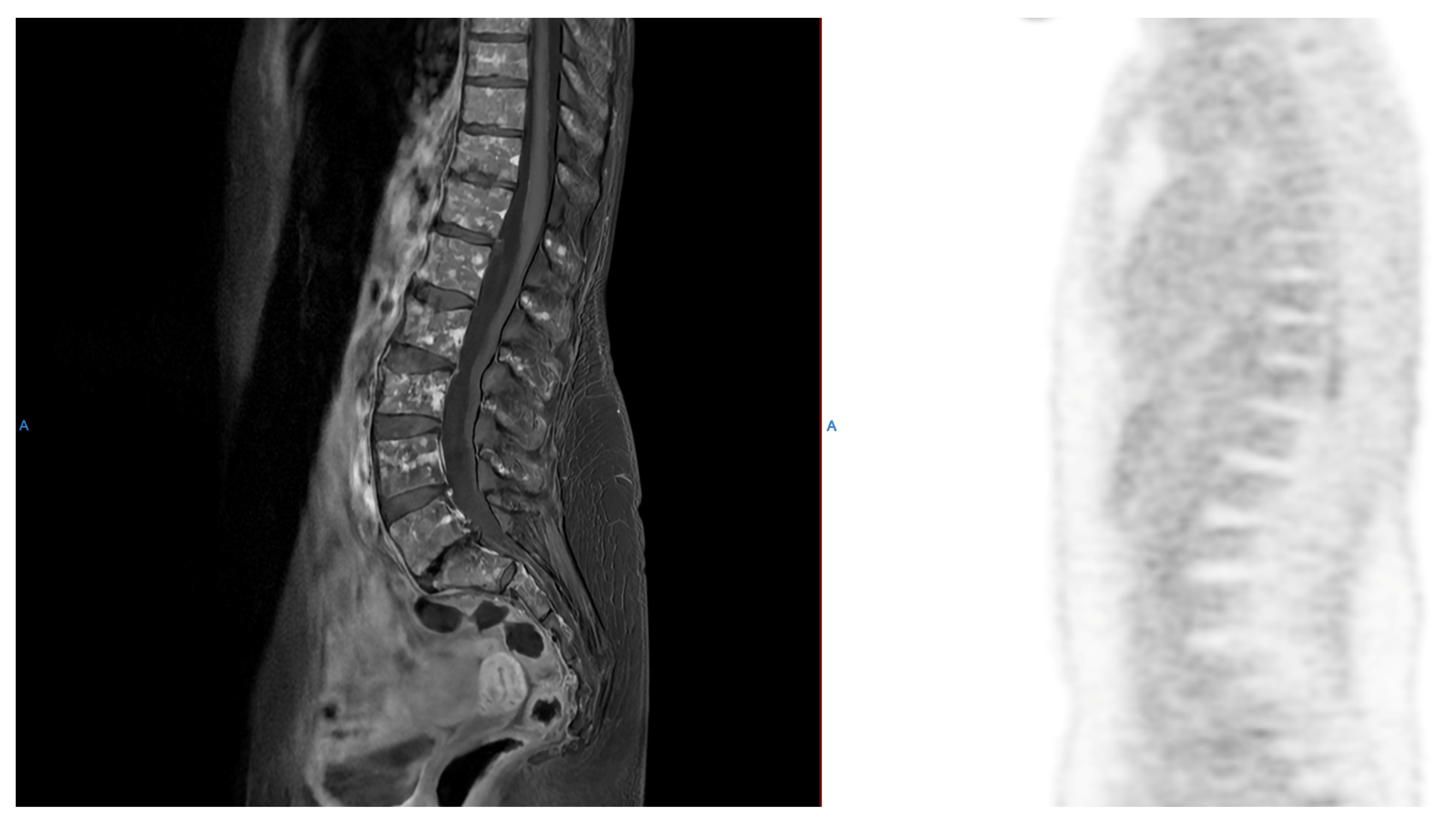
Figure 1. A typical example of a myeloma patient staging work-up with WB-MRI and FDG-PET/CT displaying the “salt-and-pepper” pattern of myeloma bone marrow infiltration on sagittal spine MRI (left), while FDG-PET/CT was negative without any corresponding uptake or extramedullary involvement (right). A, anterior.
Cancers 18 02184 g001
Figure 2. Baseline and post-treatment FDG PET/CT of a patient with multiple myeloma showing complete metabolic response. (Bottom) row: staging FDG PET/CT showing multifocal uptake in bone lytic lesions involving the axial and proximal appendicular skeleton and also a paramedullary costovertebral left mass consistent with multiple myeloma. (Upper) row: FDG PET/CT after induction with 4 cycles of Dara-VRd (daratumumab, bortezomib, lenalidomide, and dexamethasone), showing complete metabolic response of bone lytic lesions and of the paramedullary myeloma lesion. R, right; P, posterior; I, inferior.
Cancers 18 02184 g002
Figure 3. IMWG criteria for response assessment, including criteria for minimal residual disease. Adapted from Kumar et al. [20]. For MRD assessment, the first bone marrow aspirate should be sent to MRD (not for morphology), and this sample should be taken in one draw with a volume of minimally 2 mL (to obtain sufficient cells), but maximally 4–5 mL to avoid hemodilution. IMWG=International Myeloma Working Group. MRD = Minimal Residual Disease. CR = Complete Response. sCR = stringent Complete Response. VGPR = Very Good Partial Response. PR = Partial Response. MR = Minimal Response. SD = Stable Disease. PD = Progressive Disease. IF = ImmunoFixation. IHC = ImmunoHistoChemistry. NGF = Next-Generation Flow. NGS = Next-Generation Sequencing. FLC = Free Light Chain. M-protein = Myeloma protein. SPD = Sum of the Products of the maximal perpendicular Diameters of measured lesions. CRAB features = Calcium elevation, Renal failure, Anemia, Lytic bone lesions. FCM = Flow CytoMetry. SUVmax=maximum Standardized Uptake Value. MFC = Multiparameter Flow Cytometry. 18F-FDG PET = 18F-fluorodeoxyglucose PET. ASCT = Autologous Stem Cell Transplantation. * All response categories require two consecutive assessments made any time before starting any new therapy; for MRD there is no need for two consecutive assessments, but information on MRD after each treatment stage is recommended (eg, after induction, high-dose therapy/ASCT, consolidation, maintenance). MRD tests should be initiated only at the time of suspected complete response. All categories of response and MRD require no known evidence of progressive or new bone lesions if radiographic studies were performed. However, radiographic studies are not required to satisfy these response requirements except for the requirement of FDG PET if imaging MRD-negative status is reported. ** To be used only if the end point is disease-free survival. *** To be used only if the end point is disease-free survival. § If the serum and urine M-protein are unmeasurable: a ≥50% decrease in the difference between involved and uninvolved FLC levels is required in place of the M-protein criteria. If serum and urine M-protein and serum-free light assay are unmeasurable: ≥50% reduction in plasma cells is required in place of M-protein, provided baseline bone marrow plasma-cell percentage was ≥30%. In patients without measurable serum and urine M-protein levels: the difference between involved and uninvolved FLC levels (absolute increase must be >10 mg/dL). In patients without measurable serum and urine M-protein levels and without measurable involved FLC levels: bone marrow plasma-cell percentage irrespective of baseline status (absolute increase must be ≥10%). It is not used in calculation of time to progression or progression-free survival but is listed as something that can be reported optionally or for use in clinical practice.
Cancers 18 02184 g003aCancers 18 02184 g003b
Table 1. Summary of the IMPETUS criteria. Adapted from Nanni et al. [56]
Lesion TypeSiteLesions NumberGrading
DiffuseBone Marrow Deauville five-point scale

Target: uptake in lower lumbar vertebra
A = if uptake in limbs and ribs
Focal (F)Skull (S)
Spine (SP)
Extraspinal (ExP)
1 = no lesions
2 = 1 to 3
3 = 4 to 10
4 = >10
Deauville five-point scale

Target: the hottest lesion
Lytic (L)
(on CT scan)
1 = no lesions
2 = 1 to 3
3 = 4 to 10
4 = >10
Fracture (Fr)
(on CT scan)
Paramedullary (PM)
A bone lesion involving surrounding soft tissues with bone cortical interruption
Extramedullary (EM)Nodal (N):
C = Cervical
M = Mediastinal
Ax = Axillary
Rp = Retroperitoneal
Mes = Mesenteric
In = Inguinal
Deauville five-point scale

Target: the hottest lesion
Extranodal (EN):
Li = Liver
Mu = Muscle
Spl = Spleen
Oth = Others
Deauville five-point scale

Target: the hottest lesion
Table 2. EANM Focus 4 Consensus Recommendations summary. EANM Focus 4 Consensus Recommendations summary. Adapted from Nanni et al. [23].
EANM Focus 4 Consensus Recommendations
FDG PET/CT in multiple myeloma staging
Patients with suspected active multiple myeloma (with both secretory and nonsecretory disease) should undergo staging with FDG PET/CT (regardless of low-dose CT and MRI results) to assess the disease burden, identify extramedullary disease and create a baseline for response assessment.
Patients with solitary plasmacytoma located in the bone or in extramedullary areas could benefit from FDG PET/CT scanning, including those with only one lesion detected by whole-body MRI.
The PET field of view in multiple myeloma should cover from the top of the head to the feet with arms down.
There is not enough evidence supporting imaging with non-[18F]FDG tracers (e.g., [18F]fluorocholine, [11C]choline, [11C]methionine) in routine clinical practice.
FDG PET/CT in smoldering multiple myeloma
Patients with smoldering multiple myeloma should not undergo FDG PET/CT scanning except for those with only a single focal lesion in whole-body MRI, a lesion < 5 mm in low-dose CT (LDCT), or equivocal lesions in whole-body MRI or LDCT.
FDG PET/CT in monoclonal gammopathy of undetermined significance (MGUS)
Patients with MGUS should not undergo FDG PET/CT.
Sequence of FDG PET/CT scans in patients with active multiple myeloma
No definitive consensus on the appropriate timing/sequence of FDG PET/CT scans.
FDG PET/CT reporting in patients with active multiple myeloma
Reports at staging as well as during/after therapy should document the presence of lytic lesions on LDCT (number and size), fractures on LDCT and sites with a substantially increased risk of fracture, PET-positive focal lesions (FLs, grouped 0, 1–3, or >3) along with the SUVmax of the hottest FL, and the presence of increased diffuse uptake in the bone marrow.
FDG PET/CT evaluation after therapy
During or after therapy of multiple myeloma, changes in the number/size of PET-positive FLs, SUVmax/Deauville score of the hottest FL and decreases in diffuse bone marrow uptake should be reported. Bone marrow uptake should be reported as pathological when it is visually higher than the normal liver uptake.
After therapy, a complete normalization of FDG PET/CT in multiple myeloma can be seen if the uptake in previous hot FLs and bone marrow are not measurable or are visually lower than the liver and no new lytic lesions in LDCT images are present.
FDG PET/CT should be performed for minimal residual disease (MRD) assessment even in patients with negative multiparametric flow cytometry or genomic tests on bone marrow aspiration, since the combination of such techniques allows for a more accurate patient stratification.
Table 3. Comparison of standardized FDG PET/CT response criteria in multiple myeloma.
CriterionBasis/ScalePrimary UseCMR DefinitionBest-Suited Scenario
Deauville (DS)5-point visual scale vs. mediastinum and liver referenceResponse assessment (CMR)Uptake ≤ liver (DS 1–3) in all initially involved sitesDefining metabolic complete response after therapy
IMPeTUsDeauville score applied to bone marrow, focal lesions (number/site), PMD/EMD, and lytic lesions on CTStandardized baseline and response reportingDS < 4 in bone marrow and all focal lesionsStructured, reproducible reporting across centers
IMWGIntegrates PET findings with NGF/NGS and serum/marrow parametersComposite CR/MRD definitionNegative imaging plus MRD-negative marrowDefining imaging-plus-MRD-negative complete response
EANM Focus 4/2024 EANM guidelineProcedural and interpretative standards (acquisition, indications, reporting)Acquisition and reporting harmonizationNormalization of focal lesion and marrow uptake below liver, no new lytic lesionsHarmonizing acquisition and reporting protocols
Table 4. Evidence characterization for the principal applications of FDG PET/CT and emerging radiotracers in multiple myeloma response assessment.
ApplicationEvidence LevelBasis
Complete metabolic response (Deauville-defined) after therapy as a predictor of PFS/OSEstablishedMultiple large prospective cohorts and trial sub-studies (FORTE, IMAJEM, CASSIOPET); endorsed by IMWG and EANM
Residual focal lesions/persistent SUVmax > 4.2 as adverse prognostic markersEstablishedConsistent across several prospective and large retrospective series
Baseline extramedullary/paramedullary disease and >3 focal lesions as adverse prognostic factorsEstablishedMultiple cohorts; reproduced across different reading criteria
FDG PET/CT combined with bone marrow MRD (NGF/NGS) double negativity for risk stratificationEstablished/consolidatingProspective and registry data; incorporated into IMWG MRD criteria
Interim/early PET (e.g., day 7, post-induction ΔSUVmax) to guide therapyEmergingSingle trials/cohorts (IMAJEM, total therapy datasets); not yet routine practice
IMPeTUs-based standardized readingEmerging/consolidatingDeveloped and validated mainly in single-group/Italian cohorts; broader validation ongoing
Treatment intensification in PET-positive patients (e.g., post-ASCT KRd)EmergingSmall single-center series; conversion rates not yet confirmed in trials
[11C]choline, [11C]methionine, [11C]acetate, [18F]FLTInvestigationalPilot/comparative studies in small cohorts; no validated response role
[68Ga]pentixafor (CXCR4)InvestigationalProspective but early-phase diagnostic data; complementary, not validated for response
CD38/SLAMF7 immunoPET ([64Cu]daratumumab, [89Zr]isatuximab, [89Zr]elotuzumab)Investigational/preclinicalFirst-in-human or preclinical; not yet in response-assessment use
Disclaimer/Publisher’s Note: The statements, opinions and data contained in all publications are solely those of the individual author(s) and contributor(s) and not of MDPI and/or the editor(s). MDPI and/or the editor(s) disclaim responsibility for any injury to people or property resulting from any ideas, methods, instructions or products referred to in the content.