Commentary

Integrating Extracellular Matrix Dynamics and Membrane-Associated Signaling: The Role of Clusterin-LRP1 Network in Cancer Cell Migration and Tumor Progression


1
Laboratory of Biochemistry, Molecular Biology and Oncometabolism, Department of Medicine and Surgery, University of Parma, Via Volturno 39, 43125 Parma, Italy
2
Biochemistry, Biochemical Analysis & Matrix Pathobiology Research Group, Department of Chemistry, University of Patras, 26504 Patras, Greece
*
Authors to whom correspondence should be addressed.
Cancers 2026, 18(11), 1791; https://doi.org/10.3390/cancers18111791
Submission received: 26 April 2026 / Revised: 22 May 2026 / Accepted: 26 May 2026 / Published: 30 May 2026
(This article belongs to the Special Issue Feature Papers in Section "Tumor Microenvironment" (2nd Edition))

Simple Summary

Tumor progression and metastasis depend on a dynamic and reciprocal interaction between cancer cells and their surrounding environment, yet the mechanisms by which these complex processes are coordinated at the cell surface are not fully understood. Clusterin, a protein secreted outside cells, may play key roles in integrating these events, helping the assembly of key molecules on the cell membrane into a functional network that links extracellular matrix remodeling, endocytosis, and intracellular signaling. Through this organization, clusterin may promote changes in cell shape and behavior that promote cancer cell migration and invasion. By proposing this model, we aim to provide a new perspective on how these events are regulated and to highlight potential targets for future therapeutic strategies.

Abstract

Tumor cell migration relies on the integration of extracellular matrix (ECM) remodeling, cell surface signaling regulating cytoskeleton dynamics, and epithelial-to-mesenchymal transition (EMT). Clusterin (CLU), a secreted glycoprotein, is involved in extracellular proteostasis and is known to interact with members of the LDL receptor family, including low-density lipoprotein receptor-related protein 1 (LRP1). Beyond its canonical chaperone activity, CLU is involved in several biological processes, including cell survival, apoptosis, tissue remodeling, inflammation and cancer progression. On the other hand, the membrane type 1 matrix metalloproteinase (MT1-MMP), functionally linked to CD44 and LRP1, represents a key membrane-associated molecule that may control cell adhesion and receptor-mediated uptake of ECM ligands and proteases. In this article, we critically highlight a hypothetical model in which secreted CLU (sCLU) may function as the central player of a dynamic membrane-associated network integrating proteolysis, endocytosis, and intracellular signaling. Based on recent literature findings and STRING analyses, LRP1, MT1-MMP, CD44, and cell surface matrix components, such as proteoglycans (PGs) and integrins, are likely to be involved. By coordinating this membrane-associated molecular crosstalk, sCLU may integrate ECM remodeling with cytoskeletal dynamics and EMT-related programs related to invasive behavior. Overall, this framework highlights a potential mechanism through which sCLU may contribute to tumor cell plasticity and aggressiveness, suggesting new avenues for therapeutic intervention.

1. Introduction

Tumor cell migration represents a complex and tightly regulated process involving ECM remodeling, the reprogramming of signaling pathways controlling cytoskeletal dynamics, and EMT [1]. These mechanisms converge at the ECM–cell interface, in which the ECM represents not merely a natural structural bioscaffold, but it also actively modulates the availability of biochemical and mechanical cues. At the same time, the cell membrane functions as a complementary regulatory hub where these inputs are integrated through tightly controlled processes such as proteolysis, receptor trafficking, and endocytosis. Within this framework, cell–cell and cell–ECM interactions further contribute to shaping the spatial and temporal organization of signaling events, ultimately influencing cell adhesion, migration, and invasion [2].
Specific ECM components as well as membrane-associated proteins and enzymes play key roles in coordinating these events within the tumor microenvironment (TME). Among these, MT1-MMP (also known as MMP14) drives tumor migration through its ability to degrade ECM components, such as fibronectin and vitronectin, activate pro-MMP2, and promote the shedding of multiple surface proteins including LRP1, CD44, and heparan sulfate PGs (HSPGs). In addition, its cytoplasmic tail supports RhoA activation, which in turn contributes to modulating the cytoskeletal rearrangement through actin polymerization [3].
In this context, LRP1, as a type I transmembrane receptor, promotes the internalization of extracellular ligands as well as the turnover of cell membrane receptors. In this way LRP1 contributes to the regulation of proteolytic processes and ECM remodeling in the pericellular environment [4]. Notably, LRP1 promotes the endocytosis of adhesion receptors such as CD44, regulating their surface availability and contributing to the dynamic balance between cell adhesion and detachment required for efficient migration [5]. Furthermore, LRP1 acts as a signaling co-receptor by forming complexes with growth factors receptors, thereby linking membrane trafficking to intracellular signaling cascades associated with invasive behavior. In addition, its cytoplasmic tail engages adaptor and scaffold proteins involved in cytoskeletal attachment and dynamics [6].
During cancer dissemination, CD44, a major hyaluronan receptor, interacts with MMPs and stabilizes their localization to the leading edge of migrating cells, promoting spatially restricted ECM degradation. Specifically, CD44 functions as a scaffold for the assembly of MMPs with their substrates, including ECM components such as collagen, fibronectin, and laminin, as well as cell surface receptors, including CD44 itself. The interdependent relationship between MT1-MMP and CD44 highlights a functional interplay between cell adhesion and proteolytic remodeling [7]. Beyond its role in cell motility and pro-tumorigenic signaling, CD44 is a well-established cancer stem cell biomarker, strongly linked to tumor initiation, self-renewal, and resistance to therapy [8,9].
HSPGs are glycoproteins bearing covalently attached heparan sulfate chains and are mainly represented by membrane-associated PGs, such as syndecans (SDCs) and glypicans (GPCs), as well as pericellular PGs, including perlecan. Of note, HSPGs exert pleiotropic roles in cancer progression, importantly influencing cancer cell signaling and proliferation, migration, invasion, cell–matrix adhesion, and metastatic potential [10,11]. In particular, SDCs (SDC1-4) act as co-receptors for growth factors and chemokines, as well as interacting with matrix structural molecules and integrins, thus triggering intracellular pro-oncogenic signaling pathways [12,13]. Moreover, proteolytic shedding of SDC ectodomains by enzymes (i.e., MMPs) generates soluble SDCs (i.e., shed SDCs), which can act in both autocrine and paracrine manners to promote the crosstalk between cancer cells and the TME, hence driving metastasis [13,14]. Similarly, GPC1-6 function as co-receptors for a wide range of cell surface receptors and mediate cell–matrix interactions, thereby participating in multiple pathways involved in the regulation of cell fate and tumor progression [15]. Among pericellular HSPGs, perlecan contributes to ECM and basement membrane organization, regulates cell adhesion, sequesters growth factors, and modulates integrin-mediated signaling involved in cancer progression [16].
Other key extracellular molecules may further affect the activity of membrane-associated systems involved in tumor cell migration. Among these, secreted clusterin (sCLU), which is the first identified and the most prominent mammalian extracellular chaperone contributes to cellular homeostasis through three main activities: proteostasis maintenance, cytoprotection, and inflammation inhibition. Since its initial characterization, sCLU has been shown to promote protein clustering and aggregation [17,18]. Indeed, it acts as an ATP-independent chaperone that binds to misfolded proteins and facilitates their clearance through interactions with scavenger receptors, including members of the LRP family and HSPGs [19,20,21]. Furthermore, sCLU binds to membrane receptors and modulates intracellular signaling pathways involved in cell survival, stress responses, and cell migration [22,23].
The main components that contribute to this membrane-associated network, their localization and biological functions are summarized in Table 1.
To further explore the potential relationships among these molecules, we performed a STRING-based protein–protein interaction (PPI) analysis including CLU, LRP1, MT1-MMP, CD44, HSPGs, and integrins, all implicated in ECM and cell membrane dynamics (Figure 1).
In this article, based on the available literature data and STRING-based network analyses, we highlight that sCLU, once secreted into the TME, may facilitate organization of a membrane-associated signaling network by promoting the interplay of LRP1, MT1-MMP, CD44, SDCs, GPCs, perlecan, and integrins. This is expected to increase the local density and spatial proximity of these components, thereby enhancing the efficiency and coordination of their functional interplay. This network may favor the coupling of membrane trafficking, proteolytic activity, and adhesion dynamics, ultimately accelerating receptor turnover while spatially restricting ECM remodeling. In parallel, these interactions may act as integrated signaling platforms that support intracellular pathways driving cytoskeletal reorganization, EMT reprogramming, and directional migration. Within this framework, sCLU may act not only as an extracellular chaperone but also as a potential upstream player of membrane dynamics, promoting a coordinated regulation of cell–ECM interactions and pro-tumorigenic cellular programs.

2. The ECM–Cell Interface in Tumor Cell Migration

The ECM macromolecular network is recognized as a key feature of TME, acting not only as a structural scaffold but also as a dynamic interface continuously shaped through bidirectional communication with tumor cells [28]. ECM is a highly organized three-dimensional network composed of diverse structural and functional macromolecules (including collagens, laminins, fibronectin, glycosaminoglycans/PGs, and glycoproteins, among others), as well as matrix-remodeling enzymes [29]. Moreover, the ECM integrates biochemical and mechanical signals that actively regulate cell adhesion, migration, and survival. On the other hand, tumor cells remodel the matrix by altering its composition, stiffness/remodeling, and architecture. These reciprocal interactions establish a self-sustaining feedback loop that drives tumor progression and metastatic dissemination, and thus contributes to therapy resistance [30].
A critical step in cancer development is the ability of tumor cells to overcome the physical constraints imposed by the basement membrane of the ECM to invade the surrounding stromal tissue [31]. In this context, membrane-anchored and secreted enzymes act as key mediators of pericellular proteolysis [32]. Specifically, MMPs consist of a large family of zinc-dependent, calcium-containing endopeptidases that are broadly upregulated across different cancer types [33]. Their primary function is to regulate ECM remodeling and intercellular communication through the proteolysis of both matrix substrates, such as collagens, various glycoproteins, and PGs, and non-matrix substrates, including growth factors and cytokines. Importantly, the shedding of these bioactive molecules can further amplify pro-tumorigenic pathways, thereby affecting multiple features of tumor progression, including cell survival and proliferation, migration, invasion, angiogenesis, immune evasion, and metastasis [3].
Among MMPs, MT1-MMP was the first proteinase to be identified on the surface of invasive cancer cells and has since been related to poor prognosis in multiple tumor types [34,35,36]. MT1-MMP exhibits a broad spectrum of substrates, including collagen types I, II, and III, fibronectin, vitronectin, and laminins. Furthermore, it can indirectly mediate the degradation of type IV collagen through the activation of pro-MMP2 [37]. Importantly, this process is regulated by tissue inhibitor of metalloproteinases 2 (TIMP2), which exerts a dual role in tumor cell migration. At low concentrations, TIMP2 promotes pro-MMP2 activation by bridging MT1-MMP and its substrate at the cell surface, thereby enhancing pericellular proteolysis and activating downstream signaling pathways, including the MAPK cascade. In contrast, at higher concentrations, TIMP2 inhibits MMP activity by blocking their catalytic sites, ultimately restraining cell migration and invasion [38,39].
Efficient localized ECM degradation depends on the polarized distribution of MT1-MMP at the leading edge of migrating cells. Indeed, MT1-MMP accumulates at specialized cell membrane protrusions, such as invadopodia, filopodia, and lamellipodia [40]. Its surface expression is dynamically controlled by endocytic and exocytic trafficking, allowing continuous recycling and targeted delivery to sites of invasion [41]. Notably, CD44 has been reported to drive MT1-MMP localization to lamellipodia through interaction with its hemopexin domain [42]. In turn, MT1-MMP cleaves the ectodomain of CD44 to promote the detachment of cells from the surrounding matrix [43]. This process is followed by intramembrane proteolysis that leads to the release of the CD44 intracellular domain, which can move to the nucleus where it modulates the transcription of genes involved in pro-tumorigenic programs [44].
In this framework, receptor-mediated trafficking has emerged as a critical regulator for coordinated proteolysis and signaling at the ECM–cell interface. The matricellular receptor LRP1, a member of the low-density lipoprotein receptor family, is involved in the modulation of multiple ECM-related pathways, as it interacts with a wide range of extracellular ligands as well as membrane-associated and intracellular proteins [45]. In particular, it mediates the endocytosis of several proteases, such as MMP2 and MMP9, either as free enzymes or in association with soluble partners, including the pro-MMP2:TIMP2 complex [46].
LRP1 also regulates the turnover of the urokinase-type plasminogen activator system. Specifically, it promotes the internalization of complexes formed by urokinase-type plasminogen activator (uPA), its receptor (uPAR), and plasminogen activator inhibitor-1 (PAI-1), thereby reducing uPAR levels at the cell surface [47]. Through this mechanism, LRP1 modulates plasmin generation, the activation of MMPs and the consequent degradation of ECM components. Notably, LRP1 has been shown to regulate the adhesion/deadhesion balance in tumor cells by promoting the internalization of CD44 [5].
Beyond its role in endocytosis, LRP1 also functions as a signaling co-receptor. For instance, it can bind to the platelet-derived growth factor receptor-β (PDGFR-β) forming a signaling complex that regulates cancer-related pathways, including the MAPK cascade and the PI3K/Akt signaling [48]. Furthermore, the cytoplasmatic tail of LRP1 interacts with a broad range of cytoplasmatic adaptors and scaffold proteins, including a close homologue of the cytoskeletal protein Talin (TLN) and OMP25, involved in cytoskeletal attachment and cell adhesion [6]. Interestingly, MT1-MMP induces LRP1 shedding at the cell surface of malignant cells [49], feeding a dynamic feedback loop between proteolysis and membrane trafficking. Soluble LRP1 fragments have been reported to retain biological activity by acting as “decoys” that bind extracellular ligands and modulate their availability, while also contributing to the activation of intracellular signaling pathways [50].
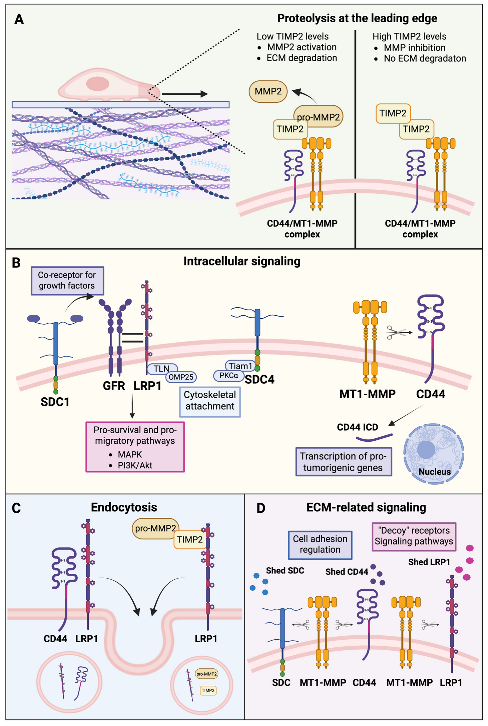
Collectively, these processes define a regulatory network at the ECM–cell interface, integrating proteolysis, receptor trafficking, and signaling to drive tumor cell migration (Figure 2).

3. The Role of sCLU in Cell Signaling and Tumor Progression

sCLU is a highly conserved, stress-responsive glycoprotein expressed in a wide range of tissues and biological fluids. The expression of sCLU is tightly and dynamically regulated at the transcriptional level, reflecting its role as a stress-responsive protein. Its gene expression is modulated by a complex interplay of signaling pathways and regulatory mechanisms, including hormones, growth factors, cytokines, and cellular stress responses such as the heat shock response and the unfolded protein response [51,52]. In addition, epigenetic mechanisms, such as DNA methylation, histone deacetylation, and microRNA-mediated regulation, contribute to fine-tuning CLU transcription in a context-dependent manner [53,54]. sCLU is synthesized through the canonical secretory pathway, beginning with translation of a precursor that includes an N-terminal signal peptide directing the protein into the endoplasmic reticulum (ER). There, the signal sequence is removed, disulfide bonds are formed, and the protein undergoes extensive glycosylation before being cleaved in the Golgi apparatus by a furin-like protease into α- and β-chains linked by disulfide bonds. The mature sCLU is then secreted as a heterodimer of two 40–45 kDa subunits. Under cellular stress, however, non-canonical intracellular CLU forms can arise due to altered processing, including failed ER translocation, retrotranslocation, alternative splicing, or alternative translation initiation [55]. Further work is required to better define the identity and function of these forms, as they have never been isolated under physiological conditions, nor have they been sequenced or crystallized [56].
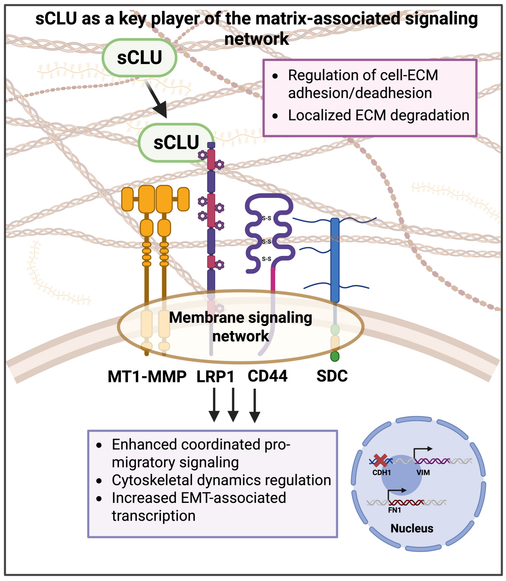
This multilayered transcriptional, post-transcriptional and translational regulation enables rapid adaptation of sCLU levels in response to cellular stress, such as oxidative damage, inflammation, and therapeutic exposure, promoting sCLU upregulation as part of a cytoprotective adaptive response [57]. Functionally, sCLU is best known as the most relevant extracellular molecular chaperone that binds misfolded proteins to prevent their aggregation and facilitate their clearance through receptor-mediated uptake [20]. This process is mainly mediated by LRP family receptors and cell-surface HSPGs [19,21]. Among these, LRP1 has emerged as a key functional receptor, coupling the endocytic clearance of sCLU-associated complexes with the activation of downstream signaling pathways that regulate cell survival and migration [23] (Figure 3).
Beyond its role in proteostasis, sCLU has been implicated in a wide range of biological processes, including lipid transport, apoptosis regulation, and tissue remodeling. Altered CLU expression has been reported in multiple malignancies, including breast [58], pancreatic [59], prostate [60], and colorectal cancer [61], where sCLU has been associated with the activation of pro-survival signaling pathways, tumor progression, metastatic dissemination, and therapy resistance. However, the role of CLU in tumorigenesis remains complex and highly context-dependent, as accumulating evidence indicates that its biological functions vary according to tumor stage, cellular localization, and microenvironmental conditions [62,63,64].
Indeed, although sCLU is frequently upregulated in advanced tumors and is generally referred to as a tumor-promoting, stress-adaptive factor, studies in CLU-deficient mice have consistently shown increased susceptibility to tumor initiation in models of prostate, brain, and skin carcinogenesis [64,65,66]. These apparently opposing observations can be reconciled by considering the fundamentally cytoprotective nature of CLU in both normal and transformed cells. In early phases of carcinogenesis, CLU exerts tissue-protective functions by limiting oxidative and proteotoxic stress, preserving extracellular homeostasis, and attenuating chronic inflammatory signaling, ultimately reducing the accumulation of DNA damage and the likelihood of malignant transformation. Consequently, complete CLU ablation may favor tumor initiation by impairing these homeostatic defense mechanisms. Conversely, once malignant transformation has occurred, cancer cells exploit the cytoprotective properties of sCLU to adapt to the hostile conditions of the TME, including hypoxia, nutrient deprivation, inflammatory stress, and therapeutic pressure. Under these conditions, persistent sCLU upregulation supports tumor cell survival, resistance to apoptosis, metastatic competence, and resistance to anticancer therapies [67].
The stage-dependent functions of CLU may also be influenced by the existence of distinct CLU isoforms characterized by different intracellular localization and biological activities [61]. Specifically, secretory/cytoplasmic CLU has largely been associated with pro-survival and tumor-promoting effects, whereas nuclear CLU has been linked to pro-apoptotic and growth-suppressive functions [68,69]. Together, these observations support the notion that the divergent roles of CLU in cancer are not contradictory but rather reflect the strong dependence of CLU biology on tumor stage, cellular context, stress conditions, and protein localization.
sCLU has been identified as a key mediator of EMT and tumor progression, acting through ERK signaling and Slug-dependent repression of epithelial markers (E-cadherin, encoded by CDH1) and upregulation of mesenchymal ones (fibronectin, FN1) [70]. Notably, sCLU is strongly upregulated downstream of TGF-β signaling during EMT in multiple cancer cell lines and represents one of the most responsive genes throughout this process [71]. Furthermore, sCLU promotes resistance to TNFα-induced cell death in breast cancer (BC) cells via NF-κB activation and subsequent upregulation of Bcl-2, thereby supporting tumor development and metastatic potential [72].
sCLU also plays a multifaceted role in the regulation of MMPs, positioning it as an important modulator of ECM turnover and tissue homeostasis, particularly at mucosal and epithelial surfaces. Mechanistically, sCLU can directly interact with several MMPs through its extracellular chaperone activity. By binding partially unfolded or stress-activated forms of these proteases, sCLU prevents their aberrant aggregation and can limit their proteolytic activity. In the case of MMP9, sCLU has been shown to form stable complexes that reduce enzymatic activity and protect key structural proteins such as tight junction components and basement membrane constituents from excessive degradation [73]. This inhibitory effect contributes to the preservation of epithelial barrier integrity under inflammatory or oxidative stress conditions. In parallel, sCLU may also regulate MMP expression at the transcriptional level in a context-dependent manner. In certain cell types, including immune and epithelial cells, sCLU has been reported to activate signaling pathways such as ERK1/2 and PI3K/Akt, leading to NF-κB-dependent upregulation of MMP9 [74,75]. In triple-negative breast cancer (TNBC) models, CLU silencing significantly impairs cell migration, invasion, tumor growth, and metastatic dissemination, in association with decreased ERK signaling and MMP9 protein levels [76,77]. This apparent duality, namely direct inhibition of MMP activity versus indirect stimulation of MMP expression, suggests that sCLU functions as a fine tuner of proteolytic balance rather than a simple inhibitor, adapting its role according to the microenvironment and the phase of tissue response (i.e., acute injury versus remodeling).
Overall, sCLU emerges as a multifunctional extracellular chaperone whose tightly regulated expression and ability to interface with proteolytic systems, such as MMPs, positions it at the crossroads of proteostasis, inflammation, and tissue remodeling. In line with this, the ability of sCLU to bind multiple extracellular partners suggests a broader involvement in organizing protein complexes at the cell surface, linking extracellular proteostasis to intracellular signaling. Through its interaction with LRP1 and other membrane-associated proteins, sCLU may promote the spatial organization of proteases and adhesion receptors at the plasma membrane, thereby modulating their activity.

4. Clusterin Guides an LRP1/MT1-MMP/CD44 Signaling Network Driving Tumor Cell Invasion

The accumulated evidence outlined above makes it plausible to suggest that sCLU may guide an LRP1/MT1-MMP/CD44/HSPGs signaling network driving tumor cell invasion (Figure 3). The binding of sCLU to LRP1 may act as a nucleation event for the organization of membrane-associated molecules. Indeed, sCLU may promote the interaction of LRP1 with proteases and adhesion receptors at the plasma membrane, thereby regulating their distribution and functional interplay. This spatial organization is expected to facilitate coordinated membrane trafficking and receptor turnover, further contributing to the dynamic regulation of receptor availability at the cell surface.
In this context, key components such as MT1-MMP and CD44 are likely to be recruited, enhancing localized ECM degradation, modulating adhesion dynamics, thereby acting as a signaling network that promotes MAPK and Akt activation, cytoskeletal remodeling, and cell motility. Consistent with these observations, proteomic analyses performed in TNBC cells revealed that CLU silencing resulted in widespread alterations in proteins implicated in cell adhesion, cytoskeletal organization, and ECM dynamics, supporting a role for sCLU in integrating ECM-derived cues with membrane receptor-mediated signaling and coordinating downstream structural and signaling networks [78].
These events also involve the modulation of Rho family GTPases, including RhoA, Rac1, and Cdc42, which coordinate membrane protrusion, contractility, and directional migration [79]. Notably, we found that CLU expression abrogation in TNBC cells is accompanied by the reduction intracellular RhoA-GTP levels [78], yielding the impaired activation of RhoA-dependent pathways that regulate actin cytoskeleton architecture. In line with this view, CD44 has been shown to engage a downstream tPA/LRP1/NF-κB signaling axis that promotes lamellipodia outgrowth and cell migration in BC cells [80]. This further supports a role for LRP1 as a key co-receptor within pro-migratory pathways, consistent with its proposed function in the sCLU-driven network described here.
Beyond the regulation of pro-migratory signaling, this membrane-associated network may also promote full or hybrid EMT-associated phenotypes/reprogramming. Notably, sCLU has been shown to interact with extracellular HSP90α to enhance LRP1 mediated Akt, ERK, and NF-κB signaling, thereby synergistically promoting EMT [23]. Similarly, the co-localization of CD44 and MT1-MMP at the leading edge has been demonstrated in cells undergoing EMT. Moreover, CD44 overexpression induces activation of the EGFR/PI3K/Akt signaling pathway while disrupting the membrane-associated E-cadherin-β-catenin complex, ultimately leading to β-catenin nuclear translocation and transcriptional activation of mesenchymal markers and invasion-related genes [81].
Collectively, available evidence may support a model in which sCLU drives the interaction of LRP1 with cell surface components contributing to proteolysis, receptor trafficking, and signaling to coordinate cytoskeletal remodeling and EMT programs that sustain tumor cell invasion. These insights also suggest that disrupting the coordinated interactions within this network may represent a promising strategy to interfere with tumor progression.

5. Syndecans as Possible Indirect Modulators of CLU/LRP1/MT1-MMP/CD44 Network in Tumor Cell Migration

As already mentioned, SDCs comprise multifunctional cell surface HSPGs, which hold central roles in cancer progression [82]. Of note, they are referred to regulate tumor cell proliferation, apoptosis, migration, invasion, angiogenesis, inflammatory signaling within the TME, as well as the maintenance of cancer stem cell properties associated with recurrence and therapeutic resistance [11]. Notably, their functions are highly context-dependent, varying with tumor type, stage, and cellular origin, and they often exert dual or even opposing effects depending on their localization and interaction partners [10,13].
In the context of cancer cell migration and invasion, SDCs act as critical modulators of cell–ECM interactions and associated signaling pathways [13]. Particularly, SDC1 regulates cell adhesion, where its loss is often associated with increased motility and invasiveness, while its extracellular domain can also participate in signaling complexes with integrins to promote cytoskeletal reorganization [13,83,84]. SDC2 has also been implicated in enhanced migratory behavior, partly through EMT-related signaling pathways [85]. Importantly, SDC4 functions as a key player in coordinating focal adhesion dynamics and actin cytoskeletal remodeling through signaling axes involving Rac1, protein kinase C alpha (PKCα), and focal adhesion kinase (FAK), thereby facilitating the formation of membrane protrusions required for efficient cell movement [27]. In contrast, SDC3 functional properties in cancer progression remains insufficiently explored; however, recent studies have demonstrated its role in BC cell migration [10].
Within this framework, the proposed CLU-associated LRP1/MT1-MMP/CD44 network driving tumor cell migration and invasion raised the possibility that HSPGs, particularly SDCs, may function as indirect modulators of this network (Figure 3). LRP1 acts as a multifunctional receptor involved in cell signaling and adhesion through interactions with various membrane-associated partners, including HSPGs such as SDC1 and GPC3, thereby influencing ligand availability and downstream signaling [27,45]. In particular, STRING-based PPI analyses revealed co-expression associations between LRP1 and SDC1, SDC2, SDC4, GPC1, GPC3, GPC4, and GPC6, as well as an experimentally validated interaction with HSPG2/perlecan (Figure 1B). STRING analysis further identified both co-expression and experimentally validated interactions among LRP1 and integrin β1, a key subunit shared by multiple integrin heterodimers (Figure 1C). It is already known that LRP1 interacts with several integrin heterodimers, including αMβ1, αMβ2, αVβ3, and αVβ5, as well as subunits αM, αL, β1, and β2, thereby regulating integrin availability/activity and, therefore, cellular adhesion/migration [4,86]. Evidence in the literature supports that LRP1–integrin interactions can modulate both integrin surface availability and functional activity [86]. In addition, integrin activation has been shown to be regulated by SDC-1 [87], while SDC-4 contributes to the dynamic assembly and disassembly of integrin-containing complexes at focal adhesion sites [88]. Specific SDC–integrin pairs, such as SDC4-α5β1, SDC1-αVβ3Vβ5, and indirect SDC2-β1 are particularly important for adhesion to ECM proteins (fibronectin and vitronectin, respectively), and for subsequent cancer cell spreading and migration [11,26,87,89]. Given the central role of HSPGs/integrins in focal adhesion formation and cell motility [26], a coordinated functional interplay between LRP1, HSPGs, and integrins may represent an indirect regulatory network in CLU-mediated cancer cell migration.
The localization and functional role of MT1-MMP within migratory and invasive cellular protrusions has been already introduced. Extending these, MT1-MMP plays a central role during invadopodia maturation by coordinating the trafficking and cell surface presentation of matrix-degrading enzymes, together with SDC1. This coordinated delivery facilitates localized ECM degradation and contributes to basement membrane disruption [3]. Importantly, the co-expression of MT1-MMP with SDC1 (also shown in Figure 1B) suggests that the former cleaves the ectodomain of SDC1, leading to its proteolytic shedding from the cell surface. Of note, in the same study, shed SDC1 ectodomain was shown to enhance fibrosarcoma cell migration on collagen-rich substrates, indicating a functional role in modulating cell–ECM interactions and promoting cell motility [90]. Collectively, these data further support the concept that CLU-associated signaling may modulate the proposed network, thereby enhancing SDC1 ectodomain shedding and coordinating tumor cell migration and invasion.
To our knowledge, limited evidence is currently available regarding the direct interaction of CD44 with integrins and the downstream signaling pathways involved in cancer initiation [91]. However, STRING-derived co-expression analyses indicate associations between CD44 and multiple integrin subunits (i.e., ITGAV, ITGAL, ITGAM, ITGB2, ITGB3, and ITGB5), along with an experimentally validated interaction with ITGB1, suggesting a potential interplay within the sCLU-proposed model (Figure 1C). Additionally, co-expression of SDC1 and CD44 (as well as STRING-based CD44/SDC4 co-expression, Figure 1C) suggests a potential functional association of the two, which may further contribute to the regulation of cell adhesion and signaling pathways implicated in cancer progression [92]. Given the established role of CD44 in cancer stem cell phenotypes and migration, it is plausible that the aforementioned interactions may influence cancer (stem) cell motility, although direct mechanistic evidence remains unexplored.

6. Therapeutic Targeting and Clinical Relevance of the sCLU/LRP1-Driven Signaling Network

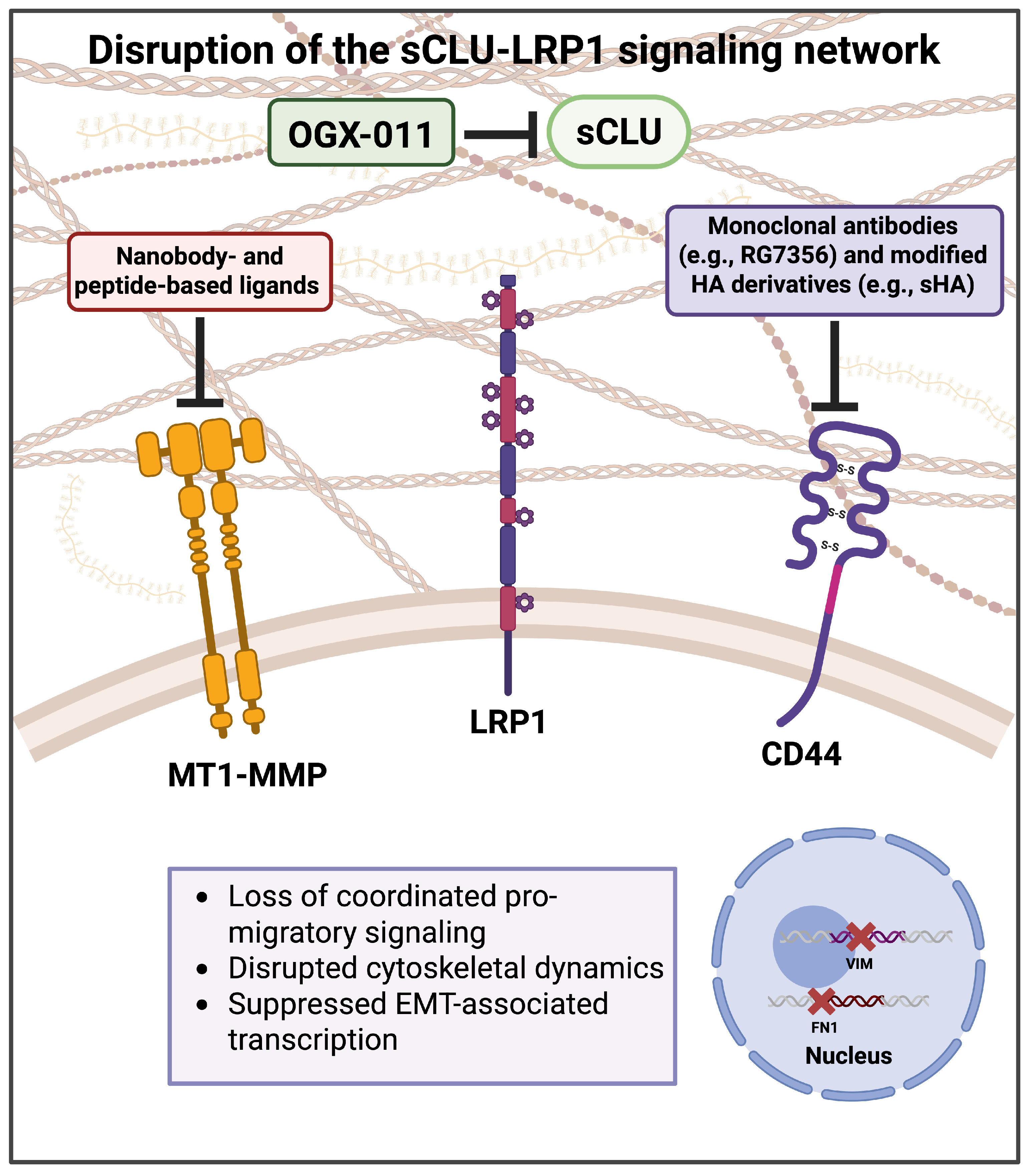
Beyond its mechanistic relevance, the identification of an sCLU-LRP1-associated signaling network may provide a conceptual framework for the development of therapeutic strategies aimed at disrupting coordinated pro-invasive programs in cancer (Figure 4).
Targeting sCLU with antisense oligonucleotides such as OGX-011 has exhibited high efficacy in hampering tumor growth, invasion, and metastatic dissemination across multiple preclinical cancer models [93,94]. In clinical settings, the therapeutic potential of CLU inhibition was tested in combination with standard chemotherapeutic treatments. Phase I and Phase II clinical studies demonstrated that OGX-011 is well tolerated and effectively reduces sCLU serum levels, leading to encouraging changes in biomarkers used as surrogate endpoints of exploratory efficacy [95,96,97]. However, despite these promising findings, two large phase III clinical trials failed to demonstrate a significant survival benefit in patients with metastatic castration-resistant prostate cancer [98,99]. These discrepancies highlight the limitations of selectively targeting sCLU within a broader and highly interconnected pro-invasive signaling network. In line with this, the sCLU-driven network described here provides a potential explanation for these outcomes, as sCLU likely acts within a broader membrane-associated signaling hub integrating receptor trafficking, proteolysis, and activation of intracellular effector molecules. Therefore, targeting CLU alone may be insufficient to fully disrupt the coordinated pro-invasive programs that sustain tumor progression, supporting the need for combinatorial strategies.
In this context, additional molecular components of the proposed pro-tumorigenic network represent attractive therapeutic targets. Although LRP1 plays a central role in mediating sCLU-driven clustering and signal integration, its wide-ranging expression across different tissues and cell types, together with its ability to bind a broad spectrum of ligands and participate in multiple biological processes, has limited its direct therapeutic targeting [100]. This, in turn, supports the exploration of alternative, more druggable nodes within the network.
A growing body of evidence supports the development of CD44-targeted strategies, including monoclonal antibodies, nanobody- and peptide-based ligands, as well as hyaluronic acid (HA)-based drug delivery systems, which aim to selectively interfere with CD44-mediated signaling [101]. Notably, the humanized monoclonal antibody RG7356 has been investigated in a phase I trial in patients with metastatic or locally advanced CD44-expressing solid malignancies [102]. While this study demonstrated an acceptable safety profile and evidence of biological activity, clinical efficacy was limited, suggesting that combination strategies may be required to improve therapeutic outcomes. Recognizing HA as a major ligand of CD44, modified HA derivatives such as sulfated HA (sHA) have emerged as promising anticancer agents that interfere with CD44 availability and signaling, thereby attenuating tumor progression, including cell migration [24,103]. Notably, sHA has been shown to attenuate prostate cancer (PC) cell migration, inhibiting PI3K/Akt signaling, and inducing transcriptional downregulation of CD44, while also reducing tumor growth and angiogenesis in PC mouse models [104]. Similarly, sHA suppresses cell proliferation, migration, and invasion, but also downregulates CD44, thereby inhibiting CD44/PI3K complex formation and impairing tumorigenic signaling pathways in bladder cancer xenografts [105].
MT1-MMP has been extensively explored as a therapeutic target. Early attempts to inhibit MT1-MMP activity relied on broad-spectrum MMP inhibitors, which target the catalytic Zn2+-binding site. Although these compounds initially showed promising anti-tumoral effects in preclinical models, they largely failed in clinical settings due to poor selectivity, dose-limiting toxicity, and off-target inhibition of physiologically important MMPs involved in tissue remodeling, wound healing, and immune regulation. In addition, functional redundancy among MMP family members and the lack of appropriate patient stratification strategies likely further contributed to the limited clinical efficacy of these approaches. Recent strategies have focused on selectively targeting MT1-MMP through nanobodies and peptide-based ligands that interfere with substrate recognition or protein–protein interactions [106]. Although these approaches have shown promising anti-invasive and anti-angiogenic effects in preclinical models, their translation into clinical benefit has remained limited. As for CD44, the modest success of MT1-MMP-targeted interventions may reflect the fact that MT1-MMP does not act as an isolated protease, but rather as part of membrane-associated multiprotein complexes involved in different processes. Within the signaling network proposed here, MT1-MMP is functionally coupled to receptors such as CD44 and LRP1, suggesting that its inhibition alone may be insufficient to disrupt the coordinated pro-invasive programs that drive tumor progression.
Although these compounds initially showed promising anti-tumoral effects in preclinical models, they largely failed in clinical settings due to poor selectivity, dose-limiting toxicities, and off-target inhibition of physiologically important MMPs involved in tissue remodeling, wound healing, and immune regulation. In addition, functional redundancy among MMP family members and the lack of appropriate patient stratification strategies likely further contributed to the limited clinical efficacy of these approaches.

7. Limitations and Future Perspectives

Despite the integrative framework proposed here, few limitations should be acknowledged. First, the model, although supported from accumulated literature data, it remains as a proposed hypothetical network where several points have to be validated experimentally. While supported by published evidence and bioinformatic interaction network analyses, the proposed membrane-associated interactions among sCLU, LRP1, MT1-MMP, CD44, and related ECM molecules have not been demonstrated as a coordinated dynamic system in space and time. Importantly, the present framework does not imply the existence of a stable or simultaneous multiprotein complex, as these interactions may occur sequentially, transiently, or in a context-dependent manner based on extracellular cues and TME conditions. Another limitation derives from the intrinsic redundancy of membrane-associated signaling and proteolytic networks, where compensatory mechanisms involving alternative receptors, proteases, or HSPGs may partially substitute for individual pathway components.
Future studies will be required to experimentally validate the proposed framework and to clarify the spatial and temporal organization of these membrane-associated interactions in tumor cells. In this context, complementary approaches such as co-immunoprecipitation, proximity ligation assays, and super-resolution microscopy could help determine whether sCLU, LRP1, MT1-MMP, CD44, and related ECM-associated molecules dynamically assemble into functionally coordinated membrane-associated platforms. In parallel, live-cell imaging may provide important insights into the spatiotemporal dynamics, trafficking behavior, and transient membrane organization of these components in real time. Moreover, the implementation of advanced experimental models, such as 3D cell cultures, will be essential to determine the biological relevance of this framework within the complex TME. Collectively, these approaches may help clarify the functional and translational relevance of this proposed membrane-associated framework in breast cancer progression.

8. Conclusions

Literature data and STRING interaction analyses support the idea that sCLU may function as an upstream regulator of a membrane-associated signaling network centered on LRP1, integrating proteolysis, endocytosis, and intracellular pathways. By engaging LRP1, sCLU may promote the assembly of MT1-MMP and CD44, thereby enhancing localized ECM degradation and modulating adhesion dynamics. In parallel, the cytoplasmic domain of LRP1 contributes to intracellular signaling pathways that support cytoskeletal remodeling and cell motility. HSPGs may further contribute to this network since they are known to interact with LRP1 and regulate the availability of TGF-β, linking in this way the ECM architecture to EMT signaling and may further modulate cytoskeletal dynamics through Rho GTPase-dependent pathways. Within this framework, CD44 is widely recognized as a marker of cancer stemness in several tumor types, linking this promigratory axis to stemness-associated traits and tumor aggressiveness. Consistently, increased expression of both CLU and LRP1 has been associated with poor prognosis, highlighting the clinical relevance of this hypothetical model. Therefore, targeting the sCLU/LRP1/MT1-MMP/CD44 network at multiple levels may represent a potential therapeutic strategy to limit tumor cell plasticity and metastatic dissemination in aggressive cancers.

Author Contributions

Data curation, writing—original draft preparation, A.C. and S.M.; figure preparation, A.C.; writing—review and editing, F.R., Z.P. and N.K.; supervision and conceptualization, F.R. and N.K. All authors have read and agreed to the published version of the manuscript.

Funding

N.K. and Z.P. acknowledge funding from the European Union Marie Skłodowska-Curie Actions (MSCA), Grant agreement ID: 101130985 “CARES—Human ECM-based platform for anti-cancer drug testing”.

Institutional Review Board Statement

Not applicable.

Informed Consent Statement

Not applicable.

Data Availability Statement

No new data were created or analyzed in this study. Data sharing is not applicable to this article.

Conflicts of Interest

The authors declare no conflicts of interest.

Abbreviations

The following abbreviations are used in this manuscript:
ECMExtracellular matrix
EMTEpithelial to mesenchymal transition
TMETumor microenvironment
MT1-MMPMembrane type 1 matrix metalloproteinase
LRP1Low-density lipoprotein receptor-related protein 1
CLUClusterin
sCLUSecreted clusterin
SDCsSyndecans
GPCsGlypicans
HSPG2Perlecan
CD44Cluster of differentiation 44
MMPsMetalloproteinases
PGsProteoglycans
TIMP2Tissue inhibitor of metalloproteinase 2
GFRsGrowth factor receptors
EREndoplasmic reticulum
BCBreast cancer
TNBCTriple-negative breast cancer
HAHyaluronic acid
sHASulfated hyaluronic acid
PCProstate cancer
PPIProtein–protein interaction

References

  1. Friedl, P.; Alexander, S. Cancer Invasion and the Microenvironment: Plasticity and Reciprocity. Cell 2011, 147, 992–1009. [Google Scholar] [CrossRef] [Scilit]
  2. Piperigkou, Z.; Mangani, S.; Koletsis, N.E.; Koutsakis, C.; Mastronikolis, N.S.; Franchi, M.; Karamanos, N.K. Principal Mechanisms of Extracellular Matrix-mediated Cell–Cell Communication in Physiological and Tumor Microenvironments. FEBS J. 2026, 293, 26–41. [Google Scholar] [CrossRef] [Scilit] [PubMed]
  3. Piperigkou, Z.; Mangani, S.; Kremmydas, S.; Koletsis, N.E.; Karamanos, N.K. A Guide to the Types, Structures, and Multifaceted Functions of Matrix Metalloproteinases in Cancer. FEBS J. 2025. [Google Scholar] [CrossRef] [Scilit]
  4. Van Gool, B.; Dedieu, S.; Emonard, H.; Roebroek, A.J.M. The Matricellular Receptor LRP1 Forms an Interface for Signaling and Endocytosis in Modulation of the Extracellular Tumor Environment. Front. Pharmacol. 2015, 6, 271. [Google Scholar] [CrossRef] [Scilit]
  5. Perrot, G.; Langlois, B.; Devy, J.; Jeanne, A.; Verzeaux, L.; Almagro, S.; Sartelet, H.; Hachet, C.; Schneider, C.; Sick, E.; et al. LRP-1–CD44, a New Cell Surface Complex Regulating Tumor Cell Adhesion. Mol. Cell. Biol. 2012, 32, 3293–3307. [Google Scholar] [CrossRef] [Scilit]
  6. Gotthardt, M.; Trommsdorff, M.; Nevitt, M.F.; Shelton, J.; Richardson, J.A.; Stockinger, W.; Nimpf, J.; Herz, J. Interactions of the Low Density Lipoprotein Receptor Gene Family with Cytosolic Adaptor and Scaffold Proteins Suggest Diverse Biological Functions in Cellular Communication and Signal Transduction. J. Biol. Chem. 2000, 275, 25616–25624. [Google Scholar] [CrossRef] [Scilit] [PubMed]
  7. Medrano-González, P.A.; Rivera-Ramírez, O.; Montaño, L.F.; Rendón-Huerta, E.P. Proteolytic Processing of CD44 and Its Implications in Cancer. Stem Cells Int. 2021, 2021, 6667735. [Google Scholar] [CrossRef] [Scilit]
  8. Loh, J.-J.; Ma, S. Hallmarks of Cancer Stemness. Cell Stem Cell 2024, 31, 617–639. [Google Scholar] [CrossRef] [Scilit]
  9. Xu, Y.; Bai, Z.; Lan, T.; Fu, C.; Cheng, P. CD44 and Its Implication in Neoplastic Diseases. MedComm 2024, 5, e554. [Google Scholar] [CrossRef] [Scilit]
  10. Habenicht, L.; Hassan, N.; Espinoza-Sànchez, N.A.; Onyeisi, J.O.S.; Győrffy, B.; Hanker, L.; Greve, B.; Götte, M. The Role of the Cell Surface Heparan Sulfate Proteoglycan Syndecan-3 in Breast Cancer Pathophysiology. Cells 2025, 14, 1612. [Google Scholar] [CrossRef] [Scilit] [PubMed]
  11. Motta, J.M.; Hassan, H.; Ibrahim, S.A. Revisiting the Syndecans: Master Signaling Regulators with Prognostic and Targetable Therapeutic Values in Breast Carcinoma. Cancers 2023, 15, 1794. [Google Scholar] [CrossRef] [Scilit]
  12. Cheng, B.; Montmasson, M.; Terradot, L.; Rousselle, P. Syndecans as Cell Surface Receptors in Cancer Biology. A Focus on Their Interaction with PDZ Domain Proteins. Front. Pharmacol. 2016, 7, 10. [Google Scholar] [CrossRef] [Scilit]
  13. Barbouri, D.; Afratis, N.; Gialeli, C.; Vynios, D.H.; Theocharis, A.D.; Karamanos, N. Syndecans as Modulators and Potential Pharmacological Targets in Cancer Progression. Front. Oncol. 2014, 4, 4. [Google Scholar] [CrossRef] [Scilit]
  14. Czarnowski, D. Syndecans in Cancer: A Review of Function, Expression, Prognostic Value, and Therapeutic Significance. Cancer Treat. Res. Commun. 2021, 27, 100312. [Google Scholar] [CrossRef] [Scilit] [PubMed]
  15. Tsao, H.-E.; Ho, M. Structural Features of Glypicans and Their Impact on Wnt Signaling in Cancer. Proteoglycan Res. 2025, 3, e70029. [Google Scholar] [CrossRef] [Scilit] [PubMed]
  16. Elgundi, Z.; Papanicolaou, M.; Major, G.; Cox, T.R.; Melrose, J.; Whitelock, J.M.; Farrugia, B.L. Cancer Metastasis: The Role of the Extracellular Matrix and the Heparan Sulfate Proteoglycan Perlecan. Front. Oncol. 2019, 9, 1482. [Google Scholar] [CrossRef] [Scilit]
  17. Blaschuk, O.; Burdzy, K.; Fritz, I.B. Purification and Characterization of a Cell-Aggregating Factor (Clusterin), the Major Glycoprotein in Ram Rete Testis Fluid. J. Biol. Chem. 1983, 258, 7714–7720. [Google Scholar] [CrossRef] [Scilit] [PubMed]
  18. Poon, S.; Easterbrook-Smith, S.B.; Rybchyn, M.S.; Carver, J.A.; Wilson, M.R. Clusterin Is an ATP−Independent Chaperone with Very Broad Substrate Specificity That Stabilizes Stressed Proteins in a Folding-Competent State. Biochemistry 2000, 39, 15953–15960. [Google Scholar] [CrossRef] [Scilit]
  19. Kounnas, M.Z.; Loukinova, E.B.; Stefansson, S.; Harmony, J.A.K.; Brewer, B.H.; Strickland, D.K.; Argraves, W.S. Identification of Glycoprotein 330 as an Endocytic Receptor for Apolipoprotein J/Clusterin. J. Biol. Chem. 1995, 270, 13070–13075. [Google Scholar] [CrossRef] [Scilit]
  20. Wyatt, A.R.; Yerbury, J.J.; Berghofer, P.; Greguric, I.; Katsifis, A.; Dobson, C.M.; Wilson, M.R. Clusterin Facilitates in Vivo Clearance of Extracellular Misfolded Proteins. Cell. Mol. Life Sci. CMLS 2011, 68, 3919–3931. [Google Scholar] [CrossRef] [Scilit]
  21. Itakura, E.; Chiba, M.; Murata, T.; Matsuura, A. Heparan Sulfate Is a Clearance Receptor for Aberrant Extracellular Proteins. J. Cell Biol. 2020, 219, e201911126. [Google Scholar] [CrossRef] [Scilit]
  22. Gil, S.Y.; Youn, B.-S.; Byun, K.; Huang, H.; Namkoong, C.; Jang, P.-G.; Lee, J.-Y.; Jo, Y.-H.; Kang, G.M.; Kim, H.-K.; et al. Clusterin and LRP2 Are Critical Components of the Hypothalamic Feeding Regulatory Pathway. Nat. Commun. 2013, 4, 1862. [Google Scholar] [CrossRef] [Scilit]
  23. Tian, Y.; Wang, C.; Chen, S.; Liu, J.; Fu, Y.; Luo, Y. Extracellular Hsp90α and Clusterin Synergistically Promote Breast Cancer Epithelial-to-Mesenchymal Transition and Metastasis via LRP1. J. Cell Sci. 2019, 132, jcs228213. [Google Scholar] [CrossRef] [Scilit]
  24. Chen, C.; Zhao, S.; Karnad, A.; Freeman, J.W. The Biology and Role of CD44 in Cancer Progression: Therapeutic Implications. J. Hematol. Oncol. 2018, 11, 64. [Google Scholar] [CrossRef] [Scilit] [PubMed]
  25. Szatmári, T.; Ötvös, R.; Hjerpe, A.; Dobra, K. Syndecan-1 in Cancer: Implications for Cell Signaling, Differentiation, and Prognostication. Dis. Markers 2015, 2015, 796052. [Google Scholar] [CrossRef] [Scilit]
  26. Mytilinaiou, M.; Nikitovic, D.; Berdiaki, A.; Kostouras, A.; Papoutsidakis, A.; Tsatsakis, A.M.; Tzanakakis, G.N. Emerging Roles of Syndecan 2 in Epithelial and Mesenchymal Cancer Progression. IUBMB Life 2017, 69, 824–833. [Google Scholar] [CrossRef] [Scilit]
  27. Keller-Pinter, A.; Gyulai-Nagy, S.; Becsky, D.; Dux, L.; Rovo, L. Syndecan-4 in Tumor Cell Motility. Cancers 2021, 13, 3322. [Google Scholar] [CrossRef] [Scilit] [PubMed]
  28. Sleeboom, J.J.F.; van Tienderen, G.S.; Schenke-Layland, K.; van der Laan, L.J.W.; Khalil, A.A.; Verstegen, M.M.A. The Extracellular Matrix as Hallmark of Cancer and Metastasis: From Biomechanics to Therapeutic Targets. Sci. Transl. Med. 2024, 16, eadg3840. [Google Scholar] [CrossRef] [Scilit]
  29. Karamanos, N.K.; Theocharis, A.D.; Piperigkou, Z.; Manou, D.; Passi, A.; Skandalis, S.S.; Vynios, D.H.; Orian-Rousseau, V.; Ricard-Blum, S.; Schmelzer, C.E.H.; et al. A Guide to the Composition and Functions of the Extracellular Matrix. FEBS J. 2021, 288, 6850–6912. [Google Scholar] [CrossRef] [Scilit] [PubMed]
  30. Huang, J.; Zhang, L.; Wan, D.; Zhou, L.; Zheng, S.; Lin, S.; Qiao, Y. Extracellular Matrix and Its Therapeutic Potential for Cancer Treatment. Signal Transduct. Target. Ther. 2021, 6, 153. [Google Scholar] [CrossRef] [Scilit]
  31. Chang, J.; Chaudhuri, O. Beyond Proteases: Basement Membrane Mechanics and Cancer Invasion. J. Cell Biol. 2019, 218, 2456–2469. [Google Scholar] [CrossRef] [Scilit] [PubMed]
  32. Piperigkou, Z.; Kyriakopoulou, K.; Koutsakis, C.; Mastronikolis, S.; Karamanos, N.K. Key Matrix Remodeling Enzymes: Functions and Targeting in Cancer. Cancers 2021, 13, 1441. [Google Scholar] [CrossRef] [Scilit]
  33. Gobin, E.; Bagwell, K.; Wagner, J.; Mysona, D.; Sandirasegarane, S.; Smith, N.; Bai, S.; Sharma, A.; Schleifer, R.; She, J.-X. A Pan-Cancer Perspective of Matrix Metalloproteases (MMP) Gene Expression Profile and Their Diagnostic/Prognostic Potential. BMC Cancer 2019, 19, 581. [Google Scholar] [CrossRef] [Scilit]
  34. Sato, H.; Takino, T.; Okada, Y.; Cao, J.; Shinagawa, A.; Yamamoto, E.; Seiki, M. A Matrix Metalloproteinase Expressed on the Surface of Invasive Tumour Cells. Nature 1994, 370, 61–65. [Google Scholar] [CrossRef] [Scilit]
  35. Têtu, B.; Brisson, J.; Wang, C.S.; Lapointe, H.; Beaudry, G.; Blanchette, C.; Trudel, D. The Influence of MMP-14, TIMP-2 and MMP-2 Expression on Breast Cancer Prognosis. Breast Cancer Res. 2006, 8, R28. [Google Scholar] [CrossRef] [Scilit]
  36. Noda, Y.; Ishida, M.; Yamaka, R.; Ueno, Y.; Sakagami, T.; Fujisawa, T.; Iwai, H.; Tsuta, K. MMP14 Expression Levels Accurately Predict the Presence of Extranodal Extensions in Oral Squamous Cell Carcinoma: A Retrospective Cohort Study. BMC Cancer 2023, 23, 142. [Google Scholar] [CrossRef] [Scilit] [PubMed]
  37. Tanaka, N.; Sakamoto, T. MT1-MMP as a Key Regulator of Metastasis. Cells 2023, 12, 2187. [Google Scholar] [CrossRef] [Scilit] [PubMed]
  38. D’Alessio, S.; Ferrari, G.; Cinnante, K.; Scheerer, W.; Galloway, A.C.; Roses, D.F.; Rozanov, D.V.; Remacle, A.G.; Oh, E.-S.; Shiryaev, S.A.; et al. Tissue Inhibitor of Metalloproteinases-2 Binding to Membrane-Type 1 Matrix Metalloproteinase Induces MAPK Activation and Cell Growth by a Non-Proteolytic Mechanism. J. Biol. Chem. 2008, 283, 87–99. [Google Scholar] [CrossRef] [Scilit]
  39. Sounni, N.E.; Rozanov, D.V.; Remacle, A.G.; Golubkov, V.S.; Noel, A.; Strongin, A.Y. Timp-2 Binding with Cellular MT1-MMP Stimulates Invasion-Promoting MEK/ERK Signaling in Cancer Cells. Int. J. Cancer J. Int. Cancer 2010, 126, 1067–1078. [Google Scholar] [CrossRef] [Scilit]
  40. Gifford, V.; Itoh, Y. MT1-MMP-Dependent Cell Migration: Proteolytic and Non-Proteolytic Mechanisms. Biochem. Soc. Trans. 2019, 47, 811–826. [Google Scholar] [CrossRef] [Scilit]
  41. Remacle, A.; Murphy, G.; Roghi, C. Membrane Type I-Matrix Metalloproteinase (MT1-MMP) Is Internalised by Two Different Pathways and Is Recycled to the Cell Surface. J. Cell Sci. 2003, 116, 3905–3916. [Google Scholar] [CrossRef] [Scilit] [PubMed]
  42. Mori, H.; Tomari, T.; Koshikawa, N.; Kajita, M.; Itoh, Y.; Sato, H.; Tojo, H.; Yana, I.; Seiki, M. CD44 Directs Membrane-Type 1 Matrix Metalloproteinase to Lamellipodia by Associating with Its Hemopexin-like Domain. EMBO J. 2002, 21, 3949–3959. [Google Scholar] [CrossRef] [Scilit]
  43. Kajita, M.; Itoh, Y.; Chiba, T.; Mori, H.; Okada, A.; Kinoh, H.; Seiki, M. Membrane-Type 1 Matrix Metalloproteinase Cleaves Cd44 and Promotes Cell Migration. J. Cell Biol. 2001, 153, 893–904. [Google Scholar] [CrossRef] [Scilit] [PubMed]
  44. Skandalis, S.S. CD44 Intracellular Domain: A Long Tale of a Short Tail. Cancers 2023, 15, 5041. [Google Scholar] [CrossRef] [Scilit]
  45. Bres, E.E.; Faissner, A. Low Density Receptor-Related Protein 1 Interactions With the Extracellular Matrix: More Than Meets the Eye. Front. Cell Dev. Biol. 2019, 7, 31. [Google Scholar] [CrossRef] [Scilit]
  46. Emonard, H.; Bellon, G.; de Diesbach, P.; Mettlen, M.; Hornebeck, W.; Courtoy, P.J. Regulation of Matrix Metalloproteinase (MMP) Activity by the Low-Density Lipoprotein Receptor-Related Protein (LRP). A New Function for an “Old Friend”. Biochimie 2005, 87, 369–376. [Google Scholar] [CrossRef] [Scilit]
  47. Czekay, R.-P.; Kuemmel, T.A.; Orlando, R.A.; Farquhar, M.G. Direct Binding of Occupied Urokinase Receptor (uPAR) to LDL Receptor-Related Protein Is Required for Endocytosis of uPAR and Regulation of Cell Surface Urokinase Activity. Mol. Biol. Cell 2001, 12, 1467–1479. [Google Scholar] [CrossRef] [Scilit]
  48. Muratoglu, S.C.; Mikhailenko, I.; Newton, C.; Migliorini, M.; Strickland, D.K. Low Density Lipoprotein Receptor-Related Protein 1 (LRP1) Forms a Signaling Complex with Platelet-Derived Growth Factor Receptor-β in Endosomes and Regulates Activation of the MAPK Pathway. J. Biol. Chem. 2010, 285, 14308–14317. [Google Scholar] [CrossRef] [Scilit]
  49. Rozanov, D.V.; Hahn-Dantona, E.; Strickland, D.K.; Strongin, A.Y. The Low Density Lipoprotein Receptor-Related Protein LRP Is Regulated by Membrane Type-1 Matrix Metalloproteinase (MT1-MMP) Proteolysis in Malignant Cells. J. Biol. Chem. 2004, 279, 4260–4268. [Google Scholar] [CrossRef] [Scilit]
  50. Gorovoy, M.; Gaultier, A.; Campana, W.M.; Firestein, G.S.; Gonias, S.L. Inflammatory Mediators Promote Production of Shed LRP1/CD91, Which Regulates Cell Signaling and Cytokine Expression by Macrophages. J. Leukoc. Biol. 2010, 88, 769–778. [Google Scholar] [CrossRef] [Scilit] [PubMed]
  51. Trougakos, I.P.; Gonos, E.S. Chapter 9: Oxidative Stress in Malignant Progression: The Role of Clusterin, a Sensitive Cellular Biosensor of Free Radicals. Adv. Cancer Res. 2009, 104, 171–210. [Google Scholar] [CrossRef] [Scilit]
  52. Loison, F.; Debure, L.; Nizard, P.; le Goff, P.; Michel, D.; le Dréan, Y. Up-Regulation of the Clusterin Gene after Proteotoxic Stress: Implication of HSF1-HSF2 Heterocomplexes. Biochem. J. 2006, 395, 223–231. [Google Scholar] [CrossRef] [Scilit]
  53. Sala, A.; Bettuzzi, S.; Pucci, S.; Chayka, O.; Dews, M.; Thomas-Tikhonenko, A. Regulation of CLU Gene Expression by Oncogenes and Epigenetic Factors Implications for Tumorigenesis. Adv. Cancer Res. 2009, 105, 115–132. [Google Scholar] [CrossRef] [Scilit]
  54. Bonacini, M.; Coletta, M.; Ramazzina, I.; Naponelli, V.; Modernelli, A.; Davalli, P.; Bettuzzi, S.; Rizzi, F. Distinct Promoters, Subjected to Epigenetic Regulation, Drive the Expression of Two Clusterin mRNAs in Prostate Cancer Cells. Biochim. Biophys. Acta BBA-Gene Regul. Mech. 2015, 1849, 44–54. [Google Scholar] [CrossRef] [Scilit]
  55. Prochnow, H.; Gollan, R.; Rohne, P.; Hassemer, M.; Koch-Brandt, C.; Baiersdörfer, M. Non-Secreted Clusterin Isoforms Are Translated in Rare Amounts from Distinct Human mRNA Variants and Do Not Affect Bax-Mediated Apoptosis or the NF-κB Signaling Pathway. PLoS ONE 2013, 8, e75303. [Google Scholar] [CrossRef] [Scilit]
  56. Rizzi, F.; Coletta, M.; Bettuzzi, S. Chapter 2: Clusterin (CLU): From One Gene and Two Transcripts to Many Proteins. Adv. Cancer Res. 2009, 104, 9–23. [Google Scholar] [CrossRef] [Scilit]
  57. Kim, J.H.; Kim, J.H.; Jun, H.O.; Yu, Y.S.; Min, B.H.; Park, K.H.; Kim, K.-W. Protective Effect of Clusterin from Oxidative Stress-Induced Apoptosis in Human Retinal Pigment Epithelial Cells. Investig. Ophthalmol. Vis. Sci. 2010, 51, 561–566. [Google Scholar] [CrossRef] [Scilit] [PubMed]
  58. Redondo, M.; Villar, E.; Torres-Muñoz, J.; Tellez, T.; Morell, M.; Petito, C.K. Overexpression of Clusterin in Human Breast Carcinoma. Am. J. Pathol. 2000, 157, 393–399. [Google Scholar] [CrossRef] [Scilit] [PubMed]
  59. Xie, M.-J.; Motoo, Y.; Su, S.-B.; Mouri, H.; Ohtsubo, K.; Matsubara, F.; Sawabu, N. Expression of Clusterin in Human Pancreatic Cancer. Pancreas 2002, 25, 234–238. [Google Scholar] [CrossRef] [Scilit] [PubMed]
  60. Miyake, H.; Nelson, C.; Rennie, P.S.; Gleave, M.E. Testosterone-Repressed Prostate Message-2 Is an Antiapoptotic Gene Involved in Progression to Androgen Independence in Prostate Cancer. Cancer Res. 2000, 60, 170–176. [Google Scholar]
  61. Pucci, S.; Bonanno, E.; Pichiorri, F.; Angeloni, C.; Spagnoli, L.G. Modulation of Different Clusterin Isoforms in Human Colon Tumorigenesis. Oncogene 2004, 23, 2298–2304. [Google Scholar] [CrossRef] [Scilit]
  62. Mazzarelli, P.; Pucci, S.; Spagnoli, L.G. CLU and Colon Cancer. The Dual Face of CLU: From Normal to Malignant Phenotype. Adv. Cancer Res. 2009, 105, 45–61. [Google Scholar] [CrossRef] [Scilit] [PubMed]
  63. Rizzi, F.; Bettuzzi, S. The Clusterin Paradigm in Prostate and Breast Carcinogenesis. Endocr. Relat. Cancer 2010, 17, R1–R17. [Google Scholar] [CrossRef] [Scilit] [PubMed]
  64. Thomas-Tikhonenko, A.; Viard-Leveugle, I.; Dews, M.; Wehrli, P.; Sevignani, C.; Yu, D.; Ricci, S.; el-Deiry, W.; Aronow, B.; Kaya, G.; et al. Myc-Transformed Epithelial Cells down-Regulate Clusterin, Which Inhibits Their Growth in Vitro and Carcinogenesis in Vivo. Cancer Res. 2004, 64, 3126–3136. [Google Scholar] [CrossRef] [Scilit]
  65. Bettuzzi, S.; Davalli, P.; Davoli, S.; Chayka, O.; Rizzi, F.; Belloni, L.; Pellacani, D.; Fregni, G.; Astancolle, S.; Fassan, M.; et al. Genetic Inactivation of ApoJ/Clusterin: Effects on Prostate Tumourigenesis and Metastatic Spread. Oncogene 2009, 28, 4344–4352. [Google Scholar] [CrossRef] [Scilit] [PubMed]
  66. Chayka, O.; Corvetta, D.; Dews, M.; Caccamo, A.E.; Piotrowska, I.; Santilli, G.; Gibson, S.; Sebire, N.J.; Himoudi, N.; Hogarty, M.D.; et al. Clusterin, a Haploinsufficient Tumor Suppressor Gene in Neuroblastomas. J. Natl. Cancer Inst. 2009, 101, 663–677. [Google Scholar] [CrossRef] [Scilit]
  67. Mamun, M.; Zheng, Y.C.; Wang, N.; Wang, B.; Zhang, Y.; Pang, J.R.; Shen, D.D.; Liu, H.M.; Gao, Y. Decoding CLU (Clusterin): Conquering Cancer Treatment Resistance and Immunological Barriers. Int. Immunopharmacol. 2024, 137, 112355. [Google Scholar] [CrossRef] [Scilit]
  68. Zhang, Q.; Teow, J.Y.; Kerishnan, J.P.; Abd Halim, A.A.; Chen, Y. Clusterin and Its Isoforms in Oral Squamous Cell Carcinoma and Their Potential as Biomarkers: A Comprehensive Review. Biomedicines 2023, 11, 1458. [Google Scholar] [CrossRef] [Scilit]
  69. Zhang, Y.; Lv, X.; Chen, L.; Liu, Y. The Role and Function of CLU in Cancer Biology and Therapy. Clin. Exp. Med. 2023, 23, 1375–1391. [Google Scholar] [CrossRef] [Scilit]
  70. Chou, T.-Y.; Chen, W.-C.; Lee, A.-C.; Hung, S.-M.; Shih, N.-Y.; Chen, M.-Y. Clusterin Silencing in Human Lung Adenocarcinoma Cells Induces a Mesenchymal-to-Epithelial Transition through Modulating the ERK/Slug Pathway. Cell. Signal. 2009, 21, 704–711. [Google Scholar] [CrossRef] [Scilit]
  71. Lenferink, A.E.G.; Cantin, C.; Nantel, A.; Wang, E.; Durocher, Y.; Banville, M.; Paul-Roc, B.; Marcil, A.; Wilson, M.R.; O’Connor-McCourt, M.D. Transcriptome Profiling of a TGF-β-Induced Epithelial-to-Mesenchymal Transition Reveals Extracellular Clusterin as a Target for Therapeutic Antibodies. Oncogene 2010, 29, 831–844. [Google Scholar] [CrossRef] [Scilit] [PubMed]
  72. Wang, Y.; Wang, X.; Zhao, H.; Liang, B.; Du, Q. Clusterin Confers Resistance to TNF-Alpha-Induced Apoptosis in Breast Cancer Cells through NF-kappaB Activation and Bcl-2 Overexpression. J. Chemother. 2012, 24, 348–357. [Google Scholar] [CrossRef] [Scilit]
  73. Jeong, S.; Ledee, D.R.; Gordon, G.M.; Itakura, T.; Patel, N.; Martin, A.; Fini, M.E. Interaction of Clusterin and Matrix Metalloproteinase-9 and Its Implication for Epithelial Homeostasis and Inflammation. Am. J. Pathol. 2012, 180, 2028–2039. [Google Scholar] [CrossRef] [Scilit]
  74. Radziwon-Balicka, A.; Santos-Martinez, M.J.; Corbalan, J.J.; O’Sullivan, S.; Treumann, A.; Gilmer, J.F.; Radomski, M.W.; Medina, C. Mechanisms of Platelet-Stimulated Colon Cancer Invasion: Role of Clusterin and Thrombospondin 1 in Regulation of the P38MAPK–MMP-9 Pathway. Carcinogenesis 2014, 35, 324–332. [Google Scholar] [CrossRef] [Scilit]
  75. Shim, Y.-J.; Kang, B.-H.; Jeon, H.-S.; Park, I.-S.; Lee, K.-U.; Lee, I.-K.; Park, G.-H.; Lee, K.-M.; Schedin, P.; Min, B.-H. Clusterin Induces Matrix Metalloproteinase-9 Expression via ERK1/2 and PI3K/Akt/NF-κB Pathways in Monocytes/Macrophages. J. Leukoc. Biol. 2011, 90, 761–769. [Google Scholar] [CrossRef] [Scilit]
  76. Li, J.; Jia, L.; Zhao, P.; Jiang, Y.; Zhong, S.; Chen, D. Stable Knockdown of Clusterin by Vector-Based RNA Interference in a Human Breast Cancer Cell Line Inhibits Tumour Cell Invasion and Metastasis. J. Int. Med. Res. 2012, 40, 545–555. [Google Scholar] [CrossRef] [Scilit] [PubMed]
  77. Niu, Z.; Li, X.; Hu, B.; Li, R.; Wang, L.; Wu, L.; Wang, X. Small Interfering RNA Targeted to Secretory Clusterin Blocks Tumor Growth, Motility, and Invasion in Breast Cancer. Acta Biochim. Biophys. Sin. 2012, 44, 991–998. [Google Scholar] [CrossRef] [Scilit]
  78. Ciringione, A.; Marozzi, M.; Belletti, S.; Lo Pinto, M.; Scilabra, S.D.; Cancemi, P.; Rizzi, F. Clusterin Promotes the Migration and Invasion of Highly Aggressive Breast Cancer Cells Through Molecular Mechanisms That Affect the Cell Cytoskeleton and Extracellular Matrix Dynamics. Int. J. Mol. Sci. 2026, 27, 1721. [Google Scholar] [CrossRef] [Scilit] [PubMed]
  79. Spiering, D.; Hodgson, L. Dynamics of the Rho-Family Small GTPases in Actin Regulation and Motility. Cell Adhes. Migr. 2011, 5, 170–180. [Google Scholar] [CrossRef] [Scilit]
  80. Qiu, Y.; Wang, H.; Guo, Q.; Liu, Y.; He, Y.; Zhang, G.; Yang, C.; Du, Y.; Gao, F. CD44s-Activated tPA/LRP1-NFκB Pathway Drives Lamellipodia Outgrowth in Luminal-Type Breast Cancer Cells. Front. Cell Dev. Biol. 2023, 11, 1224827. [Google Scholar] [CrossRef] [Scilit]
  81. Cho, S.H.; Park, Y.S.; Kim, H.J.; Kim, C.H.; Lim, S.W.; Huh, J.W.; Lee, J.H.; Kim, H.R. CD44 Enhances the Epithelial-Mesenchymal Transition in Association with Colon Cancer Invasion. Int. J. Oncol. 2012, 41, 211–218. [Google Scholar] [CrossRef] [Scilit] [PubMed]
  82. Hassan, N.; Greve, B.; Espinoza-Sánchez, N.A.; Götte, M. Cell-Surface Heparan Sulfate Proteoglycans as Multifunctional Integrators of Signaling in Cancer. Cell. Signal. 2021, 77, 109822. [Google Scholar] [CrossRef] [Scilit]
  83. Akl, M.R.; Nagpal, P.; Ayoub, N.M.; Prabhu, S.A.; Gliksman, M.; Tai, B.; Hatipoglu, A.; Goy, A.; Suh, K.S. Molecular and Clinical Profiles of Syndecan1 in Solid and Hematological Cancer for Prognosis and Precision Medicine. Oncotarget 2015, 6, 28693–28715. [Google Scholar] [CrossRef] [Scilit] [PubMed]
  84. Teng, Y.H.; Aquino, R.S.; Park, P.W. Molecular Functions of Syndecan-1 in Disease. Matrix Biol. 2012, 31, 3–16. [Google Scholar] [CrossRef] [Scilit]
  85. Yang, Y.; Cao, Y.L.; Wang, W.H.; Sen Shi, S.; Zhang, Y.Y.; Lv, B.B.; Yang, W.W.; Li, M.; Wei, D. Syndecan-2 Modulates the YAP Pathway in Epithelial-to-Mesenchymal Transition-Related Migration, Invasion, and Drug Resistance in Colorectal Cancer. Heliyon 2023, 9, e20183. [Google Scholar] [CrossRef] [Scilit]
  86. Yamamoto, K.; Scilabra, S.D.; Bonelli, S.; Jensen, A.; Scavenius, C.; Enghild, J.J.; Strickland, D.K. Novel Insights into the Multifaceted and Tissue-Specific Roles of the Endocytic Receptor LRP1. J. Biol. Chem. 2024, 300, 107521. [Google Scholar] [CrossRef] [Scilit]
  87. Beauvais, D.M.; Ell, B.J.; McWhorter, A.R.; Rapraeger, A.C. Syndecan-1 Regulates Alphavbeta3 and Alphavbeta5 Integrin Activation during Angiogenesis and Is Blocked by Synstatin, a Novel Peptide Inhibitor. J. Exp. Med. 2009, 206, 691–705. [Google Scholar] [CrossRef] [Scilit]
  88. Elfenbein, A.; Simons, M. Syndecan-4 Signaling at a Glance. J. Cell Sci. 2013, 126, 3799–3804. [Google Scholar] [CrossRef] [Scilit]
  89. Bloom, L.; Ingham, K.C.; Hynes, R.O. Fibronectin Regulates Assembly of Actin Filaments and Focal Contacts in Cultured Cells via the Heparin-Binding Site in Repeat III13. Mol. Biol. Cell 1999, 10, 1521–1536. [Google Scholar] [CrossRef] [Scilit]
  90. Endo, K.; Takino, T.; Miyamori, H.; Kinsen, H.; Yoshizaki, T.; Furukawa, M.; Sato, H. Cleavage of Syndecan-1 by Membrane Type Matrix Metalloproteinase-1 Stimulates Cell Migration. J. Biol. Chem. 2003, 278, 40764–40770. [Google Scholar] [CrossRef] [Scilit] [PubMed]
  91. Marhaba, R.; Klingbeil, P.; Nuebel, T.; Nazarenko, I.; Buechler, M.W.; Zoeller, M. CD44 and EpCAM: Cancer-Initiating Cell Markers. Curr. Mol. Med. 2008, 8, 784–804. [Google Scholar] [CrossRef] [Scilit]
  92. Ibrahim, S.A.; Gadalla, R.; El-Ghonaimy, E.A.; Samir, O.; Mohamed, H.T.; Hassan, H.; Greve, B.; El-Shinawi, M.; Mohamed, M.M.; Götte, M. Syndecan-1 Is a Novel Molecular Marker for Triple Negative Inflammatory Breast Cancer and Modulates the Cancer Stem Cell Phenotype via the IL-6/STAT3, Notch and EGFR Signaling Pathways. Mol. Cancer 2017, 16, 57. [Google Scholar] [CrossRef] [Scilit]
  93. Zellweger, T.; Miyake, H.; Cooper, S.; Chi, K.; Conklin, B.S.; Monia, B.P.; Gleave, M.E. Antitumor Activity of Antisense Clusterin Oligonucleotides Is Improved in Vitro and in Vivo by Incorporation of 2′-O-(2-Methoxy)Ethyl Chemistry. J. Pharmacol. Exp. Ther. 2001, 298, 934–940. [Google Scholar] [CrossRef] [Scilit]
  94. Cao, C.; Shinohara, E.T.; Li, H.; Niermann, K.J.; Kim, K.W.; Sekhar, K.R.; Gleave, M.; Freeman, M.; Lu, B. Clusterin as a Therapeutic Target for Radiation Sensitization in a Lung Cancer Model. Int. J. Radiat. Oncol. 2005, 63, 1228–1236. [Google Scholar] [CrossRef] [Scilit]
  95. Saad, F.; Hotte, S.; North, S.; Eigl, B.; Chi, K.; Czaykowski, P.; Wood, L.; Pollak, M.; Berry, S.; Lattouf, J.-B.; et al. Randomized Phase II Trial of Custirsen (OGX-011) in Combination with Docetaxel or Mitoxantrone as Second-Line Therapy in Patients with Metastatic Castrate-Resistant Prostate Cancer Progressing after First-Line Docetaxel: CUOG Trial P-06c. Clin. Cancer Res. 2011, 17, 5765–5773. [Google Scholar] [CrossRef] [Scilit] [PubMed]
  96. Laskin, J.J.; Nicholas, G.; Lee, C.; Gitlitz, B.; Vincent, M.; Cormier, Y.; Stephenson, J.; Ung, Y.; Sanborn, R.; Pressnail, B.; et al. Phase I/II Trial of Custirsen (OGX-011), an Inhibitor of Clusterin, in Combination with a Gemcitabine and Platinum Regimen in Patients with Previously Untreated Advanced Non-Small Cell Lung Cancer. J. Thorac. Oncol. 2012, 7, 579–586. [Google Scholar] [CrossRef] [Scilit] [PubMed]
  97. Chia, S.; Dent, S.; Ellard, S.; Ellis, P.M.; Vandenberg, T.; Gelmon, K.; Powers, J.; Walsh, W.; Seymour, L.; Eisenhauer, E.A. Phase II Trial of OGX-011 in Combination with Docetaxel in Metastatic Breast Cancer. Clin. Cancer Res. 2009, 15, 708–713. [Google Scholar] [CrossRef] [Scilit] [PubMed]
  98. Chi, K.N.; Higano, C.S.; Blumenstein, B.; Ferrero, J.-M.; Reeves, J.; Feyerabend, S.; Gravis, G.; Merseburger, A.S.; Stenzl, A.; Bergman, A.M.; et al. Custirsen in Combination with Docetaxel and Prednisone for Patients with Metastatic Castration-Resistant Prostate Cancer (SYNERGY Trial): A Phase 3, Multicentre, Open-Label, Randomised Trial. Lancet Oncol. 2017, 18, 473–485. [Google Scholar] [CrossRef] [Scilit]
  99. Beer, T.M.; Hotte, S.J.; Saad, F.; Alekseev, B.; Matveev, V.; Fléchon, A.; Gravis, G.; Joly, F.; Chi, K.N.; Malik, Z.; et al. Custirsen (OGX-011) Combined with Cabazitaxel and Prednisone versus Cabazitaxel and Prednisone Alone in Patients with Metastatic Castration-Resistant Prostate Cancer Previously Treated with Docetaxel (AFFINITY): A Randomised, Open-Label, International, Phase 3 Trial. Lancet Oncol. 2017, 18, 1532–1542. [Google Scholar] [CrossRef] [Scilit]
  100. Zou, D.; Hu, J.; Wang, Y.; He, P.; Gao, L.; Ma, H.; Yan, Y.; Li, Y.; Wang, K.; Chen, W.; et al. Decoding the Role of LRP1 in Cancers: From Molecular Mechanisms to Therapeutic Opportunities. Crit. Rev. Oncol. Hematol. 2026, 219, 105122. [Google Scholar] [CrossRef] [Scilit]
  101. Wu, S.; Tan, Y.; Li, F.; Han, Y.; Zhang, S.; Lin, X. CD44: A Cancer Stem Cell Marker and Therapeutic Target in Leukemia Treatment. Front. Immunol. 2024, 15, 1354992. [Google Scholar] [CrossRef] [Scilit] [PubMed]
  102. Menke-van der Houven van Oordt, C.W.; Gomez-Roca, C.; van Herpen, C.; Coveler, A.L.; Mahalingam, D.; Verheul, H.M.; van der Graaf, W.T.; Christen, R.; Rüttinger, D.; Weigand, S.; et al. First-in-human phase I clinical trial of RG7356, an anti-CD44 humanized antibody, in patients with advanced, CD44-expressing solid tumors. Oncotarget 2016, 7, 80046–80058. [Google Scholar] [CrossRef] [Scilit] [PubMed]
  103. Koutsakis, C.; Tavianatou, A.; Kokoretsis, D.; Karamanos, N. Sulfated Hyaluronan: A Novel Player in Cancer Therapeutic and Bioengineering Approaches. In Hyaluronan; Passi, A., Ed.; Biology of Extracellular Matrix; Springer: Cham, Switzerland, 2023; Volume 14, pp. 205–215. ISBN 978-3-031-30299-2. [Google Scholar]
  104. Benitez, A.; Yates, T.J.; Lopez, L.E.; Cerwinka, W.H.; Bakkar, A.; Lokeshwar, V.B. Targeting hyaluronidase for cancer therapy: Antitumor activity of sulfated hyaluronic acid in prostate cancer cells. Cancer Res. 2011, 71, 4085–4095. [Google Scholar] [CrossRef] [Scilit]
  105. Jordan, A.R.; Lokeshwar, S.D.; Lopez, L.E.; Hennig, M.; Chipollini, J.; Yates, T.; Hupe, M.C.; Merseburger, A.S.; Shiedlin, A.; Cerwinka, W.H.; et al. Antitumor Activity of Sulfated Hyaluronic Acid Fragments in Preclinical Models of Bladder Cancer. Oncotarget 2016, 8, 24262–24274. [Google Scholar] [CrossRef] [Scilit]
  106. Shahmoradipour, P.; Emamzadeh, R. Advances in the Use of MT1-MMP-Targeted Nanobodies and Peptides for Therapeutic Delivery in Triple-Negative Breast Cancer. Biomed. Pharmacother. 2026, 194, 118944. [Google Scholar] [CrossRef] [Scilit] [PubMed]
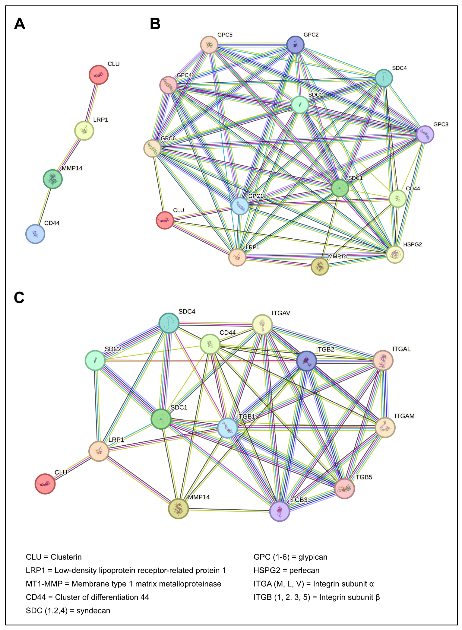
Figure 1. Protein–protein interaction (PPI) networks generated using the STRING database for the main players of the proposed signaling network. (A) PPI graph showing interactions among the main components of the proposed framework (CLU, LRP1, MT1-MMP, CD44). PPI graphs illustrating the interactions among the four main players and heparan sulfate proteoglycans (B), and syndecans and integrins (C). Network nodes represent proteins, whereas edges indicate predicted functional associations inferred from multiple evidence sources integrated within the STRING platform. Edge colors correspond to the type of supporting evidence as follows: pink, experimentally validated interactions; yellow, text mining evidence; black, co-expression evidence; blue, co-occurrence evidence; light blue, curated database interactions; light purple, protein homology. STRING database available at: https://string-db.org (version 12.0; accessed on 20 May 2026).
Cancers 18 01791 g001
Figure 2. Integrated membrane networks coordinating endocytosis, proteolysis, and signaling during tumor cell migration. Tumor cell migration relies on the coordinated interplay between ECM remodeling, membrane receptor dynamics, and intracellular signaling pathways. (A) At the leading edge of the migrating cell, MT1-MMP binds to CD44, promoting localized ECM degradation and activation of pro-MMP2 in a TIMP2-dependent manner, thereby facilitating invasion. (B) Membrane-associated complexes involving LRP1, growth factor receptors (GFRs), and syndecans (SDC1 and SDC4), contribute to signal integration at the cell surface. In this context, SDCs can function as a co-receptor for growth factors, while SDC4 is also involved in the regulation of cytoskeletal organization and adhesion dynamics through PKCα- and Rho GTPase-dependent pathways. These membrane platforms also activate pro-survival and pro-migratory signaling cascades, including MAPK and PI3K/Akt pathways. In parallel, CD44 can undergo proteolytic cleavage by MT1-MMP, releasing its intracellular domain (CD44 ICD), which is translocated to the nucleus and promotes the transcription of pro-tumorigenic genes. (C) LRP1-mediated endocytosis regulates the internalization and recycling of membrane-associated proteins, including proteases and adhesion receptors, contributing to the spatial regulation of proteolysis and receptor availability. (D) Shedding of SDCs, CD44, and LRP1 releases soluble fragments that further modulate the extracellular and pericellular landscape. Created in BioRender. Piperigkou, Z. (2026) https://BioRender.com/hgrbi8x (accessed on 20 May 2026).
Cancers 18 01791 g002
Figure 3. sCLU as a key player of the matrix-associated signaling network. sCLU promotes the formation of a membrane-associated signaling network by binding to LRP1 and facilitating the spatial co-localization of MT1-MMP, CD44 and SDCs at the cell surface. This organization enhances receptor cooperation and supports localized ECM degradation, dynamic regulation of cell–ECM adhesion, and activation of pro-migratory signaling pathways. These events converge on RhoGTPases-dependent cytoskeletal remodeling and transcriptional reprogramming associated with EMT. CDH1 = E-cadherin; VIM = Vimentin; FN1 = Fibronectin 1. Created in BioRender. Piperigkou, Z. (2026) https://BioRender.com/hd39kos (accessed on 20 May 2026).
Cancers 18 01791 g003
Figure 4. Disruption of the sCLU/LRP1-membrane associated network by targeted inhibition. Pharmacological inhibition of sCLU (i.e., OGX-011) prevents membrane cluster formation. The simultaneous targeting of key components of the network (MT1-MMP and CD44) results in loss of coordinated receptor activity and intracellular signaling, impaired cytoskeletal dynamics, and suppression of EMT-associated transcription, ultimately limiting tumor cell migration and invasion. VIM = Vimentin; FN1 = Fibronectin 1. Created in BioRender. Piperigkou, Z. (2026) https://BioRender.com/1atzxhh (accessed on 20 May 2026).
Cancers 18 01791 g004
Table 1. Localization and biological functions of key membrane-associated network (sCLU, LRP1, MT1-MMP, CD44, SDCs, GPCs, and perlecan) components.
ProteinAbbreviationLocalizationBiological FunctionsReference
Secreted clusterinsCLUExtracellular spaceExtracellular chaperone; protein clustering; stress response[18,20]
Low-density lipoprotein receptor-related protein 1LRP1Cell membrane *Endocytosis; intracellular signaling[4]
CD44CD44Cell membrane *Cell–ECM adhesion; scaffold for MMPs[24]
Membrane Type 1 Matrix MetalloproteinaseMT1-MMP (MMP14)Cell membraneECM degradation; receptor shedding; pro-MMP2 activation[3]
SyndecansSDC1Cell membrane *Cell–ECM adhesion; growth factor signaling; integrin-mediated signaling; shed SDC1 signaling[13,25]
SDC2Cell membrane *Cell–ECM adhesion; growth factor signaling; integrin-mediated signaling; shed SDC2 signaling[26]
SDC4Cell membrane *Focal adhesion formation; cytoskeletal remodeling; growth factor signaling; integrin-mediated signaling; shed SDC4 signaling[27]
GlypicansGPC1-6Cell membrane *Cell surface co-receptors; cell–matrix interactions.[15]
PerlecanHSPG2Pericellular/basement membraneECM/basement membrane organization; cell adhesion; growth factor sequestration; integrin-mediated signaling[16]
* Note: These cell surface molecules may also be detected in the extracellular space following shedding.
Disclaimer/Publisher’s Note: The statements, opinions and data contained in all publications are solely those of the individual author(s) and contributor(s) and not of MDPI and/or the editor(s). MDPI and/or the editor(s) disclaim responsibility for any injury to people or property resulting from any ideas, methods, instructions or products referred to in the content.