Enoxaparin, Tinzaparin, and Apixaban Modulate Cancer Cell Procoagulant Activity and Viability: Comparison with Quercetin

 , , ,
, , ,  , , , and
Simple Summary
Abstract
1. Introduction
2. Materials and Methods
2.1. Cell Culture
2.2. Cancer-Cell-Derived Extracellular Vesicle Isolation and Quantification
2.3. Endothelial Cell Exposure to CaCe-dEVs
2.4. Pretreatment of HUVEC with Antithrombotic Agents
2.5. Thrombin Generation Assay
2.6. Viability and Proliferation Assays
2.7. Tissue Factor Expression
2.8. Phosphatidylserine Expression
2.9. Microscopy
2.10. Statistical Analysis
3. Results
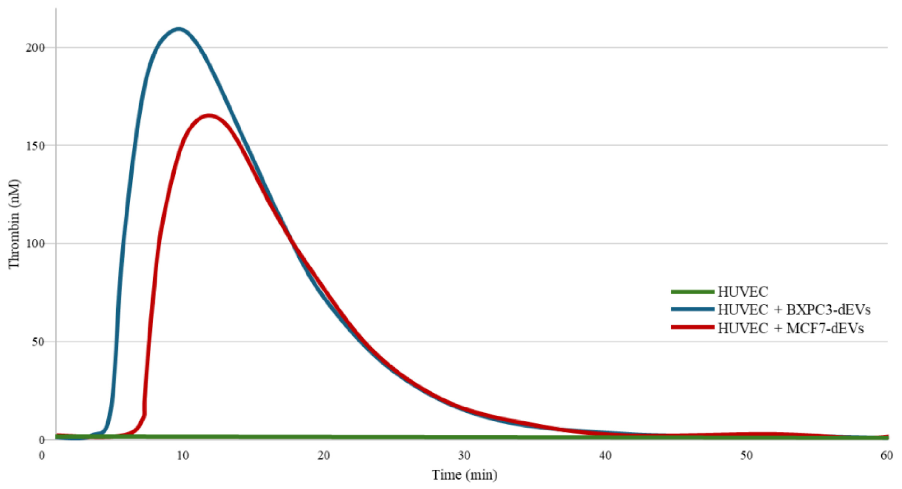
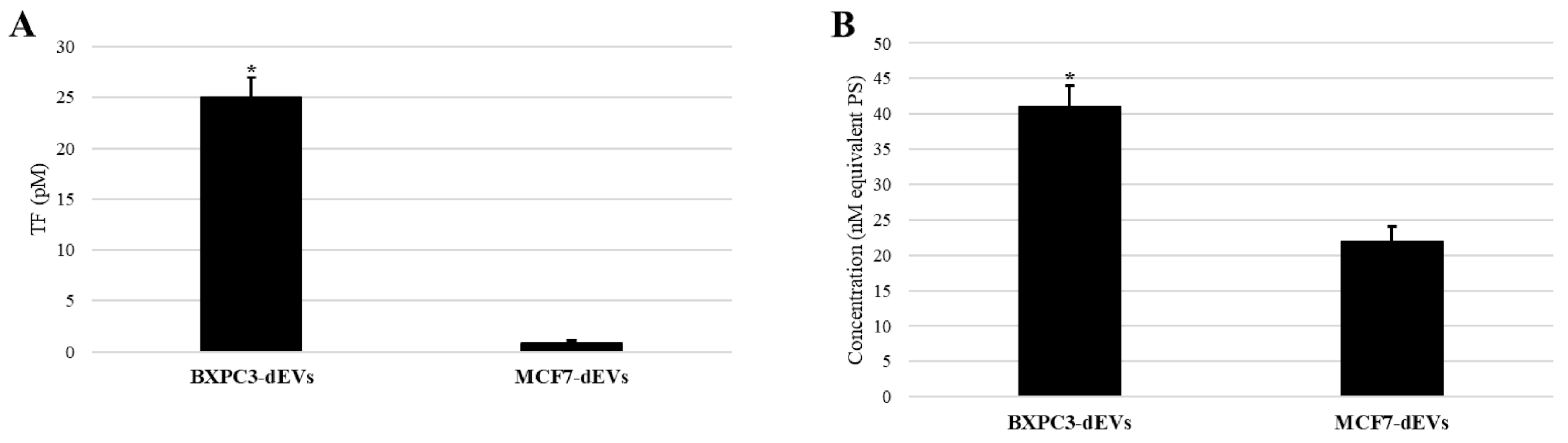
3.1. CaCe-dEVs Induced Alterations in the Procoagulant State of Endothelial Cells
3.2. CaCe-dEVs Induced Alterations in the Functional State of Endothelial Cells
3.3. Modulation of Cancer-Cell-Driven Thrombin Generation by LMWHs, Apixaban, and Quercetin
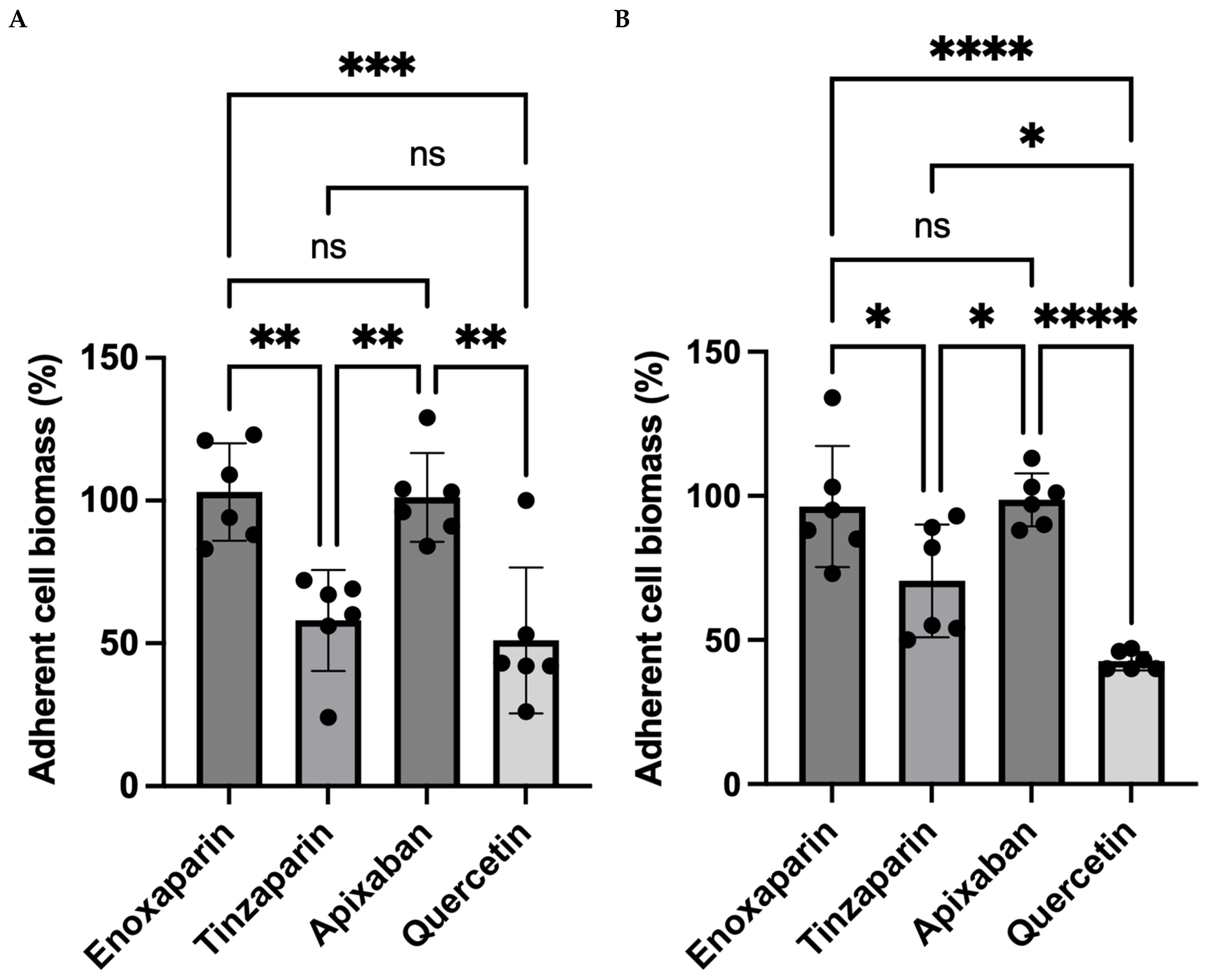
3.4. Impact of LMWHs, Apixaban, and Quercetin on Cancer Cell Viability
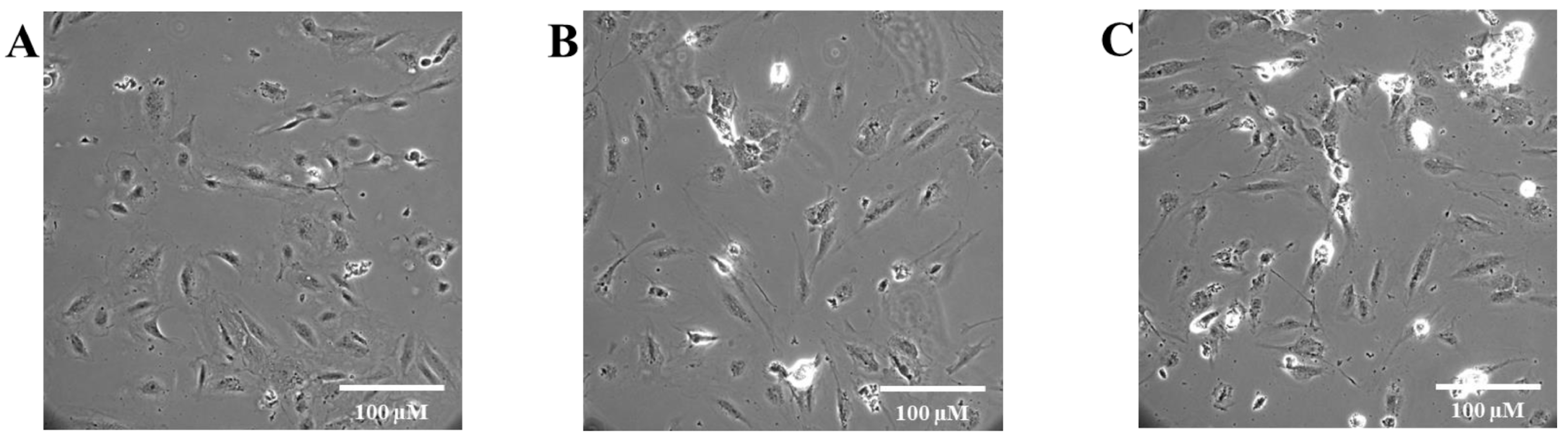
3.5. Effects of Antithrombotic Agents and Quercetin on HUVEC Morphology and Protection Against CaCe-dEVs-Induced Alterations
3.6. Effects of Enoxaparin, Tinzaparin, Apixaban, and Quercetin on HUVEC-Driven Thrombin Generation Following CaCe-dEVs Exposure
4. Discussion
- (a)
- LMWH-induced cytotoxicity on cancer cells does not represent a uniform class effect but rather reflects the specific properties of individual agents.
- (b)
- Cancer cell aggressiveness appears to influence treatment sensitivity, as BXPC3 cells exhibited greater susceptibility than MCF7 cells.
- (c)
- Effects on cell survival are distinct from effects on procoagulant potential, indicating a lack of direct correlation.
- (d)
- Reduction in cancer cell procoagulant activity by tinzaparin was comparable to that of quercetin.
- (e)
- Variability in efficacy reflects the molecular characteristics of each agent more than their anticoagulant activity.
5. Conclusions
Author Contributions
Funding
Institutional Review Board Statement
Informed Consent Statement
Data Availability Statement
Acknowledgments
Conflicts of Interest
References
- Marchetti, M.; Giaccherini, C.; Masci, G.; Russo, L.; Testa, S.; Bianchi, F.; Rossi, P.; Conti, A.; Santini, D.; Moretti, L.; et al. Thrombin generation predicts early recurrence in breast cancer patients. J. Thromb. Haemost. 2020, 18, 2220–2231. [Google Scholar] [CrossRef]
- Syrigos, K.; Grapsa, D.; Sangare, R.; Papadopoulos, A.; Kostopoulos, I.; Ioannidis, G.; Nikolaou, M.; Vassiliou, T.; Karamanos, N.; Christodoulou, C.; et al. Prospective assessment of clinical risk factors and biomarkers of hypercoagulability for the identification of patients with lung adenocarcinoma at risk for cancer-associated thrombosis: The observational ROADMAP-CAT study. Oncologist 2018, 23, 1372–1381. [Google Scholar] [CrossRef] [PubMed]
- Marchetti, M.; Russo, L.; Giaccherini, C.; Rossi, F.; Bianchi, G.; Conti, P.; Santini, D.; Moretti, L.; Testa, S.; De Luca, A.; et al. Hemostatic system activation in breast cancer: Searching for new biomarkers for cancer risk prediction and outcomes. Thromb. Res. 2022, 213, S46–S50. [Google Scholar] [CrossRef] [PubMed]
- Tran, H.C.M.; Mbemba, E.; Mourot, N.; Rousseau, A.; Morel, O.; Elalamy, I.; Gerotziafas, G.T.; Martin, F.; Lopez, A.; Dubois, C.; et al. The procoagulant signature of cancer cells drives fibrin network formation in the tumor microenvironment and impacts its quality: Implications in cancer cell migration and resistance to anticancer agents. Thromb. Res. 2024, 238, 172–183. [Google Scholar] [CrossRef] [PubMed]
- Carmeliet, P.; Jain, R.K. Molecular mechanisms and clinical applications of angiogenesis. Nature 2011, 473, 298–307. [Google Scholar] [CrossRef]
- Mehran, R.; Nilsson, M.; Khajavi, M.; Valente, M.; Eriksson, P.; Johansson, H.; Lindberg, S.; Andersson, S.; Holmberg, L.; Petersen, L.; et al. Tumor endothelial markers define novel subsets of cancer-specific circulating endothelial cells associated with antitumor efficacy. Cancer Res. 2014, 74, 2731–2741. [Google Scholar] [CrossRef]
- Lim, A.R.; Rathmell, W.K.; Rathmell, J.C. The tumor microenvironment as a metabolic barrier to effector T cells and immunotherapy. eLife 2020, 9, e55185. [Google Scholar] [CrossRef]
- Leone, P.; Malerba, E.; Susca, N.; Rossi, F.; Bianchi, G.; Conti, P.; Testa, S.; De Luca, A.; Russo, L.; Marchetti, M.; et al. Endothelial cells in tumor microenvironment: Insights and perspectives. Front. Immunol. 2024, 15, 1367875. [Google Scholar] [CrossRef]
- Becker, A.; Thakur, B.K.; Weiss, J.M.; Kim, H.S.; Peinado, H.; Lyden, D. Extracellular vesicles in cancer: Cell-to-cell mediators of metastasis. Cancer Cell 2016, 30, 836–848. [Google Scholar] [CrossRef]
- Amrane-Djedidi, R.; Rousseau, A.; Larsen, A.K.; Elalamy, I.; Van Dreden, P.; Gerotziafas, G.T. Extracellular vesicles derived from pancreatic cancer cells BXPC3 or breast cancer cells MCF7 induce a permanent procoagulant shift to endothelial cells. Thromb. Res. 2020, 187, 170–179. [Google Scholar] [CrossRef]
- Falanga, A.; Ay, C.; Di Nisio, M.; Pabinger, I.; Verso, M.; Rickles, F.R.; Valerio, L.; Heit, J.A.; Lyman, G.H.; Prandoni, P.; et al. Venous thromboembolism in cancer patients: ESMO Clinical Practice Guideline. Ann. Oncol. 2023, 34, 452–467. [Google Scholar] [CrossRef]
- Giannas, E.; Kontovounisios, C. The antineoplastic effect of heparin on colorectal cancer: A review of the literature. J. Clin. Med. 2023, 12, 7173. [Google Scholar] [CrossRef]
- Borsig, L. Antimetastatic activities of heparins and modified heparins: Experimental evidence. Thromb. Res. 2010, 125, S66–S71. [Google Scholar] [CrossRef] [PubMed]
- Norrby, K. Low-molecular-weight heparins and angiogenesis. J. Pathol. Microbiol. Immunol. 2006, 114, 79–102. [Google Scholar] [CrossRef]
- Gerotziafas, G.T.; Papageorgiou, C.; Hatmi, M.; Samama, M.M.; Elalamy, I. Clinical studies with anticoagulants to improve survival in cancer patients. Pathophysiol. Haemost. Thromb. 2008, 36, 204–211. [Google Scholar] [CrossRef] [PubMed]
- Lazo-Langner, A.; Goss, G.D.; Spaans, J.N.; Rodger, M.A. The effect of low-molecular-weight heparin on cancer survival. A systematic review and meta-analysis of randomized trials. J. Thromb. Haemost. 2007, 5, 729–737. [Google Scholar] [CrossRef]
- Huang, S.S.; Chen, W.; Vaishnani, D.K.; Huang, L.J.; Li, J.Z.; Huang, S.R.; Li, Y.Z.; Xie, Q.P. Leucine-rich repeat-containing protein 19 suppresses colorectal cancer by targeting cyclin-dependent kinase 6/E2F1 and remodeling the immune microenvironment. World J. Gastroenterol. 2025, 31, 107893. [Google Scholar] [CrossRef] [PubMed]
- Russo, V.; Falco, L.; Tessitore, V.; Lorusso, V.; Ricci, F.; Rossi, A.; Santini, D.; Moretti, L.; Bianchi, F.; Conti, P.; et al. Anti-inflammatory and anticancer effects of anticoagulant therapy in patients with malignancy. Life 2023, 13, 1888. [Google Scholar] [CrossRef]
- Guasti, L.; Squizzato, A.; Moretto, P.; Salerno, G.; Rossi, M.; De Luca, A.; Bianchi, F.; Conti, P.; Testa, S.; Marchetti, M.; et al. In vitro effects of apixaban on 5 different cancer cell lines. PLoS ONE 2017, 12, e0185035. [Google Scholar] [CrossRef]
- Gürpınar, Ö.A.; Kubat, E.; Onur, M.A. Cytotoxic activity of apixaban on HeLa cells: An in vitro study. Hacet. J. Biol. Chem. 2018, 46, 395–402. [Google Scholar] [CrossRef]
- Li, M.T.; Ke, J.; Guo, S.F.; Wang, Y.; Chen, H.; Zhao, L.; Liu, X.; Zhang, M.; Yang, P.; Liu, H.; et al. The protective effect of quercetin on endothelial cells injured by hypoxia and reoxygenation. Front. Pharmacol. 2021, 12, 732874. [Google Scholar] [CrossRef]
- Lotfi, N.; Yousefi, Z.; Golabi, M.; Ahmadi, S.; Khademi, F.; Rezai, M.; Tavakoli, H.; Salehi, M.; Jafari, F.; Asadi, M.; et al. The potential anti-cancer effects of quercetin on blood, prostate and lung cancers: An update. Front. Immunol. 2023, 14, 1077531. [Google Scholar] [CrossRef]
- Chen, W.; Li, Y.; Zhang, C.; Zhou, H.; Ma, J.; Vaishnani, D.K.; Zeng, B.; Yu, J.; Mao, H.; Zheng, J. Multi-omics and experimental validation reveal anti-HCC mechanisms of Tibetan Liuwei Muxiang Pill and quercetin. Pharmaceuticals 2025, 18, 900. [Google Scholar] [CrossRef]
- Rousseau, A.; Van Dreden, P.; Khaterchi, A.; Larsen, A.K.; Elalamy, I.; Gerotziafas, G.T. Procoagulant microparticles derived from cancer cells have determinant role in the hypercoagulable state associated with cancer. Int. J. Oncol. 2017, 51, 1793–1800. [Google Scholar] [CrossRef]
- Gerotziafas, G.T.; Galea, V.; Mbemba, E.; Douxfils, J.; Elalamy, I.; Rousseau, A.; Papageorgiou, C.; Samama, M.M.; Morel, O.; Martin, F.; et al. Tissue factor over-expression by human pancreatic cancer cells BXPC3 is related to higher prothrombotic potential as compared to breast cancer cells MCF7. Thromb. Res. 2012, 129, 779–786. [Google Scholar] [CrossRef] [PubMed]
- Théry, C.; Witwer, K.W.; Aikawa, E.; Alcaraz, M.J.; Anderson, J.D.; Andriantsitohaina, R.; Antoniou, A.; Arab, T.; Archer, F.; Atkin-Smith, G.K.; et al. Minimal information for studies of extracellular vesicles 2018 (MISEV2018): A position statement of the International Society for Extracellular Vesicles and update of the MISEV2014 guidelines. J. Extracell. Vesicles 2018, 7, 1535750. [Google Scholar] [CrossRef] [PubMed]
- Amrane-Djedidi, R. Tumor Microenvironment and Thrombogenesis—Study of the Transmission of Procoagulant Properties from Tumor Extracellular Vesicles to Endothelial Cells and Their Heritability. Ph.D. Thesis, Sorbonne University, Paris, France, 2022. [Google Scholar]
- Morikawa, S.; Baluk, P.; Kaidoh, T.; Haskell, A.; Jain, R.K.; McDonald, D.M. Abnormalities in pericytes on blood vessels and endothelial sprouts in tumors. Am. J. Pathol. 2002, 160, 985–1000. [Google Scholar] [CrossRef]
- Nagl, L.; Horvath, L.; Pircher, A.; Wolf, D.; Huber, M.; Schmidt, H.; Meier, J.; Klein, G.; Weber, R.; Wagner, T.; et al. Tumor endothelial cells (TECs) as potential immune directors of the tumor microenvironment: New findings and future perspectives. Front. Cell Dev. Biol. 2020, 8, 766. [Google Scholar] [CrossRef]
- Akino, T.; Hida, K.; Hida, Y.; Hashimoto, H.; Matsumoto, T.; Saito, Y.; Nakamura, M.; Fujita, N.; Tanaka, K.; Suzuki, T.; et al. Cytogenetic abnormalities of tumor-associated endothelial cells in human malignant tumors. Am. J. Pathol. 2009, 175, 2657–2667. [Google Scholar] [CrossRef]
- Schaaf, M.B.; Houbaert, D.; Meçe, O.; Agostinis, P. Autophagy in endothelial cells and tumor angiogenesis. Cell Death Differ. 2019, 26, 665–679. [Google Scholar] [CrossRef] [PubMed]
- Tubtimsri, S.; Chuenbarn, T.; Manmuan, S.; Wongpiyabovorn, J.; Kanchanapoom, T.; Thongkum, S.; Phonchai, S.; Charoenphol, P.; Suksaweang, P.; Somboon, T.; et al. Quercetin triggers cell apoptosis-associated ROS-mediated cell death and induces S and G2/M-phase cell cycle arrest in KON oral cancer cells. BMC Complement. Med. Ther. 2025, 25, 34. [Google Scholar] [CrossRef]
- Fareed, J.; Walenga, J.M. Why differentiate low molecular weight heparins for venous thromboembolism? Thromb. J. 2007, 5, 8. [Google Scholar] [CrossRef]
- Sarantis, P.; Bokas, A.; Papadimitropoulou, A.; Koustas, E.; Theocharis, S.; Papakotoulas, P.; Schizas, D.; Papalampros, A.; Felekouras, E.; Papavassiliou, A.G.; et al. Combinatorial treatment of tinzaparin and chemotherapy can induce a significant antitumor effect in pancreatic cancer. Int. J. Mol. Sci. 2021, 22, 7053. [Google Scholar] [CrossRef] [PubMed]
- Ungefroren, H.; Witte, D.; Fiedler, C.; Gieseler, F.; Braun, R.; Mathieu, S.; Lehnert, H.; Faendrich, F. Protease-activated receptors (PARs)—Biology and role in cancer invasion and metastasis. Cancer Metastasis Rev. 2015, 34, 347–374. [Google Scholar] [CrossRef]
- Boots, A.W.; Haenen, G.R.M.M.; Bast, A. Health effects of quercetin: From antioxidant to nutraceutical. Eur. J. Pharmacol. 2008, 585, 325–337. [Google Scholar] [CrossRef] [PubMed]
- Qi, W.; Qi, W.; Xiong, D.; Long, M. Quercetin: Its antioxidant mechanism, antibacterial properties and potential application in prevention and control of toxipathy. Molecules 2022, 27, 6545. [Google Scholar] [CrossRef]
- Biswas, P.; Dey, D.; Biswas, P.K.; Rahaman, T.I.; Saha, S.; Parvez, A.; Khan, D.A.; Lily, N.J.; Saha, K.; Sohel, M.; et al. A comprehensive analysis and anti-cancer activities of quercetin in ROS-mediated cancer and cancer stem cells. Int. J. Mol. Sci. 2022, 23, 11746. [Google Scholar] [CrossRef]
- Chen, X.; Zheng, L.; Zhang, B.; Deng, Z.; Li, H. Synergistic protection of quercetin and lycopene against oxidative stress via SIRT1-Nox4-ROS axis in HUVEC cells. Curr. Res. Food Sci. 2022, 5, 1985–1993. [Google Scholar] [CrossRef] [PubMed]
- Khorana, A.A.; Sahni, A.; Altland, O.D.; Francis, C.W. Heparin inhibition of endothelial cell proliferation and organization is dependent on molecular weight. Arterioscler. Thromb. Vasc. Biol. 2003, 23, 2110–2115. [Google Scholar] [CrossRef]
- Mousa, S.A.; Mohamed, S. Inhibition of endothelial cell tube formation by the low molecular weight heparin, tinzaparin, is mediated by tissue factor pathway inhibitor. Thromb. Haemost. 2004, 92, 627–633. [Google Scholar] [CrossRef]
- Gao, Y.; Chen, W.; Zarogoulidis, P.; Yallapu, M.M.; Nikolic, M.V.; Vaishnani, D.K.; Zheng, J.; Nedeljkovic, N.; Ye, K.; Guo, Y.; et al. Multiomics and experimental validation reveal theophylline’s mechanism targeting IL1A/ACTB/TLR4 and identify synergistic drugs in hepatocellular carcinoma. J. Pharmacol. Exp. Ther. 2026, 393, 103836. [Google Scholar] [CrossRef] [PubMed]



| Parameters | Native HUVEC | HUVEC + BXPC3-dEVs | HUVEC + MCF7-dEVs |
|---|---|---|---|
| Lag time (min) | >40 | 4.0 ± 0.2 | 6.0 ± 0.1 * |
| ETP (nM·min) | 0 | 1603 ± 367 | 1476 ± 437 |
| Peak (nM) | 0 | 214 ± 95 | 167 ± 82 |
| Parameters | Control | Enoxaparin | Tinzaparin | Apixaban | Quercetin | |||||
|---|---|---|---|---|---|---|---|---|---|---|
| Low | High | Low | High | Low | High | Low | High | Low | High | |
| (a) | ||||||||||
| Lag time (min) | 1.93 ± 0.1 | 2.33 ± 0.2 | 1.71 ± 0.0 | 2.06 ± 0.2 | 1.99 ± 0.1 | 3.00 ± 0.5 * | 1.82 ± 0.1 | 2.06 ± 0.1 | 2.27 ± 0.1 | 2.72 ± 0.1 * |
| ETP (nM·min) | 1009 ± 4.7 | 1452 ± 50 | 969 ± 28.1 | 1426 ± 90 | 1003 ± 29.5 | 1336 ± 50 | 976 ± 20.1 | 1529 ± 100 | 945 ± 37.5 | 1340 ± 50 |
| Peak (nM) | 119 ± 1.5 | 161 ± 10 | 121 ± 1.71 | 169 ± 6 | 120 ± 2.9 | 131 ± 7 * | 122 ± 1.8 | 177 ± 20 | 108 ± 4.9 | 144 ± 6 * |
| (b) | ||||||||||
| Lag time (min) | 11.49 ± 0.1 | 11.84 ± 0.5 | 10.46 ± 1.52 | 12.78 ± 1.1 | 10.9 ± 0.0 | 13.33 ± 0.5 | 11.90 ± 1.43 | 14.33 ± 1.2 * | 12.16 ± 0.83 | 18.61 ± 1 * |
| ETP (nM·min) | 652 ± 47.4 | 945 ± 0.5 | 674 ± 23.86 | 717 ± 0.5 * | 687 ± 9.41 | 735 ± 0.5 * | 672 ± 97.11 | 729 ± 7 * | 789 ± 38.11 | 649 ± 0.5 *$ |
| Peak (nM) | 41 ± 5 | 73 ± 7 | 43.63 ± 6.64 | 47 ± 5 * | 50.40 ± 0.48 | 50 ± 5 * | 51.93 ± 16.63 | 51 ± 5 * | 67.21 ± 1.03 | 43 ± 5 *$ |
| Not Exposed to CaCe-dEVs | Exposed to BXPC3-dEVs | Exposed to MCF7-dEVs | |||||||
|---|---|---|---|---|---|---|---|---|---|
| Parameters | Native HUVEC | HUVEC + Apixaban | HUVEC + Quercetin | HUVEC | HUVEC + Apixaban | HUVEC + Quercetin | HUVEC | HUVEC + Apixaban | HUVEC + Quercetin |
| Lag time (min) | 11.5 ± 2.9 | 12 ± 1.2 | 13 ± 1.0 | 4.0 ± 0.2 ++ | 5.0 ± 0.9 | 6.0 ± 1.0 *++ | 6.0 ± 0.1 *++ | 6.0 ± 0.5 | 6.5 ± 1.0 |
| ETP (nM·min) | 910 ± 150 | 940 ± 45 | 1002 ± 69 | 1603 ± 367 ++ | 1250 ± 120 | 1011 ± 60 ++ | 1476 ± 437 ++ | 1360 ± 77 | 1250 ± 190 |
| Peak (nM) | 60.7 ± 15 | 71 ± 8 | 75 ± 10 | 214 ± 95 ++ | 169 ± 6 $ | 177 ± 5 $ | 167 ± 82 *++ | 165 ± 50 | 159 ± 22 |
| Parameter | Effect of CaCe-dEVs on Endothelial Cells (HUVECs) | Effect of Antithrombotic Agents on Cancer Cells (BXPC3 and MCF7) | Protective Effects of Antithrombotic Agents and Quercetin on HUVECs Exposed to CaCe-dEVs |
|---|---|---|---|
| Procoagulant phenotype | CaCe-dEVs induced a marked endothelial procoagulant shift, characterized by
| Tinzaparin and quercetin reduced thrombin generation induced by both BXPC3 and MCF7 cells. Enoxaparin and apixaban reduced thrombin generation induced only by MCF7 cells, with no significant effect in BXPC3 cells. | Pre-exposure to apixaban and quercetin partially prevented the procoagulant shift of endothelial cells induced by exposure to BXPC3-dEVs. No significant protection was observed against MCF7-dEV-induced procoagulant shift. |
| Cell viability/proliferation | CaCe-dEVs significantly reduced endothelial cell viability and proliferation BXPC3-dEVs exerted stronger effects than MCF7-dEVs. | Tinzaparin and quercetin significantly reduced cancer cell viability in both BXPC3 and MCF7 cells. Enoxaparin and apixaban showed no significant cytotoxic effects. | No protective effect on endothelial cell viability or proliferation. |
| Cell morphology | CaCe-dEVs disrupted endothelial monolayer integrity, reduced confluence, and increased rounded/refractile cells, consistent with endothelial injury. MCF7-dEVs. induced more important morphological alteration s of endothelial cells than BXPC3-dEVs. | No significant morphological changes were assessed in cancer cells following treatment. | Pre-exposure to enoxaparin or tinzaparin failed to prevent endothelial cell morphological alterations induced by exposure to CaCe-dEVs. Apixaban limited attenuation, whereas quercetin provided modest and heterogeneous protection. |
Disclaimer/Publisher’s Note: The statements, opinions and data contained in all publications are solely those of the individual author(s) and contributor(s) and not of MDPI and/or the editor(s). MDPI and/or the editor(s) disclaim responsibility for any injury to people or property resulting from any ideas, methods, instructions or products referred to in the content. |
© 2026 by the authors. Licensee MDPI, Basel, Switzerland. This article is an open access article distributed under the terms and conditions of the Creative Commons Attribution (CC BY) license.
Share and Cite
Baghdadi, M.A.; do Carmo Las Casas, P.H.F.; Mbemba, E.; Rousseau, A.; Kempaiah, P.; Svistunov, A.A.; Bitsadze, V.; Sabbah, M.; Fareed, J.; Van Dreden, P.; et al. Enoxaparin, Tinzaparin, and Apixaban Modulate Cancer Cell Procoagulant Activity and Viability: Comparison with Quercetin. Cancers 2026, 18, 1783. https://doi.org/10.3390/cancers18111783
Baghdadi MA, do Carmo Las Casas PHF, Mbemba E, Rousseau A, Kempaiah P, Svistunov AA, Bitsadze V, Sabbah M, Fareed J, Van Dreden P, et al. Enoxaparin, Tinzaparin, and Apixaban Modulate Cancer Cell Procoagulant Activity and Viability: Comparison with Quercetin. Cancers. 2026; 18(11):1783. https://doi.org/10.3390/cancers18111783
Chicago/Turabian StyleBaghdadi, Mohammed A., Pedro Henrique Fernandes do Carmo Las Casas, Elisabeth Mbemba, Aurélie Rousseau, Prakasha Kempaiah, Andrey A. Svistunov, Victoria Bitsadze, Michèle Sabbah, Jawed Fareed, Patrick Van Dreden, and et al. 2026. "Enoxaparin, Tinzaparin, and Apixaban Modulate Cancer Cell Procoagulant Activity and Viability: Comparison with Quercetin" Cancers 18, no. 11: 1783. https://doi.org/10.3390/cancers18111783
APA StyleBaghdadi, M. A., do Carmo Las Casas, P. H. F., Mbemba, E., Rousseau, A., Kempaiah, P., Svistunov, A. A., Bitsadze, V., Sabbah, M., Fareed, J., Van Dreden, P., Trachana, V., Lefkou, E., & Gerotziafas, G. (2026). Enoxaparin, Tinzaparin, and Apixaban Modulate Cancer Cell Procoagulant Activity and Viability: Comparison with Quercetin. Cancers, 18(11), 1783. https://doi.org/10.3390/cancers18111783