Intraoperative Peritoneal Lavage for Detection of Malignant Cells: Technique, Evidence, Clinical Relevance and Future Perspectives

 , ,
, ,
Simple Summary
Abstract
1. Introduction
2. Materials and Methods
2.1. Study Design
2.2. Search Strategy
2.3. Eligibility Criteria
2.4. Selection Process
3. Results
3.1. Fluid Utilization
3.2. Timing of Lavage
3.3. Detection Method
3.4. Positive Detection Rate
3.5. Clinical Relevance of Free Cancer Cell Detection in Peritoneal Fluid
4. Discussion
4.1. Technical Variability and Its Impact
4.2. Detection of Malignant Cells: Methods and Limitations
4.3. Prognostic Implications
4.4. Clinical Implications and Cost-Effectiveness
4.5. Emerging Technologies
4.6. Limitations of This Review
4.7. Need for Standardization
5. Conclusions
Author Contributions
Funding
Data Availability Statement
Conflicts of Interest
Abbreviations
| BTC | biliary tract cancer |
| CRC | colorectal cancer |
| CRS | cytoreductive surgery |
| FISH | fluorescence in situ hybridization |
| GC | gastric adenocarcinoma |
| HE | Hematoxylin-Eosin |
| HIPEC | hyperthermic intraperitoneal chemotherapy |
| IHC | immunohistochemical staining |
| OEK | optically induced electrokinetics |
| PCI | Peritoneal Carcinomatosis Index |
| PDAC | pancreatic adenocarcinoma |
| PIPAC | pressurized intraperitoneal aerosol chemotherapy |
| p-lap | post laparotomy |
| p-lasc | post laparoscopic port insertion |
| PM | Peritoneal metastases |
| PRISMA | Preferred Reporting Items for Systematic Reviews and Meta-Analyses |
| prtr | prior tumor resection |
| ptr | post tumor resection |
| RT-PCR | reverse transcription polymerase chain reaction |
References
- Jacquet, P.; Sugarbaker, P.H. Clinical Research Methodologies in Diagnosis and Staging of Patients with Peritoneal Carcinomatosis. Cancer Treat Res. 1996, 82, 359–374. [Google Scholar] [PubMed]
- Sano, T.; Kodera, Y. Japanese Classification of Gastric Carcinoma: 3rd English Edition. Gastric Cancer 2011, 14, 101–112. [Google Scholar] [CrossRef]
- Sakoda, T.; Uemura, K.; Kondo, N.; Sumiyoshi, T.; Okada, K.; Seo, S.; Otsuka, H.; Murakami, Y.; Takahashi, S. Prognostic Value of Peritoneal Lavage Cytology in Patients with Pancreatic Ductal Adenocarcinoma Stratified by the Resectability Status. J. Gastrointest. Surg. 2021, 25, 2871–2880. [Google Scholar] [CrossRef]
- Mashiko, T.; Ogasawara, T.; Masuoka, Y.; Ei, S.; Takahashi, S.; Hirabayashi, K.; Mori, M.; Koyanagi, K.; Yamamoto, S.; Nakagohri, T. Prognostic Impact of Positive Peritoneal Lavage Cytology on Resectable Pancreatic Body and Tail Cancer: A Retrospective Study. World J. Surg. 2023, 47, 729–739. [Google Scholar] [CrossRef] [PubMed]
- Ishida, H.; Ogura, T.; Takahashi, A.; Kitamura, K.; Miyamoto, R.; Matsudaira, S.; Tanabe, M.; Kanda, H.; Kawashima, Y. Prognostic Value of Peritoneal Lavage Cytology in Potentially Resectable Pancreatic Cancer Stratified by Cytologic Status. Cancer Diagn. Progn. 2023, 3, 338–346. [Google Scholar] [CrossRef]
- Tanemura, M.; Furukawa, K.; Mikamori, M.; Asaoka, T.; Yasuoka, H.; Marukawa, D.; Urata, Y.; Yamada, D.; Kobayashi, S.; Eguchi, H. Clinical Impact of High-Quality Testing for Peritoneal Lavage Cytology in Pancreatic Cancer. Sci. Rep. 2024, 14, 10199. [Google Scholar] [CrossRef]
- Matsukuma, S.; Nagano, H.; Kobayashi, S.; Wada, H.; Seo, S.; Tsugawa, D.; Okuyama, H.; Iida, K.; Ohmura, Y.; Takeda, Y.; et al. The Impact of Peritoneal Lavage Cytology in Biliary Tract Cancer (KHBO1701): Kansai Hepato-Biliary Oncology Group. Cancer Rep. 2021, 4, e1323. [Google Scholar] [CrossRef]
- Ouchi, A.; Kinoshita, T.; Nakanishi, H.; Komori, K.; Oshiro, T.; Yoshimura, M.; Fujita, N.; Hosoda, W.; Shimizu, Y. PCR-based Quantitative Detection of Intraperitoneal Free Cancer Cells for Predicting Locoregional Recurrence after Rectal Cancer Resection. ANZ J. Surg. 2022, 92, 794–800. [Google Scholar] [CrossRef]
- Kobayashi, H.; Kotake, K.; Maeda, K.; Suto, T.; Kawasaki, M.; Ueno, H.; Komori, K.; Ozawa, H.; Koda, K.; Ohue, M.; et al. Peritoneal Lavage Cytology in Patients with Curative Resection for Stage II and III Colorectal Cancer: A Multi-Institutional Prospective Study. Ann. Gastroenterol. Surg. 2024, 8, 807–816. [Google Scholar] [CrossRef]
- Okabe, H.; Tsunoda, S.; Hosogi, H.; Hisamori, S.; Tanaka, E.; Tanaka, S.; Sakai, Y. Circulating Tumor Cells as an Independent Predictor of Survival in Advanced Gastric Cancer. Ann. Surg. Oncol. 2015, 22, 3954–3961. [Google Scholar] [CrossRef] [PubMed]
- Kano, Y.; Kosugi, S.I.; Ishikawa, T.; Otani, T.; Muneoka, Y.; Sato, Y.; Hanyu, T.; Hirashima, K.; Bamba, T.; Wakai, T. Prognostic Significance of Peritoneal Lavage Cytology at Three Cavities in Patients with Gastric Cancer. Surgery 2015, 158, 1581–1589. [Google Scholar] [CrossRef]
- Higaki, E.; Yanagi, S.; Gotohda, N.; Kinoshita, T.; Kuwata, T.; Nagino, M.; Ochiai, A.; Fujii, S. Intraoperative Peritoneal Lavage Cytology Offers Prognostic Significance for Gastric Cancer Patients with Curative Resection. Cancer Sci. 2017, 108, 978–986. [Google Scholar] [CrossRef] [PubMed]
- Sakata, T.; Takahata, T.; Kimura, T.; Yasuhara, I.; Kojima, T.; Akazai, Y.; Mimura, T.; Lefor, A.K. A Single-Institution Retrospective Analysis of Gastric Carcinoma with Positive Peritoneal Lavage Cytology and without Serosal Invasion: A Case Series. Ann. Med. Surg. 2019, 39, 10–15. [Google Scholar] [CrossRef]
- Hirabayashi, K.; Imoto, A.; Yamada, M.; Hadano, A.; Kato, N.; Miyajima, Y.; Ito, H.; Kawaguchi, Y.; Nakagohri, T.; Mine, T.; et al. Positive Intraoperative Peritoneal Lavage Cytology Is a Negative Prognostic Factor in Pancreatic Ductal Adenocarcinoma: A Retrospective Single-Center Study. Front. Oncol. 2015, 5, 182. [Google Scholar] [CrossRef]
- Suenaga, M.; Fujii, T.; Yamada, S.; Hayashi, M.; Shinjo, K.; Takami, H.; Niwa, Y.; Sonohara, F.; Shimizu, D.; Kanda, M.; et al. Peritoneal Lavage Tumor DNA as a Novel Biomarker for Predicting Peritoneal Recurrence in Pancreatic Ductal Adenocarcinoma. Ann. Surg. Oncol. 2021, 28, 2277–2286. [Google Scholar] [CrossRef] [PubMed]
- Takadate, T.; Morikawa, T.; Ishida, M.; Aoki, S.; Hata, T.; Iseki, M.; Miura, T.; Ariake, K.; Maeda, S.; Kawaguchi, K.; et al. Staging Laparoscopy Is Mandatory for the Treatment of Pancreatic Cancer to Avoid Missing Radiologically Negative Metastases. Surg. Today 2021, 51, 686–694. [Google Scholar] [CrossRef]
- Nakanishi, K.; Kanda, M.; Umeda, S.; Tanaka, C.; Kobayashi, D.; Hayashi, M.; Yamada, S.; Kodera, Y. The Levels of SYT13 and CEA MRNAs in Peritoneal Lavages Predict the Peritoneal Recurrence of Gastric Cancer. Gastric Cancer 2019, 22, 1143–1152. [Google Scholar] [CrossRef]
- Koganti, S.B.; Boddepalli, S.; Nambada, M.; Thumma, V.M.; Nagari, B.; Sastry, R.A. Positive Peritoneal Lavage Cytology–Implications for Staging and Management of Gastric Cancer. Indian J. Surg. Oncol. 2016, 7, 430–435. [Google Scholar] [CrossRef][Green Version]
- Ikoma, N.; Blum, M.; Chiang, Y.-J.; Estrella, J.S.; Roy-Chowdhuri, S.; Fournier, K.; Mansfield, P.; Ajani, J.A.; Badgwell, B.D. Yield of Staging Laparoscopy and Lavage Cytology for Radiologically Occult Peritoneal Carcinomatosis of Gastric Cancer. Ann. Surg. Oncol. 2016, 23, 4332–4337. [Google Scholar] [CrossRef]
- Zhang, X.; Liu, X.; Sun, F.; Li, S.; Gao, W.; Wang, Y. Greater Omental Milky Spot Examination for Diagnosis of Peritoneal Metastasis in Gastric Cancer Patients. J. Laparoendosc. Adv. Surg. Tech. 2017, 27, 106–109. [Google Scholar] [CrossRef] [PubMed]
- Young, S.; Ou, R.; Dehal, A.; O’Connor, V.V.; DiFronzo, L.A. Peritoneal Lavage Cytology Following Neoadjuvant Chemotherapy for Gastric Adenocarcinoma: Low Yield in Detecting Peritoneal Metastases. Am. Surg. 2022, 88, 1084–1089. [Google Scholar] [CrossRef]
- Yin, X.; Ruan, X.; Zhu, Y.; Yin, Y.; Huang, R.; Liang, C. Prediction of Peritoneal Free Cancer Cells in Gastric Cancer Patients by Golden-Angle Radial Sampling Dynamic Contrast-Enhanced Magnetic Resonance Imaging. J. Zhejiang Univ.-Sci. B 2024, 25, 617–627. [Google Scholar] [CrossRef] [PubMed]
- Gęca, K.; Rawicz-Pruszyński, K.; Mlak, R.; Sędłak, K.; Skórzewska, M.; Pelc, Z.; Małecka-Massalska, T.; Polkowski, W.P. Molecular Cytology by One-Step Nucleic Acid Amplification (Osna) Assay of Peritoneal Washings during D2 Gastrectomy in Advanced Gastric Cancer Patients: Preliminary Results. J. Clin. Med. 2021, 10, 5230. [Google Scholar] [CrossRef] [PubMed]
- Hoskovec, D.; Varga, J.; Dytrych, P.; Konecna, E.; Matek, J. Peritoneal Lavage Examination as a Prognostic Tool in Cases of Gastric Cancer. Arch. Med. Sci. 2017, 13, 612–616. [Google Scholar] [CrossRef]
- Hasbahceci, M.; Akcakaya, A.; Guler, B.; Kunduz, E.; Malya, F.; Muslumanoglu, M. Use of Peritoneal Washing Cytology for the Detection of Free Peritoneal Cancer Cells before and after Surgical Treatment of Gastric Adenocarcinoma. J. Cancer Res. Ther. 2018, 14, 1225–1229. [Google Scholar] [CrossRef]
- Cieśla, S.; Lisiecki, R.; Ławnicka, A.; Kudliński, B.; Ostrowska, P.; Davì, A.; Veroux, M.; Murawa, D. Clinical Significance of Peritoneal Fluid Examination for Free Cancer Cells in Patients Qualified for Surgery for Gastric Cancer. Front. Surg. 2021, 8, 685868. [Google Scholar] [CrossRef]
- Xuan, Y.; Gao, Q.; Wang, C.; Cai, D. Positive Peritoneal Lavage Fluid Cytology Based on Isolation by Size of Epithelial Tumor Cells Indicates a High Risk of Peritoneal Metastasis. PeerJ 2024, 12, e17602. [Google Scholar] [CrossRef]
- Takahashi, H.; Akita, H.; Wada, H.; Tomokuni, A.; Asukai, K.; Takahashi, Y.; Yanagimoto, Y.; Matsunaga, T.; Sugimura, K.; Yamamoto, K.; et al. Subclinical Cancer Cell Dissemination in Peritoneal Lavage Fluid Detected by Reverse-Transcription Polymerase Chain Reaction Identifies Patients at High Risk for Peritoneal Recurrence and Consequent Impaired Survival in the Setting of Preoperative Chemora. Surgery 2018, 164, 1168–1177. [Google Scholar] [CrossRef]
- Salamanca, I.M.G.; Jaime, M.T.E.; Penco, J.M.M.; Martínez, J.S. Role of Peritoneal Cytology in Patients with Early Stage Colorectal Cancer. Pathol. Oncol. Res. 2020, 26, 1325–1329. [Google Scholar] [CrossRef]
- Shalaby, M.; El Baradie, T.S.; Salama, M.; Shaaban, H.A.M.; Allam, R.M.; Hafiz, E.O.A.; Abdelhamed, M.A.; Attia, A. Conventional Peritoneal Cytology Lacks the Prognostic Significance of Detecting Local or Peritoneal Recurrence in Colorectal Cancer: An Egyptian Experience. JGH Open 2021, 5, 264–272. [Google Scholar] [CrossRef] [PubMed]
- Ji, Z.; Sun, J.; Wu, H.; Zhang, Q.; Peng, K.; Li, Y. Assessment of Hyperthermic Intraperitoneal Chemotherapy to Eradicate Intraperitoneal Free Cancer Cells. Transl. Oncol. 2016, 9, 18–24. [Google Scholar] [CrossRef]
- Graversen, M.; Fristrup, C.; Kristensen, T.K.; Larsen, T.R.; Pfeiffer, P.; Mortensen, M.B.; Detlefsen, S. Detection of Free Intraperitoneal Tumour Cells in Peritoneal Lavage Fluid from Patients with Peritoneal Metastasis before and after Treatment with Pressurised Intraperitoneal Aerosol Chemotherapy (PIPAC). J. Clin. Pathol. 2019, 72, 368–372. [Google Scholar] [CrossRef] [PubMed]
- Kaku, H.; Aoyagi, K.; Sudo, T.; Tanaka, Y.; Minami, T.; Isobe, T.; Kizaki, J.; Umetani, Y.; Murakami, N.; Fujita, F.; et al. Significance of Intraperitoneal-Free KRT20 and CEACAM6 MRNA Expression for Peritoneal Recurrence of Gastric Cancer. Anticancer Res. 2022, 42, 4003–4010. [Google Scholar] [CrossRef]
- Sakamoto, T.; Onda, S.; Shirai, Y.; Tsunematsu, M.; Okui, N.; Gocho, T.; Ikegami, T. Increased Incidence of Positive Peritoneal Lavage Cytology Early after Fine Needle Aspiration in Patients with Pancreatic Ducal Adenocarcinoma. Pancreatology 2023, 23, 201–203. [Google Scholar] [CrossRef]
- Coconubo, D.M.; Baskota, S.U.; Li, R.; Srivastava, P.; Cuda, J.; Khader, S. Reassessing the Optimal Volume for Malignancy Detection in Serous Fluid Cytology. Cancer Cytopathol. 2022, 130, 551–557. [Google Scholar] [CrossRef] [PubMed]
- Ranieri, D.; Raffa, S.; Parente, A.; Rossi Del Monte, S.; Ziparo, V.; Torrisi, M.R. High Adhesion of Tumor Cells to Mesothelial Monolayers Derived from Peritoneal Wash of Disseminated Gastrointestinal Cancers. PLoS ONE 2013, 8, e57659. [Google Scholar] [CrossRef] [PubMed]
- Shen, L.; Shan, Y.-S.; Hu, H.-M.; Price, T.J.; Sirohi, B.; Yeh, K.-H.; Yang, Y.-H.; Sano, T.; Yang, H.-K.; Zhang, X.; et al. Management of Gastric Cancer in Asia: Resource-Stratified Guidelines. Lancet Oncol. 2013, 14, e535–e547. [Google Scholar] [CrossRef]
- He, Q.; Zhu, J.; Wang, A.; Ji, K.; Ji, X.; Zhang, J.; Wu, X.; Li, X.; Bu, Z.; Ji, J. A Decision Analysis Comparing Three Strategies for Peritoneal Lavage Cytology Testing in Staging of Gastric Cancer in China. Cancer Med. 2020, 9, 8940–8949. [Google Scholar] [CrossRef]
- Zhang, Y.; Zhao, J.; Yu, H.; Li, P.; Liang, W.; Liu, Z.; Lee, G.B.; Liu, L.; Li, W.J.; Wang, Z. Detection and isolation of free cancer cells from ascites and peritoneal lavages using optically induced electrokinetics (OEK). Sci. Adv. 2020, 6, eaba9628, Erratum in Sci Adv. 2020, 6, eabf1937. https://doi.org/10.1126/sciadv.abf1937. [Google Scholar] [CrossRef] [PubMed]
- Yukawa, N.; Yamada, T.; Aoyama, T.; Woo, T.; Ueda, K.; Mastuda, A.; Hara, K.; Kazama, K.; Tamagawa, H.; Sato, T.; et al. Tumor DNA in Peritoneal Lavage as a Novel Biomarker for Predicting Peritoneal Recurrence in Patients with Gastric Cancer. Anticancer Res. 2023, 43, 2069–2076. [Google Scholar] [CrossRef]


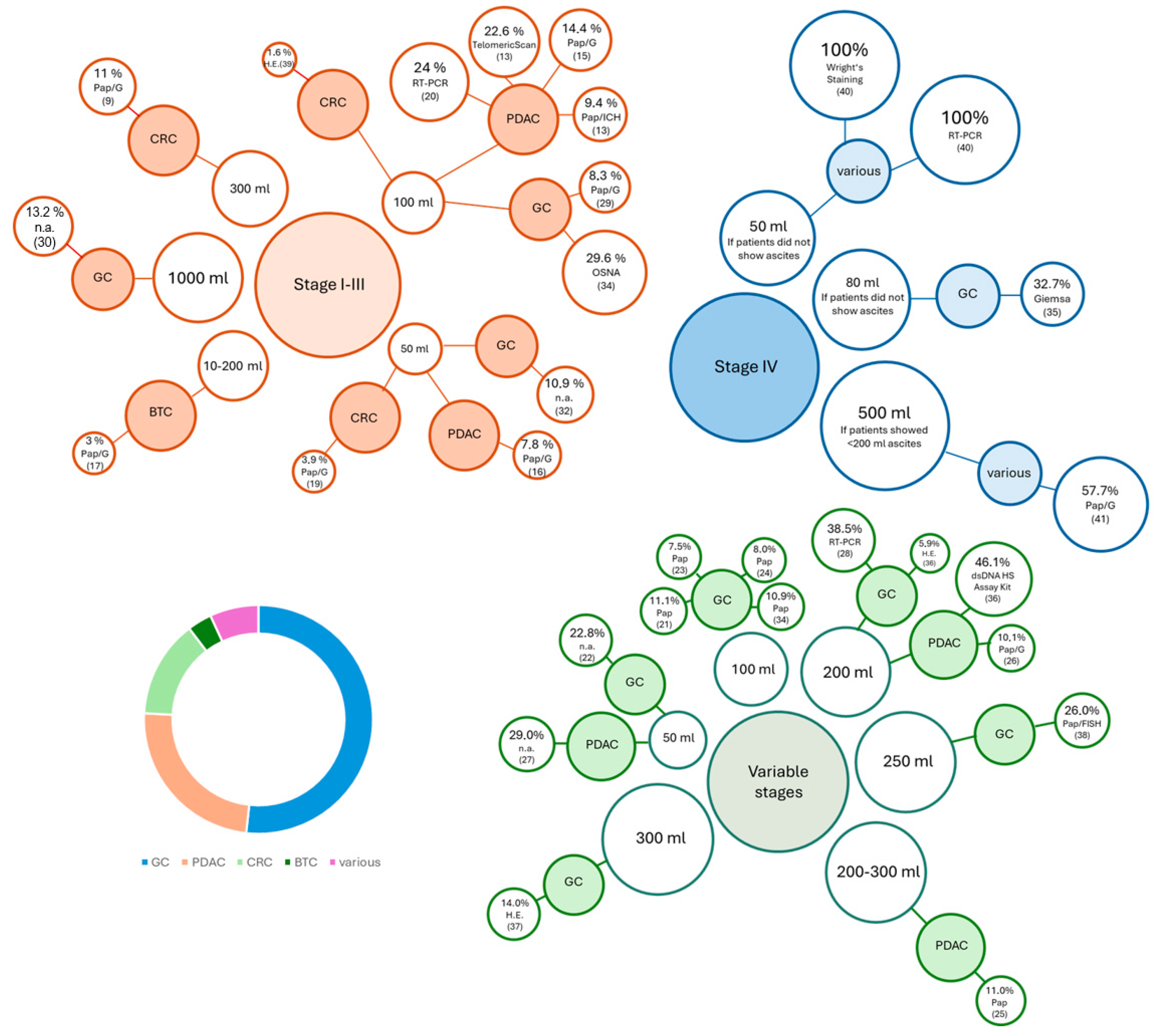
| Stage I–III | Median (Range) [%] |
| Conventional cytology (HE, Papanicolaou, Giemsa) | 8.1 (1.6–22.1) |
| Immunohistochemical staining (IHC) | 9.4 |
| RT-PCR (CEA) | 16.8 (9.5–24.0) |
| One-Step NucleicAcid Amplification (OSNA) | 29.6 |
| Telomeric Scan | 22.6 |
| Tumor entity | |
| Gastric (GC) | 16.0 (8.3–29.6) |
| Pancreatic (PDAC) | 11.9 (7.0–24.0) |
| Colorectal (CRC) | 3.9 (1.6–11.0) |
| Biliary tract (BTC) | 3 |
| Saline solution (mL) | |
| 50 | 7.8 (3.9–10.9) |
| 100 | 9.4 (1.6–29.6) |
| 300 | 11.0 |
| 1000 | 13.2 |
| Location of fluid application | |
| Tumor area | 16.6 (11.0–22.1) |
| Subhepatic space | 13.5 (3.0–24.0) |
| Ubiquitous | 8.3 (7.0–18.8) |
| Pelvic cavity | 7.8 (3.0–14.4) |
| Stage IV | median (range of detected tumor cells) [%] |
| Conventional cytology (H.E., Papanicolaou, Wright’s staining, Giemsa) | 57.7 (32.7–100) |
| RT-PCR | 100 |
| Tumor entity | |
| GC | 32.7 |
| Various | 100 (57.7–100) |
| Saline solution(mL) | |
| 50 | 100 |
| 80 | 32.7 |
| 500 | 57.7 |
| Location of fluid application | |
| Abdominal cavity | 78.9 (57.7–100) |
| Epigastrium | 32.7 |
| Tumor stage not specified | median (range) [%] |
| Conventional cytology (H.E., Papanicolaou, Giemsa) | 11.0 (10.1–26.0) |
| FISH | 26.0 |
| RT-PCR | 38.5 |
| dsDNA HS Assay | 46.1 |
| Tumor entity | |
| GC | 11.1 (7.5–38.5) |
| PDAC | 29.0 (11.0–46.1) |
| Saline solution (mL) | |
| 50 | 25.9 (22.8–29.0) |
| 200 | 38.5 (5.9–46.1) |
| 300 | 14 |
| Location of fluid application | |
| Pelvic cavity | 11.0 (7.5–38.5) |
| Ubiquitous | 25.0 (5.9–26) |
| Abdominal cavity, collected from Douglas | 46.1 |
| Article | Primary Tumor | N | n Positive (%) | Time of Lavage | Volume of Wash Fluid (Saline Solution) | Site of Lavage | Cell Detection Method |
|---|---|---|---|---|---|---|---|
| Cytology | |||||||
| Koganti et al. 2016, India [18] | GC | 60 | 5 (8.3%) | p-lap | 100 mL | subphrenic spaces, pelvic cavity | Papanicolaou and Giemsa |
| Ikoma et al. 2016, USA [19] | GC | 514 | 68 (13.2%) | n.a. | 1000 mL | n.a. | n.a. |
| Zhang et al. 2017, China [20] | GC | 136 | 30 (22.1%) | p-lap | n.a. | tumor and surrounding tissue | HE |
| Young et al. 2022, USA [21] | GC | 55 | 6 (10.9%) | n.a. | 50 mL | three abdominal quadrants | n.a. |
| Yin et al. 2024, China [22] | GC | 85 | 16 (18.8%) | p-lap | 500 mL | around transverse colon and stomach, collected around stomach, spleen and liver | microscope |
| Sakoda et al. 2021, Japan [3] | PDAC | 440 | 30 (7.0%) | p-lap | 100 mL | pelvic cavity | Papanicolaou and Giemsa |
| Mashiko et al. 2023, Japan [4] | PDAC | 97 | 14 (14.4%) | p-lap | 100 mL | abdominal cavity | Papanicolaou and Giemsa |
| Ishida et al. 2023, Japan [5] | PDAC | 231 | 18 (7.8%) | p-lap | 50 mL | pelvic cavity | Papanicolaou and Giemsa |
| Tanemura et al. 2024, Japan [6] | PDAC | 53 | 5 (9.4%) vs. 12 (22.6%) | p-lap | 100 mL | pelvic cavity | Papanicolaou/IHC vs. TelomericScan |
| Matsukuma et al. 2021, Japan [7] | BTC | 169 | 5 (3%) | p-lap | 10–200 mL | pelvic and/or subhepatic cavity | Papanicolaou and Giemsa |
| Salamanca et al. 2020, Spain [29] | CRC | 188 | 2 (1.6%) | p-lap/ p-lasc | 100 mL | n.a. | HE |
| Shalaby et al. 2021, Egypt [30] | CRC | 90 | 10 (11%) | p-lap | 300 mL | around the tumor, paracolic gutter | Papanicolaou and Giemsa |
| Ouchi et al. 2022, Japan [8] | CRC | 95 | 2 (2.1%) | prtr and ptr | n.a. | n.a. | n.a. |
| Kobayashi et al. 2024, Japan [9] | CRC | 1378 | 54 (3.9%) | prtr and ptr | 50 mL | pelvic cavity | Papanicolaou and Giemsa |
| mRNA | |||||||
| Takahashi et al. 2018, Japan (CEA) [28] | PDAC | 237 | 57 (24%) | p-lap | 100 mL | subhepatic space | RT-PCR |
| Gęca et al. 2021, Poland (CK19) [23] | GC | 27 | 7 (29.6%) | p-lap | 100 mL | n.a. | One-Step Nucleic Acid Amplification (OSNA) Assay |
| Ouchi et al. 2022, Japan (CEA) [8] | CRC | 95 | 9.50% | prtr and ptr | n.a. | n.a. | RT-PCR |
| Article | Primary Tumor | N | n Positive (%) | Time of Lavage | Volume of Wash Fluid (Saline Solution) | Site of Lavage | Cell Detection Method |
|---|---|---|---|---|---|---|---|
| Cytology | |||||||
| Hoskovec et al. 2017, Czech Republic [24] | GC | 49 | 16 (32.7%) | p-lap | 80 mL (if no ascites present) | epigastric area | Giemsa |
| Ji et al. 2016, China [31] | various | 50 | 50 (100%) | p-lap | 50 mL (if no ascites present) | abdominal cavity | Wright’s staining |
| Graversen el at. 2019, Denmark [32] | various | 26 | 15 (57.7%) | prtr | 500 mL (if patient had less than 200 mL of ascites) | abdominal cavity | Papanicolaou, Giemsa, HE |
| mRNA | |||||||
| Ji et al. 2016, China [31] | various | 50 | 50 (100%) | p-lap | 50 mL (if no ascites present) | abdominal cavity | RT-PCR |
| Article | Primary Tumor | N | n Positive (%) | Time of Lavage | Volume of Wash Fluid (Saline Solution) | Site of Lavage | Cell Detection Method |
|---|---|---|---|---|---|---|---|
| Cytology | |||||||
| Kano et al. 2015, Japan [11] | GC | 1039 | 116 (11.1%) | p-lap/ p-lasc | 100 mL | abdominal and pelvic cavity | Papanicolaou |
| Okabe et al. 2015, Japan [10] | GC | 123 | 28 (22.8%) | p-lasc | 50 mL | pelvic cavity and left subphrenic space | n.a. |
| Higaki et al. 2017, Japan [12] | GC | 911 | 68 (7.5%) | p-lap | 100 mL | pelvic cavity | Papanicolaou |
| Sakata et al. 2019, Japan [13] | GC | 973 | 78 (8.0%) | p-lap | 100 mL | right subdiaphragmal space, Douglas | Papanicolaou |
| Hasbahceci et al. 2018, Turkey [25] | GC | 34 | 2 (5.9%) | p-lap | 200 mL | ubiquitous | HE |
| Gęca et al. 2021, Poland [23] | GC | 55 | 6 (10.9%) | 1. p-lasc 2. p-lap 3. surgery | 100 mL | abdominal cavity | n.a. |
| Cieśla et al. 2021, Poland [26] | GC | 100 | 14 (14%) | p-lap/ p-lasc | 300 mL | cancer area | HE |
| Xuan et al. 2024, China [27] | GC | 50 | 13 (26.0%) | p-lasc | 250 mL | ubiquitous | Papanicolaou FISH |
| Hirabayashi et al. 2015 Japan [14] | PDAC | 162 | 18 (11%) | p-lap | 200–300 mL | pelvic cavity | Papanicolaou |
| Suenaga et al. 2021, Japan [15] | PDAC | 89 | 9 (10.1%) | p-lap | 200 mL | abdominal cavity, collected from Douglas pouch | Papanicolaou, Giemsa |
| Takadate et al. 2021, Japan [16] | PDAC | 146 | 42 (29.0%) | p-lasc | 50 mL | n.a. | n.a. |
| RT-PCR | |||||||
| Nakanishi et al. 2019, Japan [17] | GC | 187 | 72 (38.5%) | p-lap | 200 mL | pelvic cavity | RT-PCR |
| dsDNA HS Assay Kit | |||||||
| Suenaga et al. 2021, Japan [15] | PDAC | 89 | 41 (46.1%) | p-lap | 200 mL | abdominal cavity, collected from Douglas pouch | dsDNA HS Assay Kit |
Disclaimer/Publisher’s Note: The statements, opinions and data contained in all publications are solely those of the individual author(s) and contributor(s) and not of MDPI and/or the editor(s). MDPI and/or the editor(s) disclaim responsibility for any injury to people or property resulting from any ideas, methods, instructions or products referred to in the content. |
© 2026 by the authors. Licensee MDPI, Basel, Switzerland. This article is an open access article distributed under the terms and conditions of the Creative Commons Attribution (CC BY) license.
Share and Cite
Puffert, R.; Quarder, A.; Kockelmann, F.; Wirth, T.; Reineke-Plaaß, T.; Raap, M.; Schmelzle, M.; Feldbrügge, L.; Rau, B.; Köhler, F. Intraoperative Peritoneal Lavage for Detection of Malignant Cells: Technique, Evidence, Clinical Relevance and Future Perspectives. Cancers 2026, 18, 1604. https://doi.org/10.3390/cancers18101604
Puffert R, Quarder A, Kockelmann F, Wirth T, Reineke-Plaaß T, Raap M, Schmelzle M, Feldbrügge L, Rau B, Köhler F. Intraoperative Peritoneal Lavage for Detection of Malignant Cells: Technique, Evidence, Clinical Relevance and Future Perspectives. Cancers. 2026; 18(10):1604. https://doi.org/10.3390/cancers18101604
Chicago/Turabian StylePuffert, Resa, Anna Quarder, Fabian Kockelmann, Thomas Wirth, Tanja Reineke-Plaaß, Mieke Raap, Moritz Schmelzle, Linda Feldbrügge, Beate Rau, and Franziska Köhler. 2026. "Intraoperative Peritoneal Lavage for Detection of Malignant Cells: Technique, Evidence, Clinical Relevance and Future Perspectives" Cancers 18, no. 10: 1604. https://doi.org/10.3390/cancers18101604
APA StylePuffert, R., Quarder, A., Kockelmann, F., Wirth, T., Reineke-Plaaß, T., Raap, M., Schmelzle, M., Feldbrügge, L., Rau, B., & Köhler, F. (2026). Intraoperative Peritoneal Lavage for Detection of Malignant Cells: Technique, Evidence, Clinical Relevance and Future Perspectives. Cancers, 18(10), 1604. https://doi.org/10.3390/cancers18101604