Communication

Merkel Cell Polyomavirus and DNA Damage Response (DDR): Transcriptional Analysis of DDR Pathways in the Context of Merkel Cell Carcinoma


by
Sara Messina
1,†,
Domenico Mallardo
2,†,
Amedeo Ferlosio
3,
Lucia Festino
2,
Claudia Trojaniello
2,
Rossella Di Trolio
2,
Marco Ciotti
4,
Paolo Antonio Ascierto
2,5,‡,
Valeria Pietropaolo
1,*,‡ and
Sara Passerini
1,*
1
Department of Public Health and Infectious Diseases, Sapienza University of Rome, 00185 Rome, Italy
2
Unit of Melanoma, Cancer Immunotherapy and Development Therapeutics, Istituto Nazionale Tumori IRCCS “Fondazione G. Pascale”, 80131 Naples, Italy
3
Anatomic Pathology, Department of Biomedicine and Prevention, Tor Vergata University of Rome, 00133 Rome, Italy
4
Virology Unit, Polyclinic Tor Vergata Foundation, 00133 Rome, Italy
5
Dipartimento di Neuroscienze e Scienze Riproduttive ed Odontostomatologiche, Università degli Studi di Napoli “Federico II”, 80125 Naples, Italy
*
Authors to whom correspondence should be addressed.
These authors contributed equally to this work.
These authors also contributed equally to this work.
Cancers 2026, 18(10), 1592; https://doi.org/10.3390/cancers18101592
Submission received: 14 April 2026 / Revised: 6 May 2026 / Accepted: 12 May 2026 / Published: 14 May 2026
(This article belongs to the Special Issue The Role of Viruses in the Development of Cancer)

Simple Summary

Merkel cell carcinoma (MCC) is a highly aggressive cancer primarily caused by Merkel cell polyomavirus (MCPyV), which interferes with multiple cellular pathways, including the DNA Damage Response (DDR). The aim of this study was to analyze the presence and the molecular status of MCPyV in MCC samples and to assess the viral influence on the DDR pathway. We confirmed the viral etiology of MCC tumors and observed overexpression of several DDR genes including ATM, ATR, CHK1, CHK2, H2AX, Rad51, p53, and p21. Molecular differences between virus-positive and virus-negative MCCs could serve as diagnostic markers as well as allow to design specific therapeutic strategies for virus-positive MCC cancers.

Abstract

Background/Objectives: Merkel cell polyomavirus (MCPyV) is a ubiquitous virus strictly associated with Merkel cell carcinoma (MCC), a rare and aggressive skin cancer. MCPyV oncogenic properties are associated mainly with early protein expression, integration, and LT truncation. MCPyV can also interact with DNA Damage Response (DDR) mechanisms, contributing to oncogenesis and tumor progression. In this work, we investigated the correlation between MCPyV and MCC and evaluated the mRNA expression profiles of DDR genes in virus-positive and -negative tumors. Methods: A total of 19 formalin-fixed paraffin-embedded biopsies were acquired from patients diagnosed with MCC. After DNA and RNA extraction, the DNA was used for MCPyV detection via qPCR and for sequencing analysis of the early, late, and non-coding control viral regions and the extracted RNA was used for MCPyV transcripts, miRNA detection and for the evaluation of several DDR genes expression such as ATM, ATR, CHK1, CHK2, H2AX, Rad51, p53, and p21, in MCPyV-positive and -negative samples via reverse transcription, PCR, and qPCR. Results: MCPyV presence was detected in 11/19 samples, all characterized by viral integration, LT truncation, and early region expression only. Furthermore, higher mRNA levels of DDR genes were observed in MCPyV-positive tumors compared with the negative ones. Conclusions: Our findings support the role of MCPyV in MCC formation and suggest its involvement in the transcriptional regulation of DDR genes, which may influence tumor progression. Understanding the molecular interplay between MCPyV and the DDR may guide future research into plausible novel diagnostic and therapeutic strategies for virus-induced tumors.
Keywords:
MCPyV; MCC; DDR; ATM; ATR; p53

1. Introduction

The cellular system is exposed daily to endogenous and exogenous factors responsible for DNA lesions, which may compromise cellular functions [1]. In this framework, organisms have developed a complex machinery known as DNA Damage Response (DDR), involving several pathways that mediate cell cycle arrest to allow DNA repair or activate processes such as cell death or senescence, when the extent of the damage exceeds the cell’s repair capacity [2]. To overcome the wide variety of DNA lesions, cells have evolved multiple distinct DNA repair mechanisms [3]. Key components in DDR signaling are Ataxia-Telangiectasia Mutated (ATM) and Ataxia telangiectasia and Rad3-related (ATR), two protein kinases that recognize double-strand breaks (DSBs) and single-strand breaks (SSBs) respectively, and mediate the phosphorylation of several target proteins, including checkpoint kinase (CHK) 1, CHK2, and p53 [4,5]. Specifically, following DSBs, the MRN complex recruits ATM to the site of DNA damage through interaction with the NSB1 protein [6,7]. Once activated, ATM mediates phosphorylation of downstream proteins such as CHK2 and p53, leading to cell cycle arrest [3,7]. In parallel, ATR responds to SSBs and phosphorylates CHK1, which in turn phosphorylates several target proteins, many of them overlapping with CHK2 substrates [5]. For instance, p53 can be phosphorylated by either CHK1 or CHK2, thereby promoting its function as a transcription factor to induce downstream gene expression, including p21 [5].
Moreover, both activated ATM and ATR can phosphorylate the histone variant H2AX, whose phosphorylated form, γH2AX, is recognized as a general marker of DNA damage [8].
Since these pathways act collectively to preserve genomic integrity and stability, their alteration may lead to the accumulation of DNA lesions, promoting tumorigenesis [9]. Among external factors able to trigger DDR signaling, viral infections have also been recognized [1]. Notably, DDR processes are of particular interest in the case of oncogenic viruses, including Epstein–Barr virus [10], hepatitis B and C viruses [11] and Human Papillomavirus [12].
Another oncogenic virus involved in DDR manipulation is Merkel cell polyomavirus (MCPyV), a small DNA virus belonging to the Polyomaviridae family, first discovered in 2008 [13]. The viral genome is functionally divided into an early region encoding for Large T (LT), small T (sT) and 57 kT antigens (Ag) and alternative LT open reading frame (ALTO), involved in viral replication [14], and a late region encoding for structural proteins viral protein (VP) 1 and 2, and for two microRNAs (miRNAs), MCV-miR-M1-5p and MCV-miR-M1-3p, plausibly involved in the regulation of viral replication [15]. Interposed between the two coding regions is the Non-Coding Control Region (NCCR), which contains the origin of viral replication and bidirectional promoters for viral transcription [16,17].
MCPyV is widely diffused in the general population, mostly with asymptomatic infections [17,18]. However, it is also recognized as an oncogenic virus, due to its association with Merkel cell carcinoma (MCC), a rare and aggressive neuroendocrine skin cancer [13,19]. Approximately 80% of MCC cases are caused by MCPyV infection, while the remaining 20% are associated with ultraviolet (UV) exposure [20]. Specifically, virus-positive tumors exhibit clonal integration of viral DNA into the host genome [13], resulting in the expression of a truncated form of the LT protein (tLT), which loses the replicative function but maintains oncogenic properties, thus leading to increased cell proliferation and tumor development [21]. MCPyV oncogenic properties are strictly related to the capability of early proteins LT and sT to manipulate cellular processes by interacting with host proteins [22,23,24], including the DDR machinery [25]. Indeed, previous studies investigating the interplay between MCPyV and DDR revealed an activation of ATR/CHK1 and ATM/CHK2 pathways in MCPyV-positive cells, thereby promoting the phosphorylation and stabilization of the p53 protein, associated with cell cycle arrest and apoptosis [26]. Consistent with this hypothesis, Tsang et al. reported the accumulation of DDR components, including ATR, CHK1, γH2AX, and CHK2, in LT-positive nuclear foci [27]. Although some studies reported a modulation of DDR activity by LT alone, a contribution of sT in stimulating the DDR machinery has also been observed. Indeed, the expression of sT in MCPyV-positive MCC cells was found sufficient to induce the DDR signaling, leading to increased γH2AX levels and activation of the ATM pathway [28]. Notably, MCPyV may trigger DDR to promote viral replication, while also exploiting DDR activity to phosphorylate LT at Ser816, a modification associated with inhibition of cell proliferation [29]. Finally, our recent research reported overexpression of DDR genes, including ATM, ATR, CHK1, and CHK2, in MCPyV-positive MCC, suggesting a plausible role of the virus in manipulating DDR at the transcriptional level [30]. Considering these findings, our study aimed to investigate the expression profile of selected DDR genes in MCC tissues stratified by viral status.
This approach may help characterize MCPyV-associated differences in DDR gene transcriptional patterns and facilitate the identification of candidate therapeutic targets and diagnostic or prognostic biomarkers.

2. Materials and Methods

2.1. Sample Collection

Nineteen formalin-fixed paraffin-embedded (FFPE) primary tumor samples were collected from MCC patients. Specifically, ten cases (MCC1-10) (4 males, 6 females, mean age ± standard dev. 70 ± 7.58 years) were obtained from the unit of Melanoma, Cancer Immunotherapy and Development Therapeutics, Istituto Nazionale Tumori IRCCS Fondazione “G. Pascale” (Naples, Italy), and nine samples (MCC11-19) (9 males, mean age ± standard dev. 72 ± 12.85 years) were collected from the Dermatology Clinic of Tor Vergata University Hospital (Rome, Italy). All procedures were performed in accordance with the Declaration of Helsinki and were approved by the Ethics Committees of the Istituto Nazionale Tumori (Naples, Italy) (Protocol number: 32/22oss) and the Tor Vergata University (Rome, Italy) (Protocol number: 0015440/2019).

2.2. Histological Diagnosis

The histological diagnosis of MCC was established based on the observation of an expansive, nodular, or diffusely infiltrating tumor located in the dermis of subcutis, where small round blue cells with scant cytoplasm and a high nuclear/cytoplasmic ratio. Nuclei exhibit indistinct nucleoli and granular chromatin with a characteristic salt-and-pepper pattern (Figure 1). Furthermore, conspicuous mitoses and apoptotic bodies were observed. The examined tissues revealed positivity for the epithelial marker CK20 with a perinuclear dot-like pattern, CAM5.2, and pan-cytokeratins, as well as for neuroendocrine markers, including chromogranin A and synaptophysin. Negative results were observed for K7, TTF1, LCA, S100, and cdx2.

2.3. DNA Extraction

FFPE tissues were deparaffinized with xylene, and DNA was subsequently extracted using the Quick DNA FFPE Miniprep Kit (Zymo Research, Irvine, CA, USA). Total DNA was eluted in 50 µL and evaluated for suitability for polymerase chain reaction (PCR) by amplifying the β-globin gene sequences [31].

2.4. Detection of MCPyV DNA by Polymerase Chain Reaction (qPCR)

MCPyV detection and quantification were performed by quantitative PCR (qPCR) using primers for the sT sequence, as previously described [32]. Viral load was evaluated using standard curves generated from a ten-fold serial dilution of the pMCV-R17a plasmid (#24729, Addgene, Cambridge, MA, USA), which contains the complete viral genome, and results were expressed as copies per milliliter (copies/mL), based on the fixed elution volume used during extraction [32,33]. Each qPCR reaction included the plasmid pMCV-R17a as a positive control, and nuclease-free water was used as a negative control.

2.5. PCR for LTAg, VP1 and NCCR Regions and Sequencing

Samples that tested positive for MCPyV DNA were subsequently subjected to PCR targeting viral regions corresponding to LTAg (LT1, LT3), NCCR, and VP1 [13,31]. PCR assays included the pMCV-R17a as a positive control in each run, whereas nuclease-free water served as a negative control. PCR products were visualized under UV light through electrophoresis on a 2% agarose gel using SafeView reagent (Applied Biological Materials, Vancouver, BC, Canada). Furthermore, to detect NCCR and VP1 variation, the amplified products were purified using the miPCR purification kit (Metabion, Planegg, Germany) according to the manufacturer’s instructions and sequenced (Bio-Fab Research, Rome, Italy). The obtained VP1 and NCCR sequences were compared with the reference strain deposited in GeneBank, MCC350:EU375803. Then, sequence alignment was performed with Clustal W2 on the European Molecular Biology Laboratory–European Bioinformatics Institute (EMBL–EBI) website (http://www.ebi.ac.uk/Tools/msa/clustalw2/ accessed on 2 March 2026).

2.6. Sequencing Analysis of LTAg

For LTAg sequencing (151-3102 nucleotide position, GenBank strain EU375803), six primer sets were employed to perform PCR assays [31], followed by sequencing (Bio-Fab Research, Rome, Italy).

2.7. Analysis of Viral Integration Sites

MCPyV genome integration was observed using the detection of the integrated papilloma sequence (DIPS)–PCR technique, which allows the amplification and detection of the junction between viral and host DNA [33,34]. In detail, the eluted DNA was fragmented by the TaqI restriction enzyme, generating several fragments that were further ligated with enzyme-specific adaptors. The ligated fragment undergoes PCR amplification using primers that recognize viral and adaptor sequences. Sequencing analysis was conducted, and the integration sites were established using the Basic Local Alignment Search Tool (BLAST + 2.17.0) for genomic localization.

2.8. RNA Extraction and Analysis

Total RNA was extracted from FFPE biopsies with the FFPE RNA purification kit (Norgen, Thorold, ON, Canada) and eluted in 50 μL. RNA quality and concentration were assessed using spectrophotometric reading (Take3 module of the Microplate Reader BioTek SynergyHT, BioTek Instruments, Winooski, VT, USA). All samples met the predefined quality thresholds (A260/230 ≥ 1.8) and were included in analysis. Reverse transcription (RT) was performed using the SensiFAST cDNA Synthesis kit (Meridian Bioscience, Cincinnati, OH, USA) and cDNA quality was assessed through the amplification of β-globin sequences [31]. Therefore, an aliquot of cDNA was used as a template to analyze LTAg and VP1 gene expression by PCR with specific primers [31,33]. Each run included positive and negative controls. Moreover, no-RT controls were included to exclude genomic DNA contamination.

2.9. Viral miRNA Analysis

For MCPyV-encoded miRNA detection, the Applied Biosystems™ TaqMan™ MicroRNA Assays (Thermo Fisher Scientific, Waltham, MA, USA) were used. Specifically, an aliquot of cDNA was subjected to qPCR using mcv-miR-M1-5p (ID006356). Furthermore, RNU6B (ID001093) was selected as an endogenous control.

2.10. Detection of LT by Immunohistochemistry

Among the collected samples, 10 exhibited adequate material for the detection of MCPyV Large T antigen by immunohistochemistry on FFPE tissue sections. Staining was performed using the Leica BOND automated staining platform (Leica Biosystems, Wetzlar, Germany). Following antigen retrieval, sections were incubated with a mouse monoclonal antibody directed against MCPyV Large T antigen, clone CM2B4, applied at a dilution of 1:100. Immunoreactivity was visualized using a polymer-based detection system according to the manufacturer’s instructions. LT expression was assessed based on nuclear staining in tumor cells. Cases were considered positive when distinct nuclear immunoreactivity was observed in tumor cells, whereas a complete absence of nuclear staining was interpreted as negative.

2.11. Analysis of DDR Genes’ Expression

RT-qPCR was performed to measure relative levels of ATM, ATR, CHK1, CHK2, H2AX, Rad51, p53, and p21 transcripts [30,35,36,37]. The β-globin gene was used as a reference gene, selected based on its previously validated use in MCC tissues [30] and its consistent amplification across all analyzed specimens. mRNA relative levels were calculated by the comparative ΔCt method and reported as 2ΔCt.

2.12. Statistical Analysis

For statistical analysis, results from three independent experiments were expressed as the mean ± standard deviation. Both viral load and gene expression were tested using the Shapiro–Wilk test to assess normal distribution. The relative expression of DDR genes in MCPyV-positive and -negative groups was compared by Student’s t-test with Welch’s correction when necessary. p < 0.05 was considered statistically significant. Outlier analysis was also performed using the ROUT method (Q = 1%).

3. Results

3.1. Detection of MCPyV DNA by qPCR and PCR

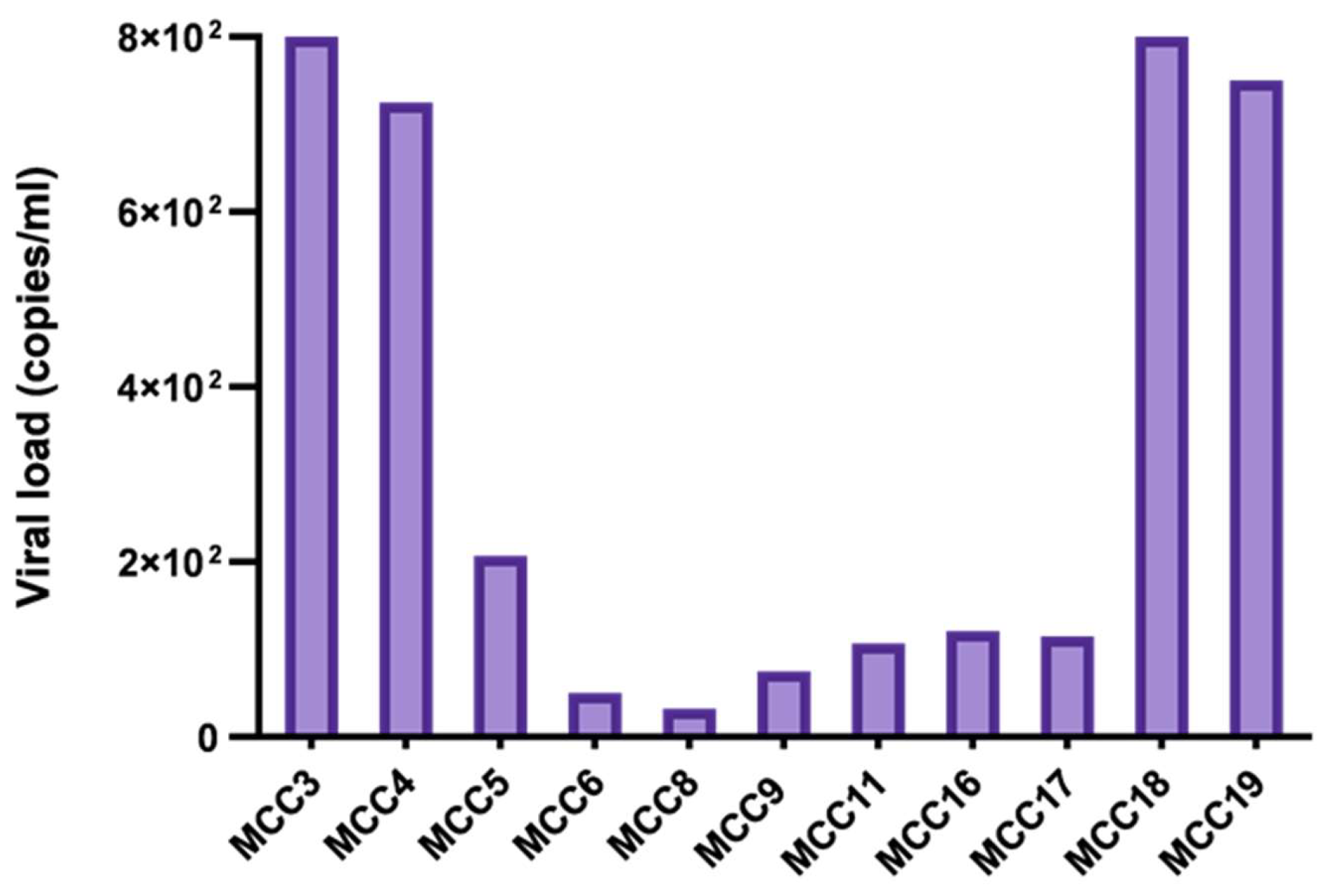
MCPyV DNA was detected in 11/19 (57.9%) patients, including 9/13 (69.2%) males and 2/6 (33.3%) females. All samples were primary tumors and displayed a mean viral load of 3.4 × 10² copies/mL (Figure 2, Table 1). The same samples also tested positive for LT1 and LT3, as well as for VP1 and NCCR amplification (Table 1).

3.2. Analysis of Viral Sequences

When analyzing NCCR and VP1 regions, canonical sequences were detected in all virus-positive samples, revealing a high degree of homology with the reference strain MCC350 (Table 1). Moreover, truncated LTAg were observed due to frameshift mutations generating stop codons, which preserve the pRb-binding motif LFCDE but eliminate the helicase domain (Table 1).

3.3. Viral Integration

Viral integration sites were identified in all virus-positive tumors. Specifically, viral junction sites varied from nuclear positions 1399 to 2952, whereas cellular junctions were mainly observed at chromosomes 5 and 6, except for two samples (MCC6 and MCC8), where integration was detected on chromosomes 4 and 1 (Table 1).

3.4. Expression of LT and VP1 Transcripts

MCC-positive tissues were further subjected to RT-PCR analysis for MCPyV LTAg and VP1 genes. In all samples, the LTAg expression has been noted, whereas the VP1 gene was not detected in any tumor (Table 2).

3.5. Viral miRNA Expression

RNA extracted from virus-positive MCCs was also tested for mcv-miR-M1-5p; however, none of the samples showed positivity to MCPyV-encoded miRNA (Table 2).

3.6. Detection of LT Protein

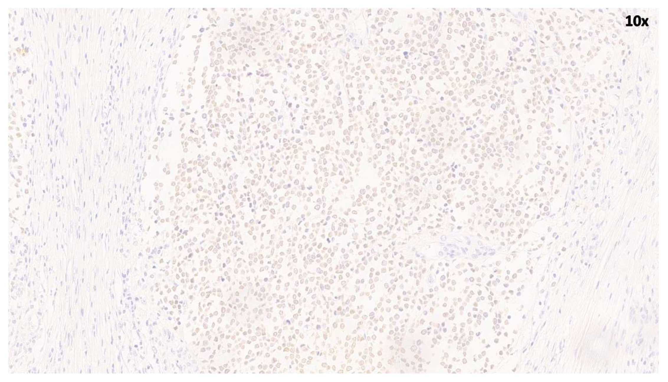
Immunohistochemical analysis was performed on 10 cases (MCC1–MCC10), including six PCR-positive and four PCR-negative cases. MCPyV LT nuclear expression was reported in five out of 10 MCC samples (Table 1). LT-positive cases showed strong and diffuse nuclear staining in tumor cells. In contrast, no immunoreactivity was observed in the remaining five MCPyV-negative samples, which were completely devoid of staining. These findings are consistent with the presence of MCPyV in a subset of MCC tumors (Figure 3).

3.7. Relative Expression of DDR Genes

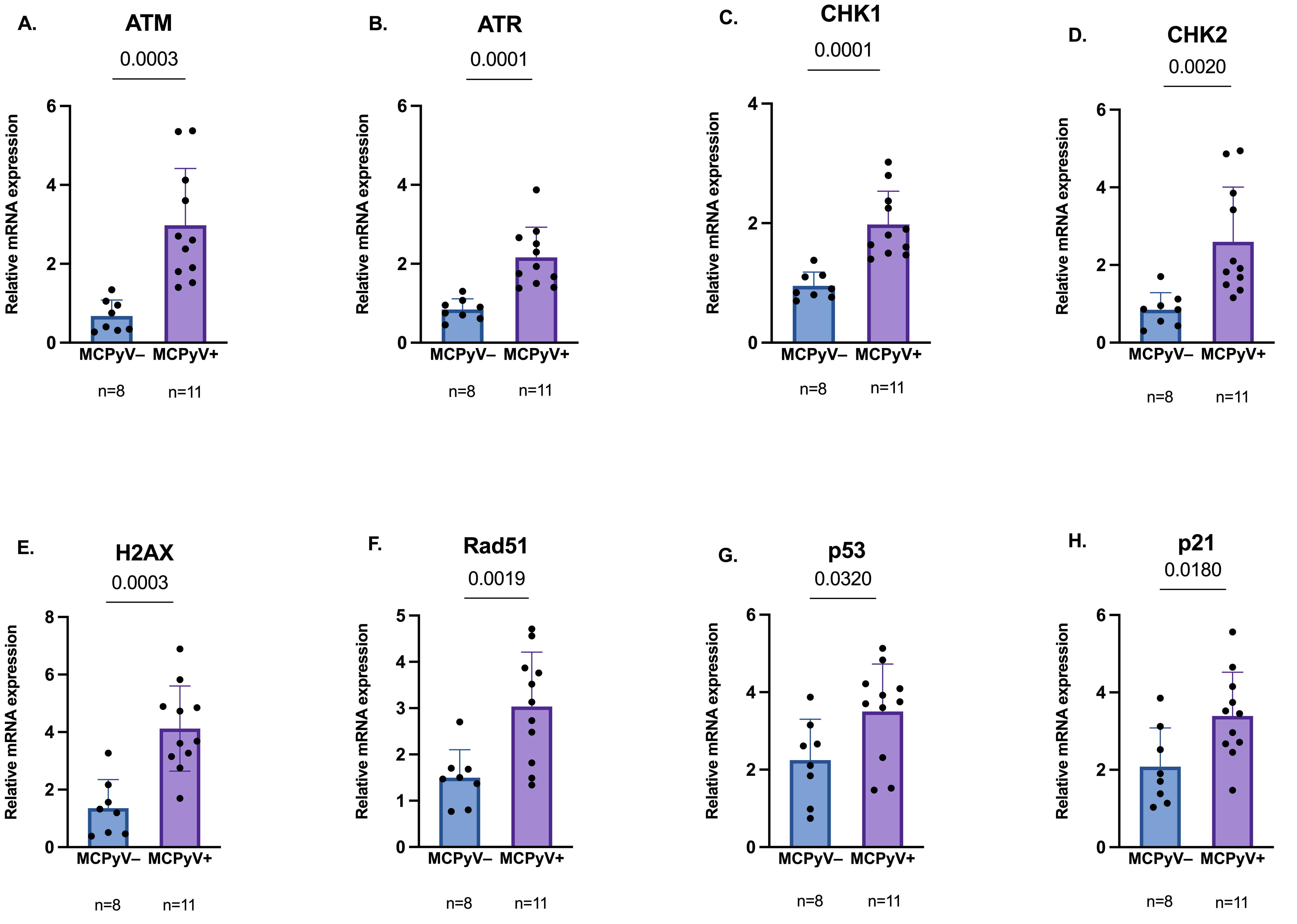
Among positive and negative MCC biopsies, a significant difference in expression levels of several DDR genes, such as H2AX, ATM, and ATR, and downstream effectors CHK1, CHK2, Rad51, p53, and p21 was observed (p < 0.05) (Figure 4).

4. Discussion

Host cells often incur DNA damage as a result of viral infection. Indeed, several viruses can induce genetic instability through DNA damage during viral replication. Moreover, some viruses can directly interfere with components of the DDR, an intricate network of proteins evolved to detect and repair DNA lesions [25,38]. Among them, MCPyV was also found to interact and regulate DDR proteins, likely promoting tolerance to viral replication in MCC [26,27,39]. Despite these observations, limited data from MCC cases do not yet establish a direct correlation between MCPyV and DDR manipulation.
In the current study, we investigated MCPyV prevalence in a cohort of MCC specimens obtained from two Italian hospitals, revealing MCPyV DNA in approximately 58% of samples and thus confirming the viral etiology of MCC formation [13]. Moreover, viral integration and LTAg truncation, as well as lack of late gene expression were reported, strongly supporting these molecular features as hallmarks of virus-mediated oncogenesis [13,19].
Specifically, numerous integration sites were detected without evidence of specific preferential sites in the MCPyV genome, whereas in the host genome, the long arm of chromosome 5 was confirmed as the most frequent localization [40,41]. Sequencing analysis showed canonical NCCR and VP1, reinforcing that mutations within these regions may not be involved in tumorigenesis [14,33].
Consistent with previous research [33], viral miRNAs were not detectable, leaving their biological relevance in cancer still unclear [42]. However, their absence, likely due to MCPyV miRNAs being expressed during the lytic infection together with late genes, combined with the lack of VP1 expression [15], indicates that viral integration disrupts transcription of the late region [17].
Once viral prevalence and molecular state in MCC specimens were assessed, the current study aimed to explore the DDR pathway at the RNA level. Consistent with our previous research suggesting a virus-mediated transcriptional activation of DDR [30], MCC tumors displayed distinct expression profiles of DDR genes, depending on viral status, with MCPyV-positive samples reporting higher ATM, ATR, CHK1 and CHK2 mRNA levels compared to negative tissues. Furthermore, an overexpression of H2AX, p53, p21 and Rad51 was reported, reinforcing the possibility that viral infection may induce changes in DDR components already at the RNA level [30,43]. The observed upregulation of ATM and ATR, alongside their downstream effectors CHK2 and CHK1, suggests that MCPyV induces multiple forms of genomic stress, mainly due to viral interference with host DNA replication and to DNA damage caused by viral integration [30]. Moreover, the concurrent activation of CHK1 and CHK2 is consistent with a plausible role of the virus in manipulating cell cycle checkpoints to maintain the host cell in a state amenable to viral replication [27,44]. Therefore, like other oncogenic viruses, MCPyV infection may induce replication stress and DDR, but simultaneously exploits this signaling to favor its own replication [45]. This fine-tuning between DDR activation and checkpoint modulation may facilitate viral replication and persistence, which can lead to genomic instability and contribute to tumorigenesis [30,46].
In addition, the overexpression of H2AX mRNA may be linked with increased DNA repair in virus-positive tumors [47]. Indeed, although γH2AX is typically discussed as a post-translational marker of DNA damage, higher H2AX mRNA levels may reflect chronic DDR activation upon viral infection, consistent with in vitro studies showing H2AX phosphorylation and ATM activation by MCPyV sT [28]. Elevated Rad51 expression further suggests enhanced homologous recombination repair in MCPyV-positive MCC [46]. Similar observations have been reported in HPV-associated malignancies, where increased Rad51 mRNA levels were detected [48].
In MCPyV-positive MCC, increased expression of p53 and p21 may reflect an adaptive response to DNA damage rather than effective tumor suppression [21]. In contrast, MCPyV-negative tumors, frequently driven by UV-induced mutation signatures and TP53 inactivation [49], may lack the coordinated activation of DDR pathways. However, despite reaching statistical significance, differences in p53 and p21 expression may have limited biological relevance given the similar distribution patterns in virus-positive and virus-negative MCCs.
Notably, these observations are limited to transcriptional changes and do not necessarily imply functional activation of the DDR signaling in vivo.
Beyond transcriptional regulation, epigenetic mechanisms may contribute to the sustained high expression of DDR genes in MCPyV-positive tumors. Consistent with this hypothesis, Cheng and colleagues reported the role of sT in binding to L-MYC to recruit the EP400 histone acetyltransferase and chromatin remodeling complex [50]. In addition, differential methylation patterns were observed between virus-positive and virus-negative MCC, suggesting that viral status may influence genome methylation [51,52]. Moreover, recent evidence suggests that MCC oncogenesis involves virus-induced changes in chromatin state and histone methylation, potentially facilitating persistent DDR gene transcription and modifying checkpoint pathways [53]. Therefore, viral components may interact with and recruit chromatin remodeling complexes, which could contribute to epigenetic modulation of DDR components. However, further studies investigating the methylation status of DDR genes are needed to confirm this hypothesis, which remains speculative.
Despite providing novel insights into the differential expression of DDR genes in MCPyV-positive and -negative MCC, our study has several limitations. The analysis based on RNA expression data may not fully reflect protein abundance or functional activity of DDR components, particularly in the case of post-translational modifications such as phosphorylation of downstream kinases CHK1 and CHK2 or γH2AX foci formation.
Moreover, the study is observational and cannot establish causality between viral status and DDR activation. Finally, the limited sample size, reflecting the rarity of MCC and the lack of a control group, including normal adjacent skin from patients and healthy controls, may not fully represent DDR modulation across tumors.
Overall, our findings showed an overexpression of DDR genes in virus-positive tumors. Since DDR genes, including ATM, CHK2, Rad51 and H2AX, were highlighted as potential markers for predicting treatment response in human tumors [36,47,54], monitoring the expression levels of DDR genes may be a candidate transcriptional signature for MCC tumors; however, any prognostic or predictive relevance remains speculative in the absence of patient outcome and requires further validation in a clinically annotated cohort.

5. Conclusions

In conclusion, the current hypothesis-generating study reports transcriptional upregulation of DDR genes in MCPyV-positive MCC tumors, suggesting viral modulation of host repair mechanisms. In light of these primarily descriptive results, additional research, including protein analysis, functional assays and clinical correlation, will be essential to validate these observations and to further explore their clinical relevance in this aggressive malignancy.

Author Contributions

Conceptualization, S.M., V.P. and S.P.; methodology, S.P. and V.P.; samples collection and resources, D.M., A.F., M.C. and P.A.A.; validation, S.P. and V.P.; formal analysis, S.P., D.M. and V.P.; investigation, S.M., D.M., A.F. and S.P.; data curation, S.P. and V.P.; writing—original draft preparation, S.M., V.P. and S.P.; writing—review and editing, S.M., D.M., A.F., L.F., C.T., R.D.T. and M.C.; P.A.A., V.P. and S.P.; supervision, S.P. and V.P.; funding acquisition, V.P. and P.A.A. All authors have read and agreed to the published version of the manuscript.

Funding

This research was funded by a grant to Valeria Pietropaolo from the Italian Ministry of University and Research (MIUR) Research Grant (RP124190255A2CB7 and RP125199A48F33A6). This work was partially supported by the Italian Ministry of Health Ricerca Corrente Funds-Linea 2/17.

Institutional Review Board Statement

This research study was performed in line with the principles of the Declaration of Helsinki. Approval was granted by the institutional review board (University Hospital Tor Vergata, Rome, Italy), the Ethics Committee of the University Hospital Tor Vergata (Rome, Italy), protocol number 0015440/2019, 1 July 2019 and the Ethics Committee of IRCCS Pascale (Naples, Italy), protocol number 32/22 oss, 9 November 2022.

Informed Consent Statement

Informed consent was obtained from all subjects involved in the study.

Data Availability Statement

Data is contained within the article.

Conflicts of Interest

The authors declare no conflicts of interest.

Abbreviations

The following abbreviations are used in this manuscript:
DDRDNA damage response 
ATMataxia-telangiectasia mutated 
ATRataxia telangiectasia and Rad3-related 
DSBdouble-strand break 
SSBssingle-strand break 
CHKcheckpoint kinase 
MCPyVMerkel cell polyomavirus 
LTlarge T 
sTsmall T 
Agantigen
ALTOalternative LT open reading frame 
VPviral protein 
miRNAmicroRNA
NCCRnon-coding control region 
MCCMerkel cell carcinoma
UVultraviolet
tLTtruncated LT 
Serserine
FFPEformalin-fixed paraffin-embedded
PCRpolymerase chain reaction 
qPCRquantitative PCR 
DIPSdetection of the integrated papilloma sequence 
RTreverse-transcribed 

References

  1. Katerji, M.; Duerksen-Hughes, P.J. DNA damage in cancer development: Special implications in viral oncogenesis. Am. J. Cancer Res. 2021, 11, 3956–3979. [Google Scholar]
  2. Clay, D.E.; Fox, D.T. DNA Damage Responses during the Cell Cycle: Insights from Model Organisms and Beyond. Genes 2021, 12, 1882. [Google Scholar] [CrossRef] [PubMed]
  3. Jackson, S.P.; Bartek, J. The DNA-damage response in human biology and disease. Nature 2009, 461, 1071–1078. [Google Scholar] [CrossRef]
  4. Helt, C.E.; Wang, W.; Keng, P.C.; Bambara, R.A. Evidence that DNA damage detection machinery participates in DNA repair. Cell Cycle 2005, 4, 529–532. [Google Scholar] [CrossRef]
  5. Blackford, A.N.; Jackson, S.P. ATM, ATR, and DNA-PK: The Trinity at the Heart of the DNA Damage Response. Mol. Cell 2017, 66, 801–817. [Google Scholar] [CrossRef]
  6. Lee, J.H.; Paull, T.T. ATM activation by DNA double-strand breaks through the Mre11-Rad50-Nbs1 complex. Science 2005, 308, 551–554. [Google Scholar] [CrossRef] [PubMed]
  7. McCarthy-Leo, C.; Darwiche, F.; Tainsky, M.A. DNA Repair Mechanisms, Protein Interactions and Therapeutic Targeting of the MRN Complex. Cancers 2022, 14, 5278. [Google Scholar] [CrossRef] [PubMed]
  8. Curtin, N.J. DNA repair dysregulation from cancer driver to therapeutic target. Nat. Rev. Cancer 2012, 12, 801–817. [Google Scholar] [CrossRef]
  9. O’Connor, M.J. Targeting the DNA Damage Response in Cancer. Mol. Cell 2015, 60, 547–560. [Google Scholar] [CrossRef]
  10. Gruhne, B.; Sompallae, R.; Masucci, M.G. Three Epstein-Barr virus latency proteins independently promote genomic instability by inducing DNA damage, inhibiting DNA repair and inactivating cell cycle checkpoints. Oncogene 2009, 28, 3997–4008. [Google Scholar] [CrossRef]
  11. Ariumi, Y.; Kuroki, M.; Dansako, H.; Abe, K.; Ikeda, M.; Wakita, T.; Kato, N. The DNA damage sensors ataxia-telangiectasia mutated kinase and checkpoint kinase 2 are required for hepatitis C virus RNA replication. J. Virol. 2008, 82, 9639–9646. [Google Scholar] [CrossRef] [PubMed]
  12. Gillespie, K.A.; Mehta, K.P.; Laimins, L.A.; Moody, C.A. Human papillomaviruses recruit cellular DNA repair and homologous recombination factors to viral replication centers. J. Virol. 2012, 86, 9520–9526. [Google Scholar] [CrossRef]
  13. Feng, H.; Shuda, M.; Chang, Y.; Moore, P.S. Clonal Integration of a Polyomavirus in Human Merkel Cell Carcinoma. Science 2008, 319, 1096–1100. [Google Scholar] [CrossRef] [PubMed]
  14. Pietropaolo, V.; Prezioso, C.; Moens, U. Merkel Cell Polyomavirus and Merkel Cell Carcinoma. Cancers 2020, 12, 1774. [Google Scholar] [CrossRef]
  15. Seo, G.J.; Chen, C.J.; Sullivan, C.S. Merkel cell polyomavirus encodes a microRNA with the ability to autoregulate viral gene expression. Virology 2009, 383, 183–187. [Google Scholar] [CrossRef]
  16. Liu, W.; You, J. Molecular Mechanisms of Merkel Cell Polyomavirus Transformation and Replication. Annu. Rev. Virol. 2020, 7, 289–307. [Google Scholar] [CrossRef] [PubMed]
  17. Yang, J.F.; Liu, W.; You, J. Characterization of molecular mechanisms driving Merkel cell polyomavirus oncogene transcription and tumorigenic potential. PLoS Pathog. 2023, 19, e1011598. [Google Scholar] [CrossRef]
  18. Bopp, L.; Wieland, U.; Hellmich, M.; Kreuter, A.; Pfister, H.; Silling, S. Natural History of Cutaneous Human Polyomavirus Infection in Healthy Individuals. Front. Microbiol. 2021, 12, 740947. [Google Scholar] [CrossRef]
  19. Ahmed, M.M.; Cushman, C.H.; DeCaprio, J.A. Merkel Cell Polyomavirus: Oncogenesis in a Stable Genome. Viruses 2021, 14, 58. [Google Scholar] [CrossRef]
  20. Tello, T.L.; Coggshall, K.; Yom, S.S.; Yu, S.S. Merkel cell carcinoma: An update and review: Current and future therapy. J. Am. Acad. Dermatol. 2018, 78, 445–454. [Google Scholar] [CrossRef]
  21. Myrda, J.; Bremm, F.; Schaft, N.; Dörrie, J. The Role of the Large T Antigen in the Molecular Pathogenesis of Merkel Cell Carc noma. Genes 2024, 15, 1127. [Google Scholar] [CrossRef] [PubMed]
  22. Shuda, M.; Kwun, H.J.; Feng, H.; Chang, Y.; Moore, P.S. Human Merkel cell polyomavirus small T antigen is an oncoprotein targeting the 4E-BP1 translation regulator. J. Clin. Investig. 2011, 121, 3623–3634. [Google Scholar] [CrossRef]
  23. An, P.; Sáenz Robles, M.T.; Pipas, J.M. Large T antigens of polyomaviruses: Amazing molecular machines. Annu. Rev. Microbiol. 2012, 66, 213–236. [Google Scholar] [CrossRef]
  24. Topalis, D.; Andrei, G.; Snoeck, R. The large tumor antigen: A “Swiss Army knife” protein possessing the functions required for the polyomavirus life cycle. Antivir. Res. 2013, 97, 122–136. [Google Scholar] [CrossRef] [PubMed]
  25. Studstill, C.J.; Mac, M.; Moody, C.A. Interplay between the DNA damage response and the life cycle of DNA tumor viruses. Tumour Virus Res. 2023, 16, 200272. [Google Scholar] [CrossRef]
  26. Li, J.; Wang, X.; Diaz, J.; Tsang, S.H.; Buck, C.B.; You, J. Merkel cell polyomavirus large T antigen disrupts host genomic integrity and inhibits cellular proliferation. J. Virol. 2013, 87, 9173–9188. [Google Scholar] [CrossRef]
  27. Tsang, S.H.; Wang, X.; Li, J.; Buck, C.B.; You, J. Host DNA damage response factors localize to merkel cell polyomavirus DNA replication sites to support efficient viral DNA replication. J. Virol. 2014, 88, 3285–3297. [Google Scholar] [CrossRef]
  28. Wu, J.H.; Narayanan, D.; Limmer, A.L.; Simonette, R.A.; Rady, P.L.; Tyring, S.K. Merkel Cell Polyomavirus Small T Antigen Induces DNA Damage Response. Intervirology 2019, 62, 96–100. [Google Scholar] [CrossRef]
  29. Li, J.; Diaz, J.; Wang, X.; Tsang, S.H.; You, J. Phosphorylation of Merkel cell polyomavirus large tumor antigen at serine 816 by ATM kinase induces apoptosis in host cells. J. Biol. Chem. 2015, 290, 1874–1884. [Google Scholar] [CrossRef]
  30. Passerini, S.; Fracella, M.; Ferlosio, A.; Moens, U.; Scagnolari, C.; Antonelli, G.; Ciotti, M.; Pietropaolo, V. Investigation of mRNA expression levels of DNA damage response genes in Merkel Cell Polyomavirus-positive Merkel Cell Carcinoma: A pilot study. Discov. Oncol. 2025, 16, 852. [Google Scholar] [CrossRef] [PubMed]
  31. Hashida, Y.; Imajoh, M.; Nemoto, Y.; Kamioka, M.; Taniguchi, A.; Taguchi, T.; Kume, M.; Orihashi, K.; Daibata, M. Detection of Merkel cell polyomavirus with a tumour-specific signature in non-small cell lung cancer. Br. J. Cancer 2013, 108, 629–637. [Google Scholar] [CrossRef]
  32. Passerini, S.; Babini, G.; Merenda, E.; Carletti, R.; Scribano, D.; Rosa, L.; Conte, A.L.; Moens, U.; Ottolenghi, L.; Romeo, U.; et al. Merkel Cell Polyomavirus in the Context of Oral Squamous Cell Carcinoma and Oral Potentially Malignant Disorders. Biomedicines 2024, 12, 709. [Google Scholar] [CrossRef]
  33. Passerini, S.; Prezioso, C.; Babini, G.; Ferlosio, A.; Cosio, T.; Campione, E.; Moens, U.; Ciotti, M.; Pietropaolo, V. Detection of Merkel cell polyomavirus (MCPyV) DNA and transcripts in Merkel cell carcinoma (MCC). Pathogens 2023, 12, 894. [Google Scholar] [CrossRef]
  34. Sastre-Garau, X.; Peter, M.; Avril, M.F.; Laude, H.; Couturier, J.; Rozenberg, F.; Almeida, A.; Boitier, F.; Carlotti, A.; Couturaud, B.; et al. Merkel cell carcinoma of the skin: Pathological and molecular evidence for a causative role of MCV in oncogenesis. J. Pathol. 2009, 218, 48–56. [Google Scholar] [CrossRef]
  35. Chappell, W.H.; Gautam, D.; Ok, S.T.; Johnson, B.A.; Anacker, D.C.; Moody, C.A. Homologous Recombination Repair Factors Rad51 and BRCA1 Are Necessary for Productive Replication of Human Papillomavirus 31. J. Virol. 2015, 90, 2639–2652. [Google Scholar] [CrossRef] [PubMed]
  36. Miser-Salihoglu, E.; Demokan, S.; Karanlik, H.; Karahalil, B.; Önder, S.; Cömert, S.; Yardim-Akaydin, S. Investigation of mRNA Expression Levels of Tip60 and Related DNA Repair Genes in Molecular Subtypes of Breast Cancer. Clin. Breast Cancer 2023, 23, 125–134. [Google Scholar] [CrossRef] [PubMed]
  37. Kang, D.; Sung, J.Y.; Hwang, H.J.; Baek, Y.; Kim, M.J.; Lim, G.E.; Kim, Y.N.; Cha, J.H.; Lee, J.S. Splicing factor SF3B4 acts as a switch in cancer cell senescence by regulating p21 mRNA stability. Cancer Lett. 2025, 615, 217530. [Google Scholar] [CrossRef]
  38. Ryan, E.L.; Hollingworth, R.; Grand, R.J. Activation of the DNA Damage Response by RNA Viruses. Biomolecules 2016, 6, 2. [Google Scholar] [CrossRef] [PubMed]
  39. Becker, J.C.; Lei, K.C. Is MCPyV-Driven DNA Damage Response the Achilles’ Heel of Merkel Cell Carcinoma? J. Investig. Dermatol. 2025, 145, 225–227. [Google Scholar] [CrossRef]
  40. Martel-Jantin, C.; Filippone, C.; Cassar, O.; Peter, M.; Tomasic, G.; Vielh, P.; Brière, J.; Petrella, T.; Aubriot-Lorton, M.H.; Mortier, L.; et al. Genetic variability and integration of Merkel cell polyomavirus in Merkel cell carcinoma. Virology 2012, 426, 134–142. [Google Scholar] [CrossRef] [PubMed]
  41. Arora, R.; Choi, J.E.; Harms, P.W.; Chandrani, P. Merkel Cell Polyomavirus in Merkel Cell Carcinoma: Integration Sites and Involvement of the KMT2D Tumor Suppressor Gene. Viruses 2020, 12, 966. [Google Scholar] [CrossRef]
  42. Konstatinell, A.; Coucheron, D.H.; Sveinbjørnsson, B.; Moens, U. MicroRNAs as Potential Biomarkers in Merkel Cell Carcinoma. Int. J. Mol. Sci. 2018, 19, 1873. [Google Scholar] [CrossRef]
  43. Passerini, S.; De Angelis, M.; Messina, S.; Scribano, D.; Ambrosi, C.; Moens, U.; Nencioni, L.; Pietropaolo, V. Respiratory Models Reveal DNA Damage Response Modulation by Merkel Cell Polyomavirus. Int. J. Mol. Sci. 2026, 27, 3449. [Google Scholar] [CrossRef]
  44. Chaurushiya, M.S.; Weitzman, M.D. Viral manipulation of DNA repair and cell cycle checkpoints. DNA Repair 2009, 8, 1166–1176. [Google Scholar] [CrossRef] [PubMed]
  45. Collins, C.D.; Stefely, M.; Mehta, K.P.M.; Spurgeon, M.E. Viruses and the host replisome: Discovering oncogenic mechanisms of small DNA tumor viruses. J. Virol. 2026, 100, e0169125. [Google Scholar] [CrossRef] [PubMed]
  46. Hollingworth, R.; Grand, R.J. Modulation of DNA Damage and Repair Pathways by Human Tumour Viruses. Viruses 2015, 7, 3201–3203, Correction in Viruses 2015, 7, 2542–2591. [Google Scholar] [CrossRef]
  47. Katsuta, E.; Sawant Dessai, A.; Ebos, J.M.; Yan, L.; Ouchi, T.; Takabe, K. H2AX mRNA expression reflects DNA repair, cell proliferation, metastasis, and worse survival in breast cancer. Am. J. Cancer Res. 2022, 12, 793–804. [Google Scholar]
  48. Lindemann, A.; Patel, A.A.; Tang, L.; Tanaka, N.; Gleber-Netto, F.O.; Bartels, M.D.; Wang, L.; McGrail, D.J.; Lin, S.Y.; Frank, S.J.; et al. Combined Inhibition of Rad51 and Wee1 Enhances Cell Killing in HNSCC Through Induction of Apoptosis Associated With Excessive DNA Damage and Replication Stress. Mol. Cancer Ther. 2021, 20, 1257–1269. [Google Scholar] [CrossRef]
  49. Goh, G.; Walradt, T.; Markarov, V.; Blom, A.; Riaz, N.; Doumani, R.; Stafstrom, K.; Moshiri, A.; Yelistratova, L.; Levinsohn, J.; et al. Mutational landscape of MCPyV-positive and MCPyV-negative Merkel cell carcinomas with implications for immunotherapy. Oncotarget 2016, 7, 3403–3415. [Google Scholar] [CrossRef]
  50. Cheng, J.; Park, D.E.; Berrios, C.; White, E.A.; Arora, R.; Yoon, R.; Branigan, T.; Xiao, T.; Westerling, T.; Federation, A.; et al. Merkel cell polyomavirus recruits MYCL to the EP400 complex to promote oncogenesis. PLoS Pathog. 2017, 13, e1006668. [Google Scholar] [CrossRef] [PubMed]
  51. Harms, P.W.; Verhaegen, M.E.; Vo, J.N.; Tien, J.C.; Pratt, D.; Su, F.; Dhanasekaran, S.M.; Cao, X.; Mangelberger, D.; VanGoor, J.; et al. Viral Status Predicts the Patterns of Genome Methylation and Decitabine Response in Merkel Cell Carcinoma. J. Investig. Dermatol. 2022, 142, 641–652. [Google Scholar] [CrossRef] [PubMed]
  52. Rotondo, J.C.; Mazziotta, C.; Lanzillotti, C.; Tognon, M.; Martini, F. Epigenetic Dysregulations in Merkel Cell Polyomavirus-Driven Merkel Cell Carcinoma. Int. J. Mol. Sci. 2021, 22, 11464. [Google Scholar] [CrossRef]
  53. Bachiri, K.; Kantar, D.; Laurent, E.M.N.; Gaboriaud, P.; Durand, L.; Drouin, A.; Chollot, M.; Schrama, D.; Houben, R.; Kervarrec, T.; et al. DNA Damage Stress Control Is a Truncated Large T Antigen and Euchromatic Histone Lysine Methyltransferase 2-Dependent Central Feature of Merkel Cell Carcinoma. J. Investig. Dermatol. 2025, 145, 400–410.e4. [Google Scholar] [CrossRef]
  54. Wu, R.; Patel, A.; Tokumaru, Y.; Asaoka, M.; Oshi, M.; Yan, L.; Ishikawa, T.; Takabe, K. High RAD51 gene expression is associated with aggressive biology and with poor survival in breast cancer. Breast Cancer Res. Treat. 2022, 193, 49–63. [Google Scholar] [CrossRef] [PubMed]
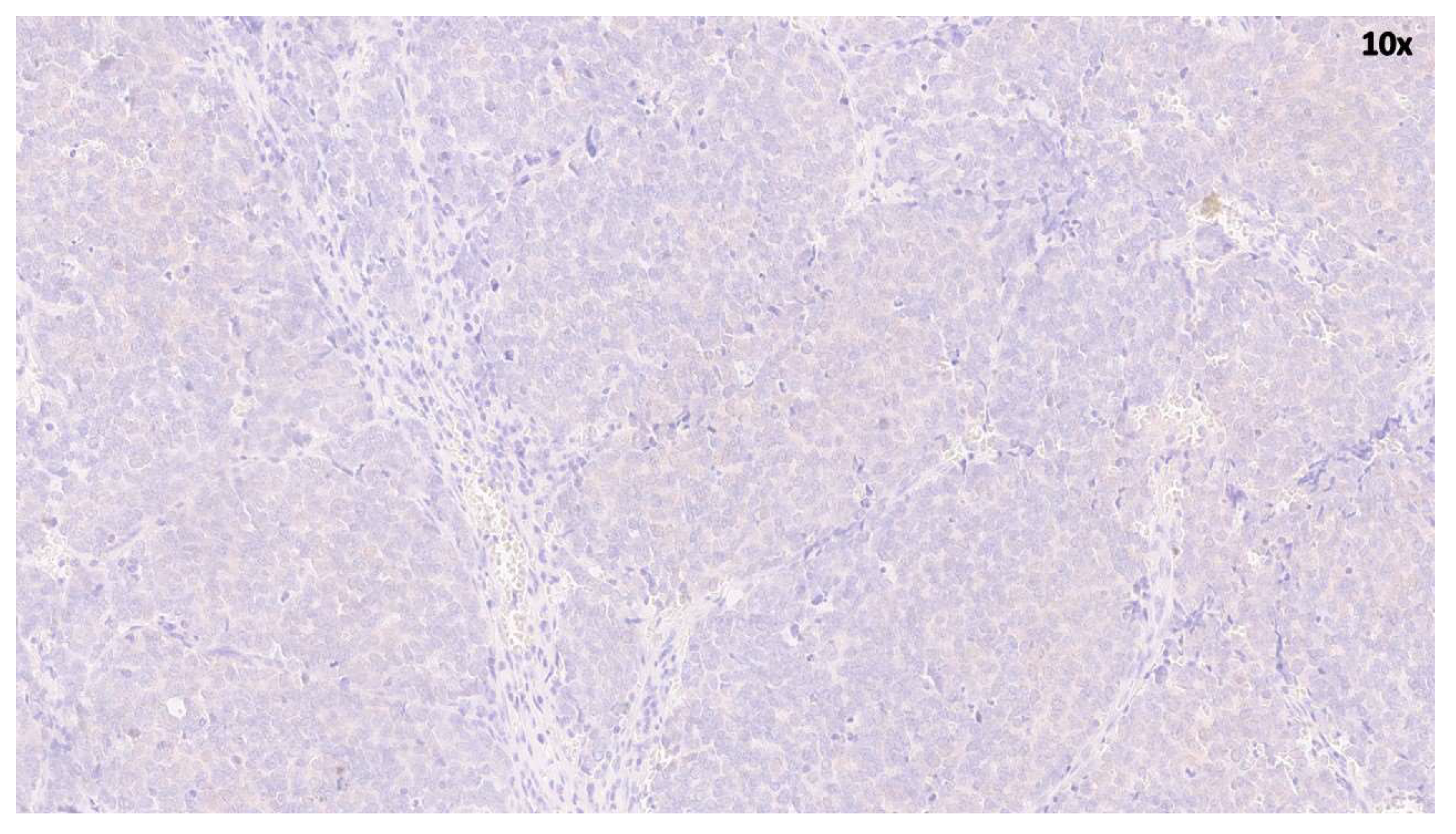
Figure 1. Low magnification (×10) of MCC section stained with hematoxylin and eosin. Typical features of MCC are evident, including the high nuclear/cytosol ratio and hyperchromatic nuclei.
Cancers 18 01592 g001
Figure 2. MCPyV load in virus-positive tissues from MCC patients.
Cancers 18 01592 g002
Figure 3. Immunohistochemical analysis of MCC showing nuclear positivity for MCPyV.
Cancers 18 01592 g003
Figure 4. Relative expression of DDR genes in MCPyV-positive (n = 11) and -negative (n = 8) MCC samples (Panel (A), ATM; Panel (B), ATR; Panel (C), CHK1; Panel (D), CHK2; Panel (E), H2AX; Panel (F), Rad51; Panel (G), p53; Panel (H), p21). Gene expression values exhibited a normal distribution (Shapiro–Wilk test; p > 0.05) and mean values of positive and negative MCCs groups were compared using an independent t-test with Welch’s correction.
Cancers 18 01592 g004
Table 1. Summary of MCPyV viral load and molecular state in virus-positive MCCs.
Sample IDSexViral LoadLTAgNCCRVP1LTAgIntegration SitesIHC
(Copies/mL)LT1LT3Viral JunctionCellular JunctionLT
MCC3F8 × 102++canonicalcanonicaltruncated1952-3′6p22.3+
MCC4M7.25 × 102++canonicalcanonicaltruncated2234-3′6p12.3+
MCC5M2.07 × 102++canonicalcanonicaltruncated1776-3′6q24.1+
MCC6F5 × 101++canonicalcanonicaltruncated2351-3′4p15.2
MCC8M3.2 × 101++canonicalcanonicaltruncated1522-3′1p36.2+
MCC9M7.5 × 101++canonicalcanonicaltruncated2952-3′5q35.1+
MCC11M1.07 × 102++canonicalcanonicaltruncated2597-3′5q11.2NA
MCC16M1.21 × 102++canonicalcanonicaltruncated1399-3′5q11.2NA
MCC17M1.15 × 102++canonicalcanonicaltruncated2843-3′5q11.2NA
MCC18M8 × 102++canonicalcanonicaltruncated5′-27385q23.1NA
MCC19M7.5 × 102++canonicalcanonicaltruncated5′-25975q11.2NA
NA: not available.
Table 2. MCPyV LTAg and VP1 transcripts and miRNAs in MCC cases.
Sample IDLTAgVP1miR-5p
MCC3+
MCC4+
MCC5+
MCC6+
MCC8+
MCC9+
MCC11+
MCC16+
MCC17+
MCC18+
MCC19+
Disclaimer/Publisher’s Note: The statements, opinions and data contained in all publications are solely those of the individual author(s) and contributor(s) and not of MDPI and/or the editor(s). MDPI and/or the editor(s) disclaim responsibility for any injury to people or property resulting from any ideas, methods, instructions or products referred to in the content.