Article

Clinical Outcomes and Prognostic Factors for Extramammary Paget’s Disease Treated with Radiation Therapy: A Multi-Institutional Observational Study

1
Department of Radiology, Graduate School of Medical Sciences, Nagoya City University, 1 Kawasumi, Mizuho-cho, Mizuho-ku, Nagoya 467-8601, Japan
2
Department of Radiation Oncology, Kitasato University Hospital, 1-15-1 Kitasato, Minami-ku, Sagamihara 252-0375, Japan
3
Department of Radiology, Aichi Medical University Hospital, 1-1 Yazako-Karimata, Nagakute 480-1195, Japan
4
Department of Radiation Oncology, Gifu University Hospital, 1-1 Yanagido, Gifu 500-1194, Japan
5
Department of Radiation Oncology, Ichinomiya Municipal Hospital, 2-2-22, Bunkyo, Ichinomiya 491-8558, Japan
6
Department of Radiation Oncology, National Hospital Organization Nagoya Medical Center, 4-1-1, Sannomaru, Nakaku, Nagoya 460-0001, Japan
7
Department of Radiology, Okazaki City Hospital, 3-1, Kouryuuji-cho Gosyoai, Okazaki 444-8553, Japan
8
Department of Geriatric and Environmental Dermatology, Nagoya City University Graduate School of Medical Sciences, 1 Kawasumi, Mizuho-cho, Mizuho-ku, Nagoya 467-8601, Japan
*
Author to whom correspondence should be addressed.
Cancers 2025, 17(9), 1507; https://doi.org/10.3390/cancers17091507
Submission received: 14 March 2025 / Revised: 24 April 2025 / Accepted: 28 April 2025 / Published: 29 April 2025
(This article belongs to the Section Clinical Research of Cancer)

Simple Summary

Extramammary Paget’s disease (EMPD) is a rare skin cancer that usually affects older adults and appears in the genital and perianal regions. This study analyzed the outcomes of radiation therapy in 81 patients with EMPD. The three-year local control, progression-free survival, and overall survival rates were 75%, 52%, and 80%, respectively. The results showed that lymph node metastasis, not having surgery, and using local radiation only without treating nearby lymph nodes were linked to worse outcomes. Side effects were generally mild, with only one patient experiencing moderate skin infection and another having severe lymphedema, while no very severe side effects occurred. These findings suggest that surgery and treating nearby lymph nodes in addition to radiation may improve outcomes for EMPD patients. Further research is needed to determine the best treatment approach, especially for older patients with poor prognostic factors.

Abstract

Background: Extramammary Paget’s disease (EMPD) is a rare cutaneous carcinoma that typically affects the elderly and is frequently observed in genital and perianal regions. We analyzed the outcomes and prognostic factors for EMPD after radiation therapy (RT). Methods: We analyzed data from 81 patients with non-metastatic EMPD who received either RT alone or in combination with surgery and/or chemotherapy. The median radiation dose was 56 Gy in 28 fractions. Local control (LC), progression-free survival (PFS), and overall survival (OS) rates were calculated using the Kaplan–Meier method. Multivariate analyses were performed using the Cox proportional hazards model. Late adverse events were evaluated by NCI-CTCAE version 5.0. Results: The median age was 78 years, and the median follow-up period was 36 months. The three-year LC, PFS, and OS rates were 75%, 52%, and 80%, respectively. Multivariate analyses identified the presence of lymph node (LN) metastasis, the absence of surgery, and the omission of elective nodal irradiation (i.e., local irradiation only) as significant factors for unfavorable LC (p = 0.01, 0.02, and 0.006) and PFS (p = 0.001, 0.04, and 0.03). LN metastasis was also a significant factor for unfavorable OS (p = 0.005). One patient developed grade 2 skin infection, and another developed grade 3 lymphedema; no grade 4 or higher toxicity was observed. Conclusions: The present results revealed prognostic factors for EMPD after RT and suggest that the absence of surgery and omission of elective nodal irradiation worsened outcomes. A prospective study is needed to establish an optimal treatment strategy for this rare malignancy, which is common in the elderly.

1. Introduction

Extramammary Paget’s disease (EMPD) is a rare cutaneous adenocarcinoma that primarily targets the genital and perianal regions, along with other areas rich in apocrine glands [1,2]. While Paget’s disease is almost exclusively diagnosed in women, with male cases accounting for less than 1% and occasionally linked to prostate cancer, invasive EMPD affects both sexes, but is more common in women [3]. Clinically, EMPD lesions frequently present with invasive redness accompanied by crusts and scaling, which sometimes resemble other skin conditions, such as eczema [4,5]. The majority of EMPD cases are typically identified as carcinoma in situ, and disease progression is generally slow. However, when Paget cells deeply infiltrate the dermis, regional lymph node (LN) metastases and distant metastases often develop and the prognosis of these patients is poor [6,7].
Although surgical excision remains the main curative approach for EMPD, its feasibility can be constrained in elderly patients, those with significant comorbidities, or individuals reluctant to undergo surgical interventions [7,8,9]. In such cases, radiation therapy (RT) is considered a viable alternative; however, clinical outcomes related to RT are underreported due to the limited number of patients receiving definitive RT [8,10]. Moreover, despite advancements in clinical outcomes for various cancers through the utilization of modern RT techniques, including intensity-modulated radiation therapy (IMRT) [11,12,13], there is a scarcity of studies documenting the application of these advanced techniques in EMPD management. Consequently, the current study aims to investigate the clinical outcomes of non-metastatic EMPD treated with RT, either as monotherapy or in conjunction with surgery and/or chemotherapy, while also identifying prognostic factors that could inform optimal treatment strategies.

2. Materials and Methods

2.1. Patient Selection and Pretreatment Evaluation

This retrospective, observational study was conducted with approval from the Institutional Review Board of our facility (approval number: 60-23-0073) and 10 other participating centers. The study followed the ethical guidelines set forth in the Declaration of Helsinki (1964) and its subsequent revisions. As this was a retrospective analysis, patient consent was acquired through an opt-out system available on the institutional website. We examined patients diagnosed with EMPD who received radiotherapy (RT), either as monotherapy or in combination with surgery and/or chemotherapy, between 2004 and 2022. Since this was a retrospective multi-institutional study, there were no strict criteria for selecting patients for RT. In general, RT was considered for patients with residual disease after surgery, those who were not surgical candidates due to advanced age or other factors. The inclusion criteria were as follows: (1) histopathologically confirmed EMPD; (2) the primary site of the lesion located outside of the breast; and (3) no evidence of distant metastasis. A total of 81 non-metastatic EMPD patients were enrolled in the study.
Clinical staging was determined based on physical examination findings and imaging studies, including computed tomography (CT) scans of the head, neck, chest, and upper abdomen. Additionally, magnetic resonance imaging (MRI) and 18F-fluorodeoxyglucose positron emission tomography (PET)/CT were performed on 6 (7%) and 18 (22%) patients, respectively. Tumor thickness was assessed based on biopsy findings and imaging studies, such as CT and MRI. Although a TNM classification system for EMPD was proposed by Ohara et al. in 2016, it was not uniformly applied in this study, as many patients were diagnosed prior to its publication [10].

2.2. RT

Excluding the 30 patients who received treatment exclusively with electron beams, 51 patients underwent CT-based radiation planning, utilizing thermoformed masks or body vacuum bag systems to ensure stable positioning during the treatment. The gross tumor volume (GTV) was defined based on clinical findings and imaging results, including CT, MRI, and/or PET/CT. For 79 patients with visible skin lesions, markers were placed around the lesion by dermatologists and radiation oncologists before performing the CT scan to improve the accuracy of GTV identification. In two other patients who underwent primary lesion resection only, the GTV was defined by lymph node metastases. Among the 47 patients (58%) who received localized irradiation, the clinical target volume (CTV) was extended up to 5 cm radially from the GTV margins. For the 34 patients (42%) who underwent elective nodal irradiation, the CTV also included the elective nodal region in addition to the CTV used for localized irradiation. Elective nodal irradiation was defined as RT that included regional LN areas based on the anatomical location of the primary lesion. For lesions in the genital, perianal or inguinal region, the irradiated nodal areas were selected from the common, internal, and external iliac, presacral, obturator, and/or inguinal regions at the discretion of the treating physician. In contrast, local irradiation was defined as RT limited to the primary lesion, without prophylactic coverage of regional LN. A circumferential margin of 3–5 mm was added around the CTV to account for positional and technical uncertainties, resulting in the planning target volume (PTV). Of these patients, 51 (63%) were treated with intensity-modulated radiation therapy (IMRT) or three-dimensional conformal radiation therapy (3DCRT), while 30 patients (37%) received electron beam therapy exclusively. IMRT plans were designed to create conformal isodose distributions that minimized exposure to adjacent normal tissues, such as the bowel, sigmoid colon, rectum, and bladder. X-ray therapy was delivered using 6–10 MV beams, combined with three-dimensional treatment planning based on tumor extent and depth. Electron beam therapy was administered with a single field using 6–12 MeV electrons. In some cases, a bolus material with a thickness of 0.5–1 cm was applied to the skin surface around the gross tumor or postoperative tumor bed to ensure adequate surface dose coverage. The median radiation dose was 56 Gy, delivered over 28 fractions (30–69 Gy in 10–37 fractions). The median dose per fraction was 2.0 Gy (1.8–3.0 Gy). Three patients received a radiation dose of 30 Gy. One of them underwent palliative irradiation and was transferred to another facility shortly after completing RT. The remaining two patients were treated at the same institution in 2005–2006 with a dose of 30 Gy. The median equivalent dose in 2 Gy fractions (EQD2) with an α/β = 10 was 56 Gy (30–71 Gy). The median radiation dose in patients who did not undergo surgery was 60 Gy (range, 30–65 Gy), while in patients who underwent surgery, the median dose was 52.3 Gy (range, 30–69 Gy).

2.3. Combination Therapy

A total of 30 patients (37%) received RT as the sole treatment, while 39 patients (48%) were treated with a combination of RT and surgery. Seven patients (9%) underwent RT in conjunction with chemotherapy, and five patients (6%) received a combination of RT, surgery, and chemotherapy.
In terms of chemotherapy regimens, six patients were administered fluorouracil and cisplatin, three received docetaxel, one was treated with fluorouracil, one with etoposide, and one with tegafur–gimeracil–oteracil potassium. Out of the 44 patients (54%) who underwent surgery, 26 patients (32%) had positive surgical margins, whereas 18 patients (22%) had negative margins. Additionally, three patients (4%) received RT before surgery, and 41 patients (51%) underwent postoperative RT.
Sentinel lymph node biopsy for EMPD has only recently been covered by national insurance; therefore, it was not performed in any of the cases included in this study.

2.4. Follow-Up Evaluation and Statistical Analysis

In the first year following RT, clinical assessments were performed every 2 to 3 months, and CT scans of the abdomen and pelvis were conducted at intervals of at least 4 months. From the second year onwards, CT imaging was performed every 6 to 12 months. PET/CT scans were utilized when there was a suspicion of recurrence. Tumor responses were evaluated using the Response Evaluation Criteria in Solid Tumors (RECIST) [8], categorizing responses as complete response (CR), partial response (PR), progressive disease (PD), or stable disease (SD).
Local control (LC), progression-free survival (PFS), and overall survival (OS) rates were estimated using the Kaplan–Meier method. LC was defined as the proportion of patients without local recurrence after the start of RT, with local recurrence referring to tumor regrowth within the primary site or the emergence of new lesions in the genital or perianal regions. PFS was defined as the proportion of patients without any recurrence or death from the initiation of RT, with follow up ending on the date of the last event-free observation. OS was defined as the proportion of surviving patients from the start of RT, irrespective of the cause of death, until the final follow up. To assess the prognostic relevance of variables, univariate analysis was performed using the Log-rank test, and multivariate analysis was conducted with the Cox proportional hazards model. Key potential prognostic factors were selected based on prior multivariate analyses, considering patient numbers and survival outcome [6,9,10,11,12,13]. All statistical evaluations were carried out using EZR (Version 1.55; Saitama Medical Center, Jichi Medical University, Japan), a graphical interface for R (The R Foundation for Statistical Computing, Vienna, Austria) [14]. Statistical significance was defined as p < 0.05. RT-related adverse events were graded according to the National Cancer Institute Common Terminology Criteria for Adverse Events (NCI-CTCAE), version 5.0.

3. Results

3.1. Patient and Treatment Characteristics

Table 1 shows the characteristics of patients and treatments. Ten patients in this study had recurrent disease after surgery, while the majority had primary disease. Twenty-one patients (26%) had LN metastases, while none had distant metastases. Clinical positive lymph node metastasis was defined as lymph nodes with a short-axis diameter of ≥10 mm on CT imaging. Of these 21 patients, 7 received local irradiation, while 14 received elective nodal irradiation. Elective nodal irradiation encompassed the prophylactic regions surrounding the primary site, including the pelvic and inguinal areas. For the patient with an axillary primary tumor, irradiation was targeted to the axillary region. Sixteen patients (20%) had a tumor ≥ 10 cm, 23 (28%) had a tumor thickness ≥ 4 mm. The cutoff values for tumor diameter and thickness were determined based on previous literature [9,10].
A Chi-square test was performed to evaluate the association between chemotherapy and tumor size (<10 cm vs. ≥10 cm), tumor thickness (<4 mm vs. ≥4 mm), and LN metastasis (no vs. yes). The analysis revealed no association between chemotherapy and tumor size (p = 0.99) or tumor thickness (p = 0.73). On the other hand, chemotherapy was primarily administered to patients with LN metastasis, resulting in a significant association (p < 0.001) and indicating a clear bias in these cases. Additionally, the chemotherapy regimens were not standardized. Therefore, univariate and multivariate analyses regarding chemotherapy were not performed.

3.2. Tumor Responses
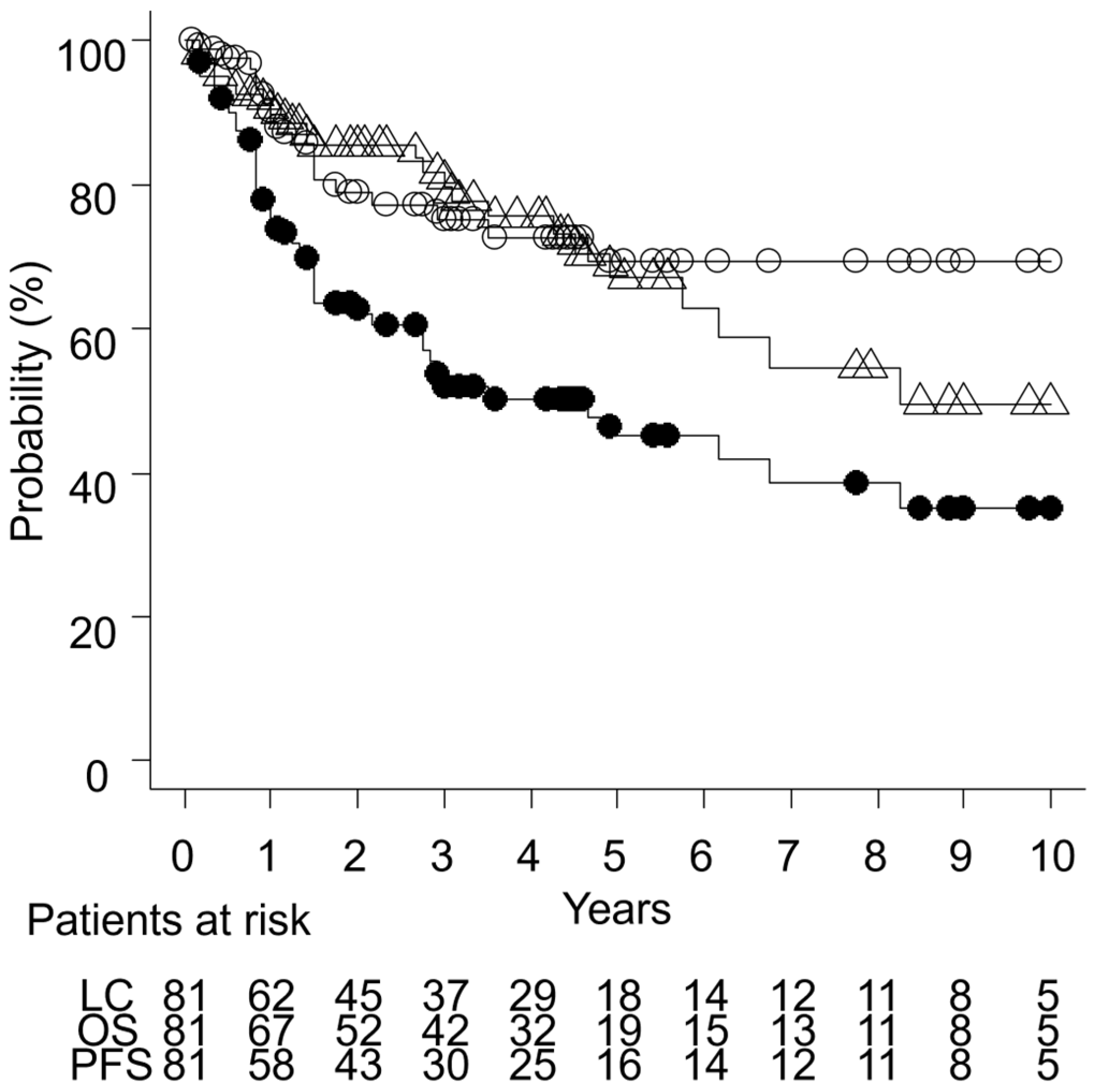
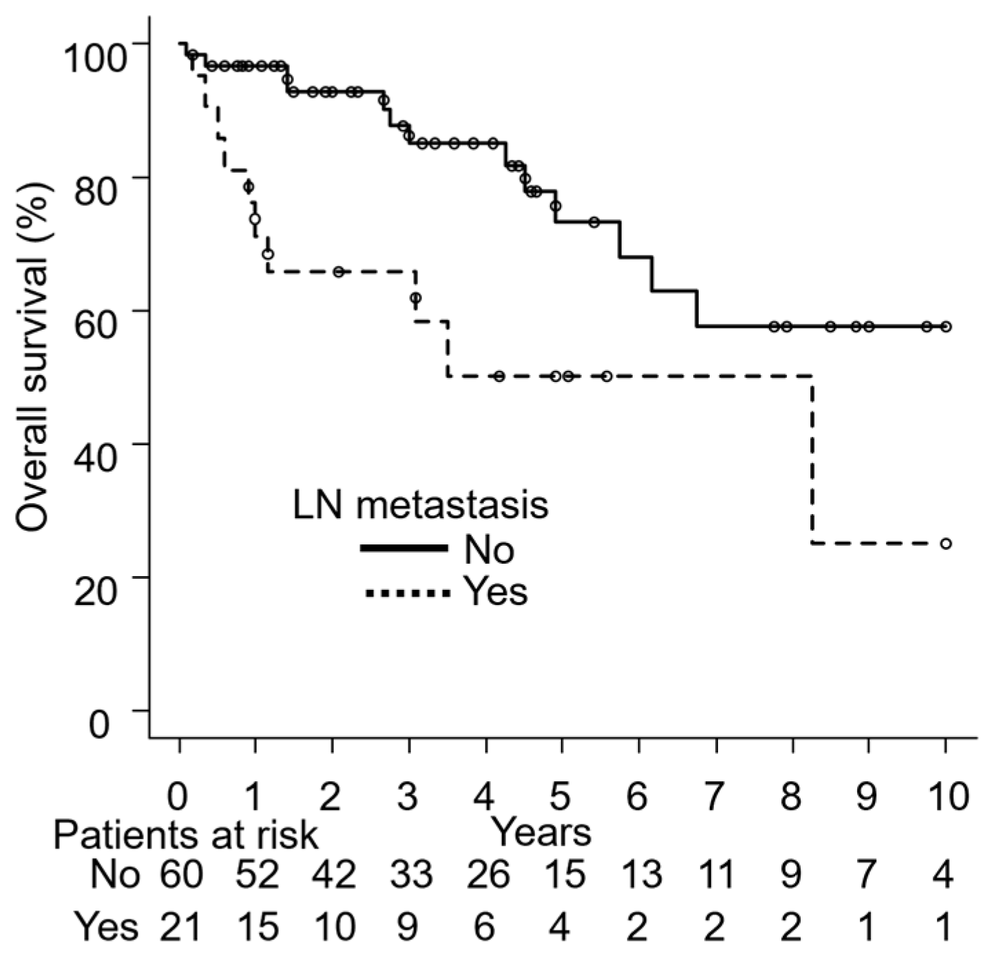
Tumor responses were not assessable in one patient (1%) due to missing data, as well as in 41 patients (51%) who had received postoperative RT. In the remaining 39 patients (48%), tumor responses were evaluated at the time of maximal tumor reduction within 1 to 3 months following RT. Among these patients, 21 (54%) achieved CR, 16 (41%) PR, and 2 (5%) exhibited PD. The response rate, calculated as the sum of CR and PR, was 95%. Both patients with PD showed progression of the primary tumor.

3.3. Outcomes

The median follow-up duration was 36 months (1–168 months), with a median follow-up of 38 months (2–168 months) for surviving patients. The three-year LC, PFS, and OS rates were 75% (95% confidence interval [CI]: 62–84%), 52% (95% CI: 40–63%), and 80% (95% CI: 68–88%), respectively, as illustrated in Figure 1.
At the time of the analysis, 23 patients (28%) had died, 12 (15%) from EMPD progression, 9 (11%) from unrelated causes (including four from lung cancer, hepatocellular carcinoma, cholangiocarcinoma, and rectal cancer, respectively, while the cause of death was unknown in the remaining five), and the cause of death was undetermined in 2 patients (2%). Local recurrence occurred in 18 patients (22%), with a median time to recurrence of 14 months (2–56 months) post-RT. Among the 37 patients who did not undergo surgery, 10 experienced local recurrence. Additionally, 12 patients (15%) developed regional LN metastasis, with a median time to onset of 17 months (9–35 months) following RT. Local recurrence was defined as tumor regrowth within the primary site. Regional LN metastasis was defined as disease progression within the elective nodal region. Distant metastasis was observed in 20 patients (25%), with the median time to onset being 17 months (2–50 months). The most common initial site of distant metastasis was the bone (9 cases), followed by the lungs (8 cases) and the liver (5 cases). Detailed information on initial distant metastasis sites is provided in Supplementary Table S1.
Moreover, seven patients (9%) developed secondary cancers after RT. Specific cases included one occurrence each of lung, breast, bile duct, kidney, and bladder cancer, along with two cases of colorectal cancer. The median time to secondary cancer onset was 29 months (6–134 months) post-RT.

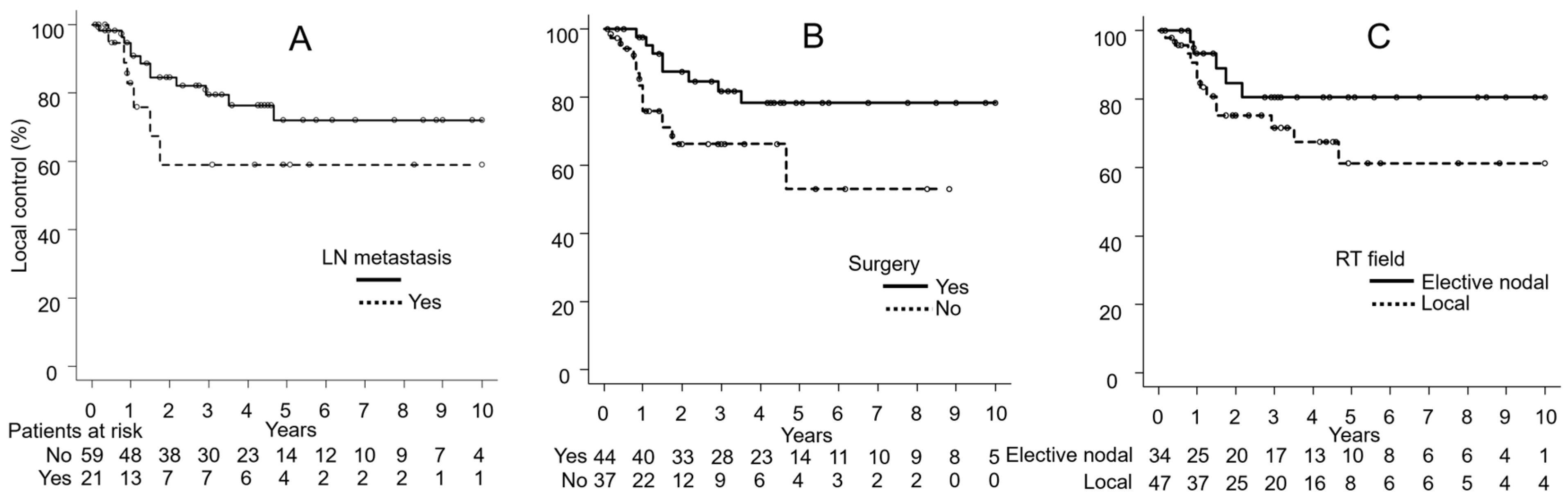
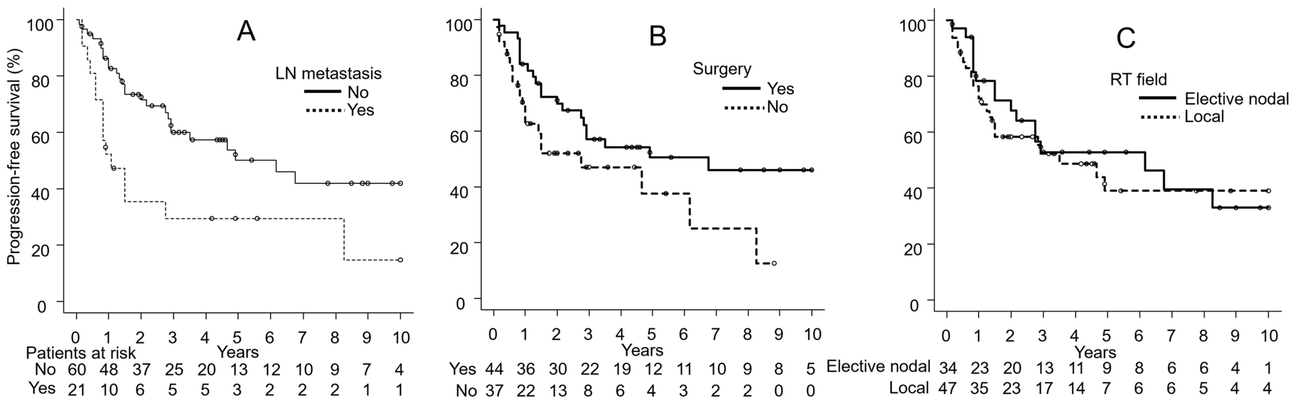
3.4. Univariate Analyses

Table 2 shows the results of the log-rank test for LC, PFS, and OS. The absence of surgery was identified as a significant factor for unfavorable LC. The LC rate was lower in patients treated without surgery than in those treated with surgery (p = 0.04). The LC rate was slightly lower in the electron beam group than in the IMRT or 3DCRT group (p = 0.06).
The presence of LN metastasis and the absence of surgery were significant factors for unfavorable PFS. The PFS rate was lower in patients with LN metastasis than in those without LN metastasis (p = 0.005). The PFS rate was slightly lower in patients treated without surgery than in those treated with surgery (p = 0.051).
The presence of LN metastasis was identified as a significant factor for unfavorable OS. The OS rate was lower in patients with LN metastasis than in those without LN metastasis (p = 0.006). The OS rate was slightly lower in patients treated without surgery than in those treated with surgery (p = 0.09).

3.5. Multivariate Analyses

Based on the number of patients and survival events, seven major potential factors were selected for the multivariable analysis: age (<78 years vs. ≥78 years), PS (0, 1 vs. 2, 3), tumor size (<10 cm vs. ≥10 cm), tumor thickness (<4 mm vs. ≥4 mm), LN metastases (no vs. yes), surgery (yes vs. no), and RT field (elective nodal vs. local). Table 3 summarizes the results obtained in multivariate analyses of LC, PFS, and OS. The presence of LN metastasis, the absence of surgery, and the omission of elective nodal irradiation, with local irradiation only, were associated with lower LC rates (LN metastasis: hazard ratio [HR] 5.56, 95% CI 1.52–20.39, p = 0.01; surgery: HR 4.29, 95% CI 1.27–14.49, p = 0.02; RT field: HR 8.54, 95% CI 1.85–39.48, p = 0.006). The presence of LN metastasis, the absence of surgery, and local irradiation were associated with lower PFS rates (LN metastasis: HR 3.77, 95% CI 1.74–8.18, p = 0.001; surgery: HR 2.13, 95% CI 1.02–4.42, p = 0.04; RT field: HR 2.46, 95% CI 1.11–5.45, p = 0.03). The presence of LN metastasis was associated with a lower OS rate (HR 3.96, 95% CI 1.51–10.37, p = 0.005).
Figure 2, Figure 3 and Figure 4 show the results of univariate analyses comparing two groups for each of the seven factors found to be significant in multivariate analyses: LC differences based on the presence or absence of LN metastasis (Figure 2A), surgery (Figure 2B), and the RT field (Figure 2C); PFS differences based on LN metastasis (Figure 3A), surgery (Figure 3B), and the RT field (Figure 3C); and OS differences by LN metastasis (Figure 4).

3.6. Adverse Events

Two patients developed grade 2 or higher late adverse events. One patient had a grade 2 skin infection, while the other had grade 3 lymphedema. The patient with the skin infection was treated with oral antibiotics. Grade 3 lymphedema was defined as the limitation of activities of daily living. The 75-year-old female patient who developed lymphedema received RT of 45 Gy in 25 fractions (1.8 Gy per fraction) with elective nodal irradiation and 20 Gy in 10 fractions with a local irradiation boost to the vulva.

4. Discussion

EMPD is a malignant neoplasm that primarily affects apocrine-rich anogenital skin [1,2]. Few studies with large numbers of patients have been conducted because of the rarity of EMPD [11,12,13]. EMPD is more frequently observed in the elderly and is slightly more common in females than in males outside of Asia; however, it is reportedly more common in males in Asia [6,7,14,15,16]. In the present study, the male-to-female ratio was 42%/58% and the median age of patients was 78 years. Although the study was conducted in Asian centers, there were more females than males. When reviewing the treatment outcomes, it is important to note that some of the patients in this study were treated with a relatively low radiation dose of 30 Gy. In the present study, 30 Gy was used for three patients, including one who received palliative irradiation. For the remaining two patients, the dose was selected based on individual clinical considerations, such as advanced age or general condition, in the absence of established dosing standards at the time. This dose was administered to patients treated in 2005–2006, at a time when the optimal radiation dose for EMPD had not yet been well established. Although radiation doses varied widely (30–69 Gy), univariate analysis did not show a significant association between total dose and outcomes such as local control or overall survival. Nevertheless, given the limited sample size and variations in treatment intent, the impact of radiation dose should be interpreted with caution.
In previous studies using RT, response rates ranged from 50 to 100% HR [17,18,19,20]. The response rate in the present study was 95% (CR, 54%; PR, 41%). RT plays a central role in the definitive treatment of patients who cannot undergo surgery and in the prevention of postoperative recurrence. Brown et al. reported the outcomes of 6 patients with EMPD treated with RT [21]. Twenty-month LC and disease-free survival (DFS) rates were both 60% and the 12-month OS rate was 100%. Brierley et al. also examined the outcomes of six patients with EMPD after RT [22]. Twenty-month LC, DFS, and OS rates were all 66%. In the present study, 3-year LC, PFS, and OS rates were 78, 52, and 80%, respectively. LC and OS rates were slightly higher in the present study than previously reported values; however, PFS was slightly lower than DFS in previous studies. Since seven patients in the present study developed cancer at different sites after RT, this difference may have been more pronounced in comparisons of DFS. However, DFS was not calculated due to insufficient data. In addition, the present study included more patients than previous studies, making a direct comparison difficult. The lower PFS compared to LC and OS in the present study may be partly explained by the relatively high incidence of distant metastases, which occurred in 25% of patients. In addition, several patients experienced regional lymph node recurrence or developed second primary cancers during the follow-up period. While these events contributed to a decrease in PFS, many patients were able to receive salvage treatment or had stable disease, which may have helped maintain overall survival. These findings highlight the importance of appropriate follow up and may suggest a potential role for systemic therapy in selected patients. Among the seven patients who developed secondary malignancies, none of the tumors arose within the irradiated field. Given the advanced age of the cohort and the long latency periods observed, these cases are more likely attributable to pre-existing risk factors rather than to RT itself.
Multivariate analyses confirmed that the presence of LN metastasis, the absence of surgery, and local irradiation significantly reduced PFS rates. The prognostic factors of the presence of LN metastasis and the absence of surgery were consistent with previous findings [6,23,24,25]. The primary treatment for EMPD is considered to be surgery [7,17,26]. In our multivariate analysis, LC rates were significantly lower in the group without surgery than in the surgery group. However, since many patients with EMPD are elderly, radical complete resection is often not feasible when the tumor is too large. In previous studies, many cases of EMPD were treated with electron beams [22,27]. Nevertheless, since it is difficult to irradiate large or deep-seated targets, including LN metastases, with electron beams, the use of techniques such as 3DCRT and IMRT has increased in recent years [17,23,28]. Although the application of IMRT for EMPD has not yet been described, its utility in increasing the dose to the target while reducing that to organs at risk has been suggested [29,30]. In the univariate analysis, LC rates were slightly lower with RT using electron beams alone than with IMRT or 3DCRT (p = 0.06). This trend may be due to the difficulty of delivering a sufficiently uniform dose to the target with electron beams. Hata et al. identified LN metastasis as a prognostic factor for EMPD and highlighted the usefulness of elective nodal irradiation for patients with LN metastasis [23]. Elective nodal irradiation with doses of 41.4–50.4 Gy may eradicate microscopic LN metastases and effectively prevent recurrence. In the present study, local irradiation significantly worsened both LC and PFS compared to elective nodal irradiation. Based on our findings, we suggest that elective nodal irradiation may offer better outcomes than local irradiation in EMPD treatment. To select the optimal radiation dose for the elective nodal field in EMPD, further studies with more patients are warranted. Additionally, previous studies identified tumor size and LN metastasis as prognostic factors in EMPD [6,9,31]. While our results for LN metastasis were consistent with previous studies, tumor size did not emerge as a significant prognostic factor in either univariate or multivariate analysis. This discrepancy may be attributed to the study’s retrospective design, as tumor size could only be categorized as ≥10 cm or <10 cm for many patients. To obtain more accurate results, prospective studies with precise tumor size measurements are necessary. Differentiating between local recurrence and regional LN metastasis remains difficult, especially in cases where the primary lesion and metastatic LN are anatomically contiguous. Further prospective studies incorporating standardized imaging criteria and pathological confirmation would be valuable in improving classification accuracy.
Patients with EMPD often develop distant metastases to the lungs, liver, and bones [17,32,33]. In the present study, 20 out of 81 patients (25%) developed distant metastases after RT and the most common organs for distant metastases were the lungs and bones. Systemic therapy may be necessary to prevent distant metastasis of EMPD; however, an effective chemotherapy regimen has yet to be established for EMPD [34,35,36]. In present study, the number of patients who received chemotherapy was small, and it was predominantly administered to patients with lymph node metastasis, resulting in a substantial selection bias. The chemotherapy regimens used were heterogeneous, and due to the limited number of patients and the retrospective nature of the study, it was not possible to assess the precise impact of chemotherapy on treatment outcomes. Moreover, the response to chemotherapy, especially in those with LN metastasis, was not consistently documented or robust enough to draw meaningful conclusions. Therefore, chemotherapy was excluded from statistical analyses. Although the role of chemotherapy in EMPD remains unclear, given the relatively high incidence of distant metastases observed in this study, further investigation into the efficacy of systemic therapy is crucial. A more comprehensive analysis, incorporating a larger number of cases with uniform chemotherapy data, could help to elucidate the potential benefits of chemotherapy, including its impact on survival rates, disease progression, and quality of life. A more detailed evaluation of clinical indications, treatment response, and long-term outcomes may provide valuable insights for future treatment strategies.
We encountered grade 2 skin infection and grade 3 lymphedema as late adverse events. In RT to the genital and perianal regions, radiation dermatitis is almost inevitable, making proper skin care essential to prevent bacterial infections. The patient who developed lymphedema had a 12 cm tumor in the vulvar region and received RT of 45 Gy in 25 fractions with elective nodal irradiation and 20 Gy in 10 fractions with a local irradiation boost to the vulva. The extensive irradiation field may have contributed to the onset of lymphedema. However, as this was a retrospective, multi-institutional study, the documentation of late toxicities was not consistent across participating centers. In many cases, detailed records regarding late adverse events were unavailable. Although techniques such as IMRT and the use of bolus material likely contributed to minimizing toxicities, the potential for underreporting—stemming from variability in documentation—should be acknowledged. To better characterize the long-term effects of RT in patients with EMPD, more consistent and detailed toxicity assessment will be essential in future studies.
The present study has several limitations inherent to a retrospective design. There were three patients whose PS was unknown due to inadequate evaluations, which may be insufficient to accurately assess survival rates. Furthermore, some patients were not followed up at the prescribed intervals, resulting in missing data at various time points. Moreover, since this was a multi-center collaborative study, the evaluation of tumor responses and adverse events requiring examinations may have been inconsistent. Furthermore, because of the retrospective design, tumor size and thickness were often assessed only dichotomously, limiting a more detailed analysis of their prognostic impact. Prospective studies with continuous or more granular measurements are warranted to better clarify their roles. In consideration of these limitations, the present results need to be interpreted with caution.

5. Conclusions

The present study identified prognostic factors for EMPD after RT, suggesting that the absence of surgery and the omission of elective nodal irradiation may negatively affect the outcomes. Given the rarity of this disease and its higher prevalence among elderly patients, especially those with LN metastasis, prospective studies are needed to establish optimal treatment strategies.

Supplementary Materials

The following supporting information can be downloaded at: https://www.mdpi.com/article/10.3390/cancers17091507/s1, Table S1. The initial sites of distant metastases.

Author Contributions

Conceptualization, M.N. (Masanari Niwa) and N.T.; methodology, M.N. (Masanari Niwa) and N.T.; software, M.N. (Masanari Niwa); data curation, M.N. (Masanari Niwa), H.K. (Hijiri Kaneko), Y.O., H.T., M.K., A.M. (Akifumi Miyakawa), S.O., T.T., D.O., A.T., N.K., S.T., M.N. (Motoki Nakamura), and H.K. (Hiroshi Kato); writing—original draft preparation, M.N. (Masanari Niwa); writing—review and editing, N.T., H.I., Y.O., M.M., A.M. (Akimichi Morita), and A.H. All authors have read and agreed to the published version of the manuscript.

Funding

This research received no external funding.

Institutional Review Board Statement

The study was conducted in accordance with the Declaration of Helsinki and approved by the Institutional Review Board of the Graduate School of Medical Sciences, Nagoya City University (protocol code 60-23-0073, approved on 10 October 2023).

Informed Consent Statement

Informed consent was obtained from all subjects involved in the study through an opt-out procedure.

Data Availability Statement

The data can be shared upon request.

Conflicts of Interest

The authors declare no conflict of interest.

Abbreviations

The following abbreviations are used in this manuscript:
EMPDExtramammary Paget’s disease
RTRadiation therapy
LCLocal control
PFSProgression-free survival
OSOverall survival
LNLymph node
CTComputed tomography
MRIMagnetic resonance imaging
PETPositron emission tomograph
GTVGross tumor volume
CTVClinical target volume
PTVPlanning target volume
IMRTIntensity-modulated radiation therapy
3DCRTThree-dimensional conformal radiation therapy
EQD2Equivalent dose in 2 Gy fractions
RECISTResponse Evaluation Criteria in Solid Tumors
CRComplete response
PRPartial response
PDProgressive disease
SDStable disease
NCI-CTCAENational Cancer Institute Common Terminology Criteria for Adverse Events
CIConfidence interval
HRHazard ratio

References

  1. Kanitakis, J. Mammary and Extramammary Paget’s Disease. J. Eur. Acad. Dermatol. Venereol. 2007, 21, 581–590. [Google Scholar] [CrossRef] [PubMed]
  2. Shepherd, V.; Davidson, E.J.; Davies-Humphreys, J. Extramammary Paget’s Disease. BJOG Int. J. Obstet. Gynaecol. 2005, 112, 273–279. [Google Scholar] [CrossRef] [PubMed]
  3. Bagby, C.M.; MacLennan, G.T. Extramammary Paget’s Disease of the Penis and Scrotum. J. Urol. 2009, 182, 2908–2909. [Google Scholar] [CrossRef] [PubMed]
  4. Karam, A.; Dorigo, O. Increased Risk and Pattern of Secondary Malignancies in Patients with Invasive Extramammary Paget Disease. Br. J. Dermatol. 2014, 170, 661–671. [Google Scholar] [CrossRef]
  5. Ishizuki, S.; Nakamura, Y. Extramammary Paget’s Disease: Diagnosis, Pathogenesis, and Treatment with Focus on Recent Developments. Curr. Oncol. 2021, 28, 2969–2986. [Google Scholar] [CrossRef] [PubMed]
  6. Hatta, N.; Yamada, M.; Hirano, T.; Fujimoto, A.; Morita, R. Extramammary Paget’s Disease: Treatment, Prognostic Factors and Outcome in 76 Patients. Br. J. Dermatol. 2008, 158, 313–318. [Google Scholar] [CrossRef]
  7. Siesling, S.; Elferink, M.A.G.; van Dijck, J.A.A.M.; Pierie, J.P.E.N.; Blokx, W.A.M. Epidemiology and Treatment of Extramammary Paget Disease in the Netherlands. Eur. J. Surg. Oncol. 2007, 33, 951–955. [Google Scholar] [CrossRef]
  8. Eisenhauer, E.A.; Therasse, P.; Bogaerts, J.; Schwartz, L.H.; Sargent, D.; Ford, R.; Dancey, J.; Arbuck, S.; Gwyther, S.; Mooney, M.; et al. New Response Evaluation Criteria in Solid Tumours: Revised RECIST Guideline (Version 1.1). Eur. J. Cancer 2009, 45, 228–247. [Google Scholar] [CrossRef]
  9. Chen, Z.; Liu, Z.; Pan, S.; Liu, J.; Zuo, S.; Wang, P. The Clinical Characteristics and Prognostic Factors of Primary Extramammary Paget’s Disease Treated with Surgery in Anogenital Regions: A Large Population Study from the SEER Database and Our Centre. J. Clin. Med. 2023, 12, 582. [Google Scholar] [CrossRef]
  10. Ohara, K.; Fujisawa, Y.; Yoshino, K.; Kiyohara, Y.; Kadono, T.; Murata, Y.; Uhara, H.; Hatta, N.; Uchi, H.; Matsushita, S.; et al. A Proposal for a TNM Staging System for Extramammary Paget Disease: Retrospective Analysis of 301 Patients with Invasive Primary Tumors. J. Dermatol. Sci. 2016, 83, 234–239. [Google Scholar] [CrossRef]
  11. Kodama, S.; Kaneko, T.; Saito, M.; Yoshiya, N.; Honma, S.; Tanaka, K. A Clinicopathologic Study of 30 Patients with Paget’s Disease of the Vulva. Gynecol. Oncol. 1995, 56, 63–70. [Google Scholar] [CrossRef] [PubMed]
  12. Feuer, G.A.; Shevchuk, M.; Calanog, A. Vulvar Paget’s Disease: The Need to Exclude an Invasive Lesion. Gynecol. Oncol. 1990, 38, 81–89. [Google Scholar] [CrossRef]
  13. Fanning, J.; Lambert, H.C.L.; Hale, T.M.; Morris, P.C.; Schuerch, C. Paget’s Disease of the Vulva: Prevalence of Associated Vulvar Adenocarcinoma, Invasive Paget’s Disease, and Recurrence after Surgical Excision. Am. J. Obstet. Gynecol. 1999, 180, 24–27. [Google Scholar] [CrossRef]
  14. Sawada, Y.; Bito, T.; Kabashima, R.; Yoshiki, R.; Hino, R.; Nakamura, M.; Shiraishi, M.; Tokura, Y. Ectopic Extramammary Paget’s Disease: Case Report and Literature Review. Acta Derm. Venereol. 2010, 90, 502–505. [Google Scholar] [CrossRef] [PubMed]
  15. Fujisawa, Y.; Yoshino, K.; Kiyohara, Y.; Kadono, T.; Murata, Y.; Uhara, H.; Hatta, N.; Uchi, H.; Matsushita, S.; Takenouchi, T.; et al. The Role of Sentinel Lymph Node Biopsy in the Management of Invasive Extramammary Paget’s Disease: Multi-Center, Retrospective Study of 151 Patients. J. Dermatol. Sci. 2015, 79, 38–42. [Google Scholar] [CrossRef] [PubMed]
  16. Chiu, C.S.; Yang, C.H.; Chen, C.H. Extramammary Paget’s Disease of the Unilateral Axilla: A Review of Seven Cases in a 20-Year Experience. Int. J. Dermatol. 2011, 50, 157–160. [Google Scholar] [CrossRef] [PubMed]
  17. Hata, M.; Koike, I.; Wada, H.; Miyagi, E.; Kasuya, T.; Kaizu, H.; Matsui, T.; Mukai, Y.; Ito, E.; Inoue, T. Radiation Therapy for Extramammary Paget’s Disease: Treatment Outcomes and Prognostic Factors. Ann. Oncol. 2014, 25, 291–297. [Google Scholar] [CrossRef]
  18. Besa, P.; Rich, T.A.; Delclos, L.; Edwards, C.L.; Ota, D.M.; Wharton, J.T. Clinical Original Contribution Extramammary Paget’s Disease of the Perineal Skin: Role of Radiotherapy. Int. J. Radiat. Oncol. Biol. Phys. 1992, 24, 73–78. [Google Scholar] [CrossRef]
  19. Itonaga, T.; Nakayama, H.; Okubo, M.; Mikami, R.; Nogi, S.; Tajima, Y.; Sugahara, S.; Tokuuye, K. Radiotherapy in Patients with Extramammary Paget’s Disease—Our Own Experience and Review of the Literature. Oncol. Res. Treat. 2014, 37, 18–22. [Google Scholar] [CrossRef]
  20. Tagliaferri, L.; Casà, C.; Macchia, G.; Pesce, A.; Garganese, G.; Gui, B.; Perotti, G.; Gentileschi, S.; Inzani, F.; Autorino, R.; et al. The Role of Radiotherapy in Extramammary Paget Disease: A Systematic Review. Int. J. Gynecol. Cancer 2018, 28, 829–839. [Google Scholar] [CrossRef]
  21. Brown, R.S.D.; Lankester, K.J.; McCormack, M.; Power, D.A.; Spittle, M.F. Radiotherapy for Perianal Paget’s Disease. Clin. Oncol. 2002, 14, 272–284. [Google Scholar] [CrossRef] [PubMed]
  22. Brierley, J.D.; Stockdale, A.D. Radiotherapy: An Effective Treatment for Extramammary Paget’s Disease. Clin. Oncol. 1991, 3, 3–5. [Google Scholar] [CrossRef]
  23. Hata, M.; Koike, I.; Wada, H.; Minagawa, Y.; Kasuya, T.; Matsui, T.; Suzuki, R.; Takano, S.; Inoue, T. Radiation Therapy for Lymph Node Metastasis from Extramammary Paget’s Disease. J. Eur. Acad. Dermatology Venereol. 2014, 28, 873–877. [Google Scholar] [CrossRef] [PubMed]
  24. Yao, H.; Xie, M.; Fu, S.; Guo, J.; Peng, Y.; Cai, Z.; Jiang, Y.; Zheng, D.; Wang, Z. Survival Analysis of Patients with Invasive Extramammary Paget Disease: Implications of Anatomic Sites. BMC Cancer 2018, 18, 403. [Google Scholar] [CrossRef] [PubMed]
  25. Ma, X.; Li, Y. Prognostic Value of Treatment Options for Extramammary Paget’s Disease: A SEER Database Analysis. Transl. Cancer Res. 2021, 10, 2873–2881. [Google Scholar] [CrossRef] [PubMed]
  26. Hata, M.; Koike, I.; Wada, H.; Miyagi, E.; Odagiri, K.; Minagawa, Y.; Kasuya, T.; Kaizu, H.; Inoue, T. Definitive Radiation Therapy for Extramammary Paget’s Disease. Anticancer Res. 2012, 32, 3315–3320. [Google Scholar]
  27. Mann, J.; Lavaf, A.; Tejwani, A.; Ross, P.; Ashamalla, H. Perianal Paget Disease Treated Definitively with Radiotherapy. Curr. Oncol. 2012, 19, 496–500. [Google Scholar] [CrossRef]
  28. Hata, M.; Koike, I.; Wada, H.; Miyagi, E.; Kasuya, T.; Kaizu, H.; Mukai, Y.; Inoue, T. Postoperative Radiation Therapy for Extramammary Paget’s Disease. Br. J. Dermatol. 2015, 172, 1014–1020. [Google Scholar] [CrossRef]
  29. Tran, M.N.; Harvey, J.A. Extensive Extramammary Paget’s Disease With Underlying Perianal Adenocarcinoma: The Role of Neoadjuvant Radiation Treatment. Cureus 2020, 2, e11966. [Google Scholar] [CrossRef] [PubMed]
  30. Choi, Y.; Park, W.; Lee, J.; Cho, E.Y.; Moon, G.H. Aggressive Clinical Course of Extramammary Paget Disease after Radiotherapy. Radiat. Oncol. J. 2014, 32, 95–98. [Google Scholar] [CrossRef]
  31. Ha, D.-L.; Ha, G.U.; Kim, J.Y.; Han, M.-H.; Lee, H.J.; Hong, D.G.; Lee, J.E.; Chung, H.Y.; Lee, S.-J. Five- and 10-Year Survival in Extramammary Paget’s Disease: A Focus on Wide Local Excision. Clin. Oncol. 2024, 20, 604–610. [Google Scholar] [CrossRef] [PubMed]
  32. Hashimoto, H.; Kaku-Ito, Y.; Furue, M.; Ito, T. The Outcome of Chemotherapy for Metastatic Extramammary Paget’s Disease. J. Clin. Med. 2021, 10, 739. [Google Scholar] [CrossRef] [PubMed]
  33. Sohn, B.S.; Kim, J.; Kim, M.; Hong, J.Y.; Lee, J.; Park, S.E.; Kim, H.; Lee, H.J.; Kang, E.J.; Lee, S.I.; et al. Treatment Outcomes of Advanced/Metastatic Extramammary Paget’s Disease in Korean Patients: KCSG-RC20-06. Cancer Med. 2023, 12, 15159–15175. [Google Scholar] [CrossRef] [PubMed]
  34. Fujisawa, Y.; Umebayashi, Y.; Otsuka, F. Metastatic Extramammary Paget’s Disease Successfully Controlled with Tumour Dormancy Therapy Using Docetaxel. Br. J. Dermatol. 2006, 154, 375–376. [Google Scholar] [CrossRef]
  35. Mochitomi, Y.; Sakamoto, R.; Gushi, A.; Hashiguchi, T.; Mera, K.; Matsushita, S.; Nishi, M.; Kanzaki, T.; Kanekura, T. Extramammary Paget’s Disease/Carcinoma Successfully Treated with a Combination Chemotherapy: Report of Two Cases. J. Dermatol. 2005, 32, 632–637. [Google Scholar] [CrossRef]
  36. Kariya, K.; Tsuji, T.; Schwartz, R.A. Trial of Low-Dose 5-Fluorouracil/Cisplatin Therapy for Advanced Extramammary Paget’s Disease. Dermatol. Surg. 2004, 30, 341–344. [Google Scholar] [CrossRef]
Figure 1. Curves of local control (LC, open circle), progression-free survival (PFS, filled circle), and overall survival (OS, triangle) for 81 patients with extramammary Paget’s disease.
Cancers 17 01507 g001
Figure 2. (A) Differences in local control by the presence or absence of lymph node (LN) metastasis, (B) the use of surgery, and (C) the radiation therapy (RT) field.
Cancers 17 01507 g002
Figure 3. (A) Differences in progression-free survival by the presence or absence of lymph node (LN) metastasis, (B) the use of surgery, and (C) the radiation therapy (RT) field.
Cancers 17 01507 g003
Figure 4. Differences in overall survival by the presence or absence of lymph node (LN) metastasis.
Cancers 17 01507 g004
Table 1. Patient and treatment characteristics.
Characteristicn = 81
Age (years)78 (50–95)
<78/≥7835 (43%)/46 (57%)
Sex
Male/female34 (42%)/47 (58%)
PS
0/1/2/3/missing27 (33%)/35 (43%)/14 (17%)/2 (2%)/3 (4%)
Primary tumor site
Genital/perianal/inguinal/axillary/genital to perianal71 (88%)/4 (5%)/1 (1%)/1 (1%)/4 (5%)
Tumor size (cm)6 (0.6–20)
<10/≥1065 (80%)/16 (20%)
Tumor thickness (mm)
<4/≥458 (72%)/23 (28%)
Lymph node metastases
No/yes60 (74%)/21 (26%)
Treatment methods
RT30 (37%)
RT + surgery39 (48%)
RT + chemotherapy *7 (9%)
RT + surgery + chemotherapy5 (6%)
RT field
Local/elective nodal47 (58%)/34 (42%)
RT methods
IMRT or 3DCRT/electron beams alone51 (63%)/30 (37%)
Total dose (Gy)56 (30–69)
EQD2 (Gy)56 (30–71)
PS = performance status, RT = radiation therapy, IMRT = intensity-modulated radiation therapy, 3DCRT = three-dimensional conformal radiation therapy, EQD2 = equivalent dose in 2 Gy fractions with α/β  =  10 Gy, *: The most common schedule is weekly dosing for 3 weeks followed by a 1-week interval.
Table 2. Results of log-rank tests for potential variables affecting each outcome.
CharacteristicVariablenLocal ControlProgression-Free SurvivalOverall Survival
3-Year Rate (%)p-Value3-Year Rate (%)p-Value3-Year Rate (%)p-Value
Age (years)<7835790.46570.18840.20
≥7846724875
SexFemale47780.42580.62770.55
Male34714484
PS0, 162780.64530.45830.73
≥216856263
Tumor size (cm)<1067750.59510.86790.42
≥1014755685
Tumor thickness (mm)<458790.63580.31800.92
≥423643880
Lymph node metastasisYes21590.18300.005660.006
No60806085
SurgeryYes44820.04570.051850.09
No37664772
RT field Local47720.18520.62770.89
Elective nodal34805383
RT methodsIMRT or 3DCRT51830.06510.35860.18
Electron beams only30655576
EQD2 (Gy)≥5645810.20520.56770.13
<5636685284
PS = performance status, RT = radiation therapy, IMRT = intensity-modulated radiation therapy, 3DCRT = three-dimensional conformal radiation therapy, EQD2 = equivalent dose in 2 Gy fractions with α/β  =  10 Gy.
Table 3. Multivariate analysis of major potential variables affecting survival.
VariableLocal ControlProgression-Free SurvivalOverall Survival
HR95% CIp-ValueHR95% CIp-ValueHR95% CIp-Value
Age (years)
0: <780.860.28–2.660.801.090.54–2.230.811.400.56–3.510.47
1: ≥78
PS
0:0, 10.850.27–2.630.770.620.31–1.230.170.570.24–1.380.22
1:2-
Tumor size (cm)
0:<102.580.69–9.650.161.490.63–3.530.370.850.23–3.140.81
1: ≥10
Tumor thickness (mm)
0: <42.450.75–8.030.141.570.77–3.210.221.210.47–3.110.69
1: ≥4
Lymph node metastasis
0: No5.561.52–20.390.013.771.74–8.180.0013.961.51–10.370.005
1: Yes
Surgery
0: Yes4.291.27–14.490.022.131.02–4.420.041.830.72–4.650.20
1: No
RT field
0: Elective nodal8.541.85–39.480.0062.461.11–5.450.031.770.66–4.770.26
1: Local
HR = hazard ratio, 95% CI = 95% confidence interval, PS = performance status, RT = radiation therapy.
Disclaimer/Publisher’s Note: The statements, opinions and data contained in all publications are solely those of the individual author(s) and contributor(s) and not of MDPI and/or the editor(s). MDPI and/or the editor(s) disclaim responsibility for any injury to people or property resulting from any ideas, methods, instructions or products referred to in the content.

