Article

Assessment of the Mechanisms of Action of Eribulin in Patients with Advanced Liposarcoma Through the Evaluation of Radiological, Functional, and Tissue Responses: A Prospective Monocentric Study (Malibu Study)

by
Maria Susanna Grimaudo
1,*,
Federico D’Orazio
2,
Salvatore Lorenzo Renne
3,4,
Maurizio D’Incalci
4,5,
Robert G. Maki
6,7,
Piergiuseppe Colombo
3,4,
Luca Balzarini
2,
Alice Laffi
1,
Armando Santoro
1,4 and
Alexia Francesca Bertuzzi
1
1
Medical Oncology and Hematology Department, IRCCS Humanitas Research Hospital, Via Alessandro Manzoni 56, 20089 Rozzano, MI, Italy
2
Radiology Department, IRCCS Humanitas Research Hospital, Via Alessandro Manzoni 56, 20089 Rozzano, MI, Italy
3
Pathology Department, IRCCS Humanitas Research Hospital, Via Alessandro Manzoni 56, 20089 Rozzano, MI, Italy
4
Department of Biomedical Sciences, Humanitas University, Via Rita Levi Montalcini 4, 20090 Pieve Emanuele, MI, Italy
5
Laboratory of Cancer Pharmacology, IRCCS Humanitas Research Hospital, Via Alessandro Manzoni 56, 20089 Rozzano, MI, Italy
6
Memorial Sloan-Kettering Cancer Center, 1275 York Ave, New York, NY 10065, USA
7
School of Medical Sciences, Weill-Cornell Medical College, 1300 York Ave, New York, NY 10065, USA
*
Author to whom correspondence should be addressed.
Cancers 2025, 17(6), 976; https://doi.org/10.3390/cancers17060976
Submission received: 26 January 2025 / Revised: 4 March 2025 / Accepted: 10 March 2025 / Published: 13 March 2025
(This article belongs to the Section Cancer Causes, Screening and Diagnosis)

Simple Summary

Eribulin is an antimitotic agent approved for the treatment of patients with advanced liposarcoma. Preclinical studies suggest that it also has a complex antitumor activity, modifying vascularization and tumor cells differentiation in various cancer types. In our prospective study we enrolled patients with pretreated advanced well differentiated/dedifferentiated and myxoid liposarcoma eligible to receive eribulin and we performed a radiological and histological assessment before and after eribulin administration. According to the published data, we found that eribulin shows non-mitotic effects, conferring tumor control in a subgroup of patients with pretreated advanced liposarcoma. Dynamic contrast-enhanced magnetic resonance imaging (DCE-MRI) is a feasible technique to identify modifications in tumor vascularization.

Abstract

Background: Liposarcoma (LPS) is one of the most frequent histotypes of soft tissue sarcoma (STS). Eribulin is a cytotoxic agent that has improved overall survival in patients with advanced LPS. Additionally, preclinical and clinical evidence suggests its influence on vascularization and cellular differentiation. Based on these data, we developed this study to investigate non-mitotic effects of eribulin in patients with advanced LPS. Methods: In this prospective monocentric observational study, we included patients with advanced LPS eligible to receive eribulin. An assessment with dynamic contrast-enhanced magnetic resonance imaging (DCE-MRI) and a biopsy were planned before treatment and after four cycles of eribulin. DCE-MRI scans were elaborated to obtain perfusion and permeability maps. Results: From September 2019 to January 2024, 11 patients were enrolled. Among them, 8/11 (73%) had successful pre- and post-treatment assessment. At the time of the analysis, 8/11 (73%) patients had disease progression and 4 (36%) had died, median progression-free survival (mPFS) was 3.3 months, and median overall survival (mOS) was 8.7 months. Among the evaluable patients, DCE-MRI perfusion decreased after eribulin treatment in patients with disease control (partial response or stable disease), while perfusion values increased in patients with progressive disease (PD). No significant change in permeability was found. Post-treatment histological changes were seen nearly in all patients, with decreased cellularity the most common change (50%), followed by vascularization modifications (20%). Conclusions: Eribulin appears to exhibit non-mitotic activity involving both vascularization and cell differentiation in LPS patients. Further studies are needed to better define these effects.

1. Introduction

Sarcomas comprise a heterogeneous group of rare tumors [1], including more than 80 subtypes according to the 2020 WHO classification [2]. Within these different entities, liposarcoma (LPS) is one of the most common types, accounting for 15–20% of STSs [2,3,4].
LPSs are malignant adipocytic tumors classified into different variants based on specific biological, molecular and clinical features [4,5]. Well-differentiated liposarcomas (WDLPS) are low-grade malignant adipocytic tumors with overall good prognosis, while dedifferentiated liposarcoma (DDLPS) and pleomorphic liposarcoma (PLPS) represent less differentiated malignant tumors with poorer prognosis [2,5,6]. Myxoid liposarcoma (MLPS) encompasses about 30% of LPS, and the prognosis is related to the degree of histological hypercellularity [2].
Most studies on systemic treatments for advanced (unresectable or metastatic) LPS have been conducted on STSs as a whole, for which anthracyclines are seen as the most active chemotherapeutic agents [7,8,9]. Specifically, for LPS, MLPS exhibits better response rates and mOS than other LPS subtypes [10].
Beyond anthracycline-based treatment, subsequent options include ifosfamide, gemcitabine (+/− docetaxel), and dacarbazine, with limited benefit [4].
Among newer chemotherapeutic agents, trabectedin and eribulin have been approved for liposarcoma patients failing other therapies [11,12]. Eribulin mesylate (eribulin) is a synthetic molecule derived from the marine sponge Halichondria okadai, which exerts antimitotic activity by the binding to the positive ends of microtubules, causing mitotic arrest and apoptosis [13,14,15]. In addition, eribulin has shown different mechanisms of action including modifications of vascularization and tumor cell differentiation [14,15]. Clinical development in advanced breast cancer led to the pivotal EMBRACE trial, in which eribulin conferred an advantage in mOS despite not improving mPFS [16]. The phase 3 pivotal trial of eribulin on pretreated LPS and leiomyosarcoma patients documented improved mOS over dacarbazine (13.5 vs. 11.5 months), with identical mPFS (2.6 vs. 2.6 months) [12]. In the prespecified subgroup analysis, the advantage in OS was maintained only in patients with LPS (15.6 vs. 8.4 months, p < 0.001), not in the leiomyosarcoma cohort. This study led to the approval of eribulin in patients with advanced LPS.
We analyzed clinical, radiological, and pathological responses to eribulin in patients with LPS, introducing dynamic contrast-enhanced magnetic resonance imaging (DCE-MRI) as additional radiological assessment in the MALIBU study (Mechanisms of Action in patients with advanced LIposarcoma treated with EriBUlin), which was started in 2018.

2. Methods

2.1. Study Objectives

The primary objective was to investigate the mechanisms of action of eribulin in patients with advanced LPS through assessment of clinical, pathological, and radiological effects. The secondary objective was to identify possible factors that could have an impact on clinical outcomes (PFS and OS).

2.2. Study Design

This was a single center, prospective, observational study that included consecutive patients eligible to receive eribulin. We planned a radiological and histological assessment before the administration of eribulin and after four cycles (12 weeks) of eribulin therapy.
Radiological assessment included a CT scan and DCE-MRI covering the lesion chosen for biopsy. The histological assessment included a core biopsy of the selected lesion accounting for both technical aspects (guaranteeing patient safety) and radiological features suggestive of high-grade areas (value reduction in apparent diffusion coefficient (ADC) maps, high contrast enhancement, low T2 signal).
Eribulin was administered intravenously following the standard protocol of the pivotal trial [12] at 1.4 mg/m2 over 2–5 min at days 1 and 8 of 21-day cycles. Chemotherapy continued until clinical or radiological progressive disease with radiological evaluation every four cycles, while biopsy was only performed at first restaging.
The inclusion criteria were as follows:
  • Histological diagnosis of liposarcoma
  • Advanced stage (unresectable or metastatic) disease treated with at least two previous lines of chemotherapy (or ineligible for anthracycline and treated with at least a previous line of chemotherapy)
  • Age ≥ 18 years
  • Ability to give informed consent
The exclusion criteria were as follows:
  • Prior eribulin treatment
  • Patients unsuitable for biopsy due to medical conditions
In terms of statistical analyses, demographic, clinical, and biological characteristics of patients, as well as response, have been summarized as numbers and percentages or as median and range, as appropriate. All evaluations were considered exploratory in nature.
CT scan images were evaluated for response using Response Evaluation Criteria in Solid Tumors (RECIST) version 1.1 [17]. DCE-MRI images were obtained using 3T scanner (Siemens, Munich, Germany). To obtain dynamic vascularization data, fast scans with T1-weighted 3D fat saturated sequences were performed with isotropic voxels, repeated during contrast administration over 6 min scanning time. To quantify perfusion and permeability, DCE-MRI were analyzed through the software Olea Sphere (version 3.0.34). We collected quantitative data measuring the values for both parameters in four regions of interest (ROI) for each patient:
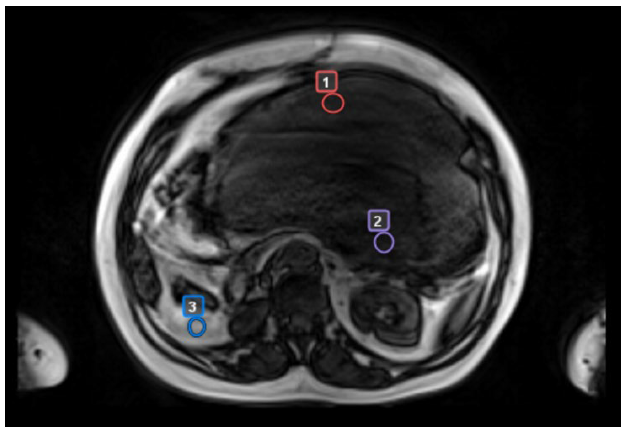
  • biopsy site (precisely identified in patients who had CT scan-guided biopsies)
  • “worst” tumor area with radiologic features suggestive of the most aggressive biology (reduction in ADC maps, high contrast enhancement, low T2 signal)
  • “best” tumor area with the least aggressive radiologic features (lack of one or more aggressive features)
  • a control area, identified as normal adipose tissue located in the same body region but separate from tumor tissue
Tissue specimens were obtained before and after treatment by core biopsy and formalin-fixed and paraffin-embedded. Hematoxylin/eosin-stained sections were submitted to morphologic evaluation of tumor tissue including histologic parameters such as cellular density, cellular atypia (pleomorphism), quantification of vascular structures, and quality of stroma.
This study was conducted in agreement with the Declaration of Helsinki and the laws and regulations of the country and according to the ICH Guideline for Good Clinical Practice. The protocol and its appendices were subject to review and approval by an Independent Ethics Committee(s) (“IEC”). All enrolled patients were informed of the aims of the study and provided written informed consent regarding the study and personal data protection.

3. Results

3.1. Patients’ Characteristics

From September 2019 to January 2024, 11 patients were enrolled (6 males and 5 females). Patient characteristics are summarized in Table 1. Median age at diagnosis was 59.2 years (range 27.8–80.0 years). A total of 9 out of 11 patients had DDLPS (82%; 1 with a prevalent WDLPS component), and 2/11 had MLPS (18%). All the assessable patients had G3 liposarcoma according to FNCLCC grading system [18], while histologic grade at diagnosis was not assessable for two patients because of an incomplete pathology report, though both had high grade primary tumors owing to the diagnosis of DDLPS. Upon diagnosis, all the patients presented with locally advanced disease and underwent surgery to remove the primary tumor. Regarding perioperative treatments, 27% of patients received perioperative radiotherapy and 45% perioperative chemotherapy.
Prior to eribulin, all patients had undergone at least two lines of chemotherapy for advanced disease, except for one patient that was ineligible for anthracyclines and was treated with trabectedin as the first-line treatment. Median time from diagnosis to the start of eribulin was 69.6 months (range 18–128 months). Most of the patients had undergone local treatments for recurrent/metastatic disease.

3.2. Clinical Evaluation

All the patients completed pre-treatment assessment and received at least one cycle of chemotherapy with eribulin. Within the included patients, 8/11 (73%) completed all assessments according to the study protocol. One patient received three cycles instead of four because of peripheral neuropathy that required treatment interruption of eribulin. A total of 3 out of 11 patients (27%) experienced clinical and radiological progression and did not participate in post-treatment assessment.
With a median follow-up of 9.2 months from the start of eribulin (range 1.3–38.5 months), 8/11 (73%) patients had disease progression and 4/11 (36%) died. In the overall population, median progression-free survival (mPFS) was 3.3 months, and median overall survival (mOS) was 8.7 months. Patients with disease control (stable disease (SD) or partial response (PR)) had longer mPFS (16.5 vs. 3.3 months) and overall survival (mOS 16.5 vs. 5.9 months) compared to patients with progressive disease (PD).
All the patients that had disease control or oligometastatic progression eligible for locoregional procedure continued treatment with eribulin, with a median duration of treatment of 3 months in the whole study population (range 1.5–36 months). One patient obtained PR after 8 months of treatment and underwent surgery to remove all visible tumor.
Treatment was generally well tolerated. Moderate-severe peripheral neuropathy was reported in two patients (one G2 peripheral neuropathy and one G3 peripheral motor neuropathy). Two patients developed G3 neutropenia. No other >G1 toxicities were observed.

3.3. Radiological Evaluation

3.3.1. RECIST Assessment (CT Scan)

A total of 8/11 patients (73%) had progressive disease (PD), 2 (18%) had SD, and 1 had PR per RECIST 1.1. [17]
Patient M09 was declared as PD at first assessment because of the detection of a new abdominal lesion, while the known target lesions remained stable according to RECIST. This patient agreed to treatment beyond progression, and continued eribulin with ablative stereotactic body radiotherapy (SBRT) on the new lesion.

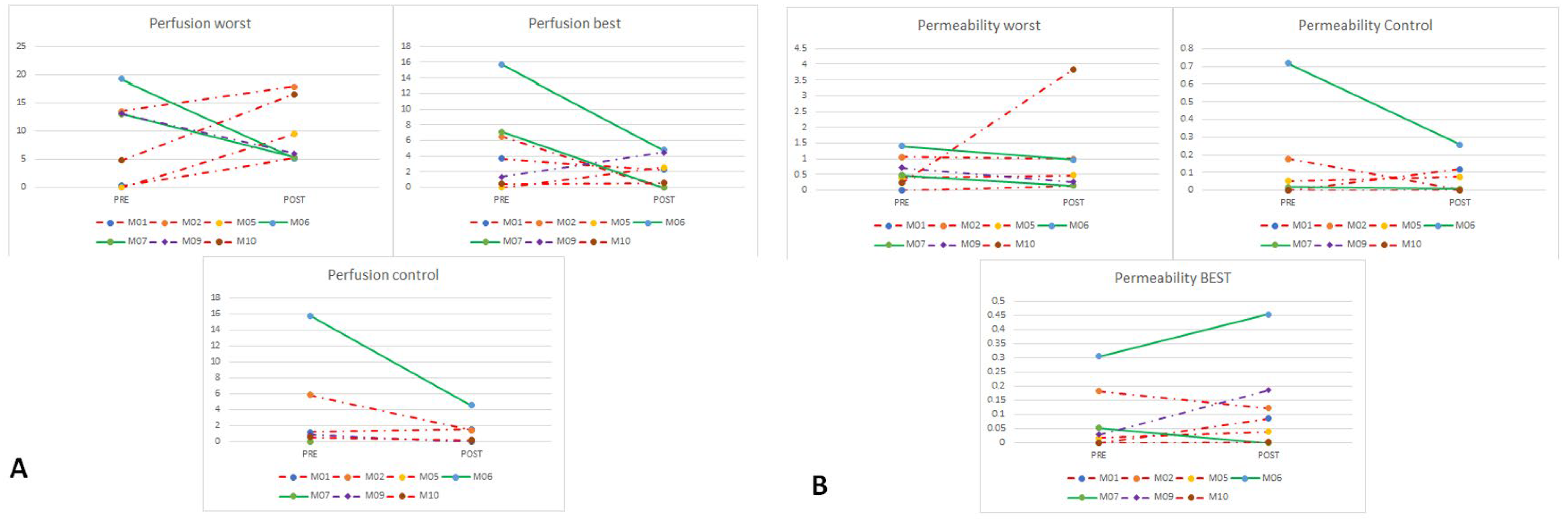
3.3.2. DCE-MRI Assessment

Among the eight patients that underwent both pre- and post-treatment DCE-MRI, four patients (50%) had PD, three (38%) had SD (including patient M09), and one (13%) had PR. Descriptive DCE-MRI evaluation is detailed in Table 2.
Generally, higher values of perfusion and permeability were detected in denser and more vascularized areas, with high discrepancies particularly in non-homogeneous lesions (Figure 1). Patients who had disease control showed overall higher pre-treatment permeability and lower perfusion and permeability after treatment. As an exception, the patient with WDLPS/DDLPS with a prevalent WDLPS component had no relevant radiological changes.
The same patient was not assessable regarding quantitative evaluation of perfusion and permeability because of technical issues pertaining to the radiology data. Moreover, more than half of the data about the precise biopsy site were missing (because an ultrasound-guided procedure had been performed, without available recorded imaging), so the graphic analysis of these data was not carried out.
We then analyzed separately the DCE-MRI perfusion and permeability values in assessable patients in “worst”, “best”, and control areas, respectively (ROIs, Figure 2).
Concerning perfusion in the “worst” areas, we found increased perfusion in patients with disease progression, while it decreased in patients with disease control. This change was not so clear in “best” areas, while perfusion values were generally lower in control areas and even lower after eribulin (Figure 3A). On the other hand, permeability decreased in patients with disease control both in “worst” and control areas, while in “best” areas a change was not evident (Figure 3B). Available perfusion and permeability data for each patient are detailed in the Supplementary Materials.

3.4. Pathology Assessment

Histological analysis was performed for the eight patients who had both pre- and post-treatment biopsies. We focused on the evaluation of the main morphological changes in cellularity, vascularization, stromal changes, and presence of cellular pleomorphism. Pathological features for each patient are summarized in Table 3.
All patients demonstrated histological modifications except the patient with WDLPS/DDLPS with a prevalent WDLPS component. A decrease in cellular density was reported in 50% of patients (both in patients with RECIST disease control or PD), while a decrease in vascular structures was detected only in one patient. One patient had an increase in blood vessels and necrosis, in line with radiological PD. Interestingly, signs of tissue maturation with adipose differentiation were found in three (38%) patients (with lipoblasts in one patient), one of whom had RECIST disease progression. Significant examples of pathological changes are reported in Figure 4. Available pre- and post-treatment histological images are shown in the Supplementary Materials.
Clinical, radiological, and pathological outcomes are reviewed in Table 4. We considered disease control (PR or SD) vs. PD in radiological exams and signs of response vs. signs of worsening (for example increased vascularization) in pathology assessment. Overall, the mean concordance between CT scan (RECIST), DCE-MRI, and histology was 83.5%, with higher discrepancies between pathological and radiological data.

4. Discussion

In this study, we explored the mechanisms of action of eribulin in a cohort of patients with advanced pretreated LPS, combining clinical, radiological, and pathological assessments, with a particular emphasis on non-mitotic effects.
Our clinical results regarding eribulin effectiveness and toxicity were consistent with existing literature, except for a lower overall survival rate (mOS 8.7 months vs. 13.5 months in Schöffski et al. [12]). Patients in our study were highly pretreated and perhaps less fit than patients usually included in clinical trials. However, the patients that achieved disease control with eribulin (three patients, 27%) had numerically longer progression-free and overall survival (mOS 16.5 months), confirming that eribulin can be effective in some patients. This positive impact was also seen in patients with stable disease (SD), so any benefit was not exclusively related to radiologic tumor shrinking.
Focusing on the responding patient subset, we explored the underlying mechanisms of action integrating radiological and pathological response features. Considering changes in vascularization analyzed through DCE-MRI, we found reduced levels of tumor perfusion after eribulin treatment in patients that achieved disease control and elevated post-treatment tumor perfusion in patients with PD. Conversely, we did not find any patterns in permeability variations, perhaps because of the small sample size and tumor heterogeneity. Preclinical and clinical models of breast cancer previously showed that eribulin causes a reduction in global tumor hypoxia, increasing vascularization in hypoxic areas, and decreasing it in more vascularized areas, a unique rebalancing of tumor blood vessel structure and organization [19,20]. Our finding could be explained considering that DCE-MRI data are drawn from a single higher grade (more vascularized) ROI, so are coherent with preclinical results [19]. More comprehensive investigations analyzing the tumor mass as a whole will be useful to better quantify vascularization.
The observed effect on tumor perfusion may explain the positive impact of eribulin on OS without affecting PFS [12,16]. It is well recognized that tumor vessels present altered structure and modified distribution compared with normal tissue [21], being vascularization is a crucial factor for tumor growth [22]. These alterations appear to favor the establishment of a tumor microenvironment (TME) that selects more aggressive tumor cells, attracts immunosuppressive cells, and compromises drug delivery [23].
While other anti-microtubule drugs cause disruption of the tumor vascular network and inhibit neoangiogenesis [24,25], eribulin normalizes tumor blood vessels’ structure and organization [19,20]. In order to support a hypothetical role of anti-antiangiogenetic drugs in STS, pazopanib provides a model. Pazopanib is the regulator-approved kinase inhibitor for soft tissue sarcomas, but LPS patients were excluded from the pivotal phase III PALETTE trial [26] based on a prior negative phase II trial [27]. However, following a centralized pathological review, two patients were reclassified and assigned to the liposarcoma cohort [27,28]. On the basis of this recategorization, pazopanib achieved the clinical activity cutoff for liposarcoma in the phase II trial [27,28], consistent with other phase II studies in which activity was seen in liposarcoma patients [29,30,31]. Taking into account these data, vascularization appears to be a targetable feature even in LPS. Our exploratory findings on correlation between tumor perfusion modifications induced by eribulin and tumor response support this hypothesis and will benefit from a larger study to confirm or refute this finding.
Epithelial-to-mesenchymal transition (EMT), the acquisition of mesenchymal characteristics in epithelial tumor cells, also plays an important role in metastatic potential [32,33]. In preclinical models and in a clinical study in breast cancer, eribulin induced an inversion of EMT reducing tumor migration and lung metastasis [34]. In sarcoma models, preclinical data showed an upregulation of adipocytic differentiation genes in liposarcoma cells after exposure to eribulin [20]. Our pathological assessment showed histological changes in all patients except for the one with WDLPS/DDLPS with a prevalent WDLPS component; we detected post-treatment morphological changes as the presence of lipoblasts and mature fat tissue features (lower grade). After treatment, we also observed a decrease in cellularity, and blood vessels changes in some patients, which did not always align with the radiological response. This discordance could be explained by tumor heterogeneity, but it can be hypothesized that modifications in both vascularization and tumor cells phenotype are necessary to achieve tumor control.
The observed pathological variations in tumor tissue and microenvironment could improve sensitivity to other therapies, suggesting a role for eribulin in earlier lines of treatment or in novel combinations. Preclinical studies showed favorable synergistic effects in association with several chemotherapeutic or targeted agents [18,35]. Based on our preliminary results, earlier administration of eribulin and/or a specific sequential treatment plan provides novel future treatment strategies to pursue.
Our study has several limitations. The small sample size and long accrual time limited us to descriptive analyses with only modest differences in study procedures such as DCE-MRI. A larger sample size with longer follow-up and more sensitive testing, such as single cell transcription analysis, would be a natural extension to this hypothesis-generating study.

5. Conclusions

Our exploratory study further supports the thesis of non-mitotic effects of eribulin on vascularization and cellular differentiation in LPS patients. As a future perspective unveiled by our study, a transcriptomic analysis in bulk tumor or in single cells could not just confirm the impact of eribulin on fat tissue differentiation and vascularity but also uncover new non-mitotic effects of eribulin. DCE-MRI appears to be a useful tool to better understand functional characteristics of tumors, especially in large heterogeneous masses as seen in sarcoma patients. Further studies are needed to confirm these results and identify patients who will benefit the most from eribulin therapy.

Supplementary Materials

The following supporting information can be downloaded at: https://www.mdpi.com/article/10.3390/cancers17060976/s1.

Author Contributions

Conceptualization, A.F.B., P.C., L.B., A.S., S.L.R., L.B., F.D., M.D. and M.S.G.; Methodology, A.F.B., S.L.R. and F.D; Software, F.D.; Validation, A.F.B., S.L.R., F.D, M.S.G., A.S. and R.G.M.; Formal Analysis, S.L.R., F.D, M.S.G. and A.F.B.; Investigation, A.F.B., M.S.G., A.L., S.L.R. and F.D.; Resources, A.S.; Data Curation, M.S.G., A.F.B., S.L.R. and F.D; Writing—Original Draft Preparation, M.S.G., A.F.B., S.L.R. and F.D.; Writing—Review and Editing, M.S.G., A.F.B., S.L.R., F.D., R.G.M. and P.C.; Visualization, F.D., M.S.G., A.F.B. and S.L.R.; Supervision, A.F.B., A.S., M.D., P.C. and L.B.; Project Administration, A.F.B.; Funding Acquisition, A.S. All authors have read and agreed to the published version of the manuscript.

Funding

Non-conditional financial support was provided by Eisai Co., Ltd. for this investigator-initiated study. Robert G Maki acknowledges research support in part by the Memorial Sloan Kettering Cancer Center Support Grant/Core Grant (P30 CA008748), Stand Up to Cancer, and Cycle for Survival.

Institutional Review Board Statement

The protocol and its appendices were subject to review and approval by the local Independent Ethics Committee (“IEC”; Approval Code: 669/18; Approval Date: 18 December 2018).

Informed Consent Statement

All enrolled patients were informed of the aims of the study and provided written informed consent regarding the study and personal data protection.

Data Availability Statement

The raw data supporting the conclusions of this article will be made available by the authors on request, according to the data protection laws.

Acknowledgments

The authors gratefully acknowledge EISAI for supporting this investigator-initiated study. The authors are also thankful to all the medical and non-medical staff of Humanitas Research Hospital that participated in the realization of this study.

Conflicts of Interest

Department of Oncology & Hematology IRCCS Humanitas Research Hospital, directed by Armando Santoro reports financial support was provided by Eisai Co Ltd. Armando Santoro reports a relationship with Eisai Co Ltd., which includes board membership and speaking and lecture fees. Other authors declare that they have no known competing financial interests or personal relationships that could have appeared to influence the work reported in this paper.

References

  1. Gronchi, A.; Miah, A.B.; Dei Tos, A.; Abecassis, N.; Bajpai, J.; Bauer, S.; Biagini, R.; Bielack, S.; Blay, J.Y.; Bolle, S.; et al. Soft tissue and visceral sarcomas: ESMO–EURACAN–GENTURIS Clinical Practice Guidelines for diagnosis, treatment and follow-up. Ann. Oncol. 2021, 32, 1348–1365. [Google Scholar] [CrossRef] [PubMed]
  2. WHO Classification of Tumours Editorial Board. Soft Tissue and Bone Tumours. In WHO Classification of Tumours Series, 5th ed.; International Agency for Research on Cancer: Lyon, France, 2020; Volume 3, Available online: https://publications.Iarc.fr/588 (accessed on 31 July 2024).
  3. Ducimetière, F.; Lurkin, A.; Ranchère-Vince, D.; Decouvelaere, A.-V.; Péoc’H, M.; Istier, L.; Chalabreysse, P.; Muller, C.; Alberti, L.; Bringuier, P.-P.; et al. Incidence of sarcoma histotypes and molecular subtypes in a prospective epidemiological study with central pathology review and molecular testing. PLoS ONE 2011, 6, e20294. [Google Scholar] [CrossRef] [PubMed]
  4. Schöffski, P. Established and experimental systemic treatment options for advanced liposarcoma. Oncol. Res. Treat. 2022, 45, 525–543. [Google Scholar] [CrossRef] [PubMed]
  5. Lee, A.T.J.; Thway, K.; Huang, P.H.; Jones, R.L. Clinical and molecular spectrum of liposarcoma. J. Clin. Oncol. 2018, 36, 151–159. [Google Scholar] [CrossRef]
  6. Sciot, R.; Gerosa, C.; Faa, G. (Eds.) Adipocytic, Vascular and Skeletal Muscle Tumors: A Practical Diagnostic Approach; Springer International Publishing: Cham, Switzerland, 2020. [Google Scholar]
  7. Sleijfer, S.; Seynaeve, C.; Verweij, J. Using single-agent therapy in adult patients with advanced soft tissue sarcoma can still be considered standard care. Oncologist 2005, 10, 833–841. [Google Scholar] [CrossRef]
  8. Santoro, A.; Tursz, T.; Mouridsen, H.; Verweij, J.; Steward, W.; Somers, R.; Buesa, J.; Casali, P.; Spooner, D.; Rankin, E. Doxorubicin vs. CYVADIC vs. doxorubicin plus ifosfamide in first-line treatment of advanced soft tissue sarcomas: A randomized study of the European Organization for Research and Treatment of Cancer Soft Tissue and Bone Sarcoma Group. J. Clin. Oncol. 1995, 13, 1537–1545. [Google Scholar] [CrossRef]
  9. Borden, E.C.; Amato, D.A.; Rosenbaum, C.; Enterline, H.T.; Shiraki, M.J.; Creech, R.H.; Lerner, H.J.; Carbone, P.P. Randomized comparison of three adriamycin regimens for metastatic soft tissue sarcomas. J. Clin. Oncol. 1987, 5, 840–850. [Google Scholar] [CrossRef]
  10. Langmans, C.; Cornillie, J.; van Cann, T.; Wozniak, A.; Hompes, D.; Sciot, R.; Debiec-Rychter, M.; Vandenbempt, I.; Schöffski, P. Retrospective Analysis of Patients with Advanced Liposarcoma in a Tertiary Referral Center. Oncol. Res. Treat. 2019, 42, 396–404. [Google Scholar] [CrossRef]
  11. Demetri, G.D.; von Mehren, M.; Jones, R.L.; Hensley, M.L.; Schuetze, S.M.; Staddon, A.; Milhem, M.; Elias, A.; Ganjoo, K.; Tawbi, H.; et al. Efficacy and safety of trabectedin or dacarbazine for metastatic liposarcoma or leiomyosarcoma after failure of conventional chemotherapy: Results of a phase III randomized multicenter clinical trial. J. Clin. Oncol. 2016, 34, 786–793. [Google Scholar] [CrossRef]
  12. Schöffski, P.; Chawla, S.; Maki, R.G.; Italiano, A.; Gelderblom, H.; Choy, E.; Grignani, G.; Camargo, V.; Bauer, S.; Rha, S.Y.; et al. Eribulin vs. dacarbazine in previously treated patients with advanced liposarcoma or leiomyosarcoma: A randomised, open-label, multicentre, phase 3 trial. Lancet 2016, 387, 1629–1637. [Google Scholar] [CrossRef]
  13. D’Incalci, M.; Galmarini, C.M. A review of trabectedin (ET-743): A unique mechanism of action. Mol. Cancer Ther. 2010, 9, 2157–2163. [Google Scholar] [CrossRef] [PubMed]
  14. Dybdal-Hargreaves, N.F.; Risinger, A.L.; Mooberry, S.L. Eribulin mesylate: Mechanism of action of a unique microtubule-targeting agent. Clin. Cancer Res. 2015, 21, 2445–2452. [Google Scholar] [CrossRef] [PubMed]
  15. Seshadri, P.; Deb, B.; Kumar, P. Multifarious targets beyond microtubules-role of eribulin in cancer therapy. Front. Biosci. (School Ed.) 2021, 13, 157–172. [Google Scholar] [CrossRef] [PubMed]
  16. Cortes, J.; O’Shaughnessy, J.; Loesch, D.; Blum, J.L.; Vahdat, L.T.; Petrakova, K.; Chollet, P.; Manikas, A.; Diéras, V.; Delozier, T.; et al. Eribulin monotherapy vs. treatment of physician’s choice in patients with metastatic breast cancer (EMBRACE): A phase 3 open-label randomised study. Lancet 2011, 377, 914–923. [Google Scholar] [CrossRef]
  17. Eisenhauer, E.A.; Therasse, P.; Bogaerts, J.; Schwartz, L.H.; Sargent, D.; Ford, R.; Dancey, J.; Arbuck, S.; Gwyther, S.; Mooney, M.; et al. New response evaluation criteria in solid tumours: Revised RECIST guideline (version 1.1). Eur. J. Cancer 2009, 45, 228–247. [Google Scholar] [CrossRef]
  18. Trojani, M.; Contesso, G.; Coindre, J.M.; Rouesse, J.; Bui, N.B.; De Mascarel, A.; Goussot, J.F.; David, M.; Bonichon, F.; Lagarde, C. Soft-tissue sarcomas of adults—Study of pathological prognostic variables and definition of a histopathological grading system. Int. J. Cancer 1984, 33, 37–42. [Google Scholar] [CrossRef]
  19. Funahashi, Y.; Okamoto, K.; Adachi, Y.; Semba, T.; Uesugi, M.; Ozawa, Y.; Tohyama, O.; Uehara, T.; Kimura, T.; Watanabe, H.; et al. Eribulin mesylate reduces tumor microenvironment abnormality by vascular remodeling in preclinical human breast cancer models. Cancer Sci. 2014, 105, 1334–1342. [Google Scholar] [CrossRef]
  20. Kawano, S.; Asano, M.; Adachi, Y.; Matsui, J. Antimitotic and Non-mitotic Effects of Eribulin Mesilate in Soft Tissue Sarcoma. Anticancer Res. 2016, 36, 1553–1561. [Google Scholar]
  21. Nagy, J.A.; Chang, S.-H.; Dvorak, A.M.; Dvorak, H.F. Why are tumour blood vessels abnormal and why is it important to know? Br. J. Cancer 2009, 100, 865–869. [Google Scholar] [CrossRef]
  22. Folkman, J. Tumor angiogenesis: Therapeutic implications. N. Engl. J. Med. 1971, 285, 1182–1186. [Google Scholar] [CrossRef]
  23. Chouaib, S.; Noman, M.Z.; Kosmatopoulos, K.; Curran, M.A. Hypoxic stress: Obstacles and opportunities for innovative immunotherapy of cancer. Oncogene 2017, 36, 439–445. [Google Scholar] [CrossRef] [PubMed]
  24. Kruczynski, A.; Poli, M.; Dossi, R.; Chazottes, E.; Berrichon, G.; Ricome, C.; Giavazzi, R.; Hill, B.T.; Taraboletti, G. Anti-angiogenic, vascular-disrupting and anti-metastatic activities of vinflunine, the latest vinca alkaloid in clinical development. Eur. J. Cancer 2006, 42, 2821–2832. [Google Scholar] [CrossRef]
  25. Hill, S.; Lonergan, S.; Denekamp, J.; Chaplin, D. Vinca alkaloids: Anti-vascular effects in a murine tumour. Eur. J. Cancer 1993, 29A, 1320–1324. [Google Scholar] [CrossRef]
  26. van der Graaf, W.T.; Blay, J.-Y.; Chawla, S.P.; Kim, D.-W.; Bui-Nguyen, B.; Casali, P.G.; Schöffski, P.; Aglietta, M.; Staddon, A.P.; Beppu, Y.; et al. Pazopanib for metastatic soft-tissue sarcoma (PALETTE): A randomised, double-blind, placebo-controlled phase 3 trial. Lancet 2012, 379, 1879–1886. [Google Scholar] [CrossRef]
  27. Sleijfer, S.; Ray-Coquard, I.; Papai, Z.; Le Cesne, A.; Scurr, M.; Schöffski, P.; Collin, F.; Pandite, L.; Marreaud, S.; De Brauwer, A.; et al. Pazopanib, a multikinase angiogenesis inhibitor, in patients with relapsed or refractory advanced soft tissue sarcoma: A phase II study from the European organisation for research and treatment of cancer-soft tissue and bone sarcoma group (EORTC study 62043). J. Clin. Oncol. 2009, 27, 3126–3132. [Google Scholar] [CrossRef]
  28. Chamberlain, F.E.; Wilding, C.; Jones, R.L.; Huang, P. Pazopanib in patients with advanced intermediate-grade or high-grade liposarcoma. Expert Opin. Investig. Drugs 2019, 28, 505–511. [Google Scholar] [CrossRef]
  29. Samuels, B.L.; Chawla, S.P.; Somaiah, N.; Staddon, A.P.; Skubitz, K.M.; Milhem, M.M.; Kaiser, P.E.; Portnoy, D.C.; Priebat, D.A.; Walker, M.S.; et al. Results of a prospective phase 2 study of pazopanib in patients with advanced intermediate-grade or high-grade liposarcoma. Cancer 2017, 123, 4640–4647. [Google Scholar] [CrossRef]
  30. Valverde, C.M.; Martin Broto, J.; Lopez-Martin, J.A.; Romagosa, C.; Sancho Marquez, M.P.; Carrasco, J.A.; Poveda, A.; Bauer, S.; Martinez-Trufero, J.; Cruz, J.; et al. Phase II clinical trial evaluating the activity and tolerability of pazopanib in patients (pts) with advanced and/or metastatic liposarcoma (LPS): A joint Spanish Sarcoma Group (GEIS) and German Interdisciplinary Sarcoma Group (GISG) Study—NCT01692496. J. Clin. Oncol. 2016, 34 (Suppl. S15), 11039. [Google Scholar] [CrossRef]
  31. Grünwald, V.; Karch, A.; Schuler, M.; Schöffski, P.; Kopp, H.-G.; Bauer, S.; Kasper, B.; Lindner, L.H.; Chemnitz, J.-M.; Crysandt, M.; et al. Randomized Comparison of Pazopanib and Doxorubicin as First-Line Treatment in Patients with Metastatic Soft Tissue Sarcoma Age 60 Years or Older: Results of a German Intergroup Study. J. Clin. Oncol. 2020, 38, 3555–3564. [Google Scholar] [CrossRef]
  32. Gavert, N.; Ben-Ze’ev, A. Epithelial–mesenchymal transition and the invasive potential of tumors. Trends Mol. Med. 2008, 14, 199–209. [Google Scholar] [CrossRef]
  33. Polyak, K.; Weinberg, R.A. Transitions between epithelial and mesenchymal states: Acquisition of malignant and stem cell traits. Nat. Rev. Cancer 2009, 9, 265–273. [Google Scholar] [CrossRef] [PubMed]
  34. Yoshida, T.; Ozawa, Y.; Kimura, T.; Sato, Y.; Kuznetsov, G.; Xu, S.; Uesugi, M.; Agoulnik, S.; Taylor, N.; Funahashi, Y.; et al. Eribulin mesilate suppresses experimental metastasis of breast cancer cells by reversing phenotype from epithelial–mesenchymal transition (EMT) to mesenchymal–epithelial transition (MET) states. Br. J. Cancer 2014, 110, 1497–1505. [Google Scholar] [CrossRef] [PubMed]
  35. Escudero, J.; Heredia-Soto, V.; Wang, Y.; Ruiz, P.; Hu, Y.; Gallego, A.; Pozo-Kreilinger, J.J.; Martinez-Marin, V.; Berjon, A.; Ortiz-Cruz, E.; et al. Eribulin activity in soft tissue sarcoma monolayer and three-dimensional cell line models: Could the combination with other drugs improve its antitumoral effect? Cancer Cell Int. 2021, 21, 646. [Google Scholar] [CrossRef] [PubMed]
Figure 1. Perfusion (A) and permeability (B) maps in a patient with an inhomogeneous retroperitoneal liposarcoma that had disease progression. The post-treatment evaluation (on the right) shows increased dimension and higher perfusion values.
Cancers 17 00976 g001
Figure 2. Example of ROI individuation in a patient with a voluminous retroperitoneal DDLPS. 1: “best” area; 2: “worst” area; 3: control area. ROI = region of interest; DDLPS = dedifferentiated liposarcoma.
Cancers 17 00976 g002
Figure 3. Perfusion (A) and permeability (B) evaluation in “worst” and “best” areas vs. control areas. Legend: patients with disease control are depicted in green; patients with progressive disease are depicted in red; the patient that had SD in the examined target lesions but had disease progression because of a new distant metastasis is depicted in purple.
Cancers 17 00976 g003
Figure 4. Post-treatment modifications (images on the right) compared to pre-treatment histology (on the left) in some responding (A) and non-responding (B) patients. Black arrows indicate blood vessels. Scale bar: 100 μm. ↑ = slight increase; ↓ = slight decrease; ↓↓ = significant decrease.
Cancers 17 00976 g004aCancers 17 00976 g004b
Table 1. Patient characteristics. FNCLCC = Fédération Nationale des Centres de Lutte le Cancer; DDLPS = dedifferentiated liposarcoma; WDLPS = well-differentiated liposarcoma; MLPS = myxoid liposarcoma; EI = epirubicin + ifosfamide; trabe = trabectedin; AI = doxorubicin + ifosfamide; A = doxorubicin; ifo = ifosfamide; gem = gemcitabine. * Tumor grade was not specified in the original pathology report; however, the finding of dedifferentiated liposarcoma in those two specimens means the tumor was either grade 2 or 3; these are both considered high grade lesions.
Patient IDAge at DiagnosisGenderHistologyPrimaryStage at DiagnosisGrade
(FNCLCC Grading System) at Diagnosis
Disease Sites at EnrollmentPrior TreatmentsTime (Months) from Diagnosis to Eribulin Treatment
M0160FDDLPS (spindle cells in myxoid stroma)retroperitoneumIIIG2 or G3 *abdomen, soft tissuesEI × 2/trabe × 922
M0244FDDLPS (lipoblastic differentiation)retroperitoneumIIIG3abdomenAI × 3/trabe × 1094
M0359MWDLPS/
DDLPS (focal hypercellularity)
retroperitoneumIIIG2 or G3 *abdomentrabe × 491
M0471MDDLPS (spindle and pleomorphic cells in myxoid stroma)retroperitoneumIIIG3abdomen, bone, lungA × 5/trabe × 490
M0534FDDLPS (spindle and pleomorphic cells)extremitiesIIIG2lung, soft tissuesAI × 6/ifo × 3/trabe × 740
M0642FDDLPS (spindle and pleomorphic cells)retroperitoneumIIIG3abdomentrabe × 4/ifo × 264
M0728FMLPS (high grade)extremitiesIIIG3abdomen, boneAI × 5/trabe × 22/
ifo × 3/gem × 3
128
M0880MDDLPS (spindle and pleomorphic cells in myxoid stroma)mediastinumIIIG3lung, liverifo × 6/trabe × 418
M0967MMLPS (high grade)extremitiesIIIG3bone, soft tissuestrabe × 40/ifo × 370
M1042MDDLPS (myxofibrosarcoma-like component)retroperitoneumIIIG3abdomentrabe × 8/ifo × 6123
M1163MDDLPS (spindle and pleomorphic cells)retroperitoneumIIIG3lungA × 3/ifo × 549
Table 2. DCE-MRI assessment. DDLPS = dedifferentiated liposarcoma; WDLPS = well-differentiated liposarcoma; MLPS = myxoid liposarcoma; PD = progressive disease; SD = stable disease; PR = partial response. Legend: patients with patient ID in red had PD, the ones in green had SD or PR, the one in blue had SD in the examined target lesions but had disease progression because of a new distant metastasis. The worst nodules were defined as tumor areas with radiologic features suggestive of the most aggressive biology (reduction in apparent diffusion coefficient (ADC) maps, high contrast enhancement, low T2 signal).
Patient IDHistologyTarget Lesions SitePre-Treatment General AssessmentPre-Treatment PerfusionPre-Treatment PermeabilityPost-Treatment General AssessmentPost-Treatment PerfusionPost-Treatment Permeability
M01DDLPSabdomenone worst nodule in bulky masshigh in worst nodulehigh in worst nodulePD (worst nodule no more detectable, overall increased bulky mass)significantly
higher
stable
M02DDLPSabdomenhomogeneous masshigh in 2 regionshigh in 2 regionsPD (1 nodule increased, 1 nodule stable)highersignificantly lower in stable nodule, remains high in increased nodule
M03WDLPS/
DDLPS
abdomenhomogeneous multifocal masseslowlowSDstable (low)stable (low)
M05DDLPSthighall high-grade nodulehigh in rim, low in center (necrosis)NAPD (overall dimension)stable (high in rim, low in center—necrosis)high (no comparison available)
M06DDLPSabdomenone worst nodule, multifocal diseasevery highvery highPRsignificantly lowersignificantly lower
M07MLPSabdomenhomogeneous noduleslowlowSDlowerlower
M09MLPStrunkhomogeneous noduleshigh in rim, low in center (necrosis)lowSDlowerlower
M10DDLPSabdomentwo worst nodules in bulky masshigh in 2 worst nodules, low in center (necrosis)high in 2 worst nodules rimsPD (increased worst nodules dimension)high in 2 worst nodules rims, low in the rest of the massstable (high in 2 worst nodules rims, low in the rest of the mass)
Table 3. Histologic assessment. DDLPS = dedifferentiated liposarcoma; WDLPS = well-differentiated liposarcoma; MLPS = myxoid liposarcoma. Legend: patients with patient ID in red had PD, and the ones in green had SD or PR, the one in blue had SD in the examined target lesions but had disease progression because of a new distant metastasis.
Patient IDHistologyPre-Treatment AssessmentPost-Treatment Assessment
M01DDLPSMyxoid stroma, low cellularity, high vascularizationSignificant cellularity reduction
M02DDLPSMany blood vessels, uneven cellularity, pleomorphic cellsSignificantly less vascularized
M03WDLPS/DDLPSAdipose tissue with slightly higher cellularity (WDLPS)No significant variations
M05DDLPSHigh cellularityLower cellularity, more blood vessels, necrosis
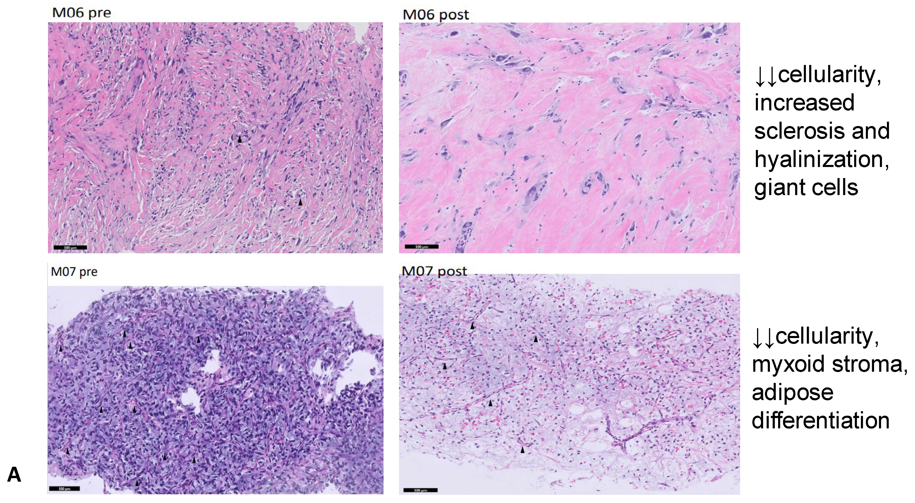
M06DDLPSHigh cellularity, scarce pleomorphismSignificant cellularity reduction, increased sclerosis and hyalinization, giant cells
M07MLPSHigh cellularity, many blood vesselsSignificant cellularity reduction, more myxoid stroma, lipoblasts and signs of adipose differentiation
M09MLPSHomogeneous MLPSIncreased sclerosis and hyalinization and signs of adipose differentiation
M10DDLPSHigh necrosis, no lipoblastsNo necrosis, signs of adipose differentiation
Table 4. Overall response assessment. DDLPS = dedifferentiated liposarcoma; WDLPS = well-differentiated liposarcoma; MLPS = myxoid liposarcoma; PD = progressive disease; SD = stable disease; PR = partial response; NA = not assessable. Legend: patients with patient ID in red had PD, the ones in green had SD or PR, the one in blue had SD in the examined target lesions but had disease progression because of a new distant metastasis. ↑ = slight increase; ↓ = slight decrease; ↓↓ = significant decrease.
Patient IDHistologyProtocol CompletionTotal Eribulin CyclesRECIST Response (Target Lesions)DCE-MRI ResponseHistology ChangesConcordance Between RECIST, MRI and Histology
M01DDLPSyes4PD (29 × 24 cm vs. 19 × 15 cm)PD↓↓ cellularity67%
M02DDLPSyes4PD (7 × 6.7 cm vs. 5.8 × 3.5 cm, 6 × 5 cm vs. 3.8 × 3.5 cm)PD↓↓ vascularization67%
M03WDLPS/
DDLPS
yes3 yearsSD (6.5 × 4.8 cm vs. 7.5 × 4.8 cm)SDnone100%
M04DDLPSno4PD (10 × 8 cm vs. 8.3 × 3.5 cm)NANANA
M05DDLPSyes4PD (9.6 × 5.4 cm vs. 6.5 × 4.3 cm)PD↓ cellularity,
↑ vascularization, necrosis
100%
M06DDLPSyes9SD (2.8 × 2 cm vs. 2.5 × 1.8 cm, 4.5 × 4.3 cm vs. 4.9 × 4.8 cm)PR↓↓ cellularity, increased sclerosis and hyalinization, giant cells100%
M07MLPSyes8PR (8.7 × 5 cm vs. 13.9 × 64, 6.3 × 4.4 cm vs. 7.8 × 7.5 cm)SD↓↓ cellularity, myxoid stroma, adipose differentiation100%
M08DDLPSno2PD (new multiple lung and mediastinum lesions, increased dimension of prior lesions) NANANA
M09MLPSyes4PD (new abdominal lesion, SD in target lesions: 9.5 × 8.2 cm vs. 9.8 × 7.8 cm, 5.8 × 3.9 cm vs. 5.6 × 4.2 cm)SDIncreased sclerosis and hyalinization, adipose differentiation67%
M10DDLPSyes3PD (9.8 × 5.3 cm vs. 6.9 × 4.9 cm, 7.9 × 4.6 cm vs. 6.5 × 4.9 cm)PDadipose differentiation67%
M11DDLPSno1PD (new clinically significant pleural effusion)NANANA
Disclaimer/Publisher’s Note: The statements, opinions and data contained in all publications are solely those of the individual author(s) and contributor(s) and not of MDPI and/or the editor(s). MDPI and/or the editor(s) disclaim responsibility for any injury to people or property resulting from any ideas, methods, instructions or products referred to in the content.

