Article

Revisiting p53 Immunohistochemical Staining and Its Prognostic Implications in Advanced EGFR-Mutated Lung Adenocarcinoma

1
Graduate Institute of Clinical Medical Sciences, College of Medicine, Chang-Gung University, Taoyuan 333323, Taiwan
2
Division of Hematology and Oncology, Department of Medicine, Chang-Gung Memorial Hospital, Chiayi 61363, Taiwan
3
Department of Medical Research and Development, Chang-Gung Memorial Hospital, Chiayi 61363, Taiwan
4
Center for General Education, Chang-Gung University of Science and Technology, Chiayi 61363, Taiwan
5
Department of Pathology, Far Eastern Memorial Hospital, New Taipei City 220216, Taiwan
6
Department of Pathology, Chang-Gung Memorial Hospital, Chiayi 61363, Taiwan
7
Health Information and Epidemiology Laboratory, Chang-Gung Memorial Hospital, Chiayi 61363, Taiwan
8
Department of Pulmonary and Critical Care Medicine, Chang-Gung Memorial Hospital, Chiayi 61363, Taiwan
*
Author to whom correspondence should be addressed.
Cancers 2025, 17(21), 3577; https://doi.org/10.3390/cancers17213577
Submission received: 7 October 2025 / Revised: 1 November 2025 / Accepted: 4 November 2025 / Published: 5 November 2025
(This article belongs to the Section Cancer Biomarkers)

Simple Summary

The areas under the receiver operating characteristic (ROC) curves with different cut-off values for p53 positivity ranged between 0.51 and 0.56. Under a cut-off value of 50% p53 immunostaining, the sensitivity and specificity of TP53 mutations were 66.7% and 45.5%, respectively, indicating an inconsistency between p53 immunostaining and TP53 mutations. Tumor p53 overexpression (≥50%) was identified as a strong prognostic factor after adjusting for other important clinical factors, such as EGFR mutation subtypes, baseline brain metastasis status, and smoking status. This study shows that p53 immunohistostaining could help spare patients from toxicity of novel combination therapy once validated and adds value to TP53 mutation analysis in the modern genomic era of EGFR-mutated lung adenocarcinoma.

Abstract

Background/Objectives: TP53 mutations in advanced epidermal growth factor receptor (EGFR)-mutated non-small cell lung cancer could worsen prognosis. Therefore, we aimed to investigate the clinical significance of TP53 mutations and p53 expression in these patients. Methods: Patients with advanced/metastatic EGFR-mutated lung adenocarcinoma treated with first-line tyrosine kinase inhibitors were retrospectively enrolled. Sanger sequencing was performed to detect TP53 mutations and immunohistochemical staining was used to verify p53 protein expression levels. Kaplan-Meier and Cox proportional hazards analyses were used to estimate survival and hazard ratio (HR) with 95% confidence interval (CI). Results: The study involved 83 patients with adequate tumor samples for TP53/p53 analysis. Patients with tumor p53 immunostaining ≥50% showed significantly better overall survival (OS) (HR: 0.49 [95% CI: 0.30–0.81], p < 0.001), but TP53 mutations were not associated with inferior progression-free survival (PFS) or OS (missense vs. wild-type [PFS, HR: 0.68 (95% CI: 0.40–1.15), p = 0.151; OS, HR: 0.88 (95% CI: 0.56–1.42), p = 0.599]). Areas under the receiver operating characteristic curves of TP53 mutations with different cut-off values for p53 positivity were 0.51–0.56. The Kaplan-Meier survival analysis revealed significant survival benefits in patients with EGFR L858R substitution and tumor p53 immunostaining ≥50% (median PFS: 8.0 vs. 5.3; median OS: 20.4 vs. 15.3 months; log-rank p = 0.025 and 0.049, respectively). Conclusions: Tumor p53 immunostaining (≥50%) was associated with better OS, especially in patients with TP53 mutations or L858R. Prospective clinical trials are required to explore the prognostic significance of p53 expression in the genomic era of TP53 mutations.

1. Introduction

The global incidence of lung cancer exceeded 2.5 million, accounting for 1.8 million deaths, in 2022 [1]. The development of therapies targeting biomarkers, such as epidermal growth factor receptor (EGFR)-activating mutations including deletions in exon 19 (Del19) and the single amino-acid substitution L858R in exon 21, has enabled personalized medicine for metastatic non-small cell lung cancer (NSCLC) [2]. Approximately 15–25% of Caucasians and 40–55% of East Asian patients with lung cancer carry EGFR mutations, and the evolution of tyrosine kinase inhibitors (TKIs) targeting EGFR mutations has improved progression-free survival (PFS) and overall survival (OS) in these patients [3,4,5]. However, the patients inevitably develop resistance to EGFR-targeted TKIs, at least partially because of the presence of other mutations along with EGFR mutations, which can influence and alter drug efficacy [6].
The human tumor suppressor gene TP53 encodes p53, which transcriptionally controls the expression of numerous target genes involved in various biological functions. However, TP53, the most commonly mutated gene in human cancers, encodes defective p53 protein, which dysregulates transcriptional activities [7]. Furthermore, TP53 mutations are among the most prevalent concomitant mutations in patients with EGFR-mutated lung cancer, and mounting evidence shows that TP53 mutations can result in intrinsic resistance and poor prognosis [8,9,10]. A recent study reported that the presence of both TP53 mutations and whole-genome doubling is associated with EGFR-TKI resistance [11]. A previous study has indicated that EGFR mutations and p53 overexpression play distinct roles in the development of small pulmonary adenocarcinomas [12].
Nevertheless, little is known about the relationship between p53 protein expression, as assayed through immunohistochemical staining, and the presence of TP53 mutations, detected via sequencing, in patients with advanced EGFR-mutated NSCLC. Therefore, in this study, we aimed to investigate the association between these old and new approaches by verifying p53 protein expression, TP53 mutations, and prognostic implications in patients with EGFR-mutated NSCLC.

2. Material and Methods

2.1. Patients and Tissue Samples

From January 2011 to June 2015, data of patients with stage IIIb/IV lung adenocarcinoma harboring EGFR mutations who were treated with frontline TKIs were retrieved from the medical records at Chang-Gung Memorial Hospital, Chia-Yi Branch, Taiwan. The following data were collected: sex, age (<70 and ≥70 years), smoking status, EGFR mutation subtypes (Del19, L858R, and other mutations), baseline brain metastasis status, and TKI types. Formalin-fixed, paraffin-embedded tissues of these patients were collected from the Chang-Gung Memorial Hospital Tissue Bank for p53 immunohistochemical staining and TP53 mutation analysis. The need for patient consent was waived owing to the retrospective nature of the study. The study was approved by the Institutional Review Board of Chang-Gung Memorial Hospital (201900059B0).

2.2. p53 Protein Expression

Immunohistochemical staining of tissue sections was performed using a p53 monoclonal antibody (Glostrup, Denmark, Leica Biosystems; Cat# NCL-L-p53-DO7, RRID: AB_563936) as described previously [13,14]. The sections were counterstained with hematoxylin and eosin (Glostrup, Denmark) and blindly reviewed by two experienced pathologists (CFF and LKF) to identify areas with the highest tumor density (80% tumor content). Positive and negative controls were used to validate the reactions, and tumor samples with >10% of tumor cells showing positive nuclear staining were considered positive controls. p53 immunohistochemical staining was assessed semi-quantitatively based on the proportion and staining intensity of positive tumor cells (Figure S1 and Table S1). In cases of discordant p53 immunostaining results, a third pathologist (CCL) was consulted. None of the pathologists were aware of the molecular, biological, or clinical details of the patients.

2.3. TP53 Mutations

The high-resolution melting technique was used to quickly and efficiently screen for TP53 mutations in exons 5–8, as previously described [15], using a 7900HT Fast Real-Time PCR System (Foster, Winston-Salem, NC, USA, Applied Biosystems). This was followed by Sanger sequencing (Tables S2 and S3). Mutations were classified as “disruptive” and “nondisruptive” according to the report by Molina-Vila et al. [15]. Also, in their analysis of the survival impact, TP53 mutations were grouped as “wild-type + disruptive” and “nondisruptive.”

2.4. Statistical Analysis

PFS was calculated as described in our previous report [16] and OS was defined as the duration from TKI treatment initiation to patient death or loss to follow-up, whichever occurred first. Survival curves were generated using the Kaplan-Meier method, and the log-rank test was used to compare the time to events between the groups. Univariate and multiple regression analyses were performed using Cox proportional hazards models. Statistical significance was set at a two-sided p value of <0.05, and all analyses were conducted using SAS version 9.4 (SAS Inc., Cary, NC, USA).

3. Results

In total, 93 samples (including 2 paired samples) were obtained for analysis. After excluding 2 patients with incomplete medical information and 5 lacking sufficient tissue for p53 immunohistochemical staining and/or TP53 mutation analysis, 83 patients with adequate formalin-fixed paraffin-embedded tissues for clinical and molecular analyses were enrolled. Of these patients, 57.8% were female individuals. The median age of the patients was 71 (range, 41–91) years; 66.3% of the patients were never smokers and 24.1% had brain metastasis at baseline. A total of 43 patients had the L858R mutation (51.8%), 25 patients had Del19 (30.1%), and 15 had other EGFR mutations (18.1%). Additionally, six patients received afatinib (7.2%), 21 received erlotinib (25.3%), and 56 received gefitinib (67.5%). A total of 39 patients (47%) had TP53 missense mutations, of whom 26 had nondisruptive mutations (V216 deletions in exon 6 [n = 11] and R282L [n = 1] and R273L [n = 1] in exon 8) and 13 had disruptive mutations (H178R in exon 5 [n = 12] and R248E in exon 7 [n = 1]) (Table 1). Regarding p53 immunohistochemical staining, 45 patients (54.2%) showed 50% positivity, 63 patients (75.9%) showed >10% positivity, and 54 patients (65.1%) showed strong or intermediate intensity (Table 1 and Table S1).
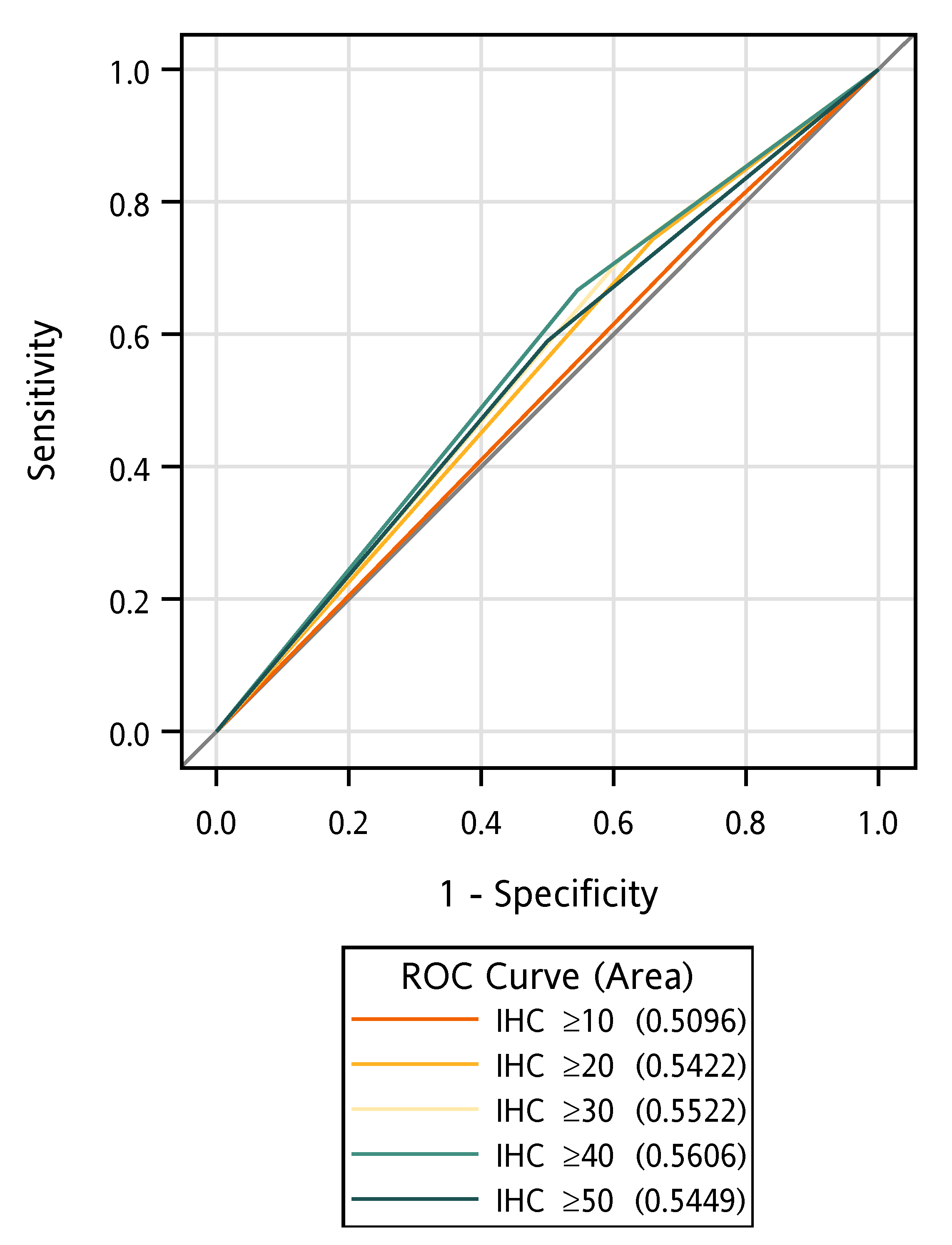
The median follow-up time was 20.3 months. The data showed that p53 immunohistochemical staining of ≥50% was associated with better OS in patients with TP53 missense mutations in exons 5–8 (n = 39) (median OS: 23.8 vs. 19.6 months, log-rank p = 0.048, Figure 1A); however, this improvement in OS was not observed in patients with immunohistochemical staining of ≥10% (median OS: 20.4 vs. 22.9 months, log-rank p = 0.679, Figure 1D). When TP53 missense mutations were subdivided into nondisruptive and disruptive mutations, there was only a trend toward improved survival in patients with nondisruptive mutations (log-rank p = 0.108, Figure 1C but not Figure B, E and F). Under a cut-off value of 10% p53 immunostaining, the sensitivity and specificity of TP53 mutations determined through Sanger sequencing were 76.9% and 25%, respectively. Under a cut-off value of 50% p53 immunostaining, the sensitivity and specificity of TP53 mutations were 66.7% and 45.5%, respectively (Table S4), indicating an inconsistency between p53 immunostaining and TP53 mutations in these samples. The areas under the receiver operating characteristic (ROC) curves with different cut-off values for p53 positivity ranged between 0.51 and 0.56 (Figure 2). No association was found between p53 immunohistochemical positivity (%) or p53 semi-quantitative staining intensity and TP53 mutations (p = 0.413 and p = 0.526, respectively). Moreover, smoking status was not associated with TP53 mutations (p = 0.695); however, p53 positivity (%) was associated with semi-quantitative staining intensity (p < 0.001).
In the univariate Cox regression analysis of PFS, older age (≥70 years) was associated with a better outcome (hazard ratio (HR): 0.58 [95% confidence interval (CI): 0.34–0.96], p = 0.035). Additionally, a trend toward better PFS was observed in patients with p53 positivity ≥50% (HR: 0.66 [95% CI: 0.40–1.08], p = 0.097), whereas patients harboring L858R showed a trend toward poor outcome (HR: 1.68 [95% CI: 0.95–2.97], p = 0.076). Moreover, regarding OS, ever/current smokers had a poor outcome (HR: 1.65 [95% CI: 1.00–2.70], p = 0.05) and p53 positivity ≥50% was associated with a better outcome (HR: 0.53 [95% CI: 0.32–0.84], p = 0.008). TP53 mutations (nondisruptive vs. disruptive plus wild-type (WT) [PFS HR: 0.93 [95% CI: 0.54–1.58], p = 0.781 and OS HR: 0.85 [95% CI: 0.51–1.42], p = 0.531] or missense vs. WT [PFS HR: 0.76 [95% CI: 0.46–1.26], p = 0.284 and OS HR: 0.83 [95% CI: 0.52–1.33], p = 0.437]) and p53 semi-quantitative staining intensity (strong/intermediate vs. weak/negative [PFS HR: 0.90 [95% CI: 0.54–1.52], p = 0.696 and OS HR: 0.77 [95% CI: 0.48–1.26], p = 0.298]) were not associated with poor survival outcomes (Table 2).
In the multiple Cox regression analyses of PFS, older age (≥70 years) was associated with a significantly better outcome (HR: 0.50 [95% CI: 0.28–0.88], p = 0.017), whereas patients harboring L858R showed a trend toward poor outcome (HR: 1.81 [95% CI: 0.99–3.30], p = 0.053). In the multiple Cox regression analyses of OS, p53 positivity ≥50% was linked to a significantly better outcome (HR: 0.49 [95% CI: 0.30–0.81], p < 0.001), whereas baseline brain metastasis was associated with a poor outcome (HR: 1.84 [95% CI: 1.03–3.30], p = 0.041). However, TP53 mutations (missense vs. WT [PFS HR: 0.68 [95% CI: 0.40–1.15], p = 0.151 and OS HR: 0.88 [95% CI: 0.56–1.42], p = 0.599]) were not associated with poor survival outcomes (Table 3). Additionally, TP53 mutations (nondisruptive vs. disruptive plus WT [PFS 0.87 [95% CI: 0.50–1.52], p = 0.633 and OS 0.99 [95% CI: 0.58–1.69], p = 0.971]) were not associated with poor survival outcomes.
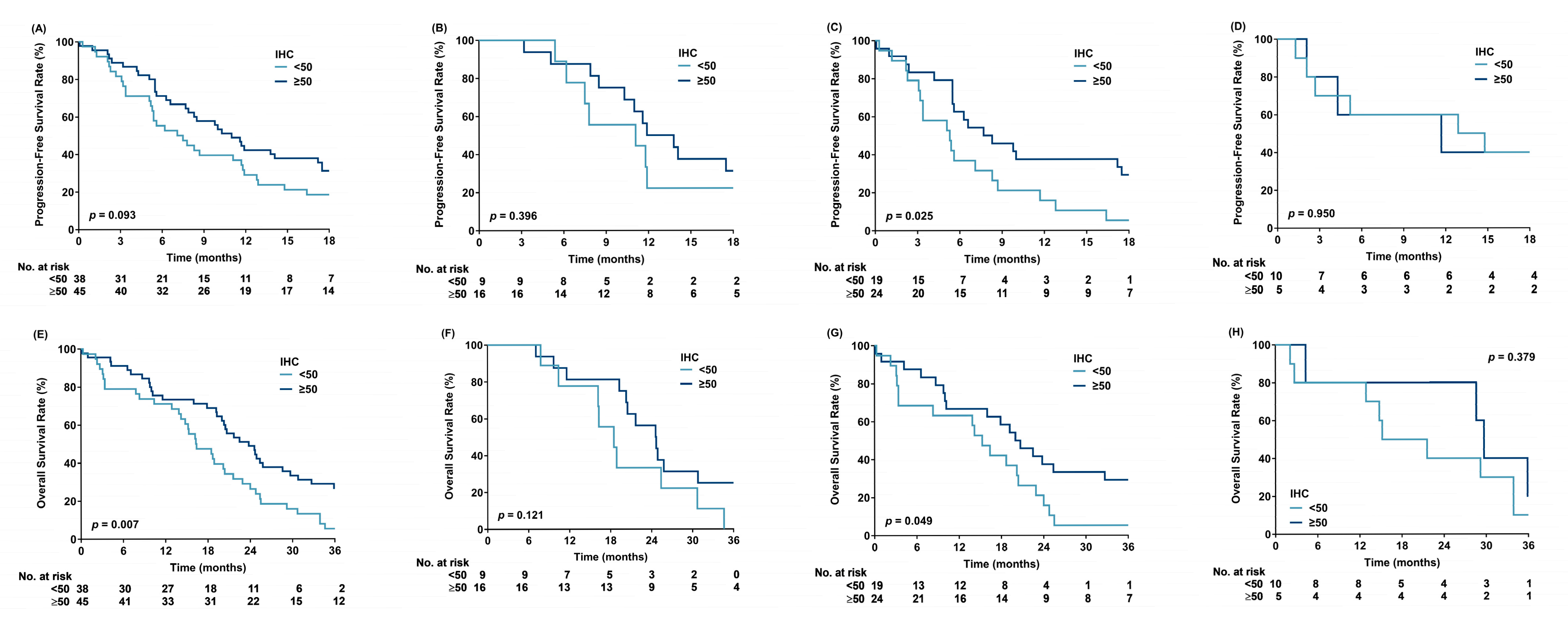
The Kaplan-Meier survival analysis of PFS showed a trend toward survival benefit in patients with p53 positivity ≥50% (median PFS: 11.3 vs. 7.3 months, log-rank p = 0.093). However, significant survival benefit was observed in patients with L858R and p53 positivity ≥50% (median PFS: 8.0 vs. 5.3 months, log-rank p = 0.025) (Figure 3A,C). A trend toward survival benefit was observed in patients with Del19 and p53 positivity ≥50% (median PFS: 12.9 vs. 11.1 months, log-rank p = 0.396) (Figure 3B). Additionally, a trend toward survival benefit was noted in patients with L858R and strong/intermediate p53 positivity (median PFS: 7.7 vs. 5.4 months, log-rank p = 0.077) (Figure S2). However, no differences in PFS were observed between patients with TP53 missense mutations and those with WT (median PFS: 11.1 vs. 7.7 months, log-rank p = 0.285), or between patients with nondisruptive mutations and those with WT plus disruptive mutations (median PFS: 10.7 vs. 8.3 months, log-rank p = 0.786) (Figures S3 and S4).
The Kaplan-Meier survival analysis of OS showed a significant survival benefit in patients with p53 positivity ≥50% (median OS: 23.8 vs. 16.4 months, log-rank p = 0.007). Among these patients, those with L858R had a significant survival benefit (median OS: 20.4 vs. 15.3 months, log-rank p = 0.049), whereas those with Del19 only showed a trend toward survival benefit (median OS: 24.7 vs. 18.5 months, log-rank p = 0.121) (Figure 3E–G). There was no survival difference in patients with mutations other than Del19 or L858R (Figure 3D,H). No differences in OS were found between patients with TP53 missense and those with WT (median OS: 21.6 vs. 19.3 months, log-rank p = 0.433) or between patients with nondisruptive mutations and those with WT plus disruptive mutations (median OS: 22.3 vs. 19.3 months, log-rank p = 0.529) (Figures S5 and S6). However, a significant OS benefit was observed in patients with Del19 and nondisruptive TP53 mutations compared with that in patients with WT plus disruptive mutations (median OS: 32.7 vs. 19.3 months, log-rank p = 0.037) (Figure S6).

4. Discussion

To our knowledge, this is the first study to investigate the association between p53 expression and TP53 mutations in patients with advanced EGFR-mutated lung adenocarcinoma. p53 immunostaining positivity of ≥50% was found to be a favorable prognostic factor for OS in the multivariate analysis of clinical factors, including EGFR mutation subtypes, baseline brain metastasis status, and smoking status [5,17,18]. Immunohistochemical methods to detect p53 were developed in the early 1990s, and because p53 is the “guardian of the genome,” it has been applied as a convenient surrogate for TP53 mutation status to predict the survival outcomes in patients with cancer [19,20,21]. However, discrepancies between p53 expression levels and TP53 mutations in lung cancer have been reported [22,23,24]. Over the past two decades, the importance of EGFR expression and mutations in lung cancer has been validated, and genomic medicine has led to improved treatments for patients with EGFR-mutated lung cancer [2,3,4,5,25,26]. Unexpectedly, p53 expression analysis using immunostaining has become largely obsolete in this field. We also observed a discordance in the present study findings when the monoclonal antibody DO-7 was used to detect p53 immunoreactivity. DO-7 can identify epitope mapping between amino acids 1 and 45 of human p53. It is recommended for the detection of both WT and mutant p53 proteins [20,27] under denaturing and non-denaturing conditions; thus, we could not confirm whether the higher expression of p53 was a WT or mutated phenotype. Furthermore, our immunohistochemical analysis data showed nuclear staining of p53 in all samples, suggesting that the transcriptional capacity of p53, which regulates genome integrity, might be preserved in these patients. Thus, a higher p53 immunoreactivity (≥50%) and nuclear localization staining might reflect the higher transcriptional activity of p53 in retaining its protective tumor growth-suppressive function. This finding is similar to that of a previous study, which showed that higher expression of p53 is a favorable prognostic factor in a subset of patients with NSCLC [28]. We found that when using a higher cut-off value of p53 immunostaining (≥50%), the rate of TP53 missense mutations was 59.0%; it increased to 76.9% under a lower cut-off value of p53 immunostaining (≥10%). The enrichment of TP53 missense mutations in patients with p53 staining ≥ 10% and the protective effect of p53 (≥50% staining) were attenuated. However, previous studies have focused solely on the prognostic implications of p53 protein expression levels or TP53 mutations. We provide evidence that evaluating both p53 protein expression level and TP53 mutations is important for predicting the outcome of NSCLC. Yoo et al. reported that EGFR mutations occur in the early stage and p53 overexpression is a late event in lung cancer development [12]. Our study further highlights that p53 positivity of ≥50% could identify a subgroup of patients with a more favorable prognosis in advanced EGFR-mutated lung adenocarcinoma.
Results for TP53 co-mutations as a prognostic factor in patients with EGFR-mutated NSCLC have been inconsistent. A recent study concluded that the development of TP53 co-mutations is a clinically relevant mechanism of EGFR-TKIs and a poor prognostic factor for OS [8]. However, in this study, there was no significant correlation between TP53 co-mutations and PFS and OS, similar to the findings of other prospective and retrospective studies in patients with EGFR-mutated lung cancer [29,30,31,32]. This discrepancy in the findings may be because we focused only on TP53 mutations from exons 5 to 8, consisting of the DNA-binding region, by direct sequencing of tumor tissues, which could have resulted in a low yield compared to that with next-generation sequencing of plasma samples [33]. Additionally, the TP53 missense mutations in our study were mainly in exon 6 (74.4%, 29/39) and exon 8 (5.1%, 2/39), which differs from the findings of other retrospective studies [34,35,36]. A recent study has indicated that TP53 mutations may have different effects on EGFR-dependent human malignancies [37]. In our study, a higher p53 protein level was associated with better clinical benefits in patients with EGFR-mutated lung adenocarcinoma. Mutated TP53 may encode defective p53 proteins by abrogating the tumor growth-suppressive function; however, the presence of TP53 mutations does not necessarily imply complete p53 inactivation. Mutant p53 may exert dominant negative effects by heterotetramerizing with WT p53 to interfere with its transcriptional activation. This may partially affect the binding of the mixed tetramer to DNA for the activation of some genes. Moreover, mutant p53 homotetramers may result in the loss of WT p53 function because of the inability to bind to p53 response elements and transactivate target genes. Furthermore, mutated p53 may confer various gain-of-function activities that affect cancer-related gene expression and possibly make the tumor more dependent on EGFR signaling [7,37], which, in turn, could make the tumor more sensitive to EGFR-TKIs.
In this study, nondisruptive TP53 mutations, which can retain some functionality of the WT p53 protein, were associated with better OS in patients with Del19 (Figure S6). However, the incidence of nondisruptive mutations was lower (33% vs. >50%) than that observed in a previous study [15]. Moreover, combination therapy with vascular endothelial growth factor inhibitors or bispecific antibodies has been shown to potentially overcome the resistance to EGFR-TKIs associated with TP53 co-mutations, but it is associated with toxicity [38,39,40]. In our study, the patients with L858R and p53 immunostaining ≥50% showed better PFS (8.0 vs. 5.3 months) and OS (20.4 vs. 15.3 months), which could spare these patients from toxicity. Although this study suggests the potential application of the traditional approach of p53 immunostaining to a modern approach of TP53 mutation analysis using evolving sequencing methods, it has certain limitations. First, the study was retrospective in nature, with a small cohort of predominately non-smokers (66.3%). This further limits the power for subgroup analyses. Nevertheless, the pathologists (FFC, KFL, and CLC) were blinded to the molecular or clinical data of the patients, individuals managing the clinical data (YRY, YY, and MHL) were blinded to the molecular data, and the person dealing with the molecular data (SFC) was blinded to the clinical data. Further prospective studies are required to confirm and validate our findings. Second, we used only the monoclonal antibody DO-7 and the cut-off value for p53 immunostaining was arbitrary. Higher expression of p53, as detected using DO-7, was associated with better outcomes in a study by Lee et al. [28], but not in a study by Pollack et al. [20]. This discrepancy raises the question that our observations could be specific to lung cancer but not universal in different human malignancies. We propose a cut-off value of 50% with observed differences in OS, and this cut-off value should also be validated in a prospective study. Additionally, we used Sanger sequencing for TP53 mutations spanning exons 5–8, which prevented us from analyzing their relationships with other important co-mutations using next-generation sequencing. Finally, the participants were enrolled from 2011 to 2015, and none of the patients received frontline osimertinib. These factors should be addressed in future studies to verify the complementary role of p53 immunostaining for individual TP53 mutations.

5. Conclusions

In conclusion, this study suggests that p53 overexpression phenotype (≥50%) can identify a subgroup of patients with a more favorable prognosis, and this finding could potentially push the boundaries of genomic medicine for patients with EGFR-mutated lung adenocarcinoma with TP53 co-mutations.

Supplementary Materials

The following supporting information can be downloaded at https://www.mdpi.com/article/10.3390/cancers17213577/s1, Figure S1: Nuclear p53 immunostaining was defined as the proportion of positive tumor cells (%); Figure S2: Progression-free survival based on p53 immunohistochemical analysis (strong/intermediate vs. weak/negative); Figure S3: Progression-free survival based on TP53 mutations (missense vs. WT); Figure S4: Progression-free survival based on TP53 mutation (nondisruptive vs. WT + disruptive); Figure S5: Overall survival based on TP53 mutation (missense vs. WT); Figure S6: Overall survival by TP53 mutation (nondisruptive vs. WT+ disruptive); Table S1: Semi-quantitative definition of p53 immunohistochemical staining based on staining intensity; Table S2: Primers used for TP53 genotyping; Table S3: Amplification conditions for TP53 genotyping; Table S4: Cross-tabulation of p53 immunohistochemical positivity (%) and missense TP53 mutations.

Author Contributions

Conceptualization, F.-C.K. and C.-S.S.; funding acquisition, F.-C.K. and C.-S.S.; methodology, F.-C.K., F.-F.C., K.-F.L., C.-L.C., S.-F.C. and C.-S.S.; project administration, F.-C.K.; resources, F.-C.K. and C.-S.S.; writing—original draft, F.-C.K., Y.-R.Y. and Y.-Y.W.; data curation, Y.-R.Y., Y.-Y.W. and M.-H.L.; software: Y.-R.Y., Y.-Y.W. and M.-H.L.; visualization, M.-H.L.; investigation, F.-F.C., K.-F.L. and C.-L.C.; validation, F.-F.C., K.-F.L., C.-L.C. and S.-F.C.; supervision, C.-S.S.; Writing—review and editing, C.-S.S. All authors have read and agreed to the published version of the manuscript.

Funding

This research was funded by a Chang Gung Research Project grant, grant number CMRPG6L0302) awarded to F. C. Kuan.

Institutional Review Board Statement

The study was approved by the Institutional Review Board of Chang-Gung Memorial Hospital (protocol code 201900059B0 and date of approval: 23 January 2019).

Informed Consent Statement

The need for informed consent was waived owing to the retrospective nature of the study.

Data Availability Statement

The raw data supporting the conclusions of this article will be made available by the authors on request.

Acknowledgments

The authors thank the Health Information and Epidemiology Laboratory, Chang-Gung Memorial Hospital, Chiayi Branch, and Taiwan Association of Cancer Research (TACR) for providing comments and assistance with data analysis.

Conflicts of Interest

The authors declare no conflicts of interest.

Abbreviations

The following abbreviations are used in this manuscript:
CIConfidence Interval
Del19Deletions in Exon 19
EGFREpidermal Growth Factor Receptor
HRHazard Ratio
NSCLCNon-Small Cell Lung Cancer
OSOverall Survival
PFSProgression-Free Survival
ROCReceiver Operating Characteristic
TKITyrosine Kinase Inhibitor
WTWild-Type

References

  1. Sung, H.; Ferlay, J.; Siegel, R.L.; Laversanne, M.; Soerjomataram, I.; Jemal, A.; Bray, F. Global Cancer Statistics 2020: GLOBOCAN Estimates of Incidence and Mortality Worldwide for 36 Cancers in 185 Countries. CA Cancer J. Clin. 2021, 71, 209–249. [Google Scholar] [CrossRef]
  2. Mok, T.S.; Wu, Y.-L.; Thongprasert, S.; Yang, C.-H.; Chu, D.-T.; Saijo, N.; Sunpaweravong, P.; Han, B.; Margono, B.; Ichinose, Y.; et al. Gefitinib or Carboplatin–Paclitaxel in Pulmonary Adenocarcinoma. N. Engl. J. Med. 2009, 361, 947–957. [Google Scholar] [CrossRef]
  3. Yang, C.-Y.; Yang, J.C.-H.; Yang, P.-C. Precision Management of Advanced Non–Small Cell Lung Cancer. Annu. Rev. Med. 2020, 71, 117–136. [Google Scholar] [CrossRef]
  4. Soria, J.-C.; Ohe, Y.; Vansteenkiste, J.; Reungwetwattana, T.; Chewaskulyong, B.; Lee, K.H.; Dechaphunkul, A.; Imamura, F.; Nogami, N.; Kurata, T.; et al. Osimertinib in Untreated EGFR-Mutated Advanced Non–Small-Cell Lung Cancer. N. Engl. J. Med. 2018, 378, 113–125. [Google Scholar] [CrossRef] [PubMed]
  5. Ramalingam, S.S.; Vansteenkiste, J.; Planchard, D.; Cho, B.C.; Gray, J.E.; Ohe, Y.; Zhou, C.; Reungwetwattana, T.; Cheng, Y.; Chewaskulyong, B.; et al. Overall Survival with Osimertinib in Untreated, EGFR-Mutated Advanced NSCLC. N. Engl. J. Med. 2020, 382, 41–50. [Google Scholar] [CrossRef] [PubMed]
  6. Blakely, C.M.; Watkins, T.B.K.; Wu, W.; Gini, B.; Chabon, J.J.; McCoach, C.E.; McGranahan, N.; Wilson, G.A.; Birkbak, N.J.; Olivas, V.R.; et al. Evolution and clinical impact of co-occurring genetic alterations in advanced-stage EGFR-mutant lung cancers. Nat. Genet. 2017, 49, 1693–1704. [Google Scholar] [CrossRef] [PubMed]
  7. Chen, X.; Zhang, T.; Su, W.; Dou, Z.; Zhao, D.; Jin, X.; Lei, H.; Wang, J.; Xie, X.; Cheng, B.; et al. Mutant p53 in cancer: From molecular mechanism to therapeutic modulation. Cell Death Dis. 2022, 13, 974. [Google Scholar] [CrossRef]
  8. Ferrara, M.G.; Belluomini, L.; Smimmo, A.; Sposito, M.; Avancini, A.; Giannarelli, D.; Milella, M.; Pilotto, S.; Bria, E. Meta-analysis of the prognostic impact of TP53 co-mutations in EGFR-mutant advanced non-small-cell lung cancer treated with tyrosine kinase inhibitors. Crit. Rev. Oncol. 2023, 184, 103929. [Google Scholar] [CrossRef]
  9. Vokes, N.I.; Chambers, E.; Nguyen, T.; Coolidge, A.; Lydon, C.A.; Le, X.; Sholl, L.; Heymach, J.V.; Nishino, M.; Van Allen, E.M.; et al. Concurrent TP53 Mutations Facilitate Resistance Evolution in EGFR-Mutant Lung Adenocarcinoma. J. Thorac. Oncol. 2022, 17, 779–792. [Google Scholar] [CrossRef]
  10. Stockhammer, P.; Grant, M.; Wurtz, A.; Foggetti, G.; Expósito, F.; Gu, J.; Zhao, H.; Choi, J.; Chung, S.; Li, F.; et al. Co-Occurring Alterations in Multiple Tumor Suppressor Genes Are Associated With Worse Outcomes in Patients With EGFR-Mutant Lung Cancer. J. Thorac. Oncol. 2023, 19, 240–251. [Google Scholar] [CrossRef]
  11. Hobor, S.; Al Bakir, M.; Hiley, C.T.; Skrzypski, M.; Frankell, A.M.; Bakker, B.; Watkins, T.B.K.; Markovets, A.; Dry, J.R.; Brown, A.P.; et al. Mixed responses to targeted therapy driven by chromosomal instability through p53 dysfunction and genome doubling. Nat. Commun. 2024, 15, 4871. [Google Scholar] [CrossRef] [PubMed]
  12. Yoo, S.B.; Chung, J.-H.; Lee, H.J.; Lee, C.-T.; Jheon, S.; Sung, S.W. Epidermal Growth Factor Receptor Mutation and p53 Overexpression during the Multistage Progression of Small Adenocarcinoma of the Lung. J. Thorac. Oncol. 2010, 5, 964–969. [Google Scholar] [CrossRef]
  13. Halasova, E.; Adamkov, M.; Matakova, T.; Kavcova, E.; Poliacek, I.; Singliar, A. Lung cancer incidence and survival in chromium exposed individuals with respect to expression of anti-apoptotic protein survivin and tumor suppressor P53 protein. Eur. J. Med. Res. 2010, 15, 55–59. [Google Scholar] [CrossRef]
  14. Chang, J.T.; Chang, G.-C.; Ko, J.-L.; Liao, H.-Y.; Liu, H.-J.; Chen, C.-C.; Su, J.-M.; Lee, H.; Sheu, G.-T. Induction of tubulin by docetaxel is associated with p53 status in human non small cell lung cancer cell lines. Int. J. Cancer 2006, 118, 317–325. [Google Scholar] [CrossRef] [PubMed]
  15. Molina-Vila, M.A.; Bertran-Alamillo, J.; Gascó, A.; Mayo-De-Las-Casas, C.; Sánchez-Ronco, M.; Pujantell-Pastor, L.; Bonanno, L.; Favaretto, A.G.; Cardona, A.F.; Vergnenègre, A.; et al. Nondisruptive p53 Mutations Are Associated with Shorter Survival in Patients with Advanced Non–Small Cell Lung Cancer. Clin. Cancer Res. 2014, 20, 4647–4659. [Google Scholar] [CrossRef] [PubMed]
  16. Kuan, F.-C.; Li, S.-H.; Wang, C.-L.; Lin, M.-H.; Tsai, Y.-H.; Yang, C.-T. Analysis of progression-free survival of first-line tyrosine kinase inhibitors in patients with non-small cell lung cancer harboring leu858Arg or exon 19 deletions. Oncotarget 2016, 8, 1343–1353. [Google Scholar] [CrossRef]
  17. Kuan, F.-C.; Kuo, L.-T.; Chen, M.-C.; Yang, C.-T.; Shi, C.-S.; Teng, D.; Lee, K.-D. Overall survival benefits of first-line EGFR tyrosine kinase inhibitors in EGFR-mutated non-small-cell lung cancers: A systematic review and meta-analysis. Br. J. Cancer 2015, 113, 1519–1528. [Google Scholar] [CrossRef]
  18. Tompkins, W.; Grady, C.B.; Hwang, W.-T.; Chandrasekhara, K.; McCoach, C.; Sun, F.; Liu, G.; Patel, D.; Nieva, J.; Herrmann, A.; et al. Characteristics of Long-Term Survivors With EGFR-Mutant Metastatic NSCLC. JTO Clin. Res. Rep. 2024, 5, 100669. [Google Scholar] [CrossRef]
  19. Toyooka, S.; Tsuda, T.; Gazdar, A.F. The TP53 gene, tobacco exposure, and lung cancer. Hum. Mutat. 2003, 21, 229–239. [Google Scholar] [CrossRef]
  20. Pollack, I.F.; Finkelstein, S.D.; Woods, J.; Burnham, J.; Holmes, E.J.; Hamilton, R.L.; Yates, A.J.; Boyett, J.M.; Finlay, J.L.; Sposto, R. Expression of p53 and Prognosis in Children with Malignant Gliomas. N. Engl. J. Med. 2002, 346, 420–427. [Google Scholar] [CrossRef]
  21. Poeta, M.L.; Manola, J.; Goldwasser, M.A.; Forastiere, A.; Benoit, N.; Califano, J.A.; Ridge, J.A.; Goodwin, J.; Kenady, D.; Saunders, J.; et al. TP53 Mutations and Survival in Squamous-Cell Carcinoma of the Head and Neck. N. Engl. J. Med. 2007, 357, 2552–2561. [Google Scholar] [CrossRef]
  22. Marchetti, A.; Buttitta, F.; Merlo, G.; Diella, F.; Pellegrini, S.; Pepe, S.; Macchiarini, P.; Chella, A.; Angeletti, C.A.; Callahan, R. p53 alterations in non-small cell lung cancers correlate with metastatic involvement of hilar and mediastinal lymph nodes. Cancer Res 1993, 53, 2846–2851. [Google Scholar] [PubMed]
  23. Battifora, H. p53 immunohistochemistry: A word of caution. Hum. Pathol. 1994, 25, 435–437. [Google Scholar] [CrossRef]
  24. Mitsudomi, T.; Oyama, T.; Nishida, K.; Ogami, A.; Osaki, T.; Nakanishi, R.; Sugio, K.; Yasumoto, K.; Sugimachi, K. p53 nuclear immunostaining and gene mutations in non-small-cell lung cancer and their effects on patient survival. Ann. Oncol. 1995, 6, S9–S13. [Google Scholar] [CrossRef]
  25. Paez, J.G.; Jänne, P.A.; Lee, J.C.; Tracy, S.; Greulich, H.; Gabriel, S.; Herman, P.; Kaye, F.J.; Lindeman, N.; Boggon, T.J.; et al. EGFR Mutations in Lung Cancer: Correlation with Clinical Response to Gefitinib Therapy. Science 2004, 304, 1497–1500. [Google Scholar] [CrossRef] [PubMed]
  26. Hirsch, F.R.; Varella-Garcia, M.; Bunn, P.A., Jr.; Di Maria, M.V.; Veve, R.; Bremnes, R.M.; Barón, A.E.; Zeng, C.; Franklin, W.A. Epidermal Growth Factor Receptor in Non–Small-Cell Lung Carcinomas: Correlation Between Gene Copy Number and Protein Expression and Impact on Prognosis. J. Clin. Oncol. 2003, 21, 3798–3807. [Google Scholar] [CrossRef]
  27. Vojtĕsek, B.; Bártek, J.; Midgley, C.A.; Lane, D.P. An immunochemical analysis of the human nuclear phosphoprotein p53. New monoclonal antibodies and epitope mapping using recombinant p53. J. Immunol. Methods 1992, 151, 237–244. [Google Scholar] [CrossRef] [PubMed]
  28. Lee, J.S.; Yoon, A.; Kalapurakal, S.K.; Ro, J.Y.; Lee, J.J.; Tu, N.; Hittelman, W.N.; Hong, W.K. Expression of p53 oncoprotein in non-small-cell lung cancer: A favorable prognostic factor. J. Clin. Oncol. 1995, 13, 1893–1903. [Google Scholar] [CrossRef]
  29. Wang, P.; Li, Y.; Lv, D.; Yang, L.; Ding, L.; Zhou, J.; Hong, W.; Chen, Y.; Zhang, D.; He, S.; et al. Mefatinib as first-line treatment of patients with advanced EGFR-mutant non-small-cell lung cancer: A phase Ib/II efficacy and biomarker study. Signal Transduct. Target. Ther. 2021, 6, 374. [Google Scholar] [CrossRef]
  30. Steendam, C.M.J.; Veerman, G.D.M.; Pruis, M.A.; Atmodimedjo, P.; Paats, M.S.; van der Leest, C.; von der Thüsen, J.H.; Yick, D.C.Y.; Hoop, E.O.-D.; Koolen, S.L.W.; et al. Plasma Predictive Features in Treating EGFR-Mutated Non-Small Cell Lung Cancer. Cancers 2020, 12, 3179. [Google Scholar] [CrossRef]
  31. Rachiglio, A.M.; Fenizia, F.; Piccirillo, M.C.; Galetta, D.; Crinò, L.; Vincenzi, B.; Barletta, E.; Pinto, C.; Ferraù, F.; Lambiase, M.; et al. The Presence of Concomitant Mutations Affects the Activity of EGFR Tyrosine Kinase Inhibitors in EGFR-Mutant Non-Small Cell Lung Cancer (NSCLC) Patients. Cancers 2019, 11, 341. [Google Scholar] [CrossRef]
  32. Hellyer, J.A.; White, M.N.; Gardner, R.M.; Cunanan, K.; Padda, S.K.; Das, M.; Ramchandran, K.; Neal, J.W.; Wakelee, H.A. Impact of Tumor Suppressor Gene Co-Mutations on Differential Response to EGFR TKI Therapy in EGFR L858R and Exon 19 Deletion Lung Cancer. Clin. Lung Cancer 2022, 23, 264–272. [Google Scholar] [CrossRef]
  33. Yu, R.; Bai, H.; Li, T.; Gao, B.; Han, J.; Chang, G.; Zhang, P.; Fei, K.; He, X.; Wang, J. TP53 mutations in circulating tumor DNA in advanced epidermal growth factor receptor-mutant lung adenocarcinoma patients treated with gefitinib. Transl. Oncol. 2021, 14, 101163. [Google Scholar] [CrossRef] [PubMed]
  34. Huang, C.-L.; Taki, T.; Adachi, M.; Konishi, T.; Higashiyama, M.; Miyake, M. Mutations in exon 7 and 8 of p53 as poor prognostic factors in patients with non-small cell lung cancer. Oncogene 1998, 16, 2469–2477. [Google Scholar] [CrossRef]
  35. Canale, M.; Petracci, E.; Delmonte, A.; Chiadini, E.; Dazzi, C.; Papi, M.; Capelli, L.; Casanova, C.; De Luigi, N.; Mariotti, M.; et al. Impact of TP53 Mutations on Outcome in EGFR-Mutated Patients Treated with First-Line Tyrosine Kinase Inhibitors. Clin. Cancer Res. 2017, 23, 2195–2202. [Google Scholar] [CrossRef] [PubMed]
  36. Aggarwal, C.; Davis, C.W.; Mick, R.; Thompson, J.C.; Ahmed, S.; Jeffries, S.; Bagley, S.; Gabriel, P.; Evans, T.L.; Bauml, J.M.; et al. Influence of TP53 Mutation on Survival in Patients With Advanced EGFR-Mutant Non–Small-Cell Lung Cancer. JCO Precis. Oncol. 2018, 2, 1–28. [Google Scholar] [CrossRef] [PubMed]
  37. Ho, T.L.F.; Lee, M.Y.; Goh, H.C.; Ng, G.Y.N.; Lee, J.J.H.; Kannan, S.; Lim, Y.T.; Zhao, T.; Lim, E.K.H.; Phua, C.Z.J.; et al. Domain-specific p53 mutants activate EGFR by distinct mechanisms exposing tissue-independent therapeutic vulnerabilities. Nat. Commun. 2023, 14, 1726. [Google Scholar] [CrossRef]
  38. Planchard, D.; Jänne, P.A.; Cheng, Y.; Yang, J.C.-H.; Yanagitani, N.; Kim, S.-W.; Sugawara, S.; Yu, Y.; Fan, Y.; Geater, S.L.; et al. Osimertinib with or without Chemotherapy in EGFR-Mutated Advanced NSCLC. N. Engl. J. Med. 2023, 389, 1935–1948. [Google Scholar] [CrossRef]
  39. Cho, B.C.; Lu, S.; Felip, E.; Spira, A.I.; Girard, N.; Lee, J.-S.; Lee, S.-H.; Ostapenko, Y.; Danchaivijitr, P.; Liu, B.; et al. Amivantamab plus Lazertinib in Previously Untreated EGFR-Mutated Advanced NSCLC. N. Engl. J. Med. 2024, 391, 1486–1498. [Google Scholar] [CrossRef]
  40. Nakagawa, K.; Nadal, E.; Garon, E.B.; Nishio, M.; Seto, T.; Yamamoto, N.; Park, K.; Shih, J.-Y.; Paz-Ares, L.; Frimodt-Moller, B.; et al. RELAY Subgroup Analyses by EGFR Ex19del and Ex21L858R Mutations for Ramucirumab Plus Erlotinib in Metastatic Non–Small Cell Lung Cancer. Clin. Cancer Res. 2021, 27, 5258–5271. [Google Scholar] [CrossRef]
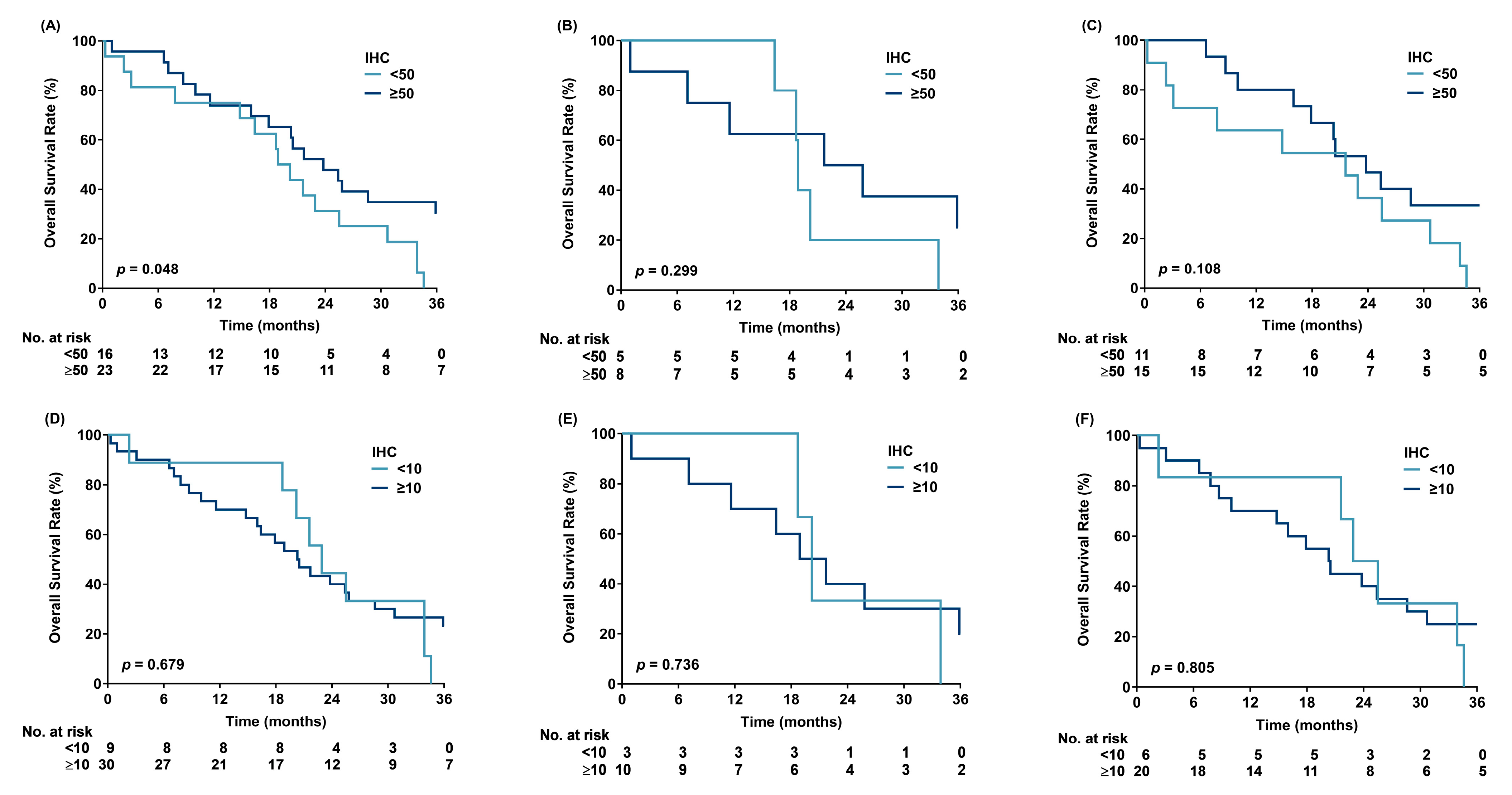
Figure 1. Overall survival based on p53 immunohistochemical positivity: (AC) (≥50% vs. <50%) and (DF) (≥10% vs. <10%). (A,D) Patients harboring missense mutations (MTs); (B,E) patients harboring disruptive MTs; (C,F) patients harboring nondisruptive MTs. IHC: immunohistochemical positivity.
Cancers 17 03577 g001
Figure 2. ROC curve by different cut-off values of p53 immunohistochemical positivity.
Cancers 17 03577 g002
Figure 3. Progression-free survival (AD) and overall survival (EH) based on p53 immunohistochemical positivity (≥50% vs. <50%). (A,E) All patients; (B,F) patients harboring Del19; (C,G) patients harboring L858R; and (D,H) patients harboring other mutations.
Cancers 17 03577 g003
Table 1. Baseline characteristics of the patients.
Variablen(%)
Total83
Sex
Female48(57.8)
Male35(42.2)
Age (years)
<7041(49.4)
≥7042(50.6)
Median (range)71(41–91)
Smoking
Never55(66.3)
Ever/Current28(33.7)
Baseline brain metastasis
No63(75.9)
Yes20(24.1)
EGFR mutations
Del1925(30.1)
L858R43(51.8)
Other15(18.1)
p53 positivity
<5038(45.8)
≥5045(54.2)
p53 mutation
Nondisruptive MT *26(31.3)
WT + Disruptive MT57(68.7)
p53 mutation
Missense39(47.0)
WT44(53.0)
p53 intensity
Strong/Intermediate54(65.1)
Weak/Neg29(34.9)
TKIs
Afatinib6(7.2)
Erlotinib21(25.3)
Gefitinib56(67.5)
* The MT refers to TP53 mutation.
Table 2. Univariate Cox regression analysis of progression-free survival and overall survival in patients with advanced EGFR-mutated adenocarcinoma.
PFSp-ValueOSp-Value
VariableHR(95% CI)HR(95% CI)
Sex
FemaleReference Reference
Male0.85(0.51–1.40)0.5171.32(0.82–2.14)0.257
Age (years)
<70Reference Reference
≥700.58(0.34–0.96)0.0351.19(0.74–1.90)0.477
Smoking
NeverReference Reference
Ever/Current1.01(0.65–1.85)0.7221.65(1.00–2.70)0.050
Baseline brain metastasis
NoReference Reference
Yes1.25(0.71–2.18)0.4401.55(0.90–2.66)0.117
EGFR mutations
Del19Reference Reference
L858R1.68(0.95–2.97)0.0761.24(0.72–2.13)0.438
Other0.94(0.42–2.09)0.8710.98(0.49–1.97)0.961
p53 positivity
<50Reference Reference
≥500.66(0.40–1.08)0.0970.53(0.32–0.84)0.008
p53 mutation
Nondisruptive MT0.93(0.54–1.58)0.7810.85(0.51–1.42)0.531
WT + Disruptive MTReference Reference
p53 mutation
Missense0.76(0.46–1.26)0.2840.83(0.52–1.33)0.437
WTReference Reference
p53 intensity
Strong/Intermediate0.90(0.54–1.52)0.6960.77(0.48–1.26)0.298
Weak/NegReference Reference
TKIs
Afatinib0.62 (0.19–2.01)0.4260.65(0.23–1.80)0.404
Erlotinib0.85(0.48–1.52)0.5870.83(0.48–1.43)0.509
GefitinibReference Reference
Table 3. Multiple Cox regression analysis of progression-free survival and overall survival in patients with advanced EGFR-mutated adenocarcinoma.
PFSp-ValueOSp-Value
VariableHRadj. *(95% CI)HRadj.(95% CI)
Sex
FemaleReference Reference
Male0.40(0.15–1.07)0.0680.84(0.33–2.11)0.707
Age (years)
<70Reference Reference
≥700.50(0.28–0.88)0.0171.59(0.93–2.72)0.088
Smoking
NeverReference Reference
Ever/Current2.11(0.77–5.81)0.1481.94(0.76–4.93)0.165
Baseline brain metastasis
NoReference Reference
Yes1.23(0.68–2.22)0.4951.84(1.03–3.30)0.041
EGFR mutations
Del19Reference Reference
L858R1.81(0.99–3.30)0.0531.15(0.66–2.01)0.623
Other1.08(0.43–2.71)0.8780.74(0.34–1.59)0.441
p53 positivity
<50Reference Reference
≥500.70(0.41–1.20)0.1940.49(0.30–0.81)<0.001
p53 mutation
Missense0.68(0.40–1.15)0.1510.88(0.56–1.42)0.599
WTReference Reference
* The HRadi refers to the hazard ratio adjusted by sex, age, smoking, baseline brain metastasis, EGFR mutations, p53 positivity and TP53 mutation.
Disclaimer/Publisher’s Note: The statements, opinions and data contained in all publications are solely those of the individual author(s) and contributor(s) and not of MDPI and/or the editor(s). MDPI and/or the editor(s) disclaim responsibility for any injury to people or property resulting from any ideas, methods, instructions or products referred to in the content.