Review

Molecular Insights on Signaling Cascades in Breast Cancer: A Comprehensive Review

1
School of Biotechnology, KIIT Deemed to Be University, Bhubaneswar 751024, India
2
School of Applied Sciences, KIIT Deemed to Be University, Bhubaneswar 751024, India
3
Kalinga Institute of Medical Sciences (KIMS), KIIT Deemed to Be University, Bhubaneswar 751024, India
*
Author to whom correspondence should be addressed.
Cancers 2025, 17(2), 234; https://doi.org/10.3390/cancers17020234
Submission received: 22 November 2024 / Revised: 27 December 2024 / Accepted: 1 January 2025 / Published: 13 January 2025
(This article belongs to the Special Issue Cell Migration and Invasion in Cancer)

Simple Summary

Breast cancer is an intricate condition that is caused by aberrant cell signaling regulation induced by the accumulation of genetic and epigenetic alterations. It is well known that various downstream signaling cascades (JAK/STAT, PI3K/Akt, and MAPK) are activated in transformed cells to regulate tumor growth, angiogenesis, metastasis, therapy failure, and stemness. However, the complex signaling networks and their crosstalk is challenged by structural alterations, aberrant gene amplification, and activation of alternative pathways.

Abstract

The complex signaling network within the breast tumor microenvironment is crucial for its growth, metastasis, angiogenesis, therapy escape, stem cell maintenance, and immunomodulation. An array of secretory factors and their receptors activate downstream signaling cascades regulating breast cancer progression and metastasis. Among various signaling pathways, the EGFR, ER, Notch, and Hedgehog signaling pathways have recently been identified as crucial in terms of breast cancer proliferation, survival, differentiation, maintenance of CSCs, and therapy failure. These receptors mediate various downstream signaling pathways such as MAPK, including MEK/ERK signaling pathways that promote common pro-oncogenic signaling, whereas dysregulation of PI3K/Akt, Wnt/β-catenin, and JAK/STAT activates key oncogenic events such as drug resistance, CSC enrichment, and metabolic reprogramming. Additionally, these cascades orchestrate an intricate interplay between stromal cells, immune cells, and tumor cells. Metabolic reprogramming and adaptations contribute to aggressive breast cancer and are unresponsive to therapy. Herein, recent insights into the novel signaling pathways operating within the breast TME that aid in their advancement are emphasized and current developments in practices targeting the breast TME to enhance treatment efficacy are reviewed.

1. Introduction

Based on GLOBOCAN 2022, approximately 20 million newly diagnosed cancers and 9.7 million cancer-associated mortalities have been reported across the globe. Breast cancer is the most frequent malignancy diagnosed in women, accounting for over 11.6% of new cases and 6.9% of deaths worldwide in 2022 [1]. The frequency of this disease has increased globally over the past two decades, despite a significant rise in survival rates. Hashim et al. reported that adjuvant therapy and mammography screening played a major role in improving survival outcomes [2]. Additionally, current chemotherapy, targeted therapy, and immunotherapeutic regimens have become instrumental in the management of advanced stages of breast cancer.
Breast cancer is composed of tumor, stromal, and immune components within the tumor microenvironment (TME), which make the tumor core conducive to promoting various oncogenic functions. Stromal cells such as cancer-associated fibroblasts (CAFs) and immune cells like tumor-associated macrophages (TAMs), T cells, and mast cells initiate the key oncogenic signaling axis in breast cancer within the TME. Further, recent reports suggest that CAF and TAM subsets also play pivotal role in promoting breast tumor oncogenesis by activation of key signaling cascades [3].
Numerous somatic and genetic abnormalities in breast cancer lead to the aberrant activation of crucial signaling cascades that regulate the differentiation of cells, tumor growth, and metastasis [4]. Genes with low mutational frequency should also be considered, as they are involved in cancer signaling pathways along with highly mutated genes. Additionally, variations in epigenetic regulators including ARID1A, KAT6A, and KMT2C can lead to altered gene expression and have a broad spread [5].
Hanahan and Weinberg identified several hallmarks of cancer, including uncontrolled proliferation, genomic instability, and invasion [6]. Targeting cell cycle-regulatory and proto-oncogenic signaling pathways, including Notch, Wnt, NF-κB, Sonic Hedgehog (SHH), ER, PI3K/Akt/mTOR, and HER2, has become an emerging area in cancer research [7].
In this review, we discuss various fundamental molecular pathways and their crosstalk within the breast TME that result in tumor growth, metastasis, immune escape, and therapy resistance. Moreover, tumor metabolism and CircRNA-, miRNA-, and lncRNA-mediated signaling are also highlighted. Furthermore, various novel therapeutic hotspots and their targeted agents in enhancing breast tumor treatment regimen are discussed here.

2. Molecular Classification of Breast Cancer

Human breast cancers are classified by a multifaceted system that provides clinical characteristics, advanced genomic profiling, and histological parameters. Tumors are defined histologically as in situ or invasive carcinomas based on the migration of transformed cells from the lobules or ducts into the stromal component surrounding it [8]. Among all, ductal carcinoma in situ (DCIS), which accounts for about 10–30%, is classified as pre-invasive breast cancer. Invasive carcinomas are categorized based on ducts and lobules such as invasive lobular carcinoma (ILC) and invasive ductal carcinoma (IDC), which account for 10–15% and 60–75% tumor cases, respectively [9]. In addition to their morphological classification, breast cancers are categorized based on ER, progesterone receptor (PR), and HER2 expression. The incidence of these subtypes varies among women based on their receptor status. For instance, about 70% of breast cancers are ER+ve malignancies, whereas HER2+ve tumors can be further classified into HER2+ve ER+ve and HER2+ve ER-ve which account for about 70% and 30%, respectively [9]. Additionally, breast cancer is classified as a luminal A and luminal B subtype. Clinically, luminal A is identified by the expression of ER and/or PR and absence of HER 2 whereas luminal B is characterized by the presence of all three receptors. Luminal B shows a worse prognosis and higher grade of tumors due to the high expression of cell proliferation marker Ki67, while luminal A tumors are clinically low grade [10]. Triple-negative breast cancer (TNBC) (represents 15% among all breast cancers) is the most aggressive and has poor response to therapy due to lack of ER, PR, and HER2 expression. It also has a range of subtypes, the majority of which express CK5 and CK14 cytokeratins as well as EGFR [9]. TNBC cancers typically have an aggressive clinical course associated with a worse chance of survival and a higher grade of diagnosis at a younger age. These cancers predispose sufferers showed early recurrence and metastasis, particularly to the lung, liver, bone, and brain, and account for an abnormally high percentage of breast cancer mortality [11]. Notably, these subtypes and clinical-pathological factors continue to be extremely important when deciding prognosis, therapy choices, and clinical trial design for effective breast cancer management.

3. Novel Treatment Regimens in Breast Cancer

3.1. Chemotherapy

Chemotherapy is one of the effective treatments for several cancers, including breast cancer [12]. In accordance with mode of interventions, chemotherapeutic drugs are classified as immunologic therapy, anti-metabolites, endocrine therapy, antimitotic agents and DNA alkylating agents [13]. Herceptin (trastuzumab), pertuzumab, and ado-trastuzumab are some of the well-known immunologic agents while methotrexate, capecitabine, 5-fluorouracil, gemcitabine, doxorubicin, palbociclib, ribociclib, and lapatinib (Tykerb) are examples of anti-metabolites; goserelin, megestrol acetate, tamoxifen, and letrozole belong to endocrine therapy; cisplatin, carboplatin, and cyclophosphamide are DNA alkylating agents; and ixabepilone, taxanes, and paclitaxel are antimitotic agents [13]. Cisplatin is shown to impede the migration of the cells in both MDA-MB-231 and MCF-7 via inhibition of early EMT [14]. Furthermore, combination of cisplatin with paclitaxel inhibited both cancer growth at primary site and metastasis without any adverse effects using in vivo breast cancer mice model [14]. Interestingly, the combination therapy of 5-fluorouracil with thymoquinone demonstrated a synergistic effect, thereby significantly enhancing apoptosis and controlling cell growth using triple-negative BT-549 and MDA-MB-231 cells [15]. An international, multicentric, phase III randomized trial, the HERA trial (BIG 1-01), showed a significant improvement in long-term disease-free survival (DFS) for cohorts administered with trastuzumab for one year after chemotherapy as compared with the observation arm or intervention of trastuzumab for two years in patients with HER2+ve early breast cancer [16]. Furthermore, another randomized phase III (PRECIOUS) clinical study evaluated the retreatment efficacy of pertuzumab in HER2+ve metastatic breast cancer patients formerly administered with pertuzumab, trastuzumab, and physician’s choice of chemotherapy (PTC). The results showed better overall survival (OS) of cohorts receiving pertuzumab retreatment in a group that was previously treated with pertuzumab, making it more effective in the third or fourth line of chemotherapy [17]. PD-L1-expressing advanced TNBC patients possessing an overall positive score of 10 or above exhibited a significant increase in OS upon receiving pembrolizumab treatment with chemotherapy when compared with the control (KEYNOTE-355) [18].

3.2. Immunotherapy

The advent of the discovery of immune checkpoint inhibitors coupled with several pieces of evidence suggesting the involvement of the immune system in shaping the tumor core and tumor immune microenvironment (TIME) have paved the way for novel and effective therapeutic strategies for breast cancer, including TNBCs [19]. The IMpassion130 trial demonstrated the efficacy of treatment with atezolizumab and nab-paclitaxel in extending progression-free survival (PFS) in patients with metastatic TNBC [20]. However, owing to the lack of clinical benefits, the FDA revoked the approval for atezolizumab in breast cancer treatment [21]. Similarly, evaluation showed that the combination of atezolizumab with paclitaxel failed to improve the PFS and OS in metastatic TNBC patients (IMpassion131) [22]. A phase Ib study involving hormone receptor-positive (HR+ve) and HER2-ve metastatic breast cancer patients receiving treatment of abemaciclib plus pembrolizumab with/without anastrozole evaluated the efficacy and safety of the combination [23]. Even though this combination of drugs showcased anti-tumor activity, it showed adverse side effects including pneumonitis led to the termination of further evaluation [22]. However, the combination of palbociclib, letrozole, and pembrolizumab in patients with HR+ve and HER2-ve stage IV metastatic breast cancer was well tolerated and demonstrated a PFS of 25.2 months [24]. The combination of pembrolizumab with trastuzumab demonstrated anti-tumor activity in patients with PD-L1+ve, metastatic, trastuzumab-resistant, HER2+ve breast cancer [25]. However, the atezolizumab plus trastuzumab emtansine combination was associated with an increase in adverse side effects rather than any significant improvement in PFS of PD-L1+ve and HER2+ve advanced breast cancer patients [26].

3.3. Targeted Therapy

Breast cancer heterogeneity demands that treatment regimens and therapies be precise and well targeted to a specific grade, subtype, and stage [27]. The addition of exemestane, an aromatase inhibitor to everolimus, an mTOR inhibitor, demonstrated tolerability, safety, and efficacy in HR+ve and HER2-ve metastatic breast cancer patients [28]. In a phase II clinical study involving postmenopausal women with HR+ve and HER2-ve metastatic breast cancer, a combination of tamoxifen with everolimus significantly increased OS and clinical benefit as compared with the control cohorts [29]. In HR+ve, HER2-ve, and PIK3CA-mutated breast cancer patients with previous endocrine treatment, the combination of α-specific PI3K inhibitor, alpelisib (BYL719), with fulvestrant demonstrated longer PFS [30]. In addition, combination of paclitaxel with pictilisib (GDC-0941)—a PI3K inhibitor with/without trastuzumab or bevacizumab—or letrozole showcased anti-tumor activity along with fewer adverse effects [31]. In HER2-ve metastatic gBRCA-associated breast cancer patients, combination of veliparib with carboplatin and paclitaxel yielded a significant improvement in PFS without altering the toxicity profile of carboplatin and paclitaxel [32]. Olaparib monotherapy in HER2-ve metastatic breast cancer patients bearing BRCA mutation showed an increase in PFS by 2.8 months and reduction in death or disease progression by 42% when compared with the standard therapy [33]. Recent FDA-approved drugs for breast cancer treatment are summarized in Table 1.

4. Oncogenic Signaling in Breast Cancer

Cell signal transduction is crucial in breast cancer development and progression. Alterations to various cell signaling cascades regulate tumor cell proliferation and survival. Dysregulation of signaling pathways, such as TGF-β, Wnt, Hedgehog, Notch, IL-6, Integrins, VEGF, HER2, EGFR, PI3K/Akt, and MAPK, leads to breast cancer cell proliferation and metastasis, as depicted in Figure 1.

4.1. Involvement of EMT in Breast Cancer Invasion and Metastasis

Epithelial-to-mesenchymal transition (EMT) is a process by which epithelial cells gain mesenchymal properties, crucial for cancer propagation and metastasis. The mesenchymal-to-epithelial transition (MET) allows for mesenchymal cells to re-differentiate into epithelial cells [34]. This transition plays a role in wound healing, fibrosis, and tumor progression, but cancer cells during EMT gain migratory abilities crucial for malignancy. They can invade other tissues through blood or lymph vessels, where they undergo MET, converting back into epithelial cells. The stem cell characteristics of EMT-derived tumor cells make them resistant to therapy, leading to the development of therapies targeting EMT for metastatic patients [35].
Transcription factors (TFs) regulate gene expression by binding to chromatin, playing a crucial role in cancer advancement and metastasis. EMT-TFs can inhibit E-cadherin, a protein facilitating cancer cell invasion [36]. Loss of E-cadherin is linked to TGF-β upregulation, reactive oxygen species, and changes in apoptotic signaling pathways [37]. Inhibiting E-cadherin increases mesenchymal cell markers like N-cadherin and vimentin. TWIST1, SLUG, SNAIL, ZEB1, ZEB2, and FOX families inhibit E-cadherin, impacting cancer initiation, progression, invasion, metastasis, and treatment resistance [38]. EMT-TF overexpression in breast cancer predicts metastasis and promotes cancer growth. TWIST1 enhances E-cadherin promoter hypermethylation and hypoacetylation, driving metastasis while knockdown of TWIST1 inhibits metastasis in breast cancer [39]. Further, SNAIL inhibits E-cadherin expression via vimentin upregulation, facilitating EMT [40]. SNAIL interacts with Suv39H1, contributing to breast cancer metastasis through epigenetic processes [41]. The ZEB family suppresses E-cadherin expression, promoting vimentin and N-cadherin expression. ZEB1 acts as an interactive partner with AP-1 to form a transactivation complex with YAP and stimulate aggressive claudin-low subtype of breast cancer [42]. These findings highlight the significance of EMT-TFs in cancer progression and underscore their potential as therapeutic targets for treating metastatic breast cancer.
Current research depicts that cells with stem cell properties are indispensable for the growth and spread of tumors [43,44]. While the exact origin of cancer stem cells (CSCs) remains elusive, biomarkers for breast cancer stem cells (BCSCs) have been identified, such as CD44+ve/CD24−ve/low cells that exhibit aggressive behavior and drug resistance [45]. Moreover, high levels of aldehyde dehydrogenase 1 (ALDH1) activity have been associated with poor breast cancer clinical outcomes [46,47]. These BCSCs show enhanced self-renewal and tumor initiation abilities and express markers like TWIST1 and FOXF2 [48]. Studies have shown that SLUG increases cytoplasmic β-catenin stability by inducing certain inflammatory factors such as IL-8 and TNF-α, while TWIST1 regulates CD24 expression, promoting stem cell characteristics in breast cancer [49,50]. Transcription factors like Lin28B and CCAAT/enhancer binding protein δ (C/EBPδ) also play important roles in the metastatic niche formation, whereas Lin28B is involved in lung metastases of breast cancer [51,52]. Additionally, IL-6 supports CSC survival and treatment resistance, while C/EBPδ contributes to CSC maintenance and metastasis through its linking with hypoxia-inducible factor-1 (HIF-1) and IL-6 [53]. Overall, understanding the role of CSCs and related transcription factors is crucial in developing targeted therapies for breast cancer metastasis.

4.2. Angiogenesis

Angiogenesis is a process of the formation of new blood vessels induced by various pro-angiogenic factors and is predominantly involved in embryonic development and wound healing in physiological conditions. In pathophysiological conditions like cancer, blood vessels carry and supply essential nutrients for promoting tumor growth, survival, proliferation, and metastasis. Further, this process is governed by pro-angiogenic and anti-angiogenic factors, where vascular endothelial growth factor (VEGF) is a pivotal player while other factors coordinate to build an environment to foster a vascular network for tumor progression and development.

4.2.1. VEGF-Dependent Angiogenesis

The VEGF family consists of seven components: placental growth factor (PlGF), VEGF-A, VEGF-B, VEGF-C, VEGF-D, VEGF-E, and snake venom vascular endothelial growth factor (svVEGF) [54]. VEGF-A is a key secretory factor that modulates cell mitosis, vascular permeability, and human endothelial functions [55]. It exists in various isoforms, including VEGF121, VEGF165, VEGF189, VEGF206, VEGF111, and VEGF145, that vary in molecular size and tissue distribution [56]. It also contributes to cell homeostasis, invasion via autocrine or paracrine mechanisms, and hematopoietic stem cell as well as tumor cell survival. Furthermore, VEGF-A is an essential angiogenesis regulator, having critical roles in tumor growth, proliferation, angiogenesis, invasion, metastasis, and treatment resistance [57]. VEGF induces angiogenesis by interacting with its primary receptors: VEGFR-1/Flt-1, VEGFR-2/KDR, and VEGFR-3/Flt-4 [58]. Zhu et al. have demonstrated that IGF-1 enhances the expression of VEGF-C via the PI3K/Akt and MAPK/ERK1/2 signaling pathways in MDA-MB-231 breast cancer cells. Additionally, they have proposed that the IGF-MAPK/ERK1/2-VEGF-C and IGF-1-PI3K/Akt-VEGF-C signaling pathways are crucial for lymph angiogenesis in breast cancer [59]. VEGFR-2 is a key mediator of angiogenesis. VEGFR-2-axis induces cell proliferation through MAPK/ERK signaling cascade as well as cell survival, migration, and vascular permeability via the FAK/PI3K/Akt signaling [60].
Breast cancer frequently exhibits overexpression and activation of HIFs, which are key regulators for cells to adjust to low cellular oxygen levels. In response to hypoxia, HIFs alter the primary transcriptional response of downstream pathways and target genes involved in glycolysis, angiogenesis, and metastasis [61,62]. Hypoxic conditions in tumors can promote growth and survival by inducing angiogenesis in response to osteopontin (OPN) and VEGF [63].
Moreover, pathways such as ILK/Akt/NF-κB/p65 axis or FOXO3a dependent MelCAM upregulation controls angiogenesis in breast cancer [63,64]. Breast tumor angiogenesis can be mediated by autocrine, juxtacrine, and paracrine mechanisms via the OPN/NRP-1/Brk/NF-κB/ATF-4 signaling pathway [65]. Von Willebrand Factor (vWF), a pro-angiogenic factor, promotes angiogenesis by triggering VEGF-A via PI3K/Akt/miR-205-5p pathway in breast cancer cells [66]. Aurora kinase A (AURKA) found in spindle microtubules and centrosomes aids cell division in normal conditions. In breast cancer, elevated AURKA levels lead to VEGF-dependent angiogenesis via the ERK pathway [67]. Moreover, deciphering these pathways may aid in the development of targeted therapies for breast cancer treatment.

4.2.2. VEGF-Independent Angiogenesis

Studies on tumor vascularity mainly focus on VEGF-dependent angiogenic therapy; however, it is also crucial to investigate VEGF-independent tumor angiogenesis. Wan et al. have reported that FOSL2 activates Wnt5a transcriptional signaling and thus promotes angiogenesis in cancer-associated fibroblasts (CAFs) [68]. CAFs play a major role in tumor growth and metastasis. This facilitates the pre-metastatic niche through lncRNA SNHG5 by enhancing the vascular permeability in breast cancer [69]. Pearson’s correlation analysis suggested a link between CD31/34 and lncRNA NR2F1-AS1 and further proved that lncRNA NR2F1-AS1 activates IGF-1/IGF-1R/ERK pathways that promotes angiogenesis and metastasis using in vitro and in vivo breast cancer models [70]. Cao et al. investigated decylubiquinone (DUb), a coenzyme that suppresses angiogenesis by targeting the ROS/p53/BAI1 signaling pathway [71].
Integrins, a type of cell adhesion receptor, play important roles in cell–cell and cell–matrix interaction in normal cells whereas they dysregulate in tumor cells. Abnormal conditions such as hypoxia and glycosylation alter the function of several integrins, resulting in the activation of key signaling cascades upon interaction with pro-tumor secretory factors such as VEGF, FGF, and PDGFR2; therefore, integrins are a novel target for angiogenesis in cancer therapy [54]. ATN-161 (Ac-PHSCN-NH2) consists of five amino acids, commonly binds to integrin (α5β1), and thereby suppresses the angiogenesis and bone metastasis in breast cancer [72]. Excessive neovascularization or vascular mimicry in breast tumor-initiating cells (BTIC) increase the occurrence of TNBC development. ASB10 (estrogen receptor ER-α trans-activated E3 ligase) ubiquitylates TEM8 (tumor endothelial marker 8); thus, the deficiency of ASB10 resulted in high accumulation of TEM8, leading to enhancements in vascular mimicry (VM). A high level of TEM8 enhances the active RhoC level, which promotes ROCK1-dependent SMAD5 phosphorylation, further enhancing the stemness and VM in breast cancer cells [73]. Vimentin, in its extracellular region, mimics as VEGF, acts as a stimulator to upregulate VEGFR-2 and PD-L1 receptors, and promotes angiogenesis [74].

4.3. Cancer Stem Cells

The multipotent capacity of BCSCs has been implicated in enhancing several hallmarks of cancer within the TME. In the breast TME, mammary stem cells (MaSCs) were primarily found in the outer basal areas and consisted of diverse subsets of BCSCs with distinct molecular signatures [75]. Similarly, other conventional stem cell markers such as CD133-, CD44-, CD49f-, and ALDH-expressing stem cells regulate various signaling networks, including the Notch, Hedgehog, and Wnt pathways, for maintenance and proliferation in breast cancer.
BCSCs exhibit oncogenic features such as tumor growth, metastasis, tumor angiogenesis, immunomodulation, and treatment resistance. For instance, BCSC secretome analysis revealed that MIF activates Wnt/β-catenin signaling to upregulate the glycolytic enzyme aldolase C through c-MYC transcription [75]. Further, Xie et al. reported that overexpression of XB130 in various malignancies is linked to poor survival and increased EMT, a necessary cellular phase for BCSC induction, and accelerated tumor initiation through the Wnt/β-catenin signaling pathway [76]. In addition, the co-transcription factor Limb–Bud-Heart (LBH) targets genes involved in Wnt/β-catenin signaling and activates stem cell transcription resulting in breast cancer metastasis [77]. A previous study found that blocking cadherin 11 downregulates Wnt/β-catenin signaling pathway, resulting in a decrease in the cancer stem-like phenotype [78]. Overexpression of CCL2 enhances stemness and macrophage polarization in breast cancer via STAT3 and Notch-1 signaling cascade, whereas silencing it increases tumor necrosis and autophagy, resulting in reduced CSC population and macrophage recruitment. The same impact was not found upon neutralization of CCL2 [79]. Further, binding of KK-LC-1 to FAT1 increases its ubiquitination and degradation. This disrupts the Hippo pathway, resulting in nuclear translocation of YAP1 and ALDH1A1 transcription. Z839878730 (Z8) is a small-molecule inhibitor that can interfere with KK-LC-1 and FAT1 interaction, resulting in reduced stemness in TNBC [80]. SDF-1/CXCL12 stimulates the NF-κB pathway, resulting in high ALDH activity and upregulation of OCT-4, Nanog, and SOX2, increasing the BCSC population, leading to the metastasis of MCF-7 cells [81]. Shan et al. discovered that overexpression of CXCL12 induces MCF-7 cells to develop BCSC phenotype through the Wnt/β-catenin pathway, leading to enhanced metastasis [82]. Polymorphonuclear myeloid-derived suppressor cells regulated by CCL20 derived from cancer cells increased stemness via the CXCL2-CXCR2 pathway [83]. Moreover, the lncRNA LINC00115 enhances chemoresistant stem-like cells, enriches stemness, and promotes metastasis via the SETDB1/PLK3/HIF1α axis in breast cancer [84].
Moreover, the link between EMT and CSCs is well established. For instance, breast cancer cells’ stemness and tumorigenicity are linked to the attainment of a hybrid or partial E/M phenotype. Liu et al. have shown the link between the heterogeneity in BCSCs and EMT. The CD24−ve/CD44+ve/ALDH+ve subpopulation with intermediate EMT state possess the maximum potential for stem cell enrichment and carcinogenesis [85]. Pasani et al. demonstrated that enrichment of intermediate E/M phenotypes promotes stemness, which is more likely to occur in these phenotypes than in “pure” E/M phenotypes using a mechanism based mathematical models [86]. Brown et al. found that CBFβ stabilized and maintained metastatic potential in three single-cell clones in the intermediate E/M stage. The study found that CBFβ had a higher prognostic value for survival outcomes than EMT score alone [87].
Tumor CSCs have diverse metabolic states and can adapt to different conditions. Somatic stem cells, embryonic stem cells, and induced pluripotent stem cells have all exhibited elevated glycolysis activity to preserve their stem cell characteristics. Luo et al. demonstrated the transition of epithelial to mesenchymal state depends on the modulation of redox metabolism [88]. By lowering ROS and ferroptosis, NRF2 mirrored ZMYND8 and improved breast cancer stemness and tumor formation. The loss of NRF2 abolished ZMYND8 effects on antioxidant genes and ROS in mammospheres. Interestingly, NRF2 directly affected ZMYND8 expression in mammospheres [88].

4.4. Tumor Metabolism

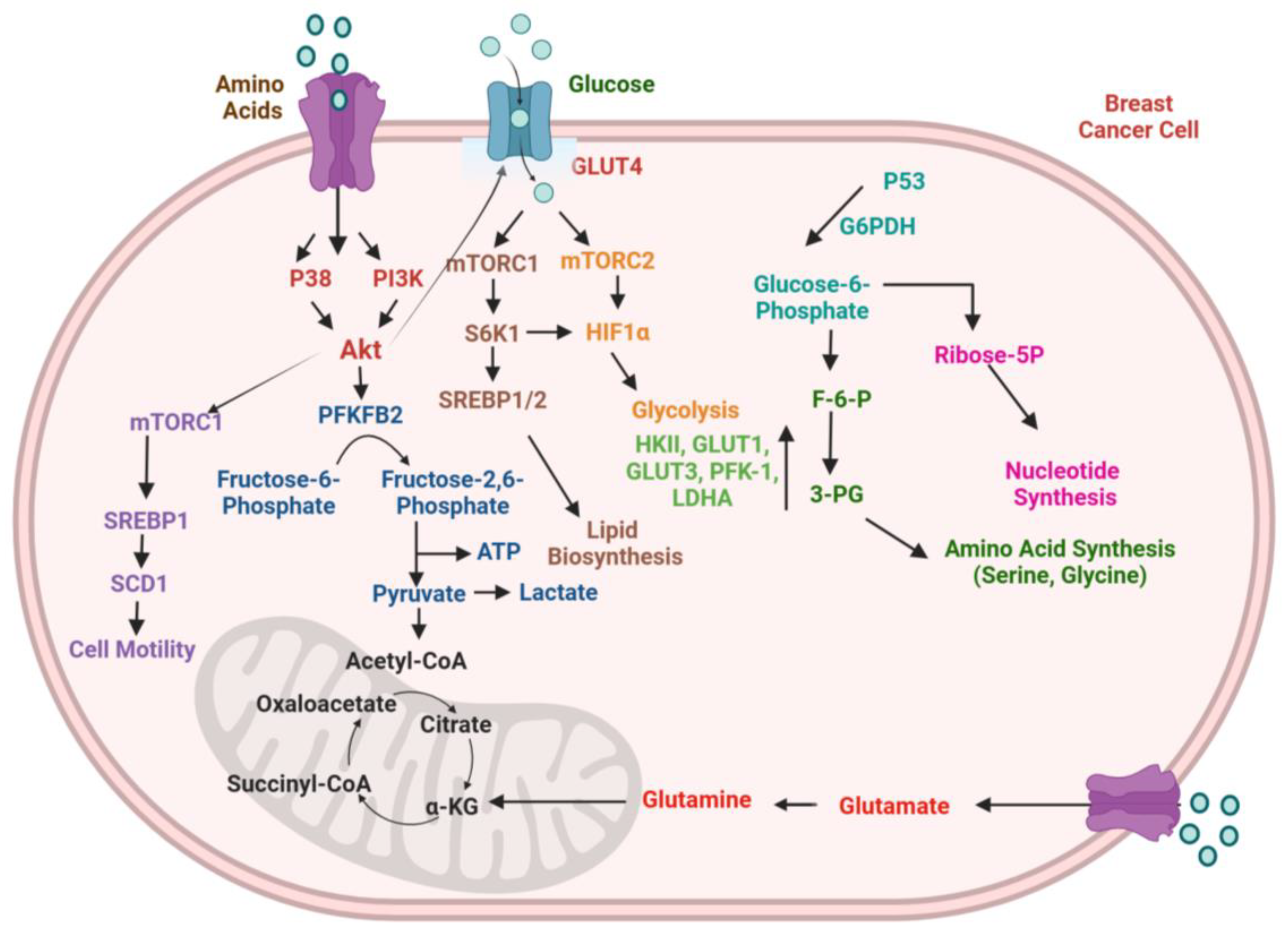
Breast cancer, like other malignancies, has been linked with metabolic reprogramming. Breast cancer manifests improved glycolysis, glutamate metabolism, pentose phosphate pathway (PPP), tricarboxylic acid (TCA) cycle, and lipid metabolism, across energy metabolic pathways. These pathways are rewired to promote breast cancer growth, proliferation, and migration [89]. HIF, TP53, c-Myc, extracellular acidification, and interactions with immune cells, CAFs, and adipocytes are a few of the intrinsic factors that have been documented to be mutated or inactivated in breast cancer cells. The underlying cause for metabolic reprogramming in the progression of breast cancer is the abnormal expression of several signaling and transcription factors associated with energy metabolism networks, as shown in Figure 2 [90].

4.4.1. Glucose Metabolism

The connection between breast cancer and aerobic glycolysis has been closely studied recently [91]. The key molecular signaling involved in the regulation of aerobic glycolysis in breast cancer are the mTOR, PI3K/Akt, and AMPK pathways. PI3K/Akt modulates the phosphorylation and activation of phosphofructokinase-2 (PFK-2) [92,93]. GLUT-1 overexpression results from activation of the PI3K/Akt signaling pathway, and this leads to its translocation from the cytoplasm to the plasma membrane [94]. Beta-estradiol (E2) activates Akt, which causes GLUT-4 to move to the plasma membrane and increases the uptake of glucose in the MCF-7 cell line [95]. PIK3CA and Akt1 gene mutations are frequently observed in breast cancer, with PIK3CA mutations primarily being detected in HER2+ve and ER+ve breast cancer [96,97]. Estrogen causes overexpression of c-myc, and approximately 80% of breast tumors are ER+ve [98,99]. Glycolysis-related enzymes like phosphofructokinase (PFK) are expressed more when HIF-1α expression is promoted and the switch to glycolysis from OXPHOS is stimulated by mTORC1 whereas mTORC2 promotes glycolysis by inducing Akt and GLUT-1-associated glucose absorption [100,101,102]. Moreover, HIF-1α boosts the expression of molecules linked to glycolysis, such as HKII, PFK-1, GLUT-1, GLUT-3, and lactate dehydrogenase (LDH) A [103]. However, p53 gene alterations are seen in the majority of malignancies, including breast cancer [104]. p53 inhibits the expression of GLUT-1, GLUT-3, GLUT-4, and phosphoglycerate mutase (PGM); thus, p53 mutation causes enhanced glycolysis in breast cancer [104]. Furthermore, p53 controls glycolysis and GLUT via mTOR and AMPK, and the PI3K/Akt/mTOR pathway stimulates the production of glycolytic enzymes and GLUT [103,105].
Lymphocytes in the TME have been linked to better prognosis and survival in various cancers and are implicated in anti-tumor immune responses. Immune cell failure, tumor cell proliferation and invasion are facilitated by abnormal glucose metabolism in the TME. The regulation of T cell function is somehow accomplished through metabolic modulation, and glucose metabolism plays a crucial role in the proliferation, activation, and function of T cells. The primary Ca2+ influx channel in T cells, for instance, is store-operated Ca2+ entry (SOCE). By controlling T cell glucose metabolism, the SOCE/calcineurin/NFAT pathway can regulate T cell development, proliferation, and function. Evidence suggests that an acidic TME, as well as elevated lactic acid, can have a major impact on macrophages [106]. For example, reduced pH in the TME can affect macrophage phenotype and functionality independently [107]. Lactic acid, in particular, produced by cancer cells, plays an important signaling role in the TME by inducing M2 polarization [108]. Furthermore, when M1 and M2 macrophages were incubated at pH 7.4 or 6.8, M2 macrophages demonstrated greater survivability and fitness in the lower pH than their M1 counterparts [109]. Pro-inflammatory M1 markers (e.g., iNOS, MCP1, IL-6) were similarly reduced at acidic pH, but M2 markers (e.g., MRC1, arginase 1 (Arg1), chitinase-3-like protein) were increased [109].

4.4.2. Lipid Metabolism

The reprogramming of lipid metabolism in cancer has been attributed to cancer-associated signaling pathway activation and interactions within the TME [110]. Lipid metabolism holds significance in tumor immunogenicity by regulating the activities of non-cancerous cells in the TME, particularly immune-associated cells [111].
In breast cancer, a decreased level of GPX4, a critical factor that regulates the oxidation of glutathione, prevents lipid peroxide formation as well as ferroptosis [112]. It also increases the production of pentaspanin protein prominin-2, which enables the formation of ferritin-containing exosomes. Both breast cancer cells and mammary epithelial cells become resistant to ferroptosis as a result of this mechanism [113]. Interestingly, GPX4 expression has shown an exceptional prognostic potential in breast cancer neoadjuvant therapy, and a high level of GPX4 is significantly correlated with metastasis-free survival [114]. Stearoyl–CoA desaturase (SCD1) is an important modulator of ferroptosis. PI3K/Akt/mTOR pathway activation results in enhanced synthesis of monounsaturated fatty acids, which leads to the increase in cell motility through downstream sterol regulatory element-binding protein 1 (SREBP1)-mediated upregulation of SCD1. This mechanism safeguards breast cancer cells from ferroptosis induced by ROS [115]. Sphingodylcholine (SPC) is a lipid mediator found in the blood that controls both apoptosis as well as autophagy. Interestingly, autophagy negatively regulates SPC-mediated apoptosis in TNBC cell lines. SPC promotes apoptosis by blocking c-JNK signal transduction and induces autophagy via the Akt/p38 signaling pathway [116].
EMT in breast cancer is fueled by various mechanisms that are influenced by the numerous lipid metabolic pathways and their intermediates. SREBP1 recruits the Snail/HDAC1/2 complex to decrease the expression of E-cadherin, and miR-18a-5p has been recognized as a possible regulator of SREBP1. SREBP1 upregulation and miR-18a-5p inhibition both notably increase the likelihood of breast cancer metastasis [117]. Sphingomyelin homeostasis is majorly regulated by the overexpression of sphingomyelin synthase 2 (SMS2) in breast cancer. By elevating TGF-β1 activity, it activates the TGF-β/Smad signaling pathway, which promotes EMT and increases breast cancer cell invasion and metastasis [118]. Targeting altered lipid metabolism processes may have potential as an anticancer treatment.

4.4.3. Immunometabolism

The abnormal metabolism in tumor cells leads to an acidic, hypoxic environment. In the aberrant metabolic microenvironment, the immune system experiences nutrient deprivation and metabolic alterations that impact their activation [119]. For example, CD28 promotes glucose absorption and glycolysis through the PI3K/Akt signaling pathway, enabling T cells to maintain their activity [105]. When T cells receive PD-1 signals, they utilize enhanced FAO to produce energy and lipolysis by elevating the expression of ATGL and CPT1A. Also, CTLA-4 may inhibit glycolysis without increasing FAO [120]. Recent investigations show that PD-1 signaling plays a role in the FAO in T cells [121]. Clinically employed checkpoint blockade antibodies against CTLA-4, PD-1, and PD-L1 restore glucose in the TME, allowing for T cell glycolysis and generation of IFN-γ. PD-L1 is a crucial regulator of tumor glucose metabolism, as demonstrated by studies indicating that inhibition of PD-L1 disrupts glycolysis by suppressing mTOR activity and decreasing glycolytic enzyme expression [122]. PD-L1 depletion can lower the rate of glycolysis by suppressing the expression of glycolytic enzymes and mTOR activity, indicating that PD-L1 may be essential for the uptake of glucose in cancer cells [122,123].

4.5. Autophagy

The essential cellular function that includes the disintegration and recycling of intracellular components is autophagy [124,125]. Based on the method by which protein enters lysosomes, autophagy may be categorized into four groups—microautophagy, macroautophagy, selective autophagy, and chaperone-mediated autophagy [126]. While cells are under stressful conditions, ULK1 is directly or indirectly activated, which phosphorylates Beclin-1, and ATGs, in turn, permits the assembly of molecular complexes and the initiation of phagophore formation [127,128]. Lower Beclin-1 expression is observed in almost 70% of breast cancer specimens [129]. As a result of Beclin-1 gene overexpression in breast cancer, MCF7 cells have been shown to increase autophagic activity; thereby, cell growth and tumor formation are reduced using in vivo breast cancer models [130]. The next stage of the autophagic pathway is the elongation stage, which involves a variety of proteins like ATG12, ATG10, ATG16, ATG7, ATG3, ATG5, LC3, and others [131]. During the maturation stage, autophagosomal membrane degradation is started by SQSTM1(p62), which interacts directly with LC3 and ubiquitination-related proteins [132].
Numerous studies have revealed an important connection between autophagy and the activation of different signaling pathways either directly or indirectly in breast cancer. One of the most extensively examined pathways associated with autophagy is the Akt-mTOR signaling pathway. Knockdown of OPN suppresses the PI3K/Akt/mTOR signaling pathway and increases autophagy via regulating the expression of αvβ3 integrin [133]. Eugenol induced apoptosis and autophagy by inhibition of the PI3K/Akt/FOXO3a signaling pathway in breast cancer cells [134]. Chaga mushroom extract (CME) treatment enhances LC3 expression and AMPK phosphorylation but decreases S6, S6K1, and mTOR phosphorylation. These findings imply that CME promotes autophagy by inhibiting the AMPK pathway [135]. The NF-κB signaling pathway is well connected with the autophagic pathway in breast cancer. It has been reported that miR-1910-3p, which targets MTMR3 and activates the NF-κB pathway within exosomes, increases proliferation, autophagy, and metastasis in breast cancer [136]. Through the JNK1-Bcl2 signaling pathway, BMP4 has been shown to cause protective autophagy and apoptosis in breast cancer [137]. Additionally, autophagy has the ability to influence the activation of some tumor signaling pathways such as the knockdown of ATG4A, which suppresses Wnt pathway-related protein expressions [138]. Thus, targeting autophagic molecular cascades could open new dimensions in the treatment of breast cancer.

5. Stromal Cell-Mediated Signaling in Breast Cancer

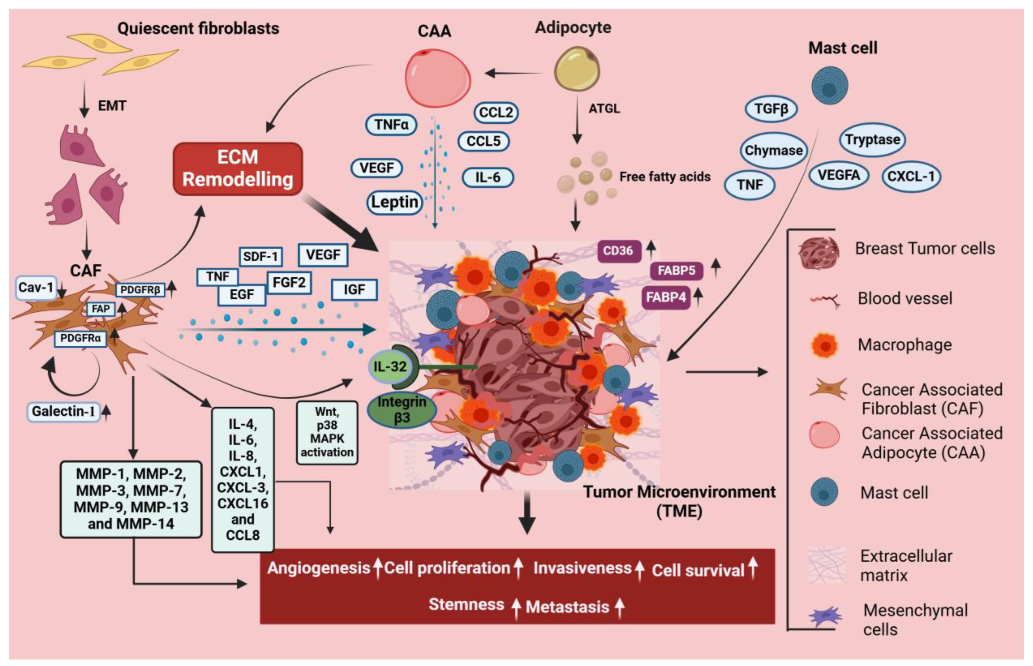
Breast cancer growth is strongly governed by stromal cells, such as mast cells, adipocytes, tumor-associated macrophages (TAMs), and CAFs, which facilitates invasion, angiogenesis, and immune suppression by promoting tumor growth through TGF-β, VEGF, and cytokine signaling, as illustrated in Figure 3. Mast cells provide pro-inflammatory signals that promote tumor growth, whereas adipocytes provide energy and secrete substances that improve cancer cell survival.

5.1. CAFs

The TME comprises numerous cell types that participate in the enhancement of tumorigenicity and modulate cancer aggressiveness. Breast cancer consists of neoplastic cells and is also witnessed with significant alterations in the TME. Several components in the breast TME suppresses immune cells, secrete soluble factors, and alter functionality of the extracellular matrix, which together act to promote breast cancer progression, anti-tumor immunity, and metastasis [139].
CAFs comprise the major part of the tumor stroma and affect the TME by aiding in cancer proliferation, angiogenesis, invasion, and metastasis [140]. Studies suggest that CAFs contribute greatly to regulating and shaping the tumor metabolism through dysregulation of several metabolic pathways, including amino acid, glucose, and lipid metabolism [141]. Activation of CAFs is an irreversible process, and almost 80% of the stromal fibroblasts in breast cancer attain an altered phenotype manifested by the secretion of elevated levels of growth factors, cytokines, and MMPs [142]. Upon activation, these CAFs interact with the neighboring tumor cells continuously, which leads to breast cancer progression by releasing growth factors such as fibroblast growth factor 2 (FGF 2), insulin-like growth factor (IGF), CXCL12 or stromal-derived growth factor (SDF-1), EGF, TNF, VEGF2, as well as cytokines and chemokines such as CCL8, CXCL16, IL-4, IL-6, IL-8, CXCL1, and CXCL3, which increase breast cancer cells’ motility [143]. The metabolic reprogramming of CAFs is initiated in the TME, involving events like the Warburg effect, shifts in Kreb cycle metabolites, and an increased rate of oxidative phosphorylation that serves as the energy source for breast tumor growth and invasion [144]. Luga et al. reported the production of exosomes by CAFs, which boost the ability of breast cancer cells to metastasize by activation of Wnt signaling [145]. Studies have shown that CAFs elevate cancer cell proliferation, invasion, and metastasis by secreting MMPs such as MMP1, MMP2, MMP3, MMP7, MMP9, MMP13, and MMP14 using in vivo and in vitro breast cancer models [143]. It has also been observed that galectin-1, highly expressed in CAFs, regulates the activation of CAFs and leads to upregulation of MMP-9, which further promotes metastasis in MDA-MB-231 cells [146]. Moreover, in breast cancer, the loss of caveolin-1 in stromal fibroblasts is observed to be an independent predictor of nodal metastasis, tumor recurrence, and poor clinical prognosis, whereas elevated levels of caveolin-1 are associated with increased survival [147,148]. Wen et al. demonstrated that CAFs produce IL-32, which further binds to integrin β3, thereby activating p38MAPK signaling, thus enhancing the expressions of fibronectin, vimentin, and N-cadherin in the breast cancer cells [149]. Tchou et al. showed that CAFs derived from HER2+ve breast cancer prominently augmented invasive properties involving pathways concerned with migration of cancer cells, and that these cells confer more invasiveness than TNBC and ER+ve cancers [150]. Choi et al. have demonstrated that CAFs enhance transmigration of breast cancer cells via the blood–brain barrier by boosting the expression of αvβ1 and α5β1 integrins [151]. Moreover, enhanced levels of all CAF-related proteins such as PDGFRα, PDGFRβ, and FAP are reportedly associated with cancer invasiveness and more likely to be found in HER2 subtypes than in TNBC [152]. Hence, targeting the tumor–stroma interaction serves as a promising ground for the advancement of therapeutics, potentially augmenting the existing treatments and preventive options for breast cancer [153].

5.2. Adipocytes

Adipocytes in the TME possess a multifaceted role in triggering the interaction between the stromal compartment and breast tumor core. The role of adipocytes in breast cancer growth is quite evident, considering that adipocytes have metabolic and endocrine functions along with the storage of fatty acids [154]. It has been observed that upon prolonged interaction with tumor cells, almost all lipid droplets disappear from adipocytes, resulting in remarkable changes in morphology, more likely towards a fibroblast-like shape [155]. The loss of lipid content from the adipocytes in tumor stroma indicates that free fatty acids are released from these cells and transferred to the tumor cells, and this transfer of fatty acids from adipocytes to tumor cells via the adipose triglyceride lipase (ATGL)-dependent lipolysis pathway boosts tumor growth in vivo and in vitro, supporting the overexpression of pro-inflammatory cytokines and proteases, which leads to the activation of cancer-associated adipocytes (CAA) [156,157]. The regulatory mechanisms of CAA in breast cancer are complex, including inflammatory adipokine secretions such as IL-1β, CCL2, TNFα, CCL5, and leptin, as well as IL-6 metabolic reprogramming by altering the metabolism of macronutrients and remodeling of the extracellular matrix [158,159]. Earlier reports revealed that fatty acids from adipocytes cause breast cancer cells to undergo metabolic reprogramming [156]. This involves an increase in mitochondrial fatty acid oxidation, uncoupled with an increase in anaerobic glycolysis or ATP synthesis [156]. It has further been observed that adipocyte-secreted free fatty acids result in the activation of AMPK in breast cancer cells, which is a key autophagy regulator [156]. Zaoui et al. have demonstrated that adipocyte-conditioned media stimulates CD36 expression in breast cancer cells, and CD36 activity contributes to adipocyte-induced cancer cell migration and invasion [160]. Obese people with breast cancer have been found to have higher levels of FABP4, which is associated with aggressiveness and stemness in breast cancer [161]. Higher levels of FABP5 in breast cancer cells that interact with adipocytes are associated with increased cancer aggressiveness [162].

5.3. Mast Cells

Mast cells play a significant role in both autoimmune and chronic inflammation of illnesses, as well as playing a role in the growth of tumors. It is widely recognized that mast cells induce neovascularization and angiogenesis by secreting VEGF, FGF-2, PDGF, and IL-6 to the tumor stroma in addition to nonclassical pro-angiogenic drivers such as proteases, particularly tryptases and chymases [163]. Mast cells are known to localize at the margins of tumor stroma, commonly around the vessels [164]. Mast cells accumulate in the TME by the release of chemoattractants by tumor cells, including SCF or CCL15 [163]. The resting state of mast cells is generally less abundant in breast tumors compared to normal tissues, whereas the activated phenotype of mast cells is seen in increased numbers in breast tumors [165]. Targeting mast cells represents a potential strategy for therapeutics because they represent the key components of immune tumor infiltration and play a crucial role in angiogenesis [166].

5.4. TAMs

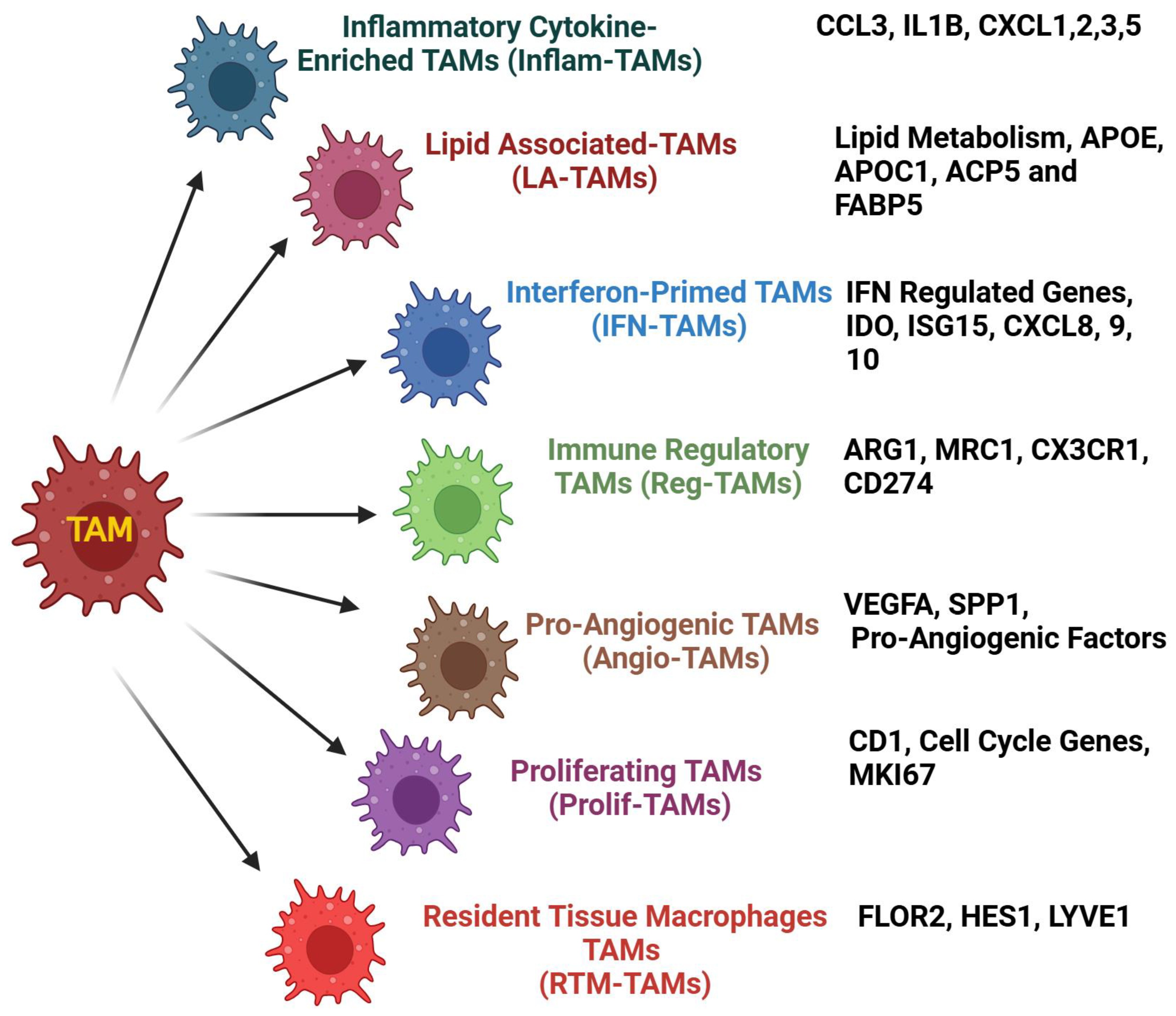
Within the TIME, TAMs showed their magnificent ability to promote cancer development. However, TAMs possess a robust tumorigenic ability that is associated with poor immune infiltration and survival in various cancers. Numerous studies have implicated the role of TAMs in enhancing several hallmarks of cancers such as tumor growth, vascularity, immune suppression, and therapy failure. Moreover, TAM heterogeneity and plasticity impact the TIME and its oncogenic functions to a greater extent. Recently, single-cell analysis revealed various TAM subsets within the TIME based on the molecular signatures and their functions. An illustration showing various TAM subsets is depicted in Figure 4.
For instance, PGRN upregulates PD-L1 expression and triggers M2 polarization via activation of STAT3, resulting in immunosuppressive PD-1/PD-L1 interaction in breast cancer [167]. TGF-β1/SMAD/HLF-activated IL-6 in TNBC cells induces a TAM-like phenotype via the JAK/STAT3 pathway and further upregulates TGF-β 1 expression, constituting a feedback circuit that promotes breast cancer ferroptosis resistance [168]. LncRNA-SNHG1 induces macrophage M2-like polarization, which leads to breast cancer growth and metastasis [169]. Transformed cells producing a high ectopic expression of ZEB1 generate lactate, which activates the PKA/CREB signaling cascade and results in the phenotype of alternatively activated (M2) macrophages in breast cancer [170]. Similarly, dysregulation of aerobic glycolysis, an increase in M2-like TAMs, and poor prognosis are clinically linked to Zeb1 expression in patients with breast cancer [170]. IL-15Rα+ve TAMs lowered CX3CL1 protein levels in tumor cells, inhibiting CD8+ve T cell recruitment via secretion of the IL-15/IL-15Rα complex (IL-15Rc). Thus, the IL-15Rc-HIF-1α-CX3CL1 signaling pathway links tumor cells with macrophages in the breast TME [171]. Moreover, TAMs induce immunotherapy resistance via secreting a rich array of cytokines and chemokines and restrict T cell infiltration in breast cancer [172]. Further, TAM-derived CCL2 triggers Ser-552 phosphorylation of β-catenin via Akt signaling, promotes EMT, and enriches cancer stemness [173]. TNBC cells expressing oncogenic multiple copies in T cell malignancy 1 (MCT-1) induces IL-6 via IL-6R and promotes macrophage polarization into M2-like macrophages while silencing MCT-1 and blocking IL-6R by tocilizumab, reducing IL-6R expression and macrophage polarization in breast cancer [174]. The heterogeneity of TAMs within the TIME is categorized based on the molecular signature and secretory factors. A recent study classified TAMs into pro-tumor and anti-tumor subsets based on the ratio of SPP1 and CXCL9 [175]. Consistent with this notion, another study showed that PD-L1+ve macrophages are immunostimulatory, while their absence creates an immunosuppressive TIME in breast cancer [176]. In addition, binding of TAM-derived CXCL1 to SOX4 promoter enhances its promoter activity via the NF-κB pathway, and silencing CXCL1 in TAMs showed a significant reduction in breast cancer progression and metastasis [177]. Similarly, CXCL1 expression is reduced by tumor cell-derived SPTBN1 and further inhibits macrophage polarization in breast cancer [178]. LYVE-1+ve TAMs are activated by IL-6, which causes them to express more immune-suppressive enzyme-like heme oxygenase-1, activates CCR5 signaling pathway, and controls the formation of nests. Additionally, LYVE-1+ve TAM formation or nest structure inhibition in gene-targeted mice improves CD8+ve T cell migration to the tumor and fosters a better response to treatment [179]. Tumor-derived lactate activates the ERK/STAT3 signaling pathway, which leads to M2 macrophage polarization in breast cancer [180]. miR-382 targets PGC-1α, reducing the TAM population with the M2 phenotype and potentially limiting breast cancer growth and metastasis [181]. EZH2 reduction resulted in DNA demethylation and subsequent elevation of miR-124-3p levels, inhibiting its target CCL2 production in tumor cells and inhibiting M2-type TAM polarization [182]. The immune-suppressive cytokine TGFβ1 and its related receptor TGFβR2 were increased in PMA-activated THP-1, which was stimulated by the CM of MDA-MB-231 cells, resulting in the cell surface expression of CD163. TNBC-induced TAMs exhibited higher expression of M1-associated genes such as CXCL10, IL-1β, and TNFα in comparison with THP-1-derived macrophages polarized by IL-4/IL-13 [183]. By improving the interaction between tumor cells and macrophages, the VEGFA/NRP-1/GAPVD1 axis facilitates the growth and cancer stemness of TNBC [184]. KLF14 inhibited the migration of breast cancer cells and the polarization of M2 macrophages via altering the signaling pathways of SOCS3/RhoA/Rock/STAT3. Further, M2 macrophage polarization was significantly reduced by either SOCS3 silencing or stimulation of RhoA/Rock/STAT3 signaling [185]. Recently, Xia et al. reported that TAM-derived IL-1β has been shown to interact with the transcription factor Yin Yang 1 (YY1), activate IL-1R2, and increase PD-L1 expression. This results in YY1 ubiquitination and proteasomal degradation via c-Fos activation, which in turn causes PD-L1 expression in both TAMs and TNBC cells. The combination of IL-1R2-neutralizing antibodies with anti-PD-1 resulted in increased anti-tumor efficacy while decreasing TAMs and exhausted CD8+ve T cells [186]. TAMs have a significant role in the immunological invasion of breast cancer by activating various signaling pathways as shown in Figure 5. TME signals cause functional reprogramming of recruited monocytes and tissue-resident macrophages, increasing cancer cell proliferation and metastasis while decreasing anti-tumor immunity. Further research is required to fully use the tumoricidal capability of macrophages, and data indicate that TAM reprogramming could complement immunotherapy in providing a robust anti-tumor response. Significant variability within the myeloid population was found by single-cell analysis, which also led to the identification of new lipid-associated macrophages (LAMs) that express PD-L1 and PD-L2, suggesting their immunoregulatory function [187]. These LAMs were found to predict poor clinical outcomes in large patient cohorts. In contrast to the macrophage polarization phenomenon, which suggests that M1 and M2 states are mutually exclusive and associated, genes were frequently expressed in the same cells. Thus, profiling with single-cell omics and spatial proteomics along with high-throughput sequencing of macrophage functional states and plasticity dictating tumorigenic ability could unravel novel therapeutic targets for the treatment modality in breast cancer.

6. Therapy Failure and Resistance

Breast cancer management is associated with a multidisciplinary approach that includes radiation therapy, chemotherapy, hormone treatment, targeted therapy, and surgery. Clonal evolution and various mutational loads with the cancer cell results in the acquiring of therapeutic resistance in clinical settings. Resistance mechanisms are achieved in two ways: (i) tumors may be intrinsically resistant prior to chemotherapy, or (ii) tumors that were previously responsive to chemotherapy can acquire resistance during treatment. Further, these resistance mechanisms, developed by various signaling cascades such as the PI3K/Akt/mTOR and RAS/MAPK/ERK pathways, can be activated in response to upstream signaling, contributing to chemoresistance [188]. A recent study showed that overexpression of Rac1 is linked with chemoresistance against multiple drugs used in neoadjuvant chemotherapy. Rac1 promotes resistance by activating the aldolase A and ERK signaling pathways, which enhance glycolysis and upregulate non-oxidative pentose phosphate pathways. This results in elevated nucleotide metabolism, helping breast cancer cells to withstand DNA damage caused by chemotherapy [189]. Liang et al. suggested that interaction of HSPB1 and IkB-α triggers HSPB1 ubiquitination-mediated degradation, which results in NF-κB signaling activation and nuclear translocation, contributing to doxorubicin (DOX) resistance. Additionally, higher levels of HSPB1 resulted in increased IL-6 secretion, further promoting the advancement of breast cancer [190]. In multidrug-resistant MCF-7 and cisplatin-resistant MDA-MB-468 cells, Akt phosphorylation, regulated by GSK3β and PTEN, is associated with cell viability, migration, and apoptosis, potentially contributing to chemoresistance in breast cancer. Moreover, GSK3β can affect cell viability through the PTEN/PI3K/Akt signaling pathway, thereby inducing chemoresistance [191]. Further investigation showed that the release of exosomes containing miR-378a-3p and miR-378d is induced by activation of the EZH2/STAT3 axis in breast cancer. Chemotherapy-resistant breast cancer cells subsequently take up these exosomes and activate the Wnt and Notch signaling in stem cells by selectively targeting DKK3 and NUMB, which ultimately leads to drug resistance [192]. Similarly, Pygo2, a co-activator of the Wnt/β-catenin pathway, was identified as the most expressed gene in resistant breast cancer cells. Subsequent studies demonstrated that Pygo2 activates the expression of MDR1 in resistant cells via Wnt/β-catenin signaling while silencing Pygo2 expression, which restored sensitivity to chemotherapy and decreased the BCSC population in response to treatment [193]. Follistatin-like 1 (FSTL1) expression is increased in MDA-MB-468 cells as well as doxorubicin-resistant MDA-MB-231 cells. However, luciferase assays indicated that miR-137 decreases FSTL1 at mRNA and protein levels. Consequently, it was demonstrated that the miR-137/FSTL1/integrinβ3/Wnt/β-catenin signaling cascade participates in the regulation of stemness and chemoresistance in breast cancer [194].
Jalalirad et al. suggested that AURKA is crucial for the TGF-β-mediated expression of SNAl1 [195]. Upregulation of SNAl1 leads to the accumulation of ALDH1 high breast tumor-initiating cells, thereby promoting chemoresistance in TNBC. Targeting both TGF-β and AURKA improved the sensitivity of patient-derived, chemoresistant TNBC cells to docetaxel-based treatment and reversed malignant plasticity [195]. Moreover, TGF-β1-enriched conditioned media generated using parental breast cancer cells that are triple-negative and estrogen receptor-positive, as well as their resistant counterparts, activate the p44/42 MAPK signaling pathway, which can enhance drug resistance and promote the CAF phenotype in dermal fibroblast. Notably, resistant breast cancer cells exhibited a greater capacity for fibroblast activation and showed an enhanced expression of mesenchymal markers as compared to parental cells in response to TGF-β1 [196]. NRP-1 has a crucial role in predicting the cellular response to drug treatment by inversely regulating ABCG2 expression. Overexpression of NRP-1 in BT-474 cells with lower ABCG2 expression sensitize these cells to adriamycin/cyclophosphamide thereby downregulating the NRP-1/ITGB3/FAK/Akt signaling. In contrast, wild-type BT-474 cells exhibit a lower expression of NRP-1 and higher ABCG2 expression, and administration of adriamycin/cyclophosphamide upregulates the NRP-1/ITGB3 cascade, thereby promoting chemoresistance [197]. Tamoxifen-resistant breast cancer cells exhibit significantly elevated levels of BARD1 and BRCA1, contributing to their resistance to DNA-damaging chemotherapy. Expression of BARD1 and BRCA1 are upregulated by activation of the PI3K/Akt pathway in these resistant breast cancer cells. Additionally, within both in vitro and in vivo models, PI3K inhibitors decrease the expression of BARD1 and BRCA1 in cells resistant to tamoxifen and re-sensitize them to cisplatin [198]. Another study uncovered that the expression of SerGlycin (SRGN) is elevated in chemoresistant breast cancer cells. The ITGA5/FAK/CREB/YAP signaling pathway is activated by extracellular SRGN, creating a positive feedback loop that is reliant on TEAD1. This mechanism induces HDAC2 expression, which helps the breast cancer cells to maintain stemness and promote chemoresistance [199]. Therefore, exploring novel resistant mechanisms induced by current therapeutics and their molecular drivers will unravel new avenues in breast cancer treatment strategies.

7. Epigenetic-Mediated Signaling

Breast cancer incidences are attributed to genetic factors, primarily involving genes like TP53, BRCA1, and BRCA2, which regulate genomic stability through cell cycle control, DNA repair, and apoptosis [200]. Apart from genetic mutations, the development of breast cancer is strongly influenced by epigenetic modifications such as histone modification and DNA methylation [201]. These epigenetic modifications exaggerate the development of breast tumors by silencing tumor suppressor genes or activating oncogenes. The reversibility of epigenetic alterations holds promising opportunities for therapeutic development. For instance, DNA methyltransferase (DNMT) and HDAC inhibitors are being investigated to restore normal gene expression patterns [202].
DNA methylation is a crucial epigenetic process that contributes significantly to maintain genomic integrity and regulate gene expression. Numerous malignancies are linked to aberrant DNA methylation, which is the covalent addition of a methyl group to CpG dinucleotides by the action of DNMT [203]. This can result in whole-genome hypomethylation in the genomic repeat regions and local hypermethylation of a gene’s promoter region in the CpG island [204]. Malignant neoplasms have been linked to both these changes. Moreover, particular hypomethylation of many genes, including signal-induced proliferation-associated 1 (SIPA1), has also been connected to the development of cancer [205].
The estrogen receptor plays an integral role in the epigenetic changes that lead to breast cancer; in particular, estradiol (E2) initiates breast cancer. E2 treatment stimulates breast cancer growth through proliferation and invasion [206]. One crucial epigenetic mechanism involves the polycomb group protein EZH2. EZH2 interacts with both ER and β-catenin, linking the estrogen and Wnt signaling pathways [207]. Wnt signaling plays a pivotal role in breast cancer development and progression. Antagonistic genes of Wnt signaling, such as SERP and DKK, are often silenced through epigenetic mechanisms like DNA methylation. This silencing leads to the continuous activation of β-catenin, promoting stem cell proliferation and contributing to poor prognosis [208,209].

8. Exosomal CircRNA-, miRNA-, and lncRNA-Mediated Signaling in Breast Cancer

Based on the ENCODE project, the majority of the human genome encodes numerous RNAs that lack translational ability [210]. Noncoding RNAs, commonly referred to as ncRNAs, are a family of RNAs that have garnered notable interest in the past few years for their critical gene regulatory functions, such as their involvement in multiple physiological activities, including apoptosis, cell cycle regulation, proliferation, migration leading to cancer progression [211]. Moreover, the advancement and metastasis of tumors are frequently linked to the deregulated expression of ncRNAs. Recent studies suggest that nc RNAs such as exosomal circular RNAs, long noncoding RNAs (lncRNAs), and miRNAs play a pivotal role in the pathogenesis and progression of breast cancer [211,212]. ncRNAs offer a potential avenue for cancer regulation through the identification of novel drug candidates. Moreover, these RNAs harbor unique properties that make them effective biomarkers for monitoring chemoresistance or recurrence in cancer management, including in breast cancer [213,214].

8.1. Exosomal Circular RNAs in Breast Cancer Pathogenesis

Exosomal circular RNAs play a pivotal role in the regulation of tumor proliferation, growth, and angiogenesis. Liang and his colleagues, based on a circRNA microarray, have reported several aberrantly expressed circRNAs, including circ-ABCB10 in breast cancer cells [215]. In addition, they also demonstrated that downregulation of circ-ABCB10 promotes G0/G1 arrest in breast cancer cells, followed by reduced clonogenicity and in vitro cellular proliferation [215]. The importance of exosomal circRNAs in metastasis and EMT has been shown previously by altering the tumor suppressor genes expression and sponging miRNAs. Studies conducted in vivo and in vitro indicate that circANKS1B promotes invasion and metastasis by triggering EMT via the TGF-β1 signaling pathway [216]. Further, a prior study demonstrated that circBCBM1 promotes the migration and progression of breast cancer. At the molecular level, circBCBM1 acts as a sponge on miR125a and influences BRD4 regulation, which results in alterations of MMP 9 expression through the SHH signaling pathway [217]. The inhibition of apoptosis and modulation of cancer-related gene expression is regulated through the circRNA/PI3K/Akt axis. Xu et al. have shown that hsa_circ_001569 was significantly upregulated in breast tumor tissues as well as cell lines [218]. As a consequence of hsa_circ_001569 silencing in these cell lines, it induced apoptosis by blocking the PI3K/Akt signaling activation, in addition to suppressing invasion and migration, as well altering the expressions of EMT markers [218].
Moreover, miR-148b-3p directly inhibits PTEN, an established tumor suppressor that causes tumor progression in breast cancer via negatively modulating the PI3K/Akt pathway [219]. Furthermore, Wang et al. have reported that the expression of circRNA_000911 is downregulated in breast cancer cells [220]. The apoptotic propensity was enhanced by sponging miR-449a and overexpressing circRNA_000911 in breast cancer cells. As a result, proliferation, invasion, and migration were reduced, thereby downregulating the Notch1 and NF-κB signaling pathways [220].

8.2. Oncogenic and Tumor Suppressor miRNAs in Breast Cancer Progression

Notably, miRNAs have been associated with various stages of breast cancer’s metastatic progression [221]. Breast cancer develops when abnormal expression of G2/S phase-regulatory proteins including EGFR and Akt signaling causes uncontrolled cell division. Many miRNAs, like miR-21, that are overexpressed in HER2+ve and triple-negative breast malignancies, target key modulators, such as PTEN, and promote tumor growth by suppressing PTEN and increasing PI3K/Akt signaling [222]. In addition to controlling PTEN expression, other miRNAs such as miRNA-93 and miRNA-106b also contribute to the aberrant activation of the PI3K/Akt pathway in breast cancer cells [223,224]. Furthermore, miR-424-5p increases PI3K and Akt activity and targets PTEN to foster the proliferation and invasion of breast cancer cells. The 3′UTR of PD-L1 mRNA can be precisely targeted by miR-424-5p, which leads to a decrease in mRNA as well as protein levels of PD-L1, thereby regulating PD-L1-driven PTEN/PI3K/Akt/mTOR pathway [225]. This observation provides additional evidence for the miRNA-driven regulation of PTEN expression. One possible therapeutic approach for breast cancer could involve targeting these miRNAs or upregulating PTEN. miR-99a attenuates mTOR signaling resulting in inhibition of breast tumor growth through the HIF1-α pathway [226].
Interestingly, miR34 family regulates breast cancer cell proliferation and is also associated with multidrug resistance in breast cancer, suggesting its dual role as a tumor suppressor and provider of oncogenic function [227]. miR-147 downregulation in breast cancer promotes Akt/mTOR signaling, while miR-200c enhances breast tumor cell sensitivity to doxorubicin and inhibits EMT and metastasis [228,229,230]. miR-204-5p upregulation inhibits breast cancer cell proliferation and metastasis by suppressing PI3K/Akt signaling through targeting PIK3CB [231].

8.3. Role of lncRNA-Mediated Signaling Pathways in Breast Cancer Development

LncRNAs, around 200 nucleotides in length, have currently been reported to harbor a significant role in the initiation and spread of breast cancer, as demonstrated by the latest studies. Extensive research has focused on investigating HOTAIR in breast cancer, a lncRNA that regulates the HOXC gene cluster and influences gene expression in various biological processes, including cancer progression [232]. Higher levels of HOTAIR expression in breast cancer tissues are associated with more aggressive tumor behavior and a poorer prognosis [233,234]. HOTAIR promotes invasion, tumor growth, and metastasis through multiple pathways, including the PI3K/Akt/mTOR signaling pathway, which is crucial for cellular metabolism [234]. Furthermore, recent investigations have demonstrated the importance of lncRNAs in regulating many signaling pathways, such as TGF-β, NF-κB, and Hedgehog, all of which have been associated with breast cancer [235,236]. Akt/PI3K/mTOR signaling cascade in breast cancer is intricately linked to other lncRNAs, such as MALAT1 and UCA1 [211,237]. Another crucial signaling cascade linked to the emergence of breast cancer is the Wnt/β-catenin pathway. LncRNAs like GAS5 and HOTAIR are thought to have an impact on the Hedgehog signaling pathway, whereas MALAT1 and HOTAIR are known to affect the TGF-β signaling pathway [238]. In addition, NF-κB signaling is influenced by lncRNAs such as HOTAIR and MEG3 in breast cancer [235].

9. Therapeutic Hotspots for Breast Cancer Treatment

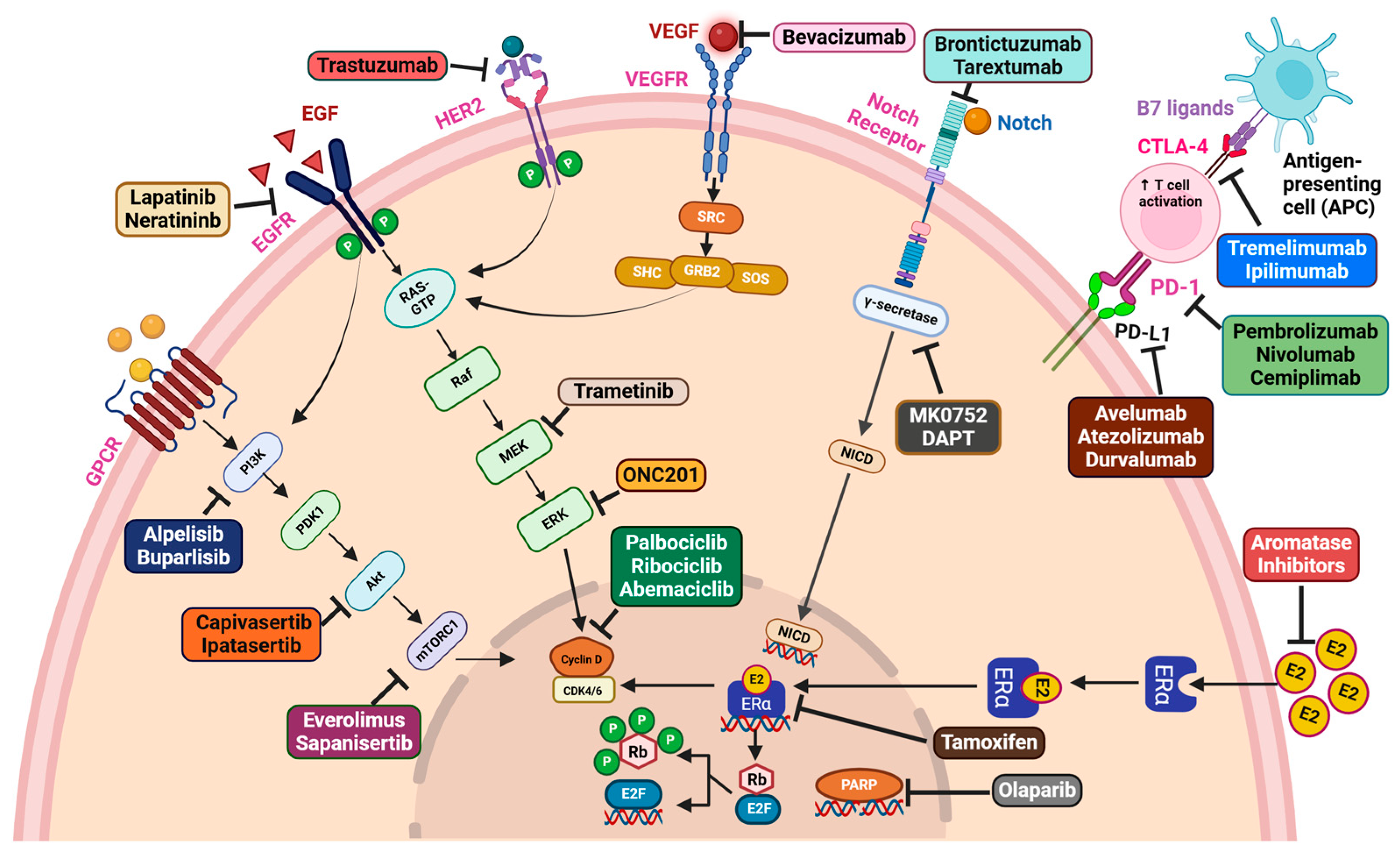
The conventional treatment strategy for breast cancer management includes radiation, surgery, and chemotherapy. Some of the major chemotherapeutic drugs and druggable molecules with their therapeutic targets are highlighted in Figure 6. Aromatase inhibitors, endocrine therapies, selective estrogen receptor degraders (SERD) and selective estrogen receptor modulators (SERM) are established approaches for personalized therapies in HR+ve breast cancer [239]. Tamoxifen was the first drug approved as a first line therapy in advanced breast cancer with higher ER status and reduces tumor recurrence [240]. Inhibitors that target PARP, HER2, PI3K, Akt, mTOR, EGF/EGFR, VEGF/VEGFR, and Notch could potentially be effective treatments to reduce the progression of breast cancer, as these pathways play key roles in various mechanisms of cancer development. Olaparib, a recognized PARP inhibitor, demonstrated greater efficacy than standard treatment in the phase III OlympiAD trial (NCT02000622). This trial conducted with olaparib alone resulted in a 2.8-month increase in PFS and reduced mortality in metastatic HER2− breast cancer patients with BRCA mutations [33]. In a phase III trial, olaparib showed an increased invasive DFS in patients with HER2-ve and gBRCA− pathogenic or likely pathogenic breast cancer (NCT02032823) [241].
One of the therapeutic hotspots gaining significant attention is the PI3K/Akt/mTOR pathway, which is known to be involved in promoting various oncogenic functions. The FDA approved alpelisib (BYL719), an p110α-specific PI3K inhibitor, as a therapeutic option for metastatic HR+ve and HER2-ve postmenopausal women [30]. Buparlisib (BKM120) is an oral pan-class I PI3K inhibitor that is under clinical trials in TNBC patients [242]. Patients with metastatic TNBC had a higher PFS rate when ipatasertib was combined with paclitaxel in the phase II LOTUS study (NCT02162719) [243]. Capivasertib, an ATP-competitive Akt inhibitor, significantly downregulates Akt signaling, as well as reduces breast tumor growth [244]. ONC201 belongs to a novel class of anticancer drugs called imipridones which is known to be a p53-independent transcriptional inducer of TNF-associated apoptosis-promoting ligand (TRAIL) [245,246,247]. ONC201 is linked to dephosphorylation of the FOXO3a and promotes the TRAIL gene expression by inactivating Akt and Erk [247]. Preclinical investigations demonstrated that ONC201 is effective in TNBC as well as non-TNBC cells that include BRCA1-deficient cells, irrespective of TRAIL sensitivity [248]. Trametinib, an MEK-specific inhibitor, has been shown to inhibit breast cancer growth [249]. Trametinib in combination with SHP099, an allosteric small-molecule inhibitor of SHP2, showed good efficacy in TNBC cells [250]. It exhibited synergistic effects across various cell lines, impeding reactivation of ERK in response to MEK inhibitors and hindering transcriptional activation [250].
Additionally, everolimus, an mTORC1 allosteric inhibitor, in conjunction with exemestane, was FDA-approved for the treatment of HR+ve and HER2-ve breast cancer. This trial including patients with ER+ve breast cancer showed that neoadjuvant letrozole treatment combined with everolimus prior to surgery had an improved clinical outcome and reduced tumor growth in contrast to letrozole treated alone [251]. Sapanisertib is a potent mTOR kinase inhibitor that selectively targets both mTORC1 and mTORC2. In a phase II trial, sapanisertib in combination with exemestane or fulvestrant exhibited therapeutic benefits in postmenopausal women with pretreated everolimus-resistant or sensitive breast cancer [252,253]. Furthermore, lapatinib and neratinib are the FDA-approved EGFR-TKIs for the treatment of HER2+ve breast cancer [254]. CDK4/6 is a crucial player in driving breast cancer progression. For targeting CDK4/6, the FDA has approved inhibitors such as palbociclib, abemaciclib, and ribociclib for the treatment of different forms of breast cancer [255]. Angiogenesis inhibitors can be applied as therapeutics to address different types of solid tumors, including breast. These inhibitors consist of small-molecule TKIs and monoclonal antibodies, primarily targeting VEGF and its receptors. In addition to bevacizumab, an anti-VEGF monoclonal antibody, paclitaxel chemotherapy increased the PFS in patients with metastatic breast cancer (NCT00028990) [256].
The breast cancer TME is generally considered as a “cold TIME”, suggesting a highly non-immunogenic condition. Various approaches to regulate the immune system have been explored in clinical trials. In addition to the recognized breast cancer subtypes, the identification of prognostic biomarkers is important for customizing immunotherapies. According to the phase II I-SPY2 trial, patients with HR+ve and HER2-ve breast cancer or TNBC showed a significantly higher pathologic response when cemiplimab, a PD-1 targeting IgG4 monoclonal antibody and fianlimab, a LAG-3 inhibitor were combined with paclitaxel (NCT01042379) [257]. Combining the PARP inhibitor olaparib with durvalumab showed promising anti-tumor activity in an open-label phase I/II trial involving patients with metastatic breast cancer who have a gBRCA mutation (NCT02734004) [258]. Ipilimumab is an IgG1 monoclonal antibody that targets CTLA-4 and exhibits substantial anti-tumor efficacy across a range of cancers. According to preclinical data, ipilimumab induces the secretion of IL-2 by TNBC cells into the TME, thereby strengthening the immunological response [259]. In phase II clinical trials, the combination therapy of ipilimumab and nivolumab along with neoadjuvant paclitaxel showed promising pathological responses in early-stage TNBC patients [260]. Tremelimumab is a humanized IgG2 monoclonal antibody that targets CTLA-4 thereby preventing tumor growth by blocking CTLA-4 and B7 interactions [261].
Anticancer vaccines are designed to stimulate antigen-specific T cell activation to target cancer cells. Breast cancer vaccines can be broadly categorized into two types. One of these targets HER2 or HER2-associated antigens and the other focuses on non-HER2-related antigens. The effectiveness of two HER2 peptide vaccines, GP2 and AE37, has been compared in a single-blind phase II trial in patients with HER2+ve breast cancer [262]. While GP2 immunization resulted in recurrence-free survival to HER2-overexpressing subgroups, AE37 vaccination conferred therapeutic benefit in TNBC and patients representing low HER2 [263]. A recombinant fowl pox vector encoding tumor-associated antigens such as carcinoembryonic antigen (CEA) and MUC1 is part of the PANVAC recombinant viral vaccine, which also comprises a second viral vector containing transgenes for cancer-associated antigens and the co-stimulatory drug TRICOM [264]. Patients with metastatic breast cancer undergoing docetaxel therapy with PANVAC demonstrated increased PFS compared to the control group in a phase II trial (NCT00179309) [264].
An increasing number of clinical trials are being conducted to develop effective breast cancer therapy, as depicted in Table 2. However, achieving the intended treatment outcome with targeted or immune therapy requires a thorough understanding of the genetic, molecular, and immunological landscape of tumor cells as well as the TME.

10. Conclusions

Breast cancer is one of the most prevailing female malignancies around the globe. Its intricate microenvironmental landscape demands further comprehensive investigation. The breast TME unveils a labyrinthine network system comprising multilevel intercellular interactions among the immune cells, stromal cells, and other cellular components. These interactions are operated by an array of molecular signaling pathways and unravel a rich tapestry of avenues for targeting a myriad of novel molecular players in finding effective therapeutic modalities. Many aberrations and genetic interplays among breast cancer subtypes are yet unknown in terms of function. To deconvolute critical molecular pathways and interconnecting nodes, rigorous functional screening (for example, utilizing CRISPR-Cas9 editing or spatial omics studies) as well as deeper analysis and data integration are warranted.
Numerous studies have dissected multiple signaling pathways and the molecules involved in aiding breast tumor growth and metastasis. For instance, our group has reported many signaling pathways regulating various oncogenic functions within the breast TME and multiple therapeutic options by targeting those molecular players [262]. One of the studies reported that tumor-derived OPN activates Twist1-dependent gene expression by binding to CD44 and αvβ3 integrins on the CAF via Akt and ERK pathway, causing tumor cells to undergo EMT [265]. In addition, Radharani et al. showed that breast tumor-activated macrophages enrich cancer stemness by upregulating CSC specific transcription factors via activating the IL-6/STAT3 pathway [266]. Recently, Panda et al. reviewed extensively OPN-regulated pathways and their downstream implications in breast and other cancers [267]. Furthermore, curcumin–chitosan nanoparticles (Cur-CHNPs), either alone or in combination with 5-FU, reduced the expression of OPN and VEGF by dysregulating PI3K and Akt activation, thereby inhibiting breast cancer migration and metastasis [268].
This review comprehensively gleaned the various fundamental signaling pathways orchestrated by numerous cellular and molecular players, focusing on the pan-breast TME. Further, this review summarized the existing in-depth analysis of current breast cancer therapies, as well as stromal- and immune-cell mediated oncogenic signaling pathways that enhance several hallmarks of breast cancer. Moreover, the multifaceted classification of breast tumors and novel therapeutic regimens have been comprehensively discussed. The array of molecular signaling regulating EMT dictates breast tumor growth and metastasis. These regulated networks within the breast TME promote tumor angiogenesis, specifically VEGF-dependent and independent pathways, CSC enrichment, tumor metabolism, therapy failure, and autophagy. In addition, the role of oncogenic pathways in the breast TME has been explored thoroughly, focusing on stromal cells such as CAFs, adipocytes, and mast cells, and immune cells such as macrophage-mediated regulatory networks resulting in breast tumor growth and metastasis. Furthermore, it has been addressed that noncoding RNAs, such as exosomal circular RNAs, lncRNAs, and miRNAs, are crucial for the initiation and spread of breast cancer. Therefore, targeting these noncoding RNAs could be a better therapeutic option for the management of breast cancer. An in-depth exploration of therapeutic modalities and their potential clinical utilities impacting numerous microenvironmental cellular and molecular components within the breast TME have been discussed.

11. Future Perspective

Although we have highlighted several regulated networks and pathways as well as their implications in breast cancer, a deeper understanding of the microenvironmental landscape is required. Furthermore, in response to these issues, we propose numerous future research challenges: First, delving into the microenvironmental cues and developing effective mono- or combination therapeutic regimens. Second, in-depth investigation of cellular and molecular players as robust biomarkers for the diagnosis of breast cancer. Third, investigating precise therapeutic options for microenvironment modulation, including targeted therapy for specific biomarkers and immunomodulatory therapy, to enhance tailored treatment and effectiveness. Exploring these future research avenues will enhance our understanding of the breast microenvironment and boost the clinical utility of microenvironmental biomarkers among molecular players, which could open new dimensions in breast cancer therapy.

Author Contributions

The authors V.K.P., B.M., S.M. (Samikshya Mahapatra), B.S., D.M., S.S. (Suryendu Saha), S.K., P.M., S.M. (Sambhunath Majhi), K.K., A.N.N., S.S. (Swarnali Saha), S.J. and G.C.K. wrote the manuscript and prepared the figures and tables together. G.C.K. conceptualized and V.K.P., B.M. and G.C.K. significantly edited the entire manuscript. All authors have read and agreed to the published version of the manuscript.

Funding

This work was supported by the Science and Engineering Research Board (SERB), Govt. of India, (Project No. JCB/2023/000011), provided to G.C.K.; the Department of Biotechnology (DBT) (Project No. BT/PR-32388/TRM/120/242/2019), Govt of India, provided to G.C.K.; and the DST INSPIRE Fellowship Program (DST INSPIRE/2021/IF210059), Govt of India, provided to V.K.P.

Acknowledgments

Figures were created using BioRender.com and accessed with a license.

Conflicts of Interest

The authors declare that they have no conflicts of interest.

References

  1. Bray, F.; Laversanne, M.; Sung, H.; Ferlay, J.; Siegel, R.L.; Soerjomataram, I.; Jemal, A. Global Cancer Statistics 2022: GLOBOCAN Estimates of Incidence and Mortality Worldwide for 36 Cancers in 185 Countries. CA Cancer J. Clin. 2024, 74, 229–263. [Google Scholar] [CrossRef] [PubMed]
  2. Hashim, D.; Boffetta, P.; La Vecchia, C.; Rota, M.; Bertuccio, P.; Malvezzi, M.; Negri, E. The Global Decrease in Cancer Mortality: Trends and Disparities. Ann. Oncol. 2016, 27, 926–933. [Google Scholar] [CrossRef]
  3. Mao, X.; Xu, J.; Wang, W.; Liang, C.; Hua, J.; Liu, J.; Zhang, B.; Meng, Q.; Yu, X.; Shi, S. Crosstalk between Cancer-Associated Fibroblasts and Immune Cells in the Tumor Microenvironment: New Findings and Future Perspectives. Mol. Cancer 2021, 20, 131. [Google Scholar] [CrossRef]
  4. Feng, Y.; Spezia, M.; Huang, S.; Yuan, C.; Zeng, Z.; Zhang, L.; Ji, X.; Liu, W.; Huang, B.; Luo, W.; et al. Breast Cancer Development and Progression: Risk Factors, Cancer Stem Cells, Signaling Pathways, Genomics, and Molecular Pathogenesis. Genes Dis. 2018, 5, 77–106. [Google Scholar] [CrossRef] [PubMed]
  5. Nolan, E.; Lindeman, G.J.; Visvader, J.E. Deciphering Breast Cancer: From Biology to the Clinic. Cell 2023, 186, 1708–1728. [Google Scholar] [CrossRef] [PubMed]
  6. Hanahan, D.; Weinberg, R.A. Hallmarks of Cancer: The next Generation. Cell 2011, 144, 646–674. [Google Scholar] [CrossRef] [PubMed]
  7. Karagiannakos, A.; Adamaki, M.; Tsintarakis, A.; Vojtesek, B.; Fåhraeus, R.; Zoumpourlis, V.; Karakostis, K. Targeting Oncogenic Pathways in the Era of Personalized Oncology: A Systemic Analysis Reveals Highly Mutated Signaling Pathways in Cancer Patients and Potential Therapeutic Targets. Cancers 2022, 14, 664. [Google Scholar] [CrossRef]
  8. Tan, P.H.; Ellis, I.; Allison, K.; Brogi, E.; Fox, S.B.; Lakhani, S.; Lazar, A.J.; Morris, E.A.; Sahin, A.; Salgado, R.; et al. The 2019 World Health Organization Classification of Tumours of the Breast. Histopathology 2020, 77, 181–185. [Google Scholar] [CrossRef]
  9. Tarantino, P.; Hamilton, E.; Tolaney, S.M.; Cortes, J.; Morganti, S.; Ferraro, E.; Marra, A.; Viale, G.; Trapani, D.; Cardoso, F.; et al. HER2-Low Breast Cancer: Pathological and Clinical Landscape. J. Clin. Oncol. 2020, 38, 1951–1962. [Google Scholar] [CrossRef] [PubMed]
  10. Braun, L.; Mietzsch, F.; Seibold, P.; Schneeweiss, A.; Schirmacher, P.; Chang-Claude, J.; Peter Sinn, H.; Aulmann, S. Intrinsic Breast Cancer Subtypes Defined by Estrogen Receptor Signalling-Prognostic Relevance of Progesterone Receptor Loss. Mod. Pathol. 2013, 26, 1161–1171. [Google Scholar] [CrossRef]
  11. Won, K.-A.; Spruck, C. Triple-negative Breast Cancer Therapy: Current and Future Perspectives (Review). Int. J. Oncol. 2020, 57, 1245–1261. [Google Scholar] [CrossRef] [PubMed]
  12. Behranvand, N.; Nasri, F.; Zolfaghari Emameh, R.; Khani, P.; Hosseini, A.; Garssen, J.; Falak, R. Chemotherapy: A Double-Edged Sword in Cancer Treatment. Cancer Immunol. Immunother. 2022, 71, 507–526. [Google Scholar] [CrossRef] [PubMed]
  13. Abotaleb, M.; Kubatka, P.; Caprnda, M.; Varghese, E.; Zolakova, B.; Zubor, P.; Opatrilova, R.; Kruzliak, P.; Stefanicka, P.; Büsselberg, D. Chemotherapeutic Agents for the Treatment of Metastatic Breast Cancer: An Update. Biomed. Pharmacother. 2018, 101, 458–477. [Google Scholar] [CrossRef] [PubMed]
  14. Wang, H.; Guo, S.; Kim, S.-J.; Shao, F.; Ho, J.W.K.; Wong, K.U.; Miao, Z.; Hao, D.; Zhao, M.; Xu, J.; et al. Cisplatin Prevents Breast Cancer Metastasis through Blocking Early EMT and Retards Cancer Growth Together with Paclitaxel. Theranostics 2021, 11, 2442–2459. [Google Scholar] [CrossRef]
  15. Zheng, M.; Mei, Z.; Junaid, M.; Tania, M.; Fu, J.; Chen, H.-C.; Khan, M.A. Synergistic Role of Thymoquinone on Anticancer Activity of 5-Fluorouracil in Triple Negative Breast Cancer Cells. Anticancer. Agents Med. Chem. 2022, 22, 1111–1118. [Google Scholar] [CrossRef] [PubMed]
  16. Cameron, D.; Piccart-Gebhart, M.J.; Gelber, R.D.; Procter, M.; Goldhirsch, A.; de Azambuja, E.; Castro, G.; Untch, M.; Smith, I.; Gianni, L.; et al. 11 Years’ Follow-up of Trastuzumab after Adjuvant Chemotherapy in HER2-Positive Early Breast Cancer: Final Analysis of the HERceptin Adjuvant (HERA) Trial. Lancet 2017, 389, 1195–1205. [Google Scholar] [CrossRef] [PubMed]
  17. Yamamoto, Y.; Iwata, H.; Taira, N.; Masuda, N.; Takahashi, M.; Yoshinami, T.; Ueno, T.; Toyama, T.; Yamanaka, T.; Takano, T.; et al. Pertuzumab Retreatment for HER2-Positive Advanced Breast Cancer: A Randomized, Open-Label Phase III Study (PRECIOUS). Cancer Sci. 2022, 113, 3169–3179. [Google Scholar] [CrossRef]
  18. Cortes, J.; Rugo, H.S.; Cescon, D.W.; Im, S.-A.; Yusof, M.M.; Gallardo, C.; Lipatov, O.; Barrios, C.H.; Perez-Garcia, J.; Iwata, H.; et al. Pembrolizumab plus Chemotherapy in Advanced Triple-Negative Breast Cancer. N. Engl. J. Med. 2022, 387, 217–226. [Google Scholar] [CrossRef]
  19. Debien, V.; De Caluwé, A.; Wang, X.; Piccart-Gebhart, M.; Tuohy, V.K.; Romano, E.; Buisseret, L. Immunotherapy in Breast Cancer: An Overview of Current Strategies and Perspectives. NPJ Breast Cancer 2023, 9, 7. [Google Scholar] [CrossRef]
  20. Schmid, P.; Adams, S.; Rugo, H.S.; Schneeweiss, A.; Barrios, C.H.; Iwata, H.; Diéras, V.; Hegg, R.; Im, S.-A.; Shaw Wright, G.; et al. Atezolizumab and Nab-Paclitaxel in Advanced Triple-Negative Breast Cancer. N. Engl. J. Med. 2018, 379, 2108–2121. [Google Scholar] [CrossRef] [PubMed]
  21. Emens, L.A.; Adams, S.; Barrios, C.H.; Diéras, V.; Iwata, H.; Loi, S.; Rugo, H.S.; Schneeweiss, A.; Winer, E.P.; Patel, S.; et al. First-Line Atezolizumab plus Nab-Paclitaxel for Unresectable, Locally Advanced, or Metastatic Triple-Negative Breast Cancer: IMpassion130 Final Overall Survival Analysis. Ann. Oncol. 2021, 32, 983–993. [Google Scholar] [CrossRef]
  22. Miles, D.; Gligorov, J.; André, F.; Cameron, D.; Schneeweiss, A.; Barrios, C.; Xu, B.; Wardley, A.; Kaen, D.; Andrade, L.; et al. Primary Results from IMpassion131, a Double-Blind, Placebo-Controlled, Randomised Phase III Trial of First-Line Paclitaxel with or without Atezolizumab for Unresectable Locally Advanced/Metastatic Triple-Negative Breast Cancer. Ann. Oncol. 2021, 32, 994–1004. [Google Scholar] [CrossRef]
  23. Rugo, H.S.; Kabos, P.; Beck, J.T.; Jerusalem, G.; Wildiers, H.; Sevillano, E.; Paz-Ares, L.; Chisamore, M.J.; Chapman, S.C.; Hossain, A.M.; et al. Abemaciclib in Combination with Pembrolizumab for HR+ve, HER2- Metastatic Breast Cancer: Phase 1b Study. NPJ Breast Cancer 2022, 8, 118. [Google Scholar] [CrossRef]
  24. Yuan, Y.; Lee, J.S.; Yost, S.E.; Frankel, P.H.; Ruel, C.; Egelston, C.A.; Guo, W.; Padam, S.; Tang, A.; Martinez, N.; et al. Phase I/II Trial of Palbociclib, Pembrolizumab and Letrozole in Patients with Hormone Receptor-Positive Metastatic Breast Cancer. Eur. J. Cancer 2021, 154, 11–20. [Google Scholar] [CrossRef] [PubMed]
  25. Loi, S.; Giobbie-Hurder, A.; Gombos, A.; Bachelot, T.; Hui, R.; Curigliano, G.; Campone, M.; Biganzoli, L.; Bonnefoi, H.; Jerusalem, G.; et al. Pembrolizumab plus Trastuzumab in Trastuzumab-Resistant, Advanced, HER2-Positive Breast Cancer (PANACEA): A Single-Arm, Multicentre, Phase 1b-2 Trial. Lancet Oncol. 2019, 20, 371–382. [Google Scholar] [CrossRef]
  26. Emens, L.A.; Esteva, F.J.; Beresford, M.; Saura, C.; De Laurentiis, M.; Kim, S.-B.; Im, S.-A.; Wang, Y.; Salgado, R.; Mani, A.; et al. Trastuzumab Emtansine plus Atezolizumab versus Trastuzumab Emtansine plus Placebo in Previously Treated, HER2-Positive Advanced Breast Cancer (KATE2): A Phase 2, Multicentre, Randomised, Double-Blind Trial. Lancet Oncol. 2020, 21, 1283–1295. [Google Scholar] [CrossRef]
  27. Nagini, S. Breast Cancer: Current Molecular Therapeutic Targets and New Players. Anticancer Agents Med. Chem. 2017, 17, 152–163. [Google Scholar] [CrossRef] [PubMed]
  28. Riccardi, F.; Colantuoni, G.; Diana, A.; Mocerino, C.; Cartenì, G.; Lauria, R.; Febbraro, A.; Nuzzo, F.; Addeo, R.; Marano, O.; et al. Exemestane and Everolimus Combination Treatment of Hormone Receptor Positive, HER2 Negative Metastatic Breast Cancer: A Retrospective Study of 9 Cancer Centers in the Campania Region (Southern Italy) Focused on Activity, Efficacy and Safety. Mol. Clin. Oncol. 2018, 9, 255–263. [Google Scholar] [CrossRef] [PubMed]
  29. Bachelot, T.; Bourgier, C.; Cropet, C.; Ray-Coquard, I.; Ferrero, J.-M.; Freyer, G.; Abadie-Lacourtoisie, S.; Eymard, J.-C.; Debled, M.; Spaëth, D.; et al. Randomized Phase II Trial of Everolimus in Combination with Tamoxifen in Patients with Hormone Receptor-Positive, Human Epidermal Growth Factor Receptor 2-Negative Metastatic Breast Cancer with Prior Exposure to Aromatase Inhibitors: A GINECO Study. J. Clin. Oncol. 2012, 30, 2718–2724. [Google Scholar] [CrossRef] [PubMed]
  30. André, F.; Ciruelos, E.; Rubovszky, G.; Campone, M.; Loibl, S.; Rugo, H.S.; Iwata, H.; Conte, P.; Mayer, I.A.; Kaufman, B.; et al. Alpelisib for PIK3CA-Mutated, Hormone Receptor-Positive Advanced Breast Cancer. N. Engl. J. Med. 2019, 380, 1929–1940. [Google Scholar] [CrossRef]
  31. Schöffski, P.; Cresta, S.; Mayer, I.A.; Wildiers, H.; Damian, S.; Gendreau, S.; Rooney, I.; Morrissey, K.M.; Spoerke, J.M.; Ng, V.W.; et al. A Phase Ib Study of Pictilisib (GDC-0941) in Combination with Paclitaxel, with and without Bevacizumab or Trastuzumab, and with Letrozole in Advanced Breast Cancer. Breast Cancer Res. 2018, 20, 109. [Google Scholar] [CrossRef] [PubMed]
  32. Diéras, V.C.; Han, H.S.; Kaufman, B.; Wildiers, H.; Friedlander, M.; Ayoub, J.-P.; Puhalla, S.L.; Bondarenko, I.; Campone, M.; Jakobsen, E.H.; et al. Phase III Study of Veliparib with Carboplatin and Paclitaxel in HER2-Negative Advanced/Metastatic gBRCA-Associated Breast Cancer. Ann. Oncol. 2019, 30, v857–v858. [Google Scholar] [CrossRef]
  33. Robson, M.; Im, S.A.; Senkus, E.; Xu, B.; Domchek, S.M.; Masuda, N.; Delaloge, S.; Li, W.; Tung, N.; Armstrong, A.; et al. Olaparib for Metastatic Breast Cancer in Patients with a Germline BRCA Mutation. N. Engl. J. Med. 2017, 377, 1700. [Google Scholar] [CrossRef] [PubMed]
  34. Brabletz, T.; Kalluri, R.; Nieto, M.A.; Weinberg, R.A. EMT in Cancer. Nat. Rev. Cancer 2018, 18, 128–134. [Google Scholar] [CrossRef] [PubMed]
  35. Tanabe, S.; Quader, S.; Cabral, H.; Ono, R. Interplay of EMT and CSC in Cancer and the Potential Therapeutic Strategies. Front. Pharmacol. 2020, 11, 904. [Google Scholar] [CrossRef] [PubMed]
  36. Saitoh, M. Transcriptional Regulation of EMT Transcription Factors in Cancer. Semin. Cancer Biol. 2023, 97, 21–29. [Google Scholar] [CrossRef]
  37. Mendonsa, A.M.; Na, T.-Y.; Gumbiner, B.M. E-Cadherin in Contact Inhibition and Cancer. Oncogene 2018, 37, 4769–4780. [Google Scholar] [CrossRef] [PubMed]
  38. Nieto, M.A.; Huang, R.Y.-J.; Jackson, R.A.; Thiery, J.P. EMT: 2016. Cell 2016, 166, 21–45. [Google Scholar] [CrossRef]
  39. Imani, S.; Hosseinifard, H.; Cheng, J.; Wei, C.; Fu, J. Prognostic Value of EMT-Inducing Transcription Factors (EMT-TFs) in Metastatic Breast Cancer: A Systematic Review and Meta-Analysis. Sci. Rep. 2016, 6, 28587. [Google Scholar] [CrossRef]
  40. Wang, H.; Sang, M.; Geng, C.; Liu, F.; Gu, L.; Shan, B. MAGE-A Is Frequently Expressed in Triple Negative Breast Cancer and Associated with Epithelial-Mesenchymal Transition. Neoplasma 2016, 63, 44–56. [Google Scholar] [CrossRef] [PubMed]
  41. Dong, C.; Wu, Y.; Wang, Y.; Wang, C.; Kang, T.; Rychahou, P.G.; Chi, Y.-I.; Evers, B.M.; Zhou, B.P. Interaction with Suv39H1 Is Critical for Snail-Mediated E-Cadherin Repression in Breast Cancer. Oncogene 2013, 32, 1351–1362. [Google Scholar] [CrossRef] [PubMed]
  42. Feldker, N.; Ferrazzi, F.; Schuhwerk, H.; Widholz, S.A.; Guenther, K.; Frisch, I.; Jakob, K.; Kleemann, J.; Riegel, D.; Bönisch, U.; et al. Genome-Wide Cooperation of EMT Transcription Factor ZEB1 with YAP and AP-1 in Breast Cancer. EMBO J. 2020, 39, e103209. [Google Scholar] [CrossRef]
  43. Song, K.; Farzaneh, M. Signaling Pathways Governing Breast Cancer Stem Cells Behavior. Stem Cell Res. Ther. 2021, 12, 245. [Google Scholar] [CrossRef] [PubMed]
  44. Su, C.; Zhang, J.; Yarden, Y.; Fu, L. The Key Roles of Cancer Stem Cell-Derived Extracellular Vesicles. Signal Transduct. Target. Ther. 2021, 6, 109. [Google Scholar] [CrossRef] [PubMed]
  45. Badve, S.; Nakshatri, H. Breast-Cancer Stem Cells-beyond Semantics. Lancet Oncol. 2012, 13, e43–e48. [Google Scholar] [CrossRef] [PubMed]
  46. Ginestier, C.; Hur, M.H.; Charafe-Jauffret, E.; Monville, F.; Dutcher, J.; Brown, M.; Jacquemier, J.; Viens, P.; Kleer, C.G.; Liu, S.; et al. ALDH1 Is a Marker of Normal and Malignant Human Mammary Stem Cells and a Predictor of Poor Clinical Outcome. Cell Stem Cell 2007, 1, 555–567. [Google Scholar] [CrossRef] [PubMed]
  47. Yamada, A.; Suzuki, C.; Shima, H.; Kida, K.; Adachi, S.; Yamamoto, S.; Narui, K.; Tanabe, M.; Shimizu, D.; Taniguchi, R.; et al. Aldehyde Dehydrogenase 1-Related Genes in Triple-Negative Breast Cancer Investigated Using Network Analysis. Anticancer Res. 2020, 40, 6733–6742. [Google Scholar] [CrossRef] [PubMed]
  48. da Silveira, W.A.; Palma, P.V.B.; Sicchieri, R.D.; Villacis, R.a.R.; Mandarano, L.R.M.; Oliveira, T.M.G.; Antonio, H.M.R.; Andrade, J.M.; Muglia, V.F.; Rogatto, S.R.; et al. Transcription Factor Networks Derived from Breast Cancer Stem Cells Control the Immune Response in the Basal Subtype. Sci. Rep. 2017, 7, 2851. [Google Scholar] [CrossRef] [PubMed]
  49. Vesuna, F.; Lisok, A.; Kimble, B.; Raman, V. Twist Modulates Breast Cancer Stem Cells by Transcriptional Regulation of CD24 Expression. Neoplasia 2009, 11, 1318–1328. [Google Scholar] [CrossRef]
  50. Storci, G.; Bertoni, S.; De Carolis, S.; Papi, A.; Nati, M.; Ceccarelli, C.; Pirazzini, C.; Garagnani, P.; Ferrarini, A.; Buson, G.; et al. Slug/β-Catenin-Dependent Proinflammatory Phenotype in Hypoxic Breast Cancer Stem Cells. Am. J. Pathol. 2013, 183, 1688–1697. [Google Scholar] [CrossRef]
  51. Qi, M.; Xia, Y.; Wu, Y.; Zhang, Z.; Wang, X.; Lu, L.; Dai, C.; Song, Y.; Xu, K.; Ji, W.; et al. Lin28B-High Breast Cancer Cells Promote Immune Suppression in the Lung Pre-Metastatic Niche via Exosomes and Support Cancer Progression. Nat. Commun. 2022, 13, 897. [Google Scholar] [CrossRef] [PubMed]
  52. Mendoza-Villanueva, D.; Balamurugan, K.; Ali, H.R.; Kim, S.-R.; Sharan, S.; Johnson, R.C.; Merchant, A.S.; Caldas, C.; Landberg, G.; Sterneck, E. The C/EBPδ Protein Is Stabilized by Estrogen Receptor α Activity, Inhibits SNAI2 Expression and Associates with Good Prognosis in Breast Cancer. Oncogene 2016, 35, 6166–6176. [Google Scholar] [CrossRef] [PubMed]
  53. Balamurugan, K.; Mendoza-Villanueva, D.; Sharan, S.; Summers, G.H.; Dobrolecki, L.E.; Lewis, M.T.; Sterneck, E. C/EBPδ Links IL-6 and HIF-1 Signaling to Promote Breast Cancer Stem Cell-Associated Phenotypes. Oncogene 2019, 38, 3765–3780. [Google Scholar] [CrossRef]
  54. Liu, Z.-L.; Chen, H.-H.; Zheng, L.-L.; Sun, L.-P.; Shi, L. Angiogenic Signaling Pathways and Anti-Angiogenic Therapy for Cancer. Signal Transduct. Target. Ther. 2023, 8, 198. [Google Scholar] [CrossRef]
  55. Senger, D.R.; Van de Water, L.; Brown, L.F.; Nagy, J.A.; Yeo, K.T.; Yeo, T.K.; Berse, B.; Jackman, R.W.; Dvorak, A.M.; Dvorak, H.F. Vascular Permeability Factor (VPF, VEGF) in Tumor Biology. Cancer Metastasis Rev. 1993, 12, 303–324. [Google Scholar] [CrossRef] [PubMed]
  56. Peach, C.J.; Mignone, V.W.; Arruda, M.A.; Alcobia, D.C.; Hill, S.J.; Kilpatrick, L.E.; Woolard, J. Molecular Pharmacology of VEGF-A Isoforms: Binding and Signalling at VEGFR2. Int. J. Mol. Sci. 2018, 19, 1264. [Google Scholar] [CrossRef] [PubMed]
  57. Ghalehbandi, S.; Yuzugulen, J.; Pranjol, M.Z.I.; Pourgholami, M.H. The Role of VEGF in Cancer-Induced Angiogenesis and Research Progress of Drugs Targeting VEGF. Eur. J. Pharmacol. 2023, 949, 175586. [Google Scholar] [CrossRef] [PubMed]
  58. Shibuya, M. Vascular Endothelial Growth Factor (VEGF) and Its Receptor (VEGFR) Signaling in Angiogenesis: A Crucial Target for Anti- and Pro-Angiogenic Therapies. Genes. Cancer 2011, 2, 1097–1105. [Google Scholar] [CrossRef] [PubMed]
  59. Zhu, C.; Qi, X.; Chen, Y.; Sun, B.; Dai, Y.; Gu, Y. PI3K/Akt and MAPK/ERK1/2 Signaling Pathways Are Involved in IGF-1-Induced VEGF-C Upregulation in Breast Cancer. J. Cancer Res. Clin. Oncol. 2011, 137, 1587–1594. [Google Scholar] [CrossRef] [PubMed]
  60. Farzaneh Behelgardi, M.; Zahri, S.; Gholami Shahvir, Z.; Mashayekhi, F.; Mirzanejad, L.; Asghari, S.M. Targeting Signaling Pathways of VEGFR1 and VEGFR2 as a Potential Target in the Treatment of Breast Cancer. Mol. Biol. Rep. 2020, 47, 2061–2071. [Google Scholar] [CrossRef]
  61. Majmundar, A.J.; Wong, W.J.; Simon, M.C. Hypoxia-Inducible Factors and the Response to Hypoxic Stress. Mol. Cell 2010, 40, 294–309. [Google Scholar] [CrossRef]
  62. Luo, S.; Jiang, Y.; Zheng, A.; Zhao, Y.; Wu, X.; Li, M.; Du, F.; Chen, Y.; Deng, S.; Chen, M.; et al. Targeting Hypoxia-Inducible Factors for Breast Cancer Therapy: A Narrative Review. Front. Pharmacol. 2022, 13, 1064661. [Google Scholar] [CrossRef]
  63. Raja, R.; Kale, S.; Thorat, D.; Soundararajan, G.; Lohite, K.; Mane, A.; Karnik, S.; Kundu, G.C. Hypoxia-Driven Osteopontin Contributes to Breast Tumor Growth through Modulation of HIF1α-Mediated VEGF-Dependent Angiogenesis. Oncogene 2014, 33, 2053–2064. [Google Scholar] [CrossRef] [PubMed]
  64. Mishra, R.; Thorat, D.; Soundararajan, G.; Pradhan, S.J.; Chakraborty, G.; Lohite, K.; Karnik, S.; Kundu, G.C. Semaphorin 3A Upregulates FOXO 3a-Dependent MelCAM Expression Leading to Attenuation of Breast Tumor Growth and Angiogenesis. Oncogene 2015, 34, 1584–1595. [Google Scholar] [CrossRef] [PubMed]
  65. Chakraborty, G.; Jain, S.; Kundu, G.C. Osteopontin Promotes Vascular Endothelial Growth Factor-Dependent Breast Tumor Growth and Angiogenesis via Autocrine and Paracrine Mechanisms. Cancer Res. 2008, 68, 152–161. [Google Scholar] [CrossRef] [PubMed]
  66. Tao, Q.; Qi, Y.; Gu, J.; Yu, D.; Lu, Y.; Liu, J.; Liang, X. Breast Cancer Cells-Derived Von Willebrand Factor Promotes VEGF-A-Related Angiogenesis through PI3K/Akt-miR-205-5p Signaling Pathway. Toxicol. Appl. Pharmacol. 2022, 440, 115927. [Google Scholar] [CrossRef] [PubMed]
  67. Xu, Y.; Wang, C.; Chen, X.; Li, Y.; Bian, W.; Yao, C. San Huang Decoction Targets Aurora Kinase A to Inhibit Tumor Angiogenesis in Breast Cancer. Integr. Cancer Ther. 2020, 19, 1534735420983463. [Google Scholar] [CrossRef] [PubMed]
  68. Wan, X.; Guan, S.; Hou, Y.; Qin, Y.; Zeng, H.; Yang, L.; Qiao, Y.; Liu, S.; Li, Q.; Jin, T.; et al. FOSL2 Promotes VEGF-Independent Angiogenesis by Transcriptionnally Activating Wnt5a in Breast Cancer-Associated Fibroblasts. Theranostics 2021, 11, 4975–4991. [Google Scholar] [CrossRef] [PubMed]
  69. Zeng, H.; Hou, Y.; Zhou, X.; Lang, L.; Luo, H.; Sun, Y.; Wan, X.; Yuan, T.; Wang, R.; Liu, Y.; et al. Cancer-Associated Fibroblasts Facilitate Premetastatic Niche Formation through lncRNA SNHG5-Mediated Angiogenesis and Vascular Permeability in Breast Cancer. Theranostics 2022, 12, 7351–7370. [Google Scholar] [CrossRef]
  70. Zhang, Q.; Li, T.; Wang, Z.; Kuang, X.; Shao, N.; Lin, Y. lncRNA NR2F1-AS1 Promotes Breast Cancer Angiogenesis through Activating IGF-1/IGF-1R/ERK Pathway. J. Cell. Mol. Med. 2020, 24, 8236. [Google Scholar] [CrossRef] [PubMed]
  71. Cao, J.; Liu, X.; Yang, Y.; Wei, B.; Li, Q.; Mao, G.; He, Y.; Li, Y.; Zheng, L.; Zhang, Q.; et al. Decylubiquinone Suppresses Breast Cancer Growth and Metastasis by Inhibiting Angiogenesis via the ROS/P53/ BAI1 Signaling Pathway. Angiogenesis 2020, 23, 325–338. [Google Scholar] [CrossRef] [PubMed]
  72. Yao, H.; Veine, D.M.; Livant, D.L. Therapeutic Inhibition of Breast Cancer Bone Metastasis Progression and Lung Colonization: Breaking the Vicious Cycle by Targeting A5β1 Integrin. Breast Cancer Res. Treat. 2016, 157, 489–501. [Google Scholar] [CrossRef]
  73. Xu, J.; Yang, X.; Deng, Q.; Yang, C.; Wang, D.; Jiang, G.; Yao, X.; He, X.; Ding, J.; Qiang, J.; et al. TEM8 Marks Neovasculogenic Tumor-Initiating Cells in Triple-Negative Breast Cancer. Nat. Commun. 2021, 12, 4413. [Google Scholar] [CrossRef]
  74. van Beijnum, J.R.; Huijbers, E.J.M.; van Loon, K.; Blanas, A.; Akbari, P.; Roos, A.; Wong, T.J.; Denisov, S.S.; Hackeng, T.M.; Jimenez, C.R.; et al. Extracellular Vimentin Mimics VEGF and Is a Target for Anti-Angiogenic Immunotherapy. Nat. Commun. 2022, 13, 2842. [Google Scholar] [CrossRef]
  75. Yan, L.; Wu, M.; Wang, T.; Yuan, H.; Zhang, X.; Zhang, H.; Li, T.; Pandey, V.; Han, X.; Lobie, P.E.; et al. Breast Cancer Stem Cells Secrete MIF to Mediate Tumor Metabolic Reprogramming That Drives Immune Evasion. Cancer Res. 2024, 84, 1270–1285. [Google Scholar] [CrossRef] [PubMed]
  76. Xie, T.; Jiang, C.; Dai, T.; Xu, R.; Zhou, X.; Su, X.; Zhao, X. Knockdown of XB130 Restrains Cancer Stem Cell-like Phenotype through Inhibition of Wnt/β-Catenin Signaling in Breast Cancer. Mol. Carcinog. 2019, 58, 1832–1845. [Google Scholar] [CrossRef]
  77. Ashad-Bishop, K.; Garikapati, K.; Lindley, L.E.; Jorda, M.; Briegel, K.J. Loss of Limb-Bud-and-Heart (LBH) Attenuates Mammary Hyperplasia and Tumor Development in MMTV-Wnt1 Transgenic Mice. Biochem. Biophys. Res. Commun. 2019, 508, 536–542. [Google Scholar] [CrossRef]
  78. Satriyo, P.B.; Bamodu, O.A.; Chen, J.-H.; Aryandono, T.; Haryana, S.M.; Yeh, C.-T.; Chao, T.-Y. Cadherin 11 Inhibition Downregulates β-Catenin, Deactivates the Canonical WNT Signalling Pathway and Suppresses the Cancer Stem Cell-Like Phenotype of Triple Negative Breast Cancer. J. Clin. Med. 2019, 8, 148. [Google Scholar] [CrossRef] [PubMed]
  79. Yoshimura, T.; Li, C.; Wang, Y.; Matsukawa, A. The Chemokine Monocyte Chemoattractant Protein-1/CCL2 Is a Promoter of Breast Cancer Metastasis. Cell Mol. Immunol. 2023, 20, 714–738. [Google Scholar] [CrossRef] [PubMed]
  80. Bu, J.; Zhang, Y.; Wu, S.; Li, H.; Sun, L.; Liu, Y.; Zhu, X.; Qiao, X.; Ma, Q.; Liu, C.; et al. KK-LC-1 as a Therapeutic Target to Eliminate ALDH+ve Stem Cells in Triple Negative Breast Cancer. Nat. Commun. 2023, 14, 2602. [Google Scholar] [CrossRef] [PubMed]
  81. Kong, L.; Guo, S.; Liu, C.; Zhao, Y.; Feng, C.; Liu, Y.; Wang, T.; Li, C. Overexpression of SDF-1 Activates the NF-κB Pathway to Induce Epithelial to Mesenchymal Transition and Cancer Stem Cell-like Phenotypes of Breast Cancer Cells. Int. J. Oncol. 2016, 48, 1085–1094. [Google Scholar] [CrossRef] [PubMed]
  82. Shan, S.; Lv, Q.; Zhao, Y.; Liu, C.; Sun, Y.; Xi, K.; Xiao, J.; Li, C. Wnt/β-Catenin Pathway Is Required for Epithelial to Mesenchymal Transition in CXCL12 over Expressed Breast Cancer Cells. Int. J. Clin. Exp. Pathol. 2015, 8, 12357–12367. [Google Scholar] [PubMed]
  83. Zhang, R.; Dong, M.; Tu, J.; Li, F.; Deng, Q.; Xu, J.; He, X.; Ding, J.; Xia, J.; Sheng, D.; et al. PMN-MDSCs Modulated by CCL20 from Cancer Cells Promoted Breast Cancer Cell Stemness through CXCL2-CXCR2 Pathway. Signal Transduct. Target. Ther. 2023, 8, 97. [Google Scholar] [CrossRef] [PubMed]
  84. Luo, F.; Zhang, M.; Sun, B.; Xu, C.; Yang, Y.; Zhang, Y.; Li, S.; Chen, G.; Chen, C.; Li, Y.; et al. LINC00115 Promotes Chemoresistant Breast Cancer Stem-like Cell Stemness and Metastasis through SETDB1/PLK3/HIF1α Signaling. Mol. Cancer 2024, 23, 60. [Google Scholar] [CrossRef]
  85. Liu, S.; Cong, Y.; Wang, D.; Sun, Y.; Deng, L.; Liu, Y.; Martin-Trevino, R.; Shang, L.; McDermott, S.P.; Landis, M.D.; et al. Breast Cancer Stem Cells Transition between Epithelial and Mesenchymal States Reflective of Their Normal Counterparts. Stem Cell Rep. 2013, 2, 78. [Google Scholar] [CrossRef] [PubMed]
  86. Pasani, S.; Sahoo, S.; Jolly, M.K. Hybrid E/M Phenotype(s) and Stemness: A Mechanistic Connection Embedded in Network Topology. J. Clin. Med. 2020, 10, 60. [Google Scholar] [CrossRef] [PubMed]
  87. Brown, M.S.; Abdollahi, B.; Wilkins, O.M.; Lu, H.; Chakraborty, P.; Ognjenovic, N.B.; Muller, K.E.; Jolly, M.K.; Christensen, B.C.; Hassanpour, S.; et al. Phenotypic Heterogeneity Driven by Plasticity of the Intermediate EMT State Governs Disease Progression and Metastasis in Breast Cancer. Sci. Adv. 2022, 8, eabj8002. [Google Scholar] [CrossRef] [PubMed]
  88. Luo, M.; Bao, L.; Xue, Y.; Zhu, M.; Kumar, A.; Xing, C.; Wang, J.E.; Wang, Y.; Luo, W. ZMYND8 Protects Breast Cancer Stem Cells against Oxidative Stress and Ferroptosis through Activation of NRF2. J. Clin. Investig. 2024, 134, e171166. [Google Scholar] [CrossRef] [PubMed]
  89. Zheng, X.; Ma, H.; Wang, J.; Huang, M.; Fu, D.; Qin, L.; Yin, Q. Energy Metabolism Pathways in Breast Cancer Progression: The Reprogramming, Crosstalk, and Potential Therapeutic Targets. Transl. Oncol. 2022, 26, 101534. [Google Scholar] [CrossRef] [PubMed]
  90. Jiao, Z.; Pan, Y.; Chen, F. The Metabolic Landscape of Breast Cancer and Its Therapeutic Implications. Mol. Diagn. Ther. 2023, 27, 349–369. [Google Scholar] [CrossRef]
  91. Wu, Z.; Wu, J.; Zhao, Q.; Fu, S.; Jin, J. Emerging Roles of Aerobic Glycolysis in Breast Cancer. Clin. Transl. Oncol. 2020, 22, 631–646. [Google Scholar] [CrossRef] [PubMed]
  92. Lee, J.-H.; Liu, R.; Li, J.; Wang, Y.; Tan, L.; Li, X.-J.; Qian, X.; Zhang, C.; Xia, Y.; Xu, D.; et al. EGFR-Phosphorylated Platelet Isoform of Phosphofructokinase 1 Promotes PI3K Activation. Mol. Cell 2018, 70, 197–210.e7. [Google Scholar] [CrossRef]
  93. Novellasdemunt, L.; Tato, I.; Navarro-Sabate, A.; Ruiz-Meana, M.; Méndez-Lucas, A.; Perales, J.C.; Garcia-Dorado, D.; Ventura, F.; Bartrons, R.; Rosa, J.L. Akt-Dependent Activation of the Heart 6-Phosphofructo-2-Kinase/Fructose-2,6-Bisphosphatase (PFKFB2) Isoenzyme by Amino Acids. J. Biol. Chem. 2013, 288, 10640–10651. [Google Scholar] [CrossRef] [PubMed]
  94. Samih, N.; Hovsepian, S.; Aouani, A.; Lombardo, D.; Fayet, G. Glut-1 Translocation in FRTL-5 Thyroid Cells: Role of Phosphatidylinositol 3-Kinase and N-Glycosylation. Endocrinology 2000, 141, 4146–4155. [Google Scholar] [CrossRef]
  95. Garrido, P.; Morán, J.; Alonso, A.; González, S.; González, C. 17β-Estradiol Activates Glucose Uptake via GLUT4 Translocation and PI3K/Akt Signaling Pathway in MCF-7 Cells. Endocrinology 2013, 154, 1979–1989. [Google Scholar] [CrossRef]
  96. Castaneda, C.A.; Cortes-Funes, H.; Gomez, H.L.; Ciruelos, E.M. The Phosphatidyl Inositol 3-Kinase/AKT Signaling Pathway in Breast Cancer. Cancer Metastasis Rev. 2010, 29, 751–759. [Google Scholar] [CrossRef]
  97. Elwy, F.; Helwa, R.; El Leithy, A.A.; Shehab El din, Z.; Assem, M.M.; Hassan, N.H.A. PIK3CA Mutations in HER2-Positive Breast Cancer Patients; Frequency and Clinicopathological Perspective in Egyptian Patients. Asian Pac. J. Cancer Prev. 2017, 18, 57–64. [Google Scholar] [CrossRef] [PubMed]
  98. Wang, C.; Mayer, J.A.; Mazumdar, A.; Fertuck, K.; Kim, H.; Brown, M.; Brown, P.H. Estrogen Induces C-Myc Gene Expression via an Upstream Enhancer Activated by the Estrogen Receptor and the AP-1 Transcription Factor. Mol. Endocrinol. 2011, 25, 1527–1538. [Google Scholar] [CrossRef]
  99. Lumachi, F.; Santeufemia, D.A.; Basso, S.M. Current Medical Treatment of Estrogen Receptor-Positive Breast Cancer. World J. Biol. Chem. 2015, 6, 231–239. [Google Scholar] [CrossRef]
  100. Düvel, K.; Yecies, J.L.; Menon, S.; Raman, P.; Lipovsky, A.I.; Souza, A.L.; Triantafellow, E.; Ma, Q.; Gorski, R.; Cleaver, S.; et al. Activation of a Metabolic Gene Regulatory Network Downstream of mTOR Complex 1. Mol. Cell 2010, 39, 171–183. [Google Scholar] [CrossRef] [PubMed]
  101. Beg, M.; Abdullah, N.; Thowfeik, F.S.; Altorki, N.K.; McGraw, T.E. Distinct Akt Phosphorylation States Are Required for Insulin Regulated Glut4 and Glut1-Mediated Glucose Uptake. eLife 2017, 6, e26896. [Google Scholar] [CrossRef]
  102. Albert, V.; Svensson, K.; Shimobayashi, M.; Colombi, M.; Muñoz, S.; Jimenez, V.; Handschin, C.; Bosch, F.; Hall, M.N. mTORC2 Sustains Thermogenesis via Akt-Induced Glucose Uptake and Glycolysis in Brown Adipose Tissue. EMBO Mol. Med. 2016, 8, 232–246. [Google Scholar] [CrossRef] [PubMed]
  103. Ghanavat, M.; Shahrouzian, M.; Deris Zayeri, Z.; Banihashemi, S.; Kazemi, S.M.; Saki, N. Digging Deeper through Glucose Metabolism and Its Regulators in Cancer and Metastasis. Life Sci. 2021, 264, 118603. [Google Scholar] [CrossRef]
  104. Chen, X.; Zhang, T.; Su, W.; Dou, Z.; Zhao, D.; Jin, X.; Lei, H.; Wang, J.; Xie, X.; Cheng, B.; et al. Mutant P53 in Cancer: From Molecular Mechanism to Therapeutic Modulation. Cell Death Dis. 2022, 13, 974. [Google Scholar] [CrossRef]
  105. Abdel-Wahab, A.F.; Mahmoud, W.; Al-Harizy, R.M. Targeting Glucose Metabolism to Suppress Cancer Progression: Prospective of Anti-Glycolytic Cancer Therapy. Pharmacol. Res. 2019, 150, 104511. [Google Scholar] [CrossRef]
  106. Zhang, S.; Zhang, X.; Yang, H.; Liang, T.; Bai, X. Hurdle or Thruster: Glucose Metabolism of T Cells in Anti-Tumour Immunity. Biochim. Biophys. Acta Rev. Cancer 2024, 1879, 189022. [Google Scholar] [CrossRef] [PubMed]
  107. El-Kenawi, A.; Gatenbee, C.; Robertson-Tessi, M.; Bravo, R.; Dhillon, J.; Balagurunathan, Y.; Berglund, A.; Vishvakarma, N.; Ibrahim-Hashim, A.; Choi, J.; et al. Acidity Promotes Tumour Progression by Altering Macrophage Phenotype in Prostate Cancer. Br. J. Cancer 2019, 121, 556–566. [Google Scholar] [CrossRef] [PubMed]
  108. Colegio, O.R.; Chu, N.-Q.; Szabo, A.L.; Chu, T.; Rhebergen, A.M.; Jairam, V.; Cyrus, N.; Brokowski, C.E.; Eisenbarth, S.C.; Phillips, G.M.; et al. Functional Polarization of Tumour-Associated Macrophages by Tumour-Derived Lactic Acid. Nature 2014, 513, 559–563. [Google Scholar] [CrossRef] [PubMed]
  109. Wang, J.X.; Choi, S.Y.C.; Niu, X.; Kang, N.; Xue, H.; Killam, J.; Wang, Y. Lactic Acid and an Acidic Tumor Microenvironment Suppress Anticancer Immunity. Int. J. Mol. Sci. 2020, 21, 8363. [Google Scholar] [CrossRef]
  110. Jin, H.-R.; Wang, J.; Wang, Z.-J.; Xi, M.-J.; Xia, B.-H.; Deng, K.; Yang, J.-L. Lipid Metabolic Reprogramming in Tumor Microenvironment: From Mechanisms to Therapeutics. J. Hematol. Oncol. 2023, 16, 103. [Google Scholar] [CrossRef] [PubMed]
  111. Zhang, S.; Lv, K.; Liu, Z.; Zhao, R.; Li, F. Fatty Acid Metabolism of Immune Cells: A New Target of Tumour Immunotherapy. Cell Death Discov. 2024, 10, 39. [Google Scholar] [CrossRef] [PubMed]
  112. Xie, Y.; Kang, R.; Klionsky, D.J.; Tang, D. GPX4 in Cell Death, Autophagy, and Disease. Autophagy 2023, 19, 2621. [Google Scholar] [CrossRef] [PubMed]
  113. Brown, C.W.; Amante, J.J.; Chhoy, P.; Elaimy, A.L.; Liu, H.; Zhu, L.J.; Baer, C.E.; Dixon, S.J.; Mercurio, A.M. Prominin2 Drives Ferroptosis Resistance by Stimulating Iron Export. Dev. Cell 2019, 51, 575–586.e4. [Google Scholar] [CrossRef] [PubMed]
  114. Sha, R.; Xu, Y.; Yuan, C.; Sheng, X.; Wu, Z.; Peng, J.; Wang, Y.; Lin, Y.; Zhou, L.; Xu, S.; et al. Predictive and Prognostic Impact of Ferroptosis-Related Genes ACSL4 and GPX4 on Breast Cancer Treated with Neoadjuvant Chemotherapy. EBioMedicine 2021, 71, 103560. [Google Scholar] [CrossRef]
  115. Wang, J.; Zhang, W.; Liu, C.; Wang, L.; Wu, J.; Sun, C.; Wu, Q. Reprogramming of Lipid Metabolism Mediates Crosstalk, Remodeling, and Intervention of Microenvironment Components in Breast Cancer. Int. J. Biol. Sci. 2024, 20, 1884–1904. [Google Scholar] [CrossRef]
  116. Ge, D.; Gao, J.; Han, L.; Li, Y.; Liu, H.-H.; Yang, W.-C.; Chang, F.; Liu, J.; Yu, M.; Zhao, J. Novel Effects of Sphingosylphosphorylcholine on the Apoptosis of Breast Cancer via Autophagy/AKT/P38 and JNK Signaling. J. Cell Physiol. 2019, 234, 11451–11462. [Google Scholar] [CrossRef] [PubMed]
  117. Zhang, N.; Zhang, H.; Liu, Y.; Su, P.; Zhang, J.; Wang, X.; Sun, M.; Chen, B.; Zhao, W.; Wang, L.; et al. SREBP1, Targeted by miR-18a-5p, Modulates Epithelial-Mesenchymal Transition in Breast Cancer via Forming a Co-Repressor Complex with Snail and HDAC1/2. Cell Death Differ. 2019, 26, 843–859. [Google Scholar] [CrossRef] [PubMed]
  118. Zheng, K.; Chen, Z.; Feng, H.; Chen, Y.; Zhang, C.; Yu, J.; Luo, Y.; Zhao, L.; Jiang, X.; Shi, F. Sphingomyelin Synthase 2 Promotes an Aggressive Breast Cancer Phenotype by Disrupting the Homoeostasis of Ceramide and Sphingomyelin. Cell Death Dis. 2019, 10, 157. [Google Scholar] [CrossRef]
  119. Zhang, D.; Xu, X.; Ye, Q. Metabolism and Immunity in Breast Cancer. Front. Med. 2021, 15, 178–207. [Google Scholar] [CrossRef] [PubMed]
  120. Beckermann, K.E.; Hongo, R.; Ye, X.; Young, K.; Carbonell, K.; Healey, D.C.C.; Siska, P.J.; Barone, S.; Roe, C.E.; Smith, C.C.; et al. CD28 Costimulation Drives Tumor-Infiltrating T Cell Glycolysis to Promote Inflammation. JCI Insight 2020, 5, e138729. [Google Scholar] [CrossRef] [PubMed]
  121. Patsoukis, N.; Bardhan, K.; Chatterjee, P.; Sari, D.; Liu, B.; Bell, L.N.; Karoly, E.D.; Freeman, G.J.; Petkova, V.; Seth, P.; et al. PD-1 Alters T-Cell Metabolic Reprogramming by Inhibiting Glycolysis and Promoting Lipolysis and Fatty Acid Oxidation. Nat. Commun. 2015, 6, 6692. [Google Scholar] [CrossRef]
  122. Chang, C.-H.; Qiu, J.; O’Sullivan, D.; Buck, M.D.; Noguchi, T.; Curtis, J.D.; Chen, Q.; Gindin, M.; Gubin, M.M.; van der Windt, G.J.W.; et al. Metabolic Competition in the Tumor Microenvironment Is a Driver of Cancer Progression. Cell 2015, 162, 1229–1241. [Google Scholar] [CrossRef] [PubMed]
  123. Saleh, R.; Taha, R.Z.; Sasidharan Nair, V.; Alajez, N.M.; Elkord, E. PD-L1 Blockade by Atezolizumab Downregulates Signaling Pathways Associated with Tumor Growth, Metastasis, and Hypoxia in Human Triple Negative Breast Cancer. Cancers 2019, 11, 1050. [Google Scholar] [CrossRef] [PubMed]
  124. Kim, H.M.; Koo, J.S. The Role of Autophagy in Breast Cancer Metastasis. Biomedicines 2023, 11, 618. [Google Scholar] [CrossRef] [PubMed]
  125. Patergnani, S.; Missiroli, S.; Morciano, G.; Perrone, M.; Mantovani, C.M.; Anania, G.; Fiorica, F.; Pinton, P.; Giorgi, C. Understanding the Role of Autophagy in Cancer Formation and Progression Is a Real Opportunity to Treat and Cure Human Cancers. Cancers 2021, 13, 5622. [Google Scholar] [CrossRef] [PubMed]
  126. Deretic, V. Autophagy in Inflammation, Infection, and Immunometabolism. Immunity 2021, 54, 437–453. [Google Scholar] [CrossRef] [PubMed]
  127. Mizushima, N.; Komatsu, M. Autophagy: Renovation of Cells and Tissues. Cell 2011, 147, 728–741. [Google Scholar] [CrossRef] [PubMed]
  128. He, C.; Klionsky, D.J. Regulation Mechanisms and Signaling Pathways of Autophagy. Annu. Rev. Genet. 2009, 43, 67–93. [Google Scholar] [CrossRef]
  129. Li, Z.; Chen, B.; Wu, Y.; Jin, F.; Xia, Y.; Liu, X. Genetic and Epigenetic Silencing of the Beclin 1 Gene in Sporadic Breast Tumors. BMC Cancer 2010, 10, 98. [Google Scholar] [CrossRef] [PubMed]
  130. Liang, X.H.; Jackson, S.; Seaman, M.; Brown, K.; Kempkes, B.; Hibshoosh, H.; Levine, B. Induction of Autophagy and Inhibition of Tumorigenesis by Beclin 1. Nature 1999, 402, 672–676. [Google Scholar] [CrossRef] [PubMed]
  131. Zhou, B.; Kreuzer, J.; Kumsta, C.; Wu, L.; Kamer, K.J.; Cedillo, L.; Zhang, Y.; Li, S.; Kacergis, M.C.; Webster, C.M.; et al. Mitochondrial Permeability Uncouples Elevated Autophagy and Lifespan Extension. Cell 2019, 177, 299–314.e16. [Google Scholar] [CrossRef]
  132. Filomeni, G.; De Zio, D.; Cecconi, F. Oxidative Stress and Autophagy: The Clash between Damage and Metabolic Needs. Cell Death Differ. 2015, 22, 377–388. [Google Scholar] [CrossRef]
  133. Zhang, H.; Guo, M.; Chen, J.-H.; Wang, Z.; Du, X.-F.; Liu, P.-X.; Li, W.-H. Osteopontin Knockdown Inhibits Av,Β3 Integrin-Induced Cell Migration and Invasion and Promotes Apoptosis of Breast Cancer Cells by Inducing Autophagy and Inactivating the PI3K/Akt/mTOR Pathway. Cell Physiol. Biochem. 2014, 33, 991–1002. [Google Scholar] [CrossRef] [PubMed]
  134. Abdullah, M.L.; Al-Shabanah, O.; Hassan, Z.K.; Hafez, M.M. Eugenol-Induced Autophagy and Apoptosis in Breast Cancer Cells via PI3K/AKT/FOXO3a Pathway Inhibition. Int. J. Mol. Sci. 2021, 22, 9243. [Google Scholar] [CrossRef] [PubMed]
  135. Lee, M.-G.; Kwon, Y.-S.; Nam, K.-S.; Kim, S.Y.; Hwang, I.H.; Kim, S.; Jang, H. Chaga Mushroom Extract Induces Autophagy via the AMPK-mTOR Signaling Pathway in Breast Cancer Cells. J. Ethnopharmacol. 2021, 274, 114081. [Google Scholar] [CrossRef]
  136. Wang, B.; Mao, J.-H.; Wang, B.-Y.; Wang, L.-X.; Wen, H.-Y.; Xu, L.-J.; Fu, J.-X.; Yang, H. Exosomal miR-1910-3p Promotes Proliferation, Metastasis, and Autophagy of Breast Cancer Cells by Targeting MTMR3 and Activating the NF-κB Signaling Pathway. Cancer Lett. 2020, 489, 87–99. [Google Scholar] [CrossRef]
  137. Deng, G.; Zeng, S.; Qu, Y.; Luo, Q.; Guo, C.; Yin, L.; Han, Y.; Li, Y.; Cai, C.; Fu, Y.; et al. BMP4 Promotes Hepatocellular Carcinoma Proliferation by Autophagy Activation through JNK1-Mediated Bcl-2 Phosphorylation. J. Exp. Clin. Cancer Res. 2018, 37, 156. [Google Scholar] [CrossRef]
  138. Li, Q.; Zan, L. Knockdown of ATG4A Inhibits Breast Cancer Progression and Promotes Tamoxifen Chemosensitivity by Suppressing Autophagy. Mol. Med. Rep. 2022, 25, 101. [Google Scholar] [CrossRef]
  139. Soysal, S.D.; Tzankov, A.; Muenst, S.E. Role of the Tumor Microenvironment in Breast Cancer. Pathobiology 2015, 82, 142–152. [Google Scholar] [CrossRef] [PubMed]
  140. Mao, Y.; Keller, E.T.; Garfield, D.H.; Shen, K.; Wang, J. Stromal Cells in Tumor Microenvironment and Breast Cancer. Cancer Metastasis Rev. 2013, 32, 303–315. [Google Scholar] [CrossRef] [PubMed]
  141. Zhu, Y.; Li, X.; Wang, L.; Hong, X.; Yang, J. Metabolic Reprogramming and Crosstalk of Cancer-Related Fibroblasts and Immune Cells in the Tumor Microenvironment. Front. Endocrinol. 2022, 13, 988295. [Google Scholar] [CrossRef]
  142. Jezierska-Drutel, A.; Rosenzweig, S.A.; Neumann, C.A. Role of Oxidative Stress and the Microenvironment in Breast Cancer Development and Progression. Adv. Cancer Res. 2013, 119, 107–125. [Google Scholar] [CrossRef] [PubMed]
  143. Fernández-Nogueira, P.; Fuster, G.; Gutierrez-Uzquiza, Á.; Gascón, P.; Carbó, N.; Bragado, P. Cancer-Associated Fibroblasts in Breast Cancer Treatment Response and Metastasis. Cancers 2021, 13, 3146. [Google Scholar] [CrossRef] [PubMed]
  144. Li, Z.; Sun, C.; Qin, Z. Metabolic Reprogramming of Cancer-Associated Fibroblasts and Its Effect on Cancer Cell Reprogramming. Theranostics 2021, 11, 8322–8336. [Google Scholar] [CrossRef] [PubMed]
  145. Luga, V.; Wrana, J.L. Tumor-Stroma Interaction: Revealing Fibroblast-Secreted Exosomes as Potent Regulators of Wnt-Planar Cell Polarity Signaling in Cancer Metastasis. Cancer Res. 2013, 73, 6843–6847. [Google Scholar] [CrossRef] [PubMed]
  146. Zhu, X.; Wang, K.; Zhang, K.; Xu, F.; Yin, Y.; Zhu, L.; Zhou, F. Galectin-1 Knockdown in Carcinoma-Associated Fibroblasts Inhibits Migration and Invasion of Human MDA-MB-231 Breast Cancer Cells by Modulating MMP-9 Expression. Acta Biochim. Biophys. Sin. 2016, 48, 462–467. [Google Scholar] [CrossRef] [PubMed]
  147. Witkiewicz, A.K.; Dasgupta, A.; Nguyen, K.H.; Liu, C.; Kovatich, A.J.; Schwartz, G.F.; Pestell, R.G.; Sotgia, F.; Rui, H.; Lisanti, M.P. Stromal Caveolin-1 Levels Predict Early DCIS Progression to Invasive Breast Cancer. Cancer Biol. Ther. 2009, 8, 1071–1079. [Google Scholar] [CrossRef]
  148. Sloan, E.K.; Ciocca, D.R.; Pouliot, N.; Natoli, A.; Restall, C.; Henderson, M.A.; Fanelli, M.A.; Cuello-Carrión, F.D.; Gago, F.E.; Anderson, R.L. Stromal Cell Expression of Caveolin-1 Predicts Outcome in Breast Cancer. Am. J. Pathol. 2009, 174, 2035–2043. [Google Scholar] [CrossRef]
  149. Wen, S.; Hou, Y.; Fu, L.; Xi, L.; Yang, D.; Zhao, M.; Qin, Y.; Sun, K.; Teng, Y.; Liu, M. Cancer-Associated Fibroblast (CAF)-Derived IL32 Promotes Breast Cancer Cell Invasion and Metastasis via Integrin Β3-P38 MAPK Signalling. Cancer Lett. 2019, 442, 320–332. [Google Scholar] [CrossRef]
  150. Tchou, J.; Kossenkov, A.V.; Chang, L.; Satija, C.; Herlyn, M.; Showe, L.C.; Puré, E. Human Breast Cancer Associated Fibroblasts Exhibit Subtype Specific Gene Expression Profiles. BMC Med. Genom. 2012, 5, 39. [Google Scholar] [CrossRef]
  151. Choi, Y.P.; Lee, J.H.; Gao, M.-Q.; Kim, B.G.; Kang, S.; Kim, S.H.; Cho, N.H. Cancer-Associated Fibroblast Promote Transmigration through Endothelial Brain Cells in Three-Dimensional in Vitro Models. Int. J. Cancer 2014, 135, 2024–2033. [Google Scholar] [CrossRef] [PubMed]
  152. Park, S.Y.; Kim, H.M.; Koo, J.S. Differential Expression of Cancer-Associated Fibroblast-Related Proteins According to Molecular Subtype and Stromal Histology in Breast Cancer. Breast Cancer Res. Treat. 2015, 149, 727–741. [Google Scholar] [CrossRef] [PubMed]
  153. Östman, A.; Augsten, M. Cancer-Associated Fibroblasts and Tumor Growth—Bystanders Turning into Key Players. Curr. Opin. Genet. Dev. 2009, 19, 67–73. [Google Scholar] [CrossRef] [PubMed]
  154. Chu, D.-T.; Phuong, T.N.T.; Tien, N.L.B.; Tran, D.-K.; Nguyen, T.-T.; Thanh, V.V.; Quang, T.L.; Minh, L.B.; Pham, V.H.; Ngoc, V.T.N.; et al. The Effects of Adipocytes on the Regulation of Breast Cancer in the Tumor Microenvironment: An Update. Cells 2019, 8, 857. [Google Scholar] [CrossRef]
  155. Bochet, L.; Lehuédé, C.; Dauvillier, S.; Wang, Y.Y.; Dirat, B.; Laurent, V.; Dray, C.; Guiet, R.; Maridonneau-Parini, I.; Le Gonidec, S.; et al. Adipocyte-Derived Fibroblasts Promote Tumor Progression and Contribute to the Desmoplastic Reaction in Breast Cancer. Cancer Res. 2013, 73, 5657–5668. [Google Scholar] [CrossRef]
  156. Wang, Y.Y.; Attané, C.; Milhas, D.; Dirat, B.; Dauvillier, S.; Guerard, A.; Gilhodes, J.; Lazar, I.; Alet, N.; Laurent, V.; et al. Mammary Adipocytes Stimulate Breast Cancer Invasion through Metabolic Remodeling of Tumor Cells. JCI Insight 2017, 2, e87489. [Google Scholar] [CrossRef]
  157. Dirat, B.; Bochet, L.; Dabek, M.; Daviaud, D.; Dauvillier, S.; Majed, B.; Wang, Y.Y.; Meulle, A.; Salles, B.; Le Gonidec, S.; et al. Cancer-Associated Adipocytes Exhibit an Activated Phenotype and Contribute to Breast Cancer Invasion. Cancer Res. 2011, 71, 2455–2465. [Google Scholar] [CrossRef] [PubMed]
  158. Dumas, J.-F.; Brisson, L. Interaction between Adipose Tissue and Cancer Cells: Role for Cancer Progression. Cancer Metastasis Rev. 2021, 40, 31–46. [Google Scholar] [CrossRef] [PubMed]
  159. Yao, H.; He, S. Multi-faceted Role of Cancer-associated Adipocytes in the Tumor Microenvironment (Review). Mol. Med. Rep. 2021, 24, 866. [Google Scholar] [CrossRef] [PubMed]
  160. Zaoui, M.; Morel, M.; Ferrand, N.; Fellahi, S.; Bastard, J.-P.; Lamazière, A.; Larsen, A.K.; Béréziat, V.; Atlan, M.; Sabbah, M. Breast-Associated Adipocytes Secretome Induce Fatty Acid Uptake and Invasiveness in Breast Cancer Cells via CD36 Independently of Body Mass Index, Menopausal Status and Mammary Density. Cancers 2019, 11, 2012. [Google Scholar] [CrossRef]
  161. Hao, J.; Zhang, Y.; Yan, X.; Yan, F.; Sun, Y.; Zeng, J.; Waigel, S.; Yin, Y.; Fraig, M.M.; Egilmez, N.K.; et al. Circulating Adipose Fatty Acid Binding Protein Is a New Link Underlying Obesity-Associated Breast/Mammary Tumor Development. Cell Metab. 2018, 28, 689–705.e5. [Google Scholar] [CrossRef]
  162. Yang, D.; Li, Y.; Xing, L.; Tan, Y.; Sun, J.; Zeng, B.; Xiang, T.; Tan, J.; Ren, G.; Wang, Y. Utilization of Adipocyte-Derived Lipids and Enhanced Intracellular Trafficking of Fatty Acids Contribute to Breast Cancer Progression. Cell Commun. Signal 2018, 16, 32. [Google Scholar] [CrossRef]
  163. Komi, D.E.A.; Redegeld, F.A. Role of Mast Cells in Shaping the Tumor Microenvironment. Clin. Rev. Allergy Immunol. 2020, 58, 313–325. [Google Scholar] [CrossRef] [PubMed]
  164. Tamma, R.; Guidolin, D.; Annese, T.; Tortorella, C.; Ruggieri, S.; Rega, S.; Zito, F.A.; Nico, B.; Ribatti, D. Spatial Distribution of Mast Cells and Macrophages around Tumor Glands in Human Breast Ductal Carcinoma. Exp. Cell Res. 2017, 359, 179–184. [Google Scholar] [CrossRef] [PubMed]
  165. Majorini, M.T.; Colombo, M.P.; Lecis, D. Few, but Efficient: The Role of Mast Cells in Breast Cancer and Other Solid Tumors. Cancer Res. 2022, 82, 1439–1447. [Google Scholar] [CrossRef] [PubMed]
  166. Ueshima, C.; Kataoka, T.R.; Hirata, M.; Furuhata, A.; Suzuki, E.; Toi, M.; Tsuruyama, T.; Okayama, Y.; Haga, H. The Killer Cell Ig-like Receptor 2DL4 Expression in Human Mast Cells and Its Potential Role in Breast Cancer Invasion. Cancer Immunol. Res. 2015, 3, 871–880. [Google Scholar] [CrossRef] [PubMed]
  167. Fang, W.; Zhou, T.; Shi, H.; Yao, M.; Zhang, D.; Qian, H.; Zeng, Q.; Wang, Y.; Jin, F.; Chai, C.; et al. Progranulin Induces Immune Escape in Breast Cancer via Up-Regulating PD-L1 Expression on Tumor-Associated Macrophages (TAMs) and Promoting CD8+ve T Cell Exclusion. J. Exp. Clin. Cancer Res. 2021, 40, 4. [Google Scholar] [CrossRef] [PubMed]
  168. Li, H.; Yang, P.; Wang, J.; Zhang, J.; Ma, Q.; Jiang, Y.; Wu, Y.; Han, T.; Xiang, D. HLF Regulates Ferroptosis, Development and Chemoresistance of Triple-Negative Breast Cancer by Activating Tumor Cell-Macrophage Crosstalk. J. Hematol. Oncol. 2022, 15, 2. [Google Scholar] [CrossRef] [PubMed]
  169. Zong, S.; Dai, W.; Guo, X.; Wang, K. LncRNA-SNHG1 Promotes Macrophage M2-like Polarization and Contributes to Breast Cancer Growth and Metastasis. Aging 2021, 13, 23169–23181. [Google Scholar] [CrossRef] [PubMed]
  170. Jiang, H.; Wei, H.; Wang, H.; Wang, Z.; Li, J.; Ou, Y.; Xiao, X.; Wang, W.; Chang, A.; Sun, W.; et al. Zeb1-Induced Metabolic Reprogramming of Glycolysis Is Essential for Macrophage Polarization in Breast Cancer. Cell Death Dis. 2022, 13, 206. [Google Scholar] [CrossRef]
  171. Zhang, W.; Zhang, Q.; Yang, N.; Shi, Q.; Su, H.; Lin, T.; He, Z.; Wang, W.; Guo, H.; Shen, P. Crosstalk between IL-15Rα+ve Tumor-Associated Macrophages and Breast Cancer Cells Reduces CD8+ve T Cell Recruitment. Cancer Commun. 2022, 42, 536–557. [Google Scholar] [CrossRef]
  172. Kundu, M.; Butti, R.; Panda, V.K.; Malhotra, D.; Das, S.; Mitra, T.; Kapse, P.; Gosavi, S.W.; Kundu, G.C. Modulation of the Tumor Microenvironment and Mechanism of Immunotherapy-Based Drug Resistance in Breast Cancer. Mol. Cancer 2024, 23, 92. [Google Scholar] [CrossRef]
  173. Chen, X.; Yang, M.; Yin, J.; Li, P.; Zeng, S.; Zheng, G.; He, Z.; Liu, H.; Wang, Q.; Zhang, F.; et al. Tumor-Associated Macrophages Promote Epithelial-Mesenchymal Transition and the Cancer Stem Cell Properties in Triple-Negative Breast Cancer through CCL2/AKT/β-Catenin Signaling. Cell Commun. Signal 2022, 20, 92. [Google Scholar] [CrossRef] [PubMed]
  174. Weng, Y.-S.; Tseng, H.-Y.; Chen, Y.-A.; Shen, P.-C.; Al Haq, A.T.; Chen, L.-M.; Tung, Y.-C.; Hsu, H.-L. MCT-1/miR-34a/IL-6/IL-6R Signaling Axis Promotes EMT Progression, Cancer Stemness and M2 Macrophage Polarization in Triple-Negative Breast Cancer. Mol. Cancer 2019, 18, 42. [Google Scholar] [CrossRef] [PubMed]
  175. Bill, R.; Wirapati, P.; Messemaker, M.; Roh, W.; Zitti, B.; Duval, F.; Kiss, M.; Park, J.C.; Saal, T.M.; Hoelzl, J.; et al. CXCL9:SPP1 Macrophage Polarity Identifies a Network of Cellular Programs That Control Human Cancers. Science 2023, 381, 515–524. [Google Scholar] [CrossRef]
  176. Wang, L.; Guo, W.; Guo, Z.; Yu, J.; Tan, J.; Simons, D.L.; Hu, K.; Liu, X.; Zhou, Q.; Zheng, Y.; et al. PD-L1-Expressing Tumor-Associated Macrophages Are Immunostimulatory and Associate with Good Clinical Outcome in Human Breast Cancer. Cell Rep. Med. 2024, 5, 101420. [Google Scholar] [CrossRef] [PubMed]
  177. Wang, N.; Liu, W.; Zheng, Y.; Wang, S.; Yang, B.; Li, M.; Song, J.; Zhang, F.; Zhang, X.; Wang, Q.; et al. CXCL1 Derived from Tumor-Associated Macrophages Promotes Breast Cancer Metastasis via Activating NF-κB/SOX4 Signaling. Cell Death Dis. 2018, 9, 880. [Google Scholar] [CrossRef]
  178. Wu, H.; Jiang, N.; Li, J.; Jin, Q.; Jin, J.; Guo, J.; Wei, X.; Wang, X.; Yao, L.; Meng, D.; et al. Tumor Cell SPTBN1 Inhibits M2 Polarization of Macrophages by Suppressing CXCL1 Expression. J. Cell Physiol. 2024, 239, 97–111. [Google Scholar] [CrossRef]
  179. Anstee, J.E.; Feehan, K.T.; Opzoomer, J.W.; Dean, I.; Muller, H.P.; Bahri, M.; Cheung, T.S.; Liakath-Ali, K.; Liu, Z.; Choy, D.; et al. LYVE-1+ve Macrophages Form a Collaborative CCR5-Dependent Perivascular Niche That Influences Chemotherapy Responses in Murine Breast Cancer. Dev. Cell 2023, 58, 1548–1561.e10. [Google Scholar] [CrossRef]
  180. Mu, X.; Shi, W.; Xu, Y.; Xu, C.; Zhao, T.; Geng, B.; Yang, J.; Pan, J.; Hu, S.; Zhang, C.; et al. Tumor-Derived Lactate Induces M2 Macrophage Polarization via the Activation of the ERK/STAT3 Signaling Pathway in Breast Cancer. Cell Cycle 2018, 17, 428–438. [Google Scholar] [CrossRef] [PubMed]
  181. Zhou, H.; Gan, M.; Jin, X.; Dai, M.; Wang, Y.; Lei, Y.; Lin, Z.; Ming, J. miR-382 Inhibits Breast Cancer Progression and Metastasis by Affecting the M2 Polarization of Tumor-associated Macrophages by Targeting PGC-1α. Int. J. Oncol. 2022, 61, 126. [Google Scholar] [CrossRef] [PubMed]
  182. Wang, Y.-F.; Yu, L.; Hu, Z.-L.; Fang, Y.-F.; Shen, Y.-Y.; Song, M.-F.; Chen, Y. Regulation of CCL2 by EZH2 Affects Tumor-Associated Macrophages Polarization and Infiltration in Breast Cancer. Cell Death Dis. 2022, 13, 748. [Google Scholar] [CrossRef] [PubMed]
  183. Pe, K.C.S.; Saetung, R.; Yodsurang, V.; Chaotham, C.; Suppipat, K.; Chanvorachote, P.; Tawinwung, S. Triple-Negative Breast Cancer Influences a Mixed M1/M2 Macrophage Phenotype Associated with Tumor Aggressiveness. PLoS ONE 2022, 17, e0273044. [Google Scholar] [CrossRef]
  184. Wang, L.; Zhang, L.; Zhao, L.; Shao, S.; Ning, Q.; Jing, X.; Zhang, Y.; Zhao, F.; Liu, X.; Gu, S.; et al. VEGFA/NRP-1/GAPVD1 Axis Promotes Progression and Cancer Stemness of Triple-Negative Breast Cancer by Enhancing Tumor Cell-Macrophage Crosstalk. Int. J. Biol. Sci. 2024, 20, 446–463. [Google Scholar] [CrossRef]
  185. Chu, J.; Hu, X.-C.; Li, C.-C.; Li, T.-Y.; Fan, H.-W.; Jiang, G.-Q. KLF14 Alleviated Breast Cancer Invasion and M2 Macrophages Polarization through Modulating SOCS3/RhoA/Rock/STAT3 Signaling. Cell Signal 2022, 92, 110242. [Google Scholar] [CrossRef]
  186. Xia, J.; Zhang, L.; Peng, X.; Tu, J.; Li, S.; He, X.; Li, F.; Qiang, J.; Dong, H.; Deng, Q.; et al. IL1R2 Blockade Alleviates Immunosuppression and Potentiates Anti-PD-1 Efficacy in Triple-Negative Breast Cancer. Cancer Res. 2024, 84, 2282–2296. [Google Scholar] [CrossRef] [PubMed]
  187. Liu, Z.; Gao, Z.; Li, B.; Li, J.; Ou, Y.; Yu, X.; Zhang, Z.; Liu, S.; Fu, X.; Jin, H.; et al. Lipid-Associated Macrophages in the Tumor-Adipose Microenvironment Facilitate Breast Cancer Progression. Oncoimmunology 2022, 11, 2085432. [Google Scholar] [CrossRef]
  188. Ziauddin, M.F.; Hua, D.; Tang, S.-C. Emerging Strategies to Overcome Resistance to Endocrine Therapy for Breast Cancer. Cancer Metastasis Rev. 2014, 33, 791–807. [Google Scholar] [CrossRef] [PubMed]
  189. Li, Q.; Qin, T.; Bi, Z.; Hong, H.; Ding, L.; Chen, J.; Wu, W.; Lin, X.; Fu, W.; Zheng, F.; et al. Rac1 Activates Non-Oxidative Pentose Phosphate Pathway to Induce Chemoresistance of Breast Cancer. Nat. Commun. 2020, 11, 1456. [Google Scholar] [CrossRef] [PubMed]
  190. Liang, Y.; Wang, Y.; Zhang, Y.; Ye, F.; Luo, D.; Li, Y.; Jin, Y.; Han, D.; Wang, Z.; Chen, B.; et al. HSPB1 Facilitates Chemoresistance through Inhibiting Ferroptotic Cancer Cell Death and Regulating NF-κB Signaling Pathway in Breast Cancer. Cell Death Dis. 2023, 14, 434. [Google Scholar] [CrossRef] [PubMed]
  191. Gao, C.; Yuan, X.; Jiang, Z.; Gan, D.; Ding, L.; Sun, Y.; Zhou, J.; Xu, L.; Liu, Y.; Wang, G. Regulation of AKT Phosphorylation by GSK3β and PTEN to Control Chemoresistance in Breast Cancer. Breast Cancer Res. Treat. 2019, 176, 291–301. [Google Scholar] [CrossRef] [PubMed]
  192. Yang, Q.; Zhao, S.; Shi, Z.; Cao, L.; Liu, J.; Pan, T.; Zhou, D.; Zhang, J. Chemotherapy-Elicited Exosomal miR-378a-3p and miR-378d Promote Breast Cancer Stemness and Chemoresistance via the Activation of EZH2/STAT3 Signaling. J. Exp. Clin. Cancer Res. 2021, 40, 120. [Google Scholar] [CrossRef] [PubMed]
  193. Zhang, Z.-M.; Wu, J.-F.; Luo, Q.-C.; Liu, Q.-F.; Wu, Q.-W.; Ye, G.-D.; She, H.-Q.; Li, B.-A. Pygo2 Activates MDR1 Expression and Mediates Chemoresistance in Breast Cancer via the Wnt/β-Catenin Pathway. Oncogene 2016, 35, 4787–4797. [Google Scholar] [CrossRef] [PubMed]
  194. Cheng, S.; Huang, Y.; Lou, C.; He, Y.; Zhang, Y.; Zhang, Q. FSTL1 Enhances Chemoresistance and Maintains Stemness in Breast Cancer Cells via Integrin Β3/Wnt Signaling under miR-137 Regulation. Cancer Biol. Ther. 2019, 20, 328–337. [Google Scholar] [CrossRef] [PubMed]
  195. Jalalirad, M.; Haddad, T.C.; Salisbury, J.L.; Radisky, D.; Zhang, M.; Schroeder, M.; Tuma, A.; Leof, E.; Carter, J.M.; Degnim, A.C.; et al. Aurora-A Kinase Oncogenic Signaling Mediates TGF-β-Induced Triple-Negative Breast Cancer Plasticity and Chemoresistance. Oncogene 2021, 40, 2509–2523. [Google Scholar] [CrossRef] [PubMed]
  196. Chandra Jena, B.; Kanta Das, C.; Banerjee, I.; Das, S.; Bharadwaj, D.; Majumder, R.; Mandal, M. Paracrine TGF-Β1 from Breast Cancer Contributes to Chemoresistance in Cancer Associated Fibroblasts via Upregulation of the P44/42 MAPK Signaling Pathway. Biochem. Pharmacol. 2021, 186, 114474. [Google Scholar] [CrossRef] [PubMed]
  197. Naik, A.; Al-Yahyaee, A.; Abdullah, N.; Sam, J.-E.; Al-Zeheimi, N.; Yaish, M.W.; Adham, S.A. Neuropilin-1 Promotes the Oncogenic Tenascin-C/Integrin Β3 Pathway and Modulates Chemoresistance in Breast Cancer Cells. BMC Cancer 2018, 18, 533. [Google Scholar] [CrossRef] [PubMed]
  198. Zhu, Y.; Liu, Y.; Zhang, C.; Chu, J.; Wu, Y.; Li, Y.; Liu, J.; Li, Q.; Li, S.; Shi, Q.; et al. Tamoxifen-Resistant Breast Cancer Cells Are Resistant to DNA-Damaging Chemotherapy Because of Upregulated BARD1 and BRCA1. Nat. Commun. 2018, 9, 1595. [Google Scholar] [CrossRef]
  199. Zhang, Z.; Qiu, N.; Yin, J.; Zhang, J.; Liu, H.; Guo, W.; Liu, M.; Liu, T.; Chen, D.; Luo, K.; et al. SRGN Crosstalks with YAP to Maintain Chemoresistance and Stemness in Breast Cancer Cells by Modulating HDAC2 Expression. Theranostics 2020, 10, 4290–4307. [Google Scholar] [CrossRef] [PubMed]
  200. Taylor, A.; Brady, A.F.; Frayling, I.M.; Hanson, H.; Tischkowitz, M.; Turnbull, C.; Side, L.; UK Cancer Genetics Group (UK-CGG). Consensus for Genes to Be Included on Cancer Panel Tests Offered by UK Genetics Services: Guidelines of the UK Cancer Genetics Group. J. Med. Genet. 2018, 55, 372–377. [Google Scholar] [CrossRef]
  201. Huang, Y.; Hong, W.; Wei, X. The Molecular Mechanisms and Therapeutic Strategies of EMT in Tumor Progression and Metastasis. J. Hematol. Oncol. 2022, 15, 129. [Google Scholar] [CrossRef] [PubMed]
  202. Schröder, R.; Illert, A.-L.; Erbes, T.; Flotho, C.; Lübbert, M.; Duque-Afonso, J. The Epigenetics of Breast Cancer—Opportunities for Diagnostics, Risk Stratification and Therapy. Epigenetics 2022, 17, 612–624. [Google Scholar] [CrossRef] [PubMed]
  203. Feng, L.; Lou, J. DNA Methylation Analysis. Methods Mol. Biol. 2019, 1894, 181–227. [Google Scholar] [CrossRef] [PubMed]
  204. Tang, Q.; Cheng, J.; Cao, X.; Surowy, H.; Burwinkel, B. Blood-Based DNA Methylation as Biomarker for Breast Cancer: A Systematic Review. Clin. Epigenetics 2016, 8, 115. [Google Scholar] [CrossRef]
  205. Lu, A.; Wang, W.; Wang-Renault, S.-F.; Ring, B.Z.; Tanaka, Y.; Weng, J.; Su, L. 5-Aza-2′-Deoxycytidine Advances the Epithelial-Mesenchymal Transition of Breast Cancer Cells by Demethylating Sipa1 Promoter-Proximal Elements. J. Cell Sci. 2020, 133, jcs236125. [Google Scholar] [CrossRef] [PubMed]
  206. Russo, J.; Fernandez, S.V.; Russo, P.A.; Fernbaugh, R.; Sheriff, F.S.; Lareef, H.M.; Garber, J.; Russo, I.H. 17-Beta-Estradiol Induces Transformation and Tumorigenesis in Human Breast Epithelial Cells. FASEB J. 2006, 20, 1622–1634. [Google Scholar] [CrossRef]
  207. Shi, B.; Liang, J.; Yang, X.; Wang, Y.; Zhao, Y.; Wu, H.; Sun, L.; Zhang, Y.; Chen, Y.; Li, R.; et al. Integration of Estrogen and Wnt Signaling Circuits by the Polycomb Group Protein EZH2 in Breast Cancer Cells. Mol. Cell Biol. 2007, 27, 5105–5119. [Google Scholar] [CrossRef] [PubMed]
  208. Garcia-Martinez, L.; Zhang, Y.; Nakata, Y.; Chan, H.L.; Morey, L. Epigenetic Mechanisms in Breast Cancer Therapy and Resistance. Nat. Commun. 2021, 12, 1786. [Google Scholar] [CrossRef] [PubMed]
  209. Kim, A.; Mo, K.; Kwon, H.; Choe, S.; Park, M.; Kwak, W.; Yoon, H. Epigenetic Regulation in Breast Cancer: Insights on Epidrugs. Epigenomes 2023, 7, 6. [Google Scholar] [CrossRef] [PubMed]
  210. ENCODE Project Consortium. An Integrated Encyclopedia of DNA Elements in the Human Genome. Nature 2012, 489, 57–74. [Google Scholar] [CrossRef] [PubMed]
  211. Schwarzenbach, H.; Gahan, P.B. Interplay between LncRNAs and microRNAs in Breast Cancer. Int. J. Mol. Sci. 2023, 24, 8095. [Google Scholar] [CrossRef] [PubMed]
  212. Pan, G.; Mao, A.; Liu, J.; Lu, J.; Ding, J.; Liu, W. Circular RNA Hsa_circ_0061825 (Circ-TFF1) Contributes to Breast Cancer Progression through Targeting miR-326/TFF1 Signalling. Cell Prolif. 2020, 53, e12720. [Google Scholar] [CrossRef] [PubMed]
  213. Adam-Artigues, A.; Garrido-Cano, I.; Carbonell-Asins, J.A.; Lameirinhas, A.; Simón, S.; Ortega-Morillo, B.; Martínez, M.T.; Hernando, C.; Constâncio, V.; Burgues, O.; et al. Identification of a Two-MicroRNA Signature in Plasma as a Novel Biomarker for Very Early Diagnosis of Breast Cancer. Cancers 2021, 13, 2848. [Google Scholar] [CrossRef] [PubMed]
  214. Giussani, M.; Ciniselli, C.M.; De Cecco, L.; Lecchi, M.; Dugo, M.; Gargiuli, C.; Mariancini, A.; Mancinelli, E.; Cosentino, G.; Veneroni, S.; et al. Circulating miRNAs as Novel Non-Invasive Biomarkers to Aid the Early Diagnosis of Suspicious Breast Lesions for Which Biopsy Is Recommended. Cancers 2021, 13, 4028. [Google Scholar] [CrossRef] [PubMed]
  215. Liang, H.-F.; Zhang, X.-Z.; Liu, B.-G.; Jia, G.-T.; Li, W.-L. Circular RNA Circ-ABCB10 Promotes Breast Cancer Proliferation and Progression through Sponging miR-1271. Am. J. Cancer Res. 2017, 7, 1566–1576. [Google Scholar] [PubMed]
  216. Zeng, K.; He, B.; Yang, B.B.; Xu, T.; Chen, X.; Xu, M.; Liu, X.; Sun, H.; Pan, Y.; Wang, S. The Pro-Metastasis Effect of circANKS1B in Breast Cancer. Mol. Cancer 2018, 17, 160. [Google Scholar] [CrossRef]
  217. Fu, B.; Liu, W.; Zhu, C.; Li, P.; Wang, L.; Pan, L.; Li, K.; Cai, P.; Meng, M.; Wang, Y.; et al. Circular RNA circBCBM1 Promotes Breast Cancer Brain Metastasis by Modulating miR-125a/BRD4 Axis. Int. J. Biol. Sci. 2021, 17, 3104–3117. [Google Scholar] [CrossRef]
  218. Xu, J.-H.; Wang, Y.; Xu, D. Hsa_circ_001569 Is an Unfavorable Prognostic Factor and Promotes Cell Proliferation and Metastasis by Modulating PI3K-AKT Pathway in Breast Cancer. Cancer Biomark. 2019, 25, 193–201. [Google Scholar] [CrossRef]
  219. Zhang, X.-Y.; Mao, L. Circular RNA Circ_0000442 Acts as a Sponge of MiR-148b-3p to Suppress Breast Cancer via PTEN/PI3K/Akt Signaling Pathway. Gene 2021, 766, 145113. [Google Scholar] [CrossRef] [PubMed]
  220. Wang, H.; Xiao, Y.; Wu, L.; Ma, D. Comprehensive Circular RNA Profiling Reveals the Regulatory Role of the circRNA-000911/miR-449a Pathway in Breast Carcinogenesis. Int. J. Oncol. 2018, 52, 743–754. [Google Scholar] [CrossRef]
  221. Sereno, M.; Videira, M.; Wilhelm, I.; Krizbai, I.A.; Brito, M.A. miRNAs in Health and Disease: A Focus on the Breast Cancer Metastatic Cascade towards the Brain. Cells 2020, 9, 1790. [Google Scholar] [CrossRef] [PubMed]
  222. Rahmani, F.; Ferns, G.A.; Talebian, S.; Nourbakhsh, M.; Avan, A.; Shahidsales, S. Role of Regulatory miRNAs of the PI3K/AKT Signaling Pathway in the Pathogenesis of Breast Cancer. Gene 2020, 737, 144459. [Google Scholar] [CrossRef] [PubMed]
  223. Li, N.; Miao, Y.; Shan, Y.; Liu, B.; Li, Y.; Zhao, L.; Jia, L. MiR-106b and miR-93 Regulate Cell Progression by Suppression of PTEN via PI3K/Akt Pathway in Breast Cancer. Cell Death Dis. 2017, 8, e2796. [Google Scholar] [CrossRef]
  224. Guney Eskiler, G.; Kazan, N.; Haciefendi, A.; Deveci Ozkan, A.; Ozdemir, K.; Ozen, M.; Kocer, H.B.; Yilmaz, F.; Kaleli, S.; Sahin, E.; et al. The Prognostic and Predictive Values of Differential Expression of Exosomal Receptor Tyrosine Kinases and Associated with the PI3K/AKT/mTOR Signaling in Breast Cancer Patients Undergoing Neoadjuvant Chemotherapy. Clin. Transl. Oncol. 2023, 25, 460–472. [Google Scholar] [CrossRef] [PubMed]
  225. Dastmalchi, N.; Hosseinpourfeizi, M.A.; Khojasteh, S.M.B.; Baradaran, B.; Safaralizadeh, R. Tumor Suppressive Activity of miR-424-5p in Breast Cancer Cells through Targeting PD-L1 and Modulating PTEN/PI3K/AKT/mTOR Signaling Pathway. Life Sci. 2020, 259, 118239. [Google Scholar] [CrossRef]
  226. Yang, Z.; Han, Y.; Cheng, K.; Zhang, G.; Wang, X. miR-99a Directly Targets the mTOR Signalling Pathway in Breast Cancer Side Population Cells. Cell Prolif. 2014, 47, 587–595. [Google Scholar] [CrossRef] [PubMed]
  227. Fu, J.; Imani, S.; Wu, M.-Y.; Wu, R.-C. MicroRNA-34 Family in Cancers: Role, Mechanism, and Therapeutic Potential. Cancers 2023, 15, 4723. [Google Scholar] [CrossRef]
  228. Lee, C.G.; McCarthy, S.; Gruidl, M.; Timme, C.; Yeatman, T.J. MicroRNA-147 Induces a Mesenchymal-to-Epithelial Transition (MET) and Reverses EGFR Inhibitor Resistance. PLoS ONE 2014, 9, e84597. [Google Scholar] [CrossRef]
  229. Song, C.; Liu, L.-Z.; Pei, X.-Q.; Liu, X.; Yang, L.; Ye, F.; Xie, X.; Chen, J.; Tang, H.; Xie, X. miR-200c Inhibits Breast Cancer Proliferation by Targeting KRAS. Oncotarget 2015, 6, 34968–34978. [Google Scholar] [CrossRef] [PubMed]
  230. Chen, Y.; Sun, Y.; Chen, L.; Xu, X.; Zhang, X.; Wang, B.; Min, L.; Liu, W. miRNA-200c Increases the Sensitivity of Breast Cancer Cells to Doxorubicin through the Suppression of E-Cadherin-Mediated PTEN/Akt Signaling. Mol. Med. Rep. 2013, 7, 1579–1584. [Google Scholar] [CrossRef] [PubMed]
  231. Hong, B.S.; Ryu, H.S.; Kim, N.; Kim, J.; Lee, E.; Moon, H.; Kim, K.H.; Jin, M.-S.; Kwon, N.H.; Kim, S.; et al. Tumor Suppressor miRNA-204-5p Regulates Growth, Metastasis, and Immune Microenvironment Remodeling in Breast Cancer. Cancer Res. 2019, 79, 1520–1534. [Google Scholar] [CrossRef]
  232. An, X.; Liu, Y. HOTAIR in Solid Tumors: Emerging Mechanisms and Clinical Strategies. Biomed. Pharmacother. 2022, 154, 113594. [Google Scholar] [CrossRef]
  233. Born, L.J.; Chang, K.-H.; Shoureshi, P.; Lay, F.; Bengali, S.; Hsu, A.T.W.; Abadchi, S.N.; Harmon, J.W.; Jay, S.M. HOTAIR-Loaded Mesenchymal Stem/Stromal Cell Extracellular Vesicles Enhance Angiogenesis and Wound Healing. Adv. Healthc. Mater. 2022, 11, e2002070. [Google Scholar] [CrossRef] [PubMed]
  234. Sadeghalvad, M.; Mansouri, K.; Mohammadi-Motlagh, H.-R.; Noorbakhsh, F.; Mostafaie, A.; Alipour, S.; Rezaei, N. Long Non-Coding RNA HOTAIR Induces the PI3K/AKT/mTOR Signaling Pathway in Breast Cancer Cells. Rev. Assoc. Med. Bras. (1992) 2022, 68, 456–462. [Google Scholar] [CrossRef] [PubMed]
  235. Liu, L.; Zhang, Y.; Lu, J. The Roles of Long Noncoding RNAs in Breast Cancer Metastasis. Cell Death Dis. 2020, 11, 749. [Google Scholar] [CrossRef]
  236. Desouky, E.M.; Khaliefa, A.K.; Hozayen, W.G.; Shaaban, S.M.; Hasona, N.A. Signature of miR-21 and MEG-2 and Their Correlation with TGF-β Signaling in Breast Cancer. Hum. Exp. Toxicol. 2023, 42, 9603271231159799. [Google Scholar] [CrossRef] [PubMed]
  237. Nuñez-Olvera, S.I.; Aguilar-Arnal, L.; Cisneros-Villanueva, M.; Hidalgo-Miranda, A.; Marchat, L.A.; Salinas-Vera, Y.M.; Ramos-Payán, R.; Pérez-Plasencia, C.; Carlos-Reyes, Á.; Puente-Rivera, J.; et al. Breast Cancer Cells Reprogram the Oncogenic lncRNAs/mRNAs Coexpression Networks in Three-Dimensional Microenvironment. Cells 2022, 11, 3458. [Google Scholar] [CrossRef] [PubMed]
  238. Maharati, A.; Moghbeli, M. Long Non-Coding RNAs as the Critical Regulators of PI3K/AKT, TGF-β, and MAPK Signaling Pathways during Breast Tumor Progression. J. Transl. Med. 2023, 21, 556. [Google Scholar] [CrossRef]
  239. Rani, A.; Stebbing, J.; Giamas, G.; Murphy, J. Endocrine Resistance in Hormone Receptor Positive Breast Cancer-From Mechanism to Therapy. Front. Endocrinol. 2019, 10, 245. [Google Scholar] [CrossRef]
  240. Howell, A.; Howell, S.J. Tamoxifen Evolution. Br. J. Cancer 2023, 128, 421–425. [Google Scholar] [CrossRef] [PubMed]
  241. Tutt, A.N.J.; Garber, J.E.; Kaufman, B.; Viale, G.; Fumagalli, D.; Rastogi, P.; Gelber, R.D.; de Azambuja, E.; Fielding, A.; Balmaña, J.; et al. Adjuvant Olaparib for Patients with BRCA1- or BRCA2-Mutated Breast Cancer. N. Engl. J. Med. 2021, 384, 2394–2405. [Google Scholar] [CrossRef]
  242. Garrido-Castro, A.C.; Saura, C.; Barroso-Sousa, R.; Guo, H.; Ciruelos, E.; Bermejo, B.; Gavilá, J.; Serra, V.; Prat, A.; Paré, L.; et al. Phase 2 Study of Buparlisib (BKM120), a Pan-Class I PI3K Inhibitor, in Patients with Metastatic Triple-Negative Breast Cancer. Breast Cancer Res. 2020, 22, 120. [Google Scholar] [CrossRef] [PubMed]
  243. Kim, S.-B.; Dent, R.; Im, S.-A.; Espié, M.; Blau, S.; Tan, A.R.; Isakoff, S.J.; Oliveira, M.; Saura, C.; Wongchenko, M.J.; et al. Ipatasertib plus Paclitaxel versus Placebo plus Paclitaxel as First-Line Therapy for Metastatic Triple-Negative Breast Cancer (LOTUS): A Multicentre, Randomised, Double-Blind, Placebo-Controlled, Phase 2 Trial. Lancet Oncol. 2017, 18, 1360–1372. [Google Scholar] [CrossRef] [PubMed]
  244. Andrikopoulou, A.; Chatzinikolaou, S.; Panourgias, E.; Kaparelou, M.; Liontos, M.; Dimopoulos, M.-A.; Zagouri, F. The Emerging Role of Capivasertib in Breast Cancer. Breast 2022, 63, 157–167. [Google Scholar] [CrossRef] [PubMed]
  245. Allen, J.E.; Kline, C.L.B.; Prabhu, V.V.; Wagner, J.; Ishizawa, J.; Madhukar, N.; Lev, A.; Baumeister, M.; Zhou, L.; Lulla, A.; et al. Discovery and Clinical Introduction of First-in-Class Imipridone ONC201. Oncotarget 2016, 7, 74380–74392. [Google Scholar] [CrossRef] [PubMed]
  246. Allen, J.E.; Krigsfeld, G.; Patel, L.; Mayes, P.A.; Dicker, D.T.; Wu, G.S.; El-Deiry, W.S. Identification of TRAIL-Inducing Compounds Highlights Small Molecule ONC201/TIC10 as a Unique Anti-Cancer Agent That Activates the TRAIL Pathway. Mol. Cancer 2015, 14, 99. [Google Scholar] [CrossRef] [PubMed]
  247. Allen, J.E.; Krigsfeld, G.; Mayes, P.A.; Patel, L.; Dicker, D.T.; Patel, A.S.; Dolloff, N.G.; Messaris, E.; Scata, K.A.; Wang, W.; et al. Dual Inactivation of Akt and ERK by TIC10 Signals Foxo3a Nuclear Translocation, TRAIL Gene Induction, and Potent Antitumor Effects. Sci. Transl. Med. 2013, 5, 171ra17. [Google Scholar] [CrossRef] [PubMed]
  248. Ralff, M.D.; Kline, C.L.B.; Küçükkase, O.C.; Wagner, J.; Lim, B.; Dicker, D.T.; Prabhu, V.V.; Oster, W.; El-Deiry, W.S. ONC201 Demonstrates Antitumor Effects in Both Triple-Negative and Non-Triple-Negative Breast Cancers through TRAIL-Dependent and TRAIL-Independent Mechanisms. Mol. Cancer Ther. 2017, 16, 1290–1298. [Google Scholar] [CrossRef]
  249. Leung, E.Y.; Kim, J.E.; Askarian-Amiri, M.; Rewcastle, G.W.; Finlay, G.J.; Baguley, B.C. Relationships between Signaling Pathway Usage and Sensitivity to a Pathway Inhibitor: Examination of Trametinib Responses in Cultured Breast Cancer Lines. PLoS ONE 2014, 9, e105792. [Google Scholar] [CrossRef]
  250. Fedele, C.; Ran, H.; Diskin, B.; Wei, W.; Jen, J.; Geer, M.J.; Araki, K.; Ozerdem, U.; Simeone, D.M.; Miller, G.; et al. SHP2 Inhibition Prevents Adaptive Resistance to MEK Inhibitors in Multiple Cancer Models. Cancer Discov. 2018, 8, 1237–1249. [Google Scholar] [CrossRef]
  251. Royce, M.; Bachelot, T.; Villanueva, C.; Özgüroglu, M.; Azevedo, S.J.; Cruz, F.M.; Debled, M.; Hegg, R.; Toyama, T.; Falkson, C.; et al. Everolimus Plus Endocrine Therapy for Postmenopausal Women With Estrogen Receptor-Positive, Human Epidermal Growth Factor Receptor 2-Negative Advanced Breast Cancer: A Clinical Trial. JAMA Oncol. 2018, 4, 977–984. [Google Scholar] [CrossRef] [PubMed]
  252. Petrossian, K.; Nguyen, D.; Lo, C.; Kanaya, N.; Somlo, G.; Cui, Y.X.; Huang, C.-S.; Chen, S. Use of Dual mTOR Inhibitor MLN0128 against Everolimus-Resistant Breast Cancer. Breast Cancer Res. Treat. 2018, 170, 499–506. [Google Scholar] [CrossRef] [PubMed]
  253. Lim, B.; Potter, D.A.; Salkeni, M.A.; Silverman, P.; Haddad, T.C.; Forget, F.; Awada, A.; Canon, J.-L.; Danso, M.; Lortholary, A.; et al. Sapanisertib Plus Exemestane or Fulvestrant in Women with Hormone Receptor-Positive/HER2-Negative Advanced or Metastatic Breast Cancer. Clin. Cancer Res. 2021, 27, 3329–3338. [Google Scholar] [CrossRef] [PubMed]
  254. Zubair, T.; Bandyopadhyay, D. Small Molecule EGFR Inhibitors as Anti-Cancer Agents: Discovery, Mechanisms of Action, and Opportunities. Int. J. Mol. Sci. 2023, 24, 2651. [Google Scholar] [CrossRef]
  255. Shah, M.; Nunes, M.R.; Stearns, V. CDK4/6 Inhibitors: Game Changers in the Management of Hormone Receptor–Positive Advanced Breast Cancer? Oncology 2018, 32, 216–222. [Google Scholar] [PubMed]
  256. Miller, K.; Wang, M.; Gralow, J.; Dickler, M.; Cobleigh, M.; Perez, E.A.; Shenkier, T.; Cella, D.; Davidson, N.E. Paclitaxel plus Bevacizumab versus Paclitaxel Alone for Metastatic Breast Cancer. N. Engl. J. Med. 2007, 357, 2666–2676. [Google Scholar] [CrossRef] [PubMed]
  257. Chocarro, L.; Blanco, E.; Arasanz, H.; Fernández-Rubio, L.; Bocanegra, A.; Echaide, M.; Garnica, M.; Ramos, P.; Fernández-Hinojal, G.; Vera, R.; et al. Clinical Landscape of LAG-3-Targeted Therapy. Immunooncol. Technol. 2022, 14, 100079. [Google Scholar] [CrossRef]
  258. Domchek, S.M.; Postel-Vinay, S.; Im, S.-A.; Park, Y.H.; Delord, J.-P.; Italiano, A.; Alexandre, J.; You, B.; Bastian, S.; Krebs, M.G.; et al. Olaparib and Durvalumab in Patients with Germline BRCA-Mutated Metastatic Breast Cancer (MEDIOLA): An Open-Label, Multicentre, Phase 1/2, Basket Study. Lancet Oncol. 2020, 21, 1155–1164. [Google Scholar] [CrossRef] [PubMed]
  259. Navarrete-Bernal, M.G.C.; Cervantes-Badillo, M.G.; Martínez-Herrera, J.F.; Lara-Torres, C.O.; Gerson-Cwilich, R.; Zentella-Dehesa, A.; Ibarra-Sánchez, M.d.J.; Esparza-López, J.; Montesinos, J.J.; Cortés-Morales, V.A.; et al. Biological Landscape of Triple Negative Breast Cancers Expressing CTLA-4. Front. Oncol. 2020, 10, 1206. [Google Scholar] [CrossRef]
  260. Loi, S.; Francis, P.A.; Zdenkowski, N.; Gebski, V.; Fox, S.B.; White, M.; Kiely, B.E.; Woodward, N.E.; Hui, R.; Redfern, A.D.; et al. Neoadjuvant Ipilimumab and Nivolumab in Combination with Paclitaxel Following Anthracycline-Based Chemotherapy in Patients with Treatment Resistant Early-Stage Triple-Negative Breast Cancer (TNBC): A Single-Arm Phase 2 Trial. J. Clin. Oncol. 2022, 40, 602. [Google Scholar] [CrossRef]
  261. Comin-Anduix, B.; Escuin-Ordinas, H.; Ibarrondo, F.J. Tremelimumab: Research and Clinical Development. Onco Targets Ther. 2016, 9, 1767–1776. [Google Scholar] [CrossRef] [PubMed]
  262. Rangaswami, H.; Bulbule, A.; Kundu, G.C. Osteopontin: Role in Cell Signaling and Cancer Progression. Trends Cell Biol. 2006, 16, 79–87. [Google Scholar] [CrossRef] [PubMed]
  263. Brown, T.A.; Mittendorf, E.A.; Hale, D.F.; Myers, J.W.; Peace, K.M.; Jackson, D.O.; Greene, J.M.; Vreeland, T.J.; Clifton, G.T.; Ardavanis, A.; et al. Prospective, Randomized, Single-Blinded, Multi-Center Phase II Trial of Two HER2 Peptide Vaccines, GP2 and AE37, in Breast Cancer Patients to Prevent Recurrence. Breast Cancer Res. Treat. 2020, 181, 391–401. [Google Scholar] [CrossRef] [PubMed]
  264. Heery, C.R.; Ibrahim, N.K.; Arlen, P.M.; Mohebtash, M.; Murray, J.L.; Koenig, K.; Madan, R.A.; McMahon, S.; Marté, J.L.; Steinberg, S.M.; et al. Docetaxel Alone or in Combination with a Therapeutic Cancer Vaccine (PANVAC) in Patients with Metastatic Breast Cancer: A Randomized Clinical Trial. JAMA Oncol. 2015, 1, 1087–1095. [Google Scholar] [CrossRef] [PubMed]
  265. Butti, R.; Nimma, R.; Kundu, G.; Bulbule, A.; Kumar, T.V.S.; Gunasekaran, V.P.; Tomar, D.; Kumar, D.; Mane, A.; Gill, S.S.; et al. Tumor-Derived Osteopontin Drives the Resident Fibroblast to Myofibroblast Differentiation through Twist1 to Promote Breast Cancer Progression. Oncogene 2021, 40, 2002–2017. [Google Scholar] [CrossRef] [PubMed]
  266. Radharani, N.N.V.; Yadav, A.S.; Nimma, R.; Kumar, T.V.S.; Bulbule, A.; Chanukuppa, V.; Kumar, D.; Patnaik, S.; Rapole, S.; Kundu, G.C. Tumor-Associated Macrophage Derived IL-6 Enriches Cancer Stem Cell Population and Promotes Breast Tumor Progression via Stat-3 Pathway. Cancer Cell Int. 2022, 22, 122. [Google Scholar] [CrossRef]
  267. Panda, V.K.; Mishra, B.; Nath, A.N.; Butti, R.; Yadav, A.S.; Malhotra, D.; Khanra, S.; Mahapatra, S.; Mishra, P.; Swain, B.; et al. Osteopontin: A Key Multifaceted Regulator in Tumor Progression and Immunomodulation. Biomedicines 2024, 12, 1527. [Google Scholar] [CrossRef]
  268. Mishra, B.; Yadav, A.S.; Malhotra, D.; Mitra, T.; Sinsinwar, S.; Radharani, N.N.V.; Sahoo, S.R.; Patnaik, S.; Kundu, G.C. Chitosan Nanoparticle-Mediated Delivery of Curcumin Suppresses Tumor Growth in Breast Cancer. Nanomaterials 2024, 14, 1294. [Google Scholar] [CrossRef]
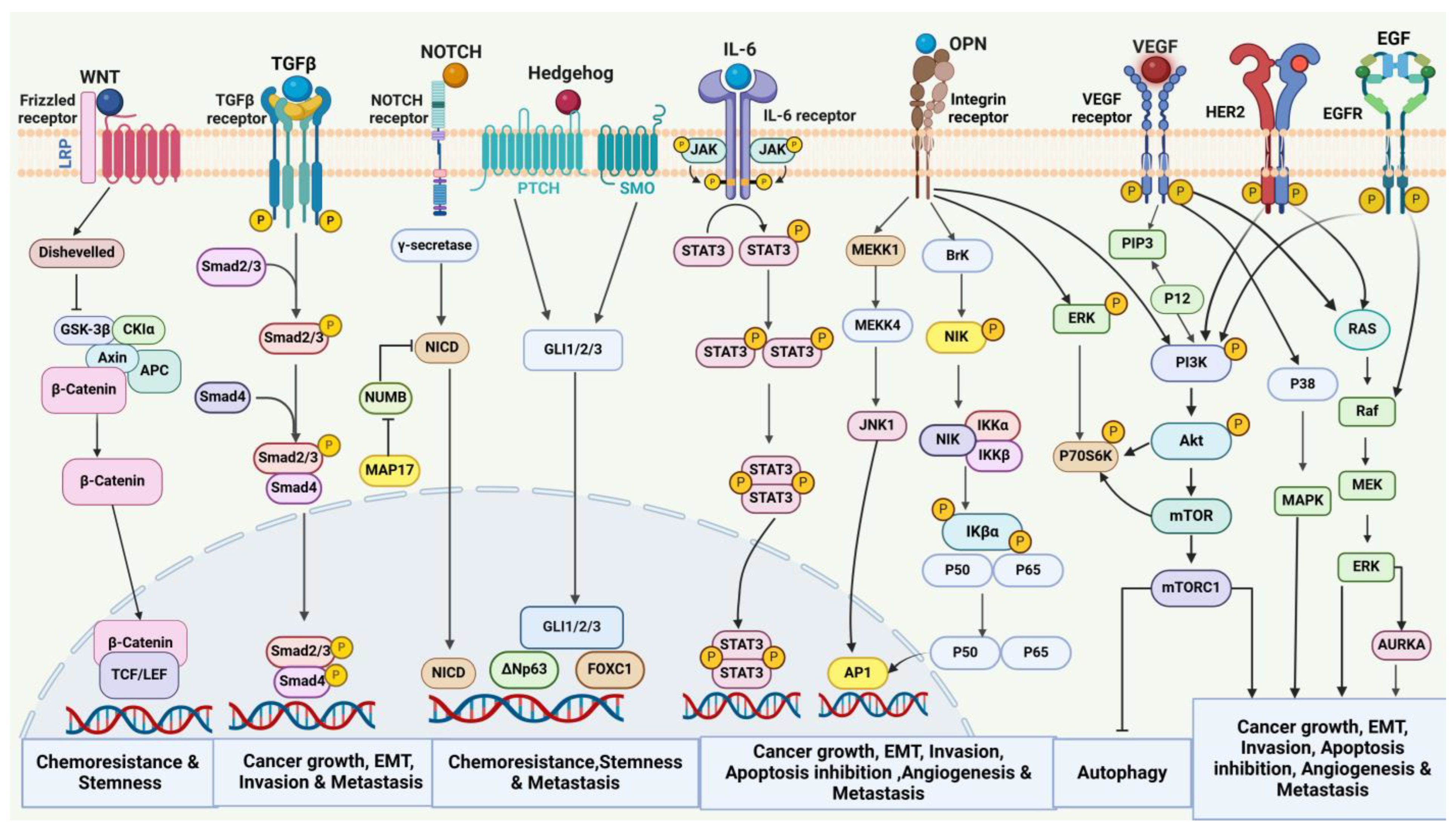
Figure 1. Various cell signaling cascades involved in breast cancer. The major signaling events, including Wnt/β-catenin, TGF-β, Notch, MAPK, Hedgehog, JAK/STAT, PI3K/Akt/mTOR, and NF-κB pathways, and their involvement in the regulation of tumor progression, survival, and metastasis. Wnt/β-catenin pathway is responsible for enhancing the stemness and promoting chemoresistance. TGF-β-mediated signaling is associated with the growth, invasion, and metastasis of breast cancer. Notch pathway as well as Hedgehog signaling induce chemoresistance and metastasis. JAK/STAT and NF-κB signaling cascades modulate breast cancer growth, invasion, metastasis, and angiogenesis. VEGFR-, HER2-, and EGFR-mediated PI3K/Akt/mTOR, MAPK, and ERK pathways have a key role in the regulation of breast tumor growth, metastasis, angiogenesis, and apoptosis.
Cancers 17 00234 g001
Figure 2. The key signaling and transcription factors associated with energy metabolism networks that serve as the foundation for metabolic reprogramming in the progression of breast cancer.
Cancers 17 00234 g002
Figure 3. Culmination of signaling pathways in the tumor stroma microenvironment involving cancer-associated fibroblasts (CAFs), adipocytes, and mast cells. CAFs release growth factors like TNF, SDF-1, VEGF, EGF, FGF2, and IGF, resulting in breast cancer progression. Secretion of several MMPs by CAFs such as MMP1, MMP2, MMP3, MMP7, MMP9, MMP13, and MMP14 are responsible for breast cancer advancement. Cytokines and chemokines released from CAFs include IL4, IL6, IL8, CXCL1, CXCL3, CXCL16, and CXCL8, which enhance breast cancer cell motility and aggressiveness. CAAs secrete inflammatory adipokines such as TNFα, leptin, CCL2, CCL5, and IL6, which modulate breast cancer progression. Mast cells contribute to angiogenesis in breast cancer by releasing growth factors such as VEGF, FGF2, and PDGF, along with proteases such as tryptases and chymases.
Cancers 17 00234 g003
Figure 4. Schematic representation of various TAM subsets based on their molecular signatures and secretory factors.
Cancers 17 00234 g004
Figure 5. Illustration depicting molecular crosstalk between TAMs and cancer cells via various signaling pathways in promoting breast tumor development and metastasis.
Cancers 17 00234 g005
Figure 6. Representative molecular targets in breast cancer, along with the corresponding immunotherapeutic and targeted treatment approach. The GPCR-mediated AKT/PI3K/mTOR signaling pathway is a major target for inhibitors such as alpelisib, buparlisib, capivasertib, ipatasertib, everolimus, and sapanisertib, used to suppress breast tumor growth. Inhibitors including trametinib and ONC201 target the EGFR-activated MEK/ERK signaling pathway. Targeting HER2 and VEGF with their respective monoclonal antibodies, trastuzumab and bevacizumab, leads to the inhibition of cell proliferation and angiogenesis. Tarextumab, a potent inhibitor of the Notch receptor, along with MK0752 and DAPT, that target γ-secretase, inactivate the Notch signaling pathway. Palbociclib, ribociclib, and abemaciclib are the CDK4/6 inhibitors that induce cell cycle arrest by suppression of CDK4/6-mediated signaling. Targeting ER with hormone therapy like tamoxifen and aromatase inhibitors can be a key strategy for treating hormone receptor-positive breast cancer. Immune checkpoint inhibitors such as pembrolizumab, nivolumab, cemiplimab, avelumab, atezolizumab, durvalumab, tremelimumab, and ipilimumab block PD-1, PD-L1, and CTLA-4, respectively, act as effective immunotherapeutic drugs.
Cancers 17 00234 g006
Table 1. Recent FDA-approved drugs for breast cancer (source: https://www.fda.gov/) (accessed on 27 December 2024).
Drug NameActive IngredientBreast Cancer Type
KISQALI
KISQALI FEMERA CO-PACK
(CO-PACKAGE)
RIBOCICLIB SUCCINATE
LETROZOLE; RIBOCICLIB SUCCINATE
HR+ve, HER2-ve breast cancer
ENHERTUFAM TRASTUZUMAB DERUXTECAN-NXKIHER2+ve breast cancer
TEPYLUTETHIOTEPABreast adenocarcinoma
IBRANCE tabletsPALBOCICLIBHR+ve, HER2-ve, advanced, or metastatic breast cancer
HALAVEN-injectionERIBULIN MESYLATEMetastatic breast cancer
TRUQAPCAPIVASERTIBHR+ve, HER2-ve, locally advanced metastatic breast cancer
ORSERDUELACESTRANT HYDROCHLORIDEER+ve, HER2-ve, ESR1-mutated, advanced, or metastatic breast cancer
VERZENIO with endocrine therapy (tamoxifen or an aromatase inhibitor or fulvestrantABEMACICLIBHR+ve, HER2-ve, node-positive early and advanced, or metastatic breast cancer
LYNPARZAOLAPARIBGermline BRCA-mutated, HER2-ve breast cancer
KEYTRUDAPEMBROLIZUMABTriple-negative breast cancer
TRODELVYSACITUZUMAB GOVITECANMetastatic triple-negative breast cancer
Table 2. Ongoing breast cancer clinical trials (Source: https://clinicaltrials.gov/) (accessed on 27 December 2024).
S. No.Breast Cancer SubtypeTherapy DetailsPhaseClinical Trial No.
1TNBCPembrolizumab, axatilimab, radiation therapyPhase 2NCT05491226
2TNBCUtidelone (UTD1) plus capecitabinePhase 2NCT06385990
3TNBCCarboplatin, docetaxel, doxorubicin, cyclophosphamide, pembrolizumabPhase 2NCT05645380
4TNBCFluzoparib + paclitaxel
Epirubicin + cyclophosphamide
Phase 2NCT05834582
5TNBCSintilimab, anlotinib, nab-paclitaxel, carboplatin, epirubicin, cyclophosphamidePhase 2NCT04877821
6TNBCCapecitabinePhase 2NCT04768426
7TNBCAnti-PD-1 monoclonal antibody, VEGFR2 tyrosine kinase inhibitorPhase 2NCT05556200
8TNBCAdebrelimab + stereotactic radiotherapy + nab-paclitaxel + carboplatin, adebrelimab + chemotherapy (nab-paclitaxel + carboplatin)Phase 2NCT06165900
9TNBCEpirubicin, cyclophosphamide, paclitaxel, carboplatin,Phase 3NCT03876886
10TNBCDeferoxamine plus chemotherapyPhase 2NCT05300958
11TNBCTiragolumab, atezolizumab and chemotherapyPhase 2NCT06175390
12TNBCEpirubicin, CTX, paclitaxel, ddEpirubicin, ddCTX, paclitaxel (with carbo), carboplatinPhase 3NCT04296175
13TNBCBL-B01D1, eribulin, vinorelbine, gemcitabine, capecitabinePhase 3NCT06382142
14TNBCOlaparib, paclitaxel + carboplatinPhase 2, 3NCT03150576
15TNBCSacituzumab govitecan-hziy (SG),
Pembrolizumab, capecitabine
Phase 3NCT05633654
16TNBCAlbumin-bound paclitaxel + carboplatin, epirubicin + docetaxelPhase 4NCT04136782
17TNBCAZD6738, olaparib, durvalumabPhase 2NCT03740893
18TNBCEribulin, LM-108, nab-paclitaxel, toripalimabPhase 2NCT06387628
19TNBCTiragolumab, atezolizumab, ipilimumabPhase 2NCT06342037
20TNBCα-lactalbumin vaccine, zymosanPhase 1NCT04674306
21TNBCCeralasertib, durvalumab, nab-paclitaxelPhase 2NCT05582538
22TNBCCapecitabine, talazoparib, pembrolizumab, inavolisibPhase 2NCT04849364
23TNBCRibociclib, bicalutamidePhase 1, 2NCT03090165
24TNBCCamrelizumab plus famitinib with/without nab-palitaxelPhase 2NCT05670925
25TNBCLenvatinib, pembrolizumabPhase 1NCT04427293
26TNBC, stage IV breast cancerAnti-HER2/HER3 dendritic cell vaccine, pembrolizumabPhase 2NCT04348747
27TNBC (stage I, II, III breast cancer)Carboplatin, cyclophosphamide, docetaxel, doxorubicin, paclitaxel, pembrolizumabPhase 3NCT05929768
28Metastatic TNBCTrilaciclib, pembrolizumab, gemcitabine, carboplatinPhase 2NCT06027268
29Metastatic TNBCGemcitabine and carboplatin plus antibiotic (moxifloxacin); gemcitabine combined with carboplatin plus placeboPhase 3NCT04722978
30TNBC, metastatic breast cancerSacituzumab govitecanPhase 3NCT05552001
31TNBC, intermediate and high-risk luminal Accelerated partial breast irradiation, chemotherapyPhase 1, 2NCT02806258
32TNBC, ductal
carcinoma in situ, lobular carcinoma
in situ
AbemaciclibPhase 2NCT03979508
33TNBC, HR+ve and HER2-ve breast cancerPembrolizumab, paclitaxel, carboplatin, cyclophosphamide, doxorubicin, capecitabinePhase 2NCT04443348
34Invasive breast cancer (HR+ve, HER2-ve, or TNBC)Cemiplimab, paclitaxel, carboplatin (not mandatory), doxorubicin, cyclophosphamidePhase 2NCT04243616
35TNBC, stage III, IV, and Recurrent Breast Cancer Avelumab, binimetinib, utomilumab, liposomal doxorubicin, sacituzumab govitecanPhase 2NCT03971409
36Metastatic TNBCAtezolizumab, bevacizumab, gemcitabine, carboplatinPhase 2NCT04739670
37TNBC, metastatic breast cancer, HER2 -ve breast cancerL-NMMAPhase 2NCT05660083
38Metastatic TNBC, stage IV breast cancer Ivermectin, balstilmabPhase 1 & 2NCT05318469
39TNBC, metastatic breast cancer and ER-low breast cancerCarboplatin, tocilizumabPhase 2NCT05846789
40Breast cancers and metastatic AR+ve TNBCPalbociclib, avelumabPhase 1NCT04360941
41Unresectable or metastatic TNBCTobemstomig, pembrolizumab, nab-paclitaxelPhase 2NCT05852691
42All types of breast cancer like HER2+ve and -ve and PR +ve, TNBCALX148, fam-trastuzumab deruxtecan-nxki, zanidatamab, tucatinibPhase 1NCT05868226
43Breast cancerAtezolizumab injection, bevacizumab, pertuzumab, trastuzumabPhase 2NCT05180006
44Breast cancerNivolumab, ipilimumabPhase 2NCT03815890
45HER2+ve, metastatic breast cancerInavolisib, Phesgo, taxane-based chemotherapyPhase 3NCT05894239
46HER2+ve metastatic
breast cancer
Atezolizumab + trastuzumab + vinorelbinePhase 2NCT04759248
47Luminal breast cancer: HER2-ve HR+veElacestrant, triptorelinPhase 2NCT05982093
48Luminal A breast cancerBreast irradiation (RT),
endocrine therapy (ET): letrozole, anastrozole, exemestane, tamoxifen
Phase 3NCT04134598
49Luminal B/HER2-ve breast cancerDalpiciclib combined with aromatase inhibitorsPhase 2NCT05640778
50Ductal carcinoma in situTamoxifen, exemestane,
letrozole, anastrazole,
testosterone + anastrazole,
elacestrant, Z-endoxifen
Phase 2NCT06075953
51Ductal carcinoma in situGranulocyte–macrophage colony-stimulating factor, multi-epitope HER2 peptide vaccine H2NVACPhase 1NCT04144023
52Ductal carcinoma in situ, postmenopausalConjugated estrogens/bazedoxifene Phase 2NCT02694809
53Invasive lobular carcinoma Fulvestrant, repotrectinibPhase 2NCT06408168
54Invasive breast lobular carcinoma NeratinibPhase 2NCT05919108
55HER2-ve
breast cancer
Doxorubicin, cyclophosphamide
Weekly paclitaxel, trastuzumab
Pertuzumab
Phase 2NCT03412643
56ER+ve
breast cancer, HER2-ve
breast cancer, metastatic breast cancer
AI + CDK4/6i,
SERD + CDK4/6i,
mTOR inhibitor + AI,
mTOR inhibitor + SERD,
mTOR inhibitor + selective estrogen receptor modulator,
PI3K inhibitor + SERD,
PI3K inhibitor + AI,
oral SERD
Phase 2NCT05826964
57HR+ve/HER2-ve metastatic breast cancerFulvestrant
Capecitabine oral product
Phase 3NCT04263298
58HER2-low, HR+ve metastatic
breast cancer
DB-1303/BNT323, capecitabine, paclitaxel,
nab-paclitaxel
Phase 3NCT06018337
59ER+ve/HER2-ve metastatic breast cancerEndocrine therapy combined with the local treatment of FES-negative lesionsPhase 3NCT06195709
60HER2-ve
breast cancer
Cyclophosphamide, fludarabine, camrelizumab, chemotherapeutic drug, ADC, or PARP inhibitorPhase 1NCT06121557
61HER2-ve
breast cancer
Epirubicin, cyclophosphamide,
docetaxel, paclitaxel
Phase 2 and 3NCT04576143
62HER2-ve
breast cancer
Cyclophosphamide,
Fludarabine, nab-paclitaxel,
Gemcitabine, carboplatin
Phase 1 and 2NCT05981001
63HER2-ve
breast cancer
Paclitaxel, carboplatin,
Cyclophosphamide/doxorubicin
Phase 2 and 3NCT05889390
64HER2-ve
breast cancer
Doxorubicin, epirubicin,
cyclophosphamide, fludarabine
Nab-paclitaxel
Phase 1NCT06121570
65ER+ve/HER2-ve breast cancerDocetaxel, carboplatin,
epirubicin, cyclophosphamide
Phase 3NCT05901428
66HR+ve/HER2-ve premenopausal breast cancerDalcelli, exemestane, gosserine
Docetaxel, epirubicin hydrochloride, cyclophosphamide
Phase 2 and 3NCT06009627
67HER2-ve breast carcinoma, HR+veCyclophosphamide,
Doxorubicin,
Durvalumab, paclitaxel
Phase 3NCT06058377
68HER2-ve early breast cancerLiposomal doxorubicin, cyclophosphamide vs. docetaxel, cyclophosphamidePhase 4NCT05302336
69HER2-ve breast cancerCamrelizumab with vinorelbine and cisplatinPhase 2NCT04848454
70HER2-ve breast
carcinoma
Cyclophosphamide,
Docetaxel
Phase 2NCT06042569
Disclaimer/Publisher’s Note: The statements, opinions and data contained in all publications are solely those of the individual author(s) and contributor(s) and not of MDPI and/or the editor(s). MDPI and/or the editor(s) disclaim responsibility for any injury to people or property resulting from any ideas, methods, instructions or products referred to in the content.