Article

Impact of Epigenome-Wide Methylation and Breast Cancer Recurrence in Women Tested Negative for BRCA Genes: The Breast Methylation Risk (BREMERI) Study

1
Department of Translational Medicine, University of Piemonte Orientale, 28100 Novara, Italy
2
Division of Cancer Prevention and Genetics, European Institute of Oncology, IEO, IRRCCS, 20141 Milan, Italy
3
Department of Medical Sciences, University of Turin, 10124 Turin, Italy
4
Department of Experimental Oncology, European Institute of Oncology, IEO, IRRCCS, 20141 Milan, Italy
*
Author to whom correspondence should be addressed.
These authors contributed equally to this work.
Cancers 2025, 17(19), 3132; https://doi.org/10.3390/cancers17193132
Submission received: 4 August 2025 / Revised: 11 September 2025 / Accepted: 24 September 2025 / Published: 26 September 2025
(This article belongs to the Section Cancer Biomarkers)

Simple Summary

Breast cancer can sometimes return after initial treatment, but predicting who is most at risk remains difficult. Our matched case–control study examined DNA methylation, defined as chemical variations in the DNA that influence gene activity. We analysed blood samples from women with breast cancer who did not carry BRCA mutations, comparing those who later had recurrence with those who remained cancer-free. Three DNA regions showed different methylation patterns between the two groups. These regions contain three genes known to contribute to important processes such as cell growth, invasion, and how the body metabolises sugar. These findings suggest that DNA methylation patterns in blood could one day help identify women at higher risk of recurrence and guide more personalised follow-up and treatment strategies.

Abstract

Background and Aim: DNA methylation may contribute to a worsening in breast cancer (BC). Methods: We conducted a matched case–control study to investigate the contribution of DNA methylation (DNAm) in breast cancer recurrence risk. Genome-wide DNAm profiles were generated from peripheral white blood cells (WBC) collected post-surgery from women with primary breast cancer BRCA wild-type, using Illumina Infinium HumanMethylationEPIC array. Cases had to experience recurrence of breast cancer or death and were matched to controls (subjects without recurrence, ratio 1:2) by age at diagnosis (+/− 5 years) and follow-up duration. Results: We identified three differentially methylated regions between the groups. Cases showed two hypomethylated regions, one upstream of the vtRNA2–1 gene (estimate −0.30, p-value < 0.005), and one in the 5′ UTR region of the FGFR2 gene (estimate −0.34, p-value < 0.028), whereas one, upstream of the RUFY1 gene (estimate 0.32, p-value < 0.015), was hypermethylated. Additionally, we identified two methylation signals, recognised as predictors of biochemical traits. The chemokine ligand 21 (unadjusted p-value < 0.03) and insulin receptor expression (unadjusted p-value < 0.04) were higher in cases than in controls. Conclusions: Our exploratory study suggests that specific DNA methylation patterns in WBCs, particularly in genes related to cellular proliferation, invasion, and glucose homeostasis, may be associated with the risk of breast cancer recurrence in BRCA wild-type women. If validated in larger cohorts, these circulating signatures may serve as blood-based biomarkers to improve risk stratification and guide tailored treatment strategies.

1. Introduction

Despite breast cancer having the highest incidence of all cancers in women worldwide, patients’ life expectancy is much higher compared to many other cancers. Breast cancer is a heterogeneous disease resulting from differences in several cancer-cell-intrinsic parameters, including genetic profile, the interplay between the genome, epigenome, and transcriptome, as well as proteome, migration and invasion capabilities, proliferation, stemness, and intrinsic cell plasticity [1]. Among breast cancer patients meeting the criteria for genetic testing of hereditary breast and ovarian cancer syndrome, approximately one-fourth are carriers of a pathogenic variant in BRCA1 or BRCA2 (BRCA) genes, whilst other less common susceptibility genes may also contribute to increased disease risk (TP53, PTEN, STK11, PALB2, ATM, and CHEK2) [2]. However, many patients with suspected hereditary breast cancer remain without a clear etiopathogenetic origin of the disease.
DNA methylation is recognised as a stable, heritable mark that can affect genome function and influence gene expression. Indeed, it is also shaped by environmental conditions. White blood cell (WBC) DNA methylation motifs have shown promise as biomarkers for assessing breast cancer susceptibility in multiple-case breast cancer families without known germline mutations [3].
Several genome-wide studies have found evidence of an association between global DNA methylation levels and an increased risk of breast cancer, detectable years before a clinical diagnosis [4,5,6]. The epigenetic machinery has been shown to play an essential role in carcinogenesis and progression through aberrant DNA methylation and histone modifications at the tissue level, exhibiting significant heterogeneity across different cancer types in this rapidly expanding field of research [7].
In breast cancer patients, specific methylation patterns could affect the risk of recurrence. In a genome-wide search for DNA methylation markers using available archival tumour samples from node-negative triple-negative breast cancer (TNBC), elevated levels of DNA methylation were associated with shorter recurrence-free intervals in triple-negative breast cancer [8], pointing to a pivotal role of methylation changes in the transition from primary breast cancer to metastatic disease by regulating key genes implicated in tumour suppression and oncogenic activity. Notably, different breast cancer subtypes exhibit different metabolic phenotypes, and altered metabolisms of glucose, lipids, or amino acids [9]. It is known that cancer cells can change their energetic strategy to support rapid growth and metastatic potential [10] and this mechanism can be fuelled by peripheral or constitutional factors.
Although epigenetic studies in cancer have focused on the effect of gene methylation mainly on breast tissue expression and metastasis [11,12], growing evidence exists on the feasibility of conducting studies on white blood cells [3,13]. Interesting results emerged from a study conducted in China [14] applying a multiplex blood-based assay targeting DNA methylation in peripheral blood mononuclear cells. Based on four methylation markers, they were able to distinguish early-stage breast cancer patients from age-matched normal females. The sensitivity of the test was 93.2%, the specificity 90.4%, with an area under the curve of 0.940.
Variations in blood DNA methylation result from a combination of internal factors, such as genetics, age, hormones, as well as external factors, including environmental toxins, smoking, stress, and lifestyle. Although more complex and indirect as a source of risk biomarkers, blood sampling, compared to tissue biopsies, offers a minimally invasive and straightforward approach for methylation analysis, suitable for clinical monitoring.
Hence, in consideration of recent evidence for an association of white blood cell methylation signatures with breast cancer risk and the hypothesis that they may have a role even in breast cancer recurrence, within a cohort of multiple-case, BRCA-negative breast cancer patients followed at the European Institute of Oncology (IEO), we performed a genome-wide DNA profiling to investigate whether specific methylation patterns in WBCs are associated with breast cancer recurrence using the Illumina MethylationEPIC BeadChip microarray for whole-genome DNA methylation profiling [15].

2. Materials and Methods

2.1. Study Design and Patient Characteristics

The present work is a retrospective case–control study of women undergoing BRCA1 and BRCA2 germline testing for breast cancer diagnosis at IEO. After oncogenetic counselling at the Division of Cancer Prevention and Genetics during the years 2001–2015, patients who were negative for BRCA testing and had a diagnosis of primary breast cancer were considered. Only women with a blood draw after surgery were eligible. Cases had to have a recurrence (ipsi- or contralateral breast cancer event, second breast tumour, metastasis) or death at least six months after surgery. No previous diagnosis of cancer was admitted. Cases were matched to controls (subjects without recurrence; ratio 1:2) by age at diagnosis (+/− 5 years) and follow-up duration. The IEO Institutional Review Board approved the study (IEO 1058), and participants signed an informed consent for research purposes.

2.2. DNA Methylation Assessment

Whole EDTA-treated blood samples were drawn and stored at −80 °C until assayed. Genomic DNA was extracted from whole blood specimens using a QIAamp DNA blood kit (Qiagen, Valencia, CA, USA) according to the manufacturer’s instructions using the automated platform “QIAcube” (Qiagen, Valencia, CA, USA) and quantified using a NanoDrop spectrophotometer (Thermo Scientific, Wilmington, DE, USA).
Samples were shipped to the Italian Institute for Genomic Medicine laboratory, where DNA samples underwent preliminary quality control before bisulphite-converting 500 ng of each sample using the EZ DNA Methylation Lightning Kit (Zymo Research, Irvine, CA, USA), following the manufacturer’s instructions.
Bisulphite-converted DNA samples were randomly arranged on the Infinium HumanMethylationEPIC BeadChip (Illumina, San Diego, CA, USA) and processed following the manufacturer’s instructions.
After amplification, hybridisation, staining, and washing, the Beadchips (referred to as the 850k chip) were scanned with the Illumina iScan SQ instrument to acquire raw image intensities, which were saved as IDATs for further processing. To mitigate the batch effect and optimise the space on the arrays, each case and one of its matched controls were analysed on the same chip, while the second control was randomly allocated to a different chip.

2.2.1. DNA Methylation Data Pre-Processing

Raw DNA methylation data generated from Illumina Infinium HumanMethylation EPIC arrays were imported and preprocessed using the ChAMP Bioconductor package (v 2.8.3) [16].
The average methylation value at each CpG locus, i.e., average “beta (β) value” ranging from 0 to 1, was computed as the ratio of the intensity of the methylated signal over the total signal (unmethylated + methylated).
To reduce artefacts and genetic confounding, initial quality control filtered probes with detection p-values > 0.01, fewer than 3 Beads in 5% of samples, mapping to multiple locations or on sex chromosomes, with known SNPs at CpG sites or single-base extensions; 742,521 CpGs passed QC.
Normalisation of probe intensities was performed using the Beta MIxture Quantile dilation (BMIQ) method to correct for probe design bias (Type I and Type II). Batch effects due to technical variability were identified and corrected using ComBat from the SVA package (v 3.56.0), using empirical Bayes frameworks while preserving the biological variation of interest.
After normalisation and batch correction, beta values were converted to M-values by logit transformation for improved statistical handling in downstream differential methylation analyses. The final preprocessed dataset was subjected to further analyses, including differential methylation position (DMP) and region (DMR) identification using ChAMP’s analytical workflows.
To estimate the proportions of major white blood cell (WBC) subtypes in our samples and adjust the statistical analysis for cellular heterogeneity among samples, we applied the Houseman reference-based deconvolution method. This approach utilises a reference matrix of methylation signatures from purified leukocyte subpopulations, modelling bulk methylation profiles as linear combinations of these reference signatures [17].

2.2.2. Differentially Methylated Analysis

The statistical analysis was structured to identify and interpret patterns of differential DNA methylation in cases vs. controls, using both site-specific and region-based approaches.
(i) Differentially Methylated Probes (DMPs):
Differential methylation at individual CpG sites was assessed using the champ.DMP functions within the ChAMP Bioconductor package (v 2.8.3). A linear modelling framework implemented via the limma package (v 3.64.3) was then applied probe-wise to test for methylation differences between cases and controls.
Empirical Bayes moderation of standard errors was applied to enhance the reliability of variance estimates, especially in small sample sizes like ours.
(ii) Differentially Methylated Regions (DMRs):
DMRs are defined as contiguous genomic regions containing multiple CpG sites that exhibit coordinated changes in methylation levels between comparison groups.
They were identified with the Bumphunter algorithm, available through the champ.DMR function in the ChAMP package (v 2.8.3). This method detects DMRs by clustering nearby CpG probes based on their genomic proximity. For each cluster, Bumphunter applies a permutation-based test to determine if regional methylation levels differ significantly between cases and controls. Since it does not depend on initial differential methylation findings at individual CpG sites, this approach is ideal for exploratory analyses.
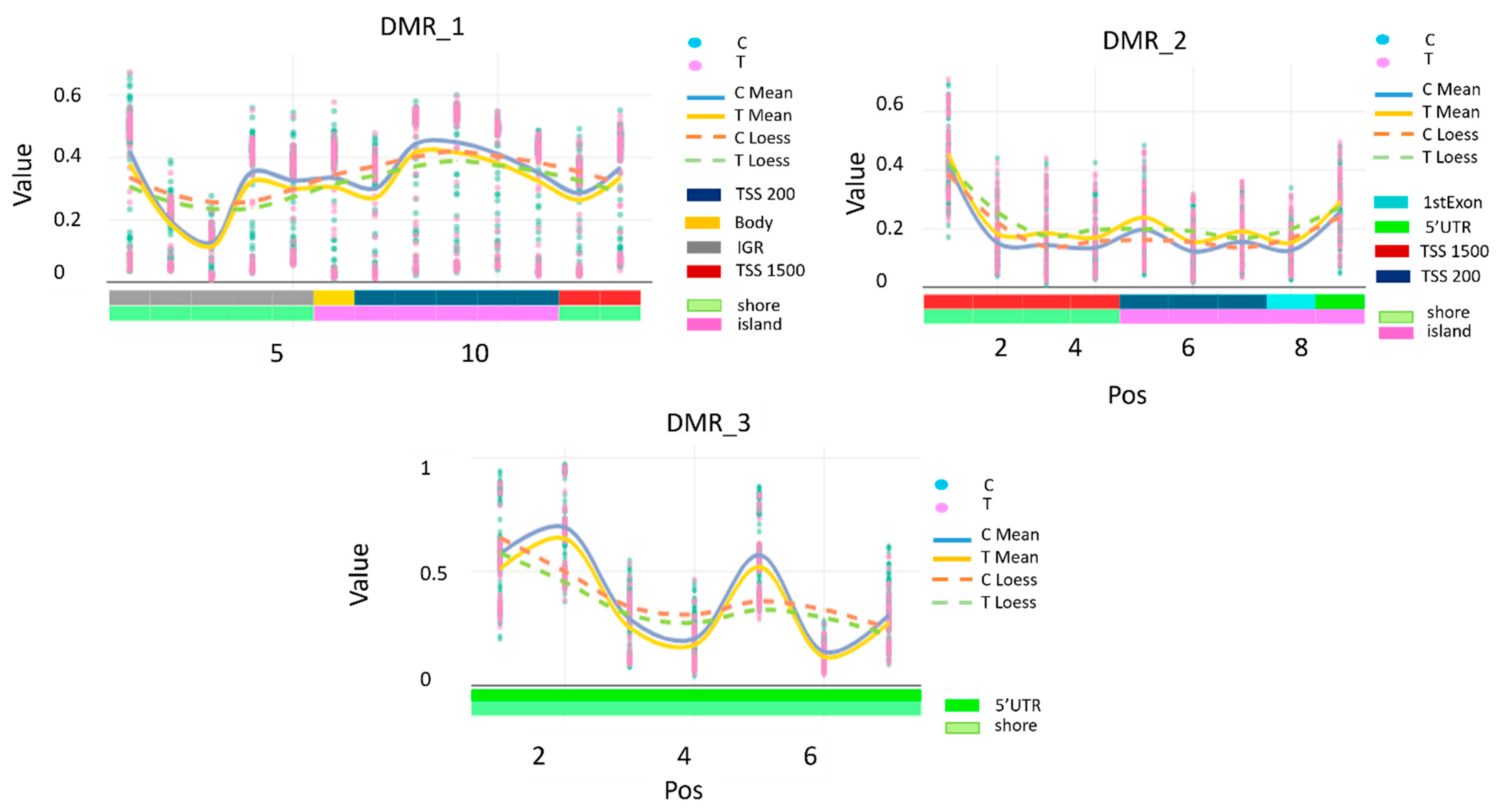
Differentially methylated regions (DMRs) were visualised using the DMR.plot function from the ChAMP Bioconductor package (v 2.8.3) with default parameters. In these plots, the X-axis corresponds to the genomic coordinates of CpG probes within each DMR, and the Y-axis represents DNA methylation β values ranging from 0 (fully unmethylated) to 1 (fully methylated). For each comparison group, individual probe-level β values (“C” and “T” tracks) are shown alongside their group-wise means (“C mean” and “T mean”) and LOESS-smoothed curves (“C loess” and “T loess”). The averaging and smoothing steps are applied for visualisation only to highlight local correlation and regional methylation trends; all statistical inference of DMRs was performed at the probe level without smoothing.
(iii) Sensitivity Analyses on DMRs:
For the three identified DMRs, we investigated possible deviation from multimodality of the distribution of methylation β values at all CpGs within each region. We used Hartigan’s dip test, as implemented in the diptest R package (v 0.77-2) [18]. CpGs showing evidence of multimodality were further categorised using the mclust R package (v 6.1.1) [19]. Briefly, mclust applies a model-based clustering framework fitting Gaussian mixture models with varying numbers of components, selecting the optimal solution based on the Bayesian Information Criterion (BIC). Finally, logistic regression analyses were repeated for each CpG using the derived categorical variables, adjusting for age, array position, and estimated WBC, for consistency with the main analysis, to evaluate robustness of the results. All statistical analyses were conducted using the open-source software R (v 4.5.1).
(iv) Differentially Methylated Blocks (DMBs):
DMBs are large genomic regions composed of clusters of CpGs, typically spanning tens to thousands of kilobases. In ChAMP, DMB analysis first groups probes within open sea regions into larger contiguous blocks by averaging methylation values of neighbouring CpGs. The Bumphunter algorithm is then applied to these blocks to identify regions showing significant methylation differences between groups using permutation testing. Compared to DMR analysis, which targets smaller, densely packed CpG clusters, DMB detection focuses on broader methylation changes, improving sensitivity to large-scale epigenetic alterations.
To account for technical variability and biological confounders, all models incorporated multiple covariates. In particular, adjustments were made for age at blood draw, chip position to control for batch effects, and for white blood cell (WBC) composition, which reflects inter-individual variability in cellular makeup—a known source of epigenetic heterogeneity in blood-based analyses.
Resulting p-values were corrected for multiple testing using the Benjamini–Hochberg false discovery rate method. Probes, regions, or blocks with adjusted p-values < 0.05 were considered statistically significant.

2.2.3. Deriving Methylation-Based Surrogates

Methylation profile scores (MPS), also known as surrogate estimators or ‘predictors’ of human traits, have been calculated using the ‘MethylDetectR’ online tool (https://shiny.igmm.ed.ac.uk/MethylDetectR, accessed on 17 July 2025).
These MPS have been calculated using machine learning methods to link DNAm data in a specific tissue (typically blood) to traits of interest, such as smoking status, epigenetic age, and concentrations of various biomarkers in plasma. These models identify CpG sites strongly correlating with the trait, assigning weights to each site’s predictive value. In our study, DNAm levels at key CpG sites were used to estimate trait values or scores for 117 human traits [18]. Linear regression was used to assess the associations between the MPS and breast cancer progression. Age at blood draw, chip position, and percentage of WBCs were controlled as potential confounders. For all tests, the significance level was set at p < 0.05, and p-values adjusted for FDR are presented.
All statistical analyses were conducted using the open-source software R (v 4.4.1).

3. Results

Overall, 428 candidate patients were considered. Figure 1 illustrates the additional exclusion criteria, reducing the cohort to 337 women. The cases were matched to controls (women without any breast cancer event or progression) by age at diagnosis (+/− 5 years), time from surgery to biobanking (+/− 5 years), and follow-up time. Eventually, 63 cases were matched to 120 controls. Cases were matched to 2 controls, except for six cases for whom only a single control was available.
In Table 1A, we present the characteristics of the study population. There was no statistically significant difference (p = 0.43) between the median age at genetic testing, which was 42 years with an interquartile range (IQ) of 37–49 years in cases (n = 63) and 45 years with an IQ of 38–50 years in the control group (n = 120). Likewise, we did not observe any statistical difference in BMI at genetic testing between groups (p = 0.69). Nonetheless, comparing BMI reported at 18 years of age, we observed a significant difference, showing a higher BMI at 18 years (p = 0.04) in cases (20, IQ 18–21) compared to controls (19, IQ 18–20). We also describe data on the use of oral contraceptives, pregnancies, menopausal status, smoking, and family history.

Epigenome-Wide Differentially Methylated Probes

After post hoc correction for FDR (adjusted p-value), no statistically significantly differentially methylated single CpG probes were recorded between groups; nonetheless, the first 100 single CpGs ordered by nominal p-value were extracted to look for trends and were assessed for biological relevance without finding any statistical significance related to breast cancer recurrence (Table S1, in the Supplementary Materials).
The clustering analysis revealed three differentially methylated regions (DMRs) associated with recurrence. DMR1 showed overall hypomethylation (estimate = −0.30 compared to the reference group, adjusted p < 0.005), DMR2 was hypermethylated (estimate = 0.32 compared to the reference group, adjusted p < 0.015), and DMR3 was hypomethylated (estimate = −0.34 compared to the reference group, adjusted p < 0.028).
Table 2 lists each CpG probe mapping within the three DMRs. DMR1 on chromosome 5 included 13 CpGs in the upstream area of the transcriptional start site of the vtRNA2–1 gene, as well as a few intergenic regions. On the same chromosome, DMR2 includes nine CpGs mapping in the upstream area of the transcriptional start site of the RUFY1 gene, one in the 1st exon, and one in the 5′UTR region of the same gene. On chromosome 10, the DMR3 included seven CpGs, covering sequences in the 5′UTR region of the FGFR2 gene.
Figure 2 shows the DNA methylation signal intensities of each CpG position across the three significant DMRs identified by ChAMP for cases vs. controls, highlighting the functional characteristics.
Given that CpGs showed evidence of multimodality (Figure S1, in the Supplementary Material), a sensitivity analysis categorising DMR1 and DMR3 was conducted to assess the robustness of the results. Logistic regression analyses, adjusting for age, array position, and estimated WBC, showed consistent results with the main analysis with ORs lower than one, even if the associations did not reach statistical significance, due to loss of statistical power (Table S2 in the Supplementary Material). The multimodal distribution of DNAm in DMR1 and DMR3 suggested a potential influence from methylation quantitative trait loci (meQTLs). To investigate this, we searched for nearby DNA sequence variants associated with the CpGs in these regions.
We consulted the EPIGEN MeQTL Database (https://epicmeqtl.kcl.ac.uk, accessed on 17 September 2025), which includes a comprehensive meQTL analysis of blood samples from 2358 individuals. According to this database, both DMR1 (6 out of 13 CpGs in the region) and DMR3 (all 7 CpGs in the region) contain genetic variants that are statistically associated with DNAm levels, with a false discovery rate (FDR) of less than 0.05 (Table S3, in the Supplementary Material).
Differentially methylated blocks (DMBs) analysis did not show any statistically significant differentially methylated blocks between groups after post hoc correction. Nevertheless, we detected a block (adjusted p-value = 0.05) that covers 327 CpG probes mapping within the genomic region of cadherin-4 (CDH4), a member of the cadherin family that encodes Ca2+ -dependent cell–cell adhesion glycoproteins, which could be of biological interest.
The surrogates’ results for the 117 MPS traits did not show statistically significant differences between cases and controls after adjusting for chip number, position, WBC percentage, and age, and after controlling for FDR. Nonetheless, we identified two traits with nominal significance: CCL21, a chemokine ligand (p-value = 0.028), and the insulin receptor (p-value = 0.041), both with a positive estimated coefficient (Table S4, in the Supplementary Material).

4. Discussion

In our study, although no association was found between isolated differentially methylated CpGs and breast cancer recurrence in BRCA-negative patients, the analysis of DMRs revealed three genomic segments that were significantly differentially methylated between cases and controls.
Two of these regions (DMR1 and DMR3) showed a multimodal distribution of DNA methylation. This pattern could reflect the influence of methylation quantitative trait loci (meQTLs), although our data do not allow us to test this directly. Future studies are required to clarify this, but it is of interest that both regions coincide with meQTLs reported in the EPIGEN MeQTL database.
To further assess the robustness of our findings, we performed a sensitivity analysis by categorising DMR1 and DMR3. Logistic regression results suggested a pattern consistent with the primary analysis, with odds ratios below one. However, the associations did not reach statistical significance, likely reflecting the reduced statistical power of this additional analysis.
Interestingly, these segments span genes involved in cell signalling, mitogenesis, differentiation, and glucose metabolism regulation, all of which play a crucial role in modulating the risk of progression and metastasis.
The VAULT 2 locus, located on chromosome 5q31, encodes vtRNA2–1, also known as non-coding 886 (nc886), because it transcribes a non-coding RNA. This gene was recently investigated in a review [19] that highlights its distinctive characteristics, namely being the only human gene known to display maternal polymorphic imprinting [20]. The vtRNA2–1 methylation pattern of an individual tends to remain stable across a lifespan [21] and similar in many somatic tissues [20,21]. Furthermore, its RNA expression is associated with its methylation status [22]. In individuals with a non-methylated vtRNA2–1, the levels of RNAs [23] were approximately two-fold compared to individuals with an imprinted locus [23]. These unique characteristics of the vtRNA locus make it particularly interesting as a blood methylation biomarker of disease.
The expression of vtRNA2–1 has been described as upregulated in many cancers, including breast and prostate cancer [19]. This overexpression correlates with progression and poor prognosis in the latter [24]. vtRNA2–1 is known to bind and modulate the activity of target proteins like Protein Kinase R (PKR), 2’-5’-oligoadenylate synthetase 1, and Dicer [25] in complex regulatory mechanisms sensed through its methylation status. Moreover, the vtRNA2–1 methylation status is associated with glucose and insulin levels during adolescence and glucose metabolism later in adulthood [22], linking methylation patterns with metabolic traits often associated with familial risk [3]. Notably, the vtRNA2–1 has been suggested to have a role in post-transcriptional regulation of epithelial barrier integrity through the inhibition of the expression of tight junction and adherens junction proteins [26]. Specifically, elevated levels of vtRNA2–1 were shown to reduce the levels of claudin 1, occludin, and E-cadherin, resulting in an impairment of the intestinal barrier. Impaired intestinal barriers can increase breast cancer prognosis by promoting chronic inflammation, facilitating bacterial translocation into the bloodstream, and influencing treatment response through gut microbiota dysbiosis, potentially increasing the risk of poor breast cancer prognosis [27]. In our study of women with breast cancer, the DMR of vtRNA2–1 was more frequently hypomethylated in cases compared to controls. Thus, we may speculate that overexpression increases the risk of recurrence. Based on the consistency between studies, this hypothesis is reasonable [28]. Nonetheless, we acknowledge that our findings should be interpreted with caution, as some studies, mainly from tissue, found that hypermethylation in the promoter region of vtRNA2–1 was associated with worse prognosis [19,29,30], because we were unable to study vtRNA2–1 expression in our cohort.
Along the same chromosomal region 5q31, another DMR segment covering the upstream area of the transcriptional start site of the RUFY1 gene was hypermethylated in cases compared to controls in our cohort. To our knowledge, very few pieces of evidence have been published on DNA methylation status in peripheral blood mononuclear cells (PBMCs) describing DMRs of RUFY1 and associations with disease. One study [31] found a DMR located on the RUFY1 gene, showing an association between increased DNA methylation in the RUFY1 promoter region and IgE-sensitised children, compared to non-sensitised children, consistently detected also in maternal PBMCs, and cord blood. An explanation linking the hypermethylation of the RUFY1 gene to its downregulation in specific tissues and respective associations with disease is still to be supported.
RUFY adaptor proteins, including RUFY1, are involved in complex biochemical crosstalk between the cytoskeleton and endosome dynamics, including the recycling of cytoplasmic materials [32]. Char and Pierre’s review [32] concluded that the dysregulated expression of RUFY proteins may severely affect cell differentiation and polarisation, contributing to the development of several cancers and neurodegenerative diseases. RUFY1 has been identified as a downstream effector of Etk protein kinase, and its principal function is to bind PIP3-containing phospholipid vesicles that participate in early endosomal membrane trafficking and P13K signalling [33], mediating cell proliferation, colony formation, apoptosis, migration, and invasion. Still, evidence of a link between the hypermethylated region of RUFY1 in WBC and reduced cell expression in any tissue type is warranted before we can speculate on any specific mechanisms.
The third genome segment contains the FGFR2 gene, which is frequently altered in tumours [34]. In our cohort of cases, we found that FGFR2 was hypomethylated in the 5’UTR island region compared to the controls. This gene plays crucial roles in development and tissue repair [34], particularly in bone and blood vessels. Aleyasin and colleagues identified the FGFR2 gene as a blood-based epigenetic biomarker in gastric cancer [13]. Consistent with our results, the FGFR2 gene exhibited considerable hypomethylation in its CpG site, suggesting an epigenetic enhancement of FGFR2 expression [35]. FGFR2 has been reported to be selectively upregulated to maintain the survival of dormant residual tumour cells [35], as well as to promote ERK1/2 signalling and ultimately recurrence.
CDH4, which encodes R-cadherin, was the only gene mapping in a block of 327 CpGs that we found differently methylated, mainly hypomethylated, in our case–cohort compared to controls, which could have some interesting biological implications. The protein is a classical cell surface glycoprotein that plays a crucial role in cell-to-cell adhesion [36]. The protein is a calcium-dependent cell–cell adhesion glycoprotein expressed in the mammary epithelial cells of the ducts and lobules [37] and involved in epithelial-to-mesenchymal transition (EMT), a crucial step towards metastasis formation. CDH4 messenger RNA levels are described to be overexpressed in breast cancer cells compared to normal cells and correlate with poor prognosis in distant metastasis-free survival [38]. Nonetheless, CDH4 was described to be repressed during EMT in breast cancer cells [37] and downregulated in breast tumour tissue [39]. However, although our finding is suggestive of further investigations, we cannot directly translate the trend of hypomethylation of the block towards an increased transcriptional activity of CDH4 in tissue. Additionally, we did not find any evidence in the literature of a link between hypomethylated regions of the CDH4 gene and reduced cell expression in any tissue type.
To better characterise our hypothesis that breast cancer recurrence is associated with altered methylation patterns, we used the methylation data to calculate a panel of surrogates for relevant metabolic traits. Regrettably, none showed a statistically significant difference after correction for multiple comparisons; nevertheless, two surrogates showed nominal significance and were estimated to be increased in cases compared to controls.
The first is Chemokine (C–C motif) ligand 21 (CCL21), a chemokine ligand that seems to play a role in breast cancer progression, mainly through its interaction with the CCR7 receptor, crucial in various processes, such as tumour cell migration, lymphangiogenesis, and immune modulation, collectively contributing to breast cancer metastasis and progression [40,41]. The second protein estimated via DNA methylation surrogates showed a significant nominal increase in the Insulin receptor (IR). It is well known that the levels of IR are significantly higher in breast cancer tissues compared with healthy ones [42]. In the last decade, the role of the receptor in breast cancer, its involvement in tumour progression, and its potential as a therapeutic target, particularly in combination with the IGF1 receptor (IGF1R) [43] have gained increasing attention.
Although CCL21, CDH4, and IR lost statistically significant associations after adjustments in our dataset, given the small sample size of our study, their established roles in immune cell recruitment, cell adhesion, and metabolic/growth signalling, respectively, provide a supportive framework for the central findings. For example, CCL21’s involvement in recruiting immunosuppressive immune cells could synergise with VTRNA2–1-mediated barrier disruption to create a microenvironment permissive to tumour progression. Similarly, CDH4’s influence on cell adhesion and migration aligns with the trafficking functions of RUFY1, while IR’s regulation of growth factor signalling may intersect with FGFR2-driven pathways. In summary, our results highlight RUFY1, FGFR2, and VTRNA2–1 as central components in a network that integrates intracellular trafficking, growth factor signalling, glucose metabolism, and epithelial barrier regulation to drive tumour progression (Table 3).
A limitation of our retrospective study is the lack of blood samples collected before breast cancer diagnosis, which prevents us from determining whether the observed methylation patterns preceded or followed surgery of the primary tumour. Additionally, we anticipated reaching a sample size of 70 cases and 140 controls. However, after applying all necessary exclusion criteria and matching, we ultimately included 63 cases and 120 controls. BRCA wild-type breast cancer is a biologically heterogeneous group, expressing different metabolic phenotypes [9]. This heterogeneity may have contributed to the lack of significant results in the probe-by-probe analysis and the reduced likelihood of detecting strong, consistent DMRs across patients. To improve our knowledge of how altered methylation influences breast cancer risk or progression, further large prospective studies are needed, focusing on the role of peripheral WBC DNA methylation patterns in unaffected women to establish the role of differently methylated regions as breast cancer risk biomarkers and collecting breast cancer tissue at diagnosis. A longitudinal follow-up of breast cancer cases would enable researchers to study the prognostic value of these biomarkers. Despite these limitations, our exploratory study revealed consistency among results linking DMRs with metabolic traits in a high-risk cohort of breast cancer patients that could benefit from tailored treatment approaches based on our findings.

5. Conclusions

Although our results are of an explorative nature and need further investigation in larger prospective cohorts, they suggest that specific epigenetic patterns in white blood cells may signal a higher risk of cancer recurrence. According to our findings, altered methylation regions of genes involved in critical cellular processes, such as cellular proliferation, tissue development and repair, and cancer invasiveness, appeared to confer a greater risk of recurrence in women diagnosed with breast cancer without known pathogenic variants in the BRCA1 and BRCA2 genes. If confirmed, these methylation patterns could serve as biomarkers in patients without detectable mutations.

Supplementary Materials

The following supporting information can be downloaded at: https://www.mdpi.com/article/10.3390/cancers17193132/s1, Table S1: Epigenome-wide differentially methylated probes listing the first 100 single CpGs ordered by nominal p-value; Table S2: Odd Ratios and 95%CI from logistic regression models using the derived categorical variables, adjusting for age, array position, and estimated WBC; Table S3: Data extracted from the EPIGEN MeQTL Database; Table S4: Methylation profile scores as surrogate estimators of human traits; Figure S1: Density plots of CpG sites within DMRs.

Author Contributions

Conceptualization, H.J., B.B. and S.G.; methodology, H.J., A.G.-G., S.P. and S.G.; validation, S.P.; formal analysis, S.P., G.C. and S.G.; investigation, H.J., D.S. and S.G.; resources, S.P., G.C., A.G.-G., V.A., D.M., M.C., M.M., I.F., D.S., S.C., C.Z., F.F. and F.B.; data curation, A.G.-G. and M.M.; writing—original draft preparation, S.P., H.J., G.C. and S.G.; writing—review and editing, S.P., H.J., G.C., A.G.-G., V.A., D.M., M.C., M.M., I.F., D.S., S.C., C.Z., F.F., F.B., B.B. and S.G.; visualisation, S.P., H.J., G.C. and S.G.; supervision, M.C. and M.M.; funding acquisition, B.B. All authors have read and agreed to the published version of the manuscript.

Funding

This work was supported by Fondazione Istituto Europeo di Oncologia e Centro Cardiologico Monzino (FIEO-CCM).

Institutional Review Board Statement

The study was conducted in accordance with the Declaration of Helsinki and approved by the Ethics Committee of the European Institute of Oncology, Milan (protocol code R1010/19—IEO 1058) on the 19 June 2019.

Informed Consent Statement

Informed consent was obtained from all subjects involved in the study.

Data Availability Statement

Internal policies and ongoing projects currently prevent us from publicly sharing the data supporting this study on open platforms. However, DNA methylation profiles of de-identified participant data underlying this article may be shared after describing the proposed use of the data and obtaining approval from the IEO Data Protection Board. The request may be sent to the corresponding author or the Director of the Division of Cancer Prevention and Genetics, IEO, Milan.

Acknowledgments

This work was supported by the Italian Ministry of Health with “Ricerca corrente” and 5xmille funds. We are grateful to Giovanni Fiorito, Clinical Bioinformatics Unit, IRCCS Istituto Giannina Gaslini, Genoa, Italy, who investigated and evidenced the multimodal distribution of two of the differentially methylated regions in our study.

Conflicts of Interest

No conflicts of interest to declare.

References

  1. Lüönd, F.; Tiede, S.; Christofori, G. Breast Cancer as an Example of Tumour Heterogeneity and Tumour Cell Plasticity during Malignant Progression. Br. J. Cancer 2021, 125, 164–175. [Google Scholar] [CrossRef]
  2. Yoshida, R. Hereditary Breast and Ovarian Cancer (HBOC): Review of Its Molecular Characteristics, Screening, Treatment, and Prognosis. Breast Cancer 2021, 28, 1167–1180. [Google Scholar] [CrossRef]
  3. Joo, J.E.; Dowty, J.G.; Milne, R.L.; Wong, E.M.; Dugué, P.-A.; English, D.; Hopper, J.L.; Goldgar, D.E.; Giles, G.G.; Southey, M.C.; et al. Heritable DNA Methylation Marks Associated with Susceptibility to Breast Cancer. Nat. Commun. 2018, 9, 867. [Google Scholar] [CrossRef]
  4. Xu, Z.; Sandler, D.P.; Taylor, J.A. Blood DNA Methylation and Breast Cancer: A Prospective Case-Cohort Analysis in the Sister Study. J. Natl. Cancer Inst. 2020, 112, 87–94. [Google Scholar] [CrossRef]
  5. Severi, G.; Southey, M.C.; English, D.R.; Jung, C.; Lonie, A.; McLean, C.; Tsimiklis, H.; Hopper, J.L.; Giles, G.G.; Baglietto, L. Epigenome-Wide Methylation in DNA from Peripheral Blood as a Marker of Risk for Breast Cancer. Breast Cancer Res. Treat. 2014, 148, 665–673. [Google Scholar] [CrossRef]
  6. van Veldhoven, K.; Polidoro, S.; Baglietto, L.; Severi, G.; Sacerdote, C.; Panico, S.; Mattiello, A.; Palli, D.; Masala, G.; Krogh, V.; et al. Epigenome-Wide Association Study Reveals Decreased Average Methylation Levels Years before Breast Cancer Diagnosis. Clin. Epigenetics 2015, 7, 67. [Google Scholar] [CrossRef]
  7. Chatterjee, A.; Rodger, E.J.; Eccles, M.R. Epigenetic Drivers of Tumourigenesis and Cancer Metastasis. Semin. Cancer Biol. 2018, 51, 149–159. [Google Scholar] [CrossRef]
  8. Fackler, M.J.; Cho, S.; Cope, L.; Gabrielson, E.; Visvanathan, K.; Wilsbach, K.; Meir-Levi, D.; Lynch, C.F.; Marks, J.; Geradts, J.; et al. DNA Methylation Markers Predict Recurrence-Free Interval in Triple-Negative Breast Cancer. NPJ Breast Cancer 2020, 6, 3. [Google Scholar] [CrossRef]
  9. Garg, P.; Singhal, G.; Horne, D.; Salgia, R.; Singhal, S.S. Metabolic Reprogramming in Breast Cancer: Pathways Driving Progression, Drug Resistance, and Emerging Therapeutics. Biochim. Biophys. Acta Rev. Cancer. 2025, 1880, 189396. [Google Scholar] [CrossRef]
  10. Wishart, D.S. Is Cancer a Genetic Disease or a Metabolic Disease? eBioMedicine 2015, 2, 478–479. [Google Scholar] [CrossRef]
  11. Bui, V.N.V.; Daugaard, T.F.; Sorensen, B.S.; Nielsen, A.L. Expression of the Non-Coding RNA Nc886 Facilitates the Development of Tyrosine Kinase Inhibitor Resistance in EGFR-Mutated Non-Small-Cell Lung Cancer Cells. Biochem. Biophys. Res. Commun. 2024, 731, 150395. [Google Scholar] [CrossRef]
  12. Vietri, M.; D’elia, G.; Benincasa, G.; Ferraro, G.; Caliendo, G.; Nicoletti, G.; Napoli, C. DNA Methylation and Breast Cancer: A Way Forward (Review). Int. J. Oncol. 2021, 59, 98. [Google Scholar] [CrossRef]
  13. Aleyasin, S.A.; Moradi, A.; Abolhasani, N.; Abdollahi, M. Investigating FGFR2 Gene as a Blood-Based Epigenetic Biomarker in Gastric Cancer. Mol. Biol. Rep. 2024, 51, 253. [Google Scholar] [CrossRef]
  14. Wang, T.; Li, P.; Qi, Q.; Zhang, S.; Xie, Y.; Wang, J.; Liu, S.; Ma, S.; Li, S.; Gong, T.; et al. A Multiplex Blood-Based Assay Targeting DNA Methylation in PBMCs Enables Early Detection of Breast Cancer. Nat. Commun. 2023, 14, 4724. [Google Scholar] [CrossRef]
  15. Pidsley, R.; Zotenko, E.; Peters, T.J.; Lawrence, M.G.; Risbridger, G.P.; Molloy, P.; Van Djik, S.; Muhlhausler, B.; Stirzaker, C.; Clark, S.J. Critical Evaluation of the Illumina MethylationEPIC BeadChip Microarray for Whole-Genome DNA Methylation Profiling. Genome Biol. 2016, 17, 208. [Google Scholar] [CrossRef] [PubMed]
  16. Morris, T.J.; Butcher, L.M.; Feber, A.; Teschendorff, A.E.; Chakravarthy, A.R.; Wojdacz, T.K.; Beck, S. ChAMP: 450k Chip Analysis Methylation Pipeline. Bioinformatics 2014, 30, 428–430. [Google Scholar] [CrossRef] [PubMed]
  17. Houseman, E.A.; Accomando, W.P.; Koestler, D.C.; Christensen, B.C.; Marsit, C.J.; Nelson, H.H.; Wiencke, J.K.; Kelsey, K.T. DNA Methylation Arrays as Surrogate Measures of Cell Mixture Distribution. BMC Bioinform. 2012, 13, 86. [Google Scholar] [CrossRef]
  18. Hillary, R.F.; Marioni, R.E. MethylDetectR: A Software for Methylation-Based Health Profiling. Wellcome Open Res. 2021, 5, 283. [Google Scholar] [CrossRef] [PubMed]
  19. Raitoharju, E.; Rajić, S.; Marttila, S. Non-Coding 886 (Nc886/VtRNA2-1), the Epigenetic Odd Duck—Implications for Future Studies. Epigenetics 2024, 19, 2332819. [Google Scholar] [CrossRef]
  20. Carpenter, B.L.; Zhou, W.; Madaj, Z.; DeWitt, A.K.; Ross, J.P.; Grønbæk, K.; Liang, G.; Clark, S.J.; Molloy, P.L.; Jones, P.A. Mother–Child Transmission of Epigenetic Information by Tunable Polymorphic Imprinting. Proc. Natl. Acad. Sci. USA 2018, 115, E11970–E11977. [Google Scholar] [CrossRef]
  21. Marttila, S.; Tamminen, H.; Rajić, S.; Mishra, P.P.; Lehtimäki, T.; Raitakari, O.; Kähönen, M.; Kananen, L.; Jylhävä, J.; Hägg, S.; et al. Methylation Status of VTRNA2-1/Nc886 Is Stable across Populations, Monozygotic Twin Pairs and in Majority of Tissues. Epigenomics 2022, 14, 1105–1124. [Google Scholar] [CrossRef]
  22. Marttila, S.; Viiri, L.E.; Mishra, P.P.; Kühnel, B.; Matias-Garcia, P.R.; Lyytikäinen, L.-P.; Ceder, T.; Mononen, N.; Rathmann, W.; Winkelmann, J.; et al. Methylation Status of Nc886 Epiallele Reflects Periconceptional Conditions and Is Associated with Glucose Metabolism through Nc886 RNAs. Clin. Epigenetics 2021, 13, 143. [Google Scholar] [CrossRef]
  23. Raitoharju, E.; Marttila, S. Commentary on “Epigenome-Wide Analysis across the Development Span of Pediatric Acute Lymphoblastic Leukemia: Backtracking to Birth”. Mol. Cancer 2025, 24, 8. [Google Scholar] [CrossRef]
  24. Pan, B.; Yu, J.; Liu, X. Upregulation of MiR-886 Indicates Poor Prognosis and Promotes Tumour Progression of Prostate Cancer. Andrologia 2022, 54, e14296. [Google Scholar] [CrossRef] [PubMed]
  25. Lee, Y.-S.; Lee, Y.S. Nc886, an RNA Polymerase III-Transcribed Noncoding RNA Whose Expression Is Dynamic and Regulated by Intriguing Mechanisms. Int. J. Mol. Sci. 2023, 24, 8533. [Google Scholar] [CrossRef] [PubMed]
  26. Ma, X.; Xiao, L.; Wen, S.J.; Yu, T.; Sharma, S.; Chung, H.K.; Warner, B.; Mallard, C.G.; Rao, J.N.; Gorospe, M.; et al. Small Noncoding Vault RNA2 -1 Disrupts Gut Epithelial Barrier Function via Interaction with HuR. EMBO Rep. 2023, 24, e54925. [Google Scholar] [CrossRef] [PubMed]
  27. Shrout, M.R.; Madison, A.A.; Renna, M.E.; Alfano, C.M.; Povoski, S.P.; Lipari, A.M.; Agnese, D.M.; Carson, W.E.; Malarkey, W.B.; Bailey, M.T.; et al. The Gut Connection: Intestinal Permeability as a Pathway from Breast Cancer Survivors’ Relationship Satisfaction to Inflammation across Treatment. Brain Behav. Immun. 2022, 100, 145–154. [Google Scholar] [CrossRef]
  28. Jang, J.J.; Kang, D.; Lee, Y.-S.; Lee, Y.S. The Versatile Roles of Nc886, a Fascinating and Peculiar Regulatory Non-Coding RNA, in Cancer. Int. J. Mol. Sci. 2024, 25, 10825. [Google Scholar] [CrossRef]
  29. Gallo, S.; Kong, E.; Ferro, I.; Polacek, N. Small but Powerful: The Human Vault RNAs as Multifaceted Modulators of Pro-Survival Characteristics and Tumorigenesis. Cancers 2022, 14, 2787. [Google Scholar] [CrossRef]
  30. Aghajani Mir, M. Vault RNAs (VtRNAs): Rediscovered Non-Coding RNAs with Diverse Physiological and Pathological Activities. Genes. Dis. 2024, 11, 772–787. [Google Scholar] [CrossRef]
  31. Acevedo, N.; Scala, G.; Merid, S.K.; Frumento, P.; Bruhn, S.; Andersson, A.; Ogris, C.; Bottai, M.; Pershagen, G.; Koppelman, G.H.; et al. DNA Methylation Levels in Mononuclear Leukocytes from the Mother and Her Child Are Associated with IgE Sensitization to Allergens in Early Life. Int. J. Mol. Sci. 2021, 22, 801. [Google Scholar] [CrossRef]
  32. Char, R.; Pierre, P. The RUFYs, a Family of Effector Proteins Involved in Intracellular Trafficking and Cytoskeleton Dynamics. Front. Cell Dev. Biol. 2020, 8, 779. [Google Scholar] [CrossRef]
  33. Kitagishi, Y.; Matsuda, S. RUFY, Rab and Rap Family Proteins Involved in a Regulation of Cell Polarity and Membrane Trafficking. Int. J. Mol. Sci. 2013, 14, 6487–6498. [Google Scholar] [CrossRef]
  34. Li, J.; Hu, K.; Huang, J.; Zhou, L.; Yan, Y.; Xu, Z. A Pancancer Analysis of the Expression Landscape and Clinical Relevance of Fibroblast Growth Factor Receptor 2 in Human Cancers. Front. Oncol. 2021, 11, 644854. [Google Scholar] [CrossRef] [PubMed]
  35. Liang, W.-W.; Lu, R.J.-H.; Jayasinghe, R.G.; Foltz, S.M.; Porta-Pardo, E.; Geffen, Y.; Wendl, M.C.; Lazcano, R.; Kolodziejczak, I.; Song, Y.; et al. Integrative Multi-Omic Cancer Profiling Reveals DNA Methylation Patterns Associated with Therapeutic Vulnerability and Cell-of-Origin. Cancer Cell 2023, 41, 1567–1585.e7. [Google Scholar] [CrossRef] [PubMed]
  36. Kaszak, I.; Witkowska-Piłaszewicz, O.; Niewiadomska, Z.; Dworecka-Kaszak, B.; Ngosa Toka, F.; Jurka, P. Role of Cadherins in Cancer—A Review. Int. J. Mol. Sci. 2020, 21, 7624. [Google Scholar] [CrossRef] [PubMed]
  37. Andrews, J.L.; Kim, A.C.; Hens, J.R. The Role and Function of Cadherins in the Mammary Gland. Breast Cancer Res. 2012, 14, 203. [Google Scholar] [CrossRef]
  38. Ku, S.-C.; Liu, H.-L.; Su, C.-Y.; Yeh, I.-J.; Yen, M.-C.; Anuraga, G.; Ta, H.D.K.; Chiao, C.-C.; Xuan, D.T.M.; Prayugo, F.B.; et al. Comprehensive Analysis of Prognostic Significance of Cadherin (CDH) Gene Family in Breast Cancer. Aging 2022, 14, 8498–8567. [Google Scholar] [CrossRef]
  39. Xu, M.; Liu, C.; Pu, L.; Lai, J.; Li, J.; Ning, Q.; Liu, X.; Deng, S. Systemic Analysis of the Expression Levels and Prognosis of Breast Cancer-Related Cadherins. Exp. Biol. Med. 2021, 246, 1706–1720. [Google Scholar] [CrossRef]
  40. Berndt, B.; Haverkampf, S.; Reith, G.; Keil, S.; Niggemann, B.; Zänker, K.S.; Dittmar, T. Fusion of CCL21 Non-Migratory Active Breast Epithelial and Breast Cancer Cells Give Rise to CCL21 Migratory Active Tumor Hybrid Cell Lines. PLoS ONE 2013, 8, e63711. [Google Scholar] [CrossRef]
  41. Rizeq, B.; Malki, M.I. The Role of CCL21/CCR7 Chemokine Axis in Breast Cancer Progression. Cancers 2020, 12, 1036. [Google Scholar] [CrossRef] [PubMed]
  42. Papa, V.; Pezzino, V.; Costantino, A.; Belfiore, A.; Giuffrida, D.; Frittitta, L.; Vannelli, G.B.; Brand, R.; Goldfine, I.D.; Vigneri, R. Elevated Insulin Receptor Content in Human Breast Cancer. J. Clin. Investig. 1990, 86, 1503–1510. [Google Scholar] [CrossRef] [PubMed]
  43. Nguyen, T.N.Q.; Jung, S.; Nguyen, H.A.; Lee, B.; Vu, S.H.; Myagmarjav, D.; Eum, H.H.; Lee, H.-O.; Jo, T.; Choi, Y.; et al. The Regulation of Insulin Receptor/Insulin-like Growth Factor 1 Receptor Ratio, an Important Factor for Breast Cancer Prognosis, by TRIP-Br1. J. Hematol. Oncol. 2022, 15, 82. [Google Scholar] [CrossRef] [PubMed]
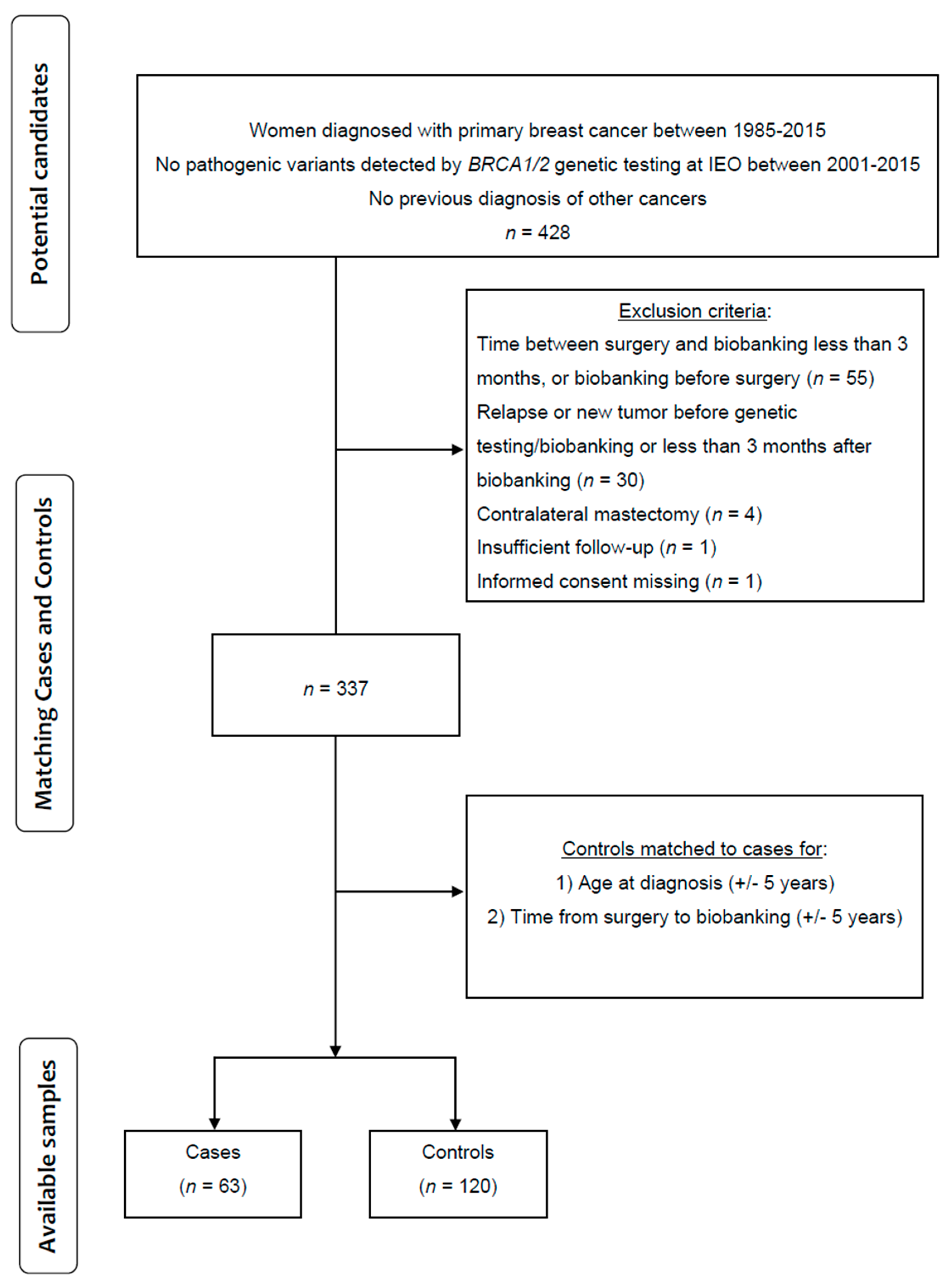
Figure 1. Patient Flow Diagram.
Cancers 17 03132 g001
Figure 2. Difference in DNA Methylation levels of the three DMRs between groups. Each panel in the figure shows the methylation signal intensities of the CpGs within each DMR for cases vs. controls. “C” track: individual values of methylated signal intensities (corresponding to cytosine at the CpG site); “T” track: individual values of unmethylated signal intensities (corresponding to thymine signal when the CpG is not methylated). “C mean” and “T mean” tracks: average values of methylated and unmethylated intensities across probes within each group, plotted to reduce probe-level noise. “C loess” and “T loess” tracks: LOESS-smoothed curves of methylated and unmethylated intensities, applied to highlight regional trends in DNA methylation that reflect the local correlation between neighbouring CpGs.
Cancers 17 03132 g002
Table 1. (A) Patients’ characteristics. Tumour characteristics are described in (B), including histotype, molecular subtype, lymph node stage, and tumour stage. (B) Tumour characteristics.
(A)
CasesControlsTotalp-Values
Total 63120183
Age at the testMedian (Q1, Q3)42 (37, 49)45 (38, 50)1830.43
BMIMedian (Q1, Q3)22.1 (20.2, 24)22.1 (20.2, 24)1820.69
BMI at 18 years oldMedian (Q1, Q3)20 (18, 21)19 (18, 20)1770.04
Time from surgery to blood draw (months)Median (Q1, Q3)12.5 (8.2, 29.2)21.5 (9.4, 52.7)1830.085
Oral contraceptiveNo24 (39.34)37 (60.66)610.32
Yes36 (31.3)79 (68.7)115
Missing7
PregnanciesNo15 (29.41)36 (70.59)510.39
Yes48 (36.36)84 (63.64)132
n. of pregnanciesNo15 (29.41)36 (70.59)510.64
One17 (34.69)32 (65.31)49
More than one31 (37.35)52 (62.65)83
menopausal statusPre48 (33.8)94 (66.2)1420.65
Peri0 (0)1 (100)1
Post14 (38.89)22 (61.11)36
Missing40
SmokingFormer15 (31.91)32 (68.09)470.68
No43 (36.75)74 (63.25)117
Yes5 (27.78)13 (72.22)18
Missing1
Family historyNo0 (0)2 (100)20.26
Breast only59 (33.91)115 (66.09)174
Breast and ovary4 (57.14)3 (42.86)7
Family history—First degree breast021 (43.75)27 (56.25)480.19
134 (31.19)75 (68.81)109
27 (28)18 (72)25
31 (100)0 (0)1
Family history—First degree ovary061 (34.08)118 (65.92)1790.61
12 (50)2 (50)4
Family history—Second degree breast020 (28.99)49 (71.01)690.39
132 (35.56)58 (64.44)90
29 (42.86)12 (57.14)21
32 (66.67)1 (33.33)3
Family history—Second degree ovary061 (34.08)118 (65.92)1790.61
12 (50)2 (50)4
(B)
CasesControlsTotalp-Values
Total 63120183
HistotypeIn situ9 (40.91)13 (59.09)220.85
Ductal48 (34.29)92 (65.71)140
Lobular4 (30.77)9 (69.23)13
Other2 (25.00)6 (75.00)8
Molecular subtypeLuminal A/B40 (35.71)72 (64.29)1120.69
Luminal B Her2+7 (31.82)15 (68.18)22
Her2+5 (31.25)11 (68.75)16
TN2 (18.18)9 (81.82)11
Missing91322
pNNegative25 (25.00)75 (75.00)1000.004 *
Positive32 (46.38)37 (53.62)69
Px6 (42.86)8 (57.14)14
pTIs8 (38.10)13 (61.90)210.045
132 (27.59)84 (72.41)116
214 (43.75)18 (56.25)32
38 (61.54)5 (38.46)13
Missing1 1
* Excluding Px from the test.
Table 2. Differentially methylated regions listing each CpG probe mapping within the three DMRs.
DMRindexChromosomeCpg Probe IDPositionStrandTypeGeneFeatureCgi
DMR_15cg07158503135415693RII IGRshore
cg04515200135415762FII IGRshore
cg13581155135415781FII IGRshore
cg11608150135415948RI IGRshore
cg06478886135416029RII IGRshore
cg04481923135416205RIIVTRNA2–1Bodyisland
cg18678645135416331RIIVTRNA2–1TSS200island
cg06536614135416381FIVTRNA2–1TSS200island
cg25340688135416398FIVTRNA2–1TSS200island
cg26896946135416405FIVTRNA2–1TSS200island
cg00124993135416412FIVTRNA2–1TSS200island
cg08745965135416529FIIVTRNA2–1TSS1500shore
DMR_25cg19626725178986131FIIRUFY1TSS1500shore
cg00080972178986291FIIRUFY1TSS1500shore
cg21226059178986404FIIRUFY1TSS1500shore
cg14820908178986412FIIRUFY1TSS1500shore
cg02136620178986620FIIRUFY1TSS200island
cg09060608178986726RIIRUFY1TSS200island
cg05457628178986728RIRUFY1TSS200island
cg22764044178986830FIIRUFY11stExonisland
cg26516362178986906FIRUFY15′UTRisland
DMR_310cg06791446123355268FIIFGFR25′UTRshore
cg25052156123355454FIIFGFR25′UTRshore
cg22633036123355576RIIFGFR25′UTRshore
cg11430259123355748RIFGFR25′UTRshore
cg02210151123356041RIIFGFR25′UTRshore
cg17681491123356205RIIFGFR25′UTRshore
cg18566515123356236RIFGFR25′UTRshore
The table describes the CpG probe IDs and positions of the three differentially methylated regions (DMR)s. The gene and the forward or reverse designation of the designed strand are reported. Type relates to the Infinium design (Infinium I applies 2 probes/locus, while Infinium II uses 1 probe/locus). The gene region features describing the CpG region are listed below in the same order as the target gene transcripts. IGR = Intergenic region, located between genes. Body = Between the ATG and stop codon; irrespective of the presence of introns, exons, TSS (transcriptional start site), or promoters. TSS200: 0–200 bases upstream of the TSS. TSS1500: 200–1500 bases upstream of the (TSS). 5′UTR = Within the 5′ untranslated region, between the TSS and the ATG start site (start codon). island = CpG island. shore = Region within 2 kb up- or downstream of a CpG island.
Table 3. A summary of the biological mechanisms epigenetically regulated in our study, genes between brackets did not reach statistical significance in our study.
MechanismGenes InvolvedBiological Impact
Cell MigrationRUFY1, (CCL21), (CDH4)Immune cell recruitment, cancer invasion
Growth SignallingFGFR2, (IR)Proliferation, metabolic dysregulation
Adhesion/JunctionsvtRNA2–1, (CDH4)EMT, barrier integrity
TraffickingRUFY1Receptor recycling, signalling modulation
Glucose regulationRUFY1, vtRNA2–1, (IR)Glucose regulation—Warburg effect
Disclaimer/Publisher’s Note: The statements, opinions and data contained in all publications are solely those of the individual author(s) and contributor(s) and not of MDPI and/or the editor(s). MDPI and/or the editor(s) disclaim responsibility for any injury to people or property resulting from any ideas, methods, instructions or products referred to in the content.