Article

Cytokine Profiling of Children, Adolescents, and Young Adults Newly Diagnosed with Sarcomas Demonstrates the Role of IL-1β in Osteosarcoma Metastasis

1
Department of Pediatrics, Albert Einstein College of Medicine, Bronx, NY 10461, USA
2
Department of Developmental and Molecular Biology, Albert Einstein College of Medicine, Bronx, NY 10461, USA
3
Moffitt Cancer Center, Tampa, FL 33612, USA
4
Department of Pediatrics-Hematology/Oncology and Bone Marrow Transplantation, University of Colorado, Aurora, CO 80045, USA
5
Connecticut Children’s Medical Center, University of Connecticut School of Medicine, Hartford, CT 06106, USA
6
Montefiore Einstein Comprehensive Cancer Center, Albert Einstein College of Medicine, Bronx, NY 10461, USA
7
Cancer Dormancy Institute, Albert Einstein College of Medicine, Bronx, NY 10461, USA
8
Marilyn and Stanley M. Katz Institute for Immunotherapy for Cancer and Inflammatory Disorders, Albert Einstein College of Medicine, Bronx, NY 10461, USA
*
Author to whom correspondence should be addressed.
Cancers 2025, 17(18), 3009; https://doi.org/10.3390/cancers17183009
Submission received: 6 August 2025 / Revised: 9 September 2025 / Accepted: 10 September 2025 / Published: 15 September 2025
(This article belongs to the Section Cancer Biomarkers)

Simple Summary

Sarcomas are a group of relatively common pediatric tumors that arise from connective tissues. This study characterizes the poorly understood immune landscape in newly diagnosed sarcoma patients by measuring levels of immune signals called cytokines. We found elevated levels of several pro-inflammatory cytokines in the plasma of these patients. Many of these were associated with inferior event-free or overall survival in patients with osteosarcoma. A particular cytokine, IL-1β, was associated with metastatic presentation and inferior event-free survival in patients with osteosarcoma. In the context of previously published preclinical work demonstrating that blocking IL-1 signaling can inhibit osteosarcoma metastasis, our work supports the development of a clinical trial testing this concept in patients with osteosarcoma.

Abstract

Background: Sarcomas are a heterogeneous group of mesenchymal tumors frequently diagnosed in pediatric and young adult patients. These tumors respond poorly to conventional immunotherapy, although the precise reason for this is not known. We sought to characterize the systemic immune response to sarcomas by measuring the levels of circulating cytokines in the plasma of newly diagnosed sarcoma patients, testing the hypothesis that the nature of a patient’s immune response to their tumor directly affects outcome. Methods: Plasma was collected from newly diagnosed, treatment-naive pediatric sarcoma patients participating in an ongoing clinical trial, MCC20320. A panel of 18 cytokines was selected, and cytokine levels were measured using the Luminex platform. Cytokine levels were analyzed based on clinicopathological parameters such as gender, age, stage, and survival. Results: We found that the cytokine profile in patients newly diagnosed with sarcoma is distinct from healthy controls, but different sarcomas were not distinguishable. Patients with osteosarcoma who had elevated levels of multiple cytokines had inferior overall survival compared to those with fewer or no elevated levels. Similarly, elevated levels of individual cytokines and chemokines, including IL-24, CXCL5, and CXCL10, were associated with inferior event-free or overall survival in patients with osteosarcoma. Perhaps most significantly, elevated IL-1β at diagnosis was associated with metastatic presentation and inferior event-free survival in patients with osteosarcoma. Conclusions: These findings suggest that pediatric sarcoma patients mount a systemic immune response that may affect event-free or overall survival. IL-1β in particular may be a valuable therapeutic target for osteosarcoma patients.

1. Introduction

Immune activity in the tumor microenvironment (TME) is widely recognized as an important contributor to oncogenic disease establishment and progression through numerous mechanisms, including modulation of tumor proliferation, immune response to tumor cells, angiogenesis, tumor cell metabolism, and dissemination of tumor cells [1,2]. Immune and non-immune cells in the TME establish a complex, dynamic network of signaling crosstalk that evolves along with the tumor. Cytokine and chemokine measurements are increasingly obtained as surrogate measurements of immune and inflammatory signaling activities because of the diversity of cell-intrinsic, innate, and adaptive immune processes that produce them; however, not all important immune signaling processes are equally well-represented by this strategy, as has been shown for type I interferon activation [3]. Moreover, interpreting the role played by any individual signal is complicated because its impact is always contextual and never isolated. The level at any particular timepoint is a composite of reactive and inductive contributions from many cell types and even potentially opposing processes, varying based on the identity and nature of the cells within the TME, the level and duration of their expression and action, and the combined impact with other signals [4,5]. This complexity necessitates studies evaluating diverse circumstances of cytokine activity in different disease endotypes and environments. It is difficult to extrapolate from one tumor type to another because each type of tumor elicits a unique immunologic response. In addition, profiling multiple cytokines simultaneously is required to fully understand tumor immune responses. Many cytokines can circulate systemically, producing dramatic effects even at a distance from the tumor. Despite this complexity, studies have found that characterizing circulating cytokines can predict disease outcomes in some cases [6,7,8].
With a unique mesenchymal lineage, sarcomas are rare and less well-studied than other solid tumors in adults; however, they are more common in children, adolescents, and young adults [9]. Because of their heterogeneous nature and relative rarity, little is known about the immune response to these tumors. In general, sarcomas are relatively unresponsive to immunotherapy, but the mechanisms underlying this relative lack of response are poorly understood [10,11].
In this study, we characterized the cytokine profiles of children, adolescents, and young adults newly diagnosed with Ewing sarcoma, osteosarcoma, rhabdomyosarcoma, and other subtypes. The goal of this work was to test the hypothesis that the nature of the immunologic response to a tumor affects patient outcome. We simultaneously quantified plasma levels of thirteen pro-tumorigenic [12,13,14,15,16,17,18,19,20,21] and five anti-tumorigenic [22,23,24,25,26] cytokines selected based on evidence suggesting that they played a role in one or more sarcomas [8,13,19,24,25,27,28,29,30,31,32,33,34,35,36,37,38,39,40]. We found that specific cytokine levels correlate with key features of disease outcome. Most significantly, we found that patients with osteosarcoma who have high levels of plasma IL-1β at diagnosis are more likely to have metastatic disease at presentation and have a shorter event-free survival (EFS) than those with lower levels of this cytokine.

2. Methods

2.1. Patients

Research subjects were enrolled in an ongoing multi-center clinical trial, MCC20320, a study evaluating blood-based biomarkers of disease response and prognosis in children, adolescents, and young adults with sarcomas. This multi-center clinical trial was approved by Advarra, and was conducted in accordance with the Ethical Principles and Guidelines for the Protection of Human Subjects of Research. Newly diagnosed patients less than or equal to 40 years old and weighing over 8 kg were enrolled at participating centers. Cohorts were defined as follows: Cohort 1: Ewing sarcoma patients with confirmed diagnosis of Ewing sarcoma (ES) with a t (11;22) EWS-FLI1 or t (21;22) EWS-ERG translocation. Cohort 2: Fusion-positive rhabdomyosarcoma (FP-RMS) patients with confirmed diagnosis of a rhabdomyosarcoma with a t (2;13) PAX3-FOXO1 or t (1;13) PAX7-FOXO1 translocation. Cohort 3: Fusion-negative rhabdomyosarcoma (FN-RMS) with a confirmed diagnosis of a rhabdomyosarcoma without a t (2;13) PAX3-FOXO1 or t (1;13) PAX7-FOXO1 translocation. Cohort 4: Confirmed patients with osteosarcoma. Cohort 5: Other translocation-driven sarcomas not included in the other 4 cohorts. Plasma from clinical waste of presumed healthy individuals was used for healthy controls. Study accrual opened in 2019 and is ongoing, but this report focuses on subjects with Ewing sarcoma and osteosarcoma—the cohorts with sufficient subject enrollment for meaningful analysis. Only treatment-naive samples drawn at the time of diagnosis were evaluated.

2.2. Plasma Extraction

Blood was collected at enrollment in a Streck Cell-free DNA BCT® tube and shipped to the University of Colorado Sarcoma Research Lab in ATS1 temperature-regulated containers (Akuratemp, Arden, NC, USA). Plasma was isolated using double-centrifugation and was frozen at −80 °C until use, as previously described by Hayashi et al. [41]. Samples were aliquoted and frozen at −80 °C and underwent no more than two freeze/thaw cycles.

2.3. Luminex Assays

All samples were measured in technical duplicates and averaged for a final concentration. IL-1β, IL-4, IL-6, IL-8, IL-10, IL-12 (p40), IL-15, IL-27, and CXCL10 were measured using kit HCYTA-60K-10. CCL21, CXCL5, IL-16, IL-24, and CXCL12 were measured using kit HCYTB-60K-06. MIF, TGF-β1, and IGF-1 were measured using HSP1MAG-63K-01, TGFBMAG-64K-01, and HIGFMAG-52K-01, respectively, all obtained from Sigma Millipore (St. Louis, MO, USA). All kits were used according to the manufacturer’s instructions. Additional dilution was required to quantify IGF-1 (1:120) and TGF-β1 (1:270). Data were acquired on a Luminex Magpix XMAP Multiplex Reader (Luminex Technologies (Madison, WI, USA), RRID:SCR_023348) and were analyzed using Belysa Immunoassay Curve Fitting Software (Millipore Sigma, Burlington, MA, USA). Interpolated concentrations of all analytes were log-transformed before analysis to reduce non-normal skew and inter-assay variability.

2.4. Statistical Analysis

Healthy controls and sarcoma cohorts were analyzed using Singular Value Decomposition (SVD) with imputation to calculate principal component analysis (PCA) with unit variance scaling applied using ClustVis software (https://biit.cs.ut.ee/clustvis/, accessed on 9 September 2025) (RRID:SCR_017133) [42]. Prediction ellipses were formed with a 95% confidence interval. ClustVis was also used to generate a heatmap of cytokine levels using unsupervised clustering on columns and rows with correlation distance and complete linkage. Based on hierarchies and visual inspection, endotypes were split into three groups, which were further analyzed for overall survival and event-free survival using a Log-rank (Mantel–Cox) test. This test, and all other statistical tests, were performed using GraphPad Prism, version 10.6.0 (GraphPad Software, La Jolla, CA, USA) (RRID:SCR_002798). All analytes in osteosarcoma and Ewing sarcoma samples were also analyzed using a Spearman r correlation matrix. Receiver operating characteristics (ROC) curves and area under the curve (AUC) were calculated for osteosarcoma and ES cohorts for analytes that were significantly altered compared to healthy controls.
Individual analyte concentrations in each cohort were compared to healthy controls using a Mann–Whitney test. Further analysis was performed on groups that had a sufficient sample size, including Ewing sarcoma, osteosarcoma, and all patients not separated by cohort, referred to as “mixed cohorts”. To explore the effect of multiple elevated or depressed cytokines in conjunction, patient outcomes to date (responsive, refractory, or relapsed disease) were distributed by the number of elevated or depressed analytes. Cytokines were considered elevated if plasma concentration was greater than the 95th percentile of the healthy cohort. Depressed levels were below the 5th percentile. Additionally, patients were grouped as 0–3 and ≥4 elevated cytokines or 0 and ≥1 depressed cytokines. Based on these groupings, event-free survival (EFS) and overall survival (OS) were analyzed with Kaplan–Meier curves with an 18-month endpoint, and hazard ratios were generated using a Log-rank (Mantel–Cox) test. EFS and OS were also determined for high and low cytokine levels, with high defined as anything at or above 75th percentile within that cohort. Further analysis was performed comparing adult (age ≥ 22) and pediatric (age ≤ 21) and localized or metastatic stage of bone sarcomas at diagnosis using the Mann–Whitney test. Since IL-1β had compelling results, we also used Fisher’s exact test to determine if there was a difference in the number of patients with metastatic or localized disease in the high or low IL-1β groups.

3. Results

A total of 123 sarcoma patients and 17 healthy subjects were included in this analysis, and they are presented in Supplementary Table S1. Median cytokine values for each cohort and their comparison to healthy controls can be found in Supplemental Table S1. When analyzed globally through PCA, all five sarcoma cohorts had overlapping clusters, but were distinguishable from healthy controls (Figure 1A). Similarly, when comparing patients’ cytokine levels through unsupervised hierarchical clustering, healthy controls clustered together. In contrast, specific sarcoma cohorts were not segregated (Figure 1B). Although patients with the same diagnosis did not cluster together in this analysis, we were able to group patients into three endotypes (Figure 1B). Group 1 was distinguished by an increase in CXCL5, CXCL12, and/or MIF. Group 3 had increased levels of IL-6, IL-8, IL-4, IL-1β, IL-15, IL-10, IL-12 p40, and/or IL-24, with relatively low levels of CCL21, CXCL10, and IGF-1. Group 2 had few distinguishing features, besides a slight increase in CXCL10 in approximately a third of patients. Although endotype grouping did not have statistically significant impact on survival, there were trends toward significance that might have been statistically significant with larger cohorts or a longer follow-up (Figure 1C). For example, patients with osteosarcoma in Endotype 1 had the worst EFS (p = 0.15), and, considering all enrolled subjects, those with Endotype 1 also had the worst OS (p = 0.15).
A Spearman correlation matrix was generated for osteosarcoma and Ewing sarcoma samples, which demonstrated several significant correlations, suggesting biologically meaningful relationships (Supplementary Figure S1). Osteosarcoma had 25 significant relationships, notably including IL-4/IL-1β (r2 = 0.58, p = 0.000048), IL-12 p40/IL-4 (r2 = 0.47, p = 0.0015), CXCL5/IL-27 (r2 = −0.48, p = 0.001), CXCL14/IL-15 (r2 = 0.66, p = 0.0000021). In Ewing sarcoma, there were 21 significant relationships, including IL-4/IL-1β (r2 = 0.55, p = 0.00048), IL-4/IL-8 (r2 = 0.68, p = 0.0000047), CXCL14/IL-15 (r2 = 0.61, p = 0.00013), and CXCL5/CXCL12 (r2 = 0.64, p = 0.000030).
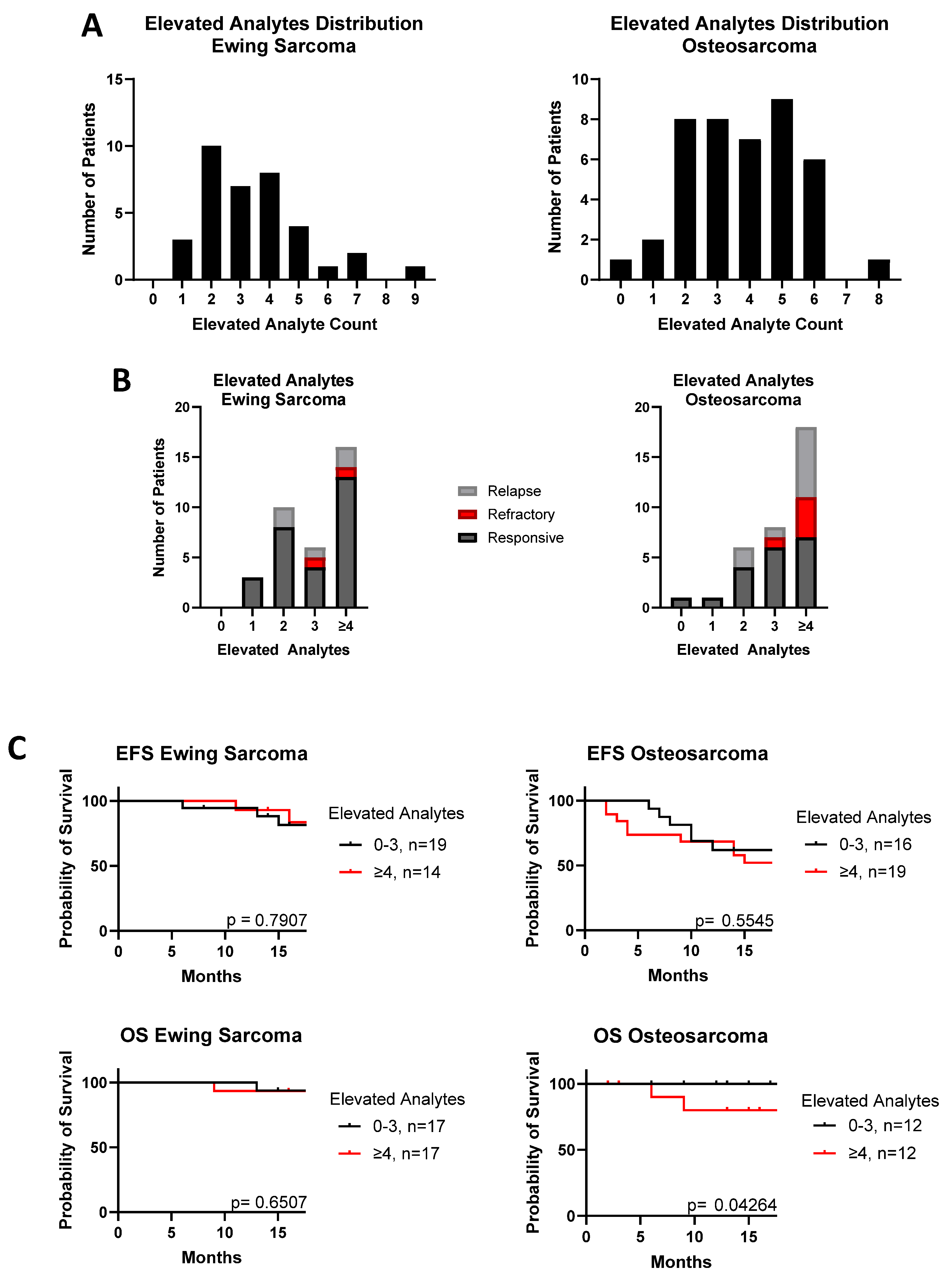
We next sought to determine if having multiple elevated cytokines had an impact on disease progression. Defining “elevated” as the upper 5% of the level of each cytokine in the healthy control cohort, 44% of Ewing sarcoma and 55% of osteosarcoma patients had four or more cytokines elevated in tandem (Figure 2A). Poor outcomes, like relapse and refractory disease, were more common among patients that had more elevated analytes in total (Figure 2B). These patients also had worse OS in osteosarcoma, but not Ewing sarcoma (Figure 2C: osteosarcoma p = 0.043; Ewing sarcoma p = 0.65); however, the impact on EFS did not reach statistical significance for either tumor type (Ewing sarcoma p = 0.79; osteosarcoma p = 0.55).
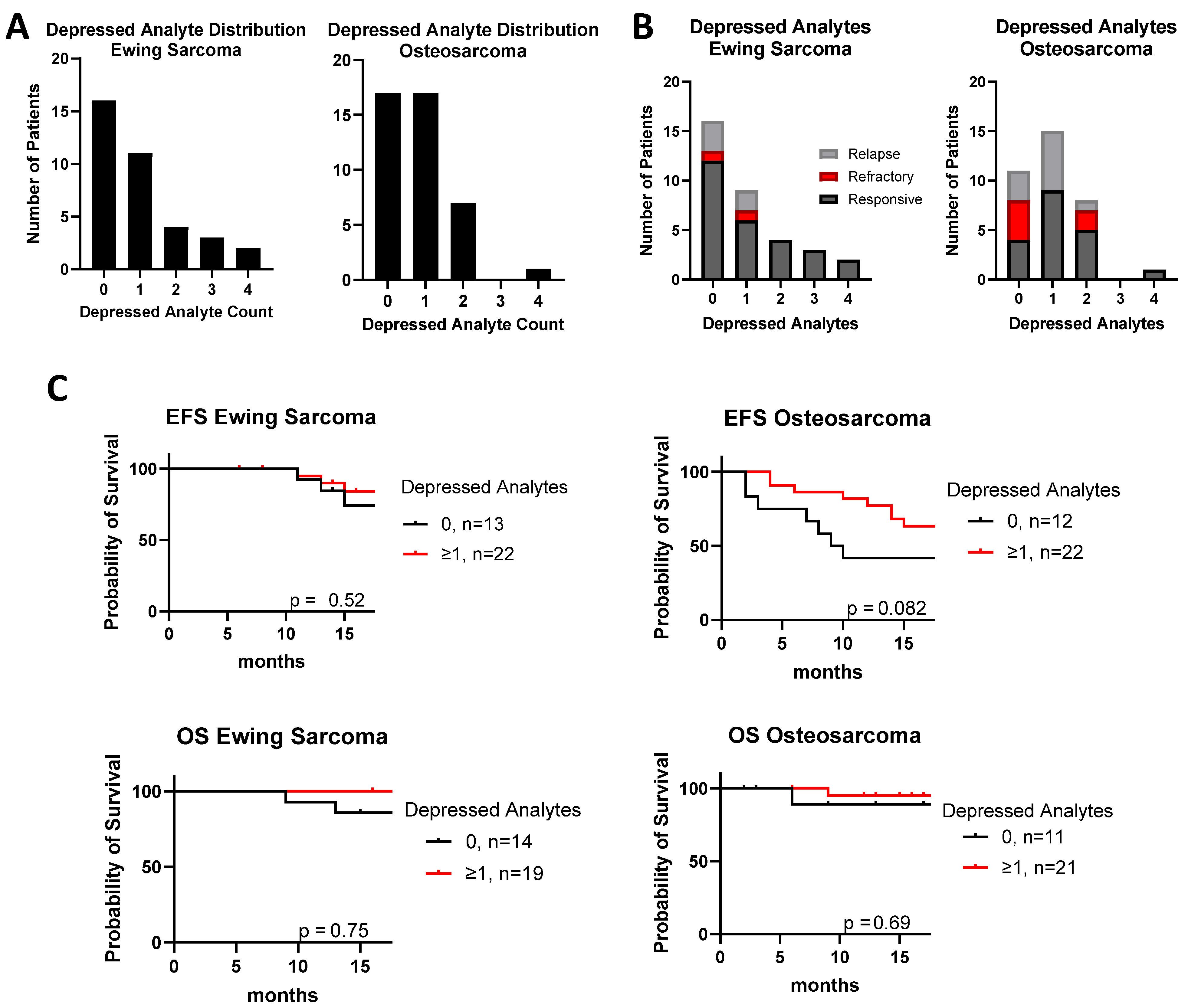
A complementary result was seen in patients with multiple depressed analytes. Defining “depressed” as the lower 5% of the cytokine level in healthy controls, we found that although only 6% of Ewing sarcoma and 2% of osteosarcoma patients had four or more cytokines depressed in total (Figure 3A), patients with poor outcomes had fewer depressed analytes in total (Figure 3B). Similarly, having fewer depressed cytokines correlated with slightly improved EFS, which approached significance for osteosarcoma (Figure 3C: Ewing sarcoma p = 0.52; osteosarcoma p = 0.082). The effect on OS was not statistically significant, probably because of small sample size (Ewing sarcoma p = 0.75; osteosarcoma p = 0.69).
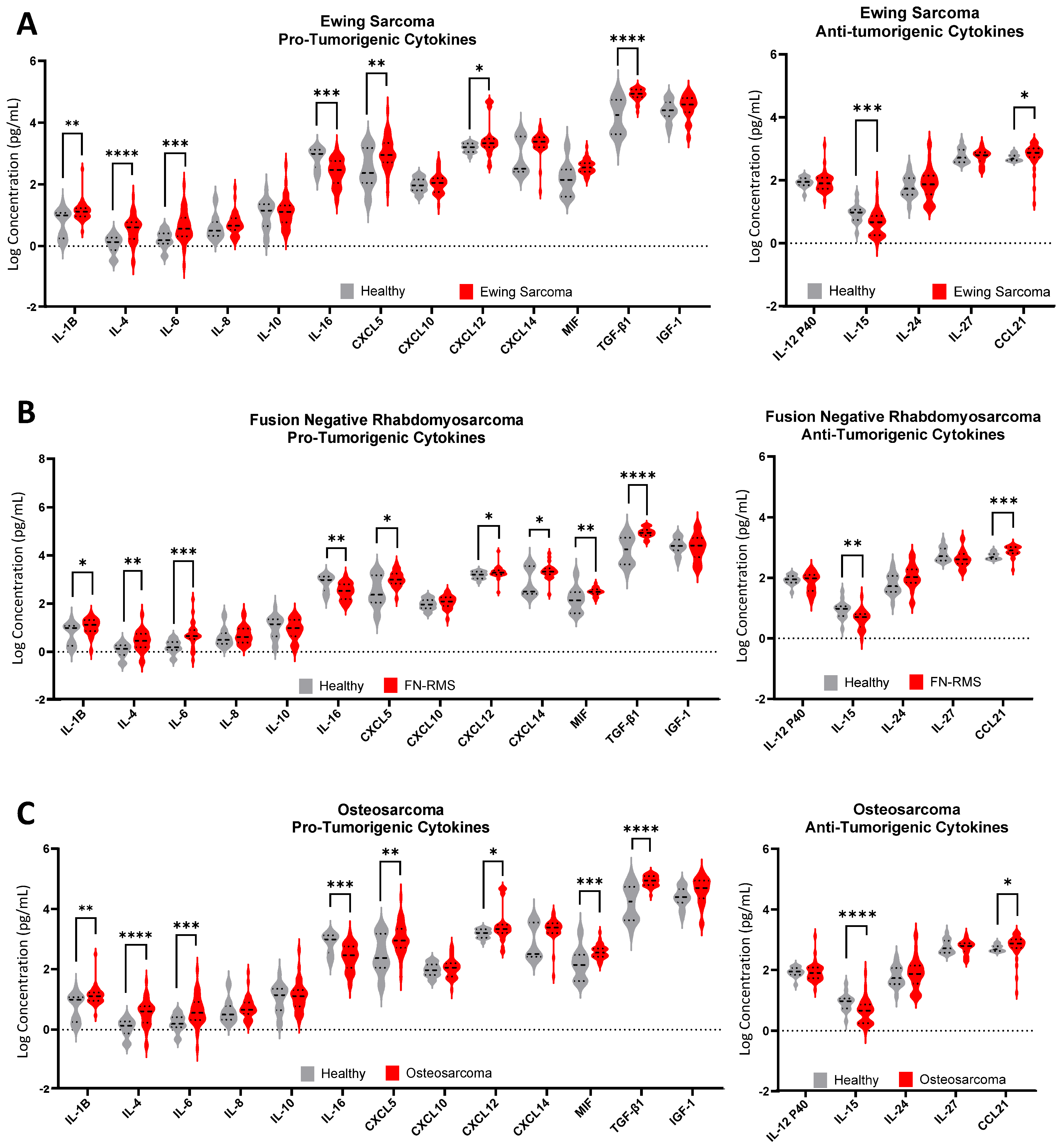
The plasma levels of individual cytokines differ in patients with sarcoma compared to healthy controls. Levels of IL-1β, IL-4, IL-6, CXCL5, CXCL12, CXCL14, MIF, IGF-1, TGFβ-1, and CCL21 were increased in one or more sarcoma cohorts compared with controls, and levels of IL-15 and IL-16 were significantly lower in one or more sarcoma cohorts compared to healthy controls (Figure 4 and Supplementary Table S2). The remaining analytes, IL-8, IL-10, IL-12 P40, IL-24, IL-27, and CXCL10 were found in similar plasma concentrations in both patients with sarcoma and healthy subjects. ROC and AUC analyses of patients with bone sarcomas demonstrated that many of the cytokines with differential expression, when compared to healthy controls, have sufficiently significant sensitivity and specificity to be considered for use as part of clinical decision-making and monitoring (Supplementary Figure S2).
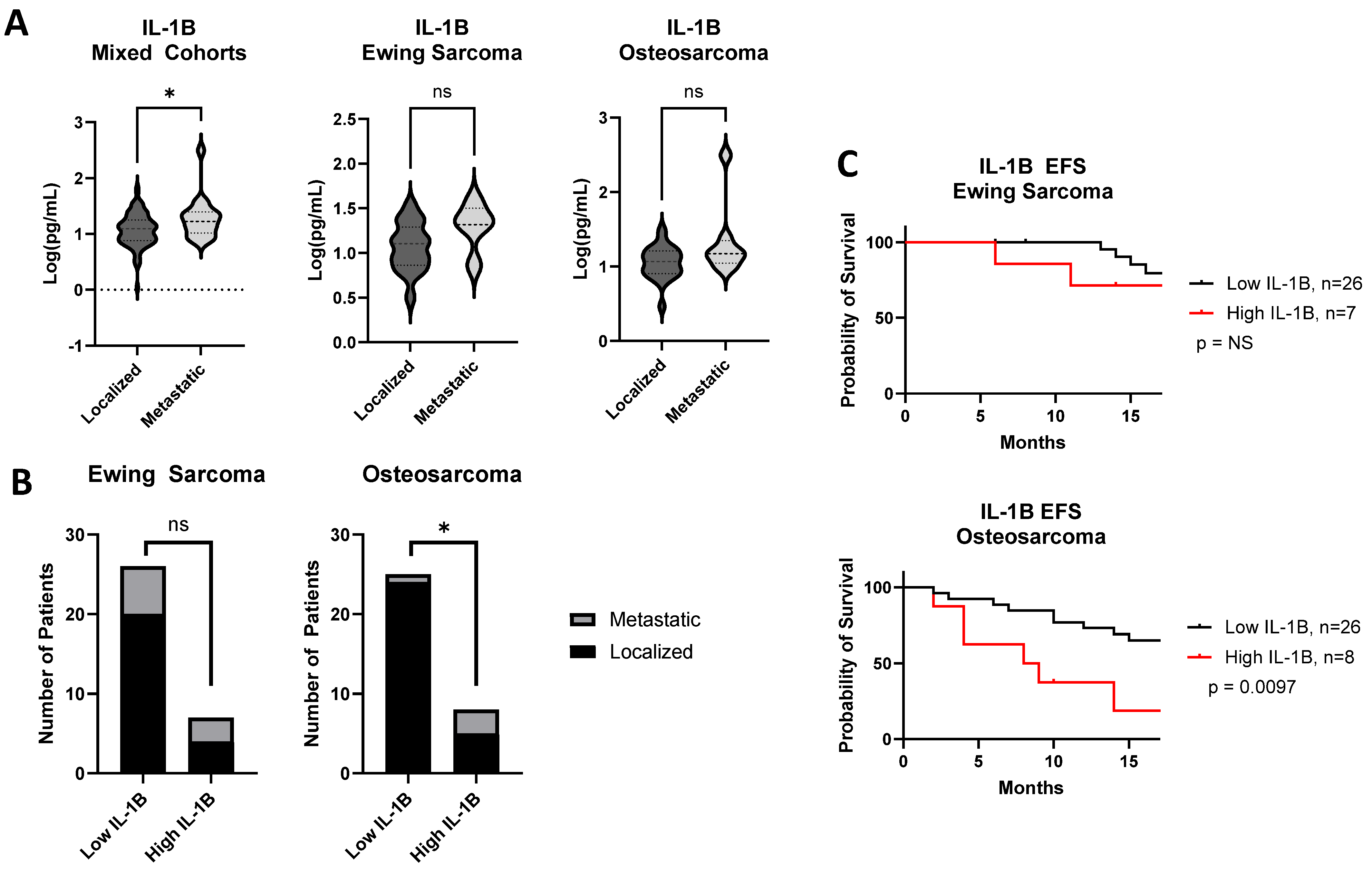
Next, we tested whether median cytokine levels in patients with Ewing sarcoma, osteosarcoma, and patients in the “mixed cohorts” subgroup correlate with clinically relevant parameters such as age, stage at diagnosis, or clinical outcome. Adult patients in the “mixed cohorts” group had statistically significantly lower levels of IL-6 (0.29 pg/mL vs. 0.64 pg/mL; p < 0.05) and higher levels of MIF (2.67 pg/mL vs. 2.49 pg/mL; p < 0.05) than pediatric patients. Similarly, MIF levels were elevated in adult patients with Ewing sarcoma compared with pediatric patients (2.83 pg/mL vs. 2.50 pg/mL; p < 0.05). No cytokine levels varied by age in the osteosarcoma cohort. Interestingly, in the “mixed cohorts” patients, those who presented with metastatic disease had a higher median level of IL-1β than those presenting with localized disease (1.23 pg/mL vs. 1.10 pg/mL; p = 0.022). Although this finding did not hold true for patients with Ewing sarcoma or osteosarcoma, in each of these cohorts there was a trend in that direction (Ewing sarcoma: 1.21 pg/mL vs. 1.07 pg/mL, p = 0.06; osteosarcoma 1.32 pg/mL vs. 1.05 pg/mL, p = 0.068). No other cytokine levels had a significant association with stage, and no median cytokine levels were associated with outcome. These and other non-significant values are displayed in Supplementary Table S3.
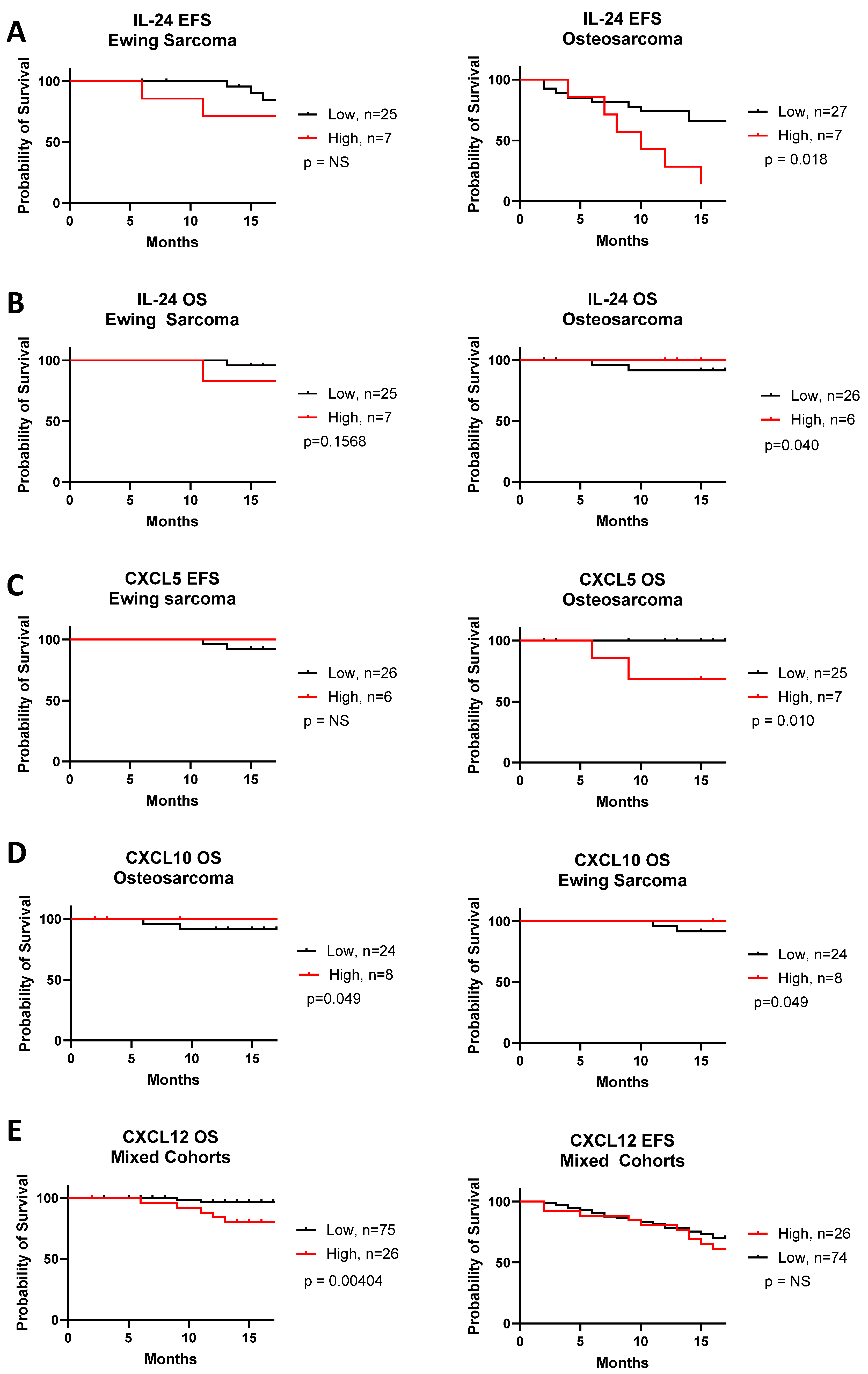
In contrast to comparisons based on median cytokine levels, high-level expression (defined as the upper 25th percentile of the cohort) of several cytokines correlated with 18-month OS and EFS (Figure 5 and Supplementary Table S4). Patients with osteosarcoma who had high IL-24 levels had poorer EFS (p = 0.018), but slightly better OS (p = 0.040). Patients with osteosarcoma showing high CXCL5 had poorer OS (p = 0.010), but not EFS. Patients with Ewing sarcoma showing high levels of CXCL10 had slightly better OS (p = 0.049), but IL-24 and CXCL5 levels had no prognostic significance. When analyzing all sarcomas unsegregated by diagnosis, we found that patients with high CXCL12 levels had shorter OS than those with low levels (p = 0.0040).
Of the cytokines examined, IL-1β emerged as the cytokine with the most significant correspondence to the features of disease progression. As mentioned above, IL-1β expression was significantly higher in the plasma of patients with metastatic disease from the mixed cohort (Figure 6A; p = 0.022). Patients with Ewing sarcoma and osteosarcoma exhibited a similar trend, but with subthreshold significance (Ewing sarcoma p = 0.060; osteosarcoma p = 0.068). Although 23.1% of patients with Ewing sarcoma who had low IL-1β had metastatic disease compared with 42.9% with high IL-1β (p = NS), in the osteosarcoma cohort, only 4% of patients with low IL-1β had metastatic disease compared to 37.5% in the high IL-1β group (Figure 6B; p = 0.036). In addition, patients with osteosarcoma who had high IL-1β levels had worse EFS than those with low IL-1β (p = 0.0097). In contrast, high IL-1β did not predict worse EFS in patients with Ewing sarcoma (Figure 6C; p = 0.36).

4. Discussion

Sarcomas represent one of the most common pediatric cancers, and treatment options remain limited, especially for patients who present with metastatic disease [10]. This group of tumors is also unresponsive to standard cancer immunotherapies, such as immune checkpoint inhibitors (ICI) [43]. This study is the first to document the potential treatment-informative value of an 18-cytokine panel in the plasma of pediatric, adolescent, and young adult patients with sarcomas. It includes information on cytokines that have been poorly studied in sarcoma peripheral blood samples, such as IL-24, IL-27, CCL21, CXCL5, and CXCL14. It also identified one cytokine, IL-1β, whose elevation is associated with activation of a pathway for which well-known clinical-grade assays and targeted therapies exist, thus offering potential for immediate evaluation in a clinical trial. We demonstrate that profiling cytokines representing diverse tumor-relevant pathways and processes can distinguish healthy subjects from patients newly diagnosed with sarcoma. This difference is meaningful and non-random, as indicated by the finding of robust AUC results in ROC graphs, as well as significant correlations between analyzed cytokines. Patients with different sarcoma diagnoses were not distinguishable from one another, indicating that this particular cytokine panel was insufficient to identify differences in the inflammatory profiles of different sarcoma subtypes, but does suggest that there may be shared immune processes active in the TME of different sarcomas. The former finding is not at all surprising since any host immune response to a tumor is predicated not only on the antigenic features of the tumor cells, but also on the host’s own immunobiology, arising from the complex interactions between their environmental exposure history and genetic background. Even the paraneoplastic responses to thymomas, a rare and highly specific immunogenic tumor with a limited set of recurrent tumor-associated genetic landscapes [44,45], can vary widely [46].
Just as mouse models may show Th1 or Th2 skewing, for example, human beings are also predisposed towards certain innate and adaptive immune responses. Indeed, genetic studies have shown that there has been positive selection for variants that once conferred resistance to life-threatening infections at a time when hygiene and antimicrobials were unavailable; these same alleles now predispose individuals to inflammatory and autoimmune conditions [47]. The inflammasome, whose activation is associated with a pathognomonic increase in downstream IL-1β and IL-18 production, as well as several distinct forms of cell death, has repeatedly been noted as one such pathway under strong evolutionary positive selection, with heterozygote advantage conferred by pro-inflammatory variation at loci such as NLRP3 or MEFV [48].
Using unsupervised hierarchical clustering, three endotypes were apparent among patients. These endotypes represent clusters of related immune cells, pathways, and processes, whether suppressive innate and adaptive immune responses elicited by tumors (Groups 1 and 2) or more mixed effects (Group 3). The group with the worst EFS and OS for osteosarcoma and Ewing sarcoma, Group 1, was defined by an increase in CXCL5, CXCL12, and MIF. These pro-tumorigenic chemokines are implicated in recruiting and activating myeloid-derived suppressor cells (MDSCs) and regulatory T cells (Tregs), immune cell populations that impair anti-tumor responses, as well as inducing angiogenesis and metastasis [16,17,49,50]. Group 2 had few visibly apparent abnormalities in cytokine expression, except for some patients with increased CXCL10 expression or decreased IL-12p40, suggesting a polarization away from helper T cell towards regulatory T cell activities. Group 3 had the longest EFS and had increased levels of both pro- and anti-tumorigenic interleukins and/or decreased expression of CXCL12, IL-16, CXCL10, and CCL21. These findings may indicate specific coordinated effects of cytokines to orchestrate an anti-tumorigenic response. These endotypes may reflect both positive and negative feedback response loops that require further, future investigation with respect to temporal cause and effect, as well as the cell populations involved. Interestingly, we also found that patients with five or more analytes elevated in conjugation were more likely to have diminished EFS. Other studies have reported similar findings in sarcomas [31,51]. We plan to follow up some of our most significant initial screening observations with an interrogation of additional pathway-specific studies to obtain the clinical and mechanistic significance of these hypothesis-generating findings.
It is interesting to note that statistically significant correlations between cytokine expression and outcomes, such as EFS and OS, are predominantly observed in patients with osteosarcoma. Osteosarcoma appears to be the only sarcoma in children, adolescents, and young adults with a track record (albeit a weak one) of response to ICI. For example, in SARC028, a clinical trial of pembrolizumab in advanced sarcomas, 5% of osteosarcoma patients had a partial response and 30% had stable disease, whereas none of the patients with Ewing sarcoma responded [52]. There have been additional case reports of patients with osteosarcoma responding to similar agents [53], but no such case reports for Ewing sarcoma. This may be because ICI primarily addresses adaptive immune pathways, whereas many of the cytokines of interest that we have elucidated herein are produced by and signal to more than just adaptive immune cells. Indeed, mifamurtide, a fully synthetic derivative of immunostimulatory muramyl dipeptide (MDP) is approved by the European Medicines Agency for the treatment of high-grade resectable non-metastatic osteosarcoma after macroscopically complete surgical resection. MDP is a bacterial cell wall derivative that binds to NOD2, leading to downstream NF-κB and MAPK signaling in monocytes and macrophages, with subsequent production of pro-inflammatory cytokines such as TNF-α, IL-1β, and IL-6 [54]. There are no comparable results in Ewing sarcoma, suggesting a fundamental difference between these bone sarcomas that is also reflected in our results.
Analysis of individual cytokine levels demonstrated robust differences in cytokine concentration in patients with sarcoma compared to healthy subjects. Furthermore, dramatic increases in expression were mostly observed in pro-tumorigenic cytokines (IL-1β, IL-4, IL-6, CXCL5, CXCL12, CXCL14 MIF, and TGF-β1), with few, if any, differences in the levels of anti-tumorigenic cytokines such as IL-12 p40, IL-24, and IL-27 when compared to healthy controls. IL-15, a potent activator of NK and T cell anti-tumor activity [23,55], was underexpressed in sarcoma cohorts. CCL21 was overexpressed, and overexpression of this chemokine, frequently considered anti-tumorigenic, may reflect a normal, coordinated immune response, with simultaneous high expression of both pro- and anti-inflammatory signaling molecules, a finding also reported in other studies [7,39]. This panel, then, suggests an environment both supportive and permissive for tumor growth and metastasis. These findings are supported by those of other studies, strengthening their role in sarcoma [13,27,30,31,51,56,57,58,59].
Our findings relate to IL-1β are particularly translationally relevant. Plasma levels of IL-1β were higher in patients with sarcoma than in healthy controls, and patients with osteosarcoma in particular with elevated IL-1β levels at diagnosis were more likely to present with metastatic disease and had a shortened EFS. For patients with osteosarcoma, metastasis is the strongest determinant for prognosis at diagnosis; patients with localized disease have a 70% five-year survival rate, which drops to <20% for patients who present with metastasis [10]. Unlike many of the cytokines we evaluated, there are mechanistic studies linking IL-1β directly to the process of metastasis. IL-1β is, in part, secreted by tumor-associated macrophages in osteosarcoma, where it has been proposed to promote metastasis, an effect which was reversed by anakinra treatment [60]. A more recent study using preclinical models of osteosarcoma showed that pulmonary metastasis is initiated by a subpopulation of disseminated tumor cells that produce cytokines such as IL-6 and CXCL8 in response to lung-epithelium-derived IL-1α [61]. Although IL-1α and IL-1β are distinct cytokines, they exert many complementary functions via signaling through the same receptor, and can both be targeted by rilonacept, a recombinant fusion protein that acts as a decoy receptor for IL-1 cytokines and is already FDA-approved for use in diverse disorders of inflammasome activation [62]. Reinecke et al. demonstrated that Anakinra, an IL-1 receptor antagonist, inhibits pulmonary metastases in their preclinical model [61], so our clinical data linking plasma IL-1β levels with metastasis and EFS (wherein metastasis is the predominant event), in the context of the mechanistic studies published by other groups, provide strong justification for clinical trials of IL-1 inhibition to prevent metastasis in patients with osteosarcoma.

5. Conclusions

In summary, our cytokine profiling of newly diagnosed patients with sarcoma uncovered several tantalizing findings worthy of further investigation. We found that plasma cytokine levels in patients newly diagnosed with sarcoma differ from those in healthy controls, but not from one sarcoma type to another. We also discovered three distinct endotypes within the sarcoma patient groups. Although these do not have statistically significant prognostic significance based on our current data, these data remain immature, and re-evaluation in the future may demonstrate prognostic or other significance. Interestingly, when comparing Ewing sarcoma and osteosarcoma—the populations with the largest representation in this study—we found correlations between certain cytokines and survival in patients with osteosarcoma, but not in patients with Ewing sarcoma. It is notable that osteosarcoma is at times responsive to ICI, whereas Ewing sarcoma is not. This highlights the importance of considering more than adaptive immune cells alone, and considering which processes of cell-intrinsic, innate, and/or adaptive immunity contribute to each tumor at each stage of its development. It remains to be seen whether the correlation between cytokines and response to immunotherapy is causal (i.e., osteosarcoma is responsive because it elicits a more robust response from the patient’s immune system) or merely correlative. Perhaps most importantly, our work, in the context of preclinical studies from other groups, supports the contribution of IL-1-mediated inflammation to osteosarcoma metastasis, and this finding justifies a clinical trial of IL-1 blockade to prevent metastatic recurrence in these patients.

Supplementary Materials

The following supporting information can be downloaded at: https://www.mdpi.com/article/10.3390/cancers17183009/s1, Figure S1: Correlation matrix of spearman correlation coefficients indicated by color gradient for each analyte in patients with (A) Osteosarcoma and (B) Ewing Sarcoma. Asterisks designate significance for that relationship where *, **, ***, and **** denote p < 0.05, p < 0.01, p < 0.001, p < 0.0001. Where no asterisks are shown, correlation was not statistically significant; Figure S2: Receiver operating characteristics curves and area under the curve (AUC) for each cytokine that was significantly different in at least one sarcoma cohort compared to the healthy cohort. Curves compare healthy to osteosarcoma (OS) in black and Ewing sarcoma (ES) in red; Table S1: Demographic characteristics of subjects included in this analysis. Not all patients had stage recorded; Table S2: Cytokine concentrations in plasma of healthy subjects and sarcoma patients at diagnosis. Median [IQR]. Statistical comparison of cohort to healthy controls was performed using the Mann-Whitney test where *, **, ***, and **** denote p < 0.05, p < 0.01, p < 0.001, p < 0.0001; Table S3: Association Between Cytokine Level and Clinicopathological Parameters. Median values displayed with significance determined by Mann-Whitney test. Hazard ratio of high to low group for overall survival (OS) and event free survival (EFS) by log rank test. p values ≤ 0.05 notated in bold. Values rounded to 2 significant figures; Table S4: Hazard ratio of high to low group for overall survival (OS) and event free survival (EFS) by log rank analysis. p values ≤ 0.05 notated in bold. Values rounded to 2 significant figures.

Author Contributions

Conceptualization: M.H., M.S.I. and D.M.L.; Methodology, D.M.L., L.K., W.K. and A.M.M.; Software, L.K., W.K. and A.M.M.; Validation, X.P.P.; Formal Analysis, L.K., D.M.L., X.P.P., W.K. and A.M.M.; Investigation, S.H., L.K., W.K., A.M.M. and D.M.L.; Resources, M.H.; Data Curation, L.K.; Writing—Original Draft Preparation, L.K. and D.M.L.; Writing—Review and Editing, L.K., X.P.P., M.H., D.M.L. and M.S.I.; Visualization, X.P.P.; Supervision, M.H. and D.M.L.; Project Administration, J.C., M.H. and D.M.L.; Funding Acquisition, M.H., M.S.I. and D.M.L. All authors have read and agreed to the published version of the manuscript.

Funding

This work was supported in part by a grant from the National Cancer Institute (5R01CA262802 to DML) and a grant from the National Pediatric Cancer Foundation (09-33693-20-09 to DML). We are grateful for support from the Einstein CFAR biomarker core (funded by grant 5P30AI124414-05) and the Montefiore Einstein Comprehensive Cancer Center support grant (2P30CA013330). Clinical trial MCC 20320 is supported by the National Pediatric Cancer Foundation.

Institutional Review Board Statement

The study was conducted according to the guidelines of the Declaration of Helsinki, and was approved by the Advarra Institutional Review Board.

Informed Consent statement

Informed consent was obtained from all subjects involved in this study.

Data Availability Statement

Original Data is available in supplemental figures. Additional raw data will be provided on reasonable request.

Conflicts of Interest

The authors declare no conflicts of interest.

References

  1. Kartikasari, A.E.R.; Huertas, C.S.; Mitchell, A.; Plebanski, M. Tumor-Induced Inflammatory Cytokines and the Emerging Diagnostic Devices for Cancer Detection and Prognosis. Front. Oncol. 2021, 11, 692142. [Google Scholar] [CrossRef]
  2. Yi, M.; Li, T.; Niu, M.; Zhang, H.; Wu, Y.; Wu, K.; Dai, Z. Targeting cytokine and chemokine signaling pathways for cancer therapy. Signal Transduct. Target. Ther. 2024, 9, 176. [Google Scholar] [CrossRef] [PubMed]
  3. Lamot, L.; Niemietz, I.; Brown, K.L. Methods for type I interferon detection and their relevance for clinical utility and improved understanding of rheumatic diseases. Clin. Exp. Rheumatol. 2019, 37, 1077–1083. [Google Scholar]
  4. Yang, H.; Zhao, L.; Zhang, Y.; Li, F. A comprehensive analysis of immune infiltration in the tumor microenvironment of osteosarcoma. Cancer Med. 2021, 10, 5696–5711. [Google Scholar] [CrossRef]
  5. Kloen, P.; Gebhardt, M.C.; Perez-Atayde, A.; Rosenberg, A.E.; Springfield, D.S.; Gold, L.I.; Mankin, H.J. Expression of transforming growth factor-β (TGF-beta) isoforms in osteosarcomas: TGF-β3 is related to disease progression. Cancer 1997, 80, 2230–2239. [Google Scholar] [CrossRef]
  6. Lee, M.H.; Laajala, E.; Kreutzman, A.; Järvinen, P.; Nísen, H.; Mirtti, T.; Hollmén, M.; Mustjoki, S. The tumor and plasma cytokine profiles of renal cell carcinoma patients. Sci. Rep. 2022, 12, 13416. [Google Scholar] [CrossRef] [PubMed]
  7. Luo, J.H.; Zhang, C.Y.; Lu, C.Y.; Guo, G.H.; Tian, Y.P.; Li, Y.L. Serum expression level of cytokine and chemokine correlates with progression of human ovarian cancer. Eur. J. Gynaecol. Oncol. 2017, 38, 33–39. [Google Scholar] [CrossRef]
  8. Munisamy, S.; Radhakrishnan, A.K.; Ramdas, P.; Samuel, P.J.; Singh, V.A. Immune Biomarkers in Blood from Sarcoma Patients: A Pilot Study. Curr. Oncol. 2022, 29, 5585–5603. [Google Scholar] [CrossRef]
  9. Helman, L.J.; Meltzer, P. Mechanisms of sarcoma development. Nat. Rev. Cancer 2003, 3, 685–694. [Google Scholar] [CrossRef]
  10. Grünewald, T.G.; Alonso, M.; Avnet, S.; Banito, A.; Burdach, S.; Cidre-Aranaz, F.; Di Pompo, G.; Distel, M.; Dorado-Garcia, H.; Garcia-Castro, J.; et al. Sarcoma treatment in the era of molecular medicine. EMBO Mol. Med. 2020, 12, e11131. [Google Scholar] [CrossRef]
  11. Wang, M.; Xia, F.; Wei, Y.; Wei, X. Molecular mechanisms and clinical management of cancer bone metastasis. Bone Res. 2020, 8, 30. [Google Scholar] [CrossRef]
  12. Yao, M.; Brummer, G.; Acevedo, D.; Cheng, N. Cytokine Regulation of Metastasis and Tumorigenicity. Adv. Cancer Res. 2016, 132, 265–367. [Google Scholar] [CrossRef]
  13. Landuzzi, L.; Ruzzi, F.; Pellegrini, E.; Lollini, P.-L.; Scotlandi, K.; Manara, M.C. IL-1 Family Members in Bone Sarcomas. Cells 2024, 13, 233. [Google Scholar] [CrossRef]
  14. Richmond, J.; Tuzova, M.; Cruikshank, W.; Center, D. Regulation of Cellular Processes by Interleukin-16 in Homeostasis and Cancer. J. Cell Physiol. 2014, 229, 139–147. [Google Scholar] [CrossRef]
  15. Mirlekar, B. Tumor promoting roles of IL-10, TGF-β, IL-4, and IL-35: Its implications in cancer immunotherapy. SAGE Open Med. 2022, 10, 1–15. [Google Scholar] [CrossRef] [PubMed]
  16. Deng, J.; Jiang, R.; Meng, E.; Wu, H. CXCL5: A coachman to drive cancer progression. Front. Oncol. 2022, 12, 944494. [Google Scholar] [CrossRef] [PubMed]
  17. Nobre, C.C.G.; De Araújo, J.M.G.; Fernandes, T.A.A.D.M.; Cobucci, R.N.O.; Lanza, D.C.F.; Andrade, V.S.; Fernandes, J.V. Macrophage Migration Inhibitory Factor (MIF): Biological Activities and Relation with Cancer. Pathol. Oncol. Res. 2017, 23, 235–244. [Google Scholar] [CrossRef]
  18. Liang, W.; Yang, C.; Peng, J.; Qian, Y.; Wang, Z. The Expression of HSPD1, SCUBE3, CXCL14 and Its Relations with the Prognosis in Osteosarcoma. Cell Biochem. Biophys. 2015, 73, 763–768. [Google Scholar] [CrossRef] [PubMed]
  19. Mancarella, C.; Morrione, A.; Scotlandi, K. Unraveling the IGF System Interactome in Sarcomas Exploits Novel Therapeutic Options. Cells 2021, 10, 2075. [Google Scholar] [CrossRef]
  20. Li, Y.; Flores, R.; Yu, A.; Okcu, M.F.; Murray, J.; Chintagumpala, M.; Hicks, J.; Lau, C.C.; Man, T.-K. Elevated expression of CXC chemokines in pediatric osteosarcoma patients. Cancer 2011, 117, 207–217. [Google Scholar] [CrossRef]
  21. Lu, J.; Song, G.; Tang, Q.; Zou, C.; Han, F.; Zhao, Z.; Yong, B.; Yin, J.; Xu, H.; Xie, X.; et al. IRX1 hypomethylation promotes osteosarcoma metastasis via induction of CXCL14/NF-κB signaling. J. Clin. Investig. 2015, 125, 1839–1856. [Google Scholar] [CrossRef]
  22. Smyth, M.J.; Taniguchi, M.; Street, S.E.A. The Anti-Tumor Activity of IL-12: Mechanisms of Innate Immunity That Are Model and Dose Dependent. J. Immunol. 2000, 165, 2665–2670. [Google Scholar] [CrossRef]
  23. Fiore, P.F.; Di Matteo, S.; Tumino, N.; Mariotti, F.R.; Pietra, G.; Ottonello, S.; Negrini, S.; Bottazzi, B.; Moretta, L.; Mortier, E.; et al. Interleukin-15 and cancer: Some solved and many unsolved questions. J. Immunother. Cancer 2020, 8, e001428. [Google Scholar] [CrossRef]
  24. Fisher, P.B. Is mda-7/IL-24 a “Magic Bullet” for Cancer? Cancer Res. 2005, 65, 10128–10138. [Google Scholar] [CrossRef]
  25. Yoshida, H.; Hunter, C.A. The Immunobiology of Interleukin-27. Annu. Rev. Immunol. 2015, 33, 417–443. [Google Scholar] [CrossRef]
  26. Lin, Y.; Sharma, S.; John, M. CCL21 Cancer Immunotherapy. Cancers 2014, 6, 1098–1110. [Google Scholar] [CrossRef]
  27. Ruka, W.; Rutkowski, P.; Kaminska, J.; Rysinska, A.; Steffen, J. Alterations of routine blood tests in adult patients with soft tissue sarcomas: Relationships to cytokine serum levels and prognostic significance. Ann. Oncol. 2001, 12, 1423–1432. [Google Scholar] [CrossRef] [PubMed]
  28. Cleary, M.M.; Bharathy, N.; Abraham, J.; Kim, J.-A.; Rudzinski, E.R.; Michalek, J.E.; Keller, C. Interleukin-4 Receptor Inhibition Targeting Metastasis Independent of Macrophages. Mol. Cancer Ther. 2021, 20, 906–914. [Google Scholar] [CrossRef] [PubMed]
  29. Li, Z.; Chen, L.; Qin, Z. Paradoxical Roles of IL-4 in Tumor Immunity. Cell Mol. Immunol. 2009, 6, 415–422. [Google Scholar] [CrossRef]
  30. Allende, C.; Higgins, B.; Johns, J. Comparison of serum cytokine concentrations between healthy dogs and canine osteosarcoma patients at the time of diagnosis. Vet. Immunol. Immunopathol. 2020, 227, 110084. [Google Scholar] [CrossRef] [PubMed]
  31. Rutkowski, P.; Kamińska, J.; Kowalska, M.; Ruka, W.; Steffen, J. Cytokine and cytokine receptor serum levels in adult bone sarcoma patients: Correlations with local tumor extent and prognosis. J. Surg. Oncol. 2003, 84, 151–159. [Google Scholar] [CrossRef]
  32. Hagi, T.; Nakamura, T.; Iino, T.; Matsubara, T.; Asanuma, K.; Matsumine, A.; Sudo, A. The diagnostic and prognostic value of interleukin-6 in patients with soft tissue sarcomas. Sci. Rep. 2017, 7, 9640. [Google Scholar] [CrossRef] [PubMed]
  33. Rademacher, M.J.; Cruz, A.; Faber, M.; Oldham, R.A.A.; Wang, D.; Medin, J.A.; Schloemer, N.J. Sarcoma IL-12 overexpression facilitates NK cell immunomodulation. Sci. Rep. 2021, 11, 8321. [Google Scholar] [CrossRef] [PubMed]
  34. Lollini, P.-L.; Palmieri, G.; De Giovanni, C.; Landuzzi, L.; Nicoletti, G.; Rossi, I.; Griffoni, C.; Frabetti, F.; Scotlandi, K.; Benini, S.; et al. Expression of interleukin 15 (IL-15) in human rhabdomyosarcoma, osteosarcoma and Ewing’s sarcoma. Int. J. Cancer 1997, 71, 732–736. [Google Scholar] [CrossRef]
  35. Rebhun, R.B.; York, D.; Cruz, S.M.; Judge, S.J.; Razmara, A.M.; Farley, L.E.; Brady, R.V.; Johnson, E.G.; Burton, J.H.; Willcox, J.; et al. Inhaled recombinant human IL-15 in dogs with naturally occurring pulmonary metastases from osteosarcoma or melanoma: A phase 1 study of clinical activity and correlates of response. J. Immunother. Cancer 2022, 10, e004493. [Google Scholar] [CrossRef]
  36. Tang, Y.-J.; Wang, J.-L.; Xie, K.-G.; Lan, C.-G. Association of interleukin 16 gene polymorphisms and plasma IL16 level with osteosarcoma risk. Sci. Rep. 2016, 6, 34607. [Google Scholar] [CrossRef]
  37. Zhuo, B.; Wang, X.; Shen, Y.; Li, J.; Li, S.; Li, Y.; Wang, R. Interleukin-24 inhibits the phenotype and tumorigenicity of cancer stem cell in osteosarcoma via downregulation Notch and Wnt/β-catenin signaling. J. Bone Oncol. 2021, 31, 100403. [Google Scholar] [CrossRef]
  38. Kushlinskii, N.E.; Timofeev, Y.S.; Solov’ev, Y.N.; Gerstein, E.S.; Lyubimova, N.V.; Bulycheva, I.V. Components of the RANK/RANKL/OPG System, IL-6, IL-8, IL-16, MMP-2, and Calcitonin in the Sera of Patients with Bone Tumors. Bull. Exp. Biol. Med. 2014, 157, 520–523. [Google Scholar] [CrossRef] [PubMed]
  39. Li, W.; Chen, F.; Gao, H.; Xu, Z.; Zhou, Y.; Wang, S.; Lv, Z.; Zhang, Y.; Xu, Z.; Huo, J.; et al. Cytokine concentration in peripheral blood of patients with colorectal cancer. Front. Immunol. 2023, 14, 1175513. [Google Scholar] [CrossRef]
  40. Khoshroo, M.; Yazdanpanah, M.J.; Yasrebi, S. Serum IL-24 Levels in Gastric and Breast Cancers and Non-cancerous Inflammations. Middle East J. Cancer 2021, 12, 183–189. [Google Scholar] [CrossRef]
  41. Hayashi, M.; Chu, D.; Meyer, C.F.; Llosa, N.J.; McCarty, G.; Morris, C.D.; Levin, A.S.; Wolinsky, J.-P.; Albert, C.M.; Steppan, D.A.; et al. Highly personalized detection of minimal Ewing sarcoma disease burden from plasma tumor DNA. Cancer 2016, 122, 3015–3023. [Google Scholar] [CrossRef]
  42. Metsalu, T.; Vilo, J. ClustVis: A web tool for visualizing clustering of multivariate data using Principal Component Analysis and heatmap. Nucleic Acids Res. 2015, 43, W566–W570. [Google Scholar] [CrossRef] [PubMed]
  43. Evdokimova, V.; Gassmann, H.; Radvanyi, L.; Burdach, S.E.G. Current State of Immunotherapy and Mechanisms of Immune Evasion in Ewing Sarcoma and Osteosarcoma. Cancers 2022, 15, 272. [Google Scholar] [CrossRef]
  44. Kelesidis, T.; Yang, O. Good’s syndrome remains a mystery after 55 years: A systematic review of the scientific evidence. Clin. Immunol. 2010, 135, 347–363. [Google Scholar] [CrossRef]
  45. Marx, A.; Belharazem, D.; Lee, D.-H.; Popovic, Z.V.; Reißfelder, C.; Schalke, B.; Schölch, S.; Ströbel, P.; Weis, C.-A.; Yamada, Y. Molecular pathology of thymomas: Implications for diagnosis and therapy. Virchows Arch. 2021, 478, 101–110. [Google Scholar] [CrossRef]
  46. Shi, Y.; Wang, C. When the Good Syndrome Goes Bad: A Systematic Literature Review. Front. Immunol. 2021, 12, 679556. [Google Scholar] [CrossRef] [PubMed]
  47. Kerner, G.; Neehus, A.-L.; Philippot, Q.; Bohlen, J.; Rinchai, D.; Kerrouche, N.; Puel, A.; Zhang, S.-Y.; Boisson-Dupuis, S.; Abel, L.; et al. Genetic adaptation to pathogens and increased risk of inflammatory disorders in post-Neolithic Europe. Cell Genom. 2023, 3, 100248. [Google Scholar] [CrossRef]
  48. Schnappauf, O.; Chae, J.J.; Kastner, D.L.; Aksentijevich, I. The Pyrin Inflammasome in Health and Disease. Front. Immunol. 2019, 10, 1745. [Google Scholar] [CrossRef] [PubMed]
  49. Dang, H.; Wu, W.; Wang, B.; Cui, C.; Niu, J.; Chen, J.; Chen, Z.; Liu, Y. CXCL5 Plays a Promoting Role in Osteosarcoma Cell Migration and Invasion in Autocrine- and Paracrine-Dependent Manners. Oncol. Res. Featur. Preclin. Clin. Cancer Ther. 2017, 25, 177–186. [Google Scholar] [CrossRef]
  50. Brylka, L.J.; Schinke, T. Chemokines in Physiological and Pathological Bone Remodeling. Front. Immunol. 2019, 10, 2182. [Google Scholar] [CrossRef]
  51. Rutkowski, P.; Kaminska, J.; Kowalska, M.; Ruka, W.; Steffen, J. Cytokine serum levels in soft tissue sarcoma patients: Correlations with clinico-pathological features and prognosis. Int. J. Cancer 2002, 100, 463–471. [Google Scholar] [CrossRef]
  52. Tawbi, H.A.; Burgess, M.; Bolejack, V.; Van Tine, B.A.; Schuetze, S.M.; Hu, J.; D’Angelo, S.; Attia, S.; Riedel, R.F.; Priebat, D.A.; et al. Pembrolizumab in advanced soft-tissue sarcoma and bone sarcoma (SARC028): A multicentre, two-cohort, single-arm, open-label, phase 2 trial. Lancet Oncol. 2017, 18, 1493–1501. [Google Scholar] [CrossRef]
  53. Li, M.; Bao, Q.; Zhang, Z.; Wang, B.; Liu, Z.; Wen, J.; Wan, R.; Shen, Y.; Zhang, W. Exceptional response to PD-1 inhibition immunotherapy in advanced metastatic osteosarcoma with tumor site infection. J. Immunother. Cancer 2022, 10, e004673. [Google Scholar] [CrossRef]
  54. Grimes, C.L.; Ariyananda, L.D.Z.; Melnyk, J.E.; O’Shea, E.K. The innate immune protein Nod2 binds directly to MDP, a bacterial cell wall fragment. J. Am. Chem. Soc. 2012, 134, 13535–13537. [Google Scholar] [CrossRef]
  55. Cai, M.; Huang, X.; Huang, X.; Ju, D.; Zhu, Y.Z.; Ye, L. Research progress of interleukin-15 in cancer immunotherapy. Front. Pharmacol. 2023, 14, 1184703. [Google Scholar] [CrossRef] [PubMed]
  56. Inoue, Y.; Inui, N.; Karayama, M.; Asada, K.; Fujii, M.; Matsuura, S.; Uto, T.; Hashimoto, D.; Matsui, T.; Ikeda, M.; et al. Cytokine profiling identifies circulating IL-6 and IL-15 as prognostic stratifiers in patients with non-small cell lung cancer receiving anti-PD-1/PD-L1 blockade therapy. Cancer Immunol. Immunother. 2023, 72, 2717–2728. [Google Scholar] [CrossRef] [PubMed]
  57. Tsukamoto, T.; Kumamoto, Y.; Miyao, N.; Masumori, N.; Takahashi, A.; Yanase, M. Interleukin-6 in Renal Cell Carcinoma. J. Urol. 1992, 148, 1778–1781. [Google Scholar] [CrossRef]
  58. Martín, F.; Santolaria, F.; Batista, N.; Milena, A.; González-Reimers, E.; Brito, M.J.; Oramas, J. CYTOKINE LEVELS (IL-6 AND IFN-γ), ACUTE PHASE RESPONSE AND NUTRITIONAL STATUS AS PROGNOSTIC FACTORS IN LUNG CANCER. Cytokine 1999, 11, 80–86. [Google Scholar] [CrossRef]
  59. Tartour, E.; Dorval, T.; Mosseri, V.; Deneux, L.; Mathiot, C.; Brailly, H.; Montero, F.; Joyeux, I.; Pouillart, P.; Fridman, W. Serum interleukin 6 and C-reactive protein levels correlate with resistance to IL-2 therapy and poor survival in melanoma patients. Br. J. Cancer 1994, 69, 911–913. [Google Scholar] [CrossRef]
  60. Han, Z.-P.; Liu, D.-B.; Wu, L.-Q.; Li, Q.; Wang, Z.-G.; Zang, X.-F. IL-1β secreted by macrophage M2 promotes metastasis of osteosarcoma via NF-κB/miR-181α-5p/RASSF1A/Wnt pathway. Transl. Cancer Res. 2020, 9, 2721–2733. [Google Scholar] [CrossRef] [PubMed]
  61. Reinecke, J.B.; Saraf, A.; Hinckley, J.; Gross, A.C.; Pommellette, H.L.; Garcia, L.J.; Cam, M.; Cannon, M.V.; Vatelle, S.; Gryder, B.E.; et al. Metastasis-initiating osteosarcoma subpopulations establish paracrine interactions with both lung and tumor cells to create a metastatic niche. Cancer Res. 2025. [Google Scholar] [CrossRef] [PubMed]
  62. Eislmayr, K.; Bestehorn, A.; Morelli, L.; Borroni, M.; Walle, L.V.; Lamkanfi, M.; Kovarik, P. Nonredundancy of IL-1α and IL-1β is defined by distinct regulation of tissues orchestrating resistance versus tolerance to infection. Sci. Adv. 2022, 8, eabj7293. [Google Scholar] [CrossRef] [PubMed]
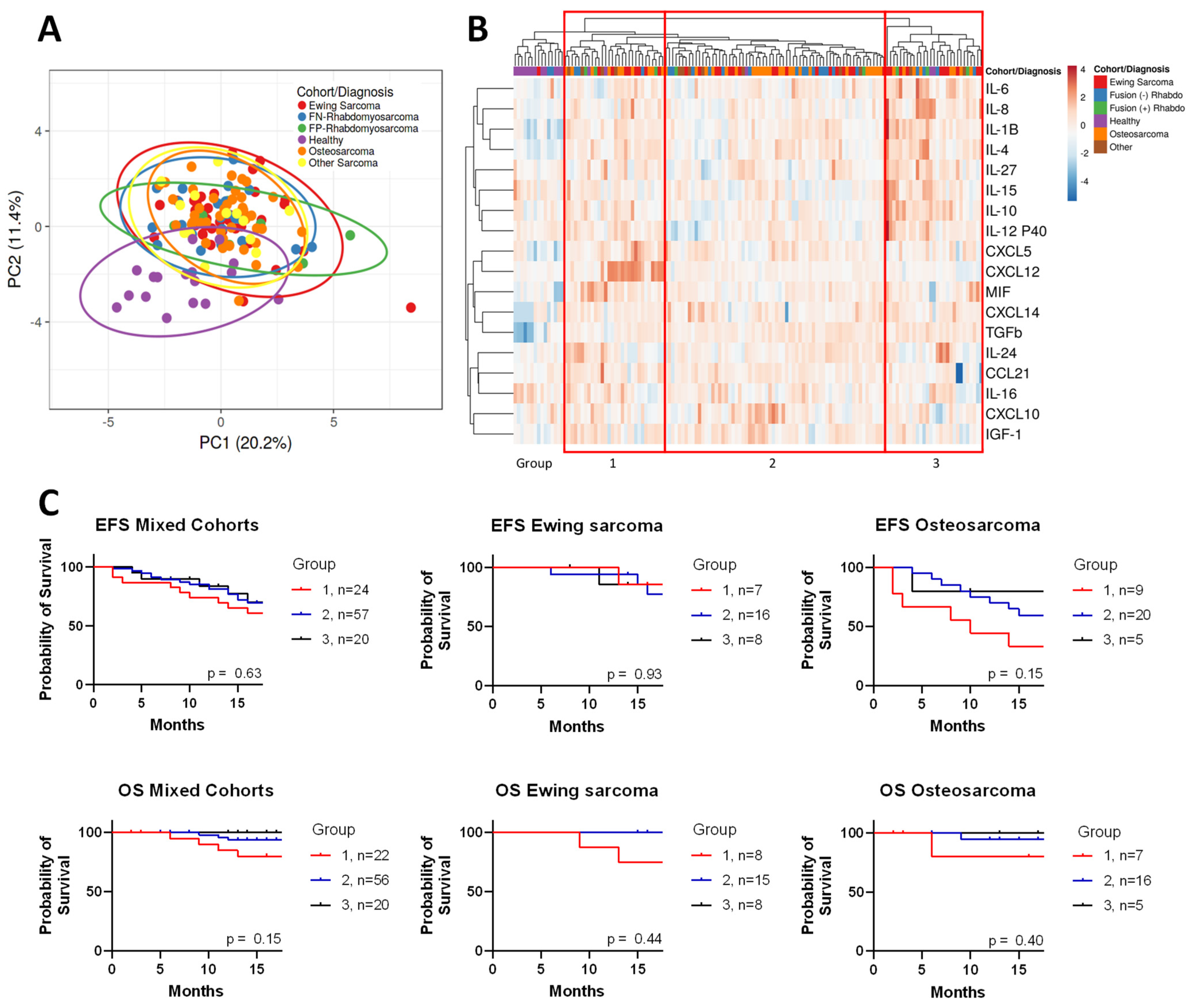
Figure 1. Unsupervised clustering of sarcomas compared to healthy controls. (A) SVD imputation to calculate principal component analysis with unit variance scaling applied. (B) Heat map of cytokine expression with cohort type; unit variance scaling is applied to rows. Imputation is used for missing value estimation. Both rows and columns are clustered using correlation distance and complete linkage. Red squares denote endotype grouping. (C) Overall Survival (OS) and event-free survival (EFS) Kaplan–Meier curves for different endotype groups. p = NS determined by Log Rank test.
Cancers 17 03009 g001
Figure 2. Comparison of outcomes based on total number of elevated analytes, defined by top 5% of healthy cohort mean. (A) Distribution of osteosarcoma and Ewing sarcoma patients by their number of elevated cytokines. (B) Clinical outcomes for patients separated by how many elevated cytokines they have. (C) Event-free survival (EFS) and overall survival (OS) curves for Ewing sarcoma and osteosarcoma patients with 0–3, or ≥4 cytokines elevated.
Cancers 17 03009 g002
Figure 3. Comparison of outcome based on total number of depressed analytes, defined by bottom 5% of the healthy cohort’s mean. (A) Distribution of osteosarcoma and Ewing sarcoma patients by their number of depressed cytokines. (B) Clinical outcomes for patients separated by how many depressed cytokines they have. (C) Event-free survival (EFS) and overall survival (OS) curves for Ewing sarcoma and osteosarcoma grouped based on number of depressed cytokines.
Cancers 17 03009 g003
Figure 4. Levels of pro-tumorigenic and anti-tumorigenic cytokines in sarcoma patients compared to healthy controls. (A) Ewing sarcoma (ES), (B) fusion-negative rhabdomyosarcoma (FN-RMS), and (C) osteosarcoma (OS). Statistical comparison using Mann–Whitney test. *, **, ***, and **** denote p < 0.05, p < 0.01, p < 0.001, p < 0.0001.
Cancers 17 03009 g004
Figure 5. Cytokines that affected event-free survival (EFS) and overall survival (OS) of osteosarcoma and Ewing sarcoma. Significance determined by log rank test.
Cancers 17 03009 g005
Figure 6. IL-1B is associated with worse outcomes in sarcomas. (A) Mann–Whitney test comparing IL-1B in patients that had metastatic disease compared to those with localized disease in all patients, not separated by diagnosis, with Ewing sarcoma and osteosarcoma. (B) Patients stratified by high (>75% of cohort) or low expression of IL-1B (≤25% of cohort). Number of those patients that had metastatic or localized disease. Statistical analysis by Fisher’s exact test. (C) Effect of IL-1B expression on event-free survival (EFS) of osteosarcoma and Ewing sarcoma. Significance determined by log rank test. *: p < 0.05. ns = not significant.
Cancers 17 03009 g006
Disclaimer/Publisher’s Note: The statements, opinions and data contained in all publications are solely those of the individual author(s) and contributor(s) and not of MDPI and/or the editor(s). MDPI and/or the editor(s) disclaim responsibility for any injury to people or property resulting from any ideas, methods, instructions or products referred to in the content.