Communication

Evaluation of Mithramycin in Combination with Chemotherapeutic Agents Against Ewing Sarcoma Cell Lines

1
Department of Microbiology, Immunology and Genetics, College of Biomedical and Translational Sciences, University of North Texas Health, Fort Worth, TX 76107, USA
2
Texas College of Osteopathic Medicine, University of North Texas Health, Fort Worth, TX 76107, USA
*
Author to whom correspondence should be addressed.
Cancers 2025, 17(18), 2977; https://doi.org/10.3390/cancers17182977
Submission received: 15 July 2025 / Revised: 28 August 2025 / Accepted: 2 September 2025 / Published: 11 September 2025
(This article belongs to the Special Issue Research on Targeted Drugs in Cancer)

Simple Summary

Ewing sarcoma primarily affects adolescents and young adults and is considered an aggressive malignancy with a poor prognosis. Mithramycin has been identified as a specific inhibitor of the EWS-FLI1 fusion protein, which is expressed in over 85% of patients with this cancer. However, its clinical application has been limited by toxicity. This study evaluated the anti-proliferative effects of Mithramycin in combination with the chemotherapeutic agents Vincristine and Etoposide. The results demonstrated a significant increase in cytotoxicity when Mithramycin was used together with chemotherapeutic agents compared to either agent alone. Further experiments were conducted to evaluate the response of Mithramycin and Etoposide combination on apoptosis. Flow cytometric analysis showed an upregulation of annexin-V-positive cells, and Western blot detection of cleaved PARP confirmed apoptosis activation. Notably, non-malignant H9C2 cardiomyocytes showed no significant reduction in proliferation with Mithramycin, Etoposide, or their combination. These findings provide preliminary evidence that Mithramycin can enhance the chemotherapeutic response in vitro. Further studies are underway to elucidate its precise mechanism of action.

Abstract

Ewing Sarcoma (ES) is a rare, malignant bone neoplasm that is primarily diagnosed in childhood and adolescence. The aggressive nature of this neoplasm requires the use of surgery, radiation and a rigorous chemotherapy regimen. Metastatic ES carries a poor prognosis, which necessitates the development of new therapeutic agents. Mithramycin was tested in targeted therapy due to its specific inhibitory effects on the EWS-FLI1 fusion protein which is present in >85% of ES tumors. We tested the combination of Mithramycin with chemotherapeutic agents vincristine (VCR) and Etoposide (Eto) for inducing higher cytotoxicity against ES cells, CHLA10 and TC205. Cardiomyocyte cell line, H9C2 was used to test the effect on non-malignant cells. Cell viability was measured using the CellTiter-Glo kit, and the combination index was evaluated to determine the type of combination response (antagonistic, additive, or synergistic). Apoptotic cells were measured post-treatment with vehicle (DMSO, control), monotherapy (mithramycin or etoposide), or combination therapy (mithramycin + etoposide) using BD LSRII flow cytometer and analyzed utilizing FlowJo software V8.0. The apoptotic protein marker c-PARP in both treatment and control groups was analyzed using Western blot analysis. The results showed higher cytotoxicity for combination treatment when compared to individual agents, and the combination index confirmed the response as synergistic. H9C2 cells did not demonstrate significant decreases in cell viability when treated with combination therapy, highlighting the specificity of the treatment toward its target tissue. Flow cytometry confirmed the underlying mechanism as upregulation of apoptosis which is further supported by an increase in effector caspases 3/7 and elevated expression of c-PARP. These in vitro assays using ES cells provide preliminary evidence for the benefit of chemotherapy and mithramycin combination.

1. Introduction

Ewing Sarcoma (ES) is the second leading cause of bone tumors in adolescents and children. It mainly affects the demographic of children and young adults from the age of 10 to 20 years old, with diagnoses most frequent in boys between 10 and 15 years old. ES is a tumor that typically develops in bone and soft tissues, particularly in long bones, the spine, ribs, and pelvis. Metastases are most commonly found in the lungs, bone, and bone marrow in up to 25% of patients at the time of diagnosis [1]. Pathologically, ES is classically characterized as a small round cell tumor. Some ES tumors have neural differentiation and can be referred to as primitive neuroectodermal tumors (PNETs). The tumors have characteristic Homer Wright Rosettes, which are cell clusters with neurofibrillary processes [2].
EWS-FLI1 fusion protein regulates oncogenes, including the ones associated with tumorigenesis [3]. This protein is expressed in >85% of ES tumors and the efficacy of Mithramycin (Mit) was tested as a targeted therapeutic agent to inhibit transcription of the EWS-FLI1 fusion protein in ES cells [4]. While mithramycin’s efficacy is evident in the compound’s inhibitory activity on the EWS–FLI1 protein, clinical application of this drug had proven to be challenging. In a previous clinical trial, both children and adults with ES received Mit alone, with average plasma concentrations of around 18 ng/mL [4]. At this concentration, many patients experienced severe elevations in alanine aminotransferase and aspartate aminotransferase, leading to discontinuation of treatment. Unfortunately, this plasma concentration was far below the required level to inhibit EWS-FLI1 transcriptional activity. The strong efficacy of Mit against ES prompts the question of how to utilize the gene-suppression activity of Mit and minimize toxicity due to drug exposure. Combination therapy of Mit and other chemotherapeutic agents provides not only a potential increase in treatment efficacy, but also the potential to lower the total dose of each individual agent and minimize toxicity.
The standard of care for ES includes combination induction chemotherapy followed by surgery and/or adjuvant chemotherapy [5,6]. In localized cases of ES, the combination of standard of care can lead to a 73% survival rate of patients, but the prognosis is poor in patients when ES has metastasized [2]. The current treatment for localized ES includes a combination of vincristine (VCR), Doxorubicin, and cyclophosphamide, with an alternating dose of ifosfamide and etoposide (Eto) every two weeks [2]. For patients with metastasized ES, the treatment includes VCR, doxorubicin, and cyclophosphamide, alternating with ifosfamide, and Eto every two weeks [2]. However, these combinatorial treatments were not reported to demonstrate improved survival of the patients or post-treatment quality-of-life. A previous clinical trial examined the efficacy of adding ifosfamide and Eto to the standard treatment regimen of VCR, doxorubicin, cyclophosphamide, and dactinomycin improved treatment outcomes. Patients with metastatic ES or PNET were treated with 9 weeks of chemotherapy before local control and then an additional 42 weeks of chemotherapy. After this regimen, some patients were treated with alternating ifosfamide and Eto. Following the clinical trial, the event-free survival was less than 30%, concluding that the addition of these agents did not increase survival outcomes; however, these agents remain a part of the standard treatment regimen [7]. Thus, finding new possible treatments that can be used as monotherapy or in combinational therapy is of utmost importance.
In recent years, there has been a growing interest in Mit’s inhibitory effects on the aberrant fusion of the transcription factor EWS-FLI1 that results from the chromosomal translocation from t (11;22) (q24;q12) [2]. This translocation results in the fusion of the constitutively expressed EWSR1 protein and the FLI1 protein. The EWSR1 protein plays a role in homologous recombination, which becomes impaired in ES due to the chromosomal translocation [8]. We hypothesize that using Mit in combination with chemotherapeutic agents will induce cytotoxicity of cancer cells through alternative mechanisms to inhibit the ESW-FLI1 fusion protein. To determine Mit’s effectiveness as a cancer therapeutic, cell viability testing was initially performed to identify proper dosages of monotherapy and combination use for Mit. Optimized doses were then tested in combination treatment to evaluate the effect on apoptosis.

2. Materials and Methods

2.1. Cell Culture

ES cell lines were obtained from Children’s Oncology Group (Lubbock, TX, USA) and H9C2 cells were purchased from ATCC (Manassas, VA, USA). CHLA-10 (EWS-FLI1-expressing) and TC205 (EWS-FLI1-non-expressing) cells, as well as cardiomyocytes (H9C2) were grown in Iscove’s Modified Dulbecco’s Media (10% FBS, 1% penicillin, 1% ITS, 4 mM glutamine, HEPES). Cell lines were grown at 37 °C and 5% CO2.

2.2. Chemicals, Reagents, and Antibodies

Mithramycin, Dimethyl sulfoxide (DMSO), Eto, VCR and β-actin antibody were obtained from Sigma-Aldrich (St. Louis, MO, USA). Cleaved poly (ADP-ribose) polymerase (c-PARP) antibody was purchased from Cell Signaling Technology (Danvers, MA, USA) and PE-Annexin V/7-AAD apoptosis kit was obtained from BD Biosciences (Franklin Lakes, NJ, USA).

2.3. Cell Viability

ES cells were grown, and 4000 cells were then plated per well in 96-well plates. After 24 h they were treated with DMSO (control) or Mit, and/or Eto/VCR. Cells were incubated at 37 °C and 5% CO2, and after 48 h, cell viability was analyzed using a CellTiter-Glo kit (Promega, Madison, WI, USA). Following the protocol outlined in the CellTiter-Glo kit, 100 µL of CellTiter-Glo reagent was added to each well of the 96-well plates and left in the dark to incubate for 30 min. Luminescence (measures the cellular ATP, which is proportional to living cells) was recorded using a Synergy HT microplate reader. Each treatment and control was measured in triplicate, and data were graphed as percent viability against concentration on a log scale. IC50 values were calculated using GraphPad Prism software V9.0. Combination index was calculated using Calcusyn software V2.0 after treatments were set in a constant-ratio design and read using CellTiter-Glo (Promega, Madison, WI, USA) [9,10]. Data was analyzed using one-way ANOVA.

2.4. Flow Cytometry: PE Annexin-V Staining

ES cells were harvested after treatment with vehicle (DMSO), or drug alone (Mit or Eto), or combination (Mit + Eto) for 48 h. The apoptotic cell population was determined following standard procedure described by the manufacturer using PE-Annexin V/7-AAD apoptosis kit. BD LSRII flow cytometer was used to measure the staining and the data was analyzed (FlowJo software V8.0). Data was analyzed using one-way ANOVA.

2.5. Western Blot: C-PARP Expression

Protein was harvested after 48 h of treatments and a BCA assay was performed to find protein concentration. Western blot data were obtained using a capillary Western analysis Jess system from Bio-Techne (Minneapolis, MN, USA). Samples were run on 25 capillary cartridges, 12-230 kDa separation from Bio-Techne. A total of 1 mg of protein lysate was loaded onto the plate, followed by primary antibodies for c-PARP (1:50) or β-actin (1:100). Loading of the plate was performed according to manufacturer instructions and subsequent separation and immunodetection occurred in the automated capillary system. Blots were generated, and the area under the curves from the chromatogram data was analyzed using Compass for Simple Western software (version 6.1). Expression of all proteins was normalized to actin. Data was analyzed using one-way ANOVA.

3. Results

3.1. Dose Response and Determination of IC50 Values

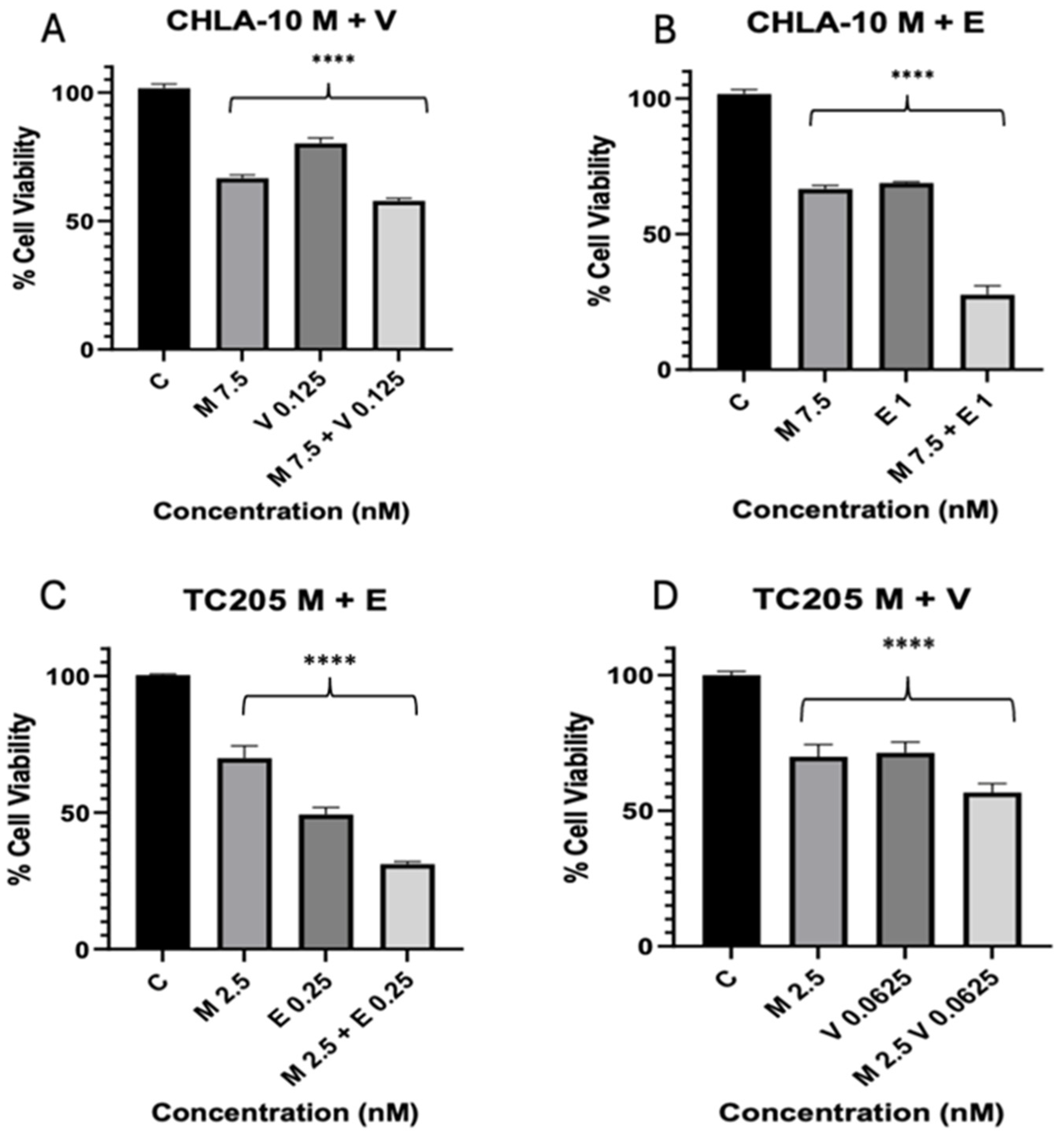
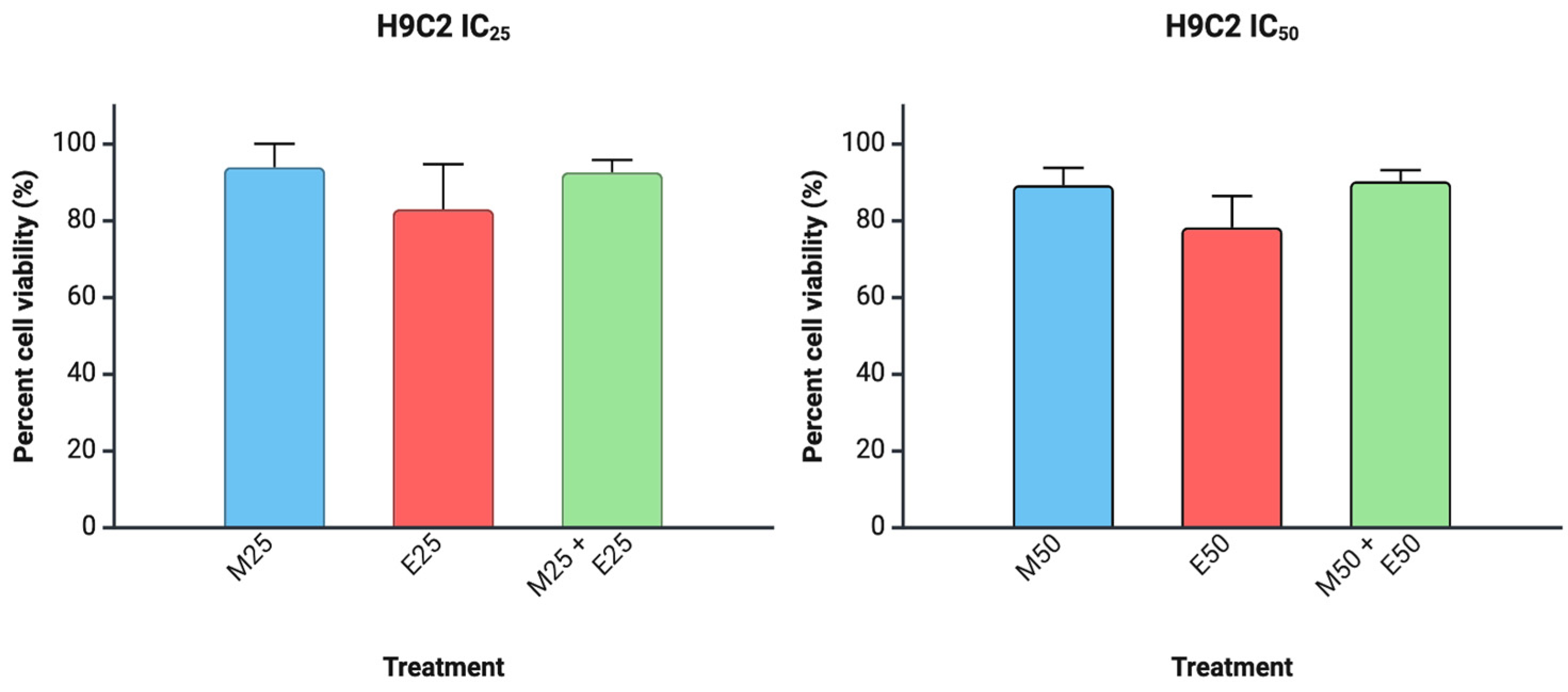
The antiproliferative effect of each drug was tested on two cell lines, TC205 and CHLA10. To establish working IC50 values, Mit, Eto, and VCR were analyzed over 48 h treatment periods, resulting in dose-dependent inhibition of cell viability. IC50 doses varied per drug and cell line (Figure 1). Combination treatments were also used at approximate IC50 values, resulting in improved reduction in cell viability for each combination tested (Figure 2). The IC50 values for CHLA10 IC50 values were 9.11, 1.25, and 0.25 nM for Mit, Eto, and VCR, respectively. For TC205, the IC50 values were 4.32, 0.25, and 0.11 nM for Mit, Eto, and VCR, respectively. Testing against non-malignant H9C2 cells (Figure 3) showed relatively little change in cell viability with Mit and Mit + Eto treatment. The only relatively greater change was observed with monotherapy IC50 Eto treatment (p = 0.055). Interestingly, an antagonistic response was observed with the Eto and Mit combination, which reduced the growth inhibition caused by Eto in H9C2 cells.

3.2. Combination Index

To study further treatment interactions, drug effect analysis using Calcusyn provided ED50 values and representative dose-effect curves to determine whether the combination produced a synergistic, antagonistic, or additive response. Details of the combination index calculation were well established and described earlier [11]. Treatment concentrations were set in a constant-ratio design and analyzed using CellTiter-Glo (Figure 4). In both CHLA10 and TC205 Mit combinations with Eto and VCR exhibited ED50 values <1, indicating synergistic drug responses in each case [12].

3.3. Mithramycin and Eto Effects on Apoptotic Cell Population

Flow cytometric analysis of TC205 cells after 48 h treatments of Mit and Eto were conducted to identify separate apoptotic cell populations. Percent change in viable cells and early and late apoptotic cell populations illustrate monotherapy and combination treatment. A positive shift in apoptosis was observed in all treatments; however, the most effective results were seen in the combination groups (Figure 5).

3.4. Cleaved PARP Expression

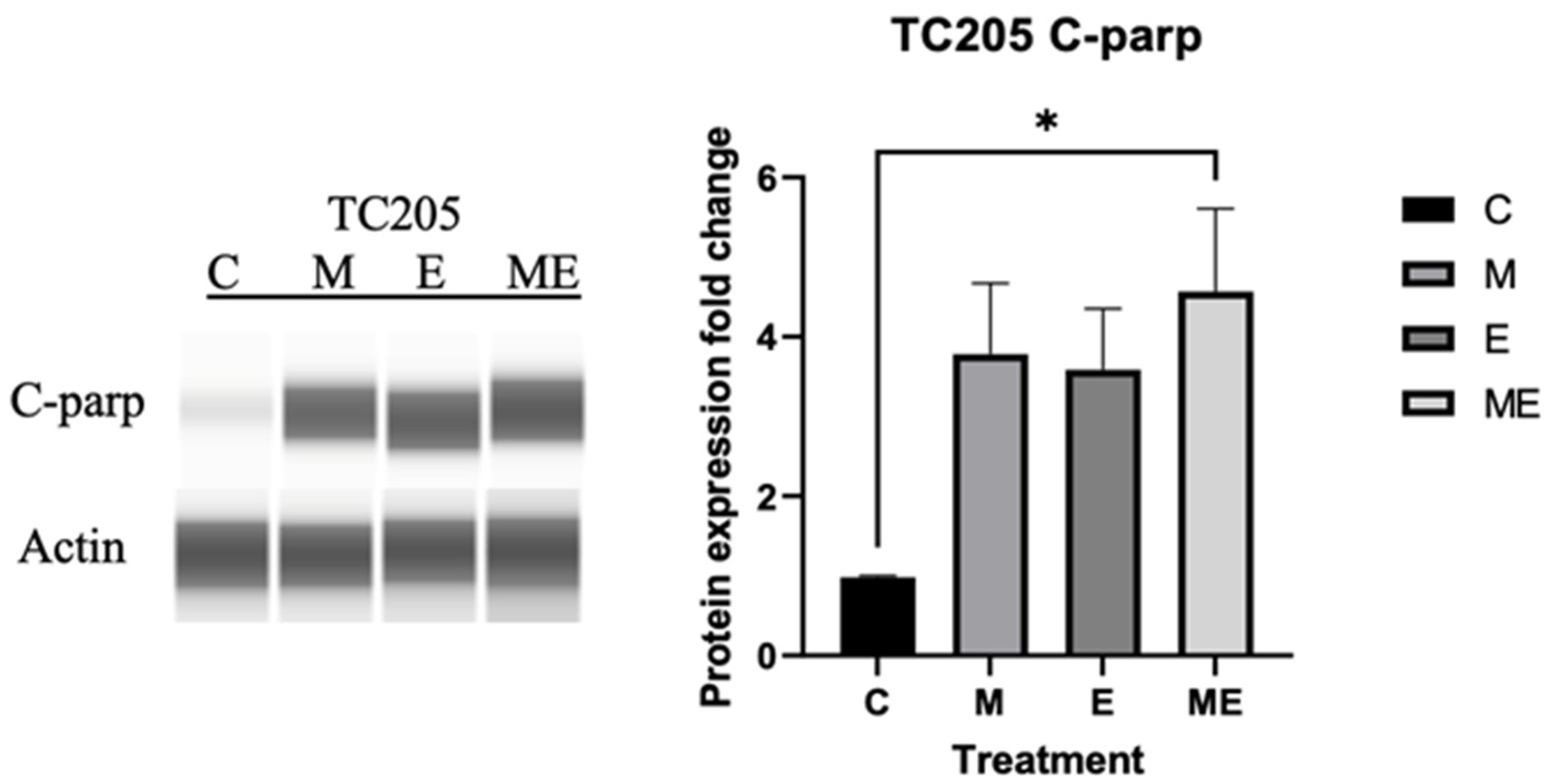
Further apoptosis-related study through Western blot analysis of c-PARP in TC205 cells followed a similar trend, with the highest C-PARP expression occurring in the combination group (Figure 6).

4. Discussion

Chemotherapeutic drugs induce high levels of toxicity in ES patients [13,14,15,16]. Although novel therapies, including immunotherapy, are in clinical testing, standard treatment options involving chemotherapy and radiation remain the most commonly used in ES therapy [17,18,19,20,21]. The majority of cancer therapies are administered as combinations of drugs within broader treatment regimens. The combination of chemotherapeutic agents with less toxic agents to improve therapeutic efficacy has been employed in multiple studies [11,22,23,24]. In ES, it is particularly important to utilize a combinatorial treatment approach [25], especially when metastasis is detected in lungs or bone marrow [26,27,28]. Thus, testing the combination of Mithramycin (Mit) with Etoposide (Eto) or Vincristine (VCR) provides not only a more effective treatment option but also better reflects actual therapeutic implementation.
The viability data of TC205 and CHLA-10 cells revealed that low-dose Mit, Eto, and VCR concentrations should be utilized for further studies. The calculated IC50 values from the cell viability assay were for the CHLA-10 cell line were 9.11, 1.25, and 0.25 nM for Mit, Eto, and VCR, respectively. For the TC205 cell line, the IC50 values were 4.32, 0.25, and 0.11 nM for Mit, Eto, and VCR, respectively. The IC50 values of Mit for the two ES cell lines tested in this study fall within the average range previously reported [29]. It has been reported that cell lines expressing the EWS-FLI1 aberrant protein are particularly sensitive to Mit [29]. The reported plasma levels in the ES clinical study were approximately 18 ng/ml [4]. In our study, we used 2.71 and 8.74 ng/mL, respectively, for TC205 and CHLA10 cells (calculated based on molecular weight and concentration). Since binding to serum proteins and absorption due to combination treatment can also influence the therapeutic effect, these doses are reasonable to test.
The results reported here show that TC205 was more sensitive than CHLA-10 to Mit at different concentrations, supporting the high sensitivity to Mit in inhibiting the EWS-FLI1 fusion gene (Figure 1A) [30]. Similarly, the results for the IC50 of Eto in this study show that lower dosages of Eto are needed for the TC205 cell line when compared to the CHLA-10 cell line (Figure 1B). Prospects for improving current therapies have been evaluated by comparing the inhibitory response of chemotherapeutic drugs, with average reported IC50 for Eto usually ranging from 0.30 µM to 1.11 µM depending on the specific ES cell line being tested [31,32,33]. Following the same pattern, the IC50 value for VCR was lower in the TC205 cell line compared with the CHLA-10 cell line (Figure 1C). The IC50 values for the ES cell lines tested in this study fall within the average IC50 values reported in previous studies. These low numbers show that VCR is effective at inhibiting the cell proliferation of ES [34].
To further improve the dosage utilized for treatment, a combinatorial therapy approach was investigated by combination dosing of Mit with Eto, as well as Mit with VCR. The combination dosing showed a decrease in viability in each instance of Mit + Eto and Mit + VCR when compared with monotherapy results (Figure 2A–D). Testing the safety of treatment doses against cardiomyocytes (H9C2) revealed minimal effect of Mit on non-malignant cells (Figure 3). In the combination, there was also slight recovery of H9C2 cells compared with Eto monotherapy, which warrants further study (Figure 3). To further explain the effective viability reduction seen in combination, we aimed to describe the drug interaction between Mit, Eto, and VCR. In both CHLA-10 and TC205 cells, combination index analysis showed a synergistic relationship (ED5 < 1) (Figure 4A,B). This synergistic relationship may be explained by the complementary behavior of their individual mechanisms.
Mithramycin inhibits the EWS-FLI1 fusion transcription factor by suppressing its transcriptional activity and altering downstream gene expression critical for tumor growth and DNA repair processes [29]. Some of the genes downregulated by Mit’s inhibition of DNA repair mechanisms can effectively block double-stranded DNA repair in the target cells, increasing the DNA damage in the cell resulting in increased apoptosis [35]. The cytotoxic effects of Eto are due to its inhibition of DNA topoisomerase II through stabilization of the DNA-topoisomerase II complex causing increased DNA breaks and damage, ultimately leading to increased cell death [31]. The combination of Mit with Eto suppresses DNA repair pathways via Mit, and the increase in DNA breaks caused by Eto further enhances the activation of apoptotic pathway initiators [31,34].
VCR can disrupt the formation of microtubule polymers during mitotic spindle assembly, resulting in the arrest of metaphase and increasing the cell death of ES dividing cells [34]. The use of vinca alkaloids, such as VCR, has been part of cancer treatment since the early stages of development, due to their M-phase-specific anti-tubulin capabilities [36]. However, the toxic effects of this family of alkaloids can be broad and affect multiple organ systems; therefore, treatment with VCR is typically performed in combinatorial treatments to achieve a lower effective dose [36]. The combination of Mit with VCR increases apoptosis through the downregulation of DNA repair mechanisms through Mit and the cell-cycle arrest caused by VCR-mediated disruption of microtubule formation.
To explain the decrease in viability of ES cell lines, we observed a marked increase in early and late apoptotic cells by flow cytometry of TC205 cells after treatment, with the combination dose offering the most significant impact on the apoptotic cell population (Figure 5). It has been reported that ES cells are particularly sensitive to inhibition of PARP, as the EWS-FLI1 oncoprotein interacts with PARP leading to increased transcriptional activation by EWS-FLI1 [37]. Treatments that target the DNA repair mechanisms can promote apoptosis through cleaving of PARP, generating the apoptosis effector protein c-PARP [38]. Western blot analysis of c-PARP, used as another identifier of apoptotic cells, showed a significant increase in all groups compared with the control group. However matching the flow cytometry data, the combination group had the highest expression of c-PARP (Figure 6). The increased expression of c-PARP confirms the flow cytometry results, showing that Mit, Eto and Mit with Eto inhibit DNA repair mechanisms in the cell, resulting in increased apoptosis.
This study has some limitations and we are continuing investigation to fulfill the gaps. It is still in the preliminary stage and limited to in vitro assays. Further validation using in vivo models is needed. Even though, cardiotoxicity with Eto is rare, it remains a serious concern especially in combination treatments [39,40]. Therefore, in this study we tested cytotoxicity against cardiomyocytes. Since Mit can affect liver, testing hepatotoxicity, along with comprehensive pharmacokinetic evaluations, will also be helpful to determine the therapeutic potential and safety profile of the combination. Other issue is using the concentration of Mit for laboratory testing aligning with the plasma levels of clinical relevance. While we show that the apoptotic process is elevated in ES cells due to treatment, the underlying molecular mechanisms responsible for this effect, particularly in the context of combination therapy, remain unclear. It is possible that multiple pathways contribute to the observed cytotoxicity. As we continue to evaluate this strategy, we will conduct experiments in detail to address outstanding issues.

5. Conclusions

Notably, there has been little improvement in treatment strategies or survival rates of ES patients over several decades; however, a few novel approaches are still in testing [41,42,43]. Using Mit, an older drug, this study provides evidence that it can enhance cytotoxicity in ES cell lines, CHLA-10 and TC205. While Mit has previously been studied for its specific inhibitory effect against EWS-FLI1 fusion protein, our findings suggest efficacy in both EWS-FLI1—expressing (CHLA-10) and non-expressing (TC205) cells. The results showed an increase in apoptosis; however, the precise molecular mechanisms responsible anti-proliferative response in Ewing sarcoma cells, particularly in combination therapy, remain unclear. Further investigations are needed to unravel underlying pathways and optimize its translational application.

Author Contributions

Conceptualization, R.B. and C.B.L.; methodology, C.B.L., L.A. and R.B.; validation, C.B.L. and R.B.; formal analysis, C.B.L. and R.B.; investigation, C.B.L., L.A. and R.B.; resources, R.B.; data curation, C.B.L.; writing—original draft preparation, C.B.L., A.O.N., K.F. and R.B.; writing—review and editing, R.B., A.O.N. and K.F.; visualization, C.B.L., A.O.N. and K.F.; supervision, R.B.; project administration, C.B.L. and R.B.; funding acquisition, R.B. All authors have read and agreed to the published version of the manuscript.

Funding

This work was partially supported by Cancer Prevention and Research Institute of Texas (award# 210046) and NIH (S21MD012472, U54MD006882).

Institutional Review Board Statement

Not applicable.

Informed Consent Statement

Not applicable.

Data Availability Statement

The data presented in this study are available in the article. Further inquiries can be directed to the corresponding author.

Acknowledgments

Authors thank Children’s Oncology Group for providing cell lines and Lesa Thomas (Texas College of Osteopathic Medicine) for her assistance in proof reading and editing the manuscript.

Conflicts of Interest

The authors declare no conflicts of interest.

Abbreviations

The following abbreviations are used in this manuscript:
c-PARPCleaved poly (ADP-ribose) polymerase (PARP)
DMSODimethyl sulfoxide
DNADeoxyribonucleic acid
EDEffective dose
ED50Median effective dose
ESEwing sarcoma
EtoEtoposide
IC50Inhibitory concentration 50
MMolar
MitMithramycin
PARPPoly (ADP-ribose) polymerase
PNETPrimitive neuroectodermal tumor
VCRVincristine

References

  1. Eaton, B.R.; Claude, L.; Indelicato, D.J.; Vatner, R.; Yeh, B.; Schwarz, R.; Laack, N. Ewing sarcoma. Pediatr. Blood Cancer 2021, 68 (Suppl. S2), e28355. [Google Scholar] [CrossRef]
  2. Choi, E.-Y.K.; Gardner, J.M.; Lucas, D.R.; McHugh, J.B.; Patel, R.M. Ewing sarcoma. Semin. Diagn. Pathol. 2014, 31, 39–47. [Google Scholar] [CrossRef]
  3. Hancock, J.D.; Lessnick, S.L. A transcriptional profiling meta-analysis reveals a core EWS-FLI gene expression signature. Cell Cycle 2008, 7, 250–256. [Google Scholar] [CrossRef]
  4. Grohar, P.J.; Glod, J.; Peer, C.J.; Sissung, T.M.; Arnaldez, F.I.; Long, L.; Figg, W.D.; Whitcomb, P.; Helman, L.J.; Widemann, B.C. A phase I/II trial and pharmacokinetic study of mithramycin in children and adults with refractory Ewing sarcoma and EWS–FLI1 fusion transcript. Cancer Chemother. Pharmacol. 2017, 80, 645–652. [Google Scholar] [CrossRef]
  5. Hesla, A.C.; Papakonstantinou, A.; Tsagkozis, P. Current Status of Management and Outcome for Patients with Ewing Sarcoma. Cancers 2021, 13, 1202. [Google Scholar] [CrossRef] [PubMed]
  6. Ben Kridis, W.; Toumi, N.; Chaari, H.; Khanfir, A.; Ayadi, K.; Keskes, H.; Boudawara, T.; Daoud, J.; Frikha, M. A Review of Ewing Sarcoma Treatment: Is it Still a Subject of Debate? Rev. Recent Clin. Trials 2017, 12, 19–23. [Google Scholar] [CrossRef]
  7. Miser, J.S.; Krailo, M.D.; Tarbell, N.J.; Link, M.P.; Fryer, C.J.; Pritchard, D.J.; Gebhardt, M.C.; Dickman, P.S.; Perlman, E.J.; Meyers, P.A.; et al. Treatment of metastatic Ewing’s sarcoma or primitive neuroectodermal tumor of bone: Evaluation of combination ifosfamide and etoposide—A Children’s Cancer Group and Pediatric Oncology Group study. J. Clin. Oncol. 2004, 22, 2873–2876. [Google Scholar] [CrossRef] [PubMed]
  8. Gorthi, A.; Romero, J.C.; Loranc, E.; Cao, L.; Lawrence, L.A.; Goodale, E.; Iniguez, A.B.; Bernard, X.; Masamsetti, V.P.; Roston, S.; et al. EWS–FLI1 increases transcription to cause R-loops and block BRCA1 repair in Ewing sarcoma. Nature 2018, 555, 387–391. [Google Scholar]
  9. Abdelrahim, M.; Baker, C.H.; Abbruzzese, J.L.; Safe, S. Tolfenamic acid and pancreatic cancer growth, angiogenesis, and Sp protein degradation. J. Natl. Cancer Inst. 2006, 98, 855–868. [Google Scholar] [CrossRef] [PubMed]
  10. Nair, V.; Pathi, S.; Jutooru, I.; Sreevalsan, S.; Basha, R.; Abdelrahim, M.; Samudio, I.; Safe, S. Metformin inhibits pancreatic cancer cell and tumor growth and downregulates Sp transcription factors. Carcinogenesis 2013, 34, 2870–2879. [Google Scholar] [CrossRef]
  11. Patil, S.; Sankpal, U.T.; Hurtado, M.; Bowman, W.P.; Murray, J.; Borgmann, K.; Ghorpade, A.; Sutphin, R.; Eslin, D.; Basha, R. Combination of clotam and vincristine enhances anti-proliferative effect in medulloblastoma cells. Gene 2019, 705, 67–76. [Google Scholar] [CrossRef] [PubMed]
  12. Huang, R.-Y.; Pei, L.; Liu, Q.; Chen, S.; Dou, H.; Shu, G.; Yuan, Z.-X.; Lin, J.; Peng, G.; Zhang, W.; et al. Isobologram Analysis: A Comprehensive Review of Methodology and Current Research. Front. Pharmacol. 2019, 10, 1222. [Google Scholar] [CrossRef]
  13. Chapuis, J.-C.; Keng, P.C.; Siemann, D.W. Activity of etoposide (VP-16) and teniposide (VM-26) in exponential and plateau phase human tumor cell cultures. Anticancer Drugs 1992, 3, 245–252. [Google Scholar] [CrossRef]
  14. Skinner, R.; Cotterill, S.J.; Stevens, M.C. Risk factors for nephrotoxicity after ifosfamide treatment in children: A UKCCSG Late Effects Group study. United Kingdom Children’s Cancer Study Group. Br. J. Cancer 2000, 82, 1636–1645. [Google Scholar]
  15. Scobioala, S.; Eich, H.T. Risk stratification of pulmonary toxicities in the combination of whole lung irradiation and high-dose chemotherapy for Ewing sarcoma patients with lung metastases: A review. Strahlenther. Onkol. 2020, 196, 495–504. [Google Scholar] [CrossRef]
  16. Ilari, I.; De Ioris, M.A.; Milano, G.M.; Pessolano, R.; De Lurentis, C.; De Sio, L.; Fidani, P.; Jenkner, A.; Cozza, R. Toxicity of high-dose chemotherapy with etoposide, thiotepa and CY in treating poor-prognosis Ewing’s sarcoma family tumors: The experience of the Bambino Gesù Children’s Hospital. Bone Marrow Transplant. 2010, 45, 1274–1280. [Google Scholar] [CrossRef] [PubMed]
  17. Subbiah, V.; Anderson, P.; Lazar, A.J.; Burdett, E.; Raymond, K.; Ludwig, J.A. Ewing’s sarcoma: Standard and experimental treatment options. Curr. Treat. Options Oncol. 2009, 10, 126–140. [Google Scholar] [CrossRef]
  18. Xie, J.; Mellado-Lagarde, M.M.; Blankenship, K.; Ganguly, D.; Twarog, N.R.; Bianski, B.; Kieffer, M.; Atkinson, S.; Sheppard, H.; Gartrell, J.; et al. The Combination of PARP and Topoisomerase 1 Inhibitors Improves Radiation Therapy for Ewing Sarcoma. Cancer Sci. 2025, 116, 1703–1714. [Google Scholar] [CrossRef] [PubMed]
  19. Geng, Y.; Yang, L.; Shao, R.; Xu, T.; Zhang, L. RNA-binding protein gene NOP58 exhibits crucial prognostic and therapeutic value in Ewing sarcoma. Hereditas 2025, 162, 76. [Google Scholar] [CrossRef] [PubMed]
  20. Aiba, H.; Aso, A.; Righi, A.; Gambarotti, M.; Cammelli, S.; Spinnato, P.; Traversari, M.; Atherley, A.; Solou, K.; Kimura, H.; et al. Comparison of histological response across patients with Ewing sarcoma treated with preoperative chemotherapy with or without preoperative radiotherapy. Eur. J. Orthop. Surg. Traumatol. 2025, 35, 251. [Google Scholar] [CrossRef]
  21. Patel, D.S.; Desai, R.T.; Pandya, A.J.; Kothari, N.R.; Patel, R.; Chorawala, M.R. Harnessing the immune responses: A new frontier in Ewing sarcoma treatment. Med. Oncol. 2025, 42, 291. [Google Scholar] [CrossRef]
  22. Li, X.; Xu, F.; Wang, R.; Shen, L.; Luo, B.; Zhou, S.; Zhang, J.; Zhang, Z.; Cao, Z.; Zhan, K.; et al. Aspirin enhances radio/chemo-therapy sensitivity in C. elegans by inducing germ cell apoptosis and suppresses RAS overactivated tumorigenesis via mtROS-mediated DNA damage and MAPK pathway. Biochem. Biophys. Res. Commun. 2024, 735, 150828. [Google Scholar] [CrossRef]
  23. Batra, H.; Pawar, S.; Bahl, D. Curcumin in combination with anti-cancer drugs: A nanomedicine review. Pharmacol. Res. 2019, 139, 91–105. [Google Scholar] [CrossRef]
  24. Sankpal, U.T.; Ingersoll, S.B.; Ahmad, S.; Holloway, R.W.; Bhat, V.B.; Simecka, J.W.; Daniel, L.; Kariali, E.; Vishwanatha, J.K.; Basha, R. Association of Sp1 and survivin in epithelial ovarian cancer: Sp1 inhibitor and cisplatin, a novel combination for inhibiting epithelial ovarian cancer cell proliferation. Tumor Biol. 2016, 37, 14259–14269. [Google Scholar] [CrossRef]
  25. Shelake, S.; Sankpal, U.T.; Eslin, D.; Bowman, W.P.; Simecka, J.W.; Raut, S.; Ray, A.; Basha, R. Clotam enhances anti-proliferative effect of vincristine in Ewing sarcoma cells. Apoptosis 2019, 24, 21–32. [Google Scholar] [CrossRef]
  26. Khanna, N.; Pandey, A.; Bajpai, J. Metastatic Ewing’s Sarcoma: Revisiting the “Evidence on the Fence”. Indian J. Med. Paediatr. Oncol. 2017, 38, 173–181. [Google Scholar] [PubMed]
  27. Ozaki, T. Diagnosis and treatment of Ewing sarcoma of the bone: A review article. J. Orthop. Sci. 2015, 20, 250–263. [Google Scholar] [CrossRef] [PubMed]
  28. Meshram, G.G.; Kaur, N.; Hura, K.S. Ewing’s sarcoma with distant metastasis: A brief note on management and emerging therapies. Clin. Pract. 2019, 9, 1111. [Google Scholar] [CrossRef] [PubMed]
  29. Lin, M.Y.; Damron, T.A.; Oest, M.E.; Horton, J.A. Mithramycin A Radiosensitizes EWS:Fli1+ Ewing Sarcoma Cells by Inhibiting Double Strand Break Repair. Int. J. Radiat. Oncol. Biol. Phys. 2021, 109, 1454–1471. [Google Scholar] [CrossRef]
  30. Orth, M.F.; Surdez, D.; Faehling, T.; Ehlers, A.C.; Marchetto, A.; Grossetête, S.; Volckmann, R.; Zwijnenburg, D.A.; Gerke, J.S.; Zaidi, S.; et al. Systematic multi-omics cell line profiling uncovers principles of Ewing sarcoma fusion oncogene-mediated gene regulation. Cell Rep. 2022, 41, 111761. [Google Scholar] [CrossRef]
  31. Grohar, P.J.; Helman, L.J. Prospects and challenges for the development of new therapies for Ewing sarcoma. Pharmacol. Ther. 2013, 137, 216–224. [Google Scholar] [CrossRef] [PubMed]
  32. Lenz, J.E.; Riester, R.; Schleicher, S.B.; Handgretinger, R.; Boehme, K.A.; Traub, F. Interaction of arsenic trioxide and etoposide in Ewing sarcoma cell lines. Oncol. Rep. 2020, 43, 337–345. [Google Scholar] [CrossRef] [PubMed]
  33. Boehme, K.A.; Nitsch, J.; Riester, R.; Handgretinger, R.; Schleicher, S.B.; Kluba, T.; Traub, F. Arsenic trioxide potentiates the effectiveness of etoposide in Ewing sarcomas. Int. J. Oncol. 2016, 49, 2135–2146. [Google Scholar] [CrossRef] [PubMed]
  34. Sabei, F.Y.; Taratula, O.; Albarqi, H.A.; Al-Fatease, A.M.; Moses, A.S.; Demessie, A.A.; Park, Y.; Vogel, W.K.; Nazzaro, E.E.; Davare, M.A.; et al. A targeted combinatorial therapy for Ewing’s sarcoma. Nanomedicine 2021, 37, 102446. [Google Scholar] [CrossRef]
  35. Lin, M.Y.; Damron, T.A.; Horton, J.A. Cell cycle arrest and apoptosis are early events in radiosensitization of EWS::FLI1 + Ewing sarcoma cells by Mithramycin A. Int. J. Radiat. Biol. 2023, 99, 1570–1583. [Google Scholar] [CrossRef]
  36. Chen, R.J.; Arora, R.D.; Menezes, R.G. Vinca Alkaloid Toxicity; StatPearls: Treasure Island, FL, USA, 2025. [Google Scholar]
  37. Vormoor, B.; Curtin, N.J. Poly(ADP-ribose) polymerase inhibitors in Ewing sarcoma. Curr. Opin. Oncol. 2014, 26, 428–433. [Google Scholar] [CrossRef]
  38. Ordonez, J.L.; Amaral, A.T.; Carcaboso, A.M.; Herrero-Martín, D.; García-Macías, M.d.C.; Sevillano, V.; Alonso, D.; Pascual-Pasto, G.; San-Segundo, L.; Vila-Ubach, M.; et al. The PARP inhibitor olaparib enhances the sensitivity of Ewing sarcoma to trabectedin. Oncotarget 2015, 6, 18875–18890. [Google Scholar] [CrossRef]
  39. Shoji, S.; Miyakita, H.; Shima, M.; Usui, Y.; Nagata, Y.; Uchida, T.; Terachi, T. Acute myocardial infarction during combined chemotherapy with bleomycin, etoposide and cisplatin for testicular cancer. Hinyokika Kiyo 2006, 52, 723–726. [Google Scholar]
  40. Herradón, E.; González, C.; González, A.; Uranga, J.A.; López-Miranda, V. Cardiovascular Toxicity Induced by Chronic Vincristine Treatment. Front. Pharmacol. 2021, 12, 692970. [Google Scholar] [CrossRef]
  41. Riggi, N.; Suvà, M.L.; Stamenkovic, I.; Longo, D.L. Ewing’s Sarcoma. N. Engl. J. Med. 2021, 384, 154–164. [Google Scholar] [CrossRef]
  42. Anderson, P.M.; Tu, Z.J.; Kilpatrick, S.E.; Trucco, M.; Hanna, R.; Chan, T. Routine EWS Fusion Analysis in the Oncology Clinic to Identify Cancer-Specific Peptide Sequence Patterns That Span Breakpoints in Ewing Sarcoma and DSRCT. Cancers 2023, 15, 1623. [Google Scholar] [CrossRef] [PubMed]
  43. Lam, P.Y.; Omer, N.; Wong, J.K.M.; Tu, C.; Alim, L.; Rossi, G.R.; Victorova, M.; Tompkins, H.; Lin, C.; Mehdi, A.M.; et al. Enhancement of anti-sarcoma immunity by NK cells engineered with mRNA for expression of a EphA2-targeted CAR. Clin. Transl. Med. 2025, 15, e70140. [Google Scholar] [CrossRef] [PubMed]
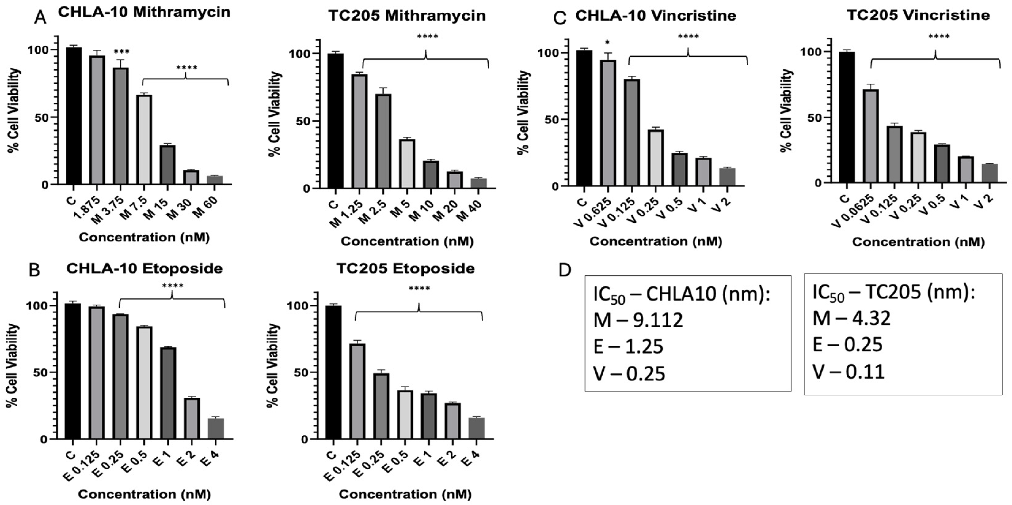
Figure 1. Monotherapy and IC50 values of CHLA-10 and TC205 ES cell lines. Cell viability results are shown for varying concentrations of monotherapy treatments of Mithramycin (Mit) and Etoposide (Eto) (AC). M: Mithramycin; E: Etoposide; V: Vincristine. IC50 values per drug per cell line are shown (D). Viability data represented are all 48 h treatments. Data were analyzed using one-way ANOVA; **** p < 0.0001, *** p < 0.0002, * p < 0.02.
Cancers 17 02977 g001
Figure 2. Combination treatments of CHLA-10 and TC205. ES cell lines treated with Mithramycin and Etoposide or Mithramycin and Vincristine (AD). M: Mithramycin; E: Etoposide; V: Vincristine. Viability data represented are all 48 h treatments. Data were analyzed using one-way ANOVA; **** p < 0.0001.
Cancers 17 02977 g002
Figure 3. Treatment effects on cardiomyocyte cell line. H9C2 cells after treatments with IC25 and IC50 doses. Mithramycin, Etoposide, and Vincristine are indicated with M, E, and V, respectively. Viability data represent 48 h treatments. Data were analyzed using one-way ANOVA.
Cancers 17 02977 g003
Figure 4. Combination index for CHLA-10 and TC205 ES cell lines. Combination treatments for TC205 (A) and CHLA-10 (B) with Mithramycin + Etoposide (ME) or Mithramycin + Vincristine (MV). ED50 values refer to effective dose 50; ED50 <1, =1, and >1 represent synergistic, additive, or antagonistic response, respectively.
Cancers 17 02977 g004
Figure 5. Effect of Mithramycin and Etoposide on apoptosis. Annexin V-PE/7-AAD flow cytometry plots and quantified results following 48 h treatment with IC25 doses for TC205 cells (AD). M: Mithramycin; E: Etoposide; V: Vincristine. Quantification of percept apoptotic cell death (E). Representative diagram of apoptosis using Annexin V-PE/7-AAD staining (F). Flow cytometry plots show the density gradient in the cell populations, with red representing highest population density, and blue representing the lowest population density. Data were analyzed using one-way ANOVA; **** p < 0.0001.
Cancers 17 02977 g005
Figure 6. Effect of Mithramycin and Etoposide on c-PARP expression. c-PARP expression was measured via Simple Western Jess system, and quantification for TC205 protein following 48 h treatment with IC25 doses was obtained in GraphPad Prism from at least 3 independent runs. M: Mithramycin; E: Etoposide; ME: Mithramycin + Etoposide. Data were analyzed using one-way ANOVA, * p < 0.03.
Cancers 17 02977 g006
Disclaimer/Publisher’s Note: The statements, opinions and data contained in all publications are solely those of the individual author(s) and contributor(s) and not of MDPI and/or the editor(s). MDPI and/or the editor(s) disclaim responsibility for any injury to people or property resulting from any ideas, methods, instructions or products referred to in the content.