Article

Characterizing Trends of Lymphedema After Axillary Lymph Node Dissection with and Without Immediate Lymphatic Reconstruction

by
Kella L. Vangsness
1,*,
Andre-Philippe Sam
2,
Jeff Chang
2,
Yash A. Mehta
2,
Michael W. Chu
3,
Mouchammed Agko
2 and
Antoine L. Carré
2
1
Division of General Surgery, Community Memorial Hospital, Ventura, CA 93033, USA
2
Division of Plastic Surgery, Department of Surgery, City of Hope, Duarte, CA 92618, USA
3
Division of Plastic and Reconstructive Surgery, Kaiser Permanente, Los Angeles, CA 90027, USA
*
Author to whom correspondence should be addressed.
Cancers 2025, 17(18), 2964; https://doi.org/10.3390/cancers17182964
Submission received: 8 July 2025 / Revised: 26 August 2025 / Accepted: 28 August 2025 / Published: 10 September 2025

Simple Summary

Breast cancer-related lymphedema is a complication following axillary lymph node dissection causing extensive morbidity and mortality. Current treatment options for lymphedema are limited. Immediate lymphatic reconstruction allows for the continued movement of lymph fluid. This is done by diverting it into veins through the creation of a lymphovenous bypass. The aim of this technique is to provide a substantial optios for patients undergoing axillary lymph node dissection.

Abstract

Background and Objectives: Breast cancer-related lymphedema (BCRL) is a complication of axillary lymph node dissection (ALND). Immediate lymphatic reconstruction (ILR) may help to decrease lymphedema after ALND by creating lymphatic bypasses. This retrospective single-institution study aimed to compare lymphedema in patients undergoing ALND with and without ILR. Materials and Methods: Bioimpedance and limb measurements determined the presence of BCRL. The categorical data that were collected and analyzed included BMI, comorbidities, BCRL onset, and number of lymphatic bypasses. Pearson’s chi-square test and multivariable logistic regression were performed to identify factors associated with the onset of lymphedema. An odds ratio compared the incidence of BCRL with and without ILR. Results: In total, 186 patients underwent ALND, 44 (24%) with ILR and 142 (76%) without. The mean number of bypasses during ILRs created was 3.54. The odds of developing lymphedema with ILR were 64% lower than for ALND alone. ILR patients who developed BCRL had a mean onset of 543 days post-operatively versus 389 days in the control group. Age, ethnicity, BMI, and bypass amount had no significant influence on lymphedema development. Conclusions: ILR was associated with lower rates of BCRL after ALND. Patients who developed lymphedema despite undergoing ILR did so 8 months later than the controls.

1. Introduction

Breast cancer is the most diagnosed cancer amongst women globally [1] and axillary lymph node dissection (ALND) is used in both the treatment and staging of breast cancer [2]. Although ALND provides therapeutic and diagnostic benefit, it can lead to the development of lymphedema, specifically known as breast cancer-related lymphedema (BCRL). BCRL is the disruption of lymphatic vessels causing lymphatic fluid accumulation in the axilla and upper extremities. Symptoms include swelling, decreased mobility in the operative arm [3], decreased activities of daily living, and decreased self-esteem [4,5].
The lymphatic system is a vital network of vessels that hold major roles in fluid homeostasis and inflammatory response [6,7]. The disruption of the lymph system in an upper extremity following axillary lymph node dissection or radiation has been heavily linked to psychological distress, poor quality of life [8], pain and functional disability [9], and prolonged wound healing [4,6]. While cures for lymphedema remain elusive due to the iatrogenic nature of its development, it is vital to employ preventative strategies.
Reconstructive surgeons currently utilize immediate lymphatic reconstruction (ILR) to reduce the risk of postoperative lymphedema. ILR employs a lymphatic microsurgical preventative healing approach (LYMPHA), a microsurgical intervention that involves creating lymphovenous bypasses at the time of ALNDs [10,11,12]. The LYMPHA procedure creates an anastomotic bypass with a standard microsurgical technique between a lymphatic channel and vein (Figure 1) to allow for continued lymph flow and prevent localized pooling. Few studies have demonstrated promising results with ILR, yet they have limited cohort sizes and short follow-up periods [13,14]. The goal of developing ILR is to employ surgical techniques to decrease the incidence of lymphedema and improve post-operative outcomes. The aim of this study is to assess if ILR is an appropriate procedure for the prophylactic treatment of lymphedema in patients who have undergone axillary node dissections with and without ILR for the treatment of breast cancer.

Immediate Lymphatic Reconstruction Technique

ILRs of lymphovenous anastomoses were performed intraoperatively following ALND at the time of the index surgery. A mixture of injectable isosulfan blue (IB) and flourescin isothiocyanate (FITC) dye was used to identify lymphatic vessels by injecting dye into the ipsilateral arm. Protocol for injection followed the Israel Deaconess Medical Center protocol [15]. Axillary veins were isolated and selected based on the desired length with minimal backflow. An ipsilateral dorsum foot vein was dissected for graft use if axillary veins were not sufficient in length. The locations of bypasses were determined by the amount and configuration of axillary lymphatic channels, as well as their proximity to venous tributaries. The maximum number of bypasses were sought for each procedure. Once created, intraoperative patency of lymphovenous anastomoses and flow of lymph fluid was evaluated through the combination IB and FITC dye under microscopic visualization.

2. Materials and Methods

This retrospective single-institution analysis was approved by the Institutional Review Board at City of Hope National Medical Center in Duarte, California (Protocol #23581, Reference #253089). Patients who underwent ALND with or without ILR between 2019 and 2023 were included. The primary outcome was the presence of lymphedema measured by SOZO (ImpediMed Limited, Brisbane, Australia). They were scored using the Lymphedema Index (L-Dex) (ImpediMed Limited, Brisbane, Australia) and upper limb circumference measurements performed by licensed lymphedema therapists. SOZO is a validated bioimpedance spectroscopy tool used for quantifying extracellular fluid shifts in lymphedema. It has high sensitivity and positive predictive value in BCRL with measurements scored using the L-Dex [16,17]. An increase of >6.5 units from baseline or >10 units from preoperative measurement is defined as subclinical lymphedema, while a score greater than +10 units is positive for lymphedema. Exclusion criteria included pre-existing lymphedema diagnoses, males, and patients who did not receive ALND.
The control group included patients who underwent ALND alone prior to 2020, as this was the year ILR was first available at the institution. The intervention group consisted of patients who were treated from 2020 onward and who underwent ALND with ILR. Clinical practices remained consistent during the study period without changes to assessment protocols. Two patients treated after 2020 received ALND only without ILR intervention due to intraoperative technical constraints, including insufficient lymphatic vessels and venous backflow. No additional patients were included or excluded based on intraoperative technical difficulties. Follow-up timelines were standardized across both groups to reduce time-related bias. The incidence and onset of lymphedema were recorded.
Patient data collected included age, race/ethnicity, body mass index (BMI), comorbidities, tumor stage, histologic grade, receptor subtype, the number of lymph nodes excised, and the number of lymphovenous bypasses created during ILR (Table 1). BMI was classified into underweight, normal, overweight, and obese, following World Health Organization criteria. For all patients, the mean BMI was 29 and age was 53. All patients received routine follow-up care and lymphedema screening approximately three months postoperatively. Assessments included a physical exam, limb measurement, and an L-Dex score.

Statistical Analysis

ALND-only patients served as the control group while those who received ILR comprised the interventional group. Patient demographics and clinical characteristics were summarized with descriptive statistics. Categorical variables were compared using Pearson’s chi-squared test to identify associations. Despite data being sparsely reported or missing due to the nature of retrospective data collection, this test was employed across all categorical variables to maintain methodological uniformity. These results were assessed with caution given the known limitations of the chi-square test when expected cell counts are <5. Despite this analysis not revealing any significant relationship, we hypothesized that the number of positive lymph nodes removed might still be associated with the incidence of lymphedema when controlling for other factors. Continuous variables were analyzed for normality using appropriate central tendency and dispersion measures based on data distribution.
Assessment of ILR and associated lymphedema development was evaluated by calculating odds ratios and 95% confidence intervals. Multivariate logistic regression analyses using a binary logistic regression was used to assess for independent predictors of lymphedema. The model included BMI and age as continuous variables. Stages were separated into groups: stage 2 versus stage 3. Stage 2 acted as the reference category, since no patients were categorized as stages 0 or 1. Meanwhile, 0–9 +LN removed was the reference category for all +LN groups. Binary categories included extranodal extension, ILR, radiation, and adjuvant chemotherapy. Covariates included the number of lymph nodes removed and the number of lymphovenous bypasses performed. The numbers of positive lymph nodes (+LN) removed were separated into three categories: 0–9, 10–15, and 16+. A p-value < 0.05 was considered statistically significant.

3. Results

Out of the 186 patients examined, 142 (76%) received ALND only and 44 (24%) patients underwent ILR. Overall, only 52 (28%) patients developed lymphedema. When separated by control versus interventional groups, 44 of 142 (31%) ALND-only patients developed lymphedema whereas only 8 of 44 (18%) of those who underwent ILR developed the condition.
The mean age of patients in the ILR cohort was 44 years old versus 56 in the ALND only group. The race/ethnicity distribution of the entire cohort included 69 patients who identified as Hispanic/Latino, 65 as Caucasian/White, 38 as Asian/Pacific Islander, and 14 as Black/African American.
Descriptive analyses using a chi-squared test assessing differences in the number of +LN removed found no statistically significant findings, only observational differences, and none that were statistically significant. There were no patients in the 16+ +LN group who underwent ILR. However, 33% of patients in the 0–9 +LN group and 20% of the 10–15 +LN group underwent ILR (X2 (2, n= 212) = 3.63, p = 0.163). These findings are taken with a note of caution due to the small sample size, unequal group size, and cells which did not meet qualifying numbers for adequate analysis.
The results of the multivariable logistic regression assessing patient demographic or clinical variables found only one statistically significant relationship in the ILR group. This was between the number of positive lymph nodes removed when groups were analyzed as categorical variables (1–9, 10–15, 16+ positive lymph nodes) (Figure 2). Groups with 10–15 and 16+ +LNs removed had a 9 and 24 times increased likelihood of being diagnosed with lymphedema, respectively. These findings are taken with a note of caution due to the unequal group sizes and small sample size. Age, BMI, race/ethnicity, mastectomy, adjuvant chemo, radiation, ILR intervention, or extranodal extension were not significantly associated with the presence of lymphedema when examined as categorical variables.
Due to the unequal group sizes, the regression model was reanalyzed with the amount of positive lymph nodes as a continuous variable. This model demonstrated three statistically significant factors associated with lymphedema development (Figure 3): the number of positive lymph nodes removed (p < 0.001), extranodal extension (p < 0.05), and ILR intervention (p < 0.05). The odds ratio for each positive lymph node removed was 1.2 (p < 0.001; 95% CI [1.08, 1.32]) suggesting that for each +LN removed there was a 20% increase in the odds of being diagnosed with lymphedema. The odds ratios for extranodal extension was 0.31 (p < 0.05; 95% CI [0.12, 0.79]) suggesting that those who had an extranodal extension diagnosis were about 70% less likely to have been diagnosed with lymphedema compared to those who had no evidence of extranodal extension. Similarly, the odds ratio for ILR was 0.36 (p < 0.05; 95% CI [0.14, 0.92]), which suggests that those who underwent ILR intervention were about 64% less likely to have a lymphedema diagnosis. The overall model for this analysis was also statistically significant (X2 (12, N = 186) = 25.96, p = 0.011).
Notably, the confidence interval is very wide, meaning that the odds ratio is statistically significant but not precise, so implications are made with a note of caution.
The onset of lymphedema developed later in those who received ILR compared to ALND only. Diagnosis was established at a mean post-operative day of 542 versus 389, respectively. The mean follow-up for patients who received ILR was 33 months compared to 39 months for controls. The mean time to end follow-up for both cohorts was at the three-year time point.

4. Discussion

Axillary lymph node excision is used for the diagnosis and treatment of breast cancer, with lymphedema being a common morbidity following surgery. Immediate lymphedema reconstruction is a microsurgical approach that maintains lymphatic flow by diverting it through the venous system. This analysis demonstrates that patients who received ILR during ALND experienced a lower overall rate of lymphedema compared to those who underwent ALND alone. Of those who underwent ILR and developed lymphedema, they went on to do so at a later date with a mean difference of 153 days. This suggests that ILR maintains continued flow and the delay of upper extremity lymphedema onset. These findings are similar to current literature [2,18].
ILR appeared to have a protective effect against lymphedema development. Only 18% of patients in the ILR group developed lymphedema compared to 31% of controls. Additionally, when all factors were held constant, undergoing ILR was shown to decrease the likelihood of developing postoperative lymphedema by 64%. A systematic review by Cook et al. [18] showed similar rates of control groups, but an even lower rate of development with ILR intervention, also in line with the current literature [12,19].
The strongest independent predictor of lymphedema development found in this analysis was the number of positive lymph nodes removed [12,19]. Groups who had 10–15 and 16+ +LNs removed had a 9 and 24 times increased likelihood of being diagnosed with lymphedema, respectively. For each +LN removed there was a 20% increase in the odds of being diagnosed with lymphedema. In contrast, this data found that extranodal extension was associated with a 70% decrease in diagnoses compared to patients who had no evidence of extension, but only when +LN was assessed as a continuous variable. The literature has shown that an increased number of positive lymph nodes is tied to advanced disease stage, yet data has not shown that late-staged tumors or clinically positive nodes are associated with lymphedema [2].
Age, race, and BMI were not factors shown to be significantly associated with lymphedema development for either control or interventional groups. Multiple studies have found no significant associations with age [20,21]. This data showed no correlation with race/ethnicity; however, Ren et al. [22] found that younger Black women experience an increased risk of lymphedema around 10 months after surgery compared to other age and race/ethnicity groupings. Kwan et al. [23] reported that African American patients faced nearly double the risk of developing BCRL compared with White/Caucasian patients. Increased BMI greater than 30 kg/m2 triples lymphedema risk [24] and obesity was found to be an independent risk factor for acquiring lymphedema [12,25]. Although this analysis did not reveal similar strong racial, age, or BMI associations, the current literature underscores the potential interplay of sociocultural factors, baseline comorbidities, and healthcare access in lymphedema risks warranting further investigation.
This analysis did not reveal any significant association between lymphedema development and radiation or chemotherapy despite a previously established increase [3,5]. Hara et al. reported a significant risk of lymphedema in patients receiving radiotherapy and, similarly, Taghian et al. noted that nodal irradiation and certain chemotherapy regimens may also contribute. A possible explanation for differing results may be due to the standardized regimen this cohort received, which could limit the capacity to detect differences. Chemotherapy, particularly neoadjuvant regimens, has been implicated in lymphedema [26,27,28], yet effects may have been diminished due to the protocol as well as the small sample size used in the analysis. Future investigations with more detailed data on individual chemotherapy agents, radiation techniques, and fractionation schedules could yield clearer insights into the relationship between these variables and BCRL.
ALND-only patients had a lymphedema rate of approximately 31%, aligning with prior studies citing 20–40% prevalence of postoperative lymphedema [4,5]. Although Kwan et al. observed a slightly lower incidence of 13.3% in their prospective cohort, differences in methodology (e.g., reliance on clinical coding rather than direct limb measurements) and demographic factors may partly account for the variation [26]. In contrast, other literature has shown a decreased lymphedema risk, which fell to 18% with prophylactic lymphatic reconstruction [13,15,29], while one small study failed to find a difference with or without ILR [29]. The ILR group took a longer time to obtain a lymphedema diagnosis, which may demonstrate that lymphatic channels were maintained and assisted with the delayed onset. However, differences in patient selection, surgical technique, and the definition of lymphedema may help to explain these inconsistencies.
Future studies can expand on this data through prospective studies with longer follow-ups and by continuing to use standardized lymphedema assessment tools (e.g., bioimpedance vs. circumferential measurements) to strengthen evidence. Additionally, a more in-depth analysis examining the cost-effectiveness [30] and impact on quality of life is warranted given that prior work suggests ILR may improve outcomes [31] and potentially reduce long-term healthcare costs [30].

Limitations

This study had limitations inherent to its retrospective design and limited sample size. Subgroups had sparse or empty cells during data collection which violated the assumptions of Pearson’s chi-square test in categorical comparisons. We acknowledge that a non-parametric test could have been more appropriate, so all results were interpreted with caution. Using larger sample sizes in future studies will allow for more robust subgroup analyses in addition to using assumption-appropriate tests.
Menopausal status was recorded but not analyzed in this study. The established influence of hormonal receptor status on lymphedema requires additional assessment as a possible confounding variable and therefore was not assessed.
Discrepancies in this data may lie in patient demographics and sample sizes. Larger prospective trials that specifically stratify patients by menopausal status and age may help clarify whether and to what extent these factors truly affect BCRL risk.

5. Conclusions

Findings suggest that ILR at the time of ALND may both lower the incidence and delay the onset of lymphedema but does not prevent its development. The increased number of positive lymph nodes removed during axillary dissection were also noted to be significantly associated with the development of lymphedema. Age, menopausal status, BMI, race/ethnicity, radiation, and chemotherapy did not reach any statistical significance in the development of lymphedema.

Author Contributions

Conceptualization, A.L.C. and J.C.; methodology, M.W.C., A.L.C. and M.A.; validation, Y.A.M.; formal analysis, A.L.C. and Y.A.M.; investigation, A.-P.S. and K.L.V.; data curation, A.-P.S. and K.L.V.; writing—original draft preparation, Y.A.M. and A.-P.S.; writing—review and editing, K.L.V.; visualization, K.L.V.; supervision, M.W.C., M.A. and J.C.; project administration, J.C. and A.L.C. All authors have read and agreed to the published version of the manuscript.

Funding

This research received no external funding.

Institutional Review Board Statement

This study was conducted in accordance with the Declaration of Helsinki and approved by the Institutional Review Board of City of Hope (Protocol #23581, Reference #253089 on 31 August 2023) under Exempt status, for which consent is not required.

Informed Consent Statement

Patient consent was waived due to retrospective data collection where all patient identifiers were removed.

Data Availability Statement

The original contributions presented in this study are included in the article. Further inquiries can be directed to the corresponding author.

Conflicts of Interest

The authors declare no conflicts of interest.

References

  1. Fahad Ullah, M. Breast cancer: Current perspectives on the disease status. In Advances in Experimental Medicine and Biology; Springer: Cham, Switzerland, 2019; pp. 51–64. [Google Scholar] [CrossRef]
  2. Beck, A.C.; Morrow, M. Axillary lymph node dissection: Dead or still alive? Breast 2023, 69, 469–475. [Google Scholar] [CrossRef] [PubMed]
  3. Hara, Y.; Otsubo, R.; Shinohara, S.; Morita, M.; Kuba, S.; Matsumoto, M.; Nagayasu, T. Lymphedema after axillary lymph node dissection in breast cancer: Prevalence and risk factors-a single-center retrospective study. Lymphat. Res. Biol. 2022, 20, 600–606. [Google Scholar] [CrossRef]
  4. Bittar, S.; Simman, R.; Lurie, F. Lymphedema: A practical approach and clinical update. Wounds 2020, 32, 86–92. [Google Scholar] [PubMed]
  5. Liao, S.; von der Weid, P.Y. Lymphatic system: An active pathway for immune protection. Semin. Cell Dev. Biol. 2015, 38, 83–89. [Google Scholar] [CrossRef]
  6. Engin, O.; Sahin, E.; Saribay, E.; Dilek, B.; Akalin, E. Risk factors for developing upper limb cellulitis after breast cancer treatment. Lymphology 2022, 55, 77–83. [Google Scholar] [CrossRef]
  7. Vignes, S.; Poizeau, F.; Dupuy, A. Cellulitis risk factors for patients with primary or secondary lymphedema. J. Vasc. Surg. Venous Lymphat. Disord. 2022, 10, 179–185. [Google Scholar] [CrossRef] [PubMed]
  8. Tamburini, B.A.J.; Padera, T.P.; Lund, A.W. Editorial: Regulation of immune function by the lymphatic vasculature. Front. Immunol. 2019, 10, 2597. [Google Scholar] [CrossRef]
  9. Johnson, L.A. In Sickness and in health: The immunological roles of the lymphatic system. Int. J. Mol. Sci. 2021, 22, 4458. [Google Scholar] [CrossRef] [PubMed]
  10. Rockson, S.G.; Rivera, K.K. Estimating the population burden of lymphedema. Ann. N. Y. Acad. Sci. 2008, 1131, 147–154. [Google Scholar] [CrossRef] [PubMed]
  11. McLaughlin, S.A.; Wright, M.J.; Morris, K.T.; Sampson, M.R.; Brockway, J.P.; Hurley, K.E.; Riedel, E.R.; Van Zee, K.J. Prevalence of lymphedema in women with breast cancer 5 years after sentinel lymph node biopsy or axillary dissection: Patient perceptions and precautionary behaviors. J. Clin. Oncol. Off. J. Am. Soc. Clin. Oncol. 2008, 26, 5220–5226. [Google Scholar] [CrossRef]
  12. Kalemikerakis, I.; Evaggelakou, A.; Kavga, A.; Vastardi, M.; Konstantinidis, T.; Govina, O. Diagnosis, treatment and quality of life in patients with cancer-related lymphedema. J. Buon. 2021, 26, 1735–1741. [Google Scholar]
  13. Taghian, N.R.; Miller, C.L.; Jammallo, L.S.; O’Toole, J.; Skolny, M.N. Lymphedema following breast cancer treatment and impact on quality of life: A review. Crit. Rev. Oncol. Hematol. 2014, 92, 227–234. [Google Scholar] [CrossRef] [PubMed]
  14. Cook, J.A.; Sinha, M.; Lester, M.; Fisher, C.S.; Sen, C.K.; Hassanein, A.H. Immediate lymphatic reconstruction to prevent breast cancer-related lymphedema: A systematic review. Adv. Wound Care 2022, 11, 382–391. [Google Scholar] [CrossRef]
  15. Coriddi, M.; Mehrara, B.; Skoracki, R.; Singhal, D.; Dayan, J.H. Immediate lymphatic reconstruction: Technical points and literature review. Plast. Reconstr. Surg. Glob. Open 2021, 9, e3431. [Google Scholar] [CrossRef]
  16. Jørgensen, M.G.; Toyserkani, N.M.; Sørensen, J.A. The effect of prophylactic lymphovenous anastomosis and shunts for preventing cancer-related lymphedema: A systematic review and meta-analysis. Microsurgery 2018, 38, 576–585. [Google Scholar] [CrossRef]
  17. Spiguel, L.; Shaw, C.; Katz, A.; Guo, L.; Chen, H.C.; Lee, B.T.; Singhal, D. Fluorescein isothiocyanate: A novel application for lymphatic surgery. Ann. Plast. Surg. 2017, 78 (Suppl. 5), S296–S298. [Google Scholar] [CrossRef] [PubMed]
  18. Coroneos, C.J.; Wong, F.C.; DeSnyder, S.M.; Shaitelman, S.F.; Schaverien, M.V. Correlation of L-Dex Bioimpedance spectroscopy with limb volume and lymphatic function in lymphedema. Lymphat. Res. Biol. 2019, 17, 301–307. [Google Scholar] [CrossRef] [PubMed]
  19. Wiser, I.; Mehrara, B.J.; Coriddi, M.; Kenworthy, E.; Cavalli, M.; Encarnacion, E.; Dayan, J.H. Preoperative Assessment of upper extremity secondary lymphedema. Cancers 2020, 12, 135. [Google Scholar] [CrossRef] [PubMed]
  20. Shah, C.; Whitworth, P.; Valente, S.; Schwarz, G.S.; Kruse, M.; Kohli, M.; Brownson, K.; Lawson, L.; Dupree, B.; Vicini, F.A. Bioimpedance spectroscopy for breast cancer-related lymphedema assessment: Clinical practice guidelines. Breast Cancer Res. Treat. 2023, 198, 1–9. [Google Scholar] [CrossRef]
  21. Norman, S.A.; Localio, A.R.; Kallan, M.J.; Weber, A.L.; Torpey, H.A.; Potashnik, S.L.; Miller, L.T.; Fox, K.R.; DeMichele, A.; Solin, L.J. Risk factors for lymphedema after breast cancer treatment. Cancer Epidemiol. Biomark. Prev. 2010, 19, 2734–2746. [Google Scholar] [CrossRef]
  22. Koelmeyer, L.A.; Gaitatzis, K.; Dietrich, M.S.; Shah, C.S.; Boyages, J.; McLaughlin, S.A.; Taback, B.; Stolldorf, D.P.; Elder, E.; Hughes, T.M.; et al. Risk factors for breast cancer-related lymphedema in patients undergoing 3 years of prospective surveillance with intervention. Cancer 2022, 128, 3408–3415. [Google Scholar] [CrossRef]
  23. Ren, Y.; Kebede, M.A.; Ogunleye, A.A.; Emerson, M.A.; Evenson, K.R.; Carey, L.A.; Troester, M.A. Burden of lymphedema in long-term breast cancer survivors by race and age. Cancer 2022, 128, 4119–4128. [Google Scholar] [CrossRef]
  24. Kwan, M.L.; Darbinian, J.; Schmitz, K.H.; Citron, R.; Partee, P.; Kutner, S.E.; Kushi, L.H. Risk factors for lymphedema in a prospective breast cancer survivorship study: The Pathways Study. Arch. Surg. 2010, 145, 1055–1063. [Google Scholar] [CrossRef]
  25. Fu, J.; Chen, R.; He, L.; Bao, L.; Lin, Z.; Jiang, W.; Zhang, J.; Wang, C.; Lin, Y. Factors affecting lymphedema after neoadjuvant chemotherapy and axillary dissection in female breast cancer patients: A retrospective cohort study based on the Chinese population. Front. Oncol. 2024, 14, 1436748. [Google Scholar] [CrossRef]
  26. Jia, M.; Pan, L.; Yang, H.; Gao, J.; Guo, F. Impact of neoadjuvant chemotherapy on breast cancer-related lymphedema after axillary lymph node dissection: A retrospective cohort study. Breast Cancer Res. Treat. 2024, 204, 223–235. [Google Scholar] [CrossRef]
  27. Lee, Y.S.; Lim, Y.C.; Yeo, J.; Kim, S.Y.; Lee, Y.J.; Ha, I.H. Risk of Lymphedema and Death after Lymph Node Dissection with Neoadjuvant and Adjuvant Treatments in Patients with Breast Cancer: An Eight-Year Nationwide Cohort Study. Healthcare 2023, 11, 1833. [Google Scholar] [CrossRef]
  28. Boccardo, F.; Casabona, F.; De Cian, F.; Friedman, D.; Murelli, F.; Puglisi, M.; Campisi, C.C.; Molinari, L.; Spinaci, S.; Dessalvi, S.; et al. Lymphatic microsurgical preventing healing approach (LYMPHA) for primary surgical prevention of breast cancer-related lymphedema: Over 4 years follow-up. Microsurgery 2014, 34, 421–424. [Google Scholar] [CrossRef] [PubMed]
  29. Chun, M.J.; Saeg, F.; Meade, A.; Kumar, T.; Toraih, E.A.; Chaffin, A.E.; Homsy, C. Immediate lymphatic reconstruction for prevention of secondary lymphedema: A meta-analysis. J. Plast. Reconstr. Aesthetic Surg. 2022, 75, 1130–1141. [Google Scholar] [CrossRef] [PubMed]
  30. Levy, A.S.; Murphy, A.I.; Ishtihar, S.; Peysakhovich, A.; Taback, B.; Grant, R.T.; Ascherman, J.A.; Feldman, S.; Rohde, C.H. Lymphatic microsurgical preventive healing approach for the primary prevention of lymphedema: A 4-year follow-up. Plast. Reconstr. Surg. 2023, 151, 413–420. [Google Scholar] [CrossRef] [PubMed]
  31. Johnson, A.R.; Asban, A.; Granoff, M.D.; Kang, C.O.; Lee, B.T.; Chatterjee, A.; Singhal, D. Is immediate lymphatic reconstruction cost-effective? Ann. Surg. 2021, 274, e581–e588. [Google Scholar] [CrossRef]
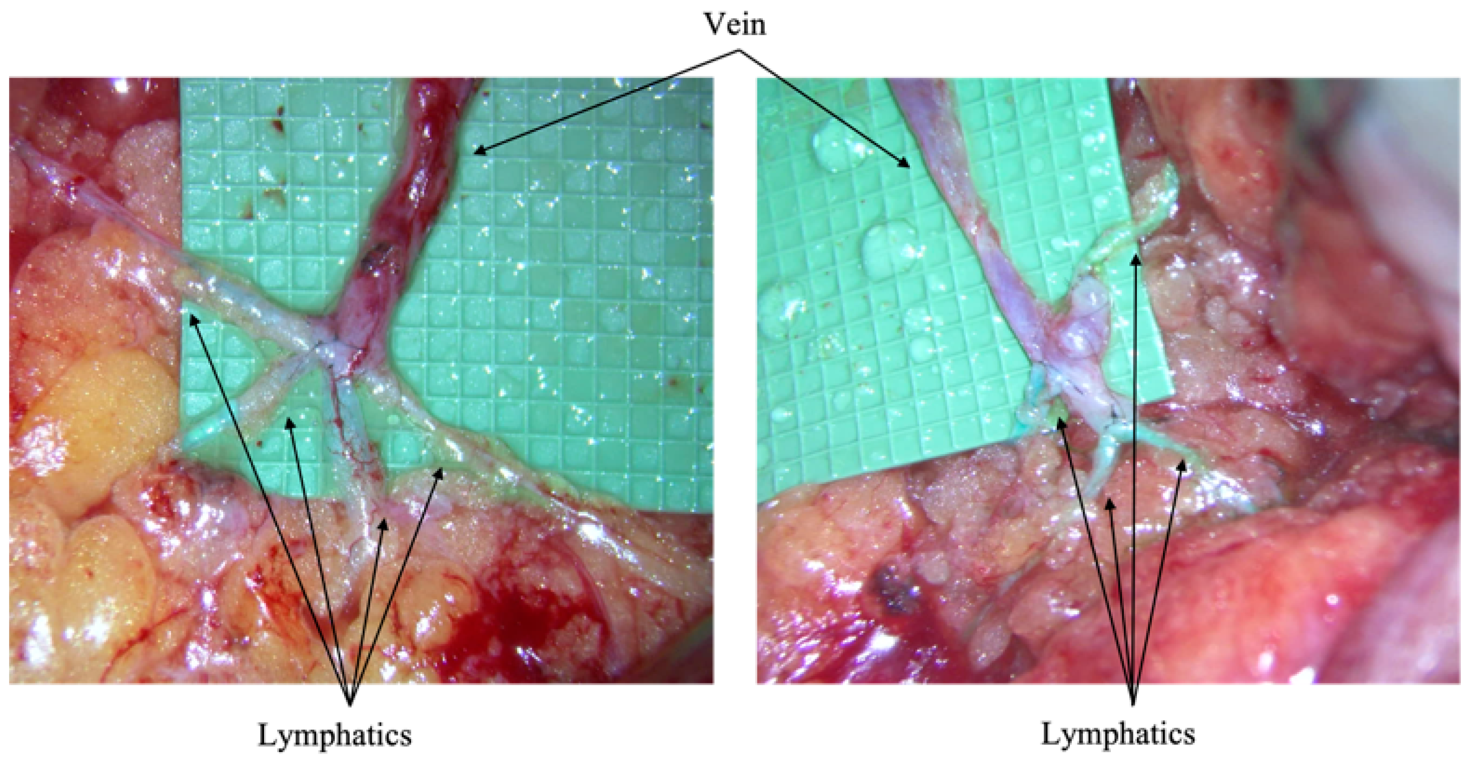
Figure 1. Above images feature examples of the LYMPHA procedure. A microsurgery bypass is created through the anastomosis of a vein (superiorly) and lymphatics (inferiorly) to allow for lymphatic flow. Intraoperative images taken from City of Hope, Duarte, California.
Cancers 17 02964 g001
Figure 2. Odds ratio plot depicting demographic and clinical variables with the number of positive lymph nodes as a categorical variable. Body mass index and age were continuous variables. Stage compared stage 2 vs. stage 3; the reference category was stage 2. Positive lymph nodes with 0–9 +LN were used as a reference category. Mastectomy: yes vs. no, with no as a reference category. Race/ethnicity is presented with Non-Hispanic white as the reference category. Extranodal extension: no vs. yes, with no as a reference category. Immediate lymphatic reconstruction: no vs. yes, with no as a reference category. Radiation: no vs. yes, with no as the reference category. Adjuvant chemotherapy: no vs. yes, with no as the reference category. ** p < 0.01 was deemed statistically significant.
Cancers 17 02964 g002
Figure 3. Odds ratio forest plot depicting demographic and clinical variables with the number of positive lymph nodes as a continuous variable. Body mass index and age were continuous variables. Stages compared stage 2 vs. stage 3: the reference category was stage 2. Positive lymph nodes were a continuous variable. Mastectomy: yes vs. no, with no as a reference category. Race/ethnicity: Non-Hispanic white was the reference category. Extranodal extension: no vs. yes, with no as the reference category. Immediate lymphatic reconstruction: no vs. yes, with no as the reference category. Radiation: no vs. yes, with no as the reference category. Adjuvant chemotherapy: no vs. yes, with no as the reference category. * p < 0.05; *** p < 0.001.
Cancers 17 02964 g003
Table 1. Patient categorical information.
CategorySubcategoryILR
n = 44
Non-ILR
n = 142
Age (years) 48 ± 1155 ± 12
Menopausal statusPremenopausal2952
Postmenopausal1590
Clinical tumor stagecT1421
cT22380
cT31226
cT4515
Clinical nodal stagecN133120
cN2516
cN366
HistologyInvasive ductal carcinoma40126
Invasive lobular carcinoma416
Grade105
22478
32059
BiomarkersER-positive HER2-negative2887
HER2-positive1131
Triple negative524
Neoadjuvant chemotherapy 38102
Initial axillary operationTargeted axillary dissection1537
Axillary lymph node dissection29105
Pathologic tumor stagepT01033
pT11647
pT21350
pT3510
pT402
Pathologic nodal stagepN01231
pN11767
pN21331
pN3213
Residual cancer burdenClass 0627
Class I29
Class II1125
Class III1533
Number of positive lymph nodes4 ± 54 ± 3
Number of lymph nodes removed16 ± 715 ± 6
Extranodal Extension1259
Adjuvant systemic therapy44134
Adjuvant radiation42139
Lymphedema844
Distant recurrence316
Mean follow-up (months)33 ± 939 ± 5
Death26
Disclaimer/Publisher’s Note: The statements, opinions and data contained in all publications are solely those of the individual author(s) and contributor(s) and not of MDPI and/or the editor(s). MDPI and/or the editor(s) disclaim responsibility for any injury to people or property resulting from any ideas, methods, instructions or products referred to in the content.